Article

Foliar Applications of Bacillus subtilis HA1 Culture Filtrate Enhance Tomato Growth and Induce Systemic Resistance against Tobacco mosaic virus Infection

1
Bioprocess Development Department, Genetic Engineering and Biotechnology Research Institute, City of Scientific Research and Technological Applications, Alexandria 21934, Egypt
2
Department of Botany and Microbiology College of Science, King Saud University, P.O. Box 2455, Riyadh 11451, Saudi Arabia
3
Centre for Agricultural Research, Plant Protection Institute, ELKH, 15 Herman Ottó Str., H-1022 Budapest, Hungary
4
Plant Protection and Biomolecular Diagnosis Department, ALCRI, City of Scientific Research and Technological Applications, Alexandria 21934, Egypt
5
Agriculture Microbiology Department, National Research Centre, Cairo 12622, Egypt
*
Author to whom correspondence should be addressed.
Horticulturae 2022, 8(4), 301; https://doi.org/10.3390/horticulturae8040301
Submission received: 28 February 2022 / Revised: 30 March 2022 / Accepted: 30 March 2022 / Published: 31 March 2022
(This article belongs to the Special Issue Sustainable Control Strategies of Plant Pathogens in Horticulture)

Abstract

:
The application of microbial products as natural biocontrol agents for inducing systemic resistance against plant viral infections represents a promising strategy for sustainable and eco-friendly agricultural applications. Under greenhouse conditions, the efficacy of the culture filtrate of Bacillus subtilis strain HA1 (Acc# OM286889) for protecting tomato plants from Tobacco mosaic virus (TMV) infection was assessed. The results showed that the dual foliar application of this culture filtrate (HA1-CF) 24 h before and 24 h after TMV inoculation was the most effective treatment for enhancing tomato plant development, with substantial improvements in shoot and root parameters. Furthermore, compared to non-treated plants, HA1-CF-treated tomato had a significant increase in total phenolic and flavonoid contents of up to 27% and 50%, respectively. In addition, a considerable increase in the activities of reactive oxygen species scavenging enzymes (PPO, SOD, and POX) and a significant decrease in non-enzymatic oxidative stress markers (H2O2 and MDA) were reported. In comparison to untreated control plants, all HA1-CF-treated plants showed a significant reduction in TMV accumulation in systemically infected tomato leaves, up to a 91% reduction at 15 dpi. The qRT-PCR results confirmed that HA1-CF stimulated the transcription of several defense-related tomato genes (PR-1, PAL, CHS, and HQT), pointing to their potential role in induced resistance against TMV. GC–MS analysis showed that phenol, 2,4-bis (1,1-dimethylethyl)-, Pyrrolo [1,2-a] pyrazine-1,4-dione, hexahydro-3-(2-methylpropyl)- and eicosane are the primary ingredient compounds in the HA1-CF ethyl acetate extract, suggesting that these molecules take part in stimulating induced systemic resistance in tomato plants. Our results imply that HA1-CF is a potential resistance inducer to control plant viral infections, a plant growth promoter, and a source of bioactive compounds for sustainable disease management.

Graphical Abstract

1. Introduction

The growing global population, as well as urbanization and global climate changes, generate an ever-increasing demand for high crop yields and improved food quality. Several strategies are being dedicated to accelerate and improve the rate of agricultural production. However, plant diseases account for significant crop losses and delay the ongoing progress of crop management [1]. Viral infections of plants represent a great threat to plant biosecurity, causing tremendous crop losses worldwide [2]. Tobacco mosaic virus (TMV) is one of the most infectious plant pathogens, attributed to its wide range of plant hosts (about 66 families with more than 900 plant species) and the severe consequences of its infection [3]. TMV is transmitted mechanically by rubbing with infected plants, contaminated agriculture tools, and/or contaminated seeds [4]. In addition, the extraordinary stability of viral particles ensures their persistence for years in e.g., fallen infected leaves or soil with full capacity for re-infection [5]. In tomatoes, TMV infection is associated with different morphological symptoms, including a systemic leaf mosaic and/or necrosis, in addition to leaf chlorosis [6]. Furthermore, the infection could develop into systemic changes in flowering organs that delay fruit ripening, affect crop productivity, and result in crop loss [7].
Controlling TMV infection is challenging and relies upon the use of resistant plant cultivars or prevention of vector spreading through intensive application of insecticides, which may cause adverse complications for human health and the environment [8]. Furthermore, pesticides eventually make their way into the surface–water system, complicating and exacerbating environmental and ecosystem issues [9,10]. For sustainability, in either agriculture or the environment, interest is growing in the application of biocontrol agents as eco-friendly substitutes for the hazardous chemicals currently applied in plant disease management strategies [11].
Plant growth-promoting rhizobacteria (PGPR) are natural rhizosphere microbiota that promote plant growth and resistance to various infections. Numerous studies have found that PGPR can improve plant growth and increase resistance to viral infection [11,12]. PGPRs enhance plant growth through promoting nutrient acquisition and the production of biomolecules involved in stress tolerance. These processes may either indirectly decrease the susceptibility of plants to infection and/or directly fight pathogens through antibiotic production or competition for essential nutrients [13,14]. Among others, strains of Bacillus sp. are widely applied as PGPRs with significant application outcomes attributed to their multiple mechanisms for infection control and plant growth stimulation mediated by numerous secondary metabolites [15,16]. However, under field conditions, the persistence and adaption of PGPR inocula to the natural microbiota are strain-dependent and heavily affected by application practices that usually lead to inconclusive outcomes [15]. Furthermore, the influence of PGPR inocula on the natural microbiota community at the site of application is unknown, with the possible potential of increasing antagonistic resistance [17,18,19]. Due to these limitations, the application of microbial culture filtrates has emerged as an eco-friendly, dependable alternative to PGPRs [15].
Biocontrol agents protect plants through the stimulation of induced systemic resistance (ISR) by activating a variety of cellular processes. These processes include (i) the up- or down-regulation of specific genes and the overexpression of specific transcription factors; (ii) changes in the levels of various compounds implicated in defense pathways and enhanced plant growth; (iii) activation of defense genes encoding reactive oxygen species (ROS) scavenging enzymes such as POX, SOD, CAT, and PPO, as well as the accumulation of extracellular pathogenesis-related (PR) proteins; and (iv) improved transport of macromolecules, phytohormones, and enzymes involved in defensive signaling [3,20,21,22,23].
The goals of the present study were to evaluate the ability of Bacillus subtilis strain HA1-culture filtrate (HA1-CF) to promote tomato growth and confer protection to TMV infection, either through direct antiviral activity or the induction of systemic resistance in tomato plants. To investigate the resistance induction mechanism, the activity of reactive oxygen species scavenging enzymes (PPO, SOD, and POX) and non-enzymatic oxidative stress markers (H2O2 and MDA), as well as total phenolic content and flavonoid content were assessed. Furthermore, the expression levels of several defense-related tomato genes, including PR-1, PR-2, PAL, CHS, and HQT, along with TMV accumulation inside tomato tissues, were also estimated. Finally, the gas chromatography–mass spectrometry (GC–MS) technique was used to screen and identify the bioactive constituents of HA1-CF.

2. Materials and Methods

2.1. Plant Material and Viral Source

Tomato (Solanum lycopersicum L.) plants of the GS 12 cultivar used during this work were purchased as virus-free seeds from the Agriculture Research Center, Egypt. The TMV strain KH1 (accession number MG264131) previously isolated from TMV-infected tomato plants, was used as a source of viral inoculum [6].

2.2. Bacterial Isolation

Bacterial isolation was conducted on nutrient agar (NA) plates with the following composition: peptone 5 g/L, yeast extract 3 g/L, NaCl 5 g/L, and agar 15 g/L [24]. Five soil samples were collected from the rhizosphere of tomato plants that appeared healthy and vigorous in different open fields in Alexandria governorate, Egypt. Soil samples were taken at a 5–15 cm root depth after removing approximately 3 cm of the soil surface. Each sample (10 g) was a mix of five samples (2 g) collected from the rhizosphere of various tomato plants at the same site. Subsequently, each sample (10 g) was suspended in 100 mL of saline solution (0.9% NaCl) for 30 min under shaking. Serial dilutions (1 × 10⁻⁴, 1 × 10⁻⁵, and 1 × 10⁻⁶) were prepared, and 100 µL of each dilution were streaked aseptically on triplicate NA plates and incubated at 30 °C for 24 h. Different bacterial colonies of 10−6 dilution were chosen, based on colony shape, colony color, and antagonistic activity against growing fungi on the NA plates. To ensure purity, the newly developed separate colonies were streaked on the same medium. To obtain culture filtrates, all isolates were cultivated separately in nutrient broth (Agar-free nutrient medium) for 48 h under shaking at 200 rpm at 30 °C. The bacterial culture filtrate (CF) was obtained as follows: centrifugation (10 min, 10,000 rpm), collection of supernatant, and filtration with a 0.45 µm pore-size syringe filter. Using the half-leaf method [25], the antiviral potency of the purified isolates was tested in Chenopodium amaranticolor plants, which serve as a local lesion host for TMV. Briefly, the upper right half of the leaves were treated with 100 µL of bacterial CF, while the left half of the leaves were treated with 100 µL of sterilized nutrient broth media. After 24 h, both halves of the leaves were mechanically inoculated with TMV. The experiment was performed in three biological replicates. According to the inhibition percentage in relation to the number of local lesions, the isolate that exhibited the most potent antiviral activity was selected for further experiments.

2.3. Isolate Identification through 16S rRNA Methodology

The isolate showing the maximum antiviral potency was subjected to molecular identification through 16S rRNA sequencing. Total genomic DNA was isolated from an overnight culture, using the Wizard Genomic DNA Purification Kit (Promega, Fitchburg, WI, USA) according to the manufacturer’s instructions. The PCR amplification of the 16S rRNA gene was conducted using two universal primers (Table 1). The amplified 16S gene was purified by a PCR purification kit (QIAGEN, Hilden, Germany) and sequenced through a Genetic Analyzer system (3130xl, Applied Biosystems, Bedford, MA, USA) using a BigDye Terminator v3.1 Cycle Sequencing kit. The annotated nucleotide sequence was analyzed using NCBI-BLAST and deposited in GenBank. The phylogenetic relationships of the potent isolate were elucidated through MEGA software (ver. 11) using an unweighted pair group method with arithmetic mean (UPGMA) and a bootstrap method with 2000 replications.

2.4. Greenhouse Experimental Design and Assessment of Growth Parameters

Under greenhouse conditions, the tomato seeds were grown in plastic pots (30 cm diameter, 29.9 cm height). Each pot was provided with 4 kg of mixed sand and clay (1:1), previously sterilized through autoclaving. Tomato plants were incubated at a day/night temperature of 28 °C/16 °C with a relative humidity of 70%. The seedlings were transplanted into new pots on the 28th day after sowing, and one week later, the leaves (two upper true leaves) of each plant were mechanically inoculated with semi-purified TMV (1 mL) as described before [31]. The experiment was carried out with five different treatments, each of which included five repetitions and five tomato plants in each pot. Each treatment had five biological replicates. Each biological replicate was a pool of 15 tomato leaves collected from the five plants (3 leaves from new systemically infected leaves/plant) in each pot. Each biological replicate was run in three technical replicates for each analysis evaluation. The five treatments were as follows: the first treatment (NT) was the mock (control) group; the second group (TMV) was the viral-infected group; the third group (T1) was tomato plants sprayed with bacterial CF 24 h before viral infection; the fourth group (T2) was tomato plants sprayed with bacterial CF 24 h after viral infection; and the final group (T3) was allocated to plants sprayed with bacterial CF two times, 24 h before and 24 h after the viral inoculation. The whole plant shoots were foliar sprayed with a handheld pressure sprayer until runoff occurred and the leaves seemed to be coated with the CF. All plants were kept under insect-proof greenhouse conditions and were daily observed for the development of mosaic symptoms over the course of 3 weeks. At 22 days post-TMV inoculation (dpi), plants from each group were collected, rinsed several times with water, and evaluated for their fresh weight (g), shoot length (cm/plant), and root length (cm/plant). The plant’s dry weight (g) was determined after drying at 50 °C for a constant weight.

2.5. Oxidative Stress Markers

2.5.1. Malondialdehyde (MDA) Determination

Malondialdehyde (MDA) levels were assessed in all treatments by using thiobarbituric acid (TBA), as in Heath and Packer [32]. Briefly, 100 mg of tomato leaf samples was ground in 1 mL of 0.1% trichloroacetic acid (TCA) and centrifuged at 10,000 rpm for 30 min. Sample supernatants (1 mL) were mixed separately with 4 mL of TBA solution (0.5% TBA: 20% TCA) and incubated at 95 °C for 30 min. The reaction was terminated through immediate immersion in ice, where the developed color was measured at 600 nm, indicating the malondialdehyde concentration (µM/g of fresh weight).

2.5.2. Hydrogen Peroxide Determination

Hydrogen peroxide (H2O2) was determined in the fresh plant samples using KI as described by Junglee et al. [33], with a few modifications. The fresh plant samples (100 mg) were separately homogenized in 0.1% TCA and centrifuged to obtain a clear homogenate. The H2O2 reaction was conducted by adding 1 mL of plant homogenate to 2 mL of KI solution (1 M KI in 10 mM phosphate buffer, pH 7.0). After 20 min, the reaction absorbance was measured at 390 nm, with results deducted using the H2O2 extinction coefficient (0.28 M−1 cm−1) and expressed as µM/g fresh weight.

2.6. Determination of Antioxidant Enzymatic Activities

2.6.1. Polyphenol Oxidase (PPO)

The PPO activity was determined by quinone methods [34]. In brief, 500 µL of crude plant extract was added to 1 mL of 50 mM quinone (in 100 mM Tris-HCl buffer pH 6.0) and incubated for 10 min at 25 °C. The reaction absorbance was measured at 420 nm where a 0.001 increase in the absorbance was equivalent to one unit of enzyme activity/min and expressed as µM/g fresh weight.

2.6.2. Superoxide Dismutase (SOD)

The SOD activity was determined through the nitroblue tetrazolium (NBT) photoreduction inhibition method with minor modifications [35]. Crude plant extract (100 µL in phosphate buffer pH 7.0) was added to 50 µM NBT, 10 µM riboflavin, 0.1 mM EDTA, 50 mM sodium carbonate, and 12 mM L-methionine. A 50 mM phosphate buffer, pH 7.6, was added to adjust the final reaction volume to 3 mL. The reaction mixtures without plant extract were considered as controls. The mixtures were exposed to fluorescent lamps for 15 min to initiate the photochemical reaction, before being placed in the dark and then measured at 560 nm. The inhibition of photochemical reduction (50%) was defined as one unit of enzyme activity [36]. The activity of SOD was expressed as µmol/g fresh weight.

2.6.3. Peroxidase (POX)

The POX activity was evaluated according to the method of Angelini et al. [37]. In brief, crude plant extract (80 µL in phosphate buffer pH 7.0) was added to 500 µL of 5 mM guaiacol and 120 µL of 1 mM hydrogen peroxide to a final volume of 1200 µL adjusted by 100 mM phosphate buffer pH 7.0. After that, the reaction was incubated for 10 min at 30 °C and the absorbance was measured at 480 nm. The extinction coefficient of ε = 26,600 M−1 cm−1 was used to calculate the results.

2.7. Determination of Total Phenolic Contents

The total phenolic content in the dried plant samples was assayed according to the Folin–Ciocalteau method [38]. Dried plant samples (0.5 g) were extracted with 80% methanol (25 mL) after shaking for 24 h. After extraction, 400 µL of the clear plant extract was added to 2 mL of Folin–Ciocalteau reagent for 5 min, and then 1.6 mL of Na2CO3 (7.5%) was added at room temperature with a vortex. The reaction mixture was incubated in the dark for 1 h and the developed color was measured at 760 nm, with the results expressed using a gallic acid standard curve.

2.8. Determination of Total Flavonoid Contents

The total flavonoid content in the plant samples was evaluated through an aluminium chloride colorimetric approach adapted from Ghosh et al. [39] as follows: 500 µL of plant extract (in phosphate buffer pH 7.0) was added to 100 µL of aluminium chloride (10%), 100 µL of potassium acetate (1 M), and 1500 µL of methanol. The final reaction volume was adjusted to 5 mL with distilled water, and incubated for 30 min at room temperature. After incubation, the absorbance was measured at 415 nm, where results were expressed using a standard curve for quercetin.

2.9. Quantitative RT-PCR (qRT-PCR) Assay and Data Analysis

2.9.1. Total RNA Extraction and cDNA Synthesis

Total RNA was extracted from around 100 mg of tomato leaves using the RNeasy plant mini kit (Qiagen, Hilden, Germany). The RNA integrity was examined by visualizing the quality of 28S and 18S rRNA bands separated by 1.2% agarose gels by electrophoresis, whereas purity (A260/A280) and concentration were determined by the SPECTRO Star Nano instrument (BMG Labtech, Ortenberg, Germany). For each sample, 1 μg of DNase I-treated RNA was used as a template to synthesize cDNA in a reverse transcription reaction using oligo (dT) and random hexamer primers, as described in previous studies [40,41]. The final cDNA product was stored at −20 °C until employment as a qRT-PCR template.

2.9.2. TMV-CP Accumulation and Defense Genes Expression Levels

The transcriptional levels of two tomato genes encoding for pathogenesis-related proteins (PR-1 and PR-2) and three genes involved in polyphenol metabolism (PAL, CHS, and HQT), as well as transcript accumulation of the TMV-coat protein gene (TMV-CP), were evaluated in all treatments and compared to controls using the qRT-PCR technique. Based on the expression levels of the TMV-CP gene and the housekeeping gene (β-actin) in the control treatment, viral accumulation levels were determined [42]. The gene expression levels were also normalized by using β-actin gene expression as a housekeeping gene. The nucleotide sequences of the primers are listed in Table 1. PCR reactions for each biological treatment were separately performed in a real-time thermocycler (Rotor-Gene 6000, QIAGEN, Germantown, MD, USA) using a SYBR Green Mix (Thermo, Foster, CA, USA) as previously described [43,44]. For each tested gene, relative expression levels were accurately calculated according to the 2–ΔΔCT method [45].

2.10. Assessment of Active Biomolecules in the Bacterial Culture Filtrate through Gas Chromatography–Mass Spectrometry

The active biomolecules in the bacterial CF were identified by gas chromatography-mass spectrometry (GC–MS) upon extraction with ethyl acetate. Ethyl acetate was added to the CF in a ratio of 1:1 and vigorously shaken for 15 min. The ethyl acetate phase was separated and concentrated in a rotatory evaporator at 50 °C. The concentrated extract was analyzed through GC–MS (TRACE 1300 Series, Thermo, Waltham, MA, USA), equipped with a split mode mass detector, with helium gas as a carrier at a flow rate of 1 mL/min. The injector was set to 250 °C and the oven was set to 60 °C for 2 min, with a scan time of 0.2 s, a mass range of 50–650 amu, and a 20-min ramp to 250 °C. During the running time of 53 min, mass spectra were obtained at 70 eV. The CF components were identified by comparing them to data in the literature and the GC–MS library.

2.11. Statistical Analysis

Using the GraphPad Prism software (version 6.01, San Diego, CA, USA), all of the obtained data was statistically evaluated using a one-way ANOVA. Significant differences were calculated according to the least significant difference (LSD) method at p ≤ 0.05 level of probability. Standard deviation (±SD) is represented numerically in tables and as a column bar in histograms. When compared to mock-inoculated tomato tissues (NT treatment), the relative expression values of more than one indicated an increase in gene accumulation, whereas values of less than one indicated a drop in expression levels.

3. Results and Discussion

3.1. Bacterial Isolation and Molecular Characterization

Among the 25 selected purified bacterial isolates, the CF of the isolate coded HA1 displayed maximum antiviral activity and hence was selected for further application in the following experiments. An NCBI-BLAST analysis revealed that the nucleotide sequence of the full length 16S RNA of HA1 has a similarity of 100% to isolates of Bacillus subtilis. B. subtilis is a naturally occurring soil microorganism that is commonly isolated from the rhizosphere of various plants [46,47]. Based on the homology results of the HA1 isolate, the isolate was putatively defined as B. subtilis and the annotated sequence was deposited in the GenBank under the accession number OM286889. In addition, a phylogenetic tree analysis, constructed through MEGA 11, revealed that HA1 is closely related to other B. subtilis strains, especially the Egyptian isolate (Acc# MT222787, strain SE05); therefore, HA1 belongs to the evolutionary lineage of B. subtilis (Figure 1).

3.2. Effect of HA1-CF on Development of TMV Symptoms and Growth Parameters

In a greenhouse experiment, the foliar application of B. subtilis strain HA1-CF significantly reduced disease severity following TMV infection, enhanced tomato plant growth, and decreased viral accumulation in all treated tomato plants (T1, T2, and T3) compared to non-treated plants (TMV treatment). The obtained results showed that the TMV treatment (i.e., tomato plants inoculated with TMV only) developed chlorosis patterns and severe mosaic symptoms at 14 dpi (Figure 2), similar to those previously described [3,8]. On the other hand, and compared to TMV plants, the symptom development of T1 (tomato plants treated with HA1-CF 24 h before TMV inoculation) and T2 (tomato plants treated with HA1-CF 24 h after TMV inoculation) plants was delayed by five and three days, respectively. Moreover, dual foliar application of HA1-CF (T3), 24 h before and after TMV inoculation, showed a 7-day delay in symptoms’ appearance. The slow increase in symptomatic plant numbers or delayed symptom development in HA1-CF-treated plants may be attributed to an obstruction of virus movement or replication. Many authors report that the foliar application of bacterial CF is associated with delays in the development of plant virus-elicited symptoms [3,11,48,49]. The NT (tomato plants treated with virus-free inoculation buffer and foliar sprayed with bacterium-free broth medium) plants showed no symptoms (Figure 2).
The growth parameter assays revealed that the T3 tomato plants gave the best results, exhibiting a significant increase in the plants’ fresh weight, up to 44% and 64% compared to NT and TMV plants, respectively (Table 2). Moreover, the T1 treatment came second, in increasing the plants’ fresh weight by 30.1% and 49% compared to NT and TMV, respectively. No significant differences were reported regarding plant dry weight between HA1-CF treatments (T1, T2, and T3) and the control treatment. However, compared to TMV plants, the three treatments enhanced dry weight by up to 28%, as reported in T3 treatment plants (Table 2). The shoot and root lengths were also improved in the three treatments (T1, T2, and T3) compared to the non-treated TMV-infected plants, with a notable enhancement in shoot and root lengths in T3, compared to control plants. Together, the results indicated stimulation and enhancement in all measured growth parameters in all treatments involving CF (T1, T2, and T3) and the superiority of the T3 treatment strategy for growth enhancement, even over non-infected plants (NT). The results are in accordance with several studies that have reported the efficacy of B. subtilis in enhancing plant growth of various plant species [50,51], mediated by a vast number of growth-promoting secondary metabolites and phytohormones [52]. Posada et al. [53] found that CFs of B. subtilis EA-CB0575, derived from vegetative cells or spores, significantly enhanced the shoot length as well as the dry weight of Musa plants, as compared to controls.

3.3. Evaluation of Oxidative Stress Markers

Elevated levels of reactive oxygen species (ROS) are a defining feature of plant virus infections [54,55,56], so the concentration of these species may be directly proportional to infection severity. In this regard, two oxidative stress markers (MDA and H2O2) were evaluated in all treatments, including those treated with HA1-CF or those not treated (Figure 3). Compared to NT plants (112 ± 5.4 and 6.8 ± 0.4 µM/g f.wt. for MDA and H2O2, respectively), the elevation of MDA (151 ± 3.7) and H2O2 (8.1 ± 0.3) levels by 35% and 19%, respectively, was obtained in TMV treatment plants (Figure 3). The findings are in accordance with results reported from many viral infections in different plants [8,57,58,59]. The elevation of H2O2 levels at the early stages of pathogenesis is a process contributing to plant resistance against viral infection [60,61]. However, unbalanced levels of ROS during e.g., the late stages of plant virus infections, lead to oxidation of vital plant cell components, such as proteins, DNA, and unsaturated fatty acids, adversely affecting the whole plant [62,63]. It was reported that the reduction of MDA maintained cell membrane integrity and stability [64]. Duan et al. [65] reported that the cell-free culture filtrate of B. amyloliquefaciens QSB-6 enhanced plant tolerance by reducing MDA accumulation inside plant tissues. The treatment with HA1-CF revealed a varied reduction in the two stress markers, related mainly to the application time. The T3 treatment exhibited the maximum reduction in MDA (119 ± 4.1 µM/g f.wt.) and H2O2 (6.4 ± 0.4 µM/g f.wt.) levels compared to that of the non-infected group levels (NT), indicating the potency of the T3 strategy in alleviating oxidative stress and lipid peroxidation in virus-infected plants.

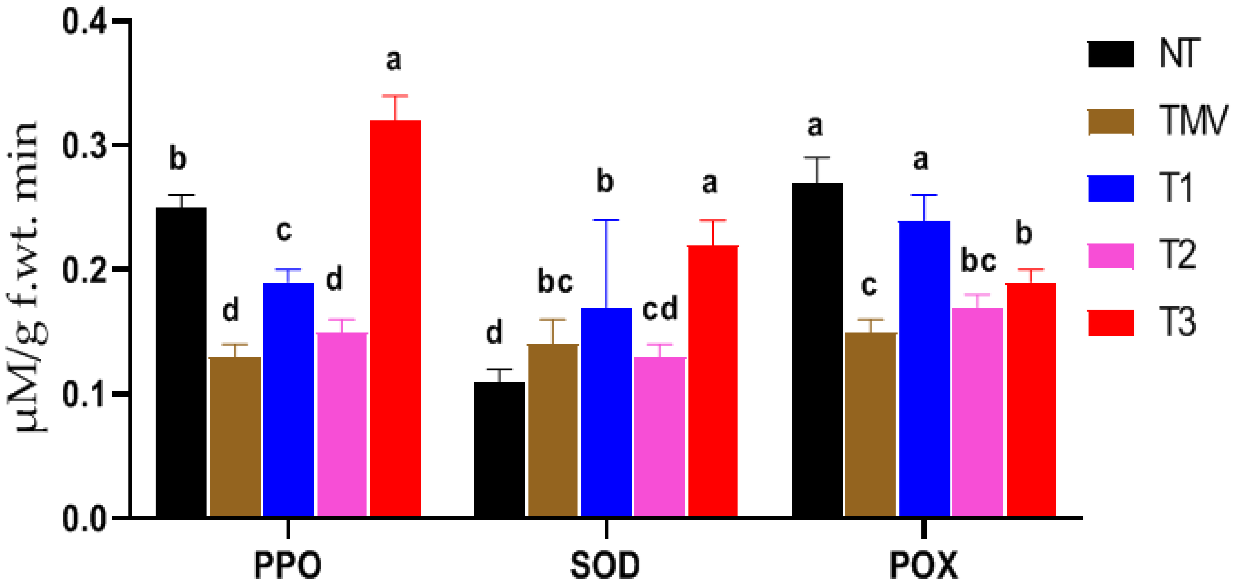
3.4. Antioxidant Enzymatic Activities

Due to the crucial role of antioxidant enzymes in plant defense mechanisms against several plant pathogens, the current work aimed to evaluate the activities of the antioxidant enzymes PPO, SOD, and POX in HA1-CF-treated and -untreated tomato plants upon TMV infection (Figure 4). The enzyme activity assay results indicated a remarkable reduction in PPO activities of more than 50% in the TMV treatment plants (0.13 ± 0.01 µM/g f.wt. min−1) compared to the NT plants (0.25 ± 0.01 µM/g f.wt. min−1). Upon treatment of tomato plants with HA1-CF, PPO activities increased in treatments T1 (0.19 ± 0.01 µM/g f.wt. min−1) as compared to the TMV treatment group. The maximum PPO activity was detected in the T3 treatment (0.32 ± 0.02 µM/g f.wt. min−1) reporting 28% increases in PPO activities compared to the NT control group (Figure 4). The measured PPO activities in the T3 plants were 2.5-fold higher than those assayed in the TMV plants, demonstrating the role of HA1-CF in up-regulation of PPO genes and/or enzymatic activities as a part of the defense mechanism against TMV infection in tomatoes. As a result, it has been reported that overexpression of PPO in various plants has defense properties against bacterial infection in tomato plants [66] and fungal infection in strawberry fruits [67]. Regarding SOD, the results indicated a slight enhancement of about 27% (0.14 ± 0.02 µM/g f.wt. min−1) in SOD activity in the TMV treatment when compared to the NT treatment (0.11 ± 0.01 µM/g f.wt. min−1), which could be attributed to an initial response of the plant defense system to oxidative stress. SOD enzymes have a major role in detoxifying reactive superoxide (O2.−) species into H2O2, which is subsequently degraded through catalases. The SOD activity was also enhanced in all HA-CF treated plants, where the maximum level was shown in T3 (0.22 ± 0.02 µM/g f.wt. min−1), followed by T1 (0.17 ± 0.07 µM/g f.wt. min−1), displaying 2- and 1.5-fold increases, respectively, compared to non-treated plants. Concerning POX activities, the results showed a significant reduction (55%) in POX activity upon TMV infection in TMV treatment (0.15 ± 0.01 µM/g f.wt. min−1), when compared to the NT plants (0.27 ± 0.02 µM/g f.wt. min−1). Compared to TMV treatment plants, the treatment of tomato plants with HA1-CF elevated the POX activities in all treatments at varying concentrations depending on the treatment strategy. The maximum POX activity (0.24 ± 0.02 µM/g f.wt. min−1) was detected in T1 plants, followed by T3, with a level of 0.19 ± 0.01 µM/g f.wt. min−1 (Figure 4). Although the highest activity of POX observed in T1 treatment was less than that reported in the control treatment, no significant changes were reported. Moreover, the increasing POX activity, which is 60% higher than that reported in the TMV treatment plants, demonstrates the efficacy of HA1-CF in improving the POX activities, resulting in boosting tomato plant tolerance under the TMV challenge. POX remarkably enhances the plant’s defense against infection by lignin synthesis, using ROS as a substrate. Lignin deposition increases the physical barrier against viral infection [68]. The obtained results are consistent with other studies that have reported the ability of Bacillus sp. to increase the activity of plant enzymes against ROS [69,70,71]. Moreover, the application of the culture filtrate of B. amyloliquefaciens considerably increased the enzyme activities of SOD, POX, and CAT of Malus hupehensis plants [65].

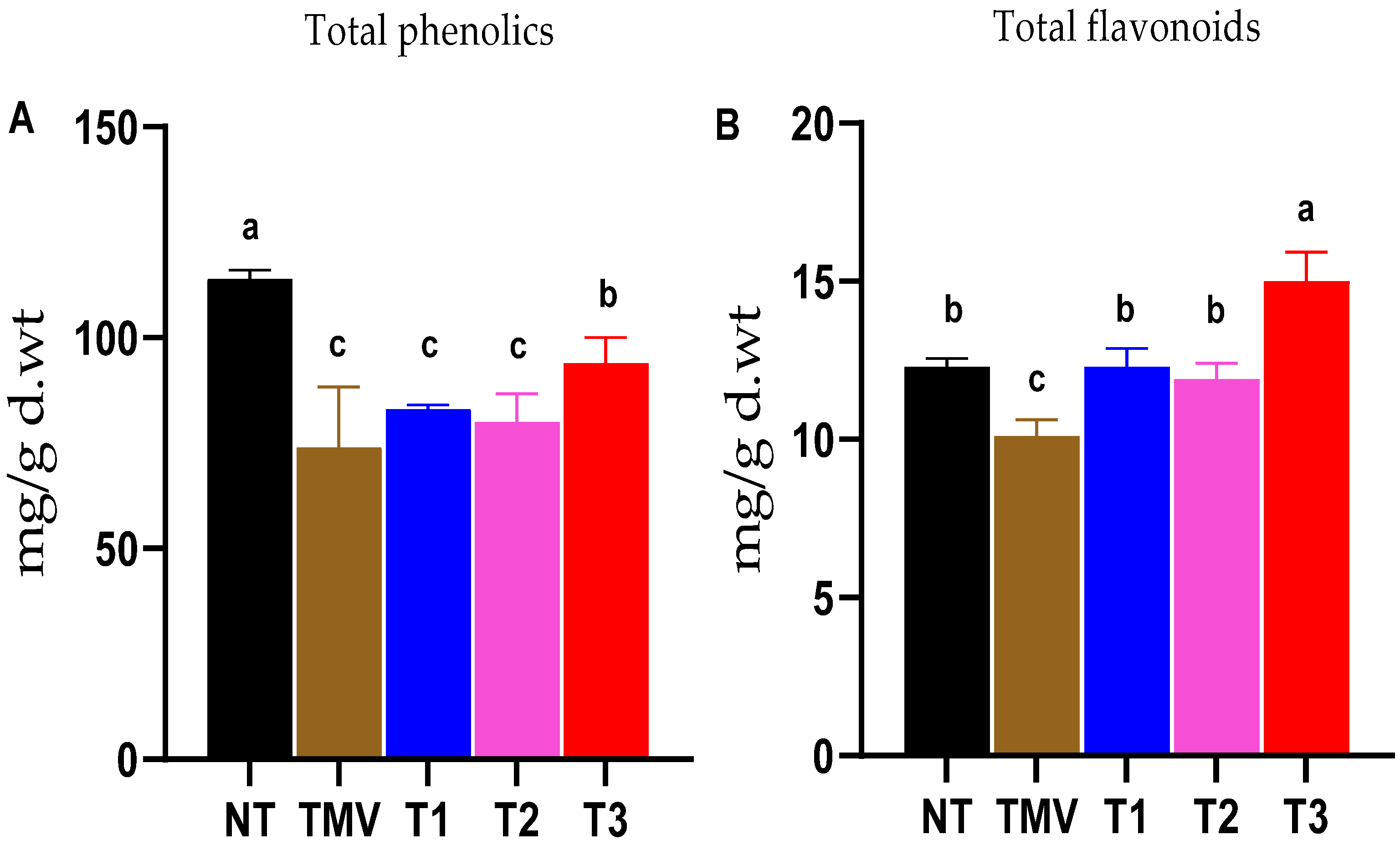
3.5. Total Phenolic and Total Flavonoid Contents

It is well known that the accumulation of polyphenolic phytochemicals, including phenolic and flavonoid compounds, is a main defense mechanism for plants to tolerate biotic and abiotic stresses, including virus infections [8,44,72]. The obtained results in the present study indicated a noticeable reduction (about 35%) in total phenolic contents in the TMV treatment plants (74 ± 14.3 mg/g d.wt.) when compared to control plants (114 ± 2.0 mg/g d.wt.). Interestingly, the T3 treatment exhibited a significant increase in tomato total phenolic contents of 27% (94 ± 6.1 mg/g d.wt.) when compared to the TMV plants (Figure 5A). No significant difference was found between TMV, T1, and T2 treatments (Figure 5A). Consequently, the dual treatment of tomato plants with HA1-CF enhanced the accumulation levels of total phenolic contents inside TMV-infected tissues. On the other hand, the total flavonoid content estimation revealed a significant reduction (18%) in flavonoid contents of the TMV plants (10.1 ± 0.53 mg/g d.wt.), compared to the NT plants (11.4 ± 2.0 mg/g d.wt.), as indicated in Figure 5B. The three HA1-CF-treated groups revealed higher flavonoid contents as compared to the TMV treatment plants. Among them, the T3 plants displayed flavonoid contents (15.0 ± 0.92 mg/g d.wt.) which were 50% and 20% higher than that of the TMV and NT groups, respectively. Notably, no significant differences were detected in total flavonoid contents in the T1 and T2 treatments when compared to the control (NT) treatment. The significant flavonoid accumulation in the T3 treatment plants indicates the importance of treatment doses (two doses in the T3 group) with respect to the treatment time (before or after infection) in the accumulation of such defense compounds.

3.6. Effect of HA1-CF on Systemic Accumulation of TMV

In line with the previously obtained results in this study, the application of HA1-CF, either before or after TMV inoculation, resulted in a considerable reduction in viral accumulation inside tomato tissues. The TMV content was calculated using the TMV-CP gene cycle threshold (Ct) value and the Ct value of the internal control β-actin gene of the tomato control treatment. The qRT-PCR results revealed that the TMV treatment exhibited the highest levels of TMV-CP transcripts, representing a 28.2-fold change, indicating the plant’s viral infection. On the other hand, the HA1-CF-treated plants exhibited relative expression levels of 3.23-, 4.35-, and 2.48-fold changes for T1, T2, and T3 treatments, respectively. These results corresponded with the appearance of the symptoms in terms of the highest and lowest concentrations of the virus inside tomato tissues. Consequently, the significant decline in TMV accumulation levels in T1, T2, and T3 tomato leaves by 88.55, 84.57, and 91.20% compared to TMV treatment plants, confirmed the biocontrol activity of the HA1-CF against TMV infection. In line with our results, the foliar application of the culture filtrate of B. licheniformis and Streptomyces sp. caused a significant reduction in the accumulation of AMV and PVY in potato plants [11,73]. Moreover, the application of Streptomyces cellulosae and B. amyloliquefaciens reduced TMV and CMV severity, and decreased viral accumulation levels in the treated leaves [3,74]. Thus, the application of HA1-CF could protect the tomato plants from TMV infection by preventing the accumulation of viral particles as well as activating the plant defense responses [3,11,48].

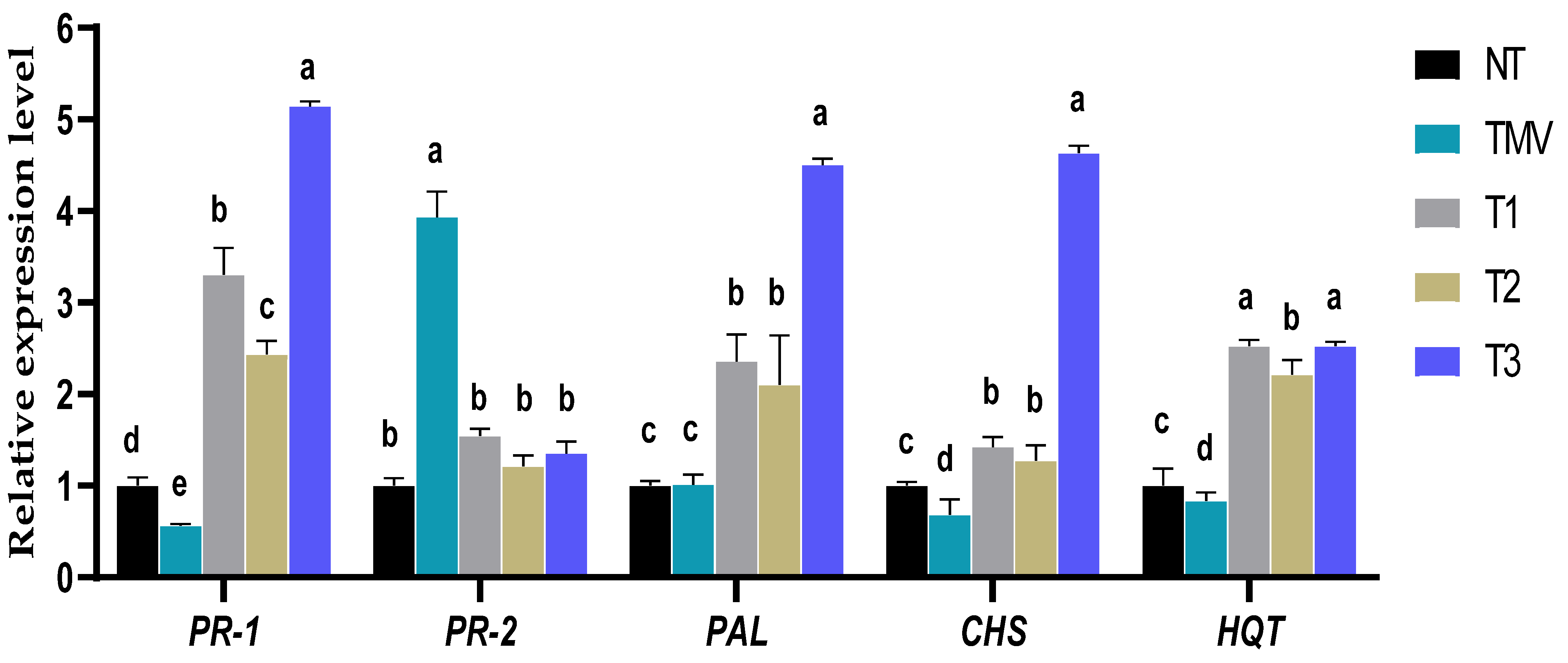
3.7. Effect of HA1-CF on Transcriptional Levels of Pathogenesis-Related Protein Genes

According to several research reports, the induction of different groups of PR proteins plays a vital role in SAR activation and is also efficient at preventing pathogen formation, multiplication, and/or spread. Among them, the genes encoding for PR-1 and PR-2 (evaluated in this study) may be the most significant markers for plant viral infection [3,8,75]. For PR-1, it was shown that the TMV treatment plants showed a significant down-regulation, with a relative expression level of 0.56-fold change lower than control treatment plants (Figure 6). Interestingly, all HA1-CF-treated plants exhibited a considerable increase in the relative expression level of PR-1, with relative expression levels of 2.43-, 3.30-, and 5.14-fold change in T2, T1, and T3, respectively, greater than control (Figure 6). It was reported that the application of the cell-free filtrate of B. velezensis increased the resistance of Datura stramonium and tomato plants against Cucumber mosaic virus and Tomato yellow leaf curl virus, respectively, by significantly elevating the expression of the PR-1 gene [48,76]. It is well known that salicylic acid (SA) plays a crucial role in stimulating plant systemic resistance through activating the plant defense system [77]. Upon viral infection, the accumulation of SA is frequently accompanied by the induction of PR-1, an SA marker gene. Meanwhile, the induction of PR-1 is associated with plant immunity activation and increasing plant resistance against pathogens [21,78]. Consequently, we propose that HA1-CF contains some elicitor secondary metabolite compounds that play a vital role in the induction of PR-1, resulting in activating systemic acquired resistance (SAR) and boosting plant resistance to viral infection.
Regarding the PR-2 transcript, the results indicated a significant induction of PR-2, observed only in TMV plants, with a relative expression level of 3.93-fold change higher than control (Figure 6). Despite HA1-CF-treated plants showing a minor up-regulation of PR-2, with relative expression levels of 1.54-, 1.21-, and 1.35-fold change in T1, T2, and T3, respectively, no significant differences were detected when compared to the control (Figure 6). The PR-2 gene encodes a protein conferring β-1,3-glucanase activity that facilitates viral translocation between plant cells across plasmodesmata, and hence TMV may induce this gene to facilitate its movement and spread through plant cells [6,79]. Previous research has shown that PR-2 is clearly induced during viral infections in potato, Arabidopsis, onion, tobacco, and tomato plants [3]. Furthermore, a lack of tobacco PR-2 expression reduced viral infection susceptibility, whereas overexpression accelerated the transmission of PVY across cells [80,81,82]. Consequently, the foliar application of HA1-CF may reduce TMV infection via lowering PR-2 expression and inhibiting long-distance movement between cells.

3.8. Effect of HA1-CF on the Transcript Levels of Polyphenolic Biosynthesis Genes

It is well known that the accumulation of polyphenolic compounds is a crucial plant defense mechanism against several biotic and abiotic stressors. They are brought to infection sites, elicit hypersensitive responses, and promote programmed cell death when plants are infected [72,83]. In higher plants, biosynthesis of polyphenolic compounds occurs mainly via the shikimate pathway, with three major routes, including the phenylpropanoid, chlorogenic, and flavonoid pathways [29,84]. Besides being the key enzyme in the first step of the phenylpropanoid pathway that is responsible for the conversion of phenylalanine to cinnamic acid, PAL plays a vital role in SA biosynthesis regulation [85,86]. Compared to the mock-inoculated (NT) tomato plants in the present study, the expression levels of PAL were significantly induced in HA1-CF-treated plants, with relative transcriptional levels of 2.36-, 2.10-, and 4.50-fold change in T1, T2, and T3, respectively, higher than the control (Figure 6). On the other hand, no significant difference was reported between TMV treatment and NT treatment plants. The obtained data revealed that the treatment of tomato plants with HA1-CF triggered the expression of PAL, which may potentially increase SA accumulation. The obtained results agree with previous reports showing that the application of the culture filtrate of B. licheniformis and B. velezensis significantly increased the expression levels of PAL and resulted in increased plant resistance against viral infections [11,48]. Consequently, we suggest that HA1-CF could be applied as an efficient ISR elicitor, boosting secondary metabolite biosynthesis, polyphenolic compounds, and SA accumulation in treated plants.
CHS is the initial enzyme in the flavonoid pathway, catalyzing the conversion of p-coumaroyl CoA to naringenin chalcones, which are the major precursor and key intermediates for the synthesis of a wide variety of flavonoids in different plant tissues [84,87]. Likewise, HQT is a strategic enzyme in the biosynthesis of chlorogenic acid. It is one of the most significant polyphenolic molecules that directly enhances plant defense, being involved in various pathogen resistances as well as displaying antioxidant properties [88,89,90]. Compared to the NT treatment plants, the untreated leaves challenged with TMV only showed a significant down-regulation of both CHS and HQT, with relative expression levels of 0.68- and 0.83-fold change, respectively (Figure 6). Thus, the down-regulation of transcriptional levels of CHS and HQT in TMV treatment plants reflected the decrease in the total flavonoid contents detected in this treatment. In this context, many previous studies have shown that the biosynthesis of flavonoid compounds, including chlorogenic acid, is suppressed inside virally infected plant tissues [8,11,75,91,92]. Notably, HA1-CF-treated leaves of T1, T2, and T3 treatments exhibited significant elevations of both CHS and HQT expression levels when compared to the control. (Figure 6). The highest expression level of CHS (4.63-fold) was reported in T3 plants, while the maximum transcriptional level of HQT was observed in both T1 and T3, with the same expression level of 2.52-fold change higher than control (Figure 6). In line with the obtained results in this study, the induction of CHS and HQT genes indicated the accumulation of the flavonoid compounds that play a role in plant resistance against pathogen infection [8,11,22]. Overall, results obtained in the present study show that the foliar application of HA1-CF activates ISR and up-regulates PR-1, PAL, CHS, and HQT. In addition, the accumulation of polyphenolic compounds in response to HA1-CF may contribute to the development of induced resistance and the suppression of TMV infection.

3.9. Identification of Bioactive Metabolites of HA1-CF by GC–MS

Because microbial secondary metabolites are the primary precursors for many biological activities, investigating such metabolites using various analytical approaches is a prerequisite for gaining a deep and comprehensive understanding of biological control toward novel applications [93]. GC–MS analysis is currently a reliable and robust analytical technique, widely applied in bioactive compound analysis and identification [94]. In the present study, the identification of bioactive compounds in the ethyl acetate extract of HA1-CF was conducted by using the mass spectrum of a GC–MS instrument. Results of our GC–MS analyses indicated the presence of 15 different components in HA1-CF; the most abundant compounds are presented in Table 3. The dominant compound detected was phenol, 2,4-bis(1,1-dimethylethyl)- at a retention time of 12.19. Interestingly, phenol, 2,4-bis (1,1-dimethylethyl)- has been reported to accumulate in plant cells under fungal and bacterial attack, and represents a major compound contributing to disease resistance in avocado and Malaysian mango kernel through inhibition of reactive oxygen species (ROS) produced by the pathogen [95,96]. TMV infection is usually associated with elevated levels of ROS [55,56], therefore, inhibition of ROS could alleviate the symptoms of viral infection. Furthermore, several amino acid residues, such as L-proline, N-valeryl, and heptadecyl ester, were detected in our analyses at a retention time of 15.54. Some additional compounds were also detected in the HA1-CF, as indicated in Table 3. The GC–MS results indicated the presence of two fatty acids, eicosane (a long-chain fatty acid) and pentadecanoic acid (asaturated fatty acid) in the HA1-CF at a retention time of 13.7 and 15.48 min, respectively. Eicosane is a biologically active compound derived from Streptomyces sp., shown to be a potent antifungal in treating Rhizoctonia solani spot disease in tobacco leaves [97]. The presence of eicosane has also been reported in other microbes [98], as well as plant extracts that exhibit major antimicrobial activities [99]. Another compound, Pyrrolo [1,2-a] pyrazine-1,4-dione, hexahydro-3-(2-methylpropyl)- was also detected at a retention time of 15.435 min (Table 3). Pyrrole and pyrrolizidines are well-known heterocyclic compounds with diverse biological activities such as antimicrobial, anticancer, antiviral, and anti-inflammatory effects [100]. The unique structures of pyrrole and pyrrolizidines with at least two different elements broaden their biological activities and applications, accounting for more than 75% of the currently applied drugs in clinical use [101]. The findings are consistent with the previously reported efficacy of B. velezensis PEA1 as a Fusarium oxysporum inhibitor, as well as an enhancer of systemic resistance to Cucumber mosaic virus in Datura stramonium, which was attributed to the presence of several different pyrrolo[1,2-a]pyrazine-1,4-dione compounds in the culture filtrate extract [48]. In addition, pyrrole and pyrrolizidines have been detected in various microbial extracts displaying potent biological activities, including antimicrobial [102], antioxidant [103], and antiviral effects [104]. Accordingly, our GC–MS analysis results revealed the presence of several biologically active compounds with reported antifungal and antibacterial activities in the HA1-CF supernatant. Importantly, the current study demonstrated a significant antiviral activity against TMV, plus the growth stimulation properties of HA1-CF in tomatoes. However, further investigations are required to directly associate the above-mentioned biological activities to the compounds we have detected in HA1-CF.

4. Conclusions

We have demonstrated that, under greenhouse conditions, the foliar application of the culture filtrate of B. subtilis strain HA1 (HA1-CF) significantly enhances tomato growth parameters and antioxidant enzyme activities (PPO, SOD, and POX) that alleviate the oxidative stress resulting from TMV infection in all treated plants compared to non-treated plants. Furthermore, HA1-CF treatments resulted in the induction of different defense-related genes (PR-1, PAL, CHS, and HQT) and a significant decline in TMV accumulation, indicating the activation of induced resistance, effective against TMV. Certain compounds detected in the ethyl acetate extract of HA1-CF (e.g., phenol, 2,4-bis (1,1-dimethylethyl)-, Pyrrolo[1,2-a]pyrazine-1,4-dione, hexahydro-3-(2-methylpropyl)-, and eicosane) could potentially be used as plant growth promoters and defense modulatory agents to protect tomato plants against TMV infection. However, further investigations are required to elucidate the different antiviral properties of HA1-CF before it can be used in open-field applications or for commercial purposes. Future research should primarily focus on optimizing and testing the culture filtrate in different plant-virus systems.

Author Contributions

Conceptualization, A.A.; methodology, H.E.-G., M.A.S. and A.A.; software, H.E.-G. and A.A.; validation, A.A.A.-A., A.A. and H.M.; formal analysis, A.A.; investigation, H.E.-G.; resources, A.A.A.-A.; data curation, H.E.-G.; writing—original draft preparation, H.E.-G. and A.A.; writing—review and editing, H.E.-G., A.A.A.-A., A.A., L.K. and H.M.; visualization, A.A.; supervision, H.M.; project administration A.A.A.-A.; funding acquisition, A.A.A.-A. and A.A. All authors have read and agreed to the published version of the manuscript.

Funding

This work was partially funded by the Science and Technology Development Fund (STDF), Egypt, Grant No 30102. This research was financially supported by the Researchers Supporting Project number (RSP2022R505), King Saud University, Riyadh, Saudi Arabia.

Institutional Review Board Statement

Not applicable.

Informed Consent Statement

Not applicable.

Data Availability Statement

Not applicable.

Acknowledgments

This paper is based upon work supported by Science, Technology & Innovation Funding Authority (STDF) under grant (30102). The authors would like to extend their appreciation to the Researchers Supporting Project number (RSP2022R505), King Saud University, Riyadh, Saudi Arabia.

Conflicts of Interest

The authors declare no conflict of interest.

References

  1. Abdelkhalek, A.; Hafez, E. Plant Viral Diseases in Egypt and Their Control. In Cottage Industry of Biocontrol Agents and Their Applications; Springer: Berlin/Heidelberg, Germany, 2020; pp. 403–421. [Google Scholar]
  2. Mumford, R.A.; Macarthur, R.; Boonham, N. The role and challenges of new diagnostic technology in plant biosecurity. Food Secur. 2016, 8, 103–109. [Google Scholar] [CrossRef]
  3. Abo-Zaid, G.A.; Matar, S.M.; Abdelkhalek, A. Induction of Plant Resistance against Tobacco Mosaic Virus Using the Biocontrol Agent Streptomyces cellulosae Isolate Actino 48. Agronomy 2020, 10, 1620. [Google Scholar] [CrossRef]
  4. McDaniel, L.; Maratos, M.; Farabaugh, J. Infection of plants by tobacco mosaic virus. Am. Biol. Teach. 1998, 60, 434–439. [Google Scholar] [CrossRef]
  5. Peng, J.; Song, K.; Zhu, H.; Kong, W.; Liu, F.; Shen, T.; He, Y. Fast detection of Tobacco Mosaic Virus infected tobacco using laser-induced breakdown spectroscopy. Sci. Rep. 2017, 7, 44551. [Google Scholar] [CrossRef]
  6. Abdelkhalek, A. Expression of tomato pathogenesis related genes in response to Tobacco mosaic virus. J. Anim. Plant Sci. 2019, 29, 1596–1602. [Google Scholar]
  7. Bazzini, A.A.; Hopp, H.E.; Beachy, R.N.; Asurmendi, S. Infection and coaccumulation of Tobacco Mosaic Virus proteins alter microRNA levels, correlating with symptom and plant development. Proc. Natl. Acad. Sci. USA 2007, 104, 12157–12162. [Google Scholar] [CrossRef] [Green Version]
  8. Abdelkhalek, A.; Al-Askar, A.A.; Alsubaie, M.M.; Behiry, S.I. First Report of Protective Activity of Paronychia argentea Extract against Tobacco Mosaic Virus Infection. Plants 2021, 10, 2435. [Google Scholar] [CrossRef] [PubMed]
  9. Sharma, A.; Kumar, V.; Shahzad, B.; Tanveer, M.; Sidhu, G.P.S.; Handa, N.; Kohli, S.K.; Yadav, P.; Bali, A.S.; Parihar, R.D.; et al. Worldwide pesticide usage and its impacts on ecosystem. SN Appl. Sci. 2019, 1, 1446. [Google Scholar] [CrossRef] [Green Version]
  10. Alengebawy, A.; Abdelkhalek, S.T.; Qureshi, S.R.; Wang, M.-Q. Heavy Metals and Pesticides Toxicity in Agricultural Soil and Plants: Ecological Risks and Human Health Implications. Toxics 2021, 9, 42. [Google Scholar] [CrossRef] [PubMed]
  11. Abdelkhalek, A.; Al-Askar, A.A.; Behiry, S.I. Bacillus licheniformis strain POT1 mediated polyphenol biosynthetic pathways genes activation and systemic resistance in potato plants against Alfalfa mosaic virus. Sci. Rep. 2020, 10, 16120. [Google Scholar] [CrossRef] [PubMed]
  12. Sorokan, A.; Cherepanova, E.; Burkhanova, G.; Veselova, S.; Rumyantsev, S.; Alekseev, V.; Mardanshin, I.; Sarvarova, E.; Khairullin, R.; Benkovskaya, G.; et al. Endophytic Bacillus spp. as a Prospective Biological Tool for Control of Viral Diseases and Non-vector Leptinotarsa decemlineata Say. in Solanum tuberosum L. Front. Microbiol. 2020, 11, 1–13. [Google Scholar] [CrossRef] [PubMed]
  13. Jiao, X.; Takishita, Y.; Zhou, G.; Smith, D.L. Plant Associated Rhizobacteria for Biocontrol and Plant Growth Enhancement. Front. Plant Sci. 2021, 12, 420. [Google Scholar] [CrossRef] [PubMed]
  14. Wang, H.; Liu, R.; You, M.P.; Barbetti, M.J.; Chen, Y. Pathogen biocontrol using plant growth-promoting bacteria (PGPR): Role of bacterial diversity. Microorganisms 2021, 9, 1988. [Google Scholar] [CrossRef] [PubMed]
  15. Pellegrini, M.; Pagnani, G.; Bernardi, M.; Mattedi, A.; Spera, D.M.; Del Gallo, M. Cell-free supernatants of plant growth-promoting bacteria: A review of their use as biostimulant and microbial biocontrol agents in sustainable agriculture. Sustainability 2020, 12, 9917. [Google Scholar] [CrossRef]
  16. Radhakrishnan, R.; Hashem, A.; Abd Allah, E.F. Bacillus: A biological tool for crop improvement through bio-molecular changes in adverse environments. Front. Physiol. 2017, 8, 1–14. [Google Scholar] [CrossRef] [PubMed]
  17. Vejan, P.; Abdullah, R.; Khadiran, T.; Ismail, S.; Nasrulhaq Boyce, A. Role of plant growth promoting rhizobacteria in agricultural sustainability—A review. Molecules 2016, 21, 573. [Google Scholar] [CrossRef] [PubMed]
  18. Saeed, Q.; Xiukang, W.; Haider, F.U.; Kučerik, J.; Mumtaz, M.Z.; Holatko, J.; Naseem, M.; Kintl, A.; Ejaz, M.; Naveed, M.; et al. Rhizosphere bacteria in plant growth promotion, biocontrol, and bioremediation of contaminated sites: A comprehensive review of effects and mechanisms. Int. J. Mol. Sci. 2021, 22, 10529. [Google Scholar] [CrossRef] [PubMed]
  19. Trabelsi, D.; Mhamdi, R. Microbial inoculants and their impact on soil microbial communities: A review. BioMed Res. Int. 2013, 2013, 863240. [Google Scholar] [CrossRef]
  20. Vitti, A.; Pellegrini, E.; Nali, C.; Lovelli, S.; Sofo, A.; Valerio, M.; Scopa, A.; Nuzzaci, M. Trichoderma harzianum T-22 induces systemic resistance in tomato infected by Cucumber mosaic virus. Front. Plant Sci. 2016, 7, 1520. [Google Scholar] [CrossRef] [PubMed] [Green Version]
  21. Abo-Zaid, G.; Abdelkhalek, A.; Matar, S.; Darwish, M.; Abdel-Gayed, M. Application of Bio-Friendly Formulations of Chitinase-Producing Streptomyces cellulosae Actino 48 for Controlling Peanut Soil-Borne Diseases Caused by Sclerotium rolfsii. J. Fungi 2021, 7, 167. [Google Scholar] [CrossRef]
  22. Heflish, A.A.; Abdelkhalek, A.; Al-Askar, A.A.; Behiry, S.I. Protective and Curative Effects of Trichoderma asperelloides Ta41 on Tomato Root Rot Caused by Rhizoctonia solani Rs33. Agronomy 2021, 11, 1162. [Google Scholar] [CrossRef]
  23. Kandan, A.; Ramiah, M.; Vasanthi, V.J.; Radjacommare, R.; Nandakumar, R.; Ramanathan, A.; Samiyappan, R. Use of Pseudomonas fluorescens-based formulations for management of tomato spotted wilt virus (TSWV) and enhanced yield in tomato. Biocontrol Sci. Technol. 2005, 15, 553–569. [Google Scholar] [CrossRef]
  24. Deng, Z.S.; Zhao, L.F.; Kong, Z.Y.; Yang, W.Q.; Lindström, K.; Wang, E.T.; Wei, G.H. Diversity of endophytic bacteria within nodules of the Sphaerophysa salsula in different regions of Loess Plateau in China. FEMS Microbiol. Ecol. 2011, 76, 463–475. [Google Scholar] [CrossRef] [PubMed]
  25. El-Dougdoug, K.A.; Ghaly, M.F.; Taha, M.A. Biological control of Cucumber mosaic virus by certain local Streptomyces isolates: Inhibitory effects of selected five Egyptian isolates. Intl. J. Virol. 2012, 8, 151–164. [Google Scholar] [CrossRef]
  26. Weisburg, W.G.; Barns, S.M.; Pelletier, D.A.; Lane, D.J. 16S ribosomal DNA amplification for phylogenetic study. J. Bacteriol. 1991, 173, 697–703. [Google Scholar] [CrossRef] [PubMed] [Green Version]
  27. Zhao, L.; Dong, J.; Hu, Z.; Li, S.; Su, X.; Zhang, J.; Yin, Y.; Xu, T.; Zhang, Z.; Chen, H. Anti-TMV activity and functional mechanisms of two sesquiterpenoids isolated from Tithonia diversifolia. Pestic. Biochem. Physiol. 2017, 140, 24–29. [Google Scholar] [CrossRef]
  28. Kavroulakis, N.; Ehaliotis, C.; Ntougias, S.; Zervakis, G.I.; Papadopoulou, K.K. Local and systemic resistance against fungal pathogens of tomato plants elicited by a compost derived from agricultural residues. Physiol. Mol. Plant Pathol. 2005, 66, 163–174. [Google Scholar] [CrossRef]
  29. André, C.M.; Schafleitner, R.; Legay, S.; Lefèvre, I.; Aliaga, C.A.A.; Nomberto, G.; Hoffmann, L.; Hausman, J.-F.; Larondelle, Y.; Evers, D. Gene expression changes related to the production of phenolic compounds in potato tubers grown under drought stress. Phytochemistry 2009, 70, 1107–1116. [Google Scholar] [CrossRef] [PubMed]
  30. Sagi, M.; Davydov, O.; Orazova, S.; Yesbergenova, Z.; Ophir, R.; Stratmann, J.W.; Fluhr, R. Plant respiratory burst oxidase homologs impinge on wound responsiveness and development in Lycopersicon esculentum. Plant Cell 2004, 16, 616–628. [Google Scholar] [CrossRef] [Green Version]
  31. Hafez, E.E.; El-Morsi, A.A.; El-Shahaby, O.A.; Abdelkhalek, A.A. Occurrence of iris yellow spot virus from onion crops in Egypt. VirusDisease 2014, 25, 455–459. [Google Scholar] [CrossRef] [PubMed] [Green Version]
  32. Heath, R.L.; Packer, L. Photoperoxidation in isolated chloroplasts: I. Kinetics and stoichiometry of fatty acid peroxidation. Arch. Biochem. Biophys. 1968, 125, 189–198. [Google Scholar] [CrossRef]
  33. Junglee, S.; Urban, L.; Sallanon, H.; Lopez-Lauri, F. Optimized Assay for Hydrogen Peroxide Determination in Plant Tissue Using Potassium Iodide. Am. J. Anal. Chem. 2014, 05, 730–736. [Google Scholar] [CrossRef] [Green Version]
  34. Cho, Y.K.; Ahn, H.K. Purification and characterization of polyphenol oxidase from potato: II. Inhibition and catalytic mechanism. J. Food Biochem. 1999, 23, 593–605. [Google Scholar] [CrossRef]
  35. Beauchamp, C.; Fridovich, I. Superoxide dismutase: Improved assays and an assay applicable to acrylamide gels. Anal. Biochem. 1971, 44, 276–287. [Google Scholar] [CrossRef]
  36. Kumar, A.; Dutt, S.; Bagler, G.; Ahuja, P.S.; Kumar, S. Engineering a thermo-stable superoxide dismutase functional at sub-zero to >50°C, which also tolerates autoclaving. Sci. Rep. 2012, 2, srep00387. [Google Scholar] [CrossRef] [Green Version]
  37. Angelini, R.; Manes, F.; Federico, R. Spatial and functional correlation between diamine-oxidase and peroxidase activities and their dependence upon de-etiolation and wounding in chick-pea stems. Planta 1990, 182, 89–96. [Google Scholar] [CrossRef] [PubMed]
  38. Singleton, V.L.; Orthofer, R.; Lamuela-Raventós, R.M. [14]Analysis of total phenols and other oxidation substrates and antioxidants by means of folin-ciocalteu reagent. In Oxidants and Antioxidants Part A; Academic Press: San Diego, CA, USA, 1999; Volume 299, pp. 152–178. ISBN 0076-6879. [Google Scholar]
  39. Ghosh, S.; Derle, A.; Ahire, M.; More, P.; Jagtap, S.; Phadatare, S.D.; Patil, A.B.; Jabgunde, A.M.; Sharma, G.K.; Shinde, V.S.; et al. Phytochemical analysis and free radical scavenging activity of medicinal plants Gnidia glauca and Dioscorea bulbifera. PLoS ONE 2013, 8, e82529. [Google Scholar] [CrossRef] [PubMed] [Green Version]
  40. Abdelkhalek, A.; Ismail, I.A.I.A.; Dessoky, E.S.E.S.; El-Hallous, E.I.E.I.; Hafez, E. A tomato kinesin-like protein is associated with Tobacco Mosaic Virus infection. Biotechnol. Biotechnol. Equip. 2019, 33, 1424–1433. [Google Scholar] [CrossRef] [Green Version]
  41. Abdelkhalek, A.; Qari, S.H.S.H.; Hafez, E. Iris yellow spot virus–induced chloroplast malformation results in male sterility. J. Biosci. 2019, 44, 142. [Google Scholar] [CrossRef] [PubMed]
  42. Abdelkhalek, A.; Al-Askar, A.A.; Arishi, A.A.; Behiry, S.I. Trichoderma hamatum Strain Th23 Promotes Tomato Growth and Induces Systemic Resistance against Tobacco Mosaic Virus. J. Fungi 2022, 8, 228. [Google Scholar] [CrossRef]
  43. Hafez, E.E.; Abdelkhalek, A.A.; Abd El-Wahab, A.S.E.-D.; Galal, F.H. Altered gene expression: Induction/suppression in leek elicited by Iris Yellow Spot Virus infection (IYSV) Egyptian isolate. Biotechnol. Biotechnol. Equip. 2013, 27, 4061–4068. [Google Scholar] [CrossRef] [Green Version]
  44. Abdelkhalek, A.; Qari, S.H.; Abu-Saied, M.A.A.-R.; Khalil, A.M.; Younes, H.A.; Nehela, Y.; Behiry, S.I. Chitosan Nanoparticles Inactivate Alfalfa Mosaic Virus Replication and Boost Innate Immunity in Nicotiana glutinosa Plants. Plants 2021, 10, 2701. [Google Scholar] [CrossRef] [PubMed]
  45. Livak, K.J.; Schmittgen, T.D. Analysis of relative gene expression data using real-time quantitative PCR and the 2− ΔΔCT method. Methods 2001, 25, 402–408. [Google Scholar] [CrossRef] [PubMed]
  46. Hashem, A.; Tabassum, B.; Abd_Allah, E.F. Bacillus subtilis: A plant-growth promoting rhizobacterium that also impacts biotic stress. Saudi J. Biol. Sci. 2019, 26, 1291–1297. [Google Scholar] [CrossRef] [PubMed]
  47. Blake, C.; Christensen, M.N.; Kovacs, A.T. Molecular aspects of plant growth promotion and protection by bacillus subtilis. Mol. Plant-Microbe Interact. 2021, 34, 15–25. [Google Scholar] [CrossRef] [PubMed]
  48. Abdelkhalek, A.; Behiry, S.I.; Al-Askar, A.A. Bacillus velezensis PEA1 Inhibits Fusarium oxysporum Growth and Induces Systemic Resistance to Cucumber Mosaic Virus. Agronomy 2020, 10, 1312. [Google Scholar] [CrossRef]
  49. Kloepper, J.W.; Ryu, C.-M.; Zhang, S. Induced systemic resistance and promotion of plant growth by Bacillus spp. Phytopathology 2004, 94, 1259–1266. [Google Scholar] [CrossRef] [PubMed] [Green Version]
  50. Qiao, J.; Yu, X.; Liang, X.; Liu, Y.; Borriss, R.; Liu, Y. Addition of plant-growth-promoting Bacillus subtilis PTS-394 on tomato rhizosphere has no durable impact on composition of root microbiome. BMC Microbiol. 2017, 17, 131. [Google Scholar] [CrossRef] [PubMed]
  51. Bokhari, A.; Essack, M.; Lafi, F.F.; Andres-Barrao, C.; Jalal, R.; Alamoudi, S.; Razali, R.; Alzubaidy, H.; Shah, K.H.; Siddique, S.; et al. Bioprospecting desert plant Bacillus endophytic strains for their potential to enhance plant stress tolerance. Sci. Rep. 2019, 9, 18154. [Google Scholar] [CrossRef]
  52. Saxena, A.K.; Kumar, M.; Chakdar, H.; Anuroopa, N.; Bagyaraj, D.J. Bacillus species in soil as a natural resource for plant health and nutrition. J. Appl. Microbiol. 2020, 128, 1583–1594. [Google Scholar] [CrossRef] [PubMed] [Green Version]
  53. Posada, L.F.; Ramírez, M.; Ochoa-Gómez, N.; Cuellar-Gaviria, T.Z.; Argel-Roldan, L.E.; Ramírez, C.A.; Villegas-Escobar, V. Bioprospecting of aerobic endospore-forming bacteria with biotechnological potential for growth promotion of banana plants. Sci. Hortic. 2016, 212, 81–90. [Google Scholar] [CrossRef]
  54. Arena, G.D.; Ramos-González, P.L.; Nunes, M.A.; Ribeiro-Alves, M.; Camargo, L.E.A.; Kitajima, E.W.; Machado, M.A.; Freitas-Astúa, J. Citrus leprosis virus C infection results in hypersensitive-like response, suppression of the JA/ET plant defense pathway and promotion of the colonization of its mite vector. Front. Plant Sci. 2016, 7, 1757. [Google Scholar] [CrossRef] [Green Version]
  55. Zhu, F.; Deng, X.-G.; Xu, F.; Jian, W.; Peng, X.-J.; Zhu, T.; Xi, D.-H.; Lin, H.-H. Mitochondrial alternative oxidase is involved in both compatible and incompatible host-virus combinations in Nicotiana benthamiana. Plant Sci. 2015, 239, 26–35. [Google Scholar] [CrossRef] [PubMed]
  56. Zhu, F.; Zhang, Q.; Che, Y.; Zhu, P.; Zhang, Q.; Ji, Z. Glutathione contributes to resistance responses to TMV through a differential modulation of salicylic acid and reactive oxygen species. Mol. Plant Pathol. 2021, 22, 1668–1687. [Google Scholar] [CrossRef] [PubMed]
  57. Jaiswal, N.; Singh, M.; Dubey, R.S.; Venkataramanappa, V.; Datta, D. Phytochemicals and antioxidative enzymes defence mechanism on occurrence of yellow vein mosaic disease of pumpkin (Cucurbita moschata). Biotech 2013, 3, 287–295. [Google Scholar] [CrossRef] [PubMed] [Green Version]
  58. Madhusudhan, K.N.; Srikanta, B.M.; Shylaja, M.D.; Prakash, H.S.; Shetty, H.S. Changes in antioxidant enzymes, hydrogen peroxide, salicylic acid and oxidative stress in compatible and incompatible host-tobamovirus interaction. J. Plant Interact. 2009, 4, 157–166. [Google Scholar] [CrossRef]
  59. Radwan, D.E.M.; Ismail, K.S. The impact of hydrogen peroxide against cucumber green mottle mosaic virus infection in watermelon plants. Polish J. Environ. Stud. 2020, 29, 3771–3782. [Google Scholar] [CrossRef]
  60. Rui, R.; Liu, S.; Karthikeyan, A.; Wang, T.; Niu, H.; Yin, J.; Yang, Y.; Wang, L.; Yang, Q.; Zhi, H. Fine-mapping and identification of a novel locus Rsc15 underlying soybean resistance to Soybean mosaic virus. Theor. Appl. Genet. 2017, 130, 2395–2410. [Google Scholar] [CrossRef] [PubMed]
  61. Yoda, H.; Yamaguchi, Y.; Sano, H. Induction of hypersensitive cell death by hydrogen peroxide produced through polyamine degradation in tobacco plants. Plant Physiol. 2003, 132, 1973–1981. [Google Scholar] [CrossRef] [Green Version]
  62. de Dios Alché, J. A concise appraisal of lipid oxidation and lipoxidation in higher plants. Redox Biol. 2019, 23, 101136. [Google Scholar] [CrossRef] [PubMed]
  63. Hernández, J.A.; Gullner, G.; Clemente-Moreno, M.J.; Künstler, A.; Juhász, C.; Díaz-Vivancos, P.; Király, L. Oxidative stress and antioxidative responses in plant–virus interactions. Physiol. Mol. Plant Pathol. 2016, 94, 134–148. [Google Scholar] [CrossRef] [Green Version]
  64. Balal, R.M.; Khan, M.M.; Shahid, M.A.; Mattson, N.S.; Abbas, T.; Ashfaq, M.; Garcia-Sanchez, F.; Ghazanfer, U.; Gimeno, V.; Iqbal, Z. Comparative studies on the physiobiochemical, enzymatic, and ionic modifications in salt-tolerant and salt-sensitive citrus rootstocks under NaCl stress. J. Am. Soc. Hortic. Sci. 2012, 137, 86–95. [Google Scholar] [CrossRef] [Green Version]
  65. Yanan, D.; Ran, C.; Rong, Z.; Jiang, W.; Xuesen, C.; Chengmiao, Y.; Zhiquan, M. Isolation, identification, and antibacterial mechanisms of Bacillus amyloliquefaciens QSB-6 and its effect on plant roots. Front. Microbiol. 2021, 12, 746799. [Google Scholar]
  66. Li, L.; Steffens, J.C. Overexpression of polyphenol oxidase in transgenic tomato plants results in enhanced bacterial disease resistance. Planta 2002, 215, 239–247. [Google Scholar] [CrossRef] [PubMed]
  67. Jia, H.; Zhao, P.; Wang, B.; Tariq, P.; Zhao, F.; Zhao, M.; Wang, Q.; Yang, T.; Fang, J. Overexpression of Polyphenol Oxidase Gene in Strawberry Fruit Delays the Fungus Infection Process. Plant Mol. Biol. Rep. 2016, 34, 592–606. [Google Scholar] [CrossRef]
  68. Mohammadi, M.; Kazemi, H. Changes in peroxidase and polyphenol oxidase activities in susceptible and resistant wheat heads inoculated with Fusarium graminearum and induced resistance. Plant Sci. 2002, 162, 491–498. [Google Scholar] [CrossRef]
  69. Khan, M.S.; Gao, J.; Chen, X.; Zhang, M.; Yang, F.; Du, Y.; Moe, T.S.; Munir, I.; Xue, J.; Zhang, X. Isolation and Characterization of Plant Growth-Promoting Endophytic Bacteria Paenibacillus polymyxa SK1 from Lilium lancifolium. Biomed Res. Int. 2020, 2020, 8650957. [Google Scholar] [CrossRef] [Green Version]
  70. Miljaković, D.; Marinković, J.; Balešević-Tubić, S. The Significance of Bacillus spp. in Disease Suppression and Growth Promotion of Field and Vegetable Crops. Microorganisms 2020, 8, 1037. [Google Scholar] [CrossRef]
  71. Rais, A.; Jabeen, Z.; Shair, F.; Hafeez, F.Y.; Hassan, M.N. Bacillus spp., a bio-control agent enhances the activity of antioxidant defense enzymes in rice against Pyricularia oryzae. PLoS ONE 2017, 12, e0187412. [Google Scholar] [CrossRef] [PubMed] [Green Version]
  72. Akyol, H.; Riciputi, Y.; Capanoglu, E.; Caboni, M.; Verardo, V. Phenolic compounds in the potato and its byproducts: An overview. Int. J. Mol. Sci. 2016, 17, 835. [Google Scholar] [CrossRef] [PubMed]
  73. Nasr-Eldin, M.; Messiha, N.; Othman, B.; Megahed, A.; Elhalag, K. Induction of potato systemic resistance against the potato virus Y (PVY NTN), using crude filtrates of Streptomyces spp. under greenhouse conditions. Egypt. J. Biol. Pest Control 2019, 29, 62. [Google Scholar] [CrossRef] [Green Version]
  74. Lee, G.H.; Ryu, C.-M. Spraying of leaf-colonizing Bacillus amyloliquefaciens protects pepper from Cucumber mosaic virus. Plant Dis. 2016, 100, 2099–2105. [Google Scholar] [CrossRef] [PubMed] [Green Version]
  75. Abdelkhalek, A.; Al-Askar, A.A. Green Synthesized ZnO Nanoparticles Mediated by Mentha Spicata Extract Induce Plant Systemic Resistance against Tobacco Mosaic Virus. Appl. Sci. 2020, 10, 5054. [Google Scholar] [CrossRef]
  76. Guo, Q.; Li, Y.; Lou, Y.; Shi, M.; Jiang, Y.; Zhou, J.; Sun, Y.; Xue, Q.; Lai, H. Bacillus amyloliquefaciens Ba13 induces plant systemic resistance and improves rhizosphere microecology against tomato yellow leaf curl virus disease. Appl. Soil Ecol. 2019, 137, 154–166. [Google Scholar] [CrossRef]
  77. Dempsey, D.M.A.; Vlot, A.C.; Wildermuth, M.C.; Klessig, D.F. Salicylic acid biosynthesis and metabolism. Arab. Book/Am. Soc. Plant Biol. 2011, 9, e0156. [Google Scholar] [CrossRef] [Green Version]
  78. Breen, S.; Williams, S.J.; Outram, M.; Kobe, B.; Solomon, P.S. Emerging Insights into the Functions of Pathogenesis-Related Protein 1. Trends Plant Sci. 2017, 22, 871–879. [Google Scholar] [CrossRef]
  79. Iglesias, V.A.; Meins, F.; Meins, F., Jr. Movement of plant viruses is delayed in a β-1, 3-glucanase-deficient mutant showing a reduced plasmodesmatal size exclusion limit and enhanced callose deposition. Plant J. 2000, 21, 157–166. [Google Scholar] [CrossRef]
  80. Otulak-Kozieł, K.; Kozieł, E.; Lockhart, B. Plant cell wall dynamics in compatible and incompatible potato response to infection caused by Potato virus Y (PVYNTN). Int. J. Mol. Sci. 2018, 19, 862. [Google Scholar] [CrossRef] [Green Version]
  81. Bucher, G.L.; Tarina, C.; Heinlein, M.; Di Serio, F.; Meins, F., Jr.; Iglesias, V.A. Local expression of enzymatically active class I β-1, 3-glucanase enhances symptoms of TMV infection in tobacco. Plant J. 2001, 28, 361–369. [Google Scholar] [CrossRef]
  82. Dobnik, D.; Baebler, Š.; Kogovšek, P.; Pompe-Novak, M.; Štebih, D.; Panter, G.; Janež, N.; Morisset, D.; Žel, J.; Gruden, K. β-1, 3-glucanase class III promotes spread of PVY NTN and improves in planta protein production. Plant Biotechnol. Rep. 2013, 7, 547–555. [Google Scholar] [CrossRef] [Green Version]
  83. Beckman, C.H. Phenolic-storing cells: Keys to programmed cell death and periderm formation in wilt disease resistance and in general defence responses in plants? Physiol. Mol. Plant Pathol. 2000, 57, 101–110. [Google Scholar] [CrossRef]
  84. Abdelkhalek, A.; Dessoky, E.S.; Hafez, E. Polyphenolic genes expression pattern and their role in viral resistance in tomato plant infected with Tobacco mosaic virus. Biosci. Res. 2018, 15, 3349–3356. [Google Scholar]
  85. Huang, J.; Gu, M.; Lai, Z.; Fan, B.; Shi, K.; Zhou, Y.-H.; Yu, J.-Q.; Chen, Z. Functional analysis of the Arabidopsis PAL gene family in plant growth, development, and response to environmental stress. Plant Physiol. 2010, 153, 1526–1538. [Google Scholar] [CrossRef] [PubMed] [Green Version]
  86. Su, H.; Song, S.; Yan, X.; Fang, L.; Zeng, B.; Zhu, Y. Endogenous salicylic acid shows different correlation with baicalin and baicalein in the medicinal plant Scutellaria baicalensis Georgi subjected to stress and exogenous salicylic acid. PLoS ONE 2018, 13, e0192114. [Google Scholar] [CrossRef] [Green Version]
  87. Kang, J.-H.; McRoberts, J.; Shi, F.; Moreno, J.E.; Jones, A.D.; Howe, G.A. The flavonoid biosynthetic enzyme chalcone isomerase modulates terpenoid production in glandular trichomes of tomato. Plant Physiol. 2014, 164, 1161–1174. [Google Scholar] [CrossRef] [Green Version]
  88. Sonnante, G.; D’Amore, R.; Blanco, E.; Pierri, C.L.; De Palma, M.; Luo, J.; Tucci, M.; Martin, C. Novel hydroxycinnamoyl-coenzyme A quinate transferase genes from artichoke are involved in the synthesis of chlorogenic acid. Plant Physiol. 2010, 153, 1224–1238. [Google Scholar] [CrossRef] [Green Version]
  89. Moglia, A.; Lanteri, S.; Comino, C.; Hill, L.; Knevitt, D.; Cagliero, C.; Rubiolo, P.; Bornemann, S.; Martin, C. Dual catalytic activity of hydroxycinnamoyl-coenzyme A quinate transferase from tomato allows it to moonlight in the synthesis of both mono- and dicaffeoylquinic acids. Plant Physiol. 2014, 166, 1777–1787. [Google Scholar] [CrossRef]
  90. Niggeweg, R.; Michael, A.J.; Martin, C. Engineering plants with increased levels of the antioxidant chlorogenic acid. Nat. Biotechnol. 2004, 22, 746. [Google Scholar] [CrossRef]
  91. Abdelkhalek, A.; Al-Askar, A.A.; Hafez, E. Differential induction and suppression of the potato innate immune system in response to Alfalfa mosaic virus infection. Physiol. Mol. Plant Pathol. 2020, 110, 101485. [Google Scholar] [CrossRef]
  92. Bazzini, A.A.; Manacorda, C.A.; Tohge, T.; Conti, G.; Rodriguez, M.C.; Nunes-Nesi, A.; Villanueva, S.; Fernie, A.R.; Carrari, F.; Asurmendi, S. Metabolic and miRNA profiling of TMV infected plants reveals biphasic temporal changes. PLoS ONE 2011, 6, e28466. [Google Scholar] [CrossRef]
  93. Ullah, A.; Bano, A.; Janjua, H.T. Microbial Secondary Metabolites and Defense of Plant Stress. Microb. Serv. Restor. Ecol. 2020, 11, 37–46. [Google Scholar] [CrossRef]
  94. Nas, F.; Aissaoui, N.; Mahjoubi, M.; Mosbah, A.; Arab, M.; Abdelwahed, S.; Khrouf, R.; Masmoudi, A.-S.; Cherif, A.; Klouche-Khelil, N. A comparative GC-MS analysis of bioactive secondary metabolites produced by halotolerant Bacillus spp. isolated from the Great Sebkha of Oran. Int. Microbiol. Off. J. Spanish Soc. Microbiol. 2021, 24, 455–470. [Google Scholar] [CrossRef] [PubMed]
  95. María Teresa, R.-C.; Rosaura, V.-G.; Elda, C.-M.; Ernesto, G.-P. The avocado defense compound phenol-2,4-bis (1,1-dimethylethyl) is induced by arachidonic acid and acts via the inhibition of hydrogen peroxide production by pathogens. Physiol. Mol. Plant Pathol. 2014, 87, 32–41. [Google Scholar] [CrossRef]
  96. Abdullah, A.S.H.; Mirghani, M.E.S.; Jamal, P. Antibacterial activity of Malaysian mango kernel. African J. Biotechnol. 2011, 10, 18739–18748. [Google Scholar] [CrossRef]
  97. Ahsan, T.; Chen, J.; Zhao, X.; Irfan, M.; Wu, Y. Extraction and identification of bioactive compounds (eicosane and dibutyl phthalate) produced by Streptomyces strain KX852460 for the biological control of Rhizoctonia solani AG-3 strain KX852461 to control target spot disease in tobacco leaf. AMB Express 2017, 7, 54. [Google Scholar] [CrossRef] [Green Version]
  98. Octarya, Z.; Novianty, R.; Suraya, N. Saryono Antimicrobial activity and GC-MS analysis of bioactive constituents of Aspergillus fumigatus 269 isolated from Sungai Pinang hot spring, Riau, Indonesia. Biodiversitas 2021, 22, 1839–1845. [Google Scholar] [CrossRef]
  99. Naeim, H.; El-Hawiet, A.; Abdel Rahman, R.A.; Hussein, A.; El Demellawy, M.A.; Embaby, A.M. Antibacterial activity of Centaurea pumilio L. Root and aerial part extracts against some multidrug resistant bacteria. BMC Complement. Med. Ther. 2020, 20, 79. [Google Scholar] [CrossRef] [Green Version]
  100. Bhardwaj, V.; Gumber, D.; Abbot, V.; Dhiman, S.; Sharma, P. Pyrrole: A resourceful small molecule in key medicinal hetero-aromatics. RSC Adv. 2015, 5, 15233–15266. [Google Scholar] [CrossRef]
  101. Sharma, S.; Kumar, D.; Singh, G.; Monga, V.; Kumar, B. Recent advancements in the development of heterocyclic anti-inflammatory agents. Eur. J. Med. Chem. 2020, 200, 112438. [Google Scholar] [CrossRef]
  102. Kumari, N.; Menghani, E.; Mithal, R. GCMS analysis of compounds extracted from actinomycetes AIA6 isolates and study of its antimicrobial efficacy. Indian J. Chem. Technol. 2019, 26, 362–370. [Google Scholar]
  103. Ser, H.-L.; Palanisamy, U.D.; Yin, W.-F.; Abd Malek, S.N.; Chan, K.-G.; Goh, B.-H.; Lee, L.-H. Presence of antioxidative agent, Pyrrolo[1,2-a]pyrazine-1,4-dione, hexahydro- in newly isolated Streptomyces mangrovisoli sp. nov. Front. Microbiol. 2015, 6, 854. [Google Scholar] [CrossRef] [PubMed] [Green Version]
  104. Pooja, S.; Aditi, T.; Naine, S.J.; Devi, C.S. Bioactive compounds from marine Streptomyces sp. VITPSA as therapeutics. Front. Biol. 2017, 12, 280–289. [Google Scholar] [CrossRef]
Figure 1. Phylogenetic tree showing the relationship of the locally isolated B. subtilis strain HA1 (shows in red rectangle) and other closely related isolates based on 16s rRNA (GenBank) nucleotide sequences. The tree was developed by the MEGA 11 program, based on the UPGMA method, with a bootstrap method of 2000 replications.
Horticulturae 08 00301 g001
Figure 2. Effect of foliar application of B. subtilis strain HA1-CF on the development of disease symptoms in tomato leaves at 14 days post-TMV inoculation. NT: mock plants; TMV: virus-infected plants; T1: tomato plants sprayed with bacterial culture filtrate 24 h before virus infection; T2: tomato plants sprayed with bacterial culture filtrate 24 h after virus infection; T3: tomato plants sprayed with bacterial culture filtrate two times, 24 h before and 24 h after virus inoculation.
Horticulturae 08 00301 g002
Figure 3. Effect of foliar application of B. subtilis strain HA1-culture filtrate on the two oxidative stress markers: MDA (A) and H2O2 (B) of tomato plants at 22 days post-TMV inoculation. NT: mock plants; TMV: virus-infected plants; T1: tomato plants sprayed with bacterial culture filtrate 24 h before viral infection; T2: tomato plants sprayed with bacterial culture filtrate 24 h after viral infection; T3: tomato plants sprayed with bacterial culture filtrate two times, 24 h before and 24 h after viral inoculation. Each column value represents the mean result obtained from five biological replicates. Significant differences were calculated using a one-way ANOVA according to the least significant difference (LSD) method at a p ≤ 0.05 level of probability with the GraphPad Prism software package. The mean values of each column with the same letter do not differ significantly.
Horticulturae 08 00301 g003
Figure 4. Effect of foliar application of B. subtilis strain HA1-culture filtrate on activities of antioxidant enzymes PPO, SOD, and POX of tomato plants at 22 days post-TMV inoculation. NT: mock plants; TMV: virus-infected plants; T1: tomato plants sprayed with bacterial culture filtrate 24 h before viral infection; T2: tomato plants sprayed with bacterial culture filtrate after 24 h of viral infection; T3: tomato plants sprayed with bacterial culture filtrate two times, 24 h before and 24 h after the viral inoculation. Each column value represents the mean result obtained from five biological replicates. Significant differences were calculated using a one-way ANOVA according to the least significant difference (LSD) method at a p ≤ 0.05 level of probability with the GraphPad Prism software package. The mean values of each column with the same letter do not differ significantly.
Horticulturae 08 00301 g004
Figure 5. Effect of foliar application of B. subtilis strain HA1-culture filtrate on the total phenolic (A) and total flavonoid (B) contents of tomato plants at 22 days post-TMV inoculation. NT: mock plants; TMV: virus-infected plants; T1: tomato plants sprayed with bacterial culture filtrate 24 h before virus infection; T2: tomato plants sprayed with bacterial culture filtrate 24 h after virus infection; T3: tomato plants sprayed with bacterial culture filtrate two times, 24 h before and 24 h after virus inoculation. Each column value represents the mean result obtained from five biological replicates. Significant differences were calculated using a one-way ANOVA according to the least significant difference (LSD) method at a p ≤ 0.05 level of probability with the GraphPad Prism software package. The mean values of each column with the same letter do not differ significantly.
Horticulturae 08 00301 g005
Figure 6. The relative expression levels of genes encoding pathogenesis-related proteins (PR-1, and PR-2) and polyphenol biosynthetic enzymes (PAL, CHS, and HQT) in tomato plants at 22 days post-TMV inoculation. NT: mock plants; TMV: virus-infected plants; T1: tomato plants sprayed with bacterial culture filtrate 24 h before virus infection; T2: tomato plants sprayed with bacterial culture filtrate after 24 h of virus infection; T3: tomato plants sprayed with bacterial culture filtrate two times, 24 h before and 24 h after virus inoculation. Each column value represents the mean result obtained from five biological replicates. Significant differences were calculated using a one-way ANOVA according to the least significant difference (LSD) method at a p ≤ 0.05 level of probability with the GraphPad Prism software package. The mean values of each column with the same letter do not differ significantly.
Horticulturae 08 00301 g006
Table 1. List of the specific primer sequences of the different genes used in qRT PCR analysis.
Primer NameAbbreviationNucleotide SequenceReferences
16S ribosomal RNA16S rRNAForward: AGAGTGATCCTGGCTCAG[26]
Reverse: GGTTACCTTGTTACGACTT
Tobacco mosaic virus-coat proteinTMV-CPForward: ACGACTGCCGAAACGTTAGA[27]
Reverse: CAAGTTGCAGGACCAGAGGT
Pathogenesis related protein-1PR-1Forward: GTTCCTCCTTGCCACCTTC[28]
Reverse: TATGCACCCCCAGCATAGTT
EndoglucanasePR-2Forward: TATAGCCGTTGGAAACGAAG[28]
Reverse: CAACTTGCCATCACATTCTG
Phenylalanine Ammonia-LyasePALForward: ACGGGTTGCCATCTAATCTGACA[29]
Reverse: CGAGCAATAAGAAGCCATCGCAAT
Chalcone SynthaseCHSForward: CACCGTGGAGGAGTATCGTAAGGC[29]
Reverse: TGATCAACACAGTTGGAAGGCG
Hydroxycinnamoyl Co A: quinate (break)hydroxycinnamoyl transferaseHQTForward: CCCAATGGCTGGAAGATTAGCTA[29]
Reverse: CATGAATCACTTTCAGCCTCAACAA
β-actinβ-actinForward: TGGCATACAAAGACAGGACAGCCT[30]
Reverse: ACTCAATCCCAAGGCCAACAGAGA
Table 2. Effect of foliar application of B. subtilis strain HA1-CF on growth parameters of tomato plants at 22 days post-TMV inoculation.
Treatment *Fresh Weight g/PlantDry Weight g/PlantShoot Length cm/PlantRoot Length cm/Plant
NT7.15 ± 0.37 bc2.24 ± 0.03 ab27.67 ± 3.3014.17 ± 1.55 ab
TMV6.28 ± 0.61 c1.89 ± 0.07 c26.17 ± 0.249.33 ± 2.63 b
T19.36 ± 2.20 ab2.27 ± 0.11 ab28.33 ± 4.5014 ± 4.55 ab
T26.47 ± 0.87 bc2.14 ± 0.15 bc27.33 ± 2.4911.33 ± 1.25 ab
T310.35 ± 1.19 a2.43 ± 0.16 a29.67 ± 2.0616.33 ± 2.87 a
* NT: mock plants; TMV: virus-infected plants; T1: tomato plants sprayed with bacterial culture filtrate 24 h before viral infection; T2: tomato plants sprayed with bacterial culture filtrate 24 h after viral infection; T3: tomato plants sprayed with bacterial culture filtrate two times, 24 h before and 24 h after viral inoculation. Each column value represents the mean result obtained from five biological replicates. Significant differences were calculated using a one-way ANOVA according to the least significant difference (LSD) method at a p ≤ 0.05 level of probability with the GraphPad Prism software package. The mean values of each column with the same letter do not differ significantly.
Table 3. The highest six compounds detected in HA1-CF as revealed with GC–MS analysis.
PeakR. Time (min.)AreaNameChemical FormulaMolecular Structure
211.960560.11Nonane, 5-(2-methylpropyl)-C13H28 Horticulturae 08 00301 i001
312.1871.592.35Phenol, 2,4-bis(1,1-dimethylethyl)-C14H22O Horticulturae 08 00301 i002
613.689475.26EicosaneC20H42 Horticulturae 08 00301 i003
1215.435535.02Pyrrolo[1,2-a]pyrazine-1,4-dione, hexahydro-3-(2-methylpropyl)-C10H16N2O2 Horticulturae 08 00301 i004
1315.481549.58Pentadecanoic acidC15H30O2 Horticulturae 08 00301 i005
1415.542835.29L-Proline, N-valeryl-, heptadecyl esterC26H49NO3 Horticulturae 08 00301 i006
Publisher’s Note: MDPI stays neutral with regard to jurisdictional claims in published maps and institutional affiliations.

