Article

Alternative Control of Tomato Wilt Using the Aqueous Extract of Calotropis procera

by
Kamal A. M. Abo-Elyousr
1,*,
Esmat F. Ali
2,3,* and
Nashwa M. A. Sallam
4
1
Department of Arid Land Agriculture, Faculty of Meteorology, Environment and Arid Land Agriculture, King Abdulaziz University, Jeddah 80208, Saudi Arabia
2
Department of Horticulture (Floriculture), Faculty of Agriculture, Assiut University, Assiut 71256, Egypt
3
Department of Biology, College of Science, Taif University, P.O. Box 11099, Taif 21944, Saudi Arabia
4
Department of Plant Pathology, Faculty of Agriculture, Assiut University, Assiut 71526, Egypt
*
Authors to whom correspondence should be addressed.
Horticulturae 2022, 8(3), 197; https://doi.org/10.3390/horticulturae8030197
Submission received: 3 February 2022 / Revised: 14 February 2022 / Accepted: 22 February 2022 / Published: 23 February 2022
(This article belongs to the Special Issue Biological Control of Pre- and Postharvest Fungal Diseases)

Abstract

:
Calotropis procera (Aiton) Dryand (commonly known as the apple of sodom, calotrope, and giant milkweed) is an evergreen, perennial shrub of the Apocynaceae family, and is mainly found in arid and semi-arid regions. Previous studies have established the toxic effects of Calotropis procera (C. procera) (Aiton). Its extract is used as one of the vital alternatives to pesticides due to its effective impacts on several pathogens and to combat insect pests that cause severe damage to several crops, but so far, its effects on combating Fusarium oxysporum f. sp. lycopersici (FOL) have not yet been evaluated. Therefore, the current study focuses on evaluating the effects of the aqueous extract of this plant on FOL by studying physicochemical indicators. In Saudi Arabia and many other countries, such disease is considered as one of the critical factors affecting tomato growth and production. For the control of Fusarium wilt disease (Fwd), many strategies have been developed, but are limited in their use. Ten isolates of the pathogens were obtained from diseased tomato plants. These isolates were able to exhibit symptoms of tomato wilt, and they varied in their virulence. Isolate No. 5 caused the highest severity of the disease (73%). According to these results, we used this isolate in the following experiment, which was identified by ITS sequences as F. oxysporum f. sp. lycopersici. Following intensive screening studies, we selected aqueous plant extract of C. procera to study its effectiveness against Fwd of tomato plants and their antifungal activity or induce systemic resistance in plants. All concentrations of C. procera extracts suppressed growth of the pathogen. The highest reduction of mycelia growth was obtained by aqueous extract at 15%, it reduced the pathogen growth to 70.2%, relative to the control. In greenhouse conditions, the aqueous C. procera extract at 5, 10, and 15% significantly reduced Fwd of the tomato compared to the infected control (52.01, 69.86, and 83.6%, respectively). The use of aqueous C. procera extract at 5, 10, and 15% significantly enhanced the fresh and dry weight of tomato plants (g plant−1) compared to inoculated plants (for fresh weight: 50, 66.6, and 86.6 and for dry weight: 60, 100, and 120%, respectively). Total phenols and flavonoids as well as antioxidant enzymes were increased in inoculated or non-inoculated tomato plants after being treated with aqueous extracts of C. procera.

1. Introduction

Tomatoes (Lycopersicum esculentum, Mill) are one of the most profitable and popular vegetables grown worldwide, are ranked second after potatoes among vegetable plants, and the first among processed crops in terms of the areas planted and the production with vegetables, globally. However, the tomato plant faces major challenges in terms of its growth and productivity, the most important of which is its infection with many plant pathogens, such as fungi, bacteria, viruses, nematodes, and biotic factors [1]. Root rots, damping-off, and wilt are among the important fungal diseases. Fusarium oxysporum is a well-known plant pathogen that causes significant harm to a variety of host plants [2]. Tomato wilt disease causes reduction in weight of tomato fruits and productivity [3,4].
Chemical control pollutes the environment and is harmful to human health, and the control effects decrease gradually due to fungicide resistance of pathogens. Bio-control has been identified as a potential and sustainable method due to less chance of environment pollution and low health risk compared with chemical controls. Several researchers have screened several plants for anti-fungal specifications, which are attributed to biologically active substances produced in the secondary metabolism of plants. Calotropis spp. for example, is known by the active ingredients in these products, albuminoids, colocynthisin phytosterol glycoside [5], C. procera leaf content alkaloids, glycosides, phenolic glycosides, and cardic glycoside [6]. In the same context, the leaf extract of C. procera has been shown to reduce the radial growth of F. oxysporum, which was isolated from cumin [7]. In addition, Baraka et al. [8] found that extracts of fruits such plant significantly reduced the infection of lupine by R. solani, F. oxysporum, and F. solani.
C. procera belongs to the Asclepiadaceae family, with a broad range of ecological amplitude. It is a salt-tolerant weed that thrives in drought-prone areas, such as lagoon borders and overgrazed pastures. It is commonly known as rubber tree, Sodom apple usher, calotrope, akando and arka. When cut or broken, all sections of the plant ooze white latex, which acts as a defense against fungus, insects, and viruses [9]. The whole plant contains a and b amyrin, giganteol, gigantin, teraxasterol, and b-sitosterol isogiganteol [10]. This plant has produced a vast number of secondary metabolites, including numerous flavonoids [11], Triterpenes [12], cardiac glycosides [13], and sterols [14]. This plant has attracted the attention of several authors for its multiple biological influences due to the fact that it contains a large number of biologically active compounds and is characterized by its toxic effects, its negative allelopathic potential, and its role in weed control [15,16]. In this regard, the active constituents of CLEs and their mode of action in disease control are still unknown due to the large number of potential molecular targets [15], the overlap of performance and interaction between them [17,18], and their interaction with a number of environmental factors [19]. Hussaien et al. [20] found that treated onion plant with Citrullus colocynthis increased phenolic content as well as antioxidant enzymes and possibly induction of resistance against onion blight disease. Many cellular components are protected by phenols and flavonoids, which play an important role in plant growth and development. Increased enzymatic activity can also lead to higher phenolic content and antioxidant enzyme activity [21]. POD activity helps in the regulation of hydrogen peroxide formation in the cell wall, which is required for the cross-linking of phenolic groups in response to external stressors [22].
The present work aimed to study the effectiveness of different concentrations of C. procera water extracts against the causal pathogen of tomato wilt F. oxysporum in vitro conditions and their effect on disease control of tomato wilt under a greenhouse experiment. The effect of the best concentration on seed germination and antioxidant enzymes (peroxidase and polyphenol oxidase), phenol, and flavonoid contents in tomato lives was also investigated. We further include the impact of the aqueous CLEs in order to identify the composition of extracts for the possibility to be used as alternative bio-pesticides to protect against Fusarium oxysporum f. sp. lycopersici.

2. Materials and Methods

2.1. Isolation of Fusarium Pathogen from Tomato Plant

Roots of tomato plants displaying wilt disease were collected from different locations in the city of Jeddah during the winter season. Infected plants were removed and washed thoroughly under running water to remove any soil particles that might have adhered to them. Then, diseased plants were cut into small pieces and dipped for 2 min in 70% ethyl alcohol to sterilize their surface and washed several times in sterile distilled water. The pieces were dried by sterilized filter paper, then transferred individually into Petri dishes (9 cm) containing about 15 mL potato dextrose agar (PDA) supplemented by streptomycin antibiotic 400 mg/L−1 to avoid any contamination with bacteria. After, Petri plates were incubated for 7 days at 27 ± 2 °C. The plates were examined daily to observe the hyphal growth in the plates. Pure cultures of developing fungi were obtained by hyphal tip technique. The fungi pure culture was incubated on PDA for 7 days in slant agar at 27 ± 2 °C. The purified fungi were identified according to Nelson et al. [23] using fungal morphological and microscopical characteristics.

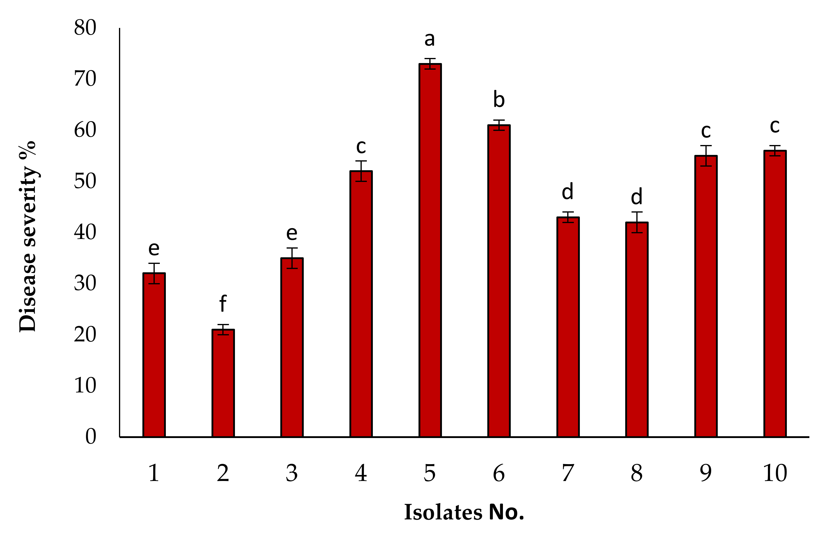
2.2. Pathogenicity Test

In our current experiment, 10 isolates were examined for their ability to cause wilting of tomato plants grown under greenhouse conditions using tomato hybrids (Super Strain-B) to evaluate the pathogenic capability of the tested fungal isolates. This study was conducted under greenhouse conditions using 30 cm sterilized pots, containing about 2 kg soil. Pots and soil were sterilized by autoclave at 121 °C. Fungal inoculum was prepared by growing each isolate fungus on sterilized grain sorghum medium (200 mL + 150 g + 4 g + 4 g + 50 g from water, grain sorghum, sucrose, and clean sand, respectively) and incubated for two weeks at 27 °C. The inoculum was mixed with the soil at 3% (w/w), and then irrigated for three times one week before transplanting to ensure even distribution and growth of each particular isolate.
Tomato seedlings (three 35-day-old seedlings) were planted in each infected pot, three replicates were used, and each one consisted of 5 pots (2 seedlings/pot). The experiment was repeated two times. After 90 days, the degree of disease was measured as was measured according to the color of brown vessels.

2.3. Determination of Disease Index

The disease index was estimated by measuring the internal discoloration (browning) area in the vascular bundle by measuring the longitudinal sector of the root using the scale described by Mousa et al. [1].
1 = 0–25% of vascular root bundles are brown, 2 = 26–50% of vascular root bundles are brown.
3 = 51–75% of vascular root bundles are brown, 4 = 76–100% of vascular root bundles are brown.
The percentage of internal discoloration was calculated by the formula:
%   of   vascular   browning = Sum   of   vascular   browning   value / 4   ×   total   number   of   plants   ×   100

2.4. Molecular Identification of F. oxysporum

The pathogenic fungal isolate, KAUK7, was purified by a single spore isolate and identified by its microscopic and microscopic properties [23]. To identify the ITS sequence, KAUK7 was sent to Solgent Company, Daejeon South Korea by DNA extraction and PCR was used to amplify the tRNA region covering ITS1 and ITS2 in two rounds [24]. The nucleotide sequence data of the ITS2 and ITS2 regions were subjected to pairwise alignment by the method of Lipman and Pearson [25] using the program “GENETYX-MAC” (Genetyx Corp., Osaka, Japan) and sequences were further analyzed using BLAST from the National Center of Biotechnology Information (NCBI) available from: https://www.ncbi.nlm.nih.gov/ (accessed on 10 January 2022).

2.5. Preparation of Plant Extracts

To conduct this study, we used the leaves of C. procera as a source of the available plant extract because this plant is a wild plant widely spread in Saudi Arabia, and the extract is an alternative to chemical pesticides and an eco-friendly substance; we sought to evaluate its effect on the Fwd control. Fresh leaves of C. procera were collected, washed, and oven dried (50 °C) for three days. After that, in a laboratory blender, 10 g of dried samples were blended for 1.5 min (Waring, commercial, USA), and 100 mL of sterilized distilled water was added and blended again. The mixture was placed in falcon tubes (50 mL) and centrifuged (3220× g) at 20 °C for 30 min. Once again, the supernatant was recuperated and centrifuged (20,817× g) at room temperature for 30 min. Then, the supernatant was filtered through 0.65 µm, and 0.45 µm membrane filters to obtain an aqueous solution free of particle waste [26]. The final supernatant was sterilized with 0.22 µm membrane filter and the AE was then considered ready to use for bioassays.
Further concentrations of extracts were prepared by diluting the 100% stock solution and stored for further application in a petri dish and greenhouse experiment.

2.6. Isolation of Phytochemical Compounds from C. procera Leaves

After collection, the fresh leaves were immediately frozen in the laboratory on dry ice. After that, the frozen leaves were ground in cryogenic grinding for three cycles (one minute each), after which the plant material (50 g) was soaked directly in 100 mL of cold water. Then, the solution was shaken at 200 rpm for 24 h at 37 °C [27,28].
Subsequently, the extract was transferred and divided into several centrifugal bottles with screw cap (100 mL utofil®PP) and centrifuged (5000 rpm) for 10 min at 4 °C to eliminate plant debris. Then, the upper layers of the extract were transferred to new storage bottles (250 mL), 3 mL of each extract was pumped into three new glass tubes (1.5 mL with caps) to reduce the loss of some volatile matter, with each glass tube (1.5 mL) containing 1 mL of the extract. Then, all tubes were covered with parafilm after closing them in screw-top vials with silicone/PTFE septum lids and stored at −20 °C until GC–MS analysis [27,28].
To separate the active components from leaf extracts, a Trace GC-ISQ quantum mass spectrometer system was used for phytochemical analysis.
Samples (one µL) were injected into a GC–MS equipped with a TG–5MS column (30 m × 0.25 mm ID, 0.25 µm film thickness), and helium gas was used as a carrier with a constant flow (1.0 mL min−1). The mass spectra were detected between 50–500 m/z.
A temperature at 50 °C was used initially for 10 min, then increased at a rate of 5 °C every minute until it reached 250 °C; the isothermal temperature was kept at 300 °C (for 2 min), and at the end, isothermal was kept at 350 °C for 10 min. Libraries of NIST, Adams, Terpenoids, and VOCs were used to identify phytochemical components by comparing known mass spectra for each compound with data stored in those libraries. The relative percentage of each component was estimated by RT and the average peak area compared with the total peak areas [28].

2.7. In Vitro Antifungal Activity

A total of 10 mL of the pre-prepared aqueous methanol extract of C. procera was placed with 50 mL of PDA (5, 10, or 15%), then 15 mL of the former were placed in sterile Petri dishes and left to solidify. Then, the plates were seeded with a 9 mm disc of 10-day-old F. oxysporum f. sp. lycopersici (FOL7) cultured on PDA and incubated at room temperature (27 °C). The percentage of radial growth inhibition of the pathogen was measured. Four replicates were used for each concentration, and each replicate consisted of five plates. The percentage decrease (Mr) in colony diameter resulting from each concentration from the extract was calculated using the technique used by Alamri et al. [29]:
Mr = M 1   M 2   /   M 1   ×   100 .
where Mr = % reduction in colony diameter, M2 = colony diameter in the treated medium, and M1= colony diameter in the untreated medium (control).

2.8. Effects of C. procera on Seeds Germination and Seedling Vigor In Vitro

The effect of pretreatment of seeds with C. procera was examined in three treatments in vitro. Three groups of tomato seeds were treated as follows: first group had 50 seeds soaked in each concentration of C. procera, the second group had 50 seeds soaked in suspension of the isolate of Fusarium KAUK7 as the infected control and the third group had the seeds soaked in sterile distilled water along with a healthy control. Each treatment (50 seeds) was repeated three times and the treated seeds were then placed on wet filter paper and incubated for one week at 28 °C. Then, germinated seeds (%), mean shoot length (MSL), and root length (MRL) were recorded. Moreover, the seedling strength index (VI) was determined using the following equation [30]:
VI = MSL + MRL   ×   germination   %

2.9. Effects of C. procera on Disease Severity under Greenhouse Condition

The F. oxysporum f. sp. lycopersici KAUK7 fungal inoculum was prepared as stated in the pathogenicity test. Plastic pots (30 cm) were sterilized seven days before planting and planted with sterile soil inoculated with the KAUK7 vaccine at 3% (w/w). Tomato seedlings were soaked in AE (5, 10 and 15%) of C. procera (best pathogen reduction and best treatment for VI %) for 30 min [31], then planted in infected pots (2 seedling/pot). Three replicates were used, each replicate consisted of 5 posts. The seedlings of the control treatment were also treated with water instead of the aqueous extract of C. procera at the same time. The wilt intensity (%) was registered after 90 days of planting.
Plants from different treatments were removed at the end of the experiment, cleaned thoroughly with running water, blotted with tissue paper, weighed to determine fresh weights, and then oven dried for 72 h to determine dry weights.

2.10. Assay Total Phenol Contents

After 15, 20, and 25 days of treatment, tomato leaves were collected and immersed in N2 liquid, then 1 g of plant material was placed in methanol (80%), and then centrifuged at 4 °C at 1000 rpm (for 20 min). Then, the pellet were discarded after adding ascorbic acid (0.1 g per 5 mL), and the homogenates were evaporated in a rotary evaporator at 65 °C and repeated three times for 5 min. In 5 mL of 80 percent methanol, the residue was dissolved. Each treatment has four replicates.
The following procedure was used to estimate phenols, as mentioned by Zhishen et al. [32]: 0.02 methanol extract, 0.5 mL Folin–Ciocalteu reagent, 0.75 mL Na2CO3 solution (20%), and 8 mL water made up the reaction mixture. In a water bath, the mixture was incubated for one hour at 37 °C. Water was used as a control. Total phenol content was measured as mg g−1 plant fresh weight using a spectrophotometric method at 767 nm. A standard curve of Gallic acid (0–5 mg) was used. Total phenol was calculated as mg Gallic acid/g of plant material.

2.11. Total Flavonoid Content

To measure the flavonoid content in each sample, a modified chromatographic method was used as determined by Zhishen et al. [32].

2.12. Antioxidant Enzyme Activity Analysis

Ten days after C. procera treatment with F. oxysporum, antioxidant enzyme activity levels were assessed, with plants inoculated only with F. oxysporum serving as controls. Next, 1 g tomato leaves were collected, washed, dried, put into a precooled mortar, mixed with 8.0 mL of 0.05 M (pH 7.8) phosphate buffer, ground into a homogenate, and centrifuged (10,000 rpm) for 15 min at 4 °C. After that, the supernatant was transferred to a new test tube, and the crude enzyme extract from tomato leaves was obtained. The activity of peroxidase (POD) was measured by guaiacol colorimetry, and the amount of enzyme required for a 0.01 increase (represented by A470) per minute was defined as one unit of POD. PPO activity was determined as mentioned by Batra and Kuhn [33].

2.13. Statistical Analysis

All tests were set up in a completely randomized design. Each experiment consisted of three replicates and was repeated twice as mentioned in each experiment. Using the statistical analysis method, data were subjected to ANOVA [34]. The LSD test was used to compare the means at p < 0.05 values.

3. Results

3.1. Isolation of the Causal Pathogen and Pathogenicity Tests

Ten isolates of diseased tomato plants that showed signs of wilting were isolated. It was found that all these isolates showed typical symptoms of tomato wilt, but varied in their virulence. Isolate No. 5 caused the highest severity of the disease, followed by isolates No. 6, 10, and 9 while isolates No. 2, 1, and 3 showed less severity than the other isolates Figure 1. For all the following experiments, isolate No. 5 was used after being identified by ITS sequencing where it caused the highest disease severity on tomato plants.

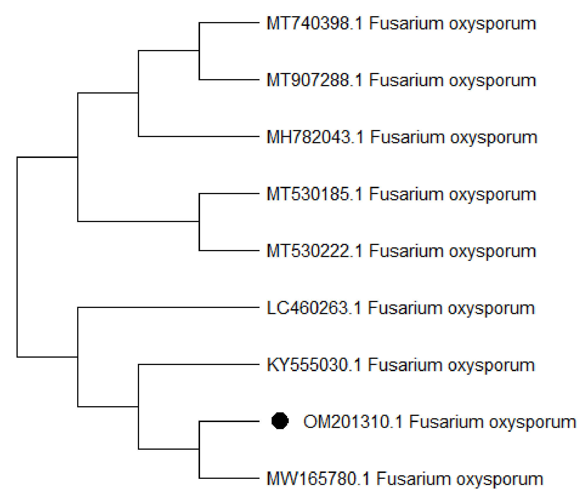
3.2. Identification of KENF7 by Using ITS Sequencing

The ITS sequence was used to characterize the FOL isolate KENF7 (ITS1 and ITS2). A BLAST search of NCBI data libraries for ITS sequence similarities yielded the highest results, with 100% sequence similarity to the F. oxysporum strain ATCC 48112 (GenBank accession No. MW165780.1). The ITS sequence for the FKAU1 isolate has been lodged in the GenBank sequence database under accession number OM201310.1 (Figure 2).

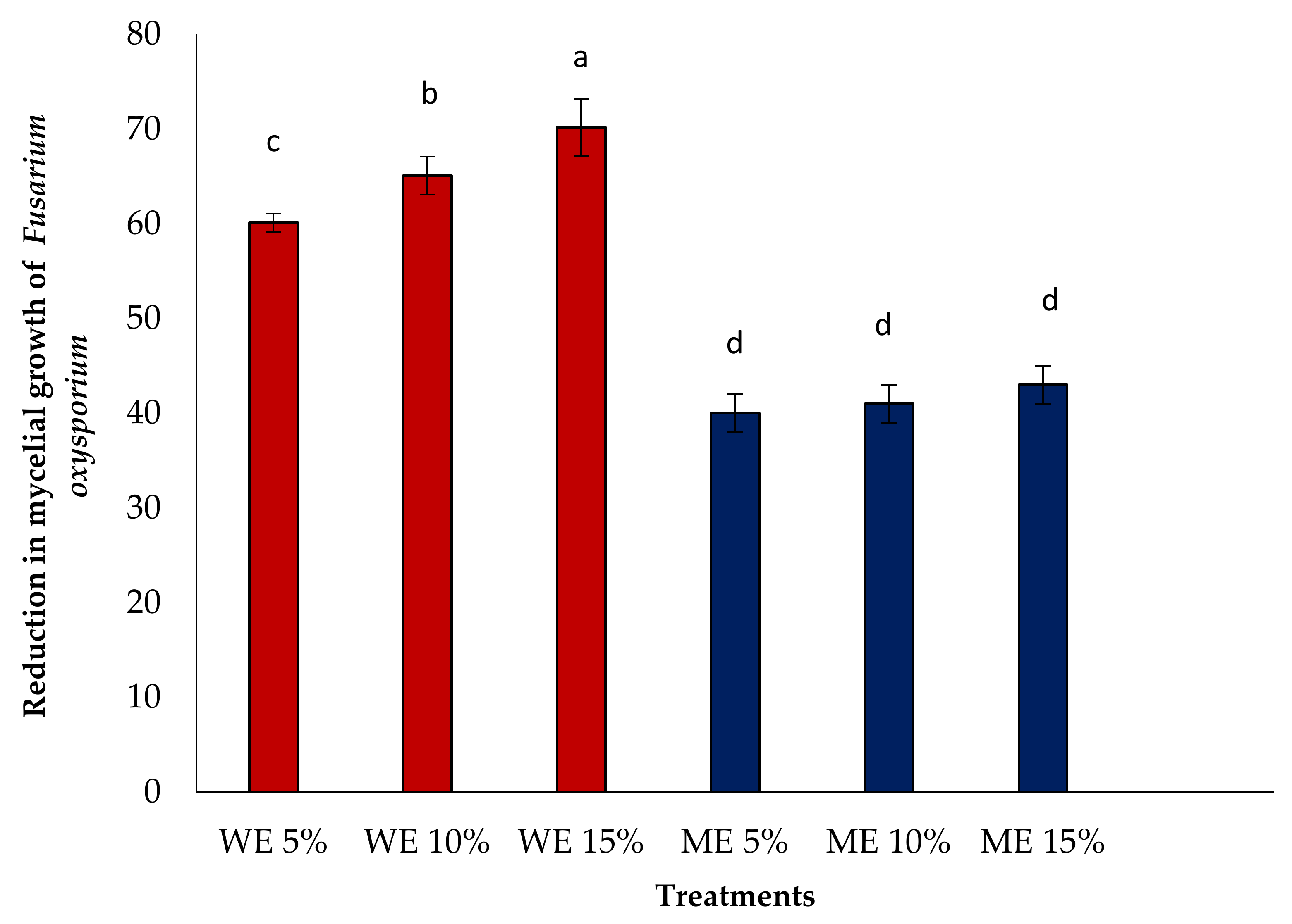
3.3. In Vitro Antifungal Activity

The results in Figure 3 show that all tested levels (5, 10, and 10%) of the aqueous extract inhibited the growth of F. oxysporum f. sp. lycopersici KENF7 with different percentages. Aqueous extracts of C. procera at concentration 15% inhibited the growth of the pathogen (70.2%) relative to the control. All concentrations of C. procera extracts also suppressed growth of the pathogen but this was lower than in the aqueous extract. The least inhibition of the pathogen was shown by the lowest concentration of the extract used.

3.4. Effect of Aqueous Extract on Seed Germination and Seedling Vigor Index

The data included in Table 1 show that treatment with C. procera extract at 15% gave the highest impact on germination (+25%) and seedling vigor (569.5%) relative to the other concentrations or control treatment. MSL and MRL were also increased by C. procera and showed the least improvement in seed germination and MSL in seeds infected with the fungus F. oxysporum f.s. lycopersici KENF7.

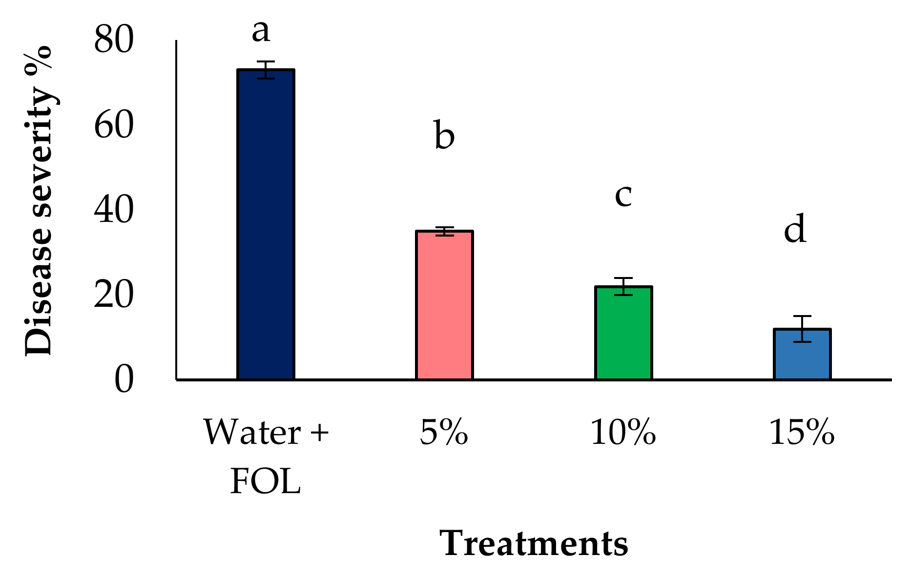
3.5. Effect of Aqueous Extract on Disease Reduction under Greenhouse

The results shown in Figure 4 and Figure 5 showed that the tested aqueous extract of C. procera at each concentration significantly reduced the incidence of Fwd disease in tomatoes under greenhouse conditions. Aqueous extract of C. procera at 15% also resulted in the highest reduction in disease severity followed by other levels.

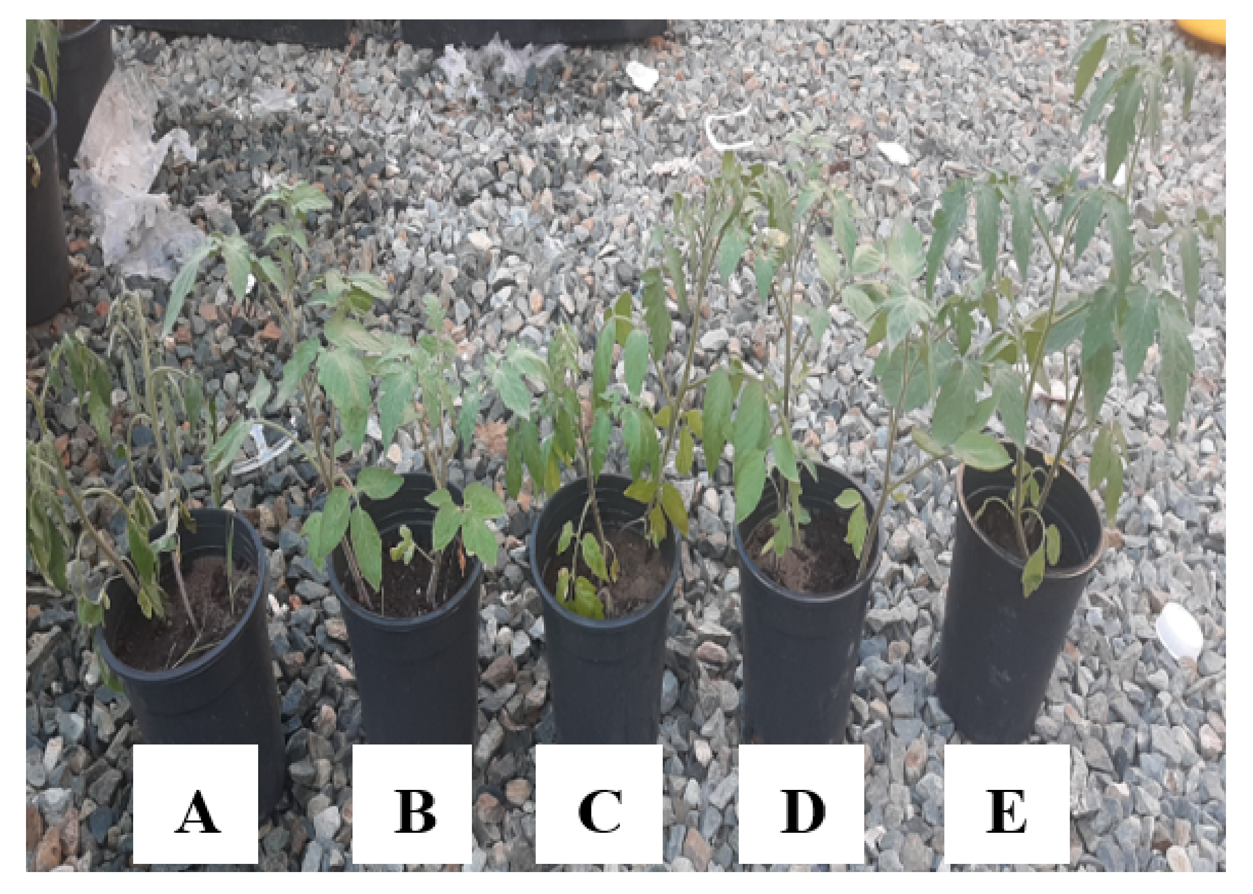
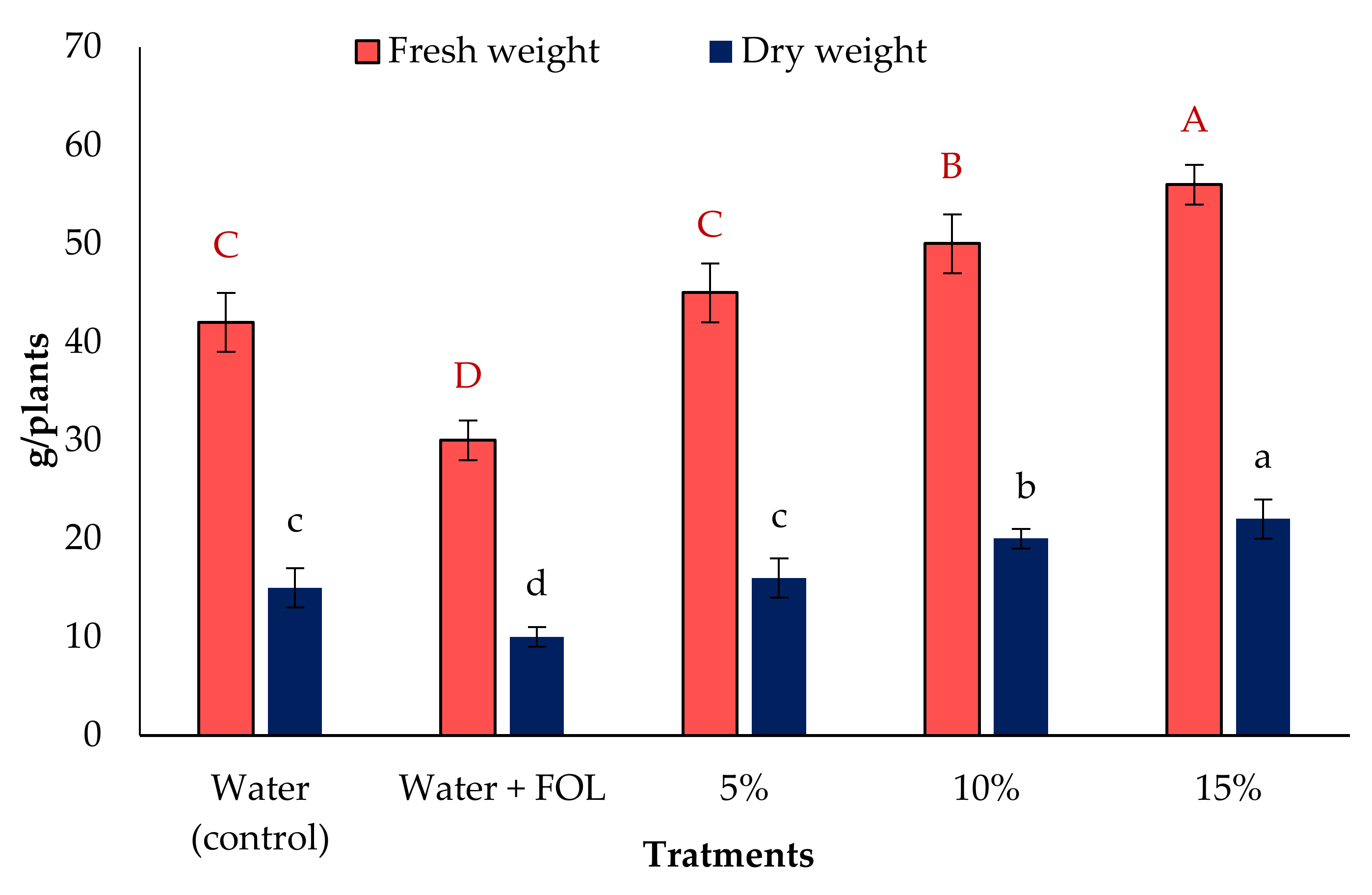
3.6. Vegetative Growth

The data illustrated in Figure 6 indicate that the highest fresh and dry weights achieved was by the aqueous extract of C. procera at 15%, and there was a significant enhancement in the fresh and dry weight of tomato plants, followed by the rates of 10 and 5%, the lowest fresh and dry weights in the infected plants.

3.7. Effect of Aqueous Extract on Enzymatic Activities

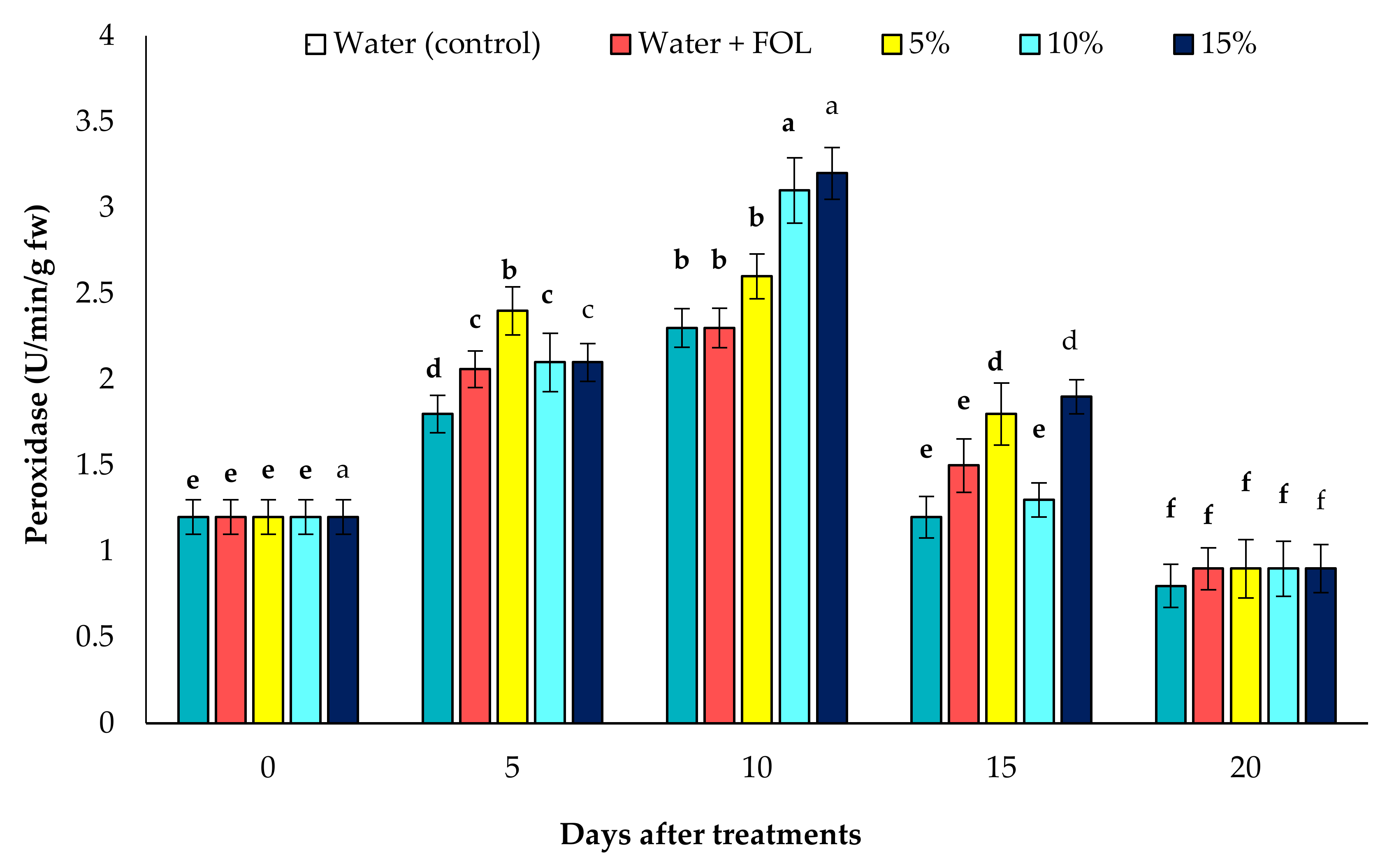
3.7.1. Activity of POD in Inoculated Plants

The results in Figure 7 show that the POD activity of treated tomato plants with the extract of C. procera was significantly (p ≤ 0.05) higher than that of untreated plants after 10 days of application. Moreover, the enzyme activity was higher when using the highest level of the extract (15%) compared to the other treatments. In general, the POD activity in the treated plant was highest after 10 days followed by after 5 days of treatment.

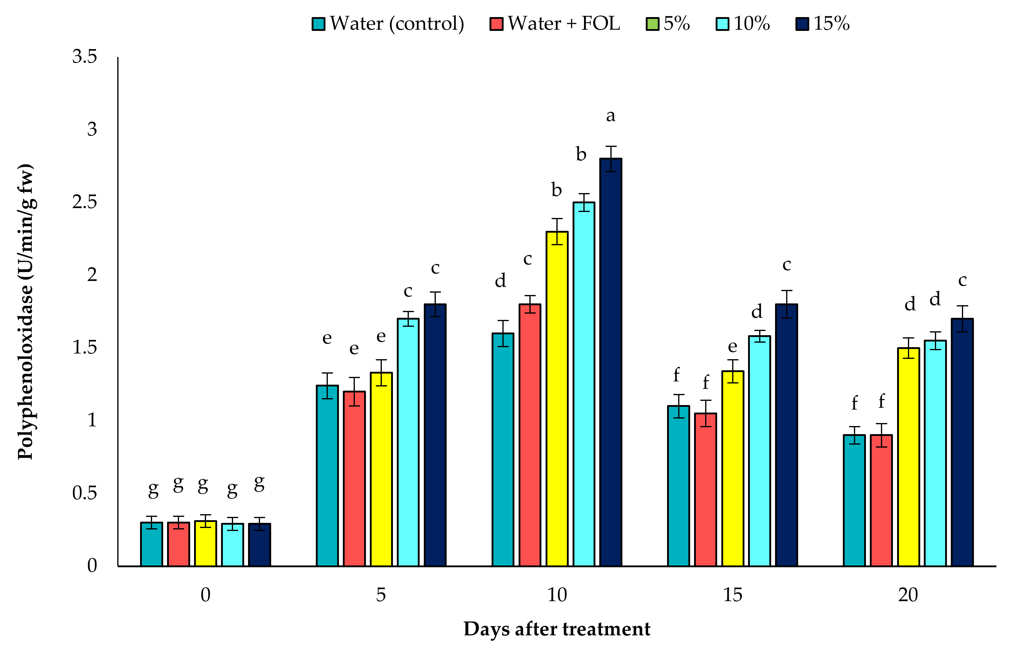
3.7.2. PPO Activity in Inoculated Plants

In comparison to control plants, C. procera induced a significant increase in the activity of the PPO enzyme at all time points, as shown in Figure 8. The largest increase in PPO activity was shown in tomato plants treated with C. procera. In general, PPO activity was highest at 10 days from C. procera application followed by 10 and 5%; the infected and healthy plant nearly gave the same activity of PPO.

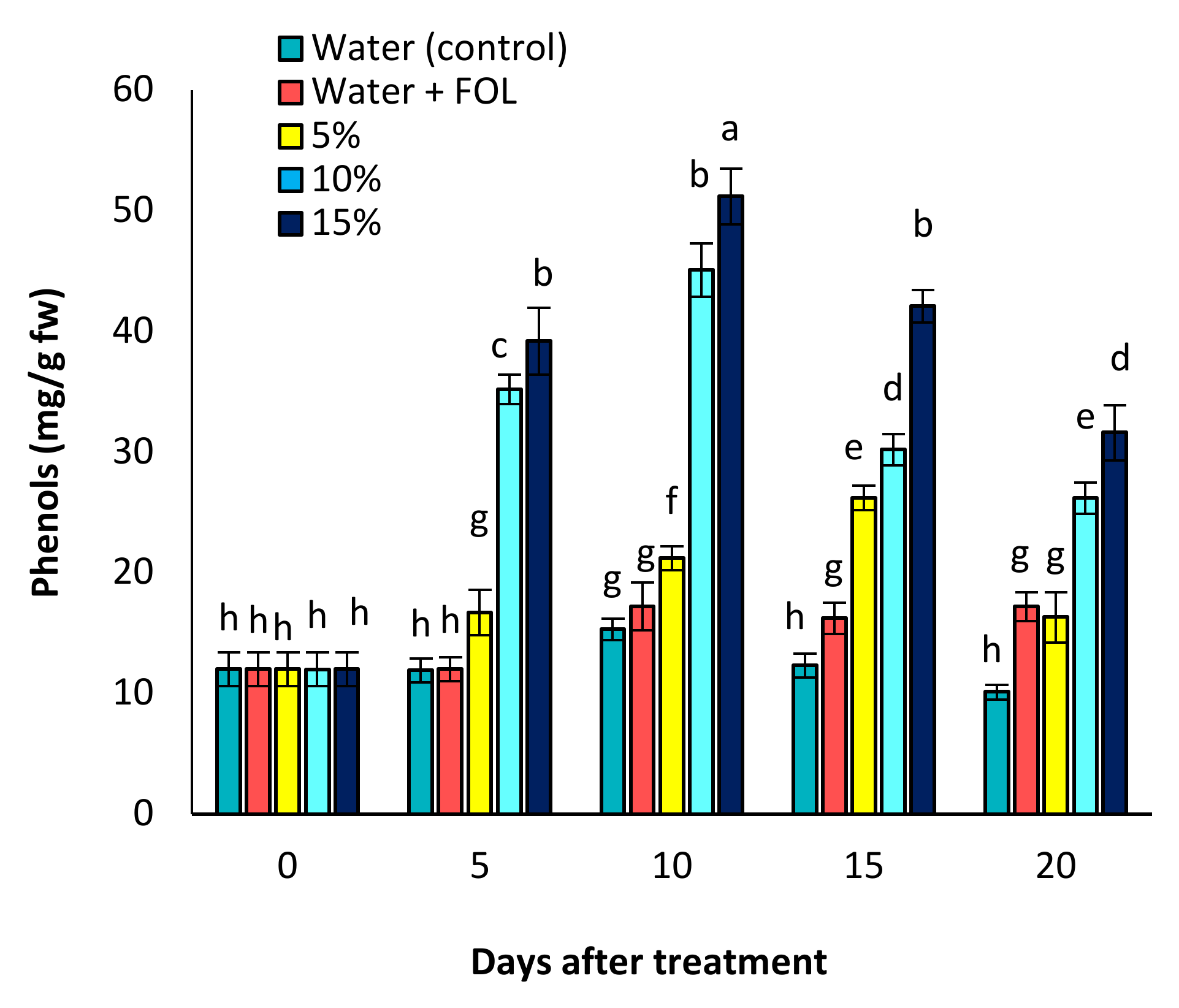
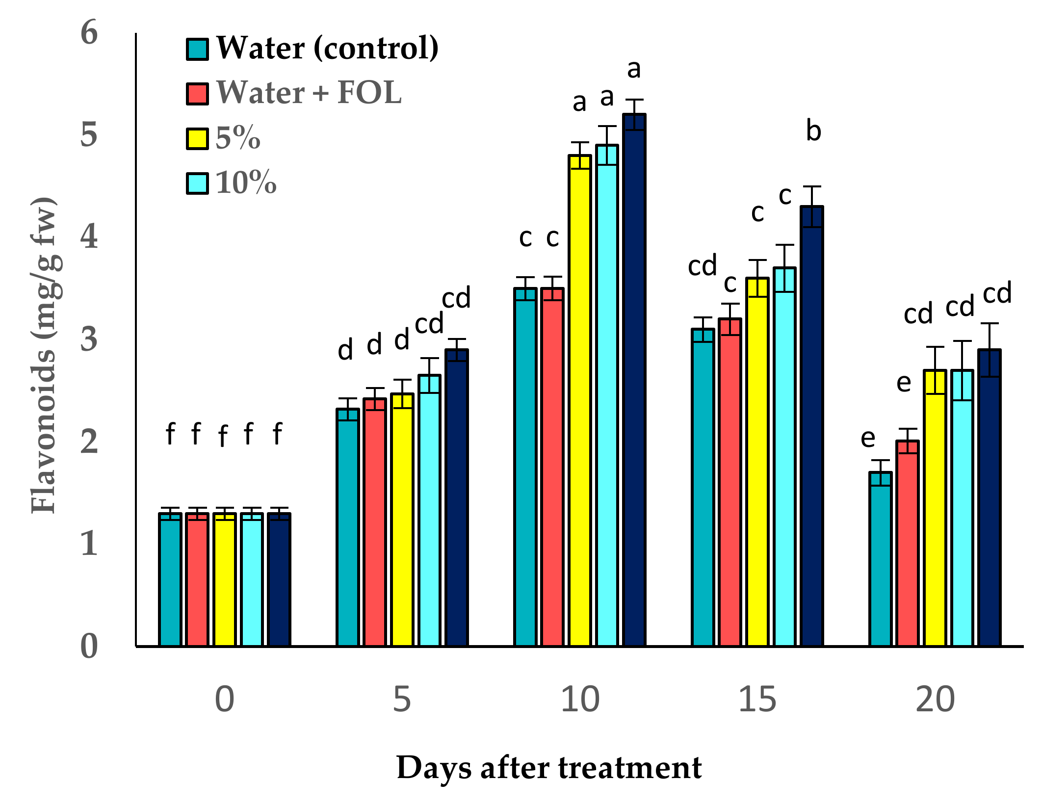
3.8. Effect of C. procera on Phenol and Flavonoid Contents

It was found from the data illustrated in Figure 9 and Figure 10 that treatment of tomato plants with C. procera extract gave the highest of flavonoids and phenol contents in inoculated or un-inoculated plants. The other treatment also increased the flavonoid and phenol contents relative to the untreated plants.

3.9. Identification of Phytochemical Components of C. procera AE

By GC–MS analysis, 31 bioactive compounds were identified in the AE of C. procera leaves. The results were recorded for the qualitative and quantitative analyses of all phytochemicals in the three extracts, shown in Table 2. In AE, among the compounds identified in AE, N-Benzylideneisopropylamine was recorded as the main compound (16.53%), followed by (+)-delta-Cadinene (12.24%), Biocytin (9.89%), Palmitic acid (6.87%), and 1, 3-Dipalmitin, TMS derivative (5.98%).

4. Discussion

Tomato plants are attacked by several soil fungi that infect them during their different growth stages, resulting in diseases, root rot, and wilt. This study focused on wilt disease, which causes reduced production of tomato plants in all countries that grow them, and it is known that the cause of this disease is the fungus Fusarium oxysporium f. s. lycopersici. Ten isolates were isolated from tomato plants in Jeddah city; all these isolates were able to cause wilt disease on tomato plants to various different degrees, and these results are in agreement with these obtained by Abo-Elyousr and Mohamed [2] who determined that fungal isolates differ in their ability to cause disease.
The main method for control of such disease is the use of fungicides [35], but the chemical control is harmful to the environment and human health, and the control effects decrease gradually due to the drug resistance of pathogens. Therefore, it was necessary to search for alternatives to chemicals through the use of natural alternatives, especially as many previous studies have recommended using plant extracts to control fungal, bacterial, and helminthic diseases [36,37]. In our study, the aqueous extract of C. procera with different levels suppressed growth of F. oxysporum f. sp. lycopersici in vitro to different degrees. AE (15%) showed the highest effect in reducing radial growth of the pathogen compared with the other levels. This was in the same context with the report of Mousa et al. [1] on F. oxysporum f. sp. lycopersici. Similarly, Kareem et al. [38] found an antimicrobial activity of the leaf extracts of C. procera against F. oxysporum. Moreover, Nikam et al. [39] used the stem, leaves, and flowers of C. procera extracts to test the bioassays for antimicrobial activities against F. oxysporum. Otherwise, Olufolaji et al. [40] reported that phytochemicals, i.e., alkaloids, glycoside, tannins, saponin, steroids, flavonoids, medicagenic acid, and phenols present in the several plants and our plant could be toxic to the fungus, thereby causing its growth inhibition. In addition, Doshi et al. [41] analyzed extracts of buds, stems, flowers, and mature leaves of C. procera. They found a number of biologically active compounds, such as triterpenoids, glycosides, tannins, alkaloids, phenolics, and saponin, that may cause toxic effects on several pathogens as our data recorded. Along the same line, several authors reported that the plant extracts affected the growth of phytopathogenic fungal as well as reduced the diseased severity, and this may be due to secondary metabolites (e.g., phenolic, flavonoids, terpenoids, and alkaloids [42,43,44]. The results of our present study showed that aqueous extract of C. procera significantly reduced wilt diseases in all tested concentrations. When testing the aqueous extract on seed germination and seedling vigor in vitro, the treatments increased seed germination, MSL, and MRL when compared to the infection control. At 15%, C. procera gave the highest seed germination and seedling vigor followed by the other levels. Many researchers have noted that plant extracts increased the seed germination as well as vigor index among neem, garlic, and ginger [45,46,47]. Our results revealed that C. procera extracts increased POD activity in inoculated tomato plants compared to untreated inoculated plants after 5, 10, and 15 days of application. Several authors have demonstrated that enhanced POD activity was related to the systemic induced resistance in plants to fungi [48,49]. In many plants, plant extracts have been used and documented to induce systemic resistance, reduce disease incidence, and thus improve plant growth and production [20,50].
Plant defense genes that are dormant in healthy uninoculated plants can induce systemic disease resistance when activated by various factors. Increased POD, PPO, and PAL enzyme activity has been shown to be effective in the treatment of a variety of fungal illnesses [51,52]. POD is involved in several plant defense processes, including lignin production and oxidative cross-linking of plant cell walls, as well as pathogen infection-induced formation of active oxygen species [53]. The increase in resistance against disease can be attributed to the increased activities of peroxidase and chitinase because both enzymes are known to increase the plant’s ability to resist diseases. In agreement with this, similar results have been reported by Shabana et al. [54], who mentioned that plant extract application (neem, cloves, and garden quinine) to wheat plant significantly reduced leaf rust infection compared to a control. In this context, phytosterols, a group of steroidal alcohols and phytochemicals naturally present in plants, have been found to induce antifungal activity against Aspergillus, Penicillium, and Fusarium [55].
The treatment of plants with C. procera at all concentrations were found to significantly increase total phenol and flavonoid content compared to an untreated control. Other studies support our findings, because phenolic compounds are toxic to pathogens and the accumulation of these bioactive ingredients at infection sites was associated with pathogen development restriction [56]. Moreover, changes in the pH of plant cell cytoplasm due to the increase of phenolics may also promote resistance, inhibiting pathogen development [57].

5. Conclusions

Tomato seedlings treated with aqueous extracts of C. procera at different concentrations reduced F. oxysporum f. sp. lycopersici radial growth in a laboratory environment as well as when used as seedling pre-treatment for tomatoes, promoting seed germination and seeding vigor. In addition, treatment with aqueous extracts increased the vegetative growth of tomatoes under greenhouse conditions. The treatment of tomato seedlings with aqueous extracts of C. procera increased the antioxidant enzymes (peroxidase and polyphenol oxidase) and increased flavonoid and phenol contents in seedlings after inoculation. Our results are promising for the use of C. procera as eco-friendly material against F. oxysporum under greenhouse conditions.

Author Contributions

Conceptualization, K.A.M.A.-E. and E.F.A.; methodology, K.A.M.A.-E., E.F.A. and N.M.A.S.; software, K.A.M.A.-E., E.F.A. and N.M.A.S.; validation, K.A.M.A.-E. and N.M.A.S.; formal analysis, K.A.M.A.-E. and E.F.A.; investigation, K.A.M.A.-E. and N.M.A.S.; resources, K.A.M.A.-E., E.F.A. and N.M.A.S.; data curation, K.A.M.A.-E. and E.F.A.; writing—original draft preparation, K.A.M.A.-E., E.F.A. and N.M.A.S.; writing—review and editing, K.A.M.A.-E. and E.F.A.; visualization, K.A.M.A.-E., E.F.A. and N.M.A.S.; supervision, K.A.M.A.-E. and E.F.A.; project administration, K.A.M.A.-E., E.F.A. and N.M.A.S.; funding acquisition, K.A.M.A.-E. All authors have read and agreed to the published version of the manuscript.

Funding

The authors extend their appreciation to the Deputyship for Research and Innovation, Ministry of Education in Saudi Arabia for funding this research work through the project number “IFPIP: 24-155-1442” and King Abdulaziz University, DSR, Jeddah, Saudi Arabia.

Institutional Review Board Statement

Not applicable.

Informed Consent Statement

Not applicable.

Data Availability Statement

Data for sequence of Fusarium oxysporum is available at website https://www.ncbi.nlm.nih.gov/nuccore/OM201310, accessed on 10 January 2022.

Conflicts of Interest

The authors declare no conflict of interest.

References

  1. Mousa, M.A.A.; Abo-Elyousr, K.A.M.; Abdel Aal, A.M.K.; Alshareef, N.O. Combination of Bacillus amyloliqefaciens and peppermint oil for control of Fusarium wilt disease of tomato. Agronomy 2021, 11, 2536. [Google Scholar] [CrossRef]
  2. Abo-Elyousr, K.A.M.; Mohammed, H. Biological Control of Fusarium Wilt in Tomato by Plant Growth-Promoting Yeasts and Rhizobacteria. Plant Pathol. J. 2009, 25, 99–204. [Google Scholar] [CrossRef]
  3. Dursun, A.; Melek, E.; Dönmez, M.F. Effects of foliar application of plant growth promoting bacterium on chemical contents, yield and growth of tomato (Lycopersicon esculentum L.) and cucumber (Cucumis sativus L.). Pak. J. Bot. 2020, 42, 3349–3356. [Google Scholar]
  4. Jabnoun-Khiareddine, H.J.; Abdallah, R.A.B.; Remadi, M.D.; Nefzi, A.; Ayed, F. Grafting tomato cultivars for soil borne diseases suppression and plant growth and yield improvement. J. Plant Pathol. Microbiol. 2019, 10, 1–8. [Google Scholar]
  5. Oliver, B.B. Medicinal Plants in Tropical West Africa; Cambridge University Press: Cambridge, UK, 1986. [Google Scholar]
  6. Yossry, A.A.; Ali, S.M.A.; Imery, S.M. Fungitoxic properties of some plant extracts against growth of soil borne disease fungi. Ann. Agric. Sci. 1998, 3, 891–909. [Google Scholar]
  7. Nidhi, S.; Trivedi, P.C. Screening of leaf extracts of some plants for their nematicidal and fungicidal properties against Meloidogyne incognita and Fusarium oxysporum. Asian J. Exp. Sci. 2002, 16, 21–28. [Google Scholar]
  8. Baraka, M.A.; Omar, S.A.; Ebtehage, E.; Zian, A.H. Controlling seedling damping-off, root rot and wilt diseases of lupine (Lupinus albus L.). Agric. Res. J. Suez Canal Univ. 2006, 6, 57–68. [Google Scholar]
  9. Deepak, D. Phytochemistry of Indian Asclepiadaceae. In The Taxonomy and Phytochemistry of the Asclepidaceae in Tropical Asia; Kiew, R., Ed.; University Pertanian: Selangor, Malaysia, 1995; pp. 33–44. [Google Scholar]
  10. Arora, S.S. Calotropis procera (Ait) R. Br.-Ak: A new and free source of fibre and renewable hydrocarbons. Agric. Mech. Asia Afr. Lat. Am. 1982, 13, 71–75. [Google Scholar]
  11. Heneidak, S.; Grayer, R.J.; Kite, G.C.; Simmonds, M.S.J. Flavonoid glycosides from Egyptian species of the tribe Asclepiadeae (Apocynaceae, subfamily Asclepiadoideae). Biochem. Sys. Ecol. 2006, 34, 575–584. [Google Scholar] [CrossRef]
  12. Bhutani, K.K.; Gupta, D.K.; Kapil, R.S. Occurrence of D/E trans stereochemistry isomeric to ursane (cis) series in a new pentacyclic triterpene from Calotropis procera. Tetrahedron Lett. 1992, 33, 7593–7596. [Google Scholar] [CrossRef]
  13. Hanna, A.G.; Shalaby, N.M.M.; Morsy, N.A.M.; Andras, A.; Toth, G.; Malik, S.; Duddeck, H. Structure of a calotropagenin-derived artifact from Calotropis procera. Magn. Reson. Chem. 2002, 40, 599–602. [Google Scholar] [CrossRef]
  14. Chundattu, S.J.; Agrawal, V.K.; Ganesh, N. Phytochemical investigation of Calotropis procera. Arab. J. Chem. 2012, 9, S230–S234. [Google Scholar] [CrossRef] [Green Version]
  15. Gulzar, A.; Siddiqui, M.B.; Bi, S. Phenolic acid allelochemicals induced morphological, ultrastructural, and cytological modification on Cassia sophera L. and Allium cepa L. Protoplasma 2016, 253, 1211–1221. [Google Scholar] [CrossRef]
  16. Hussain, F.; Rasool, A.; Aziz, K.; Raisham, S.; Aziz, S.; Badshah, L.; Hussain, W. Allelopathic inhibition of germination, seedling growth and cell division of selected plant species by Calotropis procera (Ait.) Ait. Plant Sci. Today 2020, 7, 1–8. [Google Scholar] [CrossRef]
  17. Hayat, S.; Ahmad, H.; Ali, M.; Ren, K.; Cheng, Z. Aqueous garlic extract stimulates growth and antioxidant enzymes activity of tomato (Solanum lycopersicum). Sci. Hortic. 2018, 240, 139–146. [Google Scholar] [CrossRef]
  18. Zulfiqar, F.; Younis, A.; Finnegan, P.M.; Ferrante, A. Comparison of Soaking Corms with Moringa Leaf Extract Alone or in Combination with Synthetic Plant Growth Regulators on the Growth, Physiology and Vase Life of Sword Lily. Plants 2020, 9, 1590. [Google Scholar] [CrossRef]
  19. Hayat, S.; Ahmad, H.; Nasir, M.; Khan, M.N.; Ali, M.; Hayat, K.; Khan, F.; Ma, Y.; Cheng, Z.; Khan, M.A.; et al. Some Physiological and Biochemical Mechanisms during Seed-to-Seedling Transition in Tomato as Influenced by Garlic Allelochemicals. Antioxidants 2020, 9, 235. [Google Scholar] [CrossRef] [Green Version]
  20. Hussein Mohamed, M.A.; Abo-Elyousr, K.A.M.; Hassan, M.A.H.; Hashem, M.; Hassan, E.A.; Alamri, S.A.M. Induction of defense mechanisms involved in disease resistance of onion blight disease caused by Botrytis allii. Egypt. J. Biol. Pest Control 2018, 28, 80. [Google Scholar] [CrossRef] [Green Version]
  21. Chuying, C.; Nan, C.; Jinyin, C.; Chunpeng, W. Clove Essential Oil as an Alternative Approach to Control Postharvest Blue Mold Caused by Penicillium italicum in Citrus Fruit. Biomolecules 2010, 9, 197. [Google Scholar] [CrossRef] [Green Version]
  22. Youssef, K.; Roberto, S.R.; Tiepo, A.N.; Constantino, L.V.; de Resende, J.T.V.; Abo-Elyousr, K.A.M. Salt solution treatments trigger antioxidant defense response against gray mold disease in table grapes. J. Fungi 2020, 6, 179. [Google Scholar] [CrossRef]
  23. Nelson, P.E.; Tousson, T.A.; Marasas, W.F.O. Fusarium Species: An Illustrated Manual for Identification; Pennsylvania State University Press: University Park, PA, USA, 1983. [Google Scholar]
  24. White, T.J.; Bruns, T.; Lee, S.; Taylor, J. Amplification and direct sequencing of fungal ribosomal RNA genes for phyloge-netics. In PCR Protocols. A Guide to Methods and Applications; Innis, M.A., Gelfand, D.H., Sninsky, J.J., White, T.J., Eds.; Academic Press: San Diego, CA, USA, 1990; pp. 315–322. [Google Scholar]
  25. Lipman, D.J.; Pearson, W.R. Rapid and sensitive protein similarty searches. Science 1985, 227, 1435–1441. [Google Scholar] [CrossRef] [Green Version]
  26. Ruiz-Ruiz, J.C.; Peraza-Echeverría, L.; Soto-Hernández, R.M.; San MiguelChávez, R.; Pérez-Brito, D.; Tapia-Tussell, R.; Ortiz-Vázquez, E.; Rodríguez-García, C.M. Diospyros cuneata Inhibition of Fusarium oxysporum: Aqueous Extract and its Encapsulation by Ionic Gelation. J. Plant Pathol. Microbiol. 2016, 7, 332. [Google Scholar] [CrossRef] [Green Version]
  27. Ali, M.; Reem, M.H.; Naveed, U.R.; Guangbiao, S.; Penghui, L.; Xiaochun, W.; Liang, G.; Jian, Z. De novo transcriptome sequencing and metabolite profiling analyses reveal the complex metabolic genes involved in the terpenoid biosynthesis in Blue Anise Sage (Salvia guaranitica L.). DNA Res. 2018, 25, 597–617. [Google Scholar] [CrossRef] [Green Version]
  28. Ali, M.; Penghui, L.; Guangbiao, S.; Daopu, C.; Xiaochun, W.; Jian, Z. Transcriptome and metabolite analyses reveal the complex metabolic genes involved in volatile terpenoid biosynthesis in garden sage (Salvia officinalis). Sci. Rep. 2017, 7, 16074. [Google Scholar] [CrossRef] [Green Version]
  29. Alamri, S.A.M.; Hashem, M.; Moustafa, Y.S.; Nafady, N.A.; Abo-Elyousr, K.A.M. Biological control of root rot in lettuce caused by Exserohilum rostratum and Fusarium oxysporum via induction of the defense mechanism. Biol. Control 2019, 128, 76–84. [Google Scholar] [CrossRef]
  30. Abdul Baki, A.A.; Anderson, J.D. Vigour determination in soybean seed by multiple criteria. Crop Sci. 1973, 13, 630–633. [Google Scholar] [CrossRef]
  31. Somda, I.; Leth, V.; Seeme, P. Evaluation of Lemongrass, Eucalyptus and Neem aqueous extracts for controlling seed-borne fungi of sorghum grown in Burkina Faso. World J. Agric. Sci. 2007, 3, 218–223. [Google Scholar]
  32. Zhishen, J.; Mengcheng, T.; Jianming, W. The determination of flavonoid contents in mulberry and their scavenging effects on superoxide radicals. Food Chem. 1999, 64, 555–559. [Google Scholar] [CrossRef]
  33. Batra, G.; Kuhn, C. Polyphenoloxidase and peroxidase activities associated with acquired resistance and its inhibition by 2-thiouracil in virus-infected soybean. Physiol. Plant Pathol. 1975, 5, 239–248. [Google Scholar] [CrossRef]
  34. Gomez, K.A.; Gomez, A.A. Statistical Procedures for Agricultural Research; Wiley: New York, NY, USA, 1984. [Google Scholar]
  35. Carvalho, F.P. Pesticides, environment, and food safety. Food Energy Secur. 2017, 6, 48–60. [Google Scholar] [CrossRef]
  36. Enikuomehin, O.A.; Oyedeji, E.O. Fungitoxic effect of some plant extracts against tomato fruit rot pathogens. Arch. Phytopathol. Plant Prot. 2010, 43, 233–240. [Google Scholar] [CrossRef]
  37. Abo-Elyousr, K.A.M.; Asran, M.R. Antibacterial activity of certain plant extracts against bacterial wilt of tomato. Arch. Phytopathol. Plant Prot. 2009, 42, 573–578. [Google Scholar] [CrossRef]
  38. Kareem, S.O.; Akpan, I.; Ojo, O.P. Antimicrobial Activities of Calotropis procera on Selected Pathogenic Microorganisms. Afr. J. Biomed. Res. 2008, 11, 105–110. [Google Scholar] [CrossRef]
  39. Nikam, P.S.; Jagtap, G.P.; Sontakke, P.L. Management of chickpea wilt caused by Fusarium oxysporium f. sp. ciceri. Afr. J. Agric. Res. 2007, 2, 692–697. [Google Scholar]
  40. Olufolaji, D.B.; Adeosun, B.O.; Onasanya, R.O. In vitro investigation on antifungal activity of some plant extracts against Pyricularia oryzae. Niger. J. Biotechnol. 2015, 29, 38–43. [Google Scholar] [CrossRef] [Green Version]
  41. Doshi, H.; Satodia, H.; Thakur, H.C.; Parabia, F.; Khan, A. Phytochemical screening and biological activity of Calotropis procera (Ait) R. Br. (Asclepiadaceae) against selected Bacteria and Anopheles stephansi larvae. Int. Res. Plant Res. 2011, 1, 29–33. [Google Scholar] [CrossRef]
  42. Mohamed, N.H.; El-Hadidy, A.M. Studies of biologically active constituents of Verbascum eremobium Murb. and its inducing resistance against some diseases of cucumber. Egypt. J. Phytopathol. 2008, 36, 133–150. [Google Scholar]
  43. Wink, M.; Ashour, M.L.; El-Readi, M.Z. Secondary Metabolites from Plants Inhibiting ABC Transporters and Reversing Resistance of Cancer Cells and Microbes to Cytotoxic and Antimicrobial Agents. Front. Microbiol. 2012, 3, 130. [Google Scholar] [CrossRef] [Green Version]
  44. Jan, R.; Asaf, S.; Numan, M.; Lubna; Kim, K.-M. Plant Secondary Metabolite Biosynthesis and Transcriptional Regulation in Response to Biotic and Abiotic Stress Conditions. Agronomy 2021, 11, 968. [Google Scholar] [CrossRef]
  45. Khan, M.I.; Kumar, R. Antifungal activity of leaf extract of Neem on seed mycoflora of wheat. Indian J. Seed Abs. 1992, 15, 299. [Google Scholar]
  46. Ahmed, M.; Mehbub, H.; Kamrul, H.; Dash, C.K. Efficacy of Different Plant Extract on Reducing Seed Borne Infection and Increasing Germination of Collected Rice Seed Sample. Univers. J. Plant Sci. 2013, 1, 66–73. [Google Scholar] [CrossRef]
  47. Ali, E.F.; Al-Yasi, H.M.; Issa, A.A.; Hessini, K.; Hassan, F.A.S. Ginger extract and fulvic acid foliar applications as novel practical approaches to improve the growth and productivity of Damask Rose. Plants 2022, 11, 412. [Google Scholar] [CrossRef] [PubMed]
  48. Naz, R.; Bano, A.; Nosheen, A.; Yasmin, H.; Keyani, R.; Shah, S.T.A.; Anwar, Z.; Roberts, T.H. Induction of defense-related enzymes and enhanced disease resistance in maize against Fusarium verticillioides by seed treatment with Jacaranda mimosifolia formulations. Sci. Rep. 2021, 11, 59. [Google Scholar] [CrossRef] [PubMed]
  49. Yu, Y.; Gui, Y.; Li, Z.; Jiang, C.; Guo, J.; Niu, D. Induced Systemic Resistance for Improving Plant Immunity by Beneficial Microbes. Plants 2022, 11, 386. [Google Scholar] [CrossRef]
  50. Ramamoorthy, V.; Samiyappan, R. Induction of defence-related genes in Pseudomonas fluorescens—treated chilli plants in response to infection by Colletotrichum capsici. J. Mycol. Plant Pathol. 2001, 31, 146–155. [Google Scholar]
  51. Saravanakumar, D.; Vijayakumar, C.; Kumar, N. PGPR-induced defense responses in the tea plant against blister blight disease. Crop Prot. 2007, 26, 556–565. [Google Scholar] [CrossRef]
  52. Lanubile, A.; Ferrarini, A.; Maschietto, V.; Delledonne, M.; Marocco, A.; Bellin, D. Functional genomic analysis of constitutive and inducible defense responses to Fusarium verticillioides infection in maize genotypes with contrasting ear rot resistance. BMC Genom. 2014, 15, 710. [Google Scholar] [CrossRef] [Green Version]
  53. Vanitha, S.C.; Umesha, S. Pseudomonas fluorescens mediated systemic resistance in tomato is driven through an elevated synthesis of defense enzymes. Biol. Plant. 2011, 55, 317–322. [Google Scholar] [CrossRef]
  54. Shabana, Y.M.; Mohamed, E.; Shahin, A.A.A.; El Sawy, M.M.; Draz, I.S.; Youssif, A.W. Efficacy of plant extracts in controlling wheat leaf rust disease caused by Puccinia triticina. Egypt. J. Basic Appl. Sci. 2017, 4, 67–73. [Google Scholar] [CrossRef] [Green Version]
  55. Odhav, M.B.; Mohanlall, V. Antifungal activity of stigmasterol, sitosterol and ergosterol from Bulbine natalensis Baker (Asphodelaceae) B. J. Med. Plants Res. 2012, 6, 5135–5141. [Google Scholar] [CrossRef]
  56. Abo-Elyousr, K.A.M.; Ibrahim, Y.E.; Balabel, N.M. Induction of disease defensive enzymes in response to treatment with acibenzolar-S-methyl (ASM) and Pseudomonas fluorescens Pf2 and inoculated with Ralstonia solanacearum race 3, biovar2 (phylotype II). J. Phytopathol. 2012, 160, 382–389. [Google Scholar] [CrossRef]
  57. Khaledi, N.; Taheri, P.; Tarighi, S. Antifungal activity of various essential oils against Rhizoctonia solani and Macrophomina phaseolina as major bean pathogens. J. Appl. Microbiol. 2015, 118, 704–717. [Google Scholar] [CrossRef]
Figure 1. Pathogenicity tests of 10 isolates on tomato plants. Values in the column followed by different letters indicate significant differences among treatments according to a least significant difference test (p = 0.05). Bars indicate the standard error.
Horticulturae 08 00197 g001
Figure 2. Phylogenetic analysis of Fusarium oxysporium isolate on the ITS gene. This analysis was performed using the neighbor-joining method in BLAST pairwise alignments.
Horticulturae 08 00197 g002
Figure 3. Percentage of F. oxysporum KENF7 reduction in mycelial growth caused by different concentrations (5, 10, and 15%) of C. procera in vitro. Values in the column followed by different letters indicate significant differences among treatments according to a least significant difference test (p = 0.05). WE means aqueous and ME: means ethanol extract. Bars indicate the standard error.
Horticulturae 08 00197 g003
Figure 4. Effect of different concentrations of aqueous extracts (5, 10, and 15%) of C. procera on Fusarium wilt caused by F. oxysporum KENF7 on tomato plants under greenhouse conditions. Values in the column followed by different letters indicate significant differences among treatments according to a least significant difference test (p = 0.05). Bars indicate the standard error.
Horticulturae 08 00197 g004
Figure 5. Effect of treatment with different concentrations of aqueous extracts (5, 10, and 15%) of C. procera on Fusarium wilt caused by F. oxysporum KENF7 on tomato plants under greenhouse conditions. (A) Infected control, (B) healthy control, (C) soaked tomato seedling with C. procera at 5%, (D), C. procera at 10%, and (E) C. procera at 15%.
Horticulturae 08 00197 g005
Figure 6. Effect of different concentrations of aqueous extracts (5, 10, and 15%) of C. procera on fresh and dry weight of whole plants (g plant−1) under greenhouse conditions. Values in the column followed by different letters indicate significant differences among treatments according to a least significant difference test (p = 0.05). Bars indicate the standard error.
Horticulturae 08 00197 g006
Figure 7. Effect of different concentrations of aqueous extracts (5, 10, and 15%) of C. procera on peroxidase activity (U/min g−1 FW) in leaf extracts of tomato plants after treatment with C. procera and inoculation with F. oxysporum KENF7. Values in the column followed by different letters indicate significant differences among treatments according to a least significant difference test (p = 0.05).
Horticulturae 08 00197 g007
Figure 8. Effect of different concentrations of aqueous extracts (5, 10, and 15%) of C. procera on polyphenoloxidase activity (U/min g−1 FW) in leaf extracts of tomato plants after treatment with C. procera and inoculation with F. oxysporum KENF7. Values in the column followed by different letters indicate significant differences among treatments according to a least significant difference test (p = 0.05).
Horticulturae 08 00197 g008
Figure 9. Effect of different concentrations of aqueous extracts (5, 10, and 15%) of C. procera on phenol content (phenols; mgg−1 FW] in leaf extracts of tomato plants after treatment with C. procera and inoculation with F. oxysporum KENF7. Values in the column followed by different letters indicate significant differences among treatments according to a least significant difference test (p = 0.05).
Horticulturae 08 00197 g009
Figure 10. Effect of different concentrations of aqueous extracts (5, 10, and 15%) of C. procera on flavonoids (mg g−1 FW) in leaf extracts of tomato plants after treatment with C. procera and inoculation with F. oxysporum KENF7. Values in the column followed by different letters indicate significant differences among treatments according to a least significant difference test (p = 0.05).
Horticulturae 08 00197 g010
Table 1. Effect of different concentration of aqueous extract of C. procera (5, 10, and 15%) on seed germination and seedling vigor under Fwd infection.
ConcentrationGermination (%)MSL (cm)MRL (cm)Vigor Index (VI %)
C. procera 5%75 b (+10.3)1.2 (±0.4) b4.5 (±0.9) b427.5
C. procera 10%75 b (+10.3%)1.3 (±0.2) b5.2 (±0.7) a487.5
C. procera 15%85 a (+25%)1.5 (±0.1) a5.2 (±0.5) a569.5
Infected control68 c (-)1.1 (±0.2) b3.1(±0.7) c285.6
Healthy control85 a (+25%)1.5 (±0.1) c5.1 (±0.4) a561.0
Mean shoot length (MSL), mean root length (MRL), vigor index (VI). Means (±SE) followed by different lettering indicates differences (p ≤ 0.05) between treatments determined by ANOVA LSD.
Table 2. GC–MS profile of aqueous extract from C. procera leaves.
No.Compound NameR.TFormulaM.WPeak Area (%)
1.1,3-Dipalmitin, TMS derivative4.27C38H76O5Si6405.98
2.Palmitoyl glycerol4.62C19H38O43301.36
3.Palmitic acid5.23C16H32O22566.87
4.Ethyl iso-allocholate6.14C26H44O54362.65
5.Oxiraneundecanoic acid, 3-pentyl-, methyl ester, cis8.53C19H36O33121.12
6.Astaxanthin9.18C40H52O45961.32
7.α-D-mannopyranoside9.96C15H28B2O63261.84
8.Biocytin11.79C16H28N4O4S3729.98
9.Ascaridole13.08C10H16O21682.57
10.(+)-delta-Cadinene13.89C15H2420412.24
11.N-Benzylideneisopropylamine14.57C10H13N14716.53
12.2-(3,4-dimethoxyphenyl)-3,5-dihydroxy-7-methoxy-4H-1- Benzopyran-4-one15.69C18H16O73441.24
13.5,8,11-Eicosatrienoic acid, (Z)-, TMS derivative16.84C23H42O2Si3781.67
14.All-trans-beta-Carotene18.24C40H56536.41.27
15.1-Monooleoylglycerol, 2TMS derivative18.69C27H56O4Si25001.64
16.Tristrimethylsilyl ether derivative of 1,25-dihydroxy vitamin D218.89C37H68O3Si36440.69
17.5,8,11-Eicosatriynoic acid,tert-butyldimethylsilyl ester19.36C26H42O2Si4142.26
18.Octadecanoic acid,9,10-epoxy-18-(trimethylsiloxy)-, methyl ester, cis-19.92C22H44O4Si4000.59
19.10,12,14-Nonacosatriynoic acid20.34C29H46O24260.42
20.9,12-Octadecadienoic acid(Z,Z)-, 2,3-bis[(trimethylsilyl)oxy]propyl ester20.67C27H54O4Si24982.18
21.Rhodopin21.13C40H58O5541.45
22.Palmitic acid, methyl ester21.76C17H34O22700.82
23.Methyl 2-O,3-O-bis(trimethylsilyl)-4-O,6-O- (methylboranediyl)-β-D-glucopyranoside22.79C14H31BO6Si23623.69
24.Methyl 2-O,3-O-bis(trimethylsilyl)-4-O,6-O- (methylboranediyl)-α-D-glucopyranoside23.68C14H31BO6Si23622.73
25.Glycodeoxycholic acid24.07C26H43NO54490.54
26.à-D-Glucofuranose, 6-O-(trimethylsilyl)-, cyclic 1,2:3,5-bis(butylboronate)24.25C17H34B2O6Si3840.56
27.1,25-Dihydroxyvitamin D3, TMS derivative24.58C30H52O3Si4880.56
28.Oleic acid, methyl ester24.79C19H36O22963.94
29.Glyceryl 2-linoleate25.18C27H52O4Si24961.14
30.Stigmasterol26.18C32H56OSi4841.09
31.Trilinolein31.64C57H98O68781.47
Publisher’s Note: MDPI stays neutral with regard to jurisdictional claims in published maps and institutional affiliations.

