Article

In Vitro Growth Responses of Ornamental Bananas (Musa sp.) as Affected by Light Sources

by
Wagner A. Vendrame
1,*,
Cassandre Feuille
1,
David Beleski
1 and
Paulo Mauricio Centenaro Bueno
2
1
Environmental Horticulture Department, Institute of Food and Agricultural Sciences, University of Florida, Gainesville, FL 32611, USA
2
Instituto Federal do Paraná, Palmas 8555, PR, Brazil
*
Author to whom correspondence should be addressed.
Horticulturae 2022, 8(2), 92; https://doi.org/10.3390/horticulturae8020092
Submission received: 21 December 2021 / Revised: 11 January 2022 / Accepted: 14 January 2022 / Published: 20 January 2022

Abstract

:
Light-emitting diodes (LEDs) have become very popular for the production of horticultural crops. LEDs represent an alternative lighting source to regular fluorescent (FL) bulbs, increasing the quality of plants and minimizing production costs. LEDs also provide selective light intensity and quality, suitable for commercial micropropagation. The objective of this study was to evaluate the growth and development of in vitro ornamental bananas under different light sources. Two ornamental banana varieties were selected for this study: Musa ‘Little Prince’ and Musa ‘Truly Tiny’. Light quality and intensity of three different light sources were evaluated: LED-1 (116 μmol m−2 s−1), LED-2 (90 μmol m−2 s−1), and FL (100 μmol m−2 s−1). Length and biomass of plantlets were greater under LED-1 compared to FL but not significantly different from LED-2. The fresh and dry weight of shoots and roots, number of leaves, and number and length of roots were not significantly different between treatments. Chlorophyll content was greater under LEDs. Leaf number and stomata number and size were greater under FL. Our results indicate that shoot length and biomass could be improved by optimizing light quality and intensity. Different responses to light sources between the two banana varieties also indicated a genotype effect.

1. Introduction

Light is one of the primary factors that affect in vitro plant morphogenesis [1,2]. Artificial light sources, including fluorescent lamps, high-pressure sodium lamps, metal halide lamps, and incandescent lamps, among others, have been widely used for plant tissue culture research and commercial micropropagation of several crops [3]. Cool-white fluorescent lamps remain the most used type of light source for micropropagation [4,5]. While popular, cool-white fluorescent light sources have a wide spectrum distribution (350 to 750 nm) and therefore are of low quality for promoting plant growth. In addition, energy consumption is increased, representing the second-highest cost in micropropagation after labor [6,7]. Fluorescent lights also emit heat, which can cause damage and photo-stress to plants [3]. Therefore, more efficient and cost-effective light systems that promote in vitro plant growth and development are necessary. Light-emitting diodes (LEDs) have become very popular in agriculture, particularly for the production of horticultural crops, and have been widely used in microgravity studies aiming at space life support systems [8,9]. More recently, LEDs have been incorporated into in vitro plant systems [10]. LEDs represent an alternative lighting source to regular fluorescent bulbs, increasing the quality of in vitro plantlets and minimizing the per plant production costs. LEDs provide selective light intensity and quality, are suitable for commercial micropropagation, and also allow the control of photosynthetically active radiation (PAR), providing optimal conditions for plant growth and development, including improved morphology and metabolism [11]. Additional advantages of LEDs include the longer life and smaller size of light-emitting diodes compared to fluorescent bulbs, with very little heat generation [3]. In addition, LEDs may influence the growth and production of secondary metabolites in plants, such as those reported for Pfaffia glomerata [12]. The spectral distribution of the light affects plant growth mainly via photosynthesis, and it affects plant morphology. Plant morphology is also affected by photon flux density; therefore, there may be an interaction between these effects. Understanding these effects and these interactions is required to control plant growth and morphology using LEDs [13].
Because of the high sterility and polyploidy of cultivated or domesticated banana varieties and hybrids [14], asexual propagation methods are the primary means for banana production. However, many diseases, such as black sigatoka, fusarium wilt, banana bunchy top virus, burrowing nematodes, and banana weevil borer [15], are associated with traditional propagation techniques [16,17,18]. Therefore, micropropagation has become a common practice for the production of bananas, thus assisting with the production of clean, disease-free plant material [16,19,20]. The tissue culture of bananas also allows large-scale mass in vitro clonal propagation of elite banana varieties, free of diseases. The use of LEDs in micropropagation could provide higher-quality banana plants with the benefit of reduced energy costs. LEDs have been studied previously for banana in vitro cultures [21,22,23] with a focus either on specific wavelengths, LED ratios, or lower intensities (45 to 75 μmol m−2 s−1 PPFD).
Therefore, the main objective of this study was to evaluate the growth and development of in vitro ornamental bananas as affected by different light sources, including simple LED and traditional fluorescent lighting. In addition to the growth and development parameters of two in vitro ornamental banana varieties, relative chlorophyll content, leaf stomata, and anatomy characteristics were also evaluated.

2. Materials and Methods

2.1. Plant Material and Culture Establishment
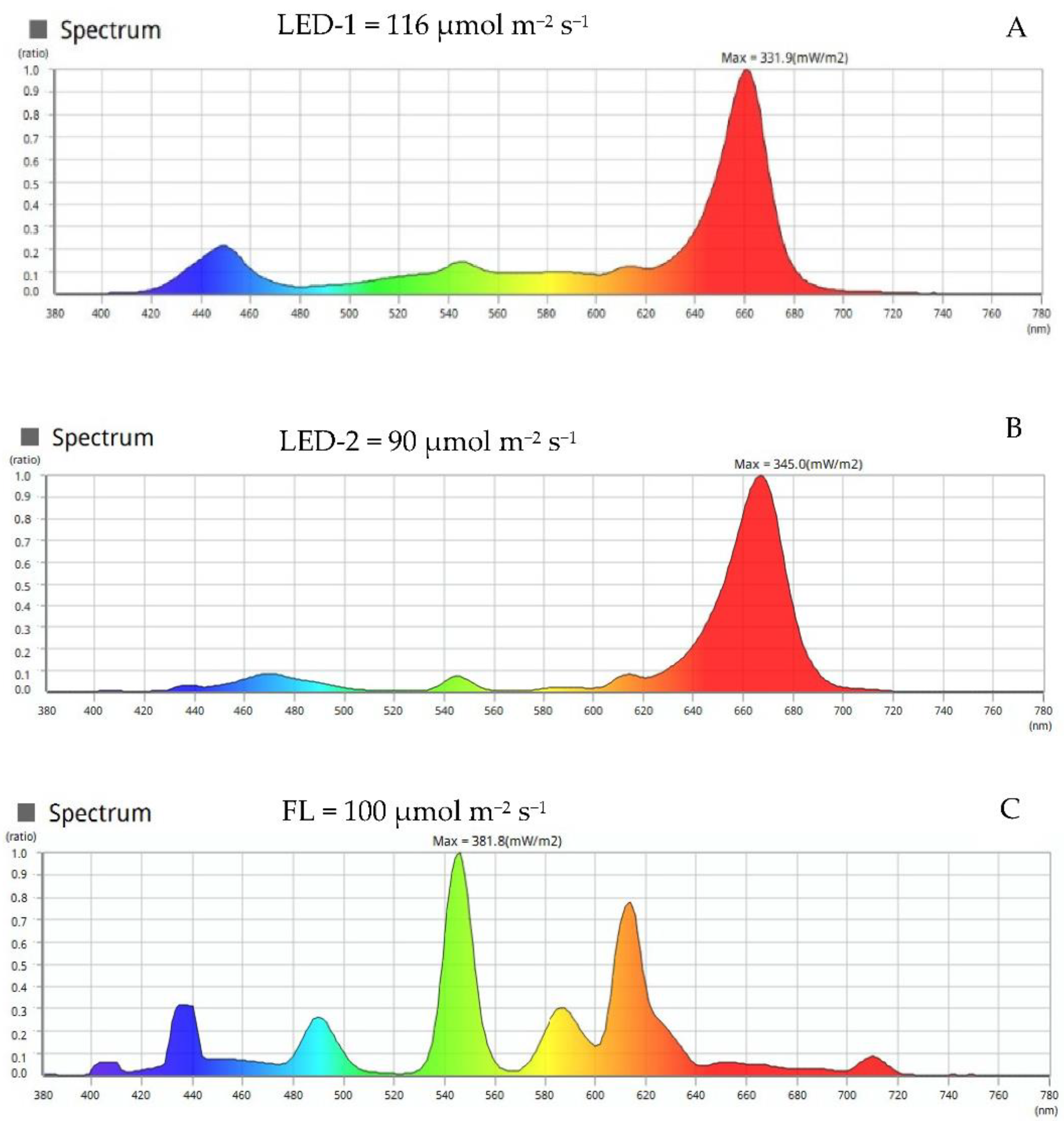
Two varieties of ornamental bananas, Musa ‘Little Prince’ and Musa ‘Truly Tiny’, were obtained from AgriStarts, Inc. (Apopka, FL, USA). They are dwarf varieties of dwarf Cavendish, with a compact habit and thick dark green leaves. Musa ‘Little Prince’ has thick trunks and is generally used as an ornamental indoors or as a landscape plant outdoors. Musa ‘Truly Tiny’ is extremely dwarf and produces the smallest edible fruits in the world. Both are desirable as ornamental plants and commonly produced via tissue culture, and have similar growth rates. In vitro plantlets were established in Murashige and Skoog (MS) culture medium [24] supplemented with benzylaminopurine (BAP; 8.8 µM) and sucrose (30 g L−1). The pH was adjusted to 5.8, and the medium was solidified with agar (agar–agar, Sigma Aldrich Inc., St. Louis, MO, USA) at 8 g L−1. A volume of 50 mL of medium was dispensed into baby food jars, and the medium was autoclaved at 121 °C and 20 psi for 20 min. In vitro banana plantlets measuring about 5 cm in height had their leaves and roots trimmed to 2 cm shoot-tips, which were used as explants. Baby food jars with polypropylene lids were sealed with parafilm, and cultures were placed in a growth chamber under controlled environmental conditions of 27 ± 2 °C and a 16 h photoperiod at 100 μmol m−2 s−1 PPFD. In vitro shoots were subcultured to fresh MS medium at four-week intervals.

2.2. Light Sources

Three different light sources were evaluated: two provided by LED lighting, LED-1 at 116 μmol m−2 s−1 (Philips GreenPower DR/B 3:1 150 43W) and LED-2 at 90 μmol m−2 s−1 (Philips GreenPower DR/W 3:1 150 33W); and one provided by fluorescent lighting (Philips 9A fluorescent bulbs 40W) at 100 μmol m−2 s−1 (FL). Photoperiod was 16/8 h (light/dark). The spectral energy distribution for the lighting used in this study is shown in Figure 1: LED-1 showed peak emissions at 440 nm and 650 nm (Figure 1A), LED-2 showed peak emissions at 440 nm and 670 nm (Figure 1B); and fluorescent lighting (FL) showed a broader spectrum with peaks in the green (550 nm), blue (440 nm), and some additional peaks in between (490 nm, 590 nm, 610 nm, and 710 nm) (Figure 1C). The intensity and composition of all light sources were measured using an LI-180 Li-Cor spectrometer (Li-Cor, Lincoln, NE, USA).

2.3. In Vitro Growth and Development

Explants from all treatments were evaluated four weeks after in vitro establishment for shoot length, root length and number, plantlet fresh and dry weight, shoot fresh and dry weight, root fresh and dry weight, root length, and number determined. Dry weight was determined by oven-drying plantlets at 70 °C until they reached constant weight. Five random plants were selected per treatment.

2.4. Relative Chlorophyll Content

Relative chlorophyll content was evaluated as SPAD value by placing the third expanded leaf of each plantlet, counted from top downwards, in a portable SPAD-502 chlorophyll meter (SPAD-502, Minolta Co., Ltd., Tokyo, Japan). Five random plantlets were selected per treatment.

2.5. Stomata Analysis

The middle third portion of the third and fourth fully expanded leaves were cut into approximately 1 cm × 1 cm sections. Impressions of the leaves were obtained by placing leaf sections on top of a thin layer of super glue (Elmer’s Products, Inc., Westerville, OH, USA), spread over microscope glass slides, and removing the leaf after drying. Impressions were obtained for both the adaxial and abaxial surfaces of the leaves. Stomata observations were performed under an optical Leica DMLB microscope (Leica microsystems, Buffalo, NY, USA) at 200× magnification. Images were recorded using a SPOT 4.7 digital camera coupled to the microscope and analyzed using the SPOT basic software (SPOT Imaging, Diagnostic Instruments, Inc., Sterling Heights, MI, USA). The number of stomata was counted under the microscope for both the abaxial and adaxial surfaces of leaves. Three different areas of 5 mm in diameter were selected as replicates for observations for each light treatment. Results were expressed as a means of counts of the three areas per mm2. The length and width of three randomly selected stomata were measured for both the adaxial and abaxial surfaces of each leaf in three different sections of each selected leaf. Five random plantlets were selected per treatment.

2.6. Anatomical Observations

Anatomical studies were conducted using the middle-third section of the second completely developed leaf. Leaf cross-sections were obtained by freehand sectioning using a steel blade, fixed in 70% FAA (formaldehyde-acetic acid-ethyl alcohol 70%) for 48 h, and then preserved in ethanol 70% (v/v). Following discoloration in sodium hypochlorite (1–1.25% active chlorine), triple-rinsing in distilled water, and staining in toluidine blue (0.05% w/v), leaf sections were subsequently fixed on semi-permanent slides with glycerinated water [25]. The slides were examined and photographed under the same light Leica DMLB microscope and the SPOT 4.7 idea digital camera and software, as described earlier. The images were evaluated by assessing five fields per repetition for each variable analyzed. The thickness of the abaxial surface epidermis (ASE), adaxial epidermis (ABE), abaxial hypodermis (AH), adaxial hypodermis (AbH), palisade parenchyma (PP), and spongy parenchyma (SP) were determined. Five random plants were selected per treatment.

2.7. Experimental Design and Statistical Analysis

The experimental design was completely randomized and consisted of six treatments (three light sources × two banana varieties), with five replications per treatment. Each replication consisted of 3 baby food flasks containing 1 in vitro plantlet per flask for a total of 90 experimental units. Data were collected and submitted to analysis of variance (ANOVA) using the R statistical analysis program, with means compared by LSD (Least Significant Difference) test at the 5% level of significance.

3. Results

3.1. In Vitro Growth and Development

The light source had no significant effect on most in vitro growth and development parameters evaluated for both banana varieties, including stem diameter, shoot and root fresh weight, leaf number, shoot and root length, and shoot and root dry weight (Table 1). However, ‘Little Prince’ plantlets produced under LED-1 and LED-2 showed larger overall plantlet fresh weight and shoot length compared to plantlets under fluorescent lights, while ‘Truly Tiny’ plantlets showed higher fresh weight only under LED-1, but no differences in shoot length.

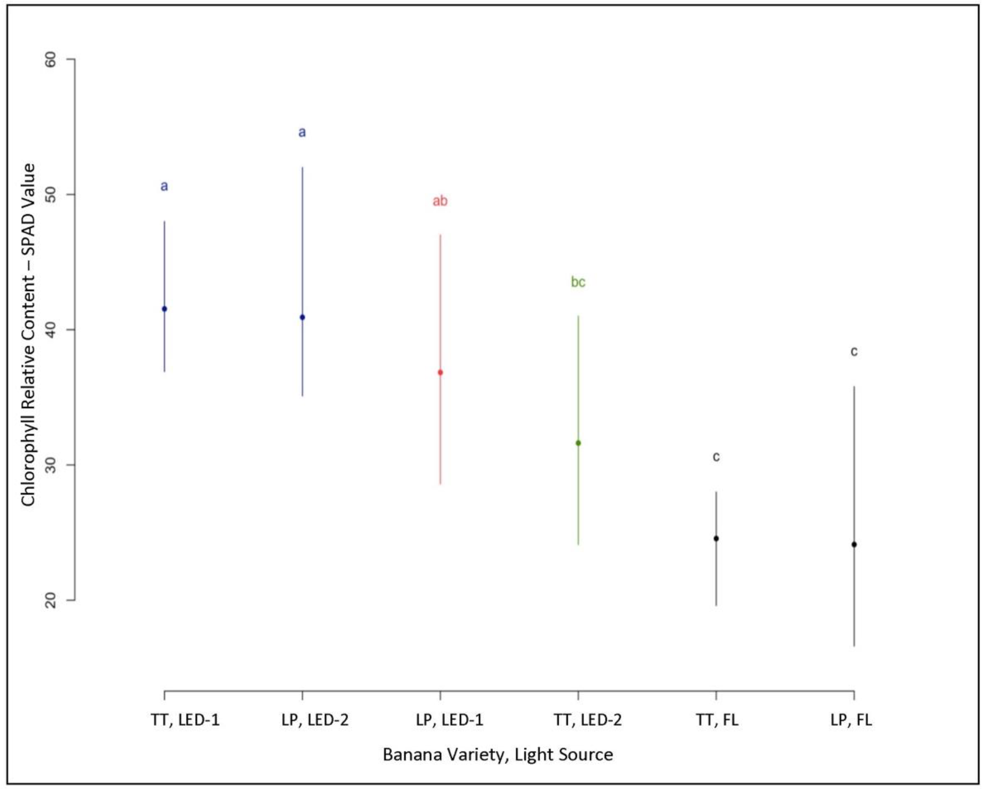
3.2. Relative Chlorophyll Content

Significant differences were shown in relative chlorophyll content among the different light sources and banana varieties (Figure 2). In general, the relative chlorophyll content of plants under LED lighting showed greater SPAD values compared to plantlets under fluorescent lamps. SPAD values were 36.84 for ‘Little Prince’ and 41.54 for ‘Truly Tiny’ under LED-1, 31.62 for ‘Truly Tiny’ and 40.92 for ‘Little Prince’ under LED-2, and 24.12 for ‘Little Prince’ and 24.56 for ‘Truly Tiny’ under fluorescent lamps (Figure 2). However, relative chlorophyll content was not significantly different between plantlets of both varieties under LED-1 and LED-2, but higher than FL, with the exception of ‘Truly Tiny’ under LED-2, which had similar values to both varieties under FL. The leaves of plantlets under LED-1 and LED-2 showed a dark green color, while the leaves of plantlets under FL had a yellowish-green color.

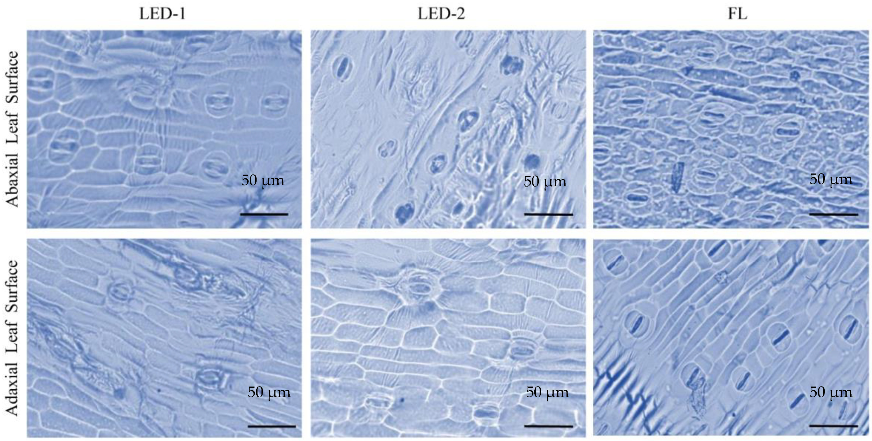
3.3. Stomata Analysis

In this study, stomata were present in both adaxial and abaxial leaf surfaces of both banana varieties under the different light sources, thus characterizing them as amphistomatic (Figure 3). Light source influenced stomata number of both banana varieties (Table 2). Some significant differences were observed between lighting treatments, following the same pattern observed for relative chlorophyll content. Overall, there were no significant differences in the effect of light sources on the adaxial number of stomata (AdSN) for both banana varieties, except for LED-2 and FL, which were slightly higher than LED-1 (Table 2). For the adaxial surface, plantlets had an average leaf stomata number per mm2 of 22.8 and 24.4 under LED-1, 28.4 and 25.8 for LED-2, and 30.0 and 32.0 under FL for Musa ‘Little Prince’ and Musa ‘Truly Tiny’, respectively (Table 2). A similar trend was observed for the abaxial number of stomata per mm2 (AbSN) for both varieties with 75.4 and 92.0 under LED-1, 67.6 and 94.2 under LED-2 and 83.6 and 101.6 under FL for Musa ‘Little Prince’ and Musa ‘Truly Tiny’, respectively (Table 2). The size of the stomata evaluated by length and width showed some variation for both adaxial and abaxial surfaces under the different light sources and for both varieties. The length of the adaxial surface stomata (AdSL) was higher for ‘Little Prince’ under FL but not significantly different for ‘Truly Tiny’ under LED-2 and FL (Table 2). The length of the abaxial surface stomata (AbSL) was higher for both varieties under LED-2 and FL (Table 2). The width of the adaxial surface stomata (AdSW) was higher for both varieties under FL, but for the abaxial surface stomata (AbSW), width showed no significant differences (Table 2).

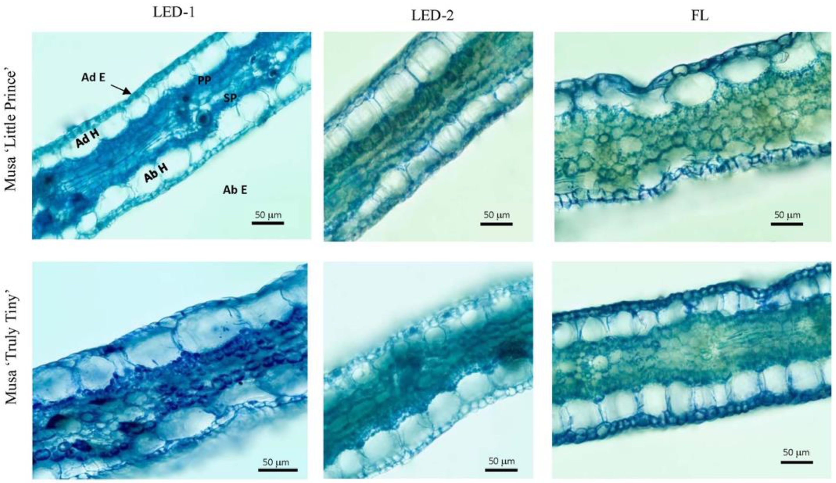
3.4. Anatomical Observations

Some parameters of leaf anatomy of in vitro plantlets were significantly affected by the light source, including abaxial epidermis (AbE), and abaxial (AbH) and adaxial (AdH) hypodermis for both varieties (Table 3). However, no significant differences were observed for adaxial epidermis (AdE) and for palisade (PP) and spongy (SP) parenchyma for both varieties (Table 3). The abaxial epidermis (AbE) was significantly thinner than the adaxial epidermis (AdE) (Table 3). As for the organization of mesophyll, the banana is dorsiventral or bifacial, with the palisade parenchyma oriented towards the adaxial epidermis and immediately below the adaxial hypodermis and the spongy parenchyma facing the abaxial epidermis (Figure 4). The palisade parenchyma was comprised of closely packed cylindrical cells, exhibiting one to two layers of cells, while the spongy parenchyma was comprised of two to three layers of spongy mesophyll cells, packed with inconspicuous air spaces (Figure 4). The cells were not well defined and had more or less a round shape under FL (Figure 4). However, no statistical differences were observed between most parameters evaluated.

4. Discussion

The maintenance of the quality of in vitro plantlets requires an environment that provides optimum conditions for growth and development [26]. Light is among some of the main factors influencing the growth and development of in vitro plantlets, including light quality, intensity, and the photoperiod [27]. In this study, our goal was to evaluate the effect of different light sources on in vitro growth and development of two ornamental banana varieties. However, for most parameters evaluated, no significant differences were observed. The stem diameter of in vitro banana plantlets in our study was not significantly affected by the light source. This contrasts with similar studies showing an increase in stem diameter of in vitro banana plantlets under LED lighting (45, 60, and 75 μmol m−2 s−1 PPF) compared to fluorescent lights (45 μmol m−2 s−1 PPF) [22], although light intensities evaluated were lower as compared to the light intensities evaluated in our study [22]. This could indicate that lower light levels are more desirable to induce a larger stem diameter in in vitro banana plantlets. Another aspect was the spectral distribution evaluated [22], where LED lighting had a 9:1 = red:blue ratio and FL had an 8:2 = red:blue ratio. Our study had similar red:blue ratios for both LEDs compared to FL, which had almost an inverse red:blue ratio and a peak in the green (Figure 1), indicating that the light spectral distribution might have influenced the results observed. Similar results were reported in rapeseed in vitro plantlets under LED lighting [28]. In contrast, the stem diameter of Cymbidium in vitro plantlets was not affected by different light sources [29], thus showing similarities to our study. In our study, however, stem diameter had a correlation with banana varieties, whereas ‘Little Prince’ showed a larger average diameter than ‘Truly Tiny’, suggesting a genotype effect. Such differences in responses confirm that the influences and mechanisms related to light quality, intensity, and photoperiod in plants are rather specific to plant species or cultivars. A similar genotype effect has been shown for two annatto (Bixa orellana) cultivars grown under different irradiance and light quality [2].
Banana in vitro plantlet fresh weight was slightly greater under LEDs compared to FL, but differences were not statistically significant between LED-1 and LED-2 for both varieties. This trend was observed when banana in vitro plantlets were cultured under LED lighting [22] as well as in rapeseed [28], where no significant differences in fresh weight of plantlets under different LED lighting were observed.
Shoot fresh and dry weight of banana in vitro plantlets were not significantly different under different light sources. Similarly, no significant differences were observed for root fresh and dry weight for both banana varieties. However, similar studies with in vitro banana [30] and Doritaenopsis [31] plantlets under LED reported higher shoot and root fresh weight.
Because the spectral energy distribution of red and blue lights aligns with chlorophyll absorption, it is generally accepted that LED lighting improves plant growth and development by enhancing the net photosynthetic rate [32,33,34]. However, in our study, the effect of LED lighting on shoot length was significantly different from FL only for one banana variety, ‘Little Prince’, reinforcing the concept of a genotype effect, as previously indicated.
There were no statistically significant differences among plantlets for root length under the different light sources evaluated in our study. These results contrast with those on banana [23] and rapeseed [28], where root length was significantly different between different light sources. However, these studies showed no significant differences among plantlet stem lengths under different light treatments, thus showing similarities with our study.
In our study, the relative chlorophyll content of banana in vitro plantlets grown under LED lighting was higher compared to fluorescent lighting. Similar results for in vitro banana plantlets were reported [35], showing that higher chlorophyll values were obtained when banana plantlets were cultured under LED lights. Similar behavior has been reported for other species, including chrysanthemum [11], Doritaenopsis [32], and rapeseed [28].
The general morphology of plantlets proved quite homogeneous under all lighting treatments evaluated. No differences were observed for leaf number among the different light treatments, and our results are similar to those reported in bananas [35] and Cymbidium. However, in similar studies with banana and Anacardium othonianum plantlets, a higher number of leaves was observed under fluorescent lamps [15,30,36]. However, strawberry in vitro plantlets showed a higher number of leaves in plantlets cultured under LED compared to fluorescent lighting [37]. The great diversity in responses in vitro among different species as affected by light intensity and quality illustrates the complexity of the topic and the need for continued studies to narrow down the best parameters for proper growth and development of plants in vitro.
Light sources influenced leaf stomata in our study. The number of stomata was generally higher on the abaxial than the adaxial leaf surface. Stomata of plantlets grown under the LED lighting were ellipsoid and closed, compared to open stomata and guard cells with round shapes in plantlets under FL (Figure 3). In general, banana plantlets grown under FL had larger numbers of stomata in both adaxial and abaxial leaf surfaces and similar higher values for length and width of stomata in adaxial leaf surfaces (Figure 3). However, for the abaxial leaf surfaces, while the length was slightly higher for FL, no differences were observed for width. Comparable results were reported in chrysanthemums [11]. However, another study with bananas [35] indicated the increased formation of stomata on both leaf surfaces (adaxial and abaxial) when banana plantlets were grown under LED compared to FL lighting.
In the present study, the epidermis of the adaxial leaf surface was thicker than the abaxial epidermis. These results corroborate a similar study of bananas in vitro [38], where the adaxial surface was thicker than the abaxial surface in leaves of in vitro-derived bananas under acclimatization. However, most of the anatomical leaf features measured in the current study were similar among the different light sources regardless of the banana variety. Similar results have been reported in pepper, showing a similar thickness in anatomical leaf features [39]. High light intensity also affected the anatomic structure in sugarcane leaves, including both adaxial and abaxial sides, and in the mesophyll and bulliform cell number [40]. However, no differences were observed in anatomic structures for ornamental bananas in our study, except for the thickness of adaxial compared to abaxial leaf surfaces.

5. Conclusions

Our results revealed that shoot mass and length could be promoted by controlling light quality and intensity. However, the effect of light quality and intensity related to plant growth and development (stem diameter, shoot and root fresh weight, shoot and root dry weight, root length, root number, and leaf number) were not evident despite contrasting reports from other studies cited here. This leaves additional work to be performed to better address the effects of light sources, intensity, and quality on the in vitro growth and development of different banana varieties. Although not directly evaluated in this study, the number of in vitro shoots produced per explant were higher for some of the cultures grown under the LED lightings, with multiple shoots produced, while no shoots were formed in cultures grown under FL. Subsequent studies will address such parameters. Different responses as demonstrated between the two banana varieties (Musa ‘Little Prince’ and Musa ‘Truly Tiny’) indicated a genotype effect as affected by different light environments. LED lighting affected the relative chlorophyll content as well as stomata size in banana in vitro plantlets. Based on the responses for both banana varieties to the different light sources evaluated in this study, we suggest that LEDs at 90 μmol m−2 s−1 could be a suitable selection for the micropropagation of ornamental bananas.

Author Contributions

Conceptualization, W.A.V.; methodology, W.A.V., C.F., D.B. and P.M.C.B.; validation, W.A.V.; formal analysis, W.A.V.; investigation, C.F. and P.M.C.B.; resources, W.A.V. and D.B.; data curation, C.F.; writing—original draft preparation, C.F.; writing—review and editing, W.A.V., D.B. and P.M.C.B.; supervision, W.A.V. and D.B.; project administration, W.A.V. and D.B.; funding acquisition, W.A.V. All authors have read and agreed to the published version of the manuscript.

Funding

This research project was funded under the USAID-AREA project at the University of Florida, Project Number G000260- 60290000-201-3300-00118870.

Institutional Review Board Statement

Not applicable.

Informed Consent Statement

Not applicable.

Data Availability Statement

Not applicable.

Acknowledgments

This study was supported by the USAID-AREA project under the Department of Environmental Horticulture, Institute of Food and Agricultural Sciences, the University of Florida.

Conflicts of Interest

The authors declare no conflict of interest.

References

  1. Rajapakse, N.C.; Shahak, Y. Light-quality manipulation by horticulture industry. Annual Plant Rev. Light Plant Dev. 2008, 30, 290. [Google Scholar]
  2. Faria, D.V.; Correia, L.N.F.; Souza, M.V.C.; Ríos-Ríos, A.M.; Vital, C.E. Irradiance and light quality affect two annatto (Bixa orellana L.) cultivars with contrasting bixin production. J. Photochem. Photobiol. 2019, 197, 111549. [Google Scholar] [CrossRef]
  3. Gupta, S.D.; Jatothu, B. Fundamentals and applications of light-emitting diodes (LEDs) in in vitro plant growth and morphogenesis. Plant Biotechnol. Rep. 2013, 7, 211–220. [Google Scholar] [CrossRef]
  4. Miyashita, Y.; Kitaya, Y.; Kozai, T.; Kimura, T. Effects of red and far-red light on the growth and morphology of potato plantlets in vitro: Using light emitting diode as a light source for micropropagation. Environ. Eff. Control. Plant Tissue Cult. 1994, 393, 189–194. [Google Scholar] [CrossRef]
  5. Kodym, A.; Zapata-Arias, F.J. Natural light as an alternative light source for the in vitro culture of banana (Musa acuminata cv.‘Grande Naine’). Plant Cell Tis. Org. Cult. 1998, 55, 141–145. [Google Scholar] [CrossRef]
  6. Dooley, J.H. Influence of Lighting Spectra on Plant Tissue Culture; American Society of Association Executives: Washington, DC, USA, 1991; No. 7530. [Google Scholar]
  7. Standaert-de Metsenaere, R.E.A. Economic considerations. Micropropag. Technol. Appl. 1991, 123–140. [Google Scholar]
  8. Mitchell, C. Plant lighting in controlled environments for space and earth applications. Acta Hortic. 2012, 956, 23–36. [Google Scholar] [CrossRef]
  9. Olle, M.; Viršilė, A. The effects of light-emitting diode lighting on greenhouse plant growth and quality. Agric. Food Sci. 2013, 22, 223–234. [Google Scholar] [CrossRef]
  10. Nhut, D.T.; Nam, N.B. Light-emitting diodes (LEDs): An artificial lighting source for biological studies. In Proceedings of the Third International Conference on the Development of Biomedical Engineering in Vietnam, Ho Chi Minh City, Vietnam, 11–14 January 2010; IFMBE Proceedings. Van Toi, V., Khoa, T.Q.D., Eds.; Springer: Berlin/Heidelberg, Germany, 2010; Volume 27, pp. 134–139. [Google Scholar]
  11. Kim, S.-J.; Hahn, E.-J.; Heo, J.-W.; Paek, K.-Y. Effects of LEDs on net photosynthetic rate, growth and leaf stomata of chrysanthemum plantlets in vitro. Sci. Hortic. 2003, 101, 143–151. [Google Scholar] [CrossRef]
  12. Silva, T.D.; Batista, D.; Fortini, E.A.; de Castro, K.M.; Felipe, S.H.S.; Fernandes, A.M.; Sousa, R.M.D.J.; Chagas, K.; da Silva, J.V.S.; Correia, L.N.D.F.; et al. Blue and red light affects morphogenesis and 20-hydroxyecdisone content of in vitro Pfaffia glomerata accessions. J. Photochem. Photobiol. B Biol. 2019, 203, 111761. [Google Scholar] [CrossRef]
  13. Jishi, T. LED lighting technique to control plant growth and morphology. In Smart Plant Factory; Kozai, T., Ed.; Springer: Singapore, 2018; pp. 211–222. [Google Scholar]
  14. Ortiz, R. Conventional banana and plantain breeding. In Proceedings of the VII International Symposium on Banana: ISHS-ProMusa Symposium on Bananas and Plantains: Towards Sustainable Global Production, Salvador, Brazil, 14 October 2011; International Society for Horticultural Science: Leuven, Belgium, 2011; Volume 986, pp. 177–194. [Google Scholar]
  15. Nguyen, Q.T.; Kozai, T. Growth of In vitro banana (Musa spp.) shoots under photomixotrophic and photoautotrophic conditions. Vitr. Cell. Dev. Biol.-Anim. 2001, 37, 824–829. [Google Scholar] [CrossRef]
  16. Wong, W.C. In vitro propagation of banana (Musa spp.): Initiation, proliferation and development of shoot-tip cultures on defined media. Plant Cell Tissue Organ Cult. 1986, 6, 159–166. [Google Scholar] [CrossRef]
  17. Bairu, M.W.; Stirk, W.A.; Doležal, K.; van Staden, J. The role of topolins in micropropagation and somaclonal variation of banana cultivars ‘Williams’ and ‘Grand Naine’ (Musa spp. AAA). Plant Cell Tis. Org. Cult. 2008, 95, 373–379. [Google Scholar] [CrossRef]
  18. Makara, A.M.; Rubaihayo, P.R.; Magambo, M.J.S. Carry-over effect of Thidiazuron on banana in vitro proliferation at different culture cycles and light incubation conditions. African J. Biotechnol. 2010, 9, 3079–3085. [Google Scholar]
  19. Ahirwar, M.K.; Sananda, M.; Singh, M.K.; Chaitali, S.; Singh, R.P. A high frequency plantlets regeneration protocol for banana (Musa paradisiaca L.) micropropagation. Asian J. Hort. 2012, 7, 397–401. [Google Scholar]
  20. Yusnita, Y.; Danial, E.; Hapsoro, D. In vitro shoot regeneration of indonesian bananas (Musa spp.) cv. ambon kuning and raja bulu, plantlet acclimatizationand field performance. AGRIVITA J. Agric. Sci. 2015, 37, 51–58. [Google Scholar] [CrossRef]
  21. Nhut, D.; Hong, L.; Watanabe, H.; Goi, M.; Tanaka, M. Growth of banana plantlets cultured in vitro under red and blue light-emitting diode (led) irradiation source. Acta Hortic. 2002, 575, 117–124. [Google Scholar] [CrossRef]
  22. Nhut, D.T.; Takamura, T.; Watanabe, H.; Tanaka, M. Efficiency of a novel culture system by using light-emitting diode (LED) on in vitro and subsequent growth of micropropagated banana plantlets. Acta Hort. 2003, 161, 121–127. [Google Scholar] [CrossRef]
  23. Trivedi, A.; Sengar, R.S. Effect of various light emittimg diodes on growth and photosynthetic pigments of banana (Musa acuminata) CV. Grande naine in vitro plantlets. Int. J. Chem. Stud. 2017, 5, 1819–1821. [Google Scholar]
  24. Murashige, T.; Skoog, F. A revised medium for rapid growth and bioassays with tobacco tissue cultures. Physiol. Plant. 1962, 15, 473–497. [Google Scholar] [CrossRef]
  25. Kraus, J.E.; Arduin, M. Manual Básico de Métodos em Morfologia Vegetal; EDUR: Seropédica, Brazil, 1997; p. 198. [Google Scholar]
  26. Chen, C. Fluorescent Lighting Distribution for Plant Micropropagation. Biosyst. Eng. 2005, 90, 295–306. [Google Scholar] [CrossRef]
  27. Aitken-Christie, J.; Kozai, T.; Smith, M.A.L. (Eds.) Automation and Environmental Control in Plant Tissue Culture; Springer Science & Business Media: Berlin/Heidelberg, Germany, 2013; p. 586. [Google Scholar]
  28. Li, H.; Tang, C.; Xu, Z. The effects of different light qualities on rapeseed (Brassica napus L.) plantlet growth and morphogenesis in vitro. Sci. Hortic. 2013, 150, 117–124. [Google Scholar] [CrossRef]
  29. Tanaka, M.; Takamura, T.; Watanabe, H.; Endo, M.; Yanagi, T.; Okamoto, K. In vitro growth of Cymbidium plantlets cultured under super bright red and blue light-emitting diodes (LEDs). J. Hort. Sci. Biotechnol. 1998, 73, 39–44. [Google Scholar] [CrossRef]
  30. Nhut, D.T.; Don, N.T.; Tanaka, M. Light-emitting diodes as an effective lighting source for in vitro banana culture. In Protocols for Micropropagation of Woody Trees and Fruits; Springer: Dordrecht, The Netherlands, 2007; pp. 527–541. [Google Scholar]
  31. Shin, K.S.; Murthy, H.N.; Heo, J.W.; Hahn, E.J.; Paek, K.Y. The effect of light quality on the growth and development of in vitro cultured Doritaenopsis plants. Acta Physiol. Plant. 2008, 30, 339–343. [Google Scholar] [CrossRef]
  32. Goins, G.D.; Yorio, N.C.; Sanwo, M.M.; Brown, C.S. Photomorphogenesis, photosynthesis, and seed yield of wheat plants grown under red light-emitting diodes (LEDs) with and without supplemental blue lighting. J. Exp. Bot. 1997, 48, 1407–1413. [Google Scholar] [CrossRef] [PubMed] [Green Version]
  33. Hoenecke, M.; Bula, R.; Tibbitts, T. Importance of ‘Blue’ Photon Levels for Lettuce Seedlings Grown under Red-light-emitting Diodes. HortScience 1992, 27, 427–430. [Google Scholar] [CrossRef] [Green Version]
  34. Tripathy, B.C.; Brown, C.S. Root-Shoot Interaction in the Greening of Wheat Seedlings Grown under Red Light. Plant Physiol. 1995, 107, 407–411. [Google Scholar] [CrossRef] [Green Version]
  35. Nascimento Vieira, L.; de Freitas Fraga, H.P.; dos Anjos, K.G.; Puttkammer, C.C.; Scherer, R.F.; da Silva, D.A.; Guerra, M.P. Light-emitting diodes (LED) increase the stomata formation and chlorophyll content in Musa acuminata (AAA) ‘Nanicão Corupá’ in vitro plantlets. Theor. Experim. Plant Physiol. 2015, 27, 91–98. [Google Scholar] [CrossRef]
  36. Rosa, M.; Neto, A.R.; Marques, V.D.O.; Silva, F.G.; De Assis, E.S.; Costa, A.C.; Dantas, L.A.; Pereira, P.S. Variations in photon flux density alter the morphophysiological and chemical characteristics of Anacardium othonianum Rizz. in vitro. Plant Cell Tissue Organ Cult. 2019, 140, 523–537. [Google Scholar] [CrossRef]
  37. Nhut, D.T.; Takamura, T.; Watanabe, H.; Okamoto, K.; Tanaka, M. Responses of strawberry plantlets cultured in vitro under superbright red and blue light-emitting diodes (LEDs). Plant Cell Tissue Organ Cult. 2003, 73, 43–52. [Google Scholar] [CrossRef]
  38. Costa, F.D.S.; de Castro, E.M.; Pasqual, M.; Pereira, J.E.S.; Oliveira, C.D. Alterações anatômicas de bananeiras micropropagadas em resposta a aclimatização ex vitro. Ciência Rural. 2009, 39, 386–392. [Google Scholar] [CrossRef]
  39. Schuerger, A.C.; Brown, C.S.; Stryjewski, E.C. Anatomical Features of Pepper Plants (Capsicum annuum L.) Grown under Red Light-emitting Diodes Supplemented with Blue or Far-red Light. Ann. Bot. 1997, 79, 273–282. [Google Scholar] [CrossRef] [PubMed]
  40. Neto, A.R.; Chagas, E.A.; Costa, B.N.S.; Chagas, P.C.; Vendrame, W.A. Photomixotrophic growth response of sugarcane plantlets with increased light intensity and design of culture vessel. Vitr. Cell Dev. Biol. Plant 2020, 56, 504–514. [Google Scholar] [CrossRef]
Figure 1. Intensity and composition of lighting as photosynthetically active radiation (PAR) for the different light treatments in this study: (A) LED-1 = 116 μmol m−2 s−1; (B) LED-2 = 90 μmol m−2 s−1; and (C) fluorescent light (FL) = 100 μmol m−2 s−1. While both LED lights peak in the red range (660–670 nm) with some blue peaks (440–470 nm), fluorescent light has a broader distribution with peaks in the green (550 nm), blue (440 nm), and some additional peaks in between (490 nm, 590 nm, 610 nm, and 710 nm). Maximum irradiance (mW m−2) is shown for each spectrum. Measurements were obtained with an LI-180 Li-Cor spectrometer.
Horticulturae 08 00092 g001
Figure 2. Relative chlorophyll content of two banana varieties, Musa Little Prince (LP) and Musa Truly Tiny (TT) after 4 weeks of in vitro culture under different light sources, LED-1 (116 μmol m−2 s−1) LED-2 (90 μmol m−2 s−1) and FL (100 μmol m−2 s−1). Values with the same letter are not significantly different at α = 0.05 level based on LSD mean separation. Bars represent standard error.
Horticulturae 08 00092 g002
Figure 3. Stomatal prints of abaxial and adaxial leaf surfaces of in vitro banana leaves (Musa ‘Little Prince’ and ‘Truly Tiny’) under different light sources. LED-1 = 116 μmol m−2 s−1; LED-2 = 90 μmol m−2 s−1, fluorescent lighting (FL) = 100 μmol m−2 s−1. Bars = 50 µm.
Horticulturae 08 00092 g003
Figure 4. Cross-section of in vitro banana leaves (Musa ‘Little Prince’ and ‘Truly Tiny’) under different light sources. LED-1 = 116 μmol m−2 s−1; LED-2 = 90 μmol m−2 s−1, fluorescent lighting (FL) = 100 μmol m−2 s−1. AdE = adaxial epidermis; AbE = abaxial epidermis; AdH = adaxial hypodermis; AbH = abaxial hypodermis; PP = palisade parenchyma; SP = spongy parenchyma. Bar = 50 μm.
Horticulturae 08 00092 g004
Table 1. Evaluation of light quality and light level effects on in vitro growth and development parameters for two ornamental banana varieties; Musa ‘Little Prince’ and Musa ‘Truly Tiny’. LED-1 = 116 μmol m−2 s−1; LED-2 = 90 μmol m−2 s−1; fluorescent light (FL) = 100 μmol m−2 s−1.
Banana
Variety
Light SourceStem Diameter *
(mm)
Plant Fresh Weight
(g)
Shoot Fresh Weight
(g)
Root Fresh Weight
(g)
Number
of Leaves
Shoot Length
(cm)
Root Length
(cm)
Number of RootsShoot Dry Weight
(g)
Root Dry Weight
(g)
Little PrinceLED-15.32 ± 0.50 a2.84 ± 0.14 a1.63 ± 0.25 a0.19 ± 0.08 a3.6 ± 0.89 a7.45 ± 0.67 a3.02 ± 0.30 a6.4 ± 2.07 a0.24 ± 0.03 a0.01 ± 0.01 a
LED-25.24 ± 0.44 a2.62 ± 0.42 ab1.94 ± 0.54 a0.34 ± 0.09 a4.0 ± 1.00 a7.33 ± 0.76 a3.70 ± 0.82 a8.2 ± 2.17 a0.28 ± 0.07 a0.03 ± 0.01 a
FL5.08 ± 0.71 a2.21 ± 0.39 c1.44 ± 0.05 a0.16 ± 0.07 a4.2 ± 0.84 a5.87 ± 0.82 b3.21 ± 0.25 a7.8 ± 3.19 a0.24 ± 0.02 a0.02 ± 0.01 a
Truly TinyLED-14.28 ± 0.55 a2.31 ± 0.16 bc1.27 ± 0.19 a0.25 ± 0.10 a3.6 ± 0.89 a6.67 ± 0.30 ab4.21 ± 0.34 a5.6 ± 1.52 a0.18 ± 0.03 a0.02 ± 0.01 a
LED-23.94 ± 0.37 a1.79 ±0.21 d1.01 ± 0.27 a0.27 ± 0.07 a3.6 ± 1.14 a6.46 ± 0.67 b4.38 ± 0.92 a5.2 ± 1.79 a0.12 ± 0.03 a0.02 ± 0.01 a
FL4.36 ± 0.92 a1.91 ± 0.38 cd1.39 ± 0.62 a0.34 ± 0.16 a4.0 ± 1.00 a6.02 ± 0.47 b4.60 ± 0.36 a6.0 ± 2.12 a0.21 ± 0.10 a0.02 ± 0.01 a
* Means within columns followed by the same letter are not significantly different at p ≤ 0.05 according to the LSD (Least Significance Difference) test. The tests were conducted separately for each parameter evaluated.
Table 2. Effects of light sources on stomata number (per mm2) and size (µm) in leaves of two ornamental banana varieties grown in vitro; Musa ‘Little Prince’ and ‘Truly Tiny’. LED-1 = 116 μmol m−2 s−1; LED-2 = 90 μmol m−2 s−1; fluorescent light (FL) = 100 μmol m−2 s−1.
Banana VarietyLight SourceAdSN *
(per mm2)
AbSN
(per mm2)
AdSL
(µm)
AbSL
(µm)
AdSW
(µm)
AbSW
(µm)
Little PrinceLED-122.8 ± 7.40 b75.4 ± 14.08 cd25.18 ± 2.57 b21.41 ± 3.19 c10.13 ± 0.94 c10.35 ± 1.25 a
LED-228.4 ± 7.67 ab67.6 ± 12.03 d25.63 ± 1.11 b27.08 ± 2.47 ab11.13 ± 0.91 bc11.35 ± 1.08 a
FL30.0 ± 6.20 ab83.6 ± 11.35 bcd28.51 ± 3.08 a27.16 ± 1.15 ab12.87 ± 0.97 a11.47 ± 1.58 a
Truly
Tiny
LED-124.4 ± 2.51 b92.0 ± 5.74 abc25.00 ± 1.89 b24.81 ± 1.65 b10.16 ± 0.61 b10.11 ± 1.72 a
LED-225.8 ± 4.66 ab94.2 ± 4.32 ab27.72 ± 2.19 ab27.68 ± 2.46 a10.00 ± 1.79 c11.05 ± 0.68 a
FL32.0 ± 3.54 a101.6 ± 21.55 a28.45 ± 0.98 a25.64 ± 1.44 ab12.02 ± 1.20 ab9.97 ± 1.18 a
* Means within columns followed by the same letter are not significantly different at p ≤ 0.05 according to the LSD (Least Significance Difference) test. The tests were conducted independently within each parameter (adaxial and abaxial stomata number, adaxial and abaxial stomata size). AdSN = Adaxial Stomata Number; AbSN = Abaxial Stomata Number; AdSL = Adaxial Stomata Length; AbSL = Abaxial Stomata Length; AdSW = Adaxial Stomata Width; AbSW = Abaxial Stomata Width.
Table 3. Effects of light source on leaf anatomy of two ornamental banana varieties grown in vitro; Musa ‘Little Prince’ and ‘Truly Tiny’. LED-1 = 116 μmol m−2 s−1; LED-2 = 90 μmol m−2 s−1; fluorescent light (FL) = 100 μmol m−2 s−1.
Banana
Variety
Light
Source
AbE (µm)AdE (µm)AbH (µm)AdH (µm)PP (µm)SP (µm)
Little
Prince
LED-115.53 ± 4.89 b17.63 ± 5.89 a59.02 ± 14.25 ab59.17 ± 5.69 c35.87 ± 9.23 a38.91 ± 9.38 ab
LED-217.70 ± 2.79 b16.40 ± 5.92 a52.38 ± 39.51 b69.31 ± 13.52 abc39.63 ± 10.75 a41.12 ± 10.29 b
FL20.66 ± 6.31 ab20.55 ± 5.28 a59.82 ± 4.46 ab64.33 ± 7.75 bc42.71 ± 11.53 a38.40 ± 11.25 ab
Truly
Tiny
LED-119.68 ± 7.05 ab15.07 ± 2.39 a61.61 ± 7.71 ab82.90 ± 14.51 a43.71 ± 11.57 a42.10 ± 8.66 ab
LED-217.73 ± 5.80 b15.08 ± 4.37 a69.01 ± 18.48 ab78.79 ± 20.14 ab35.39 ± 7.94 a33.88 ± 9.39 ab
FL25.10 ± 5.54 a18.15 ± 3.82 a62.76 ± 14.47 a69.96 ± 15.37 abc47.95 ± 13.36 a52.33 ± 17.81 a
* Means within columns followed by the same letter are not significantly different at p ≤ 0.05 according to the LSD (Least Significance Difference) test. The tests were conduct independently within each parameter (adaxial surface epidermis, abaxial surface epidermis, adaxial hypodermis, abaxial hypodermis, palisade parenchyma, spongy parenchyma). AbE = Abaxial Epidermis; AdE = Adaxial Epidermis; AbH = Abaxial Hypodermis, AdH = Adaxial Hypodermis; PP = Palisade Parenchyma; SP = Spongy Parenchyma.
Publisher’s Note: MDPI stays neutral with regard to jurisdictional claims in published maps and institutional affiliations.

