Article

The Influence of Phytohormones on the Efficiency of Callus Formation, Its Morphologically Properties and Content of Bioactive Compounds in In Vitro Cultures of Daucus carota L.

by
Anna Jadwiga Keutgen
1,*,
Magdalena Tomaszewska-Sowa
2,
Aleksander Bomberski
2 and
Norbert Keutgen
1
1
Department of Crop Sciences, Institute of Vegetables and Ornamentals, University of Natural Resources and Life Sciences, Gregor-Mendel-Str. 33, 1180 Vienna, Austria
2
Department of Agricultural Biotechnology, Bydgoszcz University of Science and Technology, Bernardyńska Street 6, 85-029 Bydgoszcz, Poland
*
Author to whom correspondence should be addressed.
Horticulturae 2022, 8(2), 100; https://doi.org/10.3390/horticulturae8020100
Submission received: 16 December 2021 / Revised: 17 January 2022 / Accepted: 20 January 2022 / Published: 23 January 2022
(This article belongs to the Section Postharvest Biology, Quality, Safety, and Technology)

Abstract

:
The significance of cultivar colour (orange and yellow), the application of MgO during field cultivation and chosen phytohormones in the callus cultivation medium are investigated in the present study, with respect to the antioxidative properties of the obtained callus. Callus cultivation are examined as an alternative method for the production of plant antioxidant compounds. Cultivar choice was most significant for callus production and the synthesis of health-promoting metabolites. The best combination, with respect to the induction efficacy and antioxidant properties measured as a synthetic value by Multidimensional Comparative Analysis (MCA), was found in the callus of cultivar ‘Flacoro’, cultivated without MgO fertilization and on a medium with kinetin (KIN) and 1-naphthaleneacetic acid (NAA) (MCA-value 0.465). The worst performance was found for cultivar ‘Yello Mello’, independent of the applied phythormones (averaged MCA-value 0.839) and for the cultivar ‘Flacoro’ fertilized with MgO and independent of growth hormones (averaged MCA-value 0.810).

1. Introduction

Many higher plants are major sources of useful secondary metabolites that are used in the pharmaceutical and agrochemical industries. The search for new plant chemicals or the increased production of desired ones is now a priority for research centres aiming at sustainability, biodiversity and environmental protection [1]. Biotechnological methods and in particular plant tissue culture, play an important role in the search for alternative forms of production of desirable compounds of medicinal nature [2,3,4,5]. Strictly controlled conditions in in vitro cultures can selectively generate many types of valuable natural products. To date, plant cell cultures produced ingredients for flavourings, aromas, fragrances, biofuels, plastics, enzymes, preservatives, cosmetics, natural colours and bioactive compounds [2,3,4]. Secondary products in plant cell culture can be produced without environmental restrictions during the entire year with less seasonal limitations. Production under these conditions is robust, predictable and independent of weather conditions. The extraction of secondary metabolites from in vitro tissues is much simpler than obtaining them from complex plant tissues [1].
Plant tissue culture techniques allow for the production of the chemical profile of a plant to be adjusted, so that it produces a compound with a potentially higher functional value. This occurs when the desired secondary metabolite is produced only in specialized plant tissues, e.g., in ginseng (Panax ginseng C. A. Mey), with in vitro cultures producing saponins, shikonin and berberine [3]. Only 30 targeted bioactive compounds are overexpressed in tissue culture compared to the content in wild relatives, but their production is mostly not economical [3,5]. In many other cases, the attempts to obtain secondary metabolites from in vitro cultures yielded results that were far from the expectations and were not suitable for commercial use [1]. In most cases, the low yield of secondary metabolites is mentioned [4]. There are three main ways to improve the yield: by enhancing the production efficacy by the standardization of the culture environment; by the application of different elicitors or abiotic stress factors specific for the synthesis of a given secondary compound or by influencing signal transduction pathways to induce higher contents of the targeted secondary compound [2,4]. In most cases, the selection of proper genotypes, media, plant hormones, growing conditions and plant organs used for the propagation were investigated.
In the case of carrot roots (Daucus carota L.), which are rich in different bioactive compounds, only few attempts were performed to increase the specific bioactive compounds, especially by in vitro callus culture [5,6,7]. The carrot is characterized by many health promoting values and by high contents of nutrients. It is characterized by anticancer, antiatherosclerotic and antimicrobial activity, and due to its content of β-carotene, high antioxidative properties [8,9,10,11,12]. As a result of the economic importance of the carrot, it is one of the most cultivated root vegetables in Europe. The worldwide cultivation area of carrots after FAO in 2019 was about 1,128,695 ha, and the yield was about 44,762,859 tons (FAO, 2019). The main carrot producers are China (21,483 kt), Uzbekistan (2770 kt), the United States (2259 kt) and the Russian Federation (1559 kt). Poland is in 9th place with 678 kt, and Austria in 40th place with 108 kt, however, with a very high production efficiency (57.5 t/ha compared to a world mean value of 28.5 t/ha). Generally, breeding techniques in recent years have mainly led to the improvement of features, such as the shape, colour and smoothness of the roots as well as to the increase in β-carotene and sugar content, which is particularly important in the consumer market [13,14]. In carrot tissues, including callus tissues, high levels of carotene (C40H56), which is a precursor of vitamin A, were found [15,16,17]. Steward (1958) [18] noted a high content of anthocyanins in carrot callus, which was confirmed by Harborne et al. [19]. There are many reports describing procedures of carrot plant regeneration in in vitro culture using different kinds of explants [16,20,21,22,23,24,25,26,27,28]. Various concentrations of 2,4–dichlorophenoxyacetic acid (2,4–D), NAA, indole-3-butyric acid, 6-benzylaminopurine and thidiazuron were applied to the cultivation medium to intensify the differentiation and growth processes [16,23,24,29]. Different amino acids precursors were also used with success to enhance the antimicrobial activity of callus cultures of different carrot organs, especially where stem callus seemed to be most effective [8]. The plant regeneration processes from the callus tissues of this species were also optimized by the application of diverse nutrients, modifying MS (Murashige and Skoog) [30] and B5 media [31]. Therefore, a further increase in the antioxidant properties as well as in the regeneration process may be expected by further improvement of in vitro carrot culture. Thus, the aim of this study is to evaluate the effect of growth regulators in the medium in combination with the genetic potential of different varieties on the content of valuable, health-promoting bioactive compounds, such as carotenoids and polyphenols, in in vitro culture from carrot storage roots. In addition, the effect of MgO applied as soil fertilizer on callus development in vitro was investigated, since Mg application improves carrot quality, as has been shown also for ‘Flacoro’ and ‘Karotan’ used in the present study [32]. The hypothesis was tested, whether an improved nutritional status of the carrot storage root would enable an explant to cope better with the stress experienced during callus cultivation.

2. Materials and Methods

The investigations were carried out in the Department of Microbiology and Food Technology and the Department of Agricultural Biotechnology, both in the Faculty of Agriculture and Biotechnology, Bydgoszcz University of Science and Technology in Poland. The investigated material was cultivated on the field of the PBS Experimental Station in Mochełek near Bydgoszcz, Poland, where the carrots were grown under mineral nutrition of N—70 kg/ha, P—80 kg/ha as P2O5 and K—100 kg/ha as K2O.
The plant material used in the experiment was the roots of three carrot varieties: ‘Karotan’, ‘Flacoro’ and ‘Yello Mello’. The selected carrot varieties differed in their morphological structure and fertility as well as in the potential content of the bioactive compounds in the roots. The colour of the carrot root is recognized as a varietal feature when it is aligned throughout the entire cross-section. The most valuable varieties are those with a darker flesh (phloem) and core (xylem) colour [33]. The variety ‘Karotan’ used in the experiments is particularly suitable for processing and perfectly suitable for storage. It is a late variety with an intense orange root colour that does not tend to accumulate nitrates. The content of total carotenoids is on average 115.8 mg kg−1 fresh mass (FM) of the content of total polyphenolics 1.94 mg kg−1 FM, and the antioxidant capacity is 1.71 mmol Fe2+ kg−1 FM measured as the Ferric Ion Reducing Ability of Plasma (FRAP) [9,10]. Another variety—‘Flacoro’—is a fertile variety characterized by a late harvest date. It is very well suitable for both processing and direct consumption. It does not accumulate nitrates, either. It can be stored for a long time. It contains on average 116.75 mg kg−1 FM of total carotenoids, 2.26 mg kg−1 FM of total polyphenolics and is characterized by a FRAP of 1.49 mmol Fe2+ kg−1 FM [9,10]. ‘Yello Mello’ is a fertile crossbreed with a long growing season. In addition to the yellow colour, it is characterized by a high homogeneity and has a tendency to greening in the head and shoulder area. It contains high concentrations of lutein, which plays an important role in the proper functioning of the human eye. Furthermore, the content of total carotenoids is on average 38.4 mg kg−1 FM, and that of total polyphenolics 1.05 mg kg−1 FM and the antioxidant capacity is 1.13 mmol Fe2+ kg−1 FM when measured as FRAP [9,10]. Carrots of this variety are recommended for juices, dried and frozen foods and as an addition to salads and snacks.
For the initiation of the in vitro experiment, the roots were harvested from plots additionally fertilized with magnesium sulphate in the amount of 90 kg MgO ha−1 applied manually in two charges (45 kg MgO ha−1 one day before sowing and 45 kg MgO ha−1 two months later), and from plots without additional fertilization. Carrot cultivation followed cereal crops, and the carrots were grown on dams. Weeding was performed manually, as required. The carrot roots were harvested on the 22nd of September before the first frosts appeared and in rainless weather. The term was chosen in order to prevent the deterioration of storage stability of the carrot. Immediately after removal from the soil, the leaves were cut off to protect the root from rapid wilting. Then, immediately after harvest, the carrots were placed in the mounds and the material was selected to initiate the in vitro cultures.

2.1. In Vitro Cultures of Carrots

The carrot roots of the chosen three varieties ‘Karotan’, ‘Flacoro’ and ‘Yello Mello’, were cleaned with a smooth brush and tap water and then were cut into 5 mm thick slices (Figure 1A), which were subjected to a chemical sterilization process. After an initial, 1 min disinfection with 70% ethyl alcohol water solution (v/v), the carrot tissues were treated with 50% of ACE (<5% bleaching agents based on chloride, Procter & Gamble DS Poland Limited, Warsaw, Poland) water solution (v/v) for 10 min and then washed three times in sterile distilled water. From such prepared material, the cambium-containing fragments were isolated (Figure 1B–D) and then plated on media (Figure 1E,F).
The sterile explants were transferred onto a modified MS medium inducing callus formation from the cambium cells. The medium contained 3% sucrose and was solidified with 0.8% agar, the pH was adjusted to 5.7 and autoclaved at 0.5 MPa at 121 °C for 25 min. Two combinations of modified MS medium were used: one containing KIN of 0.1 mg L−1 and 0.3 mg L−1 NAA, and the second medium with 1 mg L−1 2,4–D. The number of explants was in total 310, of which 130 (41.95%) turned out to be contaminated. In summary, 90 explants were available for ‘Karotan’ (52 with and 55 without MgO at the start), 46 for ‘Yello Mello’ (50 with and 51 without MgO at the start) and 44 for ‘Flacoro’ (51 each with and without MgO at the start). Consequently, it was decided to transfer 10 explants per variety and combination of the media. The in vitro cultures of the carrot callus were carried out for ten weeks in the phytotron in darkness at a temperature of ± 25 °C. The induced callus was evaluated in terms of its morphological characteristics: colour, cell coherence and surface structure. Furthermore, the FM of a single carrot calli was evaluated by weighing with a precision of 0.0001 g.

2.2. Methods of Material Preparation and Determination of the Selected Components

After 12 weeks of cultivation, the callus tissue was isolated from the individual explants and the entire callus tissue was separated from each of the initial explants. The fresh mass of each of the extracted calli was weighed with a balance. This weighed material of each callus was ground in a mortar with 3 mL of 96% ethanol, and the solution was quantitatively transferred to the test tubes. The plant material was centrifuged in a Rotin type 420R centrifuge (4500 rpm, T = 4 °C, t = 10 min).
The supernatant obtained from centrifugation was transferred to 10 mL flasks, the remaining pellet was again rinsed with 3 mL of 96% ethanol, vortexed, centrifuged as described above and combined with the first one. This step was repeated again and the supernatants were combined. The extract was filled up to 10 mL with 96% ethanol. The extracts were stored at a temperature of −80 °C until measurements.
The determinations of the total phenol contents were performed photometrically at a wavelength of 736 nm with a spectrophotometer (Shimadzu UV-1800, Kyoto, Japan) using the Folin–Ciocalteu reagent [34]. The results were presented as gallic acid equivalences (GAE) in mg L−1.
The antioxidative potential was measured as the Ferric Ion Reducing Ability of Plasma (FRAP), because it is applied widely in nutritional science [35]. This assay directly measures the antioxidants (or reductants) in a sample by exploring their absorption. FRAP was assessed colourimetrically at a wavelength of 593 nm with a spectrophotometer (Shimadzu UV-1800, Kyoto, Japan) using a modified method of Keutgen and Pawelzik [34], which is based on the method of Benzie and Strain [35]. The results are presented in mmol Fe2+ L−1.
For the determination of total carotenoids, the ethanol extract was used without any additional agents. The determination of the extract absorbance was performed at three wavelengths, 470, 653 and 666 nm, using the Eppendorf BioSpectrometer® basic (Eppendorf, Poland limited, Warszawa, Poland). The total carotenoid content was calculated in line with Keutgen [36] and expressed in mg kg−1 FM.

2.3. Statistical Evaluations

All statistical analyses were performed with the IBM SPSS Statistics version 24.0 for Windows. After testing the data of the 10 measurements for each variety for normal distribution and variance homogeneity, the mean values obtained in the different groups were compared by One-Way ANOVA at a significance level of 0.05 by Tukey b test or the Wilcoxon–Mann–Whitney test (U-test) depending on the results of the Levene’s tests. The results are presented as the mean values plus the standard deviation. The correlation coefficients were determined between the bioactive compounds and antioxidative potential using the Pearson’s coefficient at p ≤ 0.01 when the results were normally distributed.
The evaluation of the chosen genotypes on different media was achieved using Multidimensional Comparative Analysis (MCA), which deals with the comparison of multi-feature objects [37]. Using MCA, the genotypes are hierarchized from the point of view of the antioxidant properties of the carrot explants. The method of the linear ordering of the investigated objects was applied. At the same time, a matrix was created for a specific moment of time (a particular growing season), which presents the implementation of individual features (diagnostic variables) for each genotype (objects). Standardization was applied in the form of a series of transformations of this matrix, which allowed us to obtain values of features devoid of designation and on this basis to create a single synthetic variable representing the complex phenomenon. This variable is referred to as a synthetic measure of the investigated genotype, which is the final result of the MCA. The MCA values are the main criterion for organizing and ranking the examined objects. In the process of normalization, the considered diagnostic features were assigned a specific meaning for the assessment of objects. The so-called stimulants are the features for which the larger values are more desirable with respect to the object’s assessment. Destimulants are the features for which smaller values are preferred. In the presented study, the selected genotypes were rated using the following features: X1—total phenolics g kg−1 FM (stimulant—group of strong antioxidants); X2—FRAP mmol Fe2+ kg−1 FM (stimulant—describes the antioxidative potential as a result of all antioxidants present); X3—total carotenoids mg kg−1 FM (stimulant—antioxidant, pigment) and X4—callus yield g FM (stimulant—more efficient in vitro production). The lowest MCA coefficient represents the best match of all the desired characteristics, the highest one the worst.

3. Results and Discussion

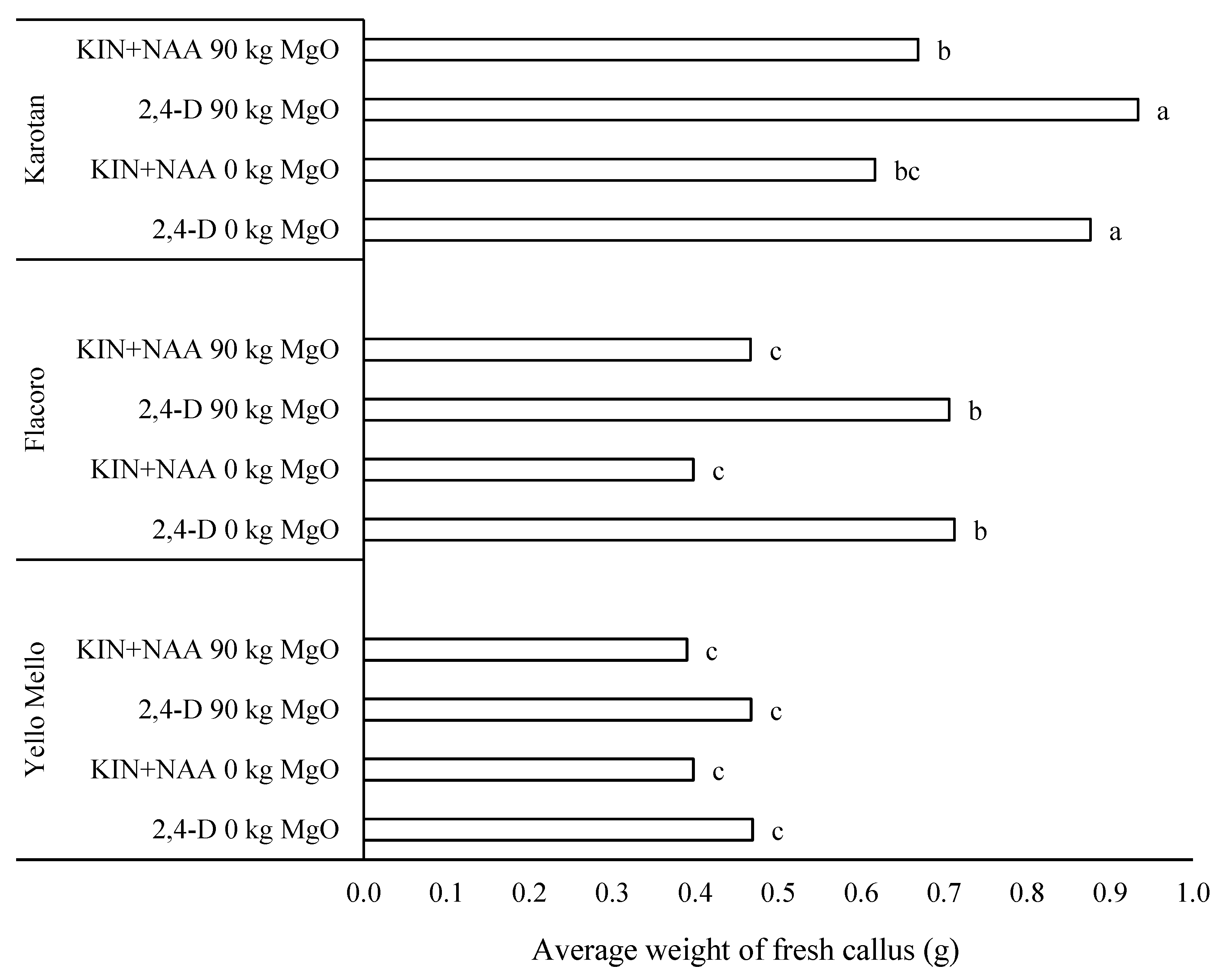
The attempt to increase the content of biologically active compounds in carrot roots by in vitro techniques is associated with difficulties in maintaining appropriate procedures and obtaining reproducible results for each cultivar. The main problems were combined with the high percentage of the contamination of explants (on average 41.95%) and the relatively low growth rate of the callus tissue. The investigated carrot callus isolated from the non-contaminated explants after 10 weeks of cultivation differed in terms of the analyzed morphological features, such as colour, cell coherence and surface structure, depending on the used medium composition and the variety. Under the influence of 2,4–D in the medium, the average FM of callus was the highest for all three varieties (on average 0.694 g FM/callus), whereas incubation on medium with the addition of KIN and NAA turned out to be less effective in terms of the induction of the callus tissue (on average 0.490 g FM/callus) independent of the carrot variety (Figure 2). These results confirmed the study of Ojha et al. [20] with Daucus carota L. subsp. Halophilus and of Mikołajczyk and Wojciechowski [38] with oilseed rape, who indicated 2,4–D as the more effective phytohormone than kinetin in in vitro cultures. Similarly, the greatest increase in fresh weight of tobacco callus was observed by Gatz et al. [39] when using 2,4–D. Higher amounts of produced somatic embryos were also observed by Mousavizadeh et al. [6] in media supplemented with 2,4–D, compared with media containing IAA.
Among the analyzed varieties in terms of the morphogenetic potential for callus formation, the best ability was revealed by the variety ‘Karotan’ (0.774 g FM/callus), which was the most appropriate genotype with respect to the induction capacity of the callus in in vitro cultures, compared to the remaining varieties used in the experiment (Figure 2). ‘Yello Mello’ was the worst genotype adapted for callus cultivation (0.431 g FM/callus). The obtained results indicate a significant impact of the carrot genotype on the regenerative potential in in vitro cultures. An influence of the variety on the efficiency of callus formation was also observed by Rabiei et al. [23], who reported significant differences in the weight of the obtained callus among the studied varieties. Variety ‘Vilmorn’ was characterized by the highest ability for inducing callus formation, where another one of four investigated cultivars—‘Nantes’—displayed the lowest mass of callus tissue. However, this variety distinguished itself with the callus of the highest morphogenetic potential, from which, as a consequence of regeneration, a large amount of plantlets was achieved. The differences in morphogenetic potential between cultivars were also observed by Oggema et al. [40], who investigated the regeneration potential of five sweet potato varieties (Ipomoea batatas L.) and showed the differences in the amount of regenerated plants from callus, indicating the cultivar ‘Mungade’ as the one with the highest ability to produce microcuttings. The variation in the amount of obtained callus and its morphological characteristics, as well as the ability to regenerate shoots, was also reported by Silvertand et al. [41]. They proved the dependence of these parameters on genotypes in studies on the regeneration of leek (Allium ampeloprasum L.) plants, which was also confirmed by histological analyses. These observations are consistent with the conclusions presented by Mikołajczyk and Wojciechowski [38], who demonstrated the differences in the regeneration efficiency in in vitro cultures in the example of two oilseed rape cultivars (Brassica napus L.).
Another experimental factor that applied, i.e., magnesium fertilization, did not significantly affect callus fresh weight production. Increasing magnesium fertilization intensified the callus formation only in the case of cultivar ‘Karotan’ on the medium, when the results of both phytohormone compositions of the medium were combined (data not shown). A positive plant response to the introduction of additional magnesium doses during carrot cultivation was also reported by Pobereżny et al. [32], who found a significant increase in the dry matter, monosaccharides and total sugar levels in the storage roots of five carrot cultivars, including ‘Flacoro’ and ‘Karotan’. Under the influence of applied MgO doses of 0, 45 and 90 kg ha−1, roots of the variety ‘Karotan’ contained the highest dry matter and both had reduced amounts of the total sugars per fresh weight [32].
Callus induced on the medium containing 2,4–D as well as KIN and NAA did not differ significantly in colour. The most intense colour was observed, as expected, in the callus tissue obtained from explants of the cultivar ‘Karotan’ (Figure 3). The examined callus was light orange in colour. The callus of the cultivar ‘Flacoro’ was characterized by a cream colour, which was not uniform, and a yellow-orange colouration was observed on some fragments. The explants of the cultivar ‘Yello Mello’ formed grey-cream coloured calli with the appearance of brown coloured sectors, which may indicate progressing necrotic processes.
The cohesion and surface structure of the calli differed according to the used growth regulators in the medium. By analyzing the cohesion of the cells, it was observed that the callus in the presence of 2,4–D was hard and the surface structure was nodule-like (Figure 3). The content of KIN and NAA in the medium resulted in a loosening of the callus surface structure. The tissue was friable under these conditions, with a small proportion of hard sectors.
The measurements of the antioxidative potential showed ‘Flacoro’ without the additional MgO fertilization and supplemented with KIN and NAA as the cultivar with the highest FRAP value (Table 1). The cultivar with the lowest value was ‘Yello Mello’ not fertilized with MgO and multiplied on medium with 2,4–D. The cultivar ‘Yello Mello’ was characterized by its yellow colour and by weaker parameters in terms of antioxidant properties related to the content of polyphenols (Table 1). These results confirm the reports of Cefola et al. [11], in which the purple variety ‘Polignano’ was characterized by the highest above-described parameters, and the yellow variety by the lowest. The antioxidative potential of carrot callus was two to three times lower, compared to raw carrot roots from the field [9,10]. However, the lower antioxidative potential of carrot callus was not directly correlated with the extent of the reduction of the content of single investigated compounds. Earlier reports [42,43] also indicated differences between the tissue polyphenol levels depending on the carrot colour and variety. The strict correlations between the total phenol content and antioxidant activity was not found in the experiment, which is in line with Ismail et al. [44]. The calculations and measurements performed for the total polyphenol content indicated the cultivar ‘Flacoro’ without additional MgO fertilization, propagated on medium with KIN and NAA as the material with the highest concentration of these compounds (Table 1). The cultivar ‘Yello Mello’ fertilized with 90 kg MgO ha−1 with 2,4–D added to the medium, showed the lowest concentration of total polyphenols (Table 1). Similar results were found by Gajewski et al. [42] for yellow-coloured carrot roots. However, it is worth noting that the achieved amounts of total polyphenols in the callus tissue were two-to-five times lower, compared to the carrot root tissue [9,10,32,42].
‘Karotan’ in combination with MgO fertilization and KIN + NAA in the medium, resulted in the highest mean carotenoids content, while in ‘Yello Mello’ with KIN + NAA in the medium and without MgO fertilization and in ‘Flacoro’ with 2,4–D and irrespective of MgO fertilization, the values were lowest (Table 1). In general, the results for all three cultivars are quite similar. High carotenoids levels were also recorded by Cefola et al. [11] studying the ‘Polignano’ carrot cultivars characterized by different storage root colour. The roots with a standard orange colour were characterized by high levels of carotenoids, compared to yellow specimens. The authors found the highest amounts of carotenoids in purple roots, which were also associated with the best results of these plants in terms of antioxidative potential and polyphenol content. However, in the comparison with the mean values of carrot roots obtained from the field, the contents obtained from the callus are on average 10 to 20 times lower (38–144 mg kg−1 FM in Keutgen et al. [10]; 10–140 mg kg−1 FM in Gajewski et al. [42]).
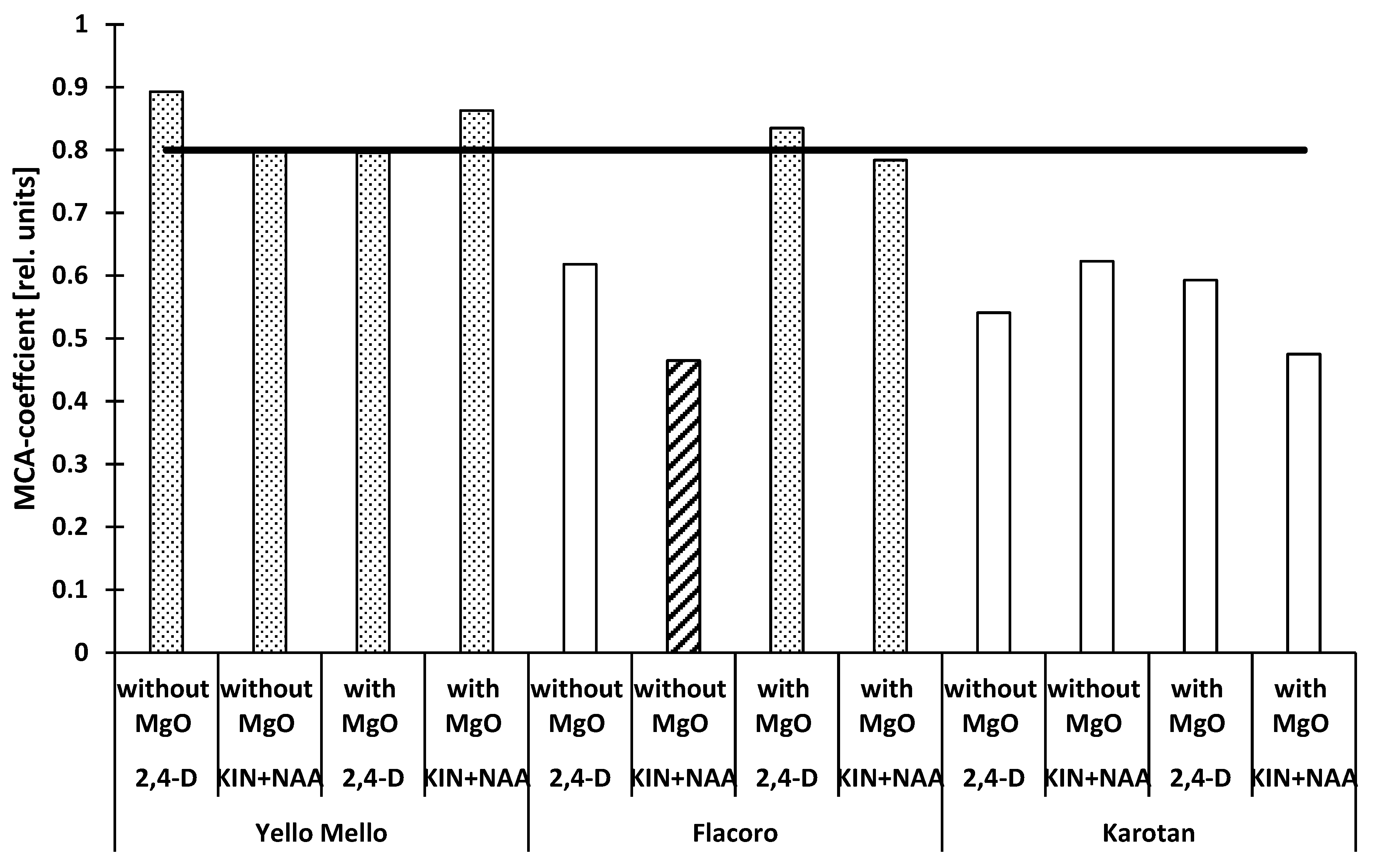
For a better evaluation of the influence of growth hormones on callus formation and the antioxidant properties of callus obtained from different carrot cultivars, a Multidimensional Comparative Analysis (MCA) was performed (Figure 4). The MCA revealed that the variety ‘Karotan’ showed the best performance, independent of the used plant hormones in the media and of the MgO application. The best performance, in this case the lowest value, was found for the cultivar ‘Flacoro’, for which the callus was obtained from the carrot roots grown without the additional application of MgO on a medium with KIN + NAA. This combination was characterized by the highest callus mass and the best antioxidant properties of the investigated factors; the synthetic measure was 0.465 (Figure 4). The worst performance (around 0.800 and above) was found for the cultivar ‘Yello Mello’, independent of the growth hormones (on average 0.839) and for the cultivar ‘Flacoro’ by application of MgO and independent of the applied phytohormones (on average 0.810). These combinations cannot be recommended for further investigations targeting high contents of health promoting compounds. In three of four variants, the cultivar ‘Yello Mello’ was characterized by rather low antioxidant properties, which were not compensated by the elevated amounts of total polyphenolics. In the case of the cultivar ‘Flacoro’, the additional application of MgO seems to mitigate stress during callus initiation and production, so the synthesis of health-promoting compounds was not enhanced, neither on a medium with 2,4–D nor on a medium with KIN + NAA.

4. Conclusions

The carrot is an important root vegetable with a wide range of antioxidant properties. However, there are not any clearly satisfactory results about the production of antioxidant properties by carrot callus obtained from in vitro cultures. The evaluation revealedthe significance of the chosen cultivar for the production of different antioxidant compounds. Less important was the influence of the used plant growth regulators. However, further investigations should include more effective elicitors, which improve the stimulation of the synthesis of the chosen health-promoting compounds and media other than MS. The use of other in vitro possibilities (not only solid media or cambial tissue) should also be considered for the development of a more effective system, which can be used independently of the vegetation season, and for the production of single or several desired antioxidant compounds. For further research, the materials of interest are the cultivars ‘Karotan’ and ‘Flacoro’, independent of the used phytohormones and the application of MgO during plant cultivation.

Author Contributions

Conceptualization, A.J.K. and N.K.; methodology, A.J.K. and N.K.; software, A.J.K. and N.K.; validation, A.J.K., N.K. and M.T.-S.; formal analysis, A.J.K., N.K., M.T.-S. and A.B.; investigation, A.J.K., N.K., M.T.-S. and A.B.; resources, A.J.K. and N.K.; data curation, A.J.K., N.K. and A.B.; writing—original draft preparation, A.J.K., N.K. and M.T.-S.; writing—review and editing, A.J.K., N.K. and M.T.-S.; visualization, A.J.K. and N.K.; supervision, A.J.K., funding acquisition, A.J.K. All authors have read and agreed to the published version of the manuscript.

Funding

This research received no external funding.

Institutional Review Board Statement

Not applicable.

Informed Consent Statement

Not applicable.

Data Availability Statement

Data sharing not applicable.

Acknowledgments

The authors would like to thank the employees of the Department of Microbiology and Food Technology, PBS Bydgoszcz, Poland for taking care of plant cultivation.

Conflicts of Interest

The authors declare no conflict of interest.

References

  1. Karuppusamy, A. A review on trends in production of secondary metabolites from higher plants by in vitro tissue, organ and cell cultures. J. Med. Plants Res. 2009, 3, 1222–1239. [Google Scholar] [CrossRef]
  2. Davies, K.M.; Espley, R.V. Opportunities and challenges for metabolic engineering of secondary metabolite pathways for improved human health characters in fruit and vegetable crops. N. Z. J. Crop Hort. 2013, 41, 154–177. [Google Scholar] [CrossRef] [Green Version]
  3. Gaosheng, H.; Jingming, J. Production of useful secondary metabolites through regulation of biosynthetic pathway in cell and tissue suspension culture of medicinal plants. In Recent Advances in Plant In Vitro Culture; Leva, A., Ed.; Intechopen: London, UK, 2012; pp. 197–200. ISBN 978-953-51-5331-3. [Google Scholar] [CrossRef] [Green Version]
  4. Shilpa, K.; Varun, K.; Lakshmi, B.S. An alternative method of natural drug production: Eliciting secondary production using plant cell culture. J. Plant Sci. 2010, 5, 222–247. [Google Scholar] [CrossRef] [Green Version]
  5. Rao, R.S.; Ravishankar, G.A. Plant tissue cultures: Chemical factories of secondary metabolites. Biotechnol. Adv. 2002, 20, 101–153. [Google Scholar] [CrossRef]
  6. Mousavizadeh, S.J.; Mashayekhi, K.; Akbarpour, V.; Kalati, H.; Ghasemi, Y. Effect of IAA and 2,4-D on somatic embryogenesis and pigments synthesis of carrot root secondary phloem. Aust. J. Agric. Res. 2010, 1, 126–131. Available online: https://www.sciencej.com/akbarpour_1_4_2010_126_131.pdf (accessed on 1 December 2021).
  7. Nishi, A.; Kurosaki, F. Daucus Carota L. (Carrot): In Vitro Production of Carotenoids and Phytoalexins; Biotechnology in Agriculture and Forestry, Medicinal and Aromatic Plants V; Bajaj, Y.P.S., Ed.; Springer: Berlin/Heidelberg, Germany, 1993; Volume 24, pp. 178–191. [Google Scholar] [CrossRef]
  8. Arafa, N.M.; Mohamed, S.S.; Aly, U.I. In vitro antimicrobial activity of carrot callus extracts as affected by tyrosine and tryptophan precursor. Int. J. PharmTech Res. 2016, 9, 121–129. Available online: https://www.sphinxsai.com/2016/ph_vol9_no9/ph01.htm (accessed on 1 December 2021).
  9. Keutgen, A.J.; Wszelaczyńska, E.; Pobereżny, J.; Kozera, W.; Knapowski, T.; Mozolewski, W. Health promoting properties of carrot (Daucus carota L.) as a function of cultivar and use of soil fertility enhancer UGmax. Ecol. Technol. 2015, XXIII, 206–210. [Google Scholar]
  10. Keutgen, A.J.; Wszelaczyńska, E.; Pobereżny, J. Influence of cultivar and UGmax on antioxidative properties of carrot roots (Daucus carota L.) and their stability during freezing process. Environ. Prot. Nat. Resour. 2014, 25, 19–22. [Google Scholar] [CrossRef] [Green Version]
  11. Cefola, M.; Pace, B.; Renna, M.; Santamaria, P.; Signore, A.; Serio, F. Compositional analysis and antioxidant profile of yellow. orange and purple Polignano carrots. Ital. J. Food Sci. 2012, 24, 284–291. Available online: https://www.researchgate.net/publication/230842053 (accessed on 1 December 2021).
  12. Arscott, S.A.; Tanumihardjo, S.A. Carrots of many colors provide basic nutrition and bioavailable phytochemicals acting as a functional food. Compr. Rev. Food Sci. Food Saf. 2010, 9, 223–239. [Google Scholar] [CrossRef]
  13. Yau, Y.-Y.; Santos, K.; Simon, P. Molecular tagging and selection for sugar type in carrot roots using co-dominant. PCR-based markers. Mol. Breed. 2005, 16, 1–10. [Google Scholar] [CrossRef]
  14. Simon, P.W.; Wolff, X.Y.; Peterson, C.E.; Kammerlohr, D.S.; Rubatzky, V.E.; Strandberg, J.O.; Basset, M.J.; White, J.M. High carotene mass carrot population. HortScience 1989, 24, 174–175. [Google Scholar]
  15. Wszelaczyńska, E.; Pobereżny, J.; Keutgen, A.J.; Szczepanek, M.; Idaszewska, N.; Brewka, J. Qualitative changes in carrot preserves depending on foliar fertilization on plants with magnesium and on selected technological processes. Food Sci. Technol. Qual. 2015, 5, 182–197. [Google Scholar] [CrossRef]
  16. Mohammed, A.A.; Al-Mallah, M.K. Determination of β-carotene in Carrot (Daucus carota L.) plants regenerated from stems callus. Rafidain J. Sci. 2013, 24, 27–36. Available online: https://www.iasj.net/iasj?func=fulltext&aId=74507 (accessed on 1 December 2021). [CrossRef]
  17. Bohm, V.; Putpitasari-Nienaber, N.L.; Ferruzzi, M.G.; Schwartz, S.J. Trolox equivalent antioxidant capacity of different geometrical isomers of α-carotene, β-carotene, lycopene and zeaxanthin. J. Agric. Food Chem. 2002, 50, 221–226. [Google Scholar] [CrossRef] [PubMed]
  18. Steward, F.C. Growth and development of cultivated cells. III. Interpretations of the growth from free cells to carrot plants. Am. J. Bot. 1958, 45, 709–713. [Google Scholar] [CrossRef]
  19. Harborne, J.B.; Mayer, A.M.; Bar-Nun, N. Identification of the major anthocyanin of carrot cells in tissue culture as cyanidin 3-(sinapoylxylosylglucosylgalactoside). Z. Naturforsch. 1983, 38, 1055–1056. [Google Scholar] [CrossRef]
  20. Butt, S.J.; Varis, S.; Nasir, I.A.; Sheraz, S.; Shahid, A.; Ali, Q. Micropropagation in advanced vegetable production: A review. Adv. Life Sci. 2015, 2, 48–57. Available online: https://www.als-journal.com/articles/vol2issue2/222.15/pdf.pdf (accessed on 1 December 2021).
  21. Kiszczak, W.; Kowalska, U.; Kapuścińska, A.; Burian, M.; Górecka, K. Effect of low temperature on in vitro androgenesis of carrot (Daucus carota L.). In Vitro Cell Dev. Biol. Plant 2015, 51, 135–142. [Google Scholar] [CrossRef]
  22. Ojha, A.; Kumar, S.; Singh, R. Plant regeneration via somatic embryogenesis from root explants of Daucus carota L., subsp. Halophilus. Med. Plant 2012, 4, 65–70. [Google Scholar] [CrossRef]
  23. Rabiei, K.; Polyakov, A.; Khodambashi, M.; Sharafova, O.; Kalashnikva, E.; Hooshmand, S.; Omidi, M. Carrot (Daucus carota L.) in vitro regeneration. Veg. Crop. Res. Bull. 2010, 73, 13–22. [Google Scholar] [CrossRef]
  24. Pant, B.; Manandhar, S. In vitro propagation of carrot (Daucus carota L.). Sci. World 2007, 5, 51–53. [Google Scholar] [CrossRef]
  25. Teruaki, S.; Kenji, K. Improvement of synchronization on carrot somatic embryo culture by controlling dissolved oxygen concentration. environment control in Biology. Adv. Life Sci. 1999, 37, 179–184. [Google Scholar]
  26. Jay, V.; Genestier, S.; Courduroux, J.C. Bioreactor studies of the effect of medium pH on carrot (Daucus carota L.) somatic embryogenesis. Plant Cell Tissue Organ Cult. 1994, 36, 205–209. [Google Scholar] [CrossRef]
  27. Fujimura, T.; Komamine, A. Involvement of endogenous auxin in somatic embryogenesis in a carrot cell suspension culture. Z. Pflanzenphysiol. 1979, 95, 13–19. [Google Scholar] [CrossRef]
  28. Chen, C.H.; Holden, D.J. Differential morphogenetic responses of carrot callus to naphthaleneacetic acid, 2, 4-Dichlorophenoxyacetic acid, and Tordon in vitro. Proc. South Dak. Acad. Sci. 1973, 52, 66–71. [Google Scholar]
  29. Tavares, A.C.; Salgueiro, L.R.; Canhoto, J.M. In vitro propagation of the wild carrot Daucus carota L. subsp. halophilus (Brot.) A. Pujadas for conservation purposes. Vitr. Cell. Dev. Biol. Plant 2010, 46, 47–56. [Google Scholar] [CrossRef] [Green Version]
  30. Murashige, T.; Skoog, F. A revised medium for rapid growth and bio assay with tobacco tissue cultures. Physiol. Plant. 1962, 15, 473–497. [Google Scholar] [CrossRef]
  31. Yau, Y.-Y.; Wang, K.Y. Increased regeneration ability of transgenic callus of carrot (Daucus carota L.) on B5-based regeneration medium. J. Appl. Hortic. 2012, 14, 152–156. [Google Scholar] [CrossRef]
  32. Pobereżny, J.; Wszelaczyńska, E.; Keutgen, A.J. Yield and chemical content of carrot storage roots depending on foliar fertilization with magnesium and duration of storage. J. Elem. 2012, 3, 479–494. [Google Scholar] [CrossRef]
  33. Michalik, B. Hodowla marchwi. In Hodowla Roślin Warzywnych; Niemirowicz-Szczytt, K., Ed.; Wyd. SGGW: Warszawa, Poland, 1993; pp. 55–77. [Google Scholar]
  34. Keutgen, A.J.; Pawelzik, E. Quality and nutritional value of strawberry fruit under long term salt stress. Food Chem. 2008, 107, 1413–1420. [Google Scholar] [CrossRef]
  35. Benzie, I.F.F.; Strain, J.J. The ferric reducing ability of plasma (FRAP) as a measure of “antioxidative power”: The FRAP assay. Anal. Biochem. 1996, 239, 70–76. [Google Scholar] [CrossRef] [PubMed] [Green Version]
  36. Keutgen, A. Modification of spinach quality by selected pre- and postharvest treatment. Beitr. Zu Agrarwiss. 2000, 24, 126. [Google Scholar]
  37. Keutgen, A.J.; Wszelaczyńska, E.; Pobereżny, J.; Przewodowska, A.; Przewodowski, W.; Milczarek, W.; Tatarowska, D.; Flis, B.; Keutgen, N. Antioxidant properties of potato tubers (Solanum tuberosum L.) as a consequence of genetic potential and growing conditions. PLoS ONE 2019, 14, e0222976. [Google Scholar] [CrossRef] [PubMed]
  38. Mikołajczyk, S.; Wojciechowski, A. Ocena zdolności regeneracyjnych in vitro wybranych genotypów rzepaku (Brassica napus L.) przy zastosowaniu różnych barw światła i regulatorów wzrostu. Rośl. Oleiste 1999, 20, 81–92. [Google Scholar]
  39. Gatz, A.; Tomaszewska-Sowa, M.; Figas, A. Changes accompanying proliferative capacity and morphology of Nicotiana tabacum L. callus in response to 2, 4-D. Acta Agrobot. 2017, 70, 4. [Google Scholar] [CrossRef] [Green Version]
  40. Oggema, J.N.; Kinyua, M.G.; Ouma, J.P.; Owuoche, J.O. Agronomic performance of locally adapted sweet potato (Ipomoea batatas (L) Lam.) cultivars derived from tissue culture regenerated plants. Afr. J. Biotechnol. 2007, 6, 1418–1425. [Google Scholar] [CrossRef]
  41. Silvertand, B.; van Rooyen, A.; Lavrijsen, P.; van Harten, A.M.; Jacobsen, E. Plant regeneration via organogenesis and somatic embryogenesis in callus cultures derived from mature zygotic embryos of leek (Allium ampeloprasum L.). Euphytica 1996, 91, 261–270. [Google Scholar] [CrossRef]
  42. Gajewski, M.; Szymczak, P.; Elkner, K.; Dąbrowska, A.; Kret, A.; Danilcenko, H. Some aspects of nutritive and biological value of carrot cultivars with orange, yellow and purple-coloured roots. Veg. Crop. Res. Bull. 2007, 67, 149–161. [Google Scholar] [CrossRef] [Green Version]
  43. Alasalvar, C.; Grigor, J.M.; Zhang, D.; Quantick, P.C.; Shahidi, F. Comparison of volatiles. phenolics. sugars. antioxidant vitamins. and sensory quality of different colored carrot varieties. J. Agric. Food Chem. 2001, 49, 1410–1416. [Google Scholar] [CrossRef]
  44. Ismail, A.; Marjan, Z.M.; Foong, C. Total antioxidant activity and phenolic content in selected vegetables. Food Chem. 2004, 87, 581–586. [Google Scholar] [CrossRef]
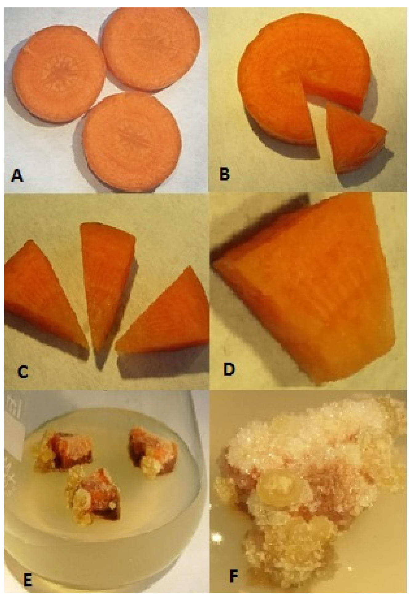
Figure 1. Process of callus induction on fragments of the ‘Karotan’ carrot root: slices before sterilization (A), isolation of a cambial fragment from the sterilized material (B), sterile carrot root fragments (C), explant containing the cambium before inoculation on medium (D), forming carrot callus on medium with 1 mg L−1 2,4–D (E) and differentiated carrot callus (F).
Horticulturae 08 00100 g001
Figure 2. Effect of magnesium fertilization and phytohormone composition of the medium during in vitro culture on the mean fresh weight of the carrot callus. Different letters indicate significant differences by Wilcoxon-Mann-Whitney-test (U-test).
Horticulturae 08 00100 g002
Figure 3. Colour, surface structure and cohesion type of carrot callus cells on media containing 2,4–D in the varieties ‘Karotan’ (A), ‘Flacoro’ (B) and ‘Yello Mello’ (C).
Horticulturae 08 00100 g003
Figure 4. MCA-coefficient of parameters of carrot callus in relation to cultivar, fertilization and medium type. The stripped column indicates the best performance of the desired callus properties, whereas the dotted columns represent the lowest efficacy of the desired callus characteristic. The black line at 0.8 represents the chosen limit of bad performance (MVA-value > 0.8) for all investigated properties.
Horticulturae 08 00100 g004
Table 1. Content of some intrinsic parameters of carrot callus in relation to cultivar, magnesium dose and type of medium.
Variety Phytohormones in the Medium MgO Fertilization (kg ha−1)
090
Antioxidative potential (FRAP) (mmol Fe2+ kg−1 of fresh callus weight)
Karotan2,4–D0.254 ± 0.008 abc0.243 ± 0.030 abcd
KIN + NAA0.259 ± 0.091 abc0.207 ± 0.051 bcd
Flacoro2,4–D0.265 ± 0.004 ab0.217 ± 0.018 bcd
KIN + NAA 0.348 ± 0.039 a0.175 ± 0.020 bcd
Yello Mello2,4–D0.138 ± 0.015 d0.157 ± 0.033 bcd
KIN + NAA0.235 ± 0.015 abcd0.149 ± 0.049 cd
Polyphenols (mg kg−1 of fresh callus weight)
Karotan2,4–D797.92 ± 0.25 abc823.89 ± 52.10 abc
KIN + NAA837.84 ± 213.07 abc878.13 ± 156.16 ab
Flacoro2,4–D899.02 ± 29.12 ab654.35 ± 120.00 bc
KIN + NAA1049.15 ± 124.48 a685.15 ± 46.43 bc
Yello Mello2,4–D653.00 ± 21.77 bc581.38 ± 45.20 c
KIN + NAA724.02 ± 32.34 bc645.13 ± 2.44 bc
Carotenoids (mg kg−1 of fresh callus weight)
Karotan2,4–D2.41 ± 0.81 ab2.44 ± 0.25 ab
KIN + NAA2.74 ± 0.58 ab 3.95 ± 1.42 a
Flacoro2,4–D1.65 ± 0.24 b1.72 ± 0.17 b
KIN + NAA2.92 ± 0.27 ab2.76 ± 0.26 ab
Yello Mello2,4–D2.31 ± 0.29 ab2.76 ± 0.27 ab
KIN + NAA1.61 ± 0.14 c2.90 ± 0.36 ab
Different letters after the mean values indicate significant differences by Tukey’s-b test or U-test at p ≤ 0.05 within the given property among the investigated genotypes, media and MgO fertilization.
Publisher’s Note: MDPI stays neutral with regard to jurisdictional claims in published maps and institutional affiliations.