Article

Enhanced Production of Apocarotenoids by Salicylic Acid Elicitation in Cell Suspension Cultures of Saffron (Crocus sativus L.)

1
Department of Plant Science, Faculty of Biological Science, Tarbiat Modares University, Tehran P.O. Box 14115-111, Iran
2
Institute of Sciences of Food Production (ISPA-CNR), 73100 Lecce, Italy
*
Authors to whom correspondence should be addressed.
Horticulturae 2022, 8(12), 1176; https://doi.org/10.3390/horticulturae8121176
Submission received: 24 November 2022 / Revised: 7 December 2022 / Accepted: 8 December 2022 / Published: 9 December 2022

Abstract

:
A cell suspension culture of saffron (Crocus sativus L.) was previously established from style-derived calli to obtain an in vitro system for crocin, an uncommon and valuable water-soluble apocarotenoid, and carotenoid production suitable for future scaling up. To shed more light on the correlation between apocarotenoid biosynthesis and key-gene expression, in this study, SA was used at 0.5 mM concentration to elicit crocin production and the effects on carotenoid production were analyzed after 6, 12, 24, and 48 h. HPLC-DAD analysis was used for total crocin quantification as well as the other carotenoids zeaxanthin, β-carotene and lutein. Quantitative RT-PCR was used to analyze the transcript levels of saffron apocarotenoid biosynthetic key genes PSY (phytoene synthase), BCH1 (β-carotene hydroxylase), and CCD2 (carotenoid cleavage dioxygenase) after SA elicitation. In saffron suspension-cultured cells elicited by SA, the carotenoid biosynthetic pathway was mostly enhanced toward crocin biosynthesis, known to exert strong biological activity and therapeutic effects, rather than lutein or xanthins. SA increased BCH1 and CCD2 gene expression 15.6 and 3.3 times, respectively, compared to the control at 24 h after elicitation. Although a dynamic change of metabolite contents and gene expression was observed during the 48 h time course in response to SA elicitation, the changes of zeaxanthin and crocin were consistent with the regulation of the corresponding genes BCH and CCD2 during the time course. In conclusion, the effects of SA on regulation of gene expression in the apocarotenoid pathway could be successfully applied for the biotechnological production of crocin.

1. Introduction

Saffron, the most expensive spice worldwide, consists of dried stigmas of Crocus sativus L. flowers, belonging to the Iridaceae family. Crocin, picrocrocin and safranal are the three major apocarotenoids present in saffron; they confer the typical characteristics of bitter taste, red color and pharmaceutical properties and are responsible of saffron quality and commercial value [1]. Saffron has also been used as a coloring agent in textiles and cosmetics since era period [2]. Saffron apocarotenoids have numerous pharmaceutical properties including antitumor, neuroprotective, anti-inflammatory, and cardioprotective activities [3].
Apocarotenoids originate from carotenoids by the oxidative tailoring of β-carotene and zeaxanthin. Among saffron apocarotenoids, crocin and its precursor crocetin have been proved to express the highest antioxidant activity and biological effects [4,5,6,7]. Crocin derives from an oxidative cleavage of zeaxanthin, through a reaction catalyzed by carotenoid cleavage dioxygenase (CCD2), identified in saffron stigma [8]. In Crocus sativus, genes involved in apocarotenoid biosynthesis were identified by comprehensive transcriptome analysis [9,10]. The biosynthesis of apocarotenoids occurs through the methylerythritol phosphate (MEP) pathway and phytoene synthase enzyme (PSY), which catalyzes the condensation of two molecules of geranylgeranyl diphosphate (GGDP) to form phytoene, being the first step of carotenoid biosynthetic pathway [11]. Another key step in apocarotenoid production is related to the formation of zeaxanthin, catalyzed by β-carotene hydroxylase (BCH). Zeaxanthin is a precursor of crocin and other zeaxanthin derivatives such as picrocrocin and safranal. In Crocus sativus plants, the accumulation of saffron apocarotenoids is regulated by the transcriptional levels of CsPSY, CsBCH, and CsCCD2 genes [12].
The in vitro production of bioactive metabolites by plant suspension cell cultures has gained increasing interest as a continuous supply strategy, independent from environmental limitations. As well, it can be a valuable alternative to the use of engineered transgenic plants which are often prevented by strict regulations mostly in European countries [13,14]. In various medicinal plants, salycilic acid (SA) has been shown to have a signaling effect on the biosynthesis of secondary metabolites through the induction of gene expression [15,16].
In our previous work, SA applied at various concentrations from 0.1 to 1 mM was shown to significantly enhance the production of crocin and phenolic compounds in saffron suspension-cultured cells [17]. Investigating the transcriptional levels of key genes involved in the carotenoid pathway toward apocarotenoid biosynthesis can help shedding light on the effects of SA application. Therefore, in this study, the quantification of saffron characteristic pigment crocin, as well as other metabolites such as lutein, β-carotene and zeaxanthin was performed after the application of SA 0.5 mM in saffron cell cultures. The expression levels of key biosynthetic genes PSY, BCH, and CCD2 evaluated by quantitative real-time PCR correlated with metabolite accumulation (Figure 1).

2. Materials and Methods

2.1. Saffron Suspension-Cultured Cells and SA Elicitation

Saffron cell suspensions were established from style originated calli as described previously [18]. MS medium [19] supplemented with NAA 3 mg L−1 and TDZ 1 mg L−1 was used for cell maintenance, according to previous studies [17,18], incubating on a rotary shaker (120 rpm) at 24 °C under dark conditions. The cells were sub-cultivated every 10 days in 500-mL Erlenmeyer flasks by transferring 15 mL of the 10 day-old suspensions into 85 mL fresh MS medium.
SA (Sigma, St. Louis, MO, USA) was first dissolved in methanol, diluted with deionized water, and filter-sterilized before adding to the culture medium. SA was applied to 3 day-old saffron suspension cultured cells at the final concentration of 0.5 mM. The same amount of methanol was added to the cultures as a control. Cell samples were collected at various time intervals and were filtered under vacuum using Miracloth filters (pore size 22–25 μm, Calbiochem, Los Angeles, CA, USA). Samples were frozen in liquid nitrogen, lyophilized overnight (Labconco, Kansas City, MO, USA) and kept at −80 until used.

2.2. Extraction and Separation of Crocin and Carotenoids from Saffron Treated Cells

To monitor the carotenoid and crocin content from saffron control and SA treated cells, the methods described by Sadler et al. [20] and Perkins-Veazie et al. [21] were optimized to extract simultaneously carotenoids and crocin. Briefly, 200 mg cell samples were extracted with 5 mL (0.05% w/v) of butylated hydroxytoluene (BHT) in acetone, 5 mL 95% ethanol, and 10 mL hexane for 15 min after stirring at 180 rpm. At the final step, 3 mL distilled water was added to the mixture and centrifuged at 4000× g for 10 min. The upper phase was discarded, recovered two times with hexane, speed-dried under vacuum, and kept at −20 for carotenoid qualifications. The lower phase, the water-soluble phase with a strong visible yellow color, was used for crocin analysis.

2.3. HPLC-DAD Analysis of Crocin and Carotenoids

Crocin was quantified by an auto-sampler HPLC device (Agilent-1290 infinity) equipped with the C18 column as described previously (Moradi et al., 2018). α-crocin (PhytoLab, Vestenbergsgreuth, Germany) was used as a pure crocin standard for the quantification of crocin at 440 nm. For carotenoids, samples were kept at −20, redissolved in ethyl acetate (50 µL), and immediately analyzed by Agilent 1100 Series HPLC system (Agilent Technologies, Waldbronn, Germany) as described by Rizzello et al. [22]. Briefly, the carotenoids were identified at a wavelength of 475 nm while comparing their retention times and UV–vis spectra to a mixed authentic standard, comprising violaxanthin, neoxanthin, chlorophyll b, lutein, zeaxanthin, and β-carotene [22].

2.4. Isolation of Nucleic Acids and cDNA Synthesis

Saffron cell samples, frozen in liquid nitrogen, were powdered using a pestle in a pre-cooled mortar and the powder was stored at −80 °C until used. DNA was extracted using DNeasy Plant Mini Kit (Qiagen, Milan, Italy).
Total RNA isolation was performed by a SV Total RNA isolation kit (Promega s.r.l., Milan, Italy) following the manufacturer’s instructions. RNA quality was evaluated by separation on 1% agarose gel. Furthermore, RNA was quantified using the NanoDrop® ND-1000 spectrophotometer (NanoDrop Technologies, Wilmington, DE, USA). About 5 μg of total RNA was treated with RQ1 RNase-Free DNase (Promega, Milano, Italy) to remove the possible genomic DNA contamination. The first strand cDNA was synthesized by ImProm-II Reverse Transcription System (Promega), according to the manufacturer’s instructions and the following PCR conditions were used: 25 °C for 5 min, 42 °C for 1 h, and 70 °C for 15 min.

2.5. Quantitative RT-PCR Analysis

The cDNA templates obtained from control and saffron cell samples were first tested for genomic DNA contamination by using gBch1f and gBch1r (Table 1) primers to amplify the region of the Bch1 gene. The region was amplified using the polymerase chain reaction (PCR) cycles according to the following conditions: pre-denaturation at 95 °C for 2 min, denaturation 95 °C for 40 sec, annealing 52 °C for 45 s, extension 72 °C for 1 min, final extension 72 °C for 7 min. The size of the amplified product obtained was compared with that obtained using genomic DNA as a template (data not shown). Quantitative RT-PCR was used to analyze the transcript levels of saffron apocarotenoid key genes CsPSY (Acc. Number, AJ888514.1), CsBCH1 (Acc. Number AJ416711.2) and CsCCD2 (Acc. Number KJ541749.1). Quantitative RT-PCR was performed by the Applied Biosystems 7500 apparatus, using a sequence-specific primer set and probes which are listed in Table 1. All the primers were purchased from PRIMM srl (Milan, Italy). The probes were labeled at the 5’-end with 6-carboxy-fluorescein (FAM) and the 3’-end with tetramethylrhodamine (TAMRA). The reaction was performed in a total volume of 25 μL, which included 12.5 μL of 2X TaqMan Universal PCR Master Mix (Applied Biosystems, Foster City, CA, USA), 0.9 μM gene-specific primers, 0.5 µL of template cDNA and a 0.2 μM probe. The cycling conditions were 95 °C for 20 s, followed by 40 cycles of 95 °C for 15 s and 60 °C for 1 min. The transcript abundance of key genes was detected in saffron cell cultures treated with 0.5 mM SA in a time course of 30 min, 90 min, 3 h and 24 h. The amplification of Crocus sativus 18S RNA (Acc. Number, AJ489273) gene was used as the endogenous control. The relative expression was normalized against Crocus sativus 18S RNA and the amount was calculated using the untreated (Ctr) samples as a calibrator, whose expression was given as equal to one. Experiments were performed in three replications. The transcripts were quantified through the comparative quantitation method 2−ΔΔCT [23].

2.6. Statistical Analysis

Data are the results of three independent experiments, reported as the mean value ± standard deviation (SD) and analyzed statistically by a one-way ANOVA along with a post hoc test using SPSS-22 software. Differences among the means were compared at the significance level of p ≤ 0.05.

3. Results

Based on the results from our previous study on the effects of SA on cell growth and crocin production in saffron suspension-cultured cells [17], SA 0.5 mM was chosen as the optimal concentration to investigate the SA effects on carotenoid contents and the expression of genes involved in the crocin biosynthetic pathway at various intervals during a 48 h time-course.

3.1. Carotenoid Contents after 0.5 mM SA Elicitation

Lutein, β-carotene, zeaxanthin and crocin contents in saffron suspension-cultured cells were analyzed at various intervals after SA 0.5 mM treatment and compared to controls. In all the analyzed samples, carotenoids revealed the highest values after 48 h. Lutein showed a nearly stable trend up to 24 h and increased by almost two-fold at 48 h both in treated and control samples. Higher lutein amounts, although not statistically significant, were observed in SA-treated cultures rather than in control ones only after 6 and 12 h. As for β-carotene, a slight decrease was observed from 6 to 12 h both in SA treated and control cultures, while after 24 h the decrease became more relevant only in control cultures. At 48 h, both treated and control cultures showed a 1.9 times increase of β-carotene amounts compared to the amounts detected at 24 h (Figure 2).
As for the content of zeaxanthin and crocin, both showed increasing levels during the time course to 48 h with the amounts measured in the treated samples always higher than the control. In particular, zeaxanthin amounts of SA-treated cultures reached 0.23 μg g−1 DW at 48 h compared to 0.15 μg g−1 DW detected in the corresponding control sample. Crocin also reached its maximum amount of 4.62 μg g−1 DW at 48 h after elicitation compared to 3.47 μg g−1 DW in untreated cultures (Figure 3).

3.2. Gene Expression of Crocin Biosynthetic Pathway after SA Treatment

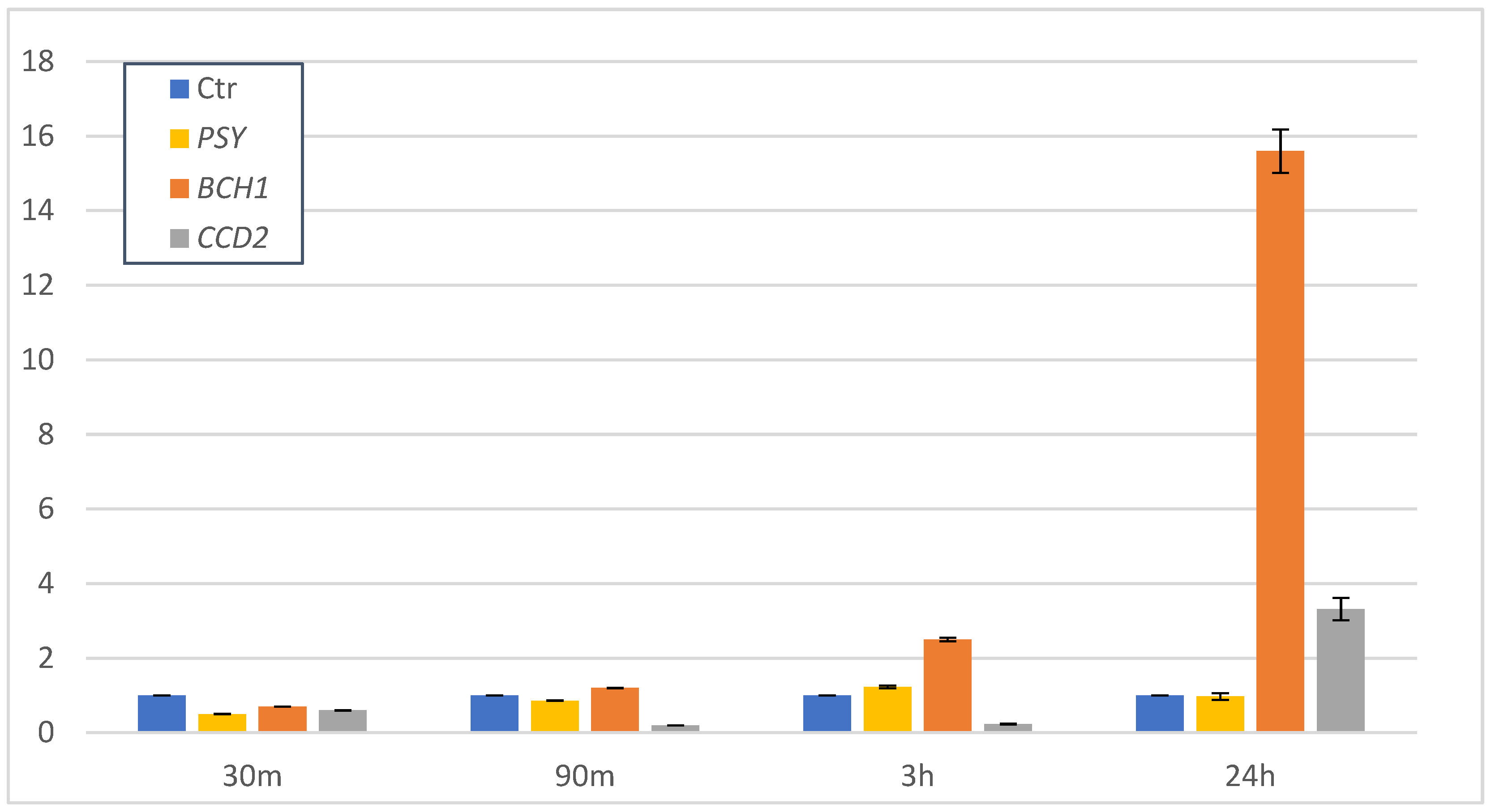
The expression of key genes responsible for the biosynthesis of apocarotenoids in C. sativus was investigated in control and 0.5 mM SA-treated suspension-cultured cells in a time course from 30 min to 24 h. Three key genes encoding the following enzymes for the biosynthesis of carotenoids were analyzed by quantitative real-time PCR (qPCR): phytoene synthase (CsPSY), which catalyzes the condensation of two molecules of geranylgeranyl pyrophosphate to form phytoene, β-carotene hydroxylase (CsBCH1), which catalyzes the hydroxylation of β-carotene to β-cryptoxanthin and zeaxanthin, and carotenoid cleavage dioxygenase (CsCCD2) a recently identified CCD family member from C. sativus, which cleaves the 7,8 and 7′,8′ double bonds of zeaxanthin to produce crocetin dialdehyde and 3-OH-β-cyclocitral; this is the first step of specific saffron crocin biosynthesis [24]. Comparative gene expression at different intervals after elicitation is shown in Figure 4.
As can be seen from the results obtained (Figure 4), early after SA elicitation, at 30 min, a decreased expression of the three genes was observed. Later on, the expression pattern of these genes was dissimilar, likely due to the different functions of the relative enzymes in the carotenoid biosynthetic pathway. As for CsPSY gene expression, only after 3 h a slight increase was detected, while after 24 h it decreased to the levels observed at early times. The expression of the CsBCH1 gene slightly increased after 90 min, reaching a 2.5-fold increase, compared to the control, 3 h after SA elicitation. Interestingly, after 24 h an even more relevant increase up to 15.6-fold compared to control cultures was detected. As for the expression of CsCCD2 gene, low levels were observed at early times up to 3 h after elicitation while a 3.32-fold enhancement compared to the control was observed at 24 h after SA elicitation.

4. Discussion

The apocarotenoid crocins are well-known for their healthful properties for humans, which include several pharmacological activities, such as anticancer effects, reducing the risk of atherosclerosis, and preventing Alzheimer’s disease [25,26]. As the traditional sources of crocins are no longer sufficient to meet current demands, efforts to increase the crocin production by using in vitro saffron cell cultures are well desirable. To this regard, shedding more light on the molecular mechanism of enhancing in vitro metabolite production by using elicitors such as SA can be very helpful.
In previous works, we established a saffron cell suspension culture from style originated calli and elicited with increasing concentrations of SA up to 1 mM. The elicited suspension cells showed an enhancement of crocin and phenolic compounds to significant levels compared to the untreated cell suspensions. Nevertheless, saffron cell growth inhibition after the addition of SA to the culture medium was observed. Particularly, the highest concentration, 1 mM SA, caused the highest stress to the cells and severely impaired cell growth of saffron cell suspension cultures [17,18].
In this study, we used SA 0.5 mM to investigate the putative correlation between the accumulation of crocin and other carotenoids, and the expression of key biosynthetic genes in saffron suspension-cultured cells, finding a correlation between apocarotenoid production and the expression of genes related to their biosynthesis.
SA is known to act as an elicitor of various metabolic pathways in plant cell cultures [27]. As expected, the addition of SA generally increased metabolite production in saffron suspension-cultured cells during a 48 h time course, although differences were observed. The content of β-carotene was significantly enhanced by SA after 24 h of treatment while zeaxanthin and crocin showed a moderate increase also at earlier times; such an increase became significantly relevant at 24 and 48 h. As for lutein, a slight enhancement effect by SA was onlyobserved early on. These differences could be explained by the carotenoid metabolic pathway, where β-carotene is a precursor of zeaxanthin and together precursors of crocin, while lutein is an end-product of the biosynthetic pathway, belonging to a different branch deriving from lycopene (Figure 1) [11].
To shed more light on the effects of SA on saffron carotenoid metabolism, we conducted a quantitative gene expression analysis of some carotenoid biosynthetic genes: phytoene synthase (PSY) as the first step of the carotenoid biosynthetic pathway, β-carotene hydroxylase (BCH) and carotenoid cleavage dioxygenase (CCD2).
It is known that biosynthesis of plant-specialized metabolites, such as isoprenoids, is transcriptionally regulated [28]. In Centella asiatica suspension cells, SA elicitation improved centelloside biosynthesis by gene regulation of the isoprenoid pathway [29]. In the juice sacs of Satsuma mandarin the elicitation of the carotenoid β-criptoxanthin was also regulated at the transcriptional level [30]. In C. sativus stigmas, the quantitative and qualitative profiles of carotenoids and apocarotenoids were studied, and the transcriptional regulation of a β-carotene hydroxylase and lycopene cyclase genes was involved in the observed changes [12,31,32]. Recently, an active transcription factor ULT1 was found to be induced by phytohormones such as SA and MeJA in the stigma part and its overexpression in saffron calli led to the increased expression of key genes CsPSY, CsPDS, CsBCH1, and CsCCD2 in crocin biosynthetic pathway [33]. In our in vitro system, the expression of CsBCH1 was moderately up-regulated after SA elicitation early at 90 min and 3 h, reaching the highest expression level after 24 h. These data suggest that SA, by increasing the expression of CsBCH1 gene, enhanced the availability of BCH enzyme, which is responsible for the conversion of β-carotene to zeaxanthin, pushing the pathway to the zeaxanthin biosynthesis towards crocin (Figure 1). This could give account of the observed zeaxanthin increase over the 48 h time-course. On the other hand, zeaxanthin is also a precursor of xanthin compounds leading to violaxanthin and neoxanthin biosynthesis. Analysis of both these metabolites was performed in saffron cell cultures, but neither violaxanthin nor neoxanthin were detected at any time point (data not shown). Interestingly, the expression of CsCCD2 gene also increased 24 h after elicitation compared to the control, and this increase was consistent with the increase of crocin metabolites at this time up to 48 h after elicitation. From these results, the metabolic pathway of carotenoids in saffron suspension cells appears to be more conducive to the production of crocin rather than to xanthin biosynthesis.
It is noteworthy that in our in vitro system it was possible to detect the expression of the gene CsCCD2 encoding for the CCD2 enzyme, which was supposed to be expressed in stigmas in correlation with crocin accumulation during stigma development [8,34]. This confirms that in vitro cell cultures have the capacity of biosynthesizing specialized metabolites such as terpenoids in adult differentiated tissues [35]. Divergent mechanisms mediate plant cell responses to elicitors such as SA, including secondary metabolites produced in cell cultures, and this can be considered as a promising process for enhancement of desired metabolites in plants as biofactories. It seems likely that exogenous SA uptake into the cells provoked a response through the stimulation of key enzymatic activities and the expression of genes involved in plant secondary metabolite biosynthetic pathways [15,16]. The generation of ROS [36] leading to the production of H2O2 [37,38], acting as a second messenger in SA signaling pathway resulted in the production of secondary metabolites in cell suspension cultures [39]. In the case of our saffron suspension-cultured cells, SA significantly enhanced the expression of CsBCH1 and CsCCD2 genes and this effect improved the carotenoid production in a time-dependent manner. Considering that the mechanism mediating plant cell response to elicitors is a complex process, a further study regarding the role of other possible factors involved in saffron cells in response to SA will help us to know which plays the most important role in crocin biosynthetic pathway.
In conclusion, the effects of salicylic acid on regulation of gene expression in the apocarotenoid pathway can be applied in biotechnological production of crocin by using in vitro saffron suspension cells. This issue can be taken into consideration also in the scaling-up process with the aim of crocin bioproduction.

Author Contributions

Conceptualization, A.D.P. and S.C.; methodology, A.D.P. and A.M.; validation, A.D.P., F.Z., G.M. and S.C.; formal analysis, A.M. and A.D.P.; resources, G.M. and S.C.; data curation, A.D.P. and S.C.; writing—original draft preparation, A.M. and S.C.; writing—review and editing, A.D.P., A.M. and S.C.; supervision, A.D.P. and S.C.; funding acquisition, G.M., F.Z. and S.C. All authors have read and agreed to the published version of the manuscript.

Funding

This research was supported by Tarbiat Modares University (Ph. D student research program) as well as by Institute of Sciences of Food Production (ISPA-CNR) as a research cooperation agreement. This work was in part funded by CNR project FOE-2019 DBA.AD003.139.

Institutional Review Board Statement

Not applicable.

Informed Consent Statement

Not applicable.

Data Availability Statement

Not applicable.

Acknowledgments

The authors thank Leone D’Amico for his valuable technical assistance.

Conflicts of Interest

The authors declare no conflict of interest.

References

  1. Srivastava, R.; Ahmed, H.; Dixit, R.K.; Dharamveer; Saraf, S.A. Crocus sativus L.: A comprehensive review. Pharmacogn. Rev. 2010, 4, 200–208. [Google Scholar] [CrossRef] [PubMed] [Green Version]
  2. Gohari, A.R.; Saeidnia, S.; Mahmoodabadi, M.K. An overview on saffron, phytochemicals, and medicinal properties. Pharmacogn. Rev. 2013, 7, 61–66. [Google Scholar] [CrossRef] [PubMed] [Green Version]
  3. Bukhari, S.I.; Manzoor, M.; Dhar, M.K. A comprehensive review of the pharmacological potential of Crocus sativus and its bioactive apocarotenoids. Biomed. Pharmacother. 2018, 98, 733–745. [Google Scholar] [CrossRef] [PubMed]
  4. Rahaiee, S.; Moini, S.; Hashemi, M.; Shojaosadati, S.A. Evaluation of antioxidant activities of bioactive compounds and various extracts obtained from saffron (Crocus sativus L.): A review. J. Food Sci. Technol. 2015, 52, 1881–1888. [Google Scholar] [CrossRef] [Green Version]
  5. Karimi, E.; Oskoueian, E.; Hendra, R.; Jaafar, H.Z. Evaluation of Crocus sativus L. stigma phenolic and flavonoid compounds and its antioxidant activity. Molecules 2010, 15, 6244–6256. [Google Scholar] [CrossRef] [Green Version]
  6. Amin, A.; Hamza, A.A.; Bajbouj, K.; Ashraf, S.S.; Daoud, S. Saffron: A potential candidate for a novel anticancer drug against hepatocellular carcinoma. Hepatology 2011, 54, 857–867. [Google Scholar] [CrossRef]
  7. Seifi, A.; Shayesteh, H. Chapter 15—Molecular biology of Crocus sativus. In Saffron; Koocheki, A., Khajeh-Hosseini, M., Eds.; Woodhead Publishing: Sawston, UK, 2020; pp. 247–258. [Google Scholar] [CrossRef]
  8. Frusciante, S.; Diretto, G.; Bruno, M.; Ferrante, P.; Pietrella, M.; Prado-Cabrero, A.; Rubio-Moraga, A.; Beyer, P.; Gomez-Gomez, L.; Al-Babili, S.; et al. Novel carotenoid cleavage dioxygenase catalyzes the first dedicated step in saffron crocin biosynthesis. Proc. Natl. Acad. Sci. USA 2014, 111, 12246–12251. [Google Scholar] [CrossRef] [Green Version]
  9. Baba, S.A.; Mohiuddin, T.; Basu, S.; Swarnkar, M.K.; Malik, A.H.; Wani, Z.A.; Abbas, N.; Singh, A.K.; Ashraf, N. Comprehensive transcriptome analysis of Crocus sativus for discovery and expression of genes involved in apocarotenoid biosynthesis. BMC Genom. 2015, 16, 698. [Google Scholar] [CrossRef] [Green Version]
  10. Yue, J.; Wang, R.; Ma, X.; Liu, J.; Lu, X.; Balaso Thakar, S.; An, N.; Liu, J.; Xia, E.; Liu, Y. Full-length transcriptome sequencing provides insights into the evolution of apocarotenoid biosynthesis in Crocus sativus. Comput. Struct. Biotechnol. J. 2020, 18, 774–783. [Google Scholar] [CrossRef]
  11. Cazzonelli, C.I.; Pogson, B.J. Source to sink: Regulation of carotenoid biosynthesis in plants. Trends Plant Sci. 2010, 15, 266–274. [Google Scholar] [CrossRef]
  12. Castillo, R.; Fernandez, J.A.; Gomez-Gomez, L. Implications of carotenoid biosynthetic genes in apocarotenoid formation during the stigma development of Crocus sativus and its closer relatives. Plant Physiol. 2005, 139, 674–689. [Google Scholar] [CrossRef] [PubMed] [Green Version]
  13. Eibl, R.; Meier, P.; Stutz, I.; Schildberger, D.; Huhn, T.; Eibl, D. Plant cell culture technology in the cosmetics and food industries: Current state and future trends. Appl. Microbiol. Biotechnol. 2018, 102, 8661–8675. [Google Scholar] [CrossRef] [PubMed] [Green Version]
  14. Paolis, A.; Frugis, G.; Giannino, D.; Iannelli, M.A.; Mele, G.; Rugini, E.; Silvestri, C.; Sparvoli, F.; Testone, G.; Mauro, M.L.; et al. Plant Cellular and Molecular Biotechnology: Following Mariotti’s Steps. Plants 2019, 8, 18. [Google Scholar] [CrossRef] [PubMed] [Green Version]
  15. Ee, S.F.; Oh, J.M.; Mohd Noor, N.; Kwon, T.R.; Mohamed-Hussein, Z.A.; Ismail, I.; Zainal, Z. Transcriptome profiling of genes induced by salicylic acid and methyl jasmonate in Polygonum minus. Mol. Biol. Rep. 2013, 40, 2231–2241. [Google Scholar] [CrossRef]
  16. Gadzovska, S.; Maury, S.; Delaunay, A.; Spasenoski, M.; Hagège, D.; Courtois, D.; Joseph, C. The influence of salicylic acid elicitation of shoots, callus, and cell suspension cultures on production of naphtodianthrones and phenylpropanoids in Hypericum perforatum L. Plant Cell Tissue Organ Cult. (PCTOC) 2013, 113, 25–39. [Google Scholar] [CrossRef]
  17. Moradi, A.; Zarinkamar, F.; De Domenico, S.; Mita, G.; Di Sansebastiano, G.P.; Caretto, S. Salycilic Acid Induces Exudation of Crocin and Phenolics in Saffron Suspension-Cultured Cells. Plants 2020, 9, 949. [Google Scholar] [CrossRef]
  18. Moradi, A.; Zarinkamar, F.; Caretto, S.; Azadi, P. Influence of thidiazuron on callus induction and crocin production in corm and style explants of Crocus sativus L. Acta Physiol. Plant. 2018, 40, 185. [Google Scholar] [CrossRef]
  19. Murashige, T.; Skoog, F. A Revised Medium for Rapid Growth and Bio Assays with Tobacco Tissue Cultures. Physiol. Plant. 1962, 15, 473–497. [Google Scholar] [CrossRef]
  20. Sadler, G.; Davis, J.; Dezman, D. Rapid Extraction of Lycopene and β-Carotene from Reconstituted Tomato Paste and Pink Grapefruit Homogenates. J. Food Sci. 1990, 55, 1460–1461. [Google Scholar] [CrossRef]
  21. Perkins-Veazie, P.; Collins, J.K.; Pair, S.D.; Roberts, W. Lycopene content differs among red-fleshed watermelon cultivars. J. Sci. Food Agric. 2001, 81, 983–987. [Google Scholar] [CrossRef]
  22. Rizzello, F.; De Paolis, A.; Durante, M.; Blando, F.; Mita, G.; Caretto, S. Enhanced Production of Bioactive Isoprenoid Compounds from Cell Suspension Cultures of Artemisia annua L. Using β-Cyclodextrins. Int. J. Mol. Sci. 2014, 15, 19092–19105. [Google Scholar] [CrossRef] [Green Version]
  23. Livak, K.J.; Schmittgen, T.D. Analysis of relative gene expression data using real-time quantitative PCR and the 2−ΔΔCT Method. Methods 2001, 25, 402–408. [Google Scholar] [CrossRef] [PubMed]
  24. Jain, M.; Srivastava, P.L.; Verma, M.; Ghangal, R.; Garg, R. De novo transcriptome assembly and comprehensive expression profiling in Crocus sativus to gain insights into apocarotenoid biosynthesis. Sci. Rep. 2016, 6, 22456. [Google Scholar] [CrossRef] [PubMed]
  25. Rahimi, M. Chemical and Medicinal Properties of Saffron. Bull. Environ. Pharmacol. Life Sci. 2015, 4, 69–81. [Google Scholar]
  26. Veisi, A.; Akbari, G.; Mard, S.A.; Badfar, G.; Zarezade, V.; Mirshekar, M.A. Role of crocin in several cancer cell lines: An updated review. Iran. J. Basic Med. Sci. 2020, 23, 3–12. [Google Scholar] [CrossRef]
  27. Gorelick, J.; Bernstein, N. Chapter Five—Elicitation: An Underutilized Tool in the Development of Medicinal Plants as a Source of Therapeutic Secondary Metabolites. In Advances in Agronomy; Sparks, D.L., Ed.; Academic Press: Cambridge, MA, USA, 2014; Volume 124, pp. 201–230. [Google Scholar]
  28. Bhat, Z.Y.; Mohiuddin, T.; Kumar, A.; Lopez-Jimenez, A.J.; Ashraf, N. Crocus transcription factors CstMYB1 and CstMYB1R2 modulate apocarotenoid metabolism by regulating carotenogenic genes. Plant Mol. Biol. 2021, 107, 49–62. [Google Scholar] [CrossRef]
  29. Loc, N.H.; Giang, N.T.; Huy, N.D. Effect of salicylic acid on expression level of genes related with isoprenoid pathway in centella (Centella asiatica (L.) Urban) cells. 3 Biotech 2016, 6, 86. [Google Scholar] [CrossRef] [Green Version]
  30. Yamamoto, R.; Ma, G.; Zhang, L.; Hirai, M.; Yahata, M.; Yamawaki, K.; Shimada, T.; Fujii, H.; Endo, T.; Kato, M. Effects of Salicylic Acid and Methyl Jasmonate Treatments on Flavonoid and Carotenoid Accumulation in the Juice Sacs of Satsuma Mandarin In Vitro. Appl. Sci. 2020, 10, 8916. [Google Scholar] [CrossRef]
  31. Ahrazem, O.; Rubio-Moraga, A.; Lopez, R.C.; Gomez-Gomez, L. The expression of a chromoplast-specific lycopene beta cyclase gene is involved in the high production of saffron’s apocarotenoid precursors. J. Exp. Bot. 2010, 61, 105–119. [Google Scholar] [CrossRef] [Green Version]
  32. Moraga, Á.R.; Rambla, J.L.; Ahrazem, O.; Granell, A.; Gómez-Gómez, L. Metabolite and target transcript analyses during Crocus sativus stigma development. Phytochemistry 2009, 70, 1009–1016. [Google Scholar] [CrossRef]
  33. Ashraf, N.; Jain, D.; Vishwakarma, R.A. Identification, cloning and characterization of an ultrapetala transcription factor CsULT1 from Crocus: A novel regulator of apocarotenoid biosynthesis. BMC Plant Biol. 2015, 15, 25. [Google Scholar] [CrossRef] [PubMed] [Green Version]
  34. Ahrazem, O.; Rubio-Moraga, A.; Berman, J.; Capell, T.; Christou, P.; Zhu, C.; Gomez-Gomez, L. The carotenoid cleavage dioxygenase CCD2 catalysing the synthesis of crocetin in spring crocuses and saffron is a plastidial enzyme. New Phytol. 2016, 209, 650–663. [Google Scholar] [CrossRef]
  35. Balestrini, R.; Brunetti, C.; Cammareri, M.; Caretto, S.; Cavallaro, V.; Cominelli, E.; De Palma, M.; Docimo, T.; Giovinazzo, G.; Grandillo, S.; et al. Strategies to Modulate Specialized Metabolism in Mediterranean Crops: From Molecular Aspects to Field. Int. J. Mol. Sci. 2021, 22, 2887. [Google Scholar] [CrossRef]
  36. Dong, J.; Wan, G.; Liang, Z. Accumulation of salicylic acid-induced phenolic compounds and raised activities of secondary metabolic and antioxidative enzymes in Salvia miltiorrhiza cell culture. J. Biotechnol. 2010, 148, 99–104. [Google Scholar] [CrossRef] [PubMed]
  37. Kawano, T. Roles of the reactive oxygen species-generating peroxidase reactions in plant defense and growth induction. Plant Cell Rep. 2003, 21, 829–837. [Google Scholar] [CrossRef]
  38. Usha Rani, P.; Jyothsna, Y. Biochemical and enzymatic changes in rice plants as a mechanism of defense. Acta Physiol. Plant. 2010, 32, 695–701. [Google Scholar] [CrossRef]
  39. Hao, W.; Guo, H.; Zhang, J.; Hu, G.; Yao, Y.; Dong, J. Hydrogen peroxide is involved in salicylic acid-elicited rosmarinic acid production in Salvia miltiorrhiza cell cultures. Sci. World J. 2014, 2014, 843764. [Google Scholar] [CrossRef]
Figure 1. A diagram of carotenoid and apocarotenoid biosynthetic pathway in Crocus sativus. PSY, phytoene synthase; BCH, β-carotene hydroxylase; CCD2, carotenoid cleavage dioxygenase.
Horticulturae 08 01176 g001
Figure 2. Time course of lutein and β-carotene contents in control (CTR) and salicylic acid (SA)-treated saffron cell suspension cultures. Common letter indicates no significant differences (p < 0.05).
Horticulturae 08 01176 g002
Figure 3. Time course of zeaxanthin and crocin contents in control (CTR) and salicylic acid (SA)-treated saffron cell suspension cultures. Common letter indicates no significant differences (p < 0.05).
Horticulturae 08 01176 g003
Figure 4. Gene expression of CsPSY, CsBCH1, and CsCCD2 after 0.5 mM salicylic acid treatment over a 30 m, 90 m, 3 h, and 24 h time course in saffron cell suspension cultures compared to untreated cell cultures (Ctr).
Horticulturae 08 01176 g004
Table 1. Primer and probe sequences.
GeneAccession NumberPrimerSequence (5’------>3’)
Cs18s FwGGCGCCAAGGAACACTTCT
AJ489273RvCTCCCTATCGTGGGACAGACA
ProbeCGTCGCGGCCCTCTCCACCT
CsPSY FwGGCCGCCCATATGACATG
AJ888514.1RvAAGGGCTGAATGTCAACTGGAA
ProbeTCGATGCTGCCTTGTCTGATACCGTCTC
CsCCD2 FwTGAGTTGGGACCTAGAAGATATGGT
KJ541749.1RvCCGTCATCCTCATCAGATTTGA
ProbeAGGCAATATTTGTGCCATGCCAACCTG
CsBCH1 FwCGACGTCTTCGCCATAATCAA
AJ416711.2RvCTGTGGAAGAAGCCGAAGTTG
ProbeTCCCCGCCATCGCCCTCC
Cs(g)
BCH1
AJ416711.2FwCCGACGTGCCCTACTTC
RvAATCCTCCTGCTCACCTC
Publisher’s Note: MDPI stays neutral with regard to jurisdictional claims in published maps and institutional affiliations.