Article

Chemical Composition, Antioxidant, Antimicrobial, Antibiofilm and Anti-Insect Activities of Jasminum grandiflorum Essential Oil

by
Lucia Galovičová
1,
Natália Čmiková
1,
Nenad Vukovic
2,
Milena Vukic
2,
Przemysław Łukasz Kowalczewski
3,
Ladislav Bakay
4 and
Miroslava Kačániová
1,5,*
1
Institute of Horticulture, Faculty of Horticulture and Landscape Engineering, Slovak University of Agriculture, Tr. A. Hlinku 2, 94976 Nitra, Slovakia
2
Department of Chemistry, Faculty of Science, University of Kragujevac, R. Domanovica 12, 34000 Kragujevac, Serbia
3
Department of Food Technology of Plant Origin, Poznań University of Life Sciences, 31 Wojska Polskiego St., 60-624 Poznań, Poland
4
Institute of Landscape Architecture, Faculty of Horticulture and Landscape Engineering, Slovak University of Agriculture, Tr. A. Hlinku 2, 94976 Nitra, Slovakia
5
Department of Bioenergy, Food Technology and Microbiology, Institute of Food Technology and Nutrition, University of Rzeszow, Zelwerowicza St. 4, 35601 Rzeszow, Poland
*
Author to whom correspondence should be addressed.
Horticulturae 2022, 8(10), 953; https://doi.org/10.3390/horticulturae8100953
Submission received: 13 September 2022 / Revised: 3 October 2022 / Accepted: 12 October 2022 / Published: 14 October 2022
(This article belongs to the Section Postharvest Biology, Quality, Safety, and Technology)

Abstract

:
The essential oil of Jasminum grandiflorum has very good biological activity. The present study aimed to analyze the chemical composition and biological activity of J. grandiflorum. The main constituents of J. grandiflorum essential oil were benzyl acetate (37%), benzyl benzoate (34.7%) and linalool (9.6%). The antioxidant activity was 58.47%, which corresponds to 220.93 TEAC. The antimicrobial activity was weak to moderate, with inhibition zones ranging from 2.33 to 5.33 mm. The lowest MIC value was against Candida glabrata. The antimicrobial activity of the vapor phase of the essential oil was significantly stronger than that of the contact application. Biofilm analysis using a MALDI-TOF MS Biotyper showed changes in the protein profile of Pseudomonas fluorescens that confirmed the inhibitory effect of J. grandiflorum. The insecticidal potential of the essential oil against Oxycarenus lavatera and Brassicogethes aeneus was also demonstrated. Due to the properties of J. grandiflorum essential oil, it could find application as a biofilm control agent for the shelf-life extension and storage of fruits and vegetables and as a possible insecticidal agent.

1. Introduction

Essential oils (EOs) are defined as secondary metabolites of plants formed by volatile compounds that impart a characteristic aroma or flavor, or both, to plants [1]. EOs are most commonly obtained from plant material by steam distillation, hydrodistillation or the mechanical cold-pressing of leaves [2]. EOs have been used for centuries in folk medicine for their antimicrobial effects, but they have also been used for many years in the cosmetic and fragrance industries, as well as in gastronomy, such as spices [3]. Another area of interest in EOs is the need for new alternatives to antimicrobials due to the increasing resistance of microorganisms to currently used drugs [4].
Jasminum grandiflorum, which belongs to the Oleaceae family, is most commonly used in the fragrance industry due to its aromatic compound content [5]. It is also known for its therapeutic effects, especially in the treatment of spasmolysis, conjunctivitis and dermatitis but also for wound care, infections and mental diseases, and for its beneficial effect in the treatment of cancer [6]. The antioxidant and antimicrobial effects of J. grandiflorum have also been observed [7].
In several in vitro studies, liquid EOs have been found to be highly effective against microorganisms, but higher concentrations are needed to achieve in vitro efficacy. This affects sensory properties if we want to use EOs as natural preservatives [8]. Vapor-phase EOs are a possible alternative when used as antimicrobial agents for food storage and preservation [9].
The present study aimed to analyze the chemical composition of J. grandiflorum essential oil and to evaluate the biological activity of this essential oil, including its antioxidant, antimicrobial and antibiofilm activity. Our study was also intended to evaluate the effects of the essential oil in the vapor phase of J. grandiflorum against microorganisms in a food model and the insecticidal potential of the vapor phase against Brassicogethes aeneus and Oxycarenus lavaterae.

2. Materials and Methods

2.1. Essential Oil

The essential oil (EO) was obtained by extracting fresh flowers from the cultivar J. grandiflorum (Hanus, s.r.o., Nitra, Slovakia). It was stored in the dark at 4 °C.

2.2. Microorganisms

Three Gram-negative bacteria (Escherichia coli CCM 3988, Haemophilus influenzae CCM 4454 and Yersinia enterocolitica CCM 7204), three Gram-positive bacteria (Streptococcus pneumoniae CCM 4501, Listeria monocytogenes CCM 4699 and Staphylococcus aureus subsp. aureus CCM 8223), and three yeasts (Candida glabrata CCM 8270, Candida tropicalis CCM 8223 and Candida albicans CCM 8261) were obtained from the Czech Collection of Microorganisms (Brno, Czech Republic). Pseudomonas fluorescens was isolated from fish samples and used for the analysis of antibiofilm activity.

2.3. Identification of Volatile Constituents by Gas Chromatography (GC) and Gas Chromatography/Mass Spectrometry (GC/MS)

The GC and GC-MS analyses were performed using an Agilent 7890A GC equipped with an inert 5975C XL EI/CI MSD and FID detector connected by a 2-way splitter of capillary flow technology with make-up (to MSD: capillary column 1.44 m × 180 μm × 0 μm at 325 °C; to FID: capillary column 0.53 m × 180 mm × 0 mm at 325 °C). An HP-5MS capillary column (30 m × 0.25 mm × 0.25 μm) was used. The temperature of the GC oven was programmed to increase from 50 °C to 70 °C (increasing rate, 4 °C/min), hold for 2 min at 70 °C, increase from 70 °C to 120 °C (increasing rate, 5 °C/min), hold for 1 min at 120 °C, and increase from 120 °C to 290 °C (increasing rate, 5 °C/min); the total run time was 52 min. Helium 5.0 was used as the carrier gas with a flow rate of 1 mL/min. The injection volume was 1 μL (10% hexane solution), while the split/splitless injector temperature was set at 280 °C, and the split ratio was set at 40.8:1; the investigated samples were analyzed in split mode. The temperatures of the MS source, MS quadrupole and MSD transfer line were set at 230 °C, 150 °C and 280 °C, respectively, and the mass scan range was 35–550 amu at 70 eV. Data acquisition started after a solvent delay time of 3.20 min for the oil sample analysis, while in the case of n-alkanes (C7–C30), the solvent delay time was 2.10 min to obtain the retention index for n-heptane (identified at 2.6 min). The components were identified based on their retention indices and comparison with reference spectra (Wiley and NIST databases) [10]. Retention indices were experimentally determined using a standard method that included the retention times of n-alkanes (C7–C34) injected under the same chromatographic conditions [11]. The percentages of the identified compounds (amounts greater than 0.1%) were derived from their peak areas.

2.4. Antioxidant Activity

The antioxidant activity of J. grandiflorum essential oil was determined using a methodology using 2,2-diphenyl-1-picrylhydrazyl (DPPH, Sigma-Aldrich, Schnelldorf, Germany). The absorbance of the stock solution (DPPH 0.025 g/L dissolved in methanol) had to be adjusted to 0.8 at 515 nm. Using a pipette, 195 μL of DPPH solution and 5 μL of the essential oil were added to a 96-well microtiter plate. The microplate was incubated in the dark for 30 min with shaking at 1000 rpm. The antioxidant activity was recalculated as the percentage of inhibition of DPPH according to the formula (A0 – AA)/A0 × 100, where A0 is the absorbance of DPPH, and AA is the absorbance of the sample.
Antioxidant activity, i.e., free radical scavenging capacity, was expressed based on the standard reference substance Trolox (Sigma Aldrich, Schnelldorf, Germany) dissolved in methanol (Uvasol® for spectroscopy, Merck, Darmstadt, Germany). Concentrations ranged from 0–100 µg/mL. According to the calibration curve of 1 μg of Trolox per 1 mL of the essential oil sample (TEAC), the total antioxidant activity was expressed.

2.5. Antimicrobial Activity of J. grandiflorum EO

In our study, we focused on the antimicrobial activity of the EO of J. grandiflorum. We used two methods to determine the antimicrobial activity of J. grandiflorum essential oil. One of the methods was the disc diffusion method, and the other was the determination of the minimum inhibitory concentration (MIC).

2.5.1. Disc Diffusion Method

The disc diffusion method consists of measuring the inhibition zones created by the EO on selected microorganisms. We prepared suitable media for bacteria (Mueller–Hinton agar (MHA), Oxoid, Basingstoke, UK) and for yeasts (Sabouraud’s dextrose agar (SDA), Oxoid, Basingstoke, UK) on Petri dishes. Bacteria and yeasts were prepared; bacteria were incubated in Mueller–Hinton broth (MHB, Oxoid, Basingstoke, UK) at 37 °C for 24 h, and yeasts were incubated in Sabouraud’s dextrose broth (SDB, Oxoid, Basingstoke, UK) at 25 °C for 24 h. From the prepared microorganisms, we adjusted the density to the desired density of 0.5 McFarland, which corresponds to 1.5×10⁸ colonies per milliliter of forming units (CFU). The Petri dishes were inoculated with 100 μL of prepared microorganisms. We placed 6 mm Blank discs (Oxoid, Basingstoke, UK) on the inoculated Petri dishes. We pipetted 10 μL of the EO onto the Blank discs. Subsequently, the Petri dishes were incubated for 24 h at the selected temperature: 37 °C for bacteria and 25 °C for yeasts. After 24 h, the radii of the inhibition zones formed by the essential oil were measured. The samples were analyzed in triplicate, the mean (n = 3) was applied to the results, and the standard deviation was calculated. Negative and positive controls were also performed. The positive controls were treated with an antibiotic (cefoxitin for Gram-positive bacteria and gentamicin for Gram-negative bacteria; Oxoid, Basingstoke, UK) or an antifungal (fluconazole; Oxoid, Basingstoke, UK) for Gram-negative and Gram-positive bacteria and yeasts, and the negative control contained only the clear Blank disc without the EO. Based on the zone of inhibition, the EO was classified as having weak (0–5 mm, *), moderate (5–8 mm, **) and strong (>8 mm, ***) antimicrobial activity.

2.5.2. Minimum Inhibitory Concentration (MIC)

The minimum inhibitory concentration is the standard method for determining antimicrobial activity. This method was performed on a 96-well plate. A spectrophotometer was used to evaluate the lowest concentration of the EO that can inhibit microorganisms. First, the selected microorganisms were cultivated. The bacteria used were cultured in Mueller–Hinton broth (MHB, Oxoid, Basingstoke, UK) at 37 °C, and yeasts were incubated in Sabouraud’s dextrose broth (SDB, Oxoid, Basingstoke, UK) at 25 °C. Microorganisms were cultured in the given culture medium for 24 h at an optical density of 0.5 McF, which is 1.5 × 10⁸ CFU/mL. Using a pipette, broth, EO, and microorganisms were added to a 96-well plate in a ratio of 2:2:1. First, 100 μL of the suitable medium was pipetted into wells: MHB for bacteria and SDB for yeasts. Then, 50 μL of the selected microorganism was pipetted into all wells. Only the first well contained 100 μL of the essential oil, and the solution in the wells was mixed using a pipette for serial dilution. The EO was added in concentrations ranging from 0.2 μL/mL to 400 μL/mL per well. Measurements were taken at the 0th hour using a Glomax spectrophotometer (Promega Inc., Madison, WI, USA). After 24 h, the 96-well plates were measured again, and the results were statistically evaluated. The pure microorganism with the medium without the addition of the essential oil was used as a control.

2.6. Antibiofilm Activity

Variations in P. fluorescens protein spectra over the course of the experiment under the influence of the application of J. grandiflorum EO were recorded using the MALDI-TOF MS Biotyper. The experiment was carried out in 50 mL polypropylene tubes. Twenty milliliters of MHB was used as the culture medium; subsequently, plastic and anticorrosion model surfaces were added to the tubes. The addition of the EO of J. grandiflorum at a concentration of 0.1% (w/v) was applied to the experimental groups. The samples were placed on a shaker with a thermostat at 37 °C and 170 rpm.
Experimental and control groups were analyzed on days 3, 5, 7, 9, 12 and 14. Using a sterile cotton swab, the biofilm was removed from the anticorrosion and plastic surfaces and smeared onto a MALDI-TOF metal plate by swabbing. The centrifugation of 300 μL of the culture medium for 1 min at 12,000 rpm yielded plating cells. The pellet of planktonic cells was washed 3 times with 30 μL of ultrapure water. The resuspended pellet in ultrapure water was plated on a 1 μL MALDI-TOF metal plate.
After drying, 1 μL of the a-cyano-4-hydroxycinnamic acid matrix (10 mg/mL) was applied to the samples and allowed to crystallize. Samples were processed using a MALDI-TOF MicroFlex (Bruker Daltonics) with linear and positive mode settings with an m/z range of 200–2000. Using automated analysis, the same similarities were used to construct a standard global spectrum (MSP). Based on the Euclidean distance, 19 speckles were generated in the MALDI Biotyper 3.0 and subsequently merged into a dendrogram [12].

2.7. In Situ Antimicrobial Activity

Six bacterial strains (Gram-positive bacteria (G+) (S. pneumoniae, L. monocytogenes and S. aureus) and Gram-negative bacteria (G) (E. coli, Y. enterocolitica and H. influenzae)), three yeasts (C. albicans, C. glabrata, and C. tropicalis), and biofilm produced by P. flourescens were used to estimate the in situ antimicrobial activity of the vapor phase of J. grandiflorum essential oil (EO). As the substrate used for the growth of the microbial species, commercially consumed food models were used—apples, pears, carrots and white radishes. Warm MHA and SDA were poured into 60 mm Petri dishes (PDs) and lids. Sliced apples, pears, carrots and white radishes (0.5 mm) were placed on agar. The inoculum was prepared as previously described. The inoculum was injected using three punctures with a bacteriological needle. Concentrations of 62.5 to 500 μL/mL were prepared by diluting the essential oil in ethyl acetate. The respective concentrations were applied sterilely in a volume of 100 μL to filter paper placed in the PD lid. After 1 min of evaporation of the remaining ethyl acetate, the lid was closed. The samples were incubated at 37 °C for 7 days. The experiment was evaluated using stereological methods in ImageJ software. The bulk density (vv) of bacterial colonies was estimated, and grid points that contained colonies (P) and those (p) that were in the reference space (growth substrate used) were counted. The volume density of the bacterial colonies was therefore calculated as follows: vv (%) = P/p. The antibacterial activity of the EO was defined as the percentage of inhibition of bacterial growth (BGI):
BGI = [(C − T)/C] × 100
where C and T are bacterial growth (expressed as v/v) in the control group and the treatment group, respectively. Negative results represent growth stimulation.

2.8. Insecticidal Activity

The insecticidal activity of J. grandiflorum was tested in the model insect species Brassicogethes aeneus and Oxycarenus lavaterae. Thirty individuals of Brassicogethes aeneus and Oxycarenus lavaterae were placed in PDs with vents. A circle of filter paper was placed in the lid of the PD. Dilutions of J. grandiflorum essential oil were made in 0.1% polysorbate to prepare the tested concentrations (50, 25, 12.5, 6.25 and 3.125%). Then, 100 µL of the respective concentration of the essential oil was applied to the filter paper placed in the PD lid. Using parafilm, the PDs were sealed around the perimeter and left at room temperature for 24 h. At the control temperature, 100 µL of 0.1% polysorbate was applied to the filter paper. After 24 h, the numbers of alive and dead individuals were evaluated. The experiment was carried out in three replicates.

2.9. Statistical Data Processing

One-way analysis of variance (ANOVA) was performed using Prism 8.0.1 (GraphPad Software, San Diego, CA, USA), followed by Tukey’s test at p < 0.05. SAS® software version 8 was used for data processing. The MIC values (concentration that caused 50% and 90% inhibition of bacterial growth) were determined by logit analysis.

3. Results

3.1. Identification of Volatile Constituents by Gas Chromatography (GC) and Gas Chromatography/Mass Spectrometry (GC/MS)

The chemical composition of a sample of J. grandiflorum L. produced in Slovakia (Table 1) was analyzed by GC and GC/MS. The main compounds identified were benzyl acetate (37%), benzyl benzoate (34.7%), linalool (9.6%), (Z)-jasmone (5%), isophytol (3.3%) and eugenol (2.1%).

3.2. Antioxidant Activity

The antioxidant activity of J. grandiflorum measured by the DPPH method was determined to be 58.47% inhibition, which corresponds to 220.93 TEAC.

3.3. Disc Diffusion Method

In our study on antimicrobial activity with the disc diffusion method, three Gram-positive bacteria (G+), three Gram-negative bacteria (G) and three yeasts, together with P. aeruginosa biofilm-forming bacteria, were tested. The best antimicrobial activity among G+, G-, and yeast was found against E. coli (3.67 ± 1.15 mm), against L. monocytogenes (5.33 ± 0.58 mm) and against C. glabrata (3.33 ± 0.58 mm), respectively. The antimicrobial activity of J. grandiflorum EO against P. aeruginosa was 3.67 ± 0.58 mm. Antibiotics showed the strongest antimicrobial effect compared to J. grandiflorum EO. The best antibiotic resistance was found in C. glabrata against fluconazole (32 ± 1.50 mm) (Table 2).

3.4. Minimum Inhibitory Concentration (MIC)

MIC 50 and MIC 90 were determined using the agar microdilution method. Low MIC 50 values (0.65–3.18 µL/mL) and MIC 90 values (0.97–5.24 µL/mL) were detected in C. glabrata, C. tropicalis and C. albicans. The mean MIC 50 (6.43 µL/mL) and MIC 90 (8.73 µL/mL) were observed in Y. enterocolitica, E. coli and L. monocytogenes. The highest values of MIC 50 and MIC 90 were determined for S. aureus, S. pneumoniae and biofilm-producing P. aeruginosa. Detailed results of the minimum inhibitory concentrations are given in Table 3.

3.5. Antibiofilm Activity

The antibiofilm effect of J. grandiflorum essential oil against the biofilm-producing bacterium P. fluorescens was evaluated using a MALDI-TOF MS Biotyper mass spectrometer. The spectra of the control group were obtained from untreated EO samples (planktonic cells and biofilm obtained from the model surface). The spectra of control planktonic cells and spectra obtained from model surfaces evolved identically. To compare the molecular differences in the biofilm with the experimental group, only the control planktonic spectrum was chosen.
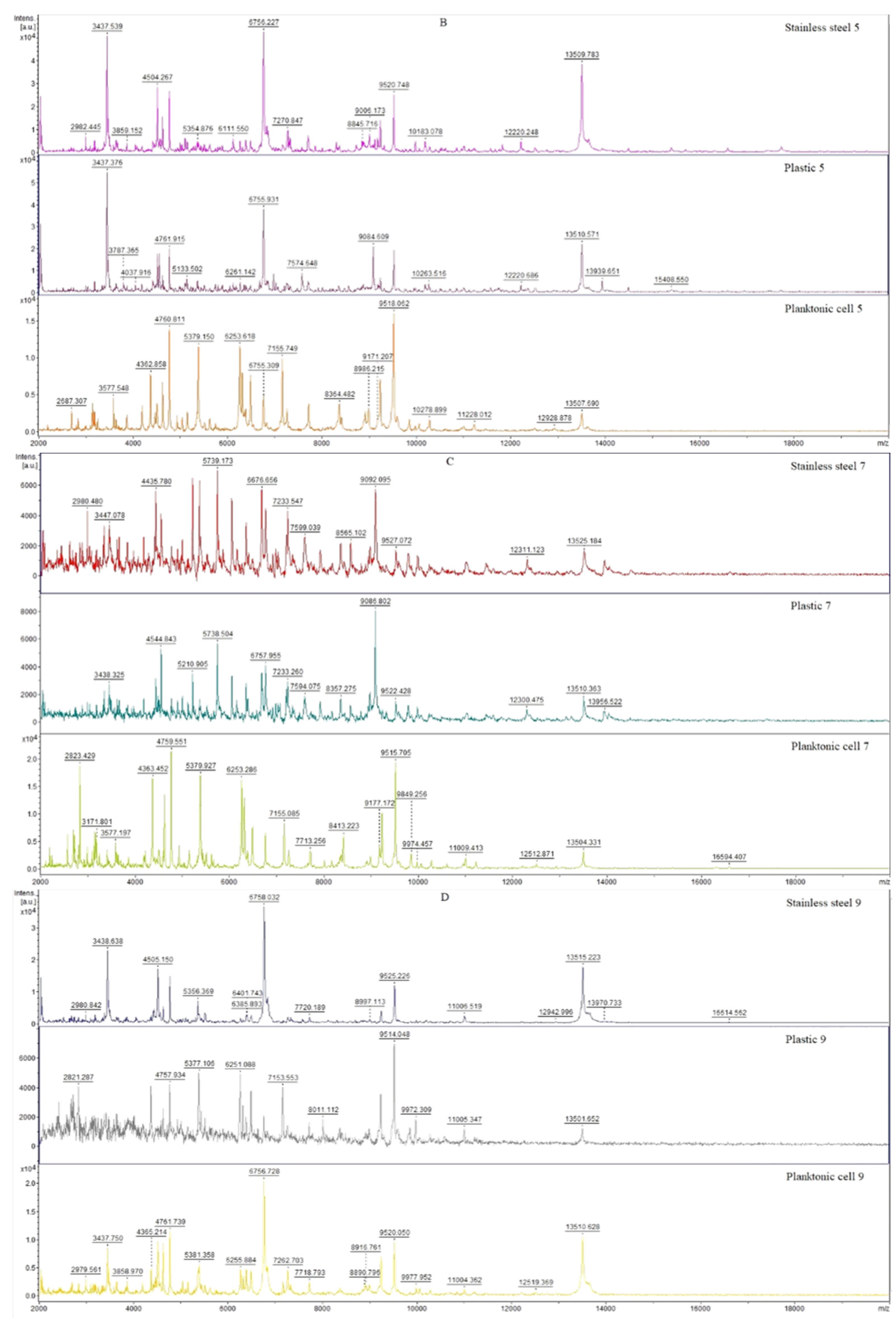
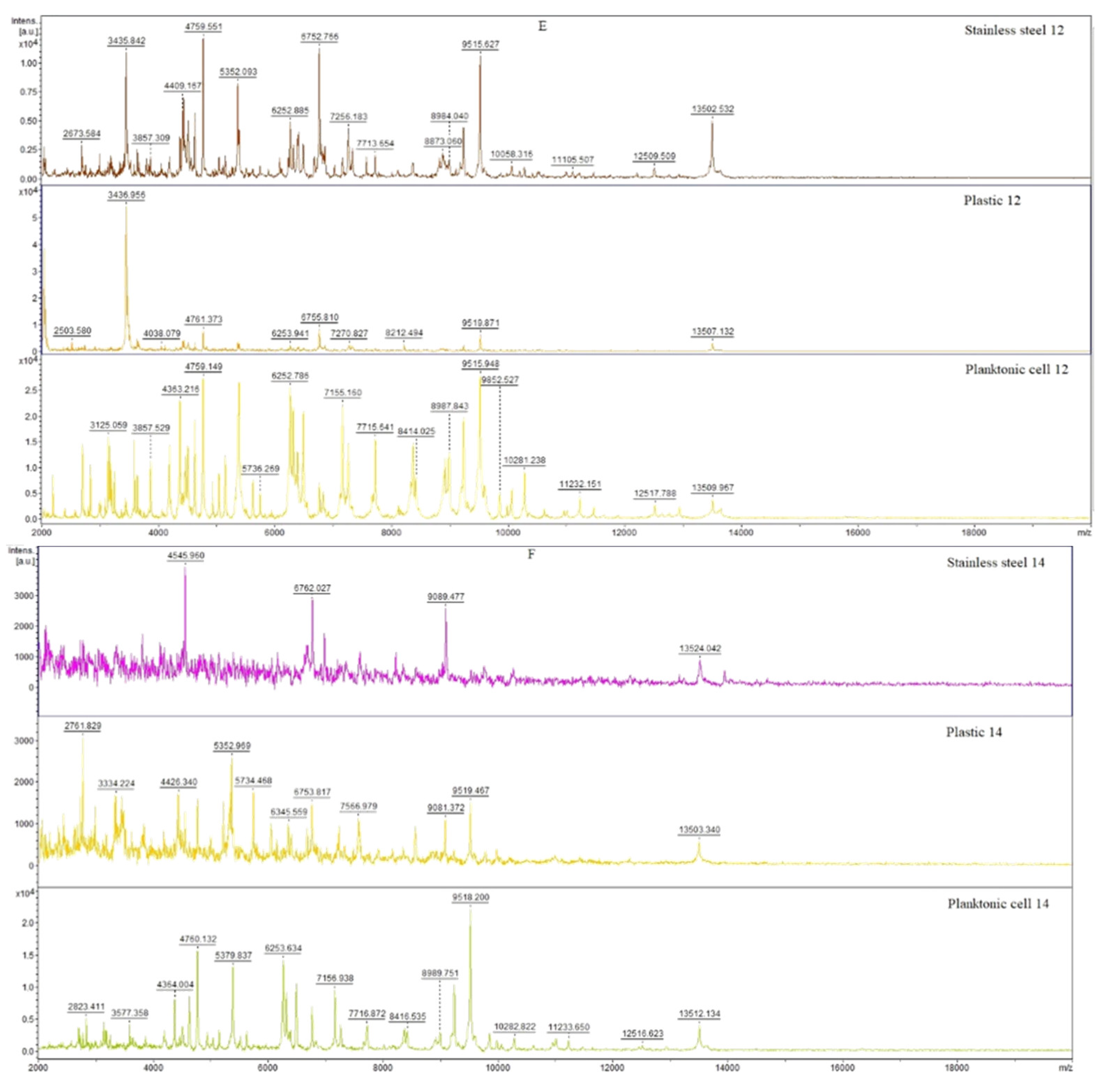
The mass spectra of the young biofilms on days 3 and 5 (Figure 1A,B) of the culture evolved similarly to the planktonic spectra, indicating a similarity in protein production between the control and experimental groups in the young biofilms. By evaluating the evolution of the spectra, we concluded that there were no significant changes in the protein profiles of the biofilms. On day 7 of the experiment, there was densification of the peaks in the experimental groups compared to the planktonic spectra, which highlighted impending changes in the protein profiles of the experimental groups. On days 9 and 12 (Figure 1D,E), we observed a pronounced effect of J. grandiflorum on the plastic surface in the experimental group. A significantly stronger inhibitory effect on the anticorrosive surface was observed only on day 14 of the experiment (Figure 1F). There were visible changes in the protein profile of the biofilm treated with J. grandiflorum EO. J. grandiflorum EO seems to affect the homeostasis of the bacterial biofilm, which helps in its inhibition, predominantly on the plastic surface.
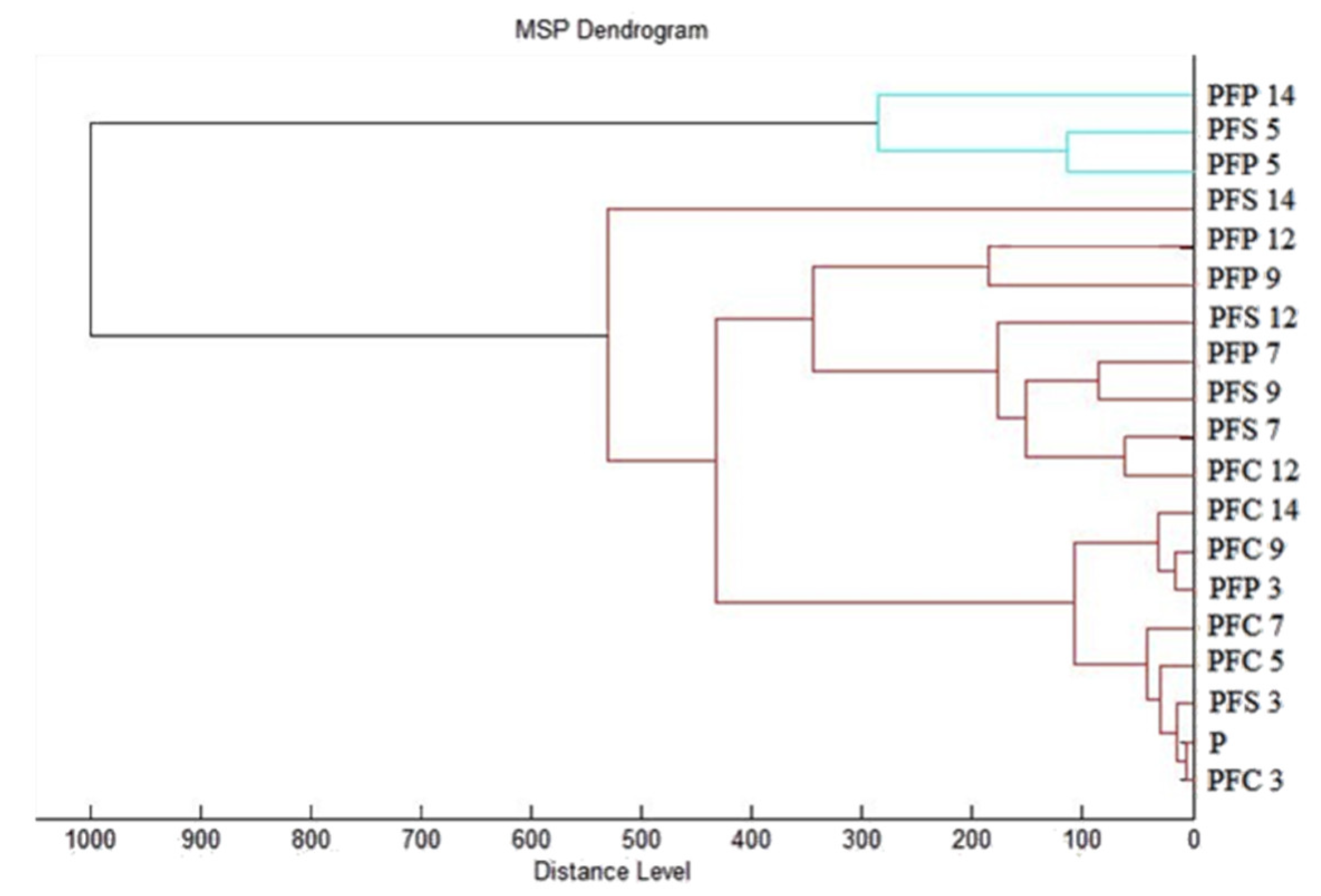
A dendrogram based on MSP distances was constructed to visually display the similarities in biofilm structure. Based on the constructed dendrogram (Figure 2), it can be observed that the early biofilm stages (PFS 3 and PFP 3) had the shortest MSP distances, along with the control and planktonic spectra. The similarity of the protein profiles in the control groups is demonstrated by the comparably short MSP distances. The early biofilm forms of the experimental group on day 3 had comparably short MSP distances to the controls, which is confirmed by the findings based on the analysis of the mass spectra. An increase in the MSP distance in the experimental group can be seen over the time course of the experiment, with a peak recorded on days 12 and 14 of the experiment. These observations are evidence of the disturbance of P. fluorescens biofilm homeostasis by the influence of J. grandiflorum essential oil.

3.6. In Situ Antimicrobial Activity

In order to further evaluate the antimicrobial potential of the EO obtained from J. grandiflorum, we performed an in situ antimicrobial analysis on apples, pears, carrots and white radishes as food models, on which we grew the same bacterial strains as those used in the evaluation of MIC 50 and MIC 90. The results are shown in Table 4. The in situ evaluation of J. grandiflorum EO revealed higher antimicrobial activity when applied to the growth of E. coli on apples, with the concentration applied (125 μL/L) showing the strongest inhibitory effect (74.45 ± 0.88%). H. influenzae showed probacterial activity at all concentrations. The highest concentration of the tested EO of J. grandiflorum had moderate effectiveness in inhibiting the growth of Y. enterocolitica on apples (63.69 ± 1.85%). Against L. monocytogenes, the inhibitory action of J. grandiflorum EO was observed only at the applied concentration of 250 μL/L (34.52 ± 0.90%), while at other concentrations, probacterial activity was observed. Among the G- bacterial strains tested, the EO showed moderate antibacterial activity at the lowest concentration when applied to E. coli (46.62 ± 1.28), and it showed probacterial activity on S. pneumoniae grown on apples. The highest antimicrobial activity against Candida yeasts was found against C. albicans at the lowest concentration of 62.5 μL/L (76.96 ± 1.53%). Against C. glabrata and C. tropicalis, the best results in apples were found with the highest concentration. The EO showed an effect on P. fluorescens biofilm-producing bacteria on apples at the lowest concentration (56.96 ± 2.29%). The in situ evaluation of G+ and G- bacteria growing on pears generally showed moderate inhibitory activity of J. grandiflorum EO. The strongest effectiveness of J. grandiflorum EO in inhibiting the growth of G bacterial strains was observed for Y. enterocolitica growing on pears in the treatment with the highest concentration applied (96.15 ± 2.31%) and for E. coli with the lowest concentration applied (87.86 ± 1.30%). The EO had moderate antimicrobial activity against S. aureus. An increase in bacterial growth was observed for G+ S. penumoniae in the treatment with J. grandiflorum EO at concentrations of 250 μL/L and 500 μL/L. Against Candida growing on pears, the tested J. grandiflorum EO showed the strongest inhibitory potential against C. albicans at the highest concentrations applied (96.53 ± 3.03%). When applied to P. flourescens grown on pears, the EO showed a probacterial effect at the lowest concentration and a moderate inhibitory effect at the highest concentration (55.81 ± 1.05%). The antibacterial activity of the vapor phase of J. grandiflorum EO on bacteria growing on carrots revealed moderate to high effects. The most sensitive to treatment with J. grandiflorum EO at the highest concentration applied was G E. coli (92.35 ± 3.57%). For H. influenzae and Y. enterocolitica, the EO had probacterial activity. For the G+ bacterial strains, the tested J. grandiflorum EO showed low antibacterial effectiveness. For L. monocytogenes, probacterial growth was observed with the two lowest concentrations of J. grandiflorum EO. However, treatment with 15.6 μL/L J. grandiflorum EO showed the strongest inhibition of S. pneumoniae (82.52 ± 1.01%), and at a concentration of 500 μL/L, it had a notable probacterial effect on S. aureus. For the yeasts, an antimicrobial effect was observed from the lowest to the highest concentrations. The best inhibitory effect was found against C. albicans at the highest concentration, with 95.30 ± 2.25%. P. flourescens biofilm-producing bacteria showed similar results to those found on pears. J. grandiflorum EO had significant antibacterial activity against G- bacteria on white radishes. E. coli was inhibited at a concentration of 125 μL/L with an inhibitory effect of 54.88 ± 0.94%, and the growth of Y. enterocolitica showed the strongest inhibition rate when treated with 62.5 μL/L (76.63 ± 2.26%), while H. influenzae was the most effectively inhibited by the vapor phase of J. grandiflorum EO at 500 μL/L (77.26 ± 1.03%). Of the G+ bacterial strains that grew on white radish, the most sensitive to treatment with J. grandiflorum EO was S. pneumoniae, with a growth inhibition rate of 32.63 ± 0.90%. Probacterial effects on the growth of S. aureus and L. monocytogenes were observed. The best inhibitory effect on white radishes was found against C. glabrata (93.45 ± 1.90%). P. flourescens was more sensitive to higher concentrations.

3.7. Insecticidal Activity

The insecticidal activity of J. grandiflorum EO was observed against Oxycarenus lavaterae and Brassicogethes aeneus. The most effective concentration against O. lavatera was found to range from 12.5 to 100% (Table 5).
Table 6 shows insecticidal activity against B. aeneus. The most effective concentration was 100%. All tested concentrations showed high or moderate insecticidal activity.

4. Discussion

The chemical composition of J. grandiflorum EO varies depending on the season. This study focused on the effect of daylight duration and temperature on growth and flowering. The content of some fragrant components (linalool, benzyl acetate, benzyl alcohol and cisjasmone) increases depending on the season, and the content of heavier components (isophytol and its ester) decreases with time. The main constituents determined in J. grandiflorum EO were acetate (37%), benzyl benzoate (34.7%), linalool (9.6%), (Z)-jasmone (5%), isophytol (3.3%) and eugenol (2.1%). These results correspond to the composition of J. grandiflorum EO in other studies [13,14,15,16]. In one of Jirovetz’s studies [17], the compounds identified in the EO of J. grandiforum from India were benzyl acetate (23.7%), benzyl benzoate (20. 7%), phytol (10.9%), linalool (8.2%), isophytol (5.5%), geranyl linalool (3.0%), methyl linoleate (2.8%) and eugenol (2.5%). Absolute jasmine showed moderate to high activity against Gram-positive and Gram-negative bacteria, as well as against yeast [18].
J. grandiflorum has received attention in recent years, and its antimicrobial and antioxidant activity has been investigated. It has been found to have anti-inflammatory, chemopreventive, antispasmodic, antimicrobial, cytoprotective, antiulcerative, antioxidant, anti-acne, and wound-healing activities, but a number of others have yet to be investigated [6]. J. grandiflorum EO was tested for antioxidant activity, i.e., the ability to scavenge free radicals, using the DPPH method. The results revealed up to 94.63% antioxidant activity, which shows different results and different antioxidant activity from those in another study [19]. In a study by Joy et al. (2008), they reported the result that the J. grandiflorum extract has better antimicrobial activity compared to other tested extracts and claimed that it can be used as an antibiotic [20].
Antimicrobial activity was determined using basic methods, namely, the disc diffusion method and minimum inhibitory concentration [17,21,22,23]. Three G- bacteria (E. coli, H. influenzae and Y. enterocolitica), three G+ bacteria (S. pneumoniae, L. monocytogenes and S. aureus), and three yeasts (C. glabrata, C. tropicalis and C. albicans) were used to determine antimicrobial activity, and P. fluorescens was used for biofilm. Against the G+ bacteria E. faecalis, jasmine showed moderate to high activity, as was also the case against the yeast Candida albicans and G- bacteria E. coli, P. aeruginosa, K. pneumoniae and Salmonella sp. [21]. Different EOs had different antimicrobial activity. Antimicrobial activity is influenced by the solvent used [24]. In the study, the antibacterial activity of the extracts was tested against twelve Gram-positive and eighteen Gram-negative bacteria at a concentration of 1000 µg/disc by the disc diffusion method. The methanolic extracts of Jasminum grandiflorum leaves showed significant antibacterial activity [25]. The chloroform extract of the leaves had significant inhibitory activity against Bacillus subtilis (25 mm), and the ethanol extract had the most significant inhibitory activity against E. coli (21 mm). The diethyl ether extract showed low inhibitory activity with an inhibition zone size of 8 mm against Streptococcus sp., as did the ethanolic extract of jasmine against P. aeruginosa and K. pneumonia [26]. Statistically significant (p ≤ 0.05) antimicrobial activity of the ethanolic extract of jasmine leaves (10 μg/mL) was observed against S. mutans and L. acidophilus. The minimum inhibitory concentration values determined were 6.25 μg/mL and 25 μg/mL [27]. Against Xanthomonas campestris and Aeromonas hydrophila, the methanolic extract of jasmine fruit showed inhibition zones of 18.33 ± 0.47 mm and 13.66 ± 0.47 mm, respectively, at 100 μg/mL [28]. Medium to high antimicrobial activity was reported against Gram-positive E. faecalis and Gram-negative E. coli, P. aeruginosa, K. pneumoniae and Salmonella species, as well as against the yeast C. albicans [23]. The jasmine essential oil also showed inhibitory activity against the mycelial growth of Collectotrichum gloeosporioides [29]. The standardization of active fraction extracts and in vivo toxicity and efficacy studies can contribute towards the development of better antimicrobial drugs. This can provide nature-friendly and inexpensive medicines available to all people of the world [30].
Biofilms are highly resistant to antibiotics and other inhibitory agents. The EOs of J. grandiflorum and other herbs could be used as an alternative to antibiotics [31]. El-Baz et al. [32] indicated the ability of J. grandiflorum EO to inhibit biofilm formation and growth. This study confirms our findings. New alternatives that can inhibit the growth and formation of biofilms need to be sought. Oliveira et al. [33] reported on the use of jasmine EO. The results of our work, as well as the results of other studies [12,34,35], confirm the potential ability of the EO to inhibit biofilm formation. Jasmine oil showed the highest antibiofilm activity, followed by cinnamon, clove and rosemary oils. Analysis showed reduced adhesion and roughness in the presence of essential oils [32].
EOs are a promising alternative to the substances currently used. Essential oils have the ability to inhibit the growth of microorganisms even in the vapor phase and could be used for food protection [36,37]. A study by Tyagi and Malik [38] showed higher antimicrobial activity in the vapor phase compared to our results. The extract of J. grandiflorum was tested for vapor-phase antimicrobial activity on apples, pears, carrots, and white radishes. Due to these properties, there is the possibility of using jasmine EOs in the vapor phase for food preservation [39]. Natural additives are considered to contribute to both quality and safety in a wide variety of foods [40]. In recent times, there has been a growing interest in safe, organic and natural products. Due to these demands, preservation techniques that improve the quality and safety of products without causing nutritional or sensory losses have started to be explored, and these essential oils are proving to be a suitable option [41]. Similar to jasmine, in time-to-kill tests, the vapor phase of the essential oil from M. piperita was found to reduce the viability of C. albicans and B. subtilis by up to 100% within 8 h [42]. Currently, the development of natural crop protection products as an alternative to synthetic fungicides is a focus [43,44]. Jasmine EO is used in sweets and candies, biscuits, snacks, chewing gum and food flavoring. It is also used in perfumery and cosmetics as an additive in lotions, soaps, air fresheners and skin care products [45].
Synthetic insecticidal compounds are used as insecticides [46]. They have relatively low toxicity to mammals and can rapidly immobilize invertebrates at low levels, making these synthetic pyrethroid insecticides almost ubiquitous [47,48]. Due to the excessive and frequent use of synthetic insecticides, new options and new substances of natural origin that have properties similar to those of synthetic insecticides are needed [47,48,49]. Therefore, there is greater interest in insecticides made from natural plant extracts that could be less toxic, such as thyme [50] and citronella [51]. In this study, the EO of J. grandiflorum, which showed repellent activity, was tested against O. lavaterae and B. aeneus. Natural products provide effective and relatively safer protection against insects, although synthetic repellents are still currently preferred [52,53]. The antibacterial activity of plants and their essential oils may be due to the presence of different active substances. Further studies are needed to characterize the isolated bioactive substances in order to develop new antibacterial drugs [54]. At this time, the use of natural products that have reliable efficacy and safety and are environmentally friendly is becoming more and more popular. EOs from various plant species have been tested and monitored for their repellent properties [53,55,56,57]. Essential oils and plant extracts are potential alternatives to synthetic insecticides. Further studies are recommended to test secondary metabolites and their formulations so that their efficacy can be improved [58,59,60].

5. Conclusions

The main constituents of J. grandiflorum essential oil were benzyl acetate (37%), benzyl benzoate (34.7%) and linalool (9.6%). The antioxidant activity of the essential oil was 58.47%, corresponding to 220.93 TEAC. We rate this antioxidant activity as high. The EO of J. grandiflorum had weak antimicrobial effects, but its antibiofilm effects were strong, as observed on different surfaces and detected by the MALDI-TOF MS Biotyper. In the vapor-phase antimicrobial activity test of the essential oil, significantly higher antibacterial effects were observed on the model foods (apples, pears, carrots and mild radishes). When evaluating insecticidal activity, we found that J. grandiflorum EO at a concentration of 12.5% and above was able to inhibit it by more than 50%. Based on our results, we have gained valuable insight into the efficacy of J. grandiflorum essential oil in the vapor phase, as these effects have been very little studied to date. The analysis of the antibiofilm activity of the essential oil by the MALDI-TOF MS Biotyper, which has been addressed by relatively few authors, is also a contribution of our work. On the basis of our findings, we hypothesize that J. grandiflorum essential oil could find application in the fight against biofilms in various industries; it is promising for use in the storage and shelf-life extension of various fruits and vegetables. The essential oil analyzed by us showed significantly stronger antimicrobial activity in the vapor phase, which is probably due to the significant proportion of volatile compounds. The vapor-phase application has a lower impact on the sensory properties of commodities and thus might be more acceptable to the consumer than the contact application.

Author Contributions

Conceptualization, L.G., N.Č. and M.K.; methodology, L.G., N.Č., N.V., M.V., P.Ł.K., L.B. and M.K.; software, L.G., N.Č. and M.K.; validation, L.G., N.Č. and M.K.; formal analysis, L.G., N.Č. and M.K.; investigation, L.G., N.Č., N.V., M.V., P.Ł.K., L.B. and M.K.; resources, M.K.; data curation, L.G. and M.K.; writing—original draft preparation, L.G., N.Č. and M.K.; writing—review and editing, L.G., N.Č. and M.K.; visualization, L.G.; supervision, M.K..; project administration, M.K.; funding acquisition, M.K. All authors have read and agreed to the published version of the manuscript.

Funding

This work has been supported by the grants of the APVV-20-0058, “The potential of the essential oils from aromatic plants for medical use and food preservation”.

Data Availability Statement

All data generated or analyzed during this study are included in this published article.

Acknowledgments

This work has been supported by grants from VEGA, no. 1/0180/20.

Conflicts of Interest

The authors declare no conflict of interest.

References

  1. Stringaro, A.; Colone, M.; Angiolella, L. Antioxidant, Antifungal, Antibiofilm, and Cytotoxic Activities of Mentha Spp. Essential Oils. Medicines 2018, 5, 112. [Google Scholar] [CrossRef] [PubMed] [Green Version]
  2. Baj, T.; Sieniawska, E.; Kowalski, R.; Wesolowskp, M.; Ulewicz-Magulska, B. Effectiveness of the deryng and clevenger-type apparatus in isolation of various types of components of essential oil from the mutelina purpurea thell. Flowers. Acta Pol. Pharm. 2015, 72, 507–515. [Google Scholar] [PubMed]
  3. Brnawi, W.I.; Hettiarachchy, N.S.; Horax, R.; Kumar-Phillips, G.; Ricke, S. Antimicrobial Activity of Leaf and Bark Cinnamon Essential Oils against Listeria Monocytogenes and Salmonella Typhimurium in Broth System and on Celery. J. Food Process. Preserv. 2019, 43, e13888. [Google Scholar] [CrossRef]
  4. Benzaid, C.; Belmadani, A.; Djeribi, R.; Rouabhia, M. The Effects of Mentha × Piperita Essential Oil on C. Albicans Growth, Transition, Biofilm Formation, and the Expression of Secreted Aspartyl Proteinases Genes. Antibiotics 2019, 8, 10. [Google Scholar] [CrossRef] [Green Version]
  5. Joulain, D. Jasminum Grandiflorum Flowers—Phytochemical Complexity and Its Capture in Extracts: A Review. Flavour Fragr. J. 2021, 36, 526–553. [Google Scholar] [CrossRef]
  6. Arun, M.; Satish, S.; Anima, P. Phytopharmacological Profile of Jasminum Grandiflorum Linn. (Oleaceae). Chin. J. Integr. Med. 2016, 22, 311–320. [Google Scholar] [CrossRef]
  7. Zhang, Y.; Sun, L.; Lai, X.; Peng, X.; Wen, S.; Zhang, Z.; Xie, Y.; Li, Q.; Chen, R.; Zheng, X.; et al. Gastroprotective Effects of Extract of Jasminum Grandiflorum L. Flower in HCl/EtOH-Induced Gastric Mucosal Ulceration Mice. Biomed. Pharmacother. 2021, 144, 112268. [Google Scholar] [CrossRef]
  8. Gutierrez, J.; Barry-Ryan, C.; Bourke, P. The Antimicrobial Efficacy of Plant Essential Oil Combinations and Interactions with Food Ingredients. Int. J. Food Microbiol. 2008, 124, 91–97. [Google Scholar] [CrossRef] [Green Version]
  9. Nadjib, B.M.; Amine, F.M.; Abdelkrim, K.; Fairouz, S.; Maamar, M. Liquid and vapour phase antibacterial activity of eucalyptus globulus essential oil = susceptibility of selected respiratory tract pathogens. Am. J. Infect. Dis. 2014, 10, 105–117. [Google Scholar] [CrossRef] [Green Version]
  10. Adams, R.P. Identification of Essential Oil Components by Gas Chromatography Mass Spectroscopy; Allured Publishing Corporation: Carol Stream, IL, USA, 2007; ISBN 978-1-932633-21-4. [Google Scholar]
  11. Vandendool, H.; Kratz, P.D. A Generalization of the Retention Index System Including Linear Temperature Programmed Gas-Liquid Partition Chromatography. J. Chromatogr. 1963, 11, 463–471. [Google Scholar] [CrossRef]
  12. Kačániová, M.; Terentjeva, M.; Galovičová, L.; Ivanišová, E.; Štefániková, J.; Valková, V.; Borotová, P.; Kowalczewski, P.Ł.; Kunová, S.; Felšöciová, S.; et al. Biological Activity and Antibiofilm Molecular Profile of Citrus aurantium Essential Oil and Its Application in a Food Model. Molecules 2020, 25, 3956. [Google Scholar] [CrossRef] [PubMed]
  13. Bauer, K.; Garbe, D.; Surburg, H. Common Fragrance and Flavor Materials; Wiley-VCH Verlag GmbH: Weinheim, Germany, 2001; ISBN 978-3-527-30364-9. [Google Scholar]
  14. Rout, P.K.; Naik, S.N.; Rao, Y.R. Composition of Absolutes of Jasminum Sambac L. Flowers Fractionated with Liquid CO 2 and Methanol and Comparison with Liquid CO2 Extract. J. Essent. Oil Res. 2010, 22, 398–406. [Google Scholar] [CrossRef]
  15. Rao, Y.R.; Rout, P.K. Geographical Location and Harvest Time Dependent Variation in the Composition of Essential Oils of Jasminum Sambac. (L.) Aiton. J. Essent. Oil Res. 2003, 15, 398–401. [Google Scholar] [CrossRef]
  16. Tamogami, S.; Awano, K.; Kitahara, T. Analysis of the Enantiomeric Ratios of Chiral Components in Absolute Jasmine. Flavour Fragr. J. 2001, 16, 161–163. [Google Scholar] [CrossRef]
  17. Jirovetz, L.; Buchbauer, G.; Schweiger, T.; Denkova, Z.; Slavchev, A.; Stoyanova, A.; Schmidt, E.; Geissler, M. Chemical Composition, Olfactory Evaluation and Antimicrobial Activities of Jasminum Grandiflorum L. Absolute from India. Nat. Prod. Commun. 2007, 2, 1934578X0700200411. [Google Scholar] [CrossRef] [Green Version]
  18. Luo, H.; Lin, S.; Ren, F.; Wu, L.; Chen, L.; Sun, Y. Antioxidant and Antimicrobial Capacity of Chinese Medicinal Herb Extracts in Raw Sheep Meat. J. Food Prot. 2007, 70, 1440–1445. [Google Scholar] [CrossRef]
  19. Wang, H.-F.; Yih, K.-H.; Huang, K.-F. Comparative Study of the Antioxidant Activity of Forty-Five Commonly Used Essential Oils and Their Potential Active Components. J. Food Drug Anal. 2020, 18, 8. [Google Scholar] [CrossRef]
  20. Balkrishna, A.; Rohela, A.; Kumar, A.; Kumar, A.; Arya, V.; Thakur, P.; Oleksak, P.; Krejcar, O.; Verma, R.; Kumar, D.; et al. Mechanistic Insight into Antimicrobial and Antioxidant Potential of Jasminum Species: A Herbal Approach for Disease Management. Plants 2021, 10, 1089. [Google Scholar] [CrossRef]
  21. Jirovetz, L. Antimicrobial Testings, Gas Chromatographic Analysis and Olfactory Evaluation of an Essential Oil of Hop Cones (Humulus Lupulus L.) from Bavaria and Some of Its Main Compounds. Sci. Pharm. 2006, 74, 189–201. [Google Scholar] [CrossRef] [Green Version]
  22. Jirovetz, L.; Buchbauer, G.; Denkova, Z.; Stoyanova, A.; Murgov, I.; Schmidt, E.; Geissler, M. Antimicrobial Testinas and Gas Chromatoaraphic Analvsis of Pure Oxvaenated Monoterpenes 1.8-Cineole, α-Terpineol, Terpinen-4-Ol and Camphor as Well as Target Comoounds in Essential Oils of Pine (Pinus Pinaster), Rosemary (Rosmarinus Officinalis), Tea Tree (Melaleuca Alternifolia). Sci. Pharm. 2005, 73, 27–39. [Google Scholar] [CrossRef]
  23. Jirovetz, L.; Buchbauer, G.; Schmidt, E.; Stoyanova, A.S.; Denkova, Z.; Nikolova, R.; Geissler, M. Purity, Antimicrobial Activities and Olfactoric Evaluations of Geraniol/Nerol and Various of Their Derivatives. J. Essent. Oil Res. 2007, 19, 288–291. [Google Scholar] [CrossRef]
  24. Bharathi, P.R.; Sripathi, S.K.; Lakshmi, A.N. Jasminum grandiflorum linn.–an update review. Int. J. Pharm. Sci. Res. 2020, 11, 1994–2010. [Google Scholar]
  25. Ali, S.T.; Ayub, A.; Ali, S.N. Antibacterial Activity Of Methanolic Extracts From Some Selected Medicinal Plants. Fuuast J. Biol. 2017, 7, 123–125. [Google Scholar]
  26. Shekhar, S. Evaluation of antimicrobial activity of jasminum species using solvent extracts against clinical pathogens. World J. Pharm. Pharm. Sci. 2015, 4, 11. [Google Scholar]
  27. Nagarajappa, R.; Batra, M.; Sharda, A.J.; Asawa, K.; Sanadhya, S.; Daryani, H.; Ramesh, G. Antimicrobial Effect of Jasminum grandiflorum L. and Hibiscus Rosa-Sinensis L. Extracts Against Pathogenic Oral Microorganisms--An In Vitro Comparative Study. Oral Health Prev. Dent. 2015, 13, 341–348. [Google Scholar] [CrossRef]
  28. Britto, A.J.; Grecelin, H. Efficacy of Fruits of Jasminum grandiflorum Linn. against Plant and Animal Pathogens. Asian J. Pharm. Clin. Res. 2011, 4, 74–75. [Google Scholar]
  29. Nidiry, E.; Srivastava, H. A Comparative Study of the Antifungal Activities of Absolutes of Jasmine and Tuberose and Their Constituents. Indian Perfum. 2007, 51, 53–54. [Google Scholar]
  30. Khond, M.; Bhosale, J.D.; Arif, T.; Padhi, M.M.; Dabur, R. Screening of Some Selected Medicinal Plants Extracts for In-Vitro Antimicrobial Activity. Middle East J. Sci. Res. 2009, 4, 271–278. [Google Scholar]
  31. El-Tarabily, K.A.; El-Saadony, M.T.; Alagawany, M.; Arif, M.; Batiha, G.E.; Khafaga, A.F.; Elwan, H.A.M.; Elnesr, S.S.; El-Hack, M.E.A. Using Essential Oils to Overcome Bacterial Biofilm Formation and Their Antimicrobial Resistance. Saudi J. Biol. Sci. 2021, 28, 5145–5156. [Google Scholar] [CrossRef]
  32. El-Baz, A.M.; Mosbah, R.A.; Goda, R.M.; Mansour, B.; Sultana, T.; Dahms, T.E.S.; El-Ganiny, A.M. Back to Nature: Combating Candida Albicans Biofilm, Phospholipase and Hemolysin Using Plant Essential Oils. Antibiotics 2021, 10, 81. [Google Scholar] [CrossRef]
  33. de Oliveira Carvalho, I.; Purgato, G.A.; Píccolo, M.S.; Pizziolo, V.R.; Coelho, R.R.; Diaz-Muñoz, G.; Alves Nogueira Diaz, M. In Vitro Anticariogenic and Antibiofilm Activities of Toothpastes Formulated with Essential Oils. Arch. Oral Biol. 2020, 117, 104834. [Google Scholar] [CrossRef]
  34. Galovičová, L.; Borotová, P.; Valková, V.; Vukovic, N.L.; Vukic, M.; Terentjeva, M.; Štefániková, J.; Ďúranová, H.; Kowalczewski, P.Ł.; Kačániová, M. Thymus serpyllum Essential Oil and Its Biological Activity as a Modern Food Preserver. Plants 2021, 10, 1416. [Google Scholar] [CrossRef]
  35. Valková, V.; Ďúranová, H.; Galovičová, L.; Borotová, P.; Vukovic, N.L.; Vukic, M.; Kačániová, M. Cymbopogon citratus Essential Oil: Its Application as an Antimicrobial Agent in Food Preservation. Agronomy 2022, 12, 155. [Google Scholar] [CrossRef]
  36. López, P.; Sánchez, C.; Batlle, R.; Nerín, C. Vapor-Phase Activities of Cinnamon, Thyme, and Oregano Essential Oils and Key Constituents against Foodborne Microorganisms. J. Agric. Food Chem. 2007, 55, 4348–4356. [Google Scholar] [CrossRef]
  37. Nedorostova, L.; Kloucek, P.; Kokoska, L.; Stolcova, M.; Pulkrabek, J. Antimicrobial Properties of Selected Essential Oils in Vapour Phase against Foodborne Bacteria. Food Control 2009, 20, 157–160. [Google Scholar] [CrossRef]
  38. Tyagi, A.K.; Malik, A. Antimicrobial Potential and Chemical Composition of Eucalyptus Globulus Oil in Liquid and Vapour Phase against Food Spoilage Microorganisms. Food Chem. 2011, 126, 228–235. [Google Scholar] [CrossRef]
  39. Ahmed, N.; Hanani, Y.A.; Ansari, S.Y.; Anwar, S. Jasmine (Jasminum Sambac L., Oleaceae) Oils. In Essential Oils in Food Preservation, Flavor and Safety; Elsevier: Amsterdam, The Netherlands, 2016; pp. 487–494. ISBN 978-0-12-416641-7. [Google Scholar]
  40. Viuda-Martos, M.; Ruiz-Navajas, Y.; Fernández-López, J.; Pérez-Álvarez, J. Antifungal Activity of Lemon (Citrus lemon L.), Mandarin (Citrus reticulata L.), Grapefruit (Citrus paradisi L.) and Orange (Citrus sinensis L.) Essential Oils. Food Control 2008, 19, 1130–1138. [Google Scholar] [CrossRef]
  41. Ju, J.; Xie, Y.; Yu, H.; Guo, Y.; Cheng, Y.; Qian, H.; Yao, W. Synergistic Interactions of Plant Essential Oils with Antimicrobial Agents: A New Antimicrobial Therapy. Crit. Rev. Food Sci. Nutr. 2022, 62, 1740–1751. [Google Scholar] [CrossRef]
  42. Tyagi, A.K.; Malik, A. Antimicrobial Potential and Chemical Composition of Mentha Piperita Oil in Liquid and Vapour Phase against Food Spoiling Microorganisms. Food Control 2011, 22, 1707–1714. [Google Scholar] [CrossRef]
  43. Combrinck, S.; Regnier, T.; Kamatou, G.P.P. In Vitro Activity of Eighteen Essential Oils and Some Major Components against Common Postharvest Fungal Pathogens of Fruit. Ind. Crops Prod. 2011, 33, 344–349. [Google Scholar] [CrossRef]
  44. Stević, T.; Berić, T.; Šavikin, K.; Soković, M.; Gođevac, D.; Dimkić, I.; Stanković, S. Antifungal Activity of Selected Essential Oils against Fungi Isolated from Medicinal Plant. Ind. Crops Prod. 2014, 55, 116–122. [Google Scholar] [CrossRef]
  45. Hernandez, E. Essential oils | Distillation. In Encyclopedia of Separation Science; Elsevier: Amsterdam, The Netherlands, 2000; pp. 2739–2744. ISBN 978-0-12-226770-3. [Google Scholar]
  46. Noosidum, A.; Chareonviriyaphap, T.; Chandrapatya, A. Synergistic Repellent and Irritant Effect of Combined Essential Oils on Aedes Aegypti (L.) Mosquitoes. J. Vector Ecol. 2014, 39, 298–305. [Google Scholar] [CrossRef]
  47. Jirakanjanakit, N.; Leemingsawat, S.; Thongrungkiat, S.; Apiwathnasorn, C.; Singhaniyom, S.; Bellec, C.; Dujardin, J.P. Influence of Larval Density or Food Variation on the Geometry of the Wing of Aedes (Stegomyia) Aegypti: Geometry of the Wing of Aedes (Stegomyia) Aegypti. Trop. Med. Int. Health 2007, 12, 1354–1360. [Google Scholar] [CrossRef] [PubMed]
  48. Thanispong, K.; Sathantriphop, S.; Chareonviriyaphap, T. Insecticide Resistance of Aedes Aegypti and Culex Quinquefasciatus in Thailand. J. Pestic. Sci. 2008, 33, 351–356. [Google Scholar] [CrossRef] [Green Version]
  49. Chanda, E. Optimizing Strategic Insecticide Resistance Management Planning in Malaria Vectors. In Insecticides Resistance; Trdan, S., Ed.; InTech: London, UK, 2016; ISBN 978-953-51-4591-2. [Google Scholar]
  50. Park, B.-S.; Choi, W.-S.; Kim, J.-H.; Kim, K.-H.; Lee, S.-E. Monoterpenes from thyme (Thymus vulgaris) as potential mosquito repellents. J. Am. Mosq. Control Assoc. 2005, 21, 80–83. [Google Scholar] [CrossRef]
  51. Yang, P.; Ma, Y. Repellent Effect of Plant Essential Oils against Aedes albopictus. J. Vector Ecol. J. Soc. Vector Ecol. 2005, 30, 231–234. [Google Scholar]
  52. Fradin, M.S.; Day, J.F. Comparative Efficacy of Insect Repellents against Mosquito Bites. N. Engl. J. Med. 2002, 347, 13–18. [Google Scholar] [CrossRef]
  53. Sritabutra, D.; Soonwera, M.; Waltanachanobon, S.; Poungjai, S. Evaluation of Herbal Essential Oil as Repellents against Aedes aegypti (L.) and Anopheles dirus Peyton & Harrion. Asian Pac. J. Trop. Biomed. 2011, 1, S124–S128. [Google Scholar] [CrossRef]
  54. Sachan, S.; Paarakh, P.; Gavani, U. Antibacterial Activity of Jasminum Grandiflorum Linn Leaves. J. Pharm. Res. 2009, 2, 1206–1207. [Google Scholar]
  55. Noosidum, A.; Prabaripai, A.; Chareonviriyaphap, T.; Chandrapatya, A. Excito-Repellency Properties of Essential Oils from Melaleuca leucadendron L., Litsea cubeba (Lour.) Persoon, and Litsea salicifolia (Nees) on Aedes aegypti (L.) Mosquitoes. J. Vector Ecol. 2008, 33, 305–312. [Google Scholar] [CrossRef]
  56. de Boer, H.; Vongsombath, C.; Pålsson, K.; Björk, L.; Jaenson, T.G.T. Botanical Repellents and Pesticides Traditionally Used Against Hematophagous Invertebrates in Lao People’s Democratic Republic: A Comparative Study of Plants Used in 66 Villages. J. Med. Entomol. 2010, 47, 400–414. [Google Scholar] [CrossRef]
  57. Suwansirisilp, K.; Visetson, S.; Prabaripai, A.; Tanasinchayakul, S.; Grieco, J.P.; Bangs, M.J.; Chareonviriyaphap, T. Behavioral Responses of Aedes Aegypti and Culex Quinquefasciatus (Diptera: Culicidae) to Four Essential Oils in Thailand. J. Pest Sci. 2013, 86, 309–320. [Google Scholar] [CrossRef]
  58. Tawatsin, A.; Wratten, S.D.; Scott, R.R.; Thavara, U.; Techadamrongsin, Y. Repellency of Volatile Oils from Plants against Three Mosquito Vectors. J. Vector Ecol. J. Soc. Vector Ecol. 2001, 26, 76–82. [Google Scholar]
  59. Tuetun, B.; Choochote, W.; Kanjanapothi, D.; Rattanachanpichai, E.; Chaithong, U.; Chaiwong, P.; Jitpakdi, A.; Tippawangkosol, P.; Riyong, D.; Pitasawat, B. Repellent Properties of Celery, Apium graveolens L., Compared with Commercial Repellents, against Mosquitoes under Laboratory and Field Conditions. Trop. Med. Int. Health 2005, 10, 1190–1198. [Google Scholar] [CrossRef]
  60. Kim, B.-S.; Yi, H.; Chun, J.; Cha, C.-J. Genome Sequence of Type Strain of Staphylococcus aureus Subsp. aureus. Gut Pathog. 2014, 6, 6. [Google Scholar] [CrossRef]
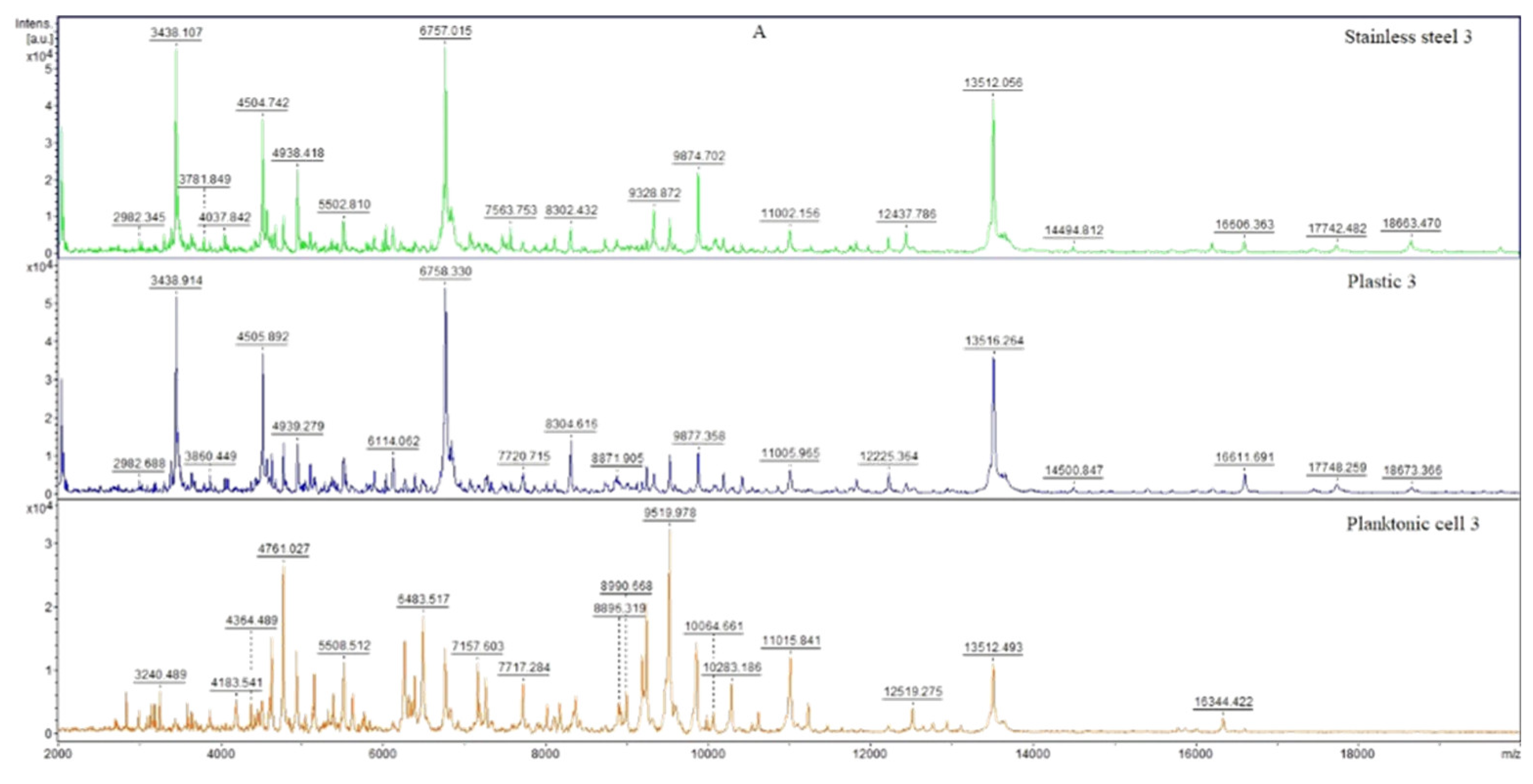
Figure 1. MALDI-TOF mass spectra of P. fluorescens during the development of the biofilm: (A) 3rd day, (B) 5th day, (C) 7th day, (D) 9th day, (E) 12th day, and (F) 14th day.
Horticulturae 08 00953 g001aHorticulturae 08 00953 g001bHorticulturae 08 00953 g001c
Figure 2. Dendrogram of P. fluorescens generated using MSPs of planktonic cells and the control. PF, P. fluorescens; C, control; S, stainless steel; P, plastic; and only P, planktonic cells.
Horticulturae 08 00953 g002
Table 1. Identification of volatile constituents of J. grandiflorum essential oil.
No.Compound%RI (lit.)RI (calc.)
1(Z)-b-Ocimene1.1801801
2m-Methylphenol0.3855855
3(E)-Hexenyl propionate0.2902900
4Linalool9.6960965
5Benzyl acetate37.0973976
62-Undecanone0.5977978
7Eugenol2.1979981
8(Z)-Jasmone5.0985986
9(E,E)-a-Farnesene0.9988991
10Caryophyllenyl alcohol1.9991997
11(Z)-Methyl jasmonate0.39981004
12Benzyl benzoate34.710241026
13(Z,Z)-Farnesyl acetone0.610311035
14Methyl hexadecanoate0.810351037
15Isophytol3.310421046
16(E)-Phytol acetate1.410961099
99.7
Table 2. Antimicrobial activity of J. grandiflorum EO with the disc diffusion method (in mm).
MicroorganismsInhibition Zone (mm)Activity of EOATB (mm)
Yersinia enterocolitica3.00 ± 0.00*26 ± 2.00
Haemophilus influenzae2.33 ± 0.58*30 ± 3.00
Escherichia coli3.67 ± 1.15*27 ± 0.50
Listeria monocytogenes5.33 ± 0.58**24 ± 1.50
Staphylococcus aureus3.67 ± 0.58*29 ± 1.00
Streptococcus pneumoniae2.33 ± 0.58*27 ± 2.00
Candida albicans2.67 ± 0.58*24 ± 2.00
Candida tropicalis2.33 ± 0.58*30 ± 1.00
Candida glabrata3.33 ± 0.58*32 ± 1.50
Pseudomonas fluorescens biofilm3.67 ± 0.58*26 ± 1.00
Weak (0–5 mm, *) and moderate (5–8 mm, **) antimicrobial activity.
Table 3. Minimal inhibition concentrations of J. grandiflorum EO (in µL/mL).
MicroorganismMIC 50MIC 90
Yersinia enterocolitica6.438.73
Haemophilus influenzae11.3615.26
Escherichia coli6.438.73
Listeria monocytogenes6.438.73
Staphylococcus aureus11.3615.26
Streptococcus pneumoniae11.3615.26
Candida albicans3.185.24
Candida tropicalis3.185.24
Candida glabrata0.650.97
Pseudomonas fluorescens biofilm11.3615.26
Table 4. In situ antibacterial activity of the EO vapor phase of J. grandiflorum against microbial strains growing on selected food models.
Food ModelBacteriaMicrobial Growth Inhibition (%)
Concentration of Leaves EO
62.5 (μL/L)125 (μL/L)250 (μL/L)500 (μL/L)
AppleGram-negative
E. coli−66.70 ± 0.98 a74.45 ± 0.88 b,a54.48 ± 0.84 c,b,a25.00 ± 2.45 d,c,b,a
H. influenzae−64.91 ± 2.40 a−54.63 ± 1.58 b,a−25.43 ± 1.62 c,b,a−16.06 ± 0.26 d,c,b,a
Y. enterocolitica34.37 ± 0.61 a2.96 ± 0.62 b,a56.63 ± 1.13 c,b,a63.69 ± 1.85 d,c,b,a
Gram-positive
L. monocytogenes−45.22 ± 1.82 a−23.99 ± 1.59 b,a34.52 ± 0.90 c,b,a−66.89 ± 1.00 d,c,b,a
S. aureus46.62 ± 1.28 a35.06 ± 1.35 b,a11.71 ± 1.06 c,b,a3.67 ± 0.95 d,c,b,a
S. pneumoniae−77.11 ± 1.66 a−46.78 ± 1.11 b,a−36.30 ± 1.66 c,b,a−13.00 ± 0.59 d,c,b,a
Yeasts
C. albicans76.96 ± 1.53 a7.11 ± 0.47 b,a24.81 ± 0.89 c,b,a45.84 ± 0.99 d,c,b,a
C. glabrata13.60 ± 1.06 a24.66 ± 1.00 b,a31.85 ± 1.42 c,b,a64.63 ± 1.44 d,c,b,a
C. tropicalis7.03 ± 0.83 a−5.48 ± 1.22 b,a43.93 ± 1.84 c,b,a66.32 ± 2.30 d,c,b,a
Pseudomonas fluorescens biofilm56.96 ± 2.29 a−7.77 ± 0.94 b,a32.84 ± 1.65 c,b,a43.91 ± 1.80 d,c,b,a
PearGram-negative
E. coli87.86 ± 1.30 a24.70 ± 2.45 b,a53.20 ± 2.57 c,b,a24.18 ± 1.66 d,c,a
H. influenzae−77.98 ± 1.25 a−54.33 ± 1.11 b,a−33.92 ± 0.36 c,b,a42.70 ± 0.91 d,c,b,a
Y. enterocolitica28.71 ± 3.47 a−32.07 ± 0.53 b,a72.09 ± 3.07 c,b,a96.15 ± 2.31 d,c,b,a
Gram-positive
L. monocytogenes42.42 ± 1.14 a34.58 ± 0.89 b,a−24.69 ± 1.10 c,b,a2.78 ± 0.56 d,c,b,a
S. aureus7.21 ± 1.69 a12.82 ± 1.32 b,a27.43 ± 2.50 c,b,a35.04 ± 1.41 d,c,b,a
S. pneumoniae−34.03 ± 1.10 a−16.22 ± 0.48 b,a33.27 ± 1.17 c,b,a86.32 ± 2.10 d,c,b,a
Yeasts
C. albicans13.08 ± 1.96 a26.57 ± 3.90 b,a50.31 ± 1.83 c,b,a96.53 ± 3.03 d,c,b,a
C. glabrata8.30 ± 0.56 a18.00 ± 1.12 b,a31.74 ± 1.32 c,b,a65.04 ± 1.54 d,c,b,a
C. tropicalis34.41 ± 2.09 a24.42 ± 0.66 b,a16.84 ± 0.98 c,b,a8.88 ± 0.58 d,c,b,a
Pseudomonas fluorescens biofilm−66.88 ± 0.96 a−56.70 ± 1.23 b,a44.44 ± 0.88 c,b,a55.81 ± 1.05 d,c,b,a
CarrotGram-negative
E. coli12.32 ± 1.97 a24.70 ± 2.45 b,a53.20 ± 2.57 c,b,a92.35 ± 3.57 d,c,b,a
H. influenzae−78.18 ± 1.50 a−23.56 ± 0.62 b,a−63.93 ± 0.91 c,b,a−44.25 ± 0.99 d,c,b,a
Y. enterocolitica14.88 ± 2.29 a26.15 ± 1.92 b,a−24.36 ± 2.11 c,b,a−4.55 ± 0.88 d,c,b,a
Gram-positive
L. monocytogenes−5.03 ± 0.64 a−1.98 ± 0.29 b,a2.86 ± 0.46 c,b,a5.43 ± 0.23 d,c,b,a
S. aureus8.67 ± 1.09 a−46.11 ± 1.53 b,a−55.52 ± 1.28 c,b,a−6.07 ± 0.59 d,c,b,a
S. pneumoniae−54.41 ± 2.33 a−66.33 ± 2.01 b,a67.66 ± 0.83 c,b,a82.52 ± 1.01 d,c,b,a
Yeasts
C. albicans−6.25 ± 0.50 a−11.77 ± 1.05 b,a54.55 ± 1.00 c,b,a95.30 ± 2.25 d,c,b,a
C. glabrata25.03 ± 1.57 a34.00 ± 4.66 b,a44.62 ± 2.01 c,b,a68.46 ± 0.71 d,c,b,a
C. tropicalis68.46 ± 3.13 a6.29 ± 0.44 b,a14.63 ± 0.92 c,b,a33.10 ± 2.00 d,c,b,a
Pseudomonas fluorescens biofilm−5.95 ± 0.24 a7.28 ± 0.39 b,a44.51 ± 1.48 c,b,a55.33 ± 1.49 d,c,b,a
White radishGram-negative
E. coli−35.32 ± 1.67 a54.88 ± 0.94 b,a22.59 ± 0.95 c,b,a12.93 ± 0.57 d,c,b,a
H. influenzae6.33 ± 0.51 a16.15 ± 1.11 b,a35.27 ± 0.86 c,b,a77.26 ± 1.03 d,c,b,a
Y. enterocolitica76.63 ± 2.26 a54.33 ± 1.01 b,a33.44 ± 1.12 c,b,a15.60 ± 1.22 d,c,b,a
Gram-positive
L. monocytogenes7.96 ± 0.69 a−23.53 ± 2.16 b,a−44.66 ± 1.00 c,b,a−34.26 ± 0.70 d,c,b,a
S. aureus77.11 ± 1.67 a21.63 ± 1.06 b,a−36.07 ± 1.50 c,b,a−23.37 ± 1.47 d,c,b,a
S. pneumoniae8.19 ± 0.56 a13.18 ± 1.41 b,a21.85 ± 1.72 c,b,a32.63 ± 0.90 d,c,b,a
Yeasts
C. albicans5.15 ± 0.18 a15.62 ± 1.06 b,a26.43 ± 1.05 c,b,a35.65 ± 1.12 d,c,b,a
C. glabrata73.99 ± 1.42 a65.76 ± 1.64 b,a84.04 ± 1.54 c,b,a93.45 ± 1.90 d,c,b,a
C. tropicalis56.96 ± 2.29 a−7.77 ± 0.94 b,a32.84 ± 1.65 c.b.a43.91 ± 1.80 d,c,b,a
Pseudomonas fluorescens biofilm5.54 ± 0.90 a7.85 ± 0.28 b13.03 ± 0.37 c,b,a88.40 ± 1.50 d,c,b,a
a,b,c,d Different letters within the same column denote significant differences (p < 0.05).
Table 5. Insecticidal activity against O. lavaterae.
Concentration (%)Number of Living IndividualsNumber of Dead IndividualsInsecticidal Activity (%)
100030100
50030100
25030100
12.5030100
6.25121860
Control group3000
Table 6. Insecticidal activity against B. aeneus.
Concentration (%)Number of Living IndividualsNumber of Dead IndividualsInsecticidal Activity (%)
100030100
5032790
2592170
12.5151550
6.25181240
Control group3000
Publisher’s Note: MDPI stays neutral with regard to jurisdictional claims in published maps and institutional affiliations.