Article

Assessment of Brassicaceae Seeds Quality by X-ray Analysis

1
Laboratory Analysis Center, Federal Scientific Vegetable Center, 14 Selektsionnaya Street, 143080 Odintsovo, Russia
2
Plant Lightphysiology and Agroecosystem Bioproductivity Department, Agrophysical Research Institute, 14 Grazhdanskiy pr., 195220 Saint Petersburg, Russia
3
Electronic Instruments and Devices Department, Saint Petersburg Electrotechnical University “LETI”, Ul. Professora Popova, 5, 197022 Saint Petersburg, Russia
4
All-Russian Scientific Research Institute of the Confectionery Industry—Branch of the V. M. Gorbatov Federal Research Center for Food Systems, 20 Elektrozavodskaya St., 107076 Moscow, Russia
*
Author to whom correspondence should be addressed.
Horticulturae 2022, 8(1), 29; https://doi.org/10.3390/horticulturae8010029
Submission received: 30 November 2021 / Revised: 23 December 2021 / Accepted: 24 December 2021 / Published: 28 December 2021
(This article belongs to the Special Issue Advances in Brassica Crops Genomics and Breeding)

Abstract

:
A serious problem of vegetable production is the quality of sown seeds. In this regard, assessment of seed quality before sowing and storage is of great practical interest. The modern level of scientific research requires the use of instrumental automated methods of seed quality evaluation, allowing to obtain more information and in a shorter time. The material for the study was a variety of samples from the collection of Brassica oleracea L., var. capitata, Raphanus sativus L., var. radicula, and Lepidium sativum L. seeds from the Federal Scientific Center of Vegetable Breeding and the Timofeev Selection Station. Digital X-ray images of seeds were obtained using a mobile X-ray diagnostic device PRDU-02. Automatic analysis of digital X-ray images was performed in the software “VideoTesT-Morphology 5.2.” The following latent defects of cabbage seeds of economic importance were revealed and identified: irregular darkening, significant “patterning” with deep separation of embryo parts, “angularity of seeds” leading to the loss of their viability. Automatic analysis of digital X-ray images of seeds confirmed the informativeness of brightness indices of digital X-ray images, as well as shape indices. Their connection with sowing qualities of the studied seeds was established.

Graphical Abstract

1. Introduction

Poor seed quality sometimes delays the development of domestic crop production. Modern technologies that provide direct and precise sowing of seeds in the ground, eliminating the thinning of crops, as well as methods for adjusting plant stands, require uniform seeds with high field germination.
During the period of generative development, plants are particularly demanding to the conditions of light, heat and moisture supply [1]. However, climatic conditions in most regions of our country are unfavorable for seed production, poor technical equipment of seed production and post-harvest treatment has a negative impact.
The situation in seed production of vegetable crops is particularly acute. Large morphobiological diversity, increased heat demand of the majority of species requires a careful approach to the organization of seed production.
In practice, seed growers constantly have to deal with the heterogeneity of seeds. On the one hand, seed variety, or heterogeneity is a biologically useful phenomenon, developed in the process of evolution, which ensures the stability and reliability of the population, necessary for the survival of species [2,3]. At the same time, seed variability is often undesirable for the practice of agricultural production. Unevenness of seedlings, different productivity of plants and heterogeneity of products in terms of quality is largely determined by the poor quality of seeds.
Cabbage vegetable crops, white cabbage in particular, play a leading role in vegetable growing in Russia, both traditionally and in modern conditions. More than 20% of the sown areas under vegetables are occupied by cabbage in Russia [4,5]. Cabbage accounts for one fourth of the vegetable ration of the Russian population. The demand for white cabbage seeds in the country is more than 50 tons annually. Both domestic and foreign companies compete for the market. Seed production is carried out all over the world, where there are optimal natural and economic conditions.
The seeds of cruciferous crops (Brassicaceae family) are high in fat oil (33–40%) and protein (25–36%). They are the main nutrients of the seed [1]. At the same time, seeds have a thin, hydrophilic shell, which is easily broken in a humid atmosphere, as well as in case of indelicate drying, which does not contribute to their “durability”. Fats become accessible to oxidation, the seed becomes open to fungal infection, its seeding and productive properties are reduced. In this regard, it is of great practical interest to assess the quality of seeds before sowing and putting in storage.
Traditional methods of seed quality control are mostly labor-intensive, time-consuming and lead to destruction of the analyzed sample. Modern scientific research enables the use of automated methods of seed quality evaluation, allowing to obtain more information and in a shorter time.
The method of radiography of seeds favorably differs from others simplicity of use, expeditiousness and non-destructive nature, which allows applying other, morphometric methods for the analysis of seeds quality. The method is standardized: ISO 6639/4-87; GOST 28666.4-90; GOST R 59603-2021 [6,7,8].
In recent years, in a joint work of the staff of the Federal Scientific Vegetable Center (FSVC), Agrophysical Research Institute (ARI) and St. Petersburg Electrotechnical University (SPbETU), “Methodological guidelines for radiographic analysis of vegetable crops seed quality” were developed based on a large-scale experiment on the seeds of 25 species of vegetable crops belonging to 10 botanical families [9,10,11].
Large-scale studies on the radiographic analysis of seeds, including vegetable crops, are also conducted abroad. Various radiographic characteristics of seeds of cucumber [12], watermelon [13], tomato [14,15], Capsicum annuum L. [16] and broccoli cabbage [17], using automated digital X-ray image analysis techniques [15,16,18].
Research objective: study of latent deficiency of Brassicaceae seeds and its relation with sowing qualities.

2. Materials and Methods

For the radiographic analysis, the collection of seeds from Federal Scientific Vegetable Center and Timofeev Breeding Station of N.N. Timofeev of Timiryazev Russian State Agrarian Academy (RSAA) was used (Table 1).
Analysis of the internal structure of seeds was performed according to the “Methodology of radiography in agriculture and crop production” [19] and “Methodological guidelines for radiographic analysis of vegetable seeds quality” [9] at the Department of Electronic Instruments and Devices, SPbETU. Radiographic imaging of seeds was performed on a mobile X-ray diagnostic device PRDU-02 and an X-ray microscope RM-1, produced by JSC “ELTECH-Med”, (Russia). Seed sampling: 50 seeds (5 rows of 10 seeds) of each analyzed sample (Figure 1).
Shooting mode is set depending on the size of the seeds: voltage 16–20 kV, current strength 98–105 μA, exposure 3–5 s. Microfocus imaging, unlike contact imaging, allows obtaining sharp, contrast X-ray images with high magnification, without loss of quality. The latent image on the plate is digitized in a special scanner “DIGORA PCT” (SOREDEX, Finland), from where the image is transmitted to the computer screen for editing, analyzing and archiving (Figure 2).
Identification and classification of hidden seed defects was performed by visual transcription by an operator radiographer. The following parameters were used in the visual transcription of seed X-rays: normal, empty, germ partitioning of germ parts with soft partitioning, patterned, angulated, with irregular shading.
Additionally, software processing of digital X-ray images of seeds was carried out using morphometry software “VideoTesT-Morphology 5.2. (“Argus-BIO”), produced by “ArgusSoft”, Ltd., Saint-Petersburg, Russia. The following parameters were analyzed: roundness, (dimensionless value) (1), circle factor (dimensionless value) (2), average brightness (brightness units) and brightness deviation (brightness units).
F r = 4 A f 2 π ,
where A—Area, f—maximal Feret diameter
F c = 4 π A P 2 ,
where A—Area, P—perimeter
Then, individual germination of seeds was assessed using filter paper and Petri dishes under controlled temperature according to GOST 12038-84 [4]. Seeds were germinated on moistened filter paper at 25 °C in the light. The emergence rate of white cabbage seeds was determined after three days, seed germination—8 days; the emergence rate of garden cress seeds was determined after 3 days, seed germination—5 days. We took pictures of seeds, seedlings and plants with a professional camera CANON-5D with a macro lens CANON-100 with a resolution of 12–24 megapixels in the FSVC photo laboratory, combined with digital morphometry of seedlings (root and sprout length), carried out with the use of morphometry software “VideoTesT-Morphology 5.2. (“Argus-BIO”).
Statistical analysis (calculation of Spearman correlation coefficients) was performed in Statistica 10, TIBCO, Palo Alto, CA, USA.

3. Results

3.1. Inspection of Brassica oleracea L., var. Capitata Seeds

The results of radiographic analysis of seeds of eight samples of white cabbage of different origin and production sites, revealed a great heterogeneity of their internal structure, (Figure 3). Even a general glance at the radiograph is able to determine the heterogeneity of seeds. The details of the internal structure of the seeds are differently arranged and give out different drawings in the image.
Analysis of Table 2 allows us to indicate a general tendency of connection between X-ray features and seed viability. It consists in the following: as a rule, seeds, which look uniformly light or with insignificant detailing on the images, sprout well. Basically, the number of normal seeds from the point of view of radiographic analysis coincides with the number of germinated seeds. Seeds from the group “germ partitioning of germ parts with soft partitioning” germinated (Table 2).
X-ray seed quality indices were established visual analysis of numerous of different varieties of cabbage seeds and their comparison with the results of laboratory germination.
We present a selection of radiographic images of seeds and photos of sprouts, where the X-ray image of a particular seed and its behavior under conditions favorable for germination are compared.
The obvious separation of the embryo parts and irregular shadows on the projection of seed #1 were reflected in its delayed germination and in the abnormality of the seedling (Figure 4).
A softer separation of the embryo parts of seed #8 without additional shadows does not lead to a deterioration in the quality of the seedling (Figure 5).
The pronounced “patterning” with deep separation of seed embryo parts and reduced values of the index “average brightness” # 38, 40 indicate their non-viability (Figure 6, Table 3).
Irregular shading on the projection of seed # 46, confirmed by low values of the indices “average brightness” and “standard deviation of brightness” are also evidence of its non-viability (Figure 7, Table 4).
Angular shape of a seed in most cases indicates its non-viability (Figure 8). As a result of automatic analysis of digital X-ray images of white cabbage seeds, it was found that the trait “angularity” of the seed can be described by such indicators as roundness and circle factor.
In seeds with the trait “angularity” the values of these indicators are significantly lower than in normally formed seeds (Table 5). Seeding qualities of angular seeds in most cases are impaired (Figure 8).
Surface mycota of seeds are clearly distinguished on radiographs due to their dense consistency (Figure 9). Although they do not particularly affect seed germination, their negative effect may appear in the further development of the plant.
Thus, the main X-ray traits of cabbage seeds, which can be used to judge about their seeding qualities, were revealed. They are as follows: clear separation of embryo details indicates reduced seed viability (Figure 4). Irregular darkening (Figure 7) in the images also indicates poor seed quality. Expressed “patterning” with deep separation of seed embryo parts (Figure 6) as an indicator of loss or reduction of nutritive tissue, respectively, leads to seed death. The images often show angular seeds (Figure 8). They look as such only on the X-ray images, mainly due to some desiccation of the cotyledons, while outwards they have almost regular round shape. In our experiment, about 75% of such seeds do not germinate. The patterns revealed are probabilistic. Further research is required to find out the reasons for this relationship.
The high information value of the method of seed radiography provides an opportunity for multidimensional analysis. Radiography of seeds as a “non-destructive” method is especially convenient in breeding practice, when working with small lots of breeding or collection material.

3.2. Inspection of Raphanus sativus L., var. Radicula Seeds

Populations and homogeneous radish seeds were analyzed and fully preserved for further work. As a rule, seeds of inbred lines are obtained piecemeal and it is not allowed to waste them for analysis. In such cases, the method of radiography is indispensable, as it provides complete preservation of the analyzed seed sample. Seeds of variety Aria (I0) in the X-ray image (Figure 10a) have low, but sufficiently uniform optical density of projection and during germination gave complete root crops. On the contrary, the internal structure of seeds of inbred generation I3 looks more “patterned,” with pronounced darkening revealing voids and loss of embryo body density (Figure 10b). When germinated, such seeds were unable to form complete root crops, an inbred depression expressed by reproductive dysfunction.

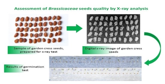
3.3. Inspection of Lepidium sativum L., Seeds

The manifestation of defects, anomalies, and flaws on the X-ray projection of seeds can be very diverse. Some of them are even species-specific. Here is an example of one of them. Seeds of cress varieties Prestige, Flagman of FSVC selection and “Mechta Derbenta” obtained at the Breeding Station named after N.N. Timofeev were taken for radiographic analysis. Seeds of Prestige and Flagman varieties were multiplied in the FSVC experimental field (Moscow Region), and “Mechta Derbenta” in Derbent district of the Republic of Dagestan.
Visual analysis of X-ray radiographs of cress seeds showed the following. Seeds of Prestige and Flagman varieties propagated in conditions of temperate zone (Moscow region) look mostly light on X-ray projections, indicating their completeness and fullness (Figure 11). Regular longitudinal shadows are associated with the anatomical structure of the seed. In laboratory germination, they showed a high degree of germination.
A scanned sample of cress seeds is shown in Figure 12. Analysis of X-ray images of seeds of variety “Mechta Derbenta” revealed unusual “patterning” of images not revealed on seeds of other crops. In the X-ray image presented, 10 of 50 analyzed seeds were found (Figure 13). As a result of laboratory germination, 12 seeds did not germinate (Figure 14). All seeds with “patterned projections” (# 9, 14, 16, 18, 19, 22, 27, 42, 43, 45) failed to germinate. Two seeds (# 11 and 34) with normal dense projection but relatively small size did not germinate yet. It is noteworthy that such significant internal injuries are not reflected in the external appearance of the seeds (Figure 12), hence, have a hidden nature.
The results of automatic analysis of digital X-ray images of garden cress seeds revealed that the indicator “average brightness” is quite informative with respect to the trait “patterning of internal structure” of seeds. With the increase of “patterning” the average brightness of digital X-ray images decreases (Table 6 and Table S1), and the sowing quality of seeds (Figure 15) deteriorates. The green border shows the automatic classification of the seeds as the best in brightness of the digital X-ray images, the yellow is the intermediate class.
Spearman correlation coefficients (p < 0.05) were, respectively: between average brightness of digital X-ray images and shoot length r = 0.34; standard deviation of brightness and shoot length r = −0.38; average brightness of digital X-ray images and root length r = 0.38; standard deviation of brightness and root length r = −0.40.
So, according to the results of analysis of a large number of X-ray images, X-ray trait directly related to the viability of seeds was established. Regular “patterning” of the internal structure of garden cress seeds on the X-ray projection is evidence of their non-viability. By means of X-ray quality analysis, it is possible to quickly and without loss of the seeds themselves to give a conclusion about the viability of a batch of damaged seeds. According to Table 7, the results of radiographic analysis of seeds almost coincide with the results of their laboratory germination: 74 and 77% of germination, respectively. In this case, the informative and fast performance of the radiographic method is obvious.
It is noteworthy that this X-ray trait has so far only been detected on cress seeds and only on the lot of southern reproduction. This is the result of the work of insect pests. The rapeseed moth (Meligethes aeneus (Fabricius, 1775) pests on the seeds of cultivated cruciferous plants [20,21]. During bud formation, they invade plants and feed on the inner parts of buds and flowers, eating stamens, pistils, petals and pollen. As a result, incomplete seeds develop and seed production decreases.

4. Discussion

The possibilities of radiographic analysis of seeds are not limited to the above examples. With the help of the method, it is possible to determine the degree of seed fullness, injuries, the presence of internal (hidden) germination and other defects and abnormalities of the internal structure.
The experimental data obtained demonstrate the effectiveness of the method of microfocus X-ray radiography for the analysis of the germination of cabbage seeds related to the peculiarities of their internal structure. The radiographic analysis of the internal structure of wheat and barley grain [22], seeds of ornamental apple tree forms [23], tomato [15], pepper [16], eggplant [24] tomato and melon [18] showed high information value of the method and speed of its execution.
Analysis of images of seed material, in particular, evaluation of various shape indices for classification of seed species diversity, is widely enough applied in seed science [25,26,27]. Regarding the application of the X-ray method, it should be noted that the works of other researchers are mainly devoted to the visualization and identification of individual, but quite common types of hidden defects, for example—incomplete seed [28], hidden insect infestation [29] and embryo defects [14].
At the same time, cabbage seeds have specific X-ray traits, such as, “angularity of seeds”, indicating the incompleteness of hoarding tissue. Just as specific is the trait of “regular patterning” of the internal structure of cress seeds, a consequence of damage by insect pests, directly related to their viability. It should be noted that earlier the study of the features of the internal structure of cabbage seeds was of a trial nature [30]. Thus, in the work of Gusakova (1997) [31], Derunov (2004) [30] the sign of “unfulfilled” cabbage seeds, determined by the radiographic method, indicating the non-viability of seeds, was shown. From the few modern works we can cite the example of the study of Abud et al. (2018) [17], which shows the influence of features and defects of the internal structure of broccoli seeds on their vigor. We tried to give the work a more systematic character and analyzed the defectiveness of cabbage seeds not only visually, but also using the means of computer morphometry. Defects and deficiencies in the internal structure of cabbage seeds were identified and classified by us, the degree of their impact on seed viability was shown [10]. This approach allows us to objectify the express-analysis of cabbage seeds quality when they are fully preserved, to extract information about defects and disadvantages of internal structure, which significantly complements the traditional methods used in seed control.

5. Conclusions

The method of seed radiography makes it possible to identify and record defects and abnormalities of the internal structure of the seeds of vegetable cruciferous crops according to a number of economically significant features.
The detected defects and abnormalities of seed development are well correlated with their viability, which increases the practicality of the method. The high informative value of the method makes it possible not only to establish the degree of viability of the analyzed seeds, but also to identify the causes of its reduction.
Automatic analysis of digital X-ray images of seeds is an effective tool in the objectification of such traits of their hidden defects as irregular darkening, angularity and patterning of the internal structure.
The advantage of the method is its rapid application, integrity and safety of the studied material, which is especially important when working with small lots of seeds of collection and breeding material. By recording and archiving the results of analysis, it is possible to trace the change in the quality of seeds over the entire period of their storage.

Supplementary Materials

The following supporting information can be downloaded at: https://www.mdpi.com/article/10.3390/horticulturae8010029/s1, Table S1. Results of automatic analysis of digital X-ray images of Lepidium sativum L. seeds Mechta Derbenta variety.

Author Contributions

Conceptualization and methodology F.M. and N.P. (Nikolay Priyatkin); software, N.P. (Nikolay Priyatkin); validation F.M. and N.P. (Nikolay Potrakhov); formal analysis, F.M and N.P. (Nikolay Priyatkin); investigation, F.M.; resources, N.P. (Nikolay Potrakhov); data curation, F.M.; writing—original draft preparation, F.M. and S.B.; writing—review and editing, S.B.; visualization, F.M., N.P. (Nikolay Priyatkin) and N.P. (Nikolay Potrakhov); supervision, project administration and funding acquisition, Y.C. All authors have read and agreed to the published version of the manuscript.

Funding

The study was financially supported by the Ministry of Education and Science of the Russian Federation (agreement no. 075-15-2020-805 from 2 October 2020).

Informed Consent Statement

Not applicable.

Data Availability Statement

Not applicable.

Acknowledgments

We express our gratitude to V.O. Kosov for help in X-ray imaging of seed samples and to L.P. Velikanov for help in visual analysis of latent seed defects.

Conflicts of Interest

The authors declare no conflict of interest.

References

  1. Ludilov, V.A.; Alekseev, Y.B.; Timina, O.O. Practical Seed Production of Vegetable Crops with the Basics of Seed Science; Ludilov, V.A., Alekseev, Y.B., Eds.; Scientific Publishing House KMMC: Moscow, Russia, 2011; p. 220. (In Russian) [Google Scholar]
  2. Bukharov, A.F. Heterogeneity of seeds: Theory and practice (review). Veg. Russ. 2020, 2, 23–31. (In Russian) [Google Scholar]
  3. Probert, R.J.; Hay, F. Keeping Seeds Alive/Seed Technology and Its Biological Basis; Black, M., Bewley, D., Eds.; Sheffield Academic Press: Sheffield, UK, 2000; pp. 184–225. [Google Scholar]
  4. Litvinov, S.S.; Razin, A.F.; Mescheryakova, R.A.; Lebedeva, N.N.; Razin, A.A. Safety of vegetable products. Potatoes Veg. 2017, 4, 22–23. (In Russian) [Google Scholar]
  5. Monakhos, S.G.; Monakhos, G.F. Selection of white cabbage (Brassica oleracea L.) for resistance to keel. Rep. Timiryazev Agric. Acad. 2016, 288, 473–477. (In Russian) [Google Scholar]
  6. ISO6639/4 -87. Cereals and legumes. In Determination of Hidden Insect Infestations. Part. 4. Accelerated Methods; Standards Publishers: Moskow, Russia, 1987. (In Russian) [Google Scholar]
  7. GOST 28666.4-90. Cereals and legumes. In Determination of Latent Insect Infestation. Part. 4. Accelerated Methods; Standards Publishers: Moskow, Russia, 1990. (In Russian) [Google Scholar]
  8. GOST R 59603-2021. Agricultural Seeds. Methods of Digital X-ray; Standartinform: Moscow, Russia, 2021; p. 16. (In Russian) [Google Scholar]
  9. Musaev, F.B.; Antoshkina, M.S.; Arkhipov, M.V.; Velikanov, L.P.; Gusakova, L.P.; Bessonov, V.B.; Gryaznov, A.Y.; Zhamova, K.K.; Kosov, V.O.; Potrakhov, E.N.; et al. X-ray Quality Analysis of Seeds of Vegetable Crops/Methodological Guidelines; Publishing house of St. Petersburg State Electrotechnical University “LETI”: Moscow, Russia, 2015; p. 42. (In Russian) [Google Scholar]
  10. Musaev, F.B.; Potrakhov, N.N.; Beletskiy, S.L. BriefAtlas of Radiographic Traits of Seeds of Vegetable Crops; Federal Scientific Center for Vegetable Growing: Moscow, Russia, 2017; p. 40. (In Russian) [Google Scholar]
  11. Musaev, F.B.; Soldatenko, A.V.; Krivenkov, L.V.; Bukharov, A.F.; Beletskiy, S.L.; Kezimana, P. Assessment of vegetable seeds quality by micro-focus X-ray analysis. Res. Crops 2020, 21, 604–610. [Google Scholar] [CrossRef]
  12. Gomes-Junior, F.G.; Chiquito, A.A.; Marcos-Filho, J. Semi-automated assessment of the embryonic area of cucumber seeds and its relationship to germination and seedling length. J. Seed Sci. 2013, 35, 183–189. [Google Scholar] [CrossRef] [Green Version]
  13. Ahmed, M.R.; Yasmin, J.; Park, E.; Kim, G.; Kim, M.S.; Wakholi, C.; Mo, C.; Cho, B.K. Classification of watermelon seeds using morphological patterns of X-ray imaging: A comparison of conventional machine learning and deep learning. Sensors 2020, 20, 6753. [Google Scholar] [CrossRef] [PubMed]
  14. Van der Burg, W.J.; Aartse, J.W.; van Zwol, R.A.; Bino, R.J. Predicting tomato seedling morphology by X-ray analysis of seeds. J. Amer. Soc. Hort. Sci. 1994, 119, 258–263. [Google Scholar] [CrossRef]
  15. Marcos-Filho, J.; Gomes-Junior, F.G.; Bernett, M.A.; Wells, A.A.; Stieve, S. Using tomato analyzer software to determine embryo size in X-rayed seeds. Rev. Bras. Sementes 2010, 32, 146–153. [Google Scholar] [CrossRef]
  16. Dell’Aquila, A. Pepper seed germination assessed by combined X-radiography and computer-aided imaging analysis. J. Biologia. Plantarum. 2007, 51, 777–781. [Google Scholar] [CrossRef]
  17. Abud, H.F.; Cicero, S.M.; Gomes Junior, F.G. Radiographic images and relationship of the internal morphology and physiological potential of broccoli seeds. Acta Scientiarum. Agron. 2018, 40, e34950. [Google Scholar] [CrossRef]
  18. Bruggink, H.; van Duijn, B. X-Ray based seed image analysis. Seed Test. Int. 2017, 153, 45–50. [Google Scholar]
  19. Arkhipov, M.V.; Alekseeva, D.I.; Batygin, N.F.; Velikanov, L.P.; Gusakova, L.P.; Derunov, I.V.; Zheludkov, A.G.; Nikolenko, V.F.; Nikitina, L.I.; Savin, V.N.; et al. Method of Radiography in Agriculture and Crop Production; Manual; Publishing House of Russian Academy of Agricultural Sciences: Moskov, Russia, 2001; p. 93. (In Russian) [Google Scholar]
  20. Lychkovskaya, I.Y.; Slukin, A.S. Distribution of phytophage insects on oil-bearing cabbage crops under conditions of dry vegetation period. Rep. Russ. Acad. Agric. Sci. 2013, 1, 28–30. (In Russian) [Google Scholar]
  21. Hokkanen, H.M.T. Biological and agrotechnical control of the rape blossom beetle Meligethes aeneus (Coleoptera, Nitidulidae). Actaentomol. Fenn. 1989, 53, 25–29. [Google Scholar]
  22. Arkhipov, M.V.; Priyatkin, N.S.; Gusakova, L.P.; Potrakhov, N.N.; Gryaznov, A.Y.; Bessonov, V.B.; Obodovsky, A.V.; Staroverov, N.E. X-Ray Computer Methods for Studying the Structural Integrity of Seeds and Their Importance in Modern Seed Science. Tech. Phys. 2019, 64, 582–592. [Google Scholar] [CrossRef]
  23. Tkachenko, K.G.; Staroverov, N.E.; Gryaznov, A.Y. Radiographic study of fruit and seed quality. Hortus Bot. 2018, 13, 52–66. (In Russian) [Google Scholar] [CrossRef]
  24. Silva, V.N.; Cicero, S.M.; Bennett, M. Relationship between eggplant seed morphology and germination. Rev. Bras. Sementes 2012, 34, 597–604. [Google Scholar] [CrossRef] [Green Version]
  25. Orru, M.; Grillo, O.; Venora, G.; Bacchetta, G. Computer vision as a method complementary to molecular analysis: Grapevine cultivar seeds case study. Comptes. Rendus. Biol. 2012, 335, 602–615. [Google Scholar] [CrossRef]
  26. Lo Bianco, M.; Grillo, O.; Canadas, E.; Venora, G.; Bacchetta, G. Inter- and intraspecic diversity in Cistus L. (Cistaceae) seeds, analysed with computer vision techniques. Plant Biol. 2017, 19, 183–190. [Google Scholar] [CrossRef] [PubMed]
  27. Loddo, A.; Loddo, M.; Di Ruberto, C. A Novel deep learning based approach for seed image classification and retrieval. Comput. Electron. Agric. 2021, 187, 106269. [Google Scholar] [CrossRef]
  28. Severiano, R.L.; Pinheiro, P.R.; Gomes Junior, F.G.; de Medeiros, A.D.; Pereira, M.D. X-ray test on passion fruit seeds submitted to different aryl removal methods. Comun. Sci. 2018, 9, 356–362. [Google Scholar] [CrossRef] [Green Version]
  29. De Medeiros, A.D.; Zavala-León, M.J.; de Oliveira Araújo, J.; Pereira, M.D.; dos Santos, D.C.F.; Silva, D.L.J. Relationship between internal morphology and physiological quality of Leucaena leucocephala seeds using image analysis. Rev. Árvore 2019, 43, 1–9. [Google Scholar] [CrossRef]
  30. Derunov, I.V. Radiographic Study of Seeds of Various Agricultural Crops and Products of Their Processing: Abstract of Ph; Candidate of Biological Sciences ARI: Saint Petersburg, Russia, 2004; p. 22. (In Russian) [Google Scholar]
  31. Gusakova, L.P. X-Ray and Cytophotometric Analysis of the Viability of Agricultural Crop Seeds: Extended Abstract of Cand; Science Dissertation: Saint-Petersburg, Russia, 1997; p. 20. (In Russian) [Google Scholar]
Figure 1. Preparation of seeds for radiographic study: (a) white cabbage, (b) radish.
Horticulturae 08 00029 g001
Figure 2. Functional scheme of receiving and processing of X-ray images: 1—radiation source (X-ray tube), 2—object of irradiation (seed), 3—receiver (CCD), 4—signal processing unit (special scanner), 5—personal computer.
Horticulturae 08 00029 g002
Figure 3. Radiographic image of Brassica oleracea L., var. capitata seeds.
Horticulturae 08 00029 g003
Figure 4. Radiography of seeds and photograph of white cabbage sprouts with “separation of embryo parts” clearly visible in seed number 1.
Horticulturae 08 00029 g004
Figure 5. Radiography of seeds and photograph of white cabbage seedlings “soft separation of embryo elements”.
Horticulturae 08 00029 g005
Figure 6. Radiography of seeds and photograph of white cabbage sprouts with “patterning of internal structure” (seed number 38 and 40 only).
Horticulturae 08 00029 g006
Figure 7. Radiography of seeds and photograph of Brassica oleracea L., var. capitata seedlings with “irregular shading” trait.
Horticulturae 08 00029 g007
Figure 8. Radiography of seeds and photograph of Brassica oleracea L., var. capitata seedlings with angular seed shape.
Horticulturae 08 00029 g008
Figure 9. Radiography of infected seeds and photograph of Brassica oleracea L., var. capitata seedlings.
Horticulturae 08 00029 g009
Figure 10. Reproductive dysfunction of Raphanus sativus L., var. radicula plants due to defects in internal seed structure: (a) population seeds (I0) and their complete crop; (b) seeds of inbred line (I3) and unformed product swelling.
Horticulturae 08 00029 g010
Figure 11. Fragment of X-ray image of Lepidium sativum L. seeds of Prestige variety.
Horticulturae 08 00029 g011
Figure 12. Photo of Lepidium sativum L. seeds of Mechta Derbenta variety.
Horticulturae 08 00029 g012
Figure 13. X-ray image of Lepidium sativum L. seeds of Mechta Derbenta variety.
Horticulturae 08 00029 g013
Figure 14. Photo of Lepidium sativum L. seedlings of Mechta Derbenta variety.
Horticulturae 08 00029 g014
Figure 15. Results of digital morphometry of Lepidium sativum L. seedlings Mechta Derbenta variety, preliminary analyzed by X-ray technique.
Horticulturae 08 00029 g015
Table 1. Seed collection for radiographic analysis.
SampleVarietyMaintainerProduction Area
Brassica oleracea L., var. capitataAmager 611FSVCDerbent, Russia
Brassica oleracea L., var. capitataBelorusskaya 455FSVCMoscow Region, Russia
Brassica oleracea L., var. capitataMoskovskaya pozdnyaya 15FSVCMoscow Region, Russia
Brassica oleracea L., var. capitataPodarok 2500FSVCDerbent, Russia
Brassica oleracea L., var. capitataF1 MalakhitRSAAItaly
Brassica oleracea L., var. capitataF1 TransferRSAAItaly
Brassica oleracea L., var. capitataF1 ValentinaRSAAItaly
Brassica oleracea L., var. capitataF1 ValentinaRSAAAustralia
Raphanus sativus L., var. radiculaAriyaFSVCMoscow Region, Russia
Raphanus sativus L., var. radiculaPink-red with a white tipFSVCMoscow Region, Russia
Raphanus sativus L., var. radiculaNiger, population I3FSVCMoscow Region, Russia
Raphanus sativus L., var. radiculaInbred line I4FSVCMoscow Region, Russia
Lepidium sativum L.,PrestigeFSVCMoscow Region, Russia
Lepidium sativum L.,FlagmanFSVCMoscow Region, Russia
Lepidium sativum L.,Mechta DerbentaRSAADerbent, Russia
Table 2. Comparison of the results of radiographic analysis and laboratory germination of Brassica oleracea L., var. capitata seeds.
ParametersVarieties
AmagerBelorusskayaMoskovskaya PozdnyayaPodarok
Trait according to the results of radiographic analysis, %
Normal45414256
Empty00140
Germ partitioning of germ parts with soft partitioning11889
Patterned1716522
Angulated13232210
With irregular shading141293
Germination, %
Germinated54524862
Sprouting16141118
Not germinated30343920
Table 3. Analysis of brightness parameters of digital X-ray images of Brassica oleracea L., var. capitata seeds with the trait “patterning of internal structure”.
ParameterSeed ID
383940
Average Brightness, brightness units162184162
Brightness Deviation, brightness units394245
Table 4. Analysis of brightness parameters of digital X-ray images of Brassica oleracea L., var. capitata seeds with the trait “irregular shading”.
ParameterSeed ID
454647
Average Brightness, brightness units165142193
Brightness Deviation, brightness units423547
Table 5. Analysis of the shape of digital X-ray images of angular Brassica oleracea L., var. capitata seeds.
ParameterSeed ID
141816
Roundness, nondimensional value0.7340.6470.890
Circle factor, nondimensional value0.9270.9070.991
Table 6. Results of automatic analysis of digital X-ray images of Lepidium sativum L. seeds Mechta Derbenta variety.
Parameters of X-ray Pattern of SeedSeed ID
41424344454647484950
Horticulturae 08 00029 i001
Average Brightness, Brightness Units184182175185168184184188187184
Brightness Standard Deviation, Brightness Units25332326272828262630
Table 7. Comparison of methods for analyzing the quality of Lepidium sativum L. seeds.
ReplicationsX-ray Analysis, %Laboratory
Germination, %
NormalInsects DamagedIrregular Shadings
176141080
27220874
36436072
48512382
Mean74.020.55.077.0
Publisher’s Note: MDPI stays neutral with regard to jurisdictional claims in published maps and institutional affiliations.

