Article

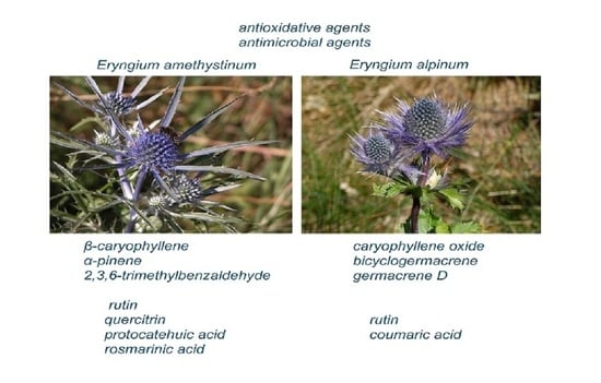
Phytochemical Traits and Biological Activity of Eryngium amethystinum and E. alpinum (Apiaceae)

1
Faculty of Pharmacy and Biochemistry, University of Zagreb, A. Kovačića 1, HR-10000 Zagreb, Croatia
2
Slovenian Institute of Hop Research and Brewing, Cesta Žalskega tabora 2, SI-3310 Žalec, Slovenia
3
Križevci College of Agriculture, M. Demerca 1, HR-48260 Križevci, Croatia
4
Faculty of Science, University of Split, Ruđera Boškovića 33, HR-21000 Split, Croatia
*
Author to whom correspondence should be addressed.
Horticulturae 2021, 7(10), 364; https://doi.org/10.3390/horticulturae7100364
Submission received: 12 August 2021 / Revised: 27 September 2021 / Accepted: 30 September 2021 / Published: 5 October 2021
(This article belongs to the Collection Prospects of Using Wild Plant Species in Horticulture)

Abstract

:
Eryngium amethystinum L. is a wild vegetable used in Croatia. Both E. amethystinum and E. alpinum L. are decorative plants that can also be used in horticulture. To find out the hidden qualities of these two species, the following biological activities were studied: essential oil (EO) composition, analyzed by gas chromatography and gas chromatography with mass spectrometry (GC, GC–MS); phenolic compound content (PC), analyzed by high performance liquid chromatography (HPLC); total phenols as well as total flavonoids, analyzed by ultraviolet–visible spectrophotometry (UV/Vis); antioxidants, analyzed by 2,2-diphenyl-1-picrylhydrazyl radical-scavenging activity (DPPH), β-carotene-linoleic acid assay, chelating activity and reducing power of the extracts; and antimicrobial evaluation by micro-dilution assay and inhibition of Candida albicans blastospore germination. The major constituents of EO were β-caryophyllene (15.2%), α-pinene (10.2%) and 2,3,6-trimethylbenzaldehyde (9.3%) for E. amethystinum and caryophyllene oxide (27.9%), bicyclogermacrene (13.2%) and germacrene D (8.2%) for E. alpinum. The methanol extracts of both species showed a broad spectrum of antibacterial and antifungal activity with minimum inhibitory concentrations (MIC) less than or equal to 1.944 and 1.11 mg/mL, respectively.

Graphical Abstract

1. Introduction

The genus Eryngium L. (family Apiacea) includes 230–250 herbs widespread in America, Eurasia, Africa, Malesia and Australia [1]. Twenty-six species of Eryngium have been recorded in Europe [2] and five in Croatia [3]. Some Eryngium species have been used as remedies, especially in folk medicine, and as horticultural plants. Eryngium creticum Lam. has been used in folk medicine in Palestine as a diuretic and emmenagogue, and for the treatment of kidney stones and infections, skin diseases and tumours [4]; E. foetidum L. for the treatment of respiratory diseases (cold, cough, asthma, sinusitis) and for diarrhoea and rheumatism in traditional medicine of the Democratic Republic of São Tomé and Principe [5]; E. billardieri Delar. for the healing of inflammation in Turkey [6]; E. campestre L. as a diuretic and emmenagogue, in respiratory system and gastric diseases, skin diseases and periodontosis [7]; and E. planum L. for cough [7]. In the folk medicine of Balkan peoples, E. campestre L. is used against hepatitis [8]. In addition, the root, young shoots and leaves of E. amethystinum L., E. campestre and E. maritimum are wild vegetables that are consumed in northern Dalmatia, Croatia [9]. People cook the young shoots and prepare them as asparagus. The young leaves are picked and cooked together with other wild vegetables [9]. Chemical research on the genus Eryngium has detected the presence of essential oils (EO) [5,10,11,12], saponins [6,13] and phenolic compounds [13].
Oxidative stress is a condition included in the pathogenesis of various illnesses such as neurodegenerative diseases, cancer, diabetes mellitus, rheumatoid arthritis, and cardiovascular diseases [14]. Plant life produces many secondary metabolites with antioxidant properties that are considered to be protective against oxidative damage. Among these plant secondary metabolites, phenolic compounds have a particularly prominent position, as they are believed to be responsible for the lower frequency of coronary diseases in communities that consume polyphenol-rich foods [15]. In addition, constant intake of polyphenols has been shown to be associated with a lower frequency of stomach, pancreatic, lung, and possibly breast cancer [16]. In addition to their health-promoting effects, polyphenols also have the ability to protect fatty acids from oxidative decay.
E. amethystinum is the most widely distributed Eryngium species in Croatia, and grows on dry, skeletal, limestone soils. Due to its decorative appearance and resistance to drought, it can also be used as an ornamental species in areas with a dry climate. Considering its use as food and in view of its possible use as an ornamental plant, it is useful to know what this species contains. In order to compare the chemical composition of E. amethystinum with another Eryngium species, we chose the beautiful mountain plant E. alpinum L., which is much less common in Croatia and grows on fresh, moist, mostly calcareous soils in mountainous areas. Thus, the goal of this investigation is to analyze the content of essential oil and phenolic compounds, as well as the antioxidant and antimicrobial activity of both Eryngium amethystinum and E. alpinum.

2. Materials and Methods

2.1. Herbal Material

Plant cuttings from ten randomly selected wild plants from one locality per species of Eryngium amethystinum and E. alpinum (Apiaceae) were collected during their flowering season in June and August of 2019 in Croatia. The GPS coordinates and altitude were 44°32′21″ N, 15°09′51″ E, 970 m a.s.l. (E. amethystinum) and 44°45′56″ N, 14°59′15″ E, 1560 m a.s.l. (E. alpinum). Voucher specimens of E. amethystinum (voucher specimen No. HFK-HR-11-2019) and E. alpinum (voucher specimen No. HFK-HR-34-2019) from plant materials were deposited at the “Fran Kušan” Herbarium, University of Zagreb, Faculty of Pharmacy and Biochemistry, Zagreb, Croatia.
The plant samples were protected from direct sunlight and air-dried at 22 °C at a humidity of 60% for 15 days.

2.2. Microorganisms

Microbial strains: Staphylococcus aureus ATCC (American Type Culture Collections; Rockville, USA) 6538, Escherichia coli ATCC 10535, Candida albicans ATCC 10231, C. albicans MFBF (Collection of the Department of Microbiology Faculty of Pharmacy and Biochemistry University of Zagreb, Croatia) 40630/2, C. parapsilosis MFBF 4800, C. krusei MFBF 429, C. glabrata MFBF 3309 and Microsporum gypseum MFBF S3) from the collection of microorganisms of the University of Zagreb (Faculty of Pharmacy and Biochemistry, Department of Microbiology) were used. Sabouraud 2% (w/v)-glucose agar, Müller–Hinton agar and broth were purchased from Merck (Germany). The RPMI 1640 broth was purchased from the Sigma-Aldrich Chemical Co. (St. Louis, MO, USA).

2.3. Gas Chromatography and Mass Spectrometry (GC and GC–MS)

2.3.1. Sample Extract Preparation

Dried flowering above ground plant parts (100 g) were subjected to hydrodistillation in Clevenger apparatus for 3 h. After that, the obtained EO was immediately dried with anhydrous sodium sulphate.

2.3.2. GC, GC–MS Conditions

The chromatographic conditions during analysis were as follows: carrier gas helium at 1 mL·min−1, temperature of injector of 250 °C, while the flame ionization detector temperature was 300 °C. The temperature programme of the VF-5MS column was: isothermal at 60 °C for 3 min, then up to 246 °C at a rate of 3 °C·min−1 and finally remaining isothermal for 25 min. The injection quantity was set at 1 μL while the split ratio was set up to 1:20. The mass spectrometry conditions were: ion temperature 200 °C; mass scan range: 40–350 mass units; ionisation voltage 70 eV. Analyses were performed in duplicate. Each peak was determined by comparing of their retention indices (for VF-5MS based on C8-C40 n-alkanes) with those of the homemade library, original samples, and by comparison of their mass spectra from the literature [17,18], NIST02 (Gaithersburg, MD, USA), and Wiley 9 mass spectrometry (Wiley, New York, NY, USA) mass spectral database. A home library was constructed from original commercially available chemicals and from the major compounds of several EOs from our previous investigations. The percentage of each component was obtained as averages of peaks area from GC and GC–MS using the normalisation method but without correction factors. Finally, the percentage of each compound was calculated as mean from three both GC and GC–MS analyses.

2.4. HPLC Analysis

2.4.1. Sample Extract Preparation

Ultrasonic extraction was performed at 25 °C for 60 min with 500 mg of powdered plant material and 20 mL of 80% ethanol. The each obtained extract was filtered and the filtrate was diluted to a volume of 25.0 mL using 80% ethanol. After that, extracts were filtered using a 0.45 μm PTFE 25 mm filter (Restek, Bad Homburg, Germany). Finally, 5 μL of each prepared extract was inserted into the HPLC instrument for analysis.

2.4.2. Preparation of Standard Solutions

The solutions of all tested standard compounds were obtained according to Kremer et al. [19]. Briefly, stock solutions of each standard were separately diluted in a solution of methanol and water (1:1, v/v) to obtain a concentration of exactly 1.0 mg/mL. After that, the working solution was diluted with a mixture of methanol and water (1:1, v/v) to obtain a concentration of 0.01 mg/mL. The standard mixture was obtained by the diluting all stock solution separately to a final concentration of 0.01 mg/mL using the same mixture of solvents.

2.4.3. HPLC Conditions

HPLC analysis of investigated samples was done using an HPLC system Agilent 1100 Series (Agilent, Santa Clara, CA, USA) and the procedure described by Kremer et al. [19] and Čeh et al. [20]. The Agilent Zorbax Eclipse XDB-C18 reversed-phase packed column (5 μm,150 mm × 4.6 mm) was utilized for separation. The separation was carried out at a temperature of exactly 30 °C. A gradient elution was applied on the chromatographic system. The gradient elution mobile phase consisted of water with acetic acid (pH value of 2.50; solvent A) and acetonitrile (solvent B). The mobile phase used with the program was as follows: starting with 85% solvent A and 15% solvent B, then from 15% solvent B to 22.5% B over a period of 15 min, followed by 40% solvent B for a duration of 10 min and then staying consistent for another period of 5 min. Initial conditions were adjusted after 5 min. The volume of injection for all investigated plant extracts and standards was 5 μL, while the flow rate was set to 1.0 mL/min. A diode array detector was used for identification of compounds. A wavelength of 280 nm was used for the detection of chrysin, naringenin, vanillic, syringic, gallic and protocatehuic acid. On the other hand, a wavelength of 320 nm was applied for detection of ferulic, p-coumaric, sinapic, rosmarinic and caffeic acid, while a wavelength of 370 nm was used for the detection of quercetin and rutin.
Determination of specific compounds was done by comparison of unknown peaks in the plant extracts with the retention times of the standards. The method of standard addition was used to avoid errors in the interpretation of results. External standards were used for quantification.

2.5. Total Phenol and Total Flavonoid Content

For determination of total flavonoid and polyphenol content, antimicrobial as well as antioxidant activity, extracts of flowers, stems and leaves of both species were prepared. Extract preparation was performed by ultrasonication of 2.5 g of powdered material with 8 mL of 80% ethanol at 30 °C for 30 min. The obtained samples were filtered using Whatman paper No. 542. All plant samples were re-extracted two times. After that, each combined filtrate was evaporated to dryness under diminished pressure and stored in the dark in a fridge at +4 °C until use. The total polyphenol content in the obtained extracts was detected with the use of the Folin–Ciocalteu colorimetric method described by Singleton et al. [21]. The calibration curve of gallic acid was constructed and the results were expressed as gallic acid equivalents (mg/g). On the other hand, the determination of flavonoid content was made using the method of Kumazawa et al. [22]. The obtained results were calculated and expressed as quercetin equivalents in mg/g.

2.6. Antioxidant Capacity

2.6.1. DPPH Radical-Scavenging Activity

The method described by Zovko Končić et al. [23], with some minor modifications, was performed to investigate DPPH radical-scavenging activity. In short, 1 mL of each extract was mixed with 1 mL of 0.16 mM methanolic DPPH solution. The reaction solution was incubated over a period of 30 min at room temperature. After incubation, the degree of absorbance was determined at a wavelength of 517 nm using methanol as a blank.
RSA (radical scavenging activity) was obtained according to the equation RSA = (1 − Asample/Acontrol) × 100. In this equation, Acontrol denotes the absorbance degree of the methanol (which is used as a control), while Asample denotes the absorbance degree of the examined extract. Butylated hydroxyanisole (BHA) was taken as a radical scavenging standard. The radical-scavenging activity of DPPH was expressed as EC50, denoting the concentration at which 50 percent of free radicals were scavenged from DPPH.

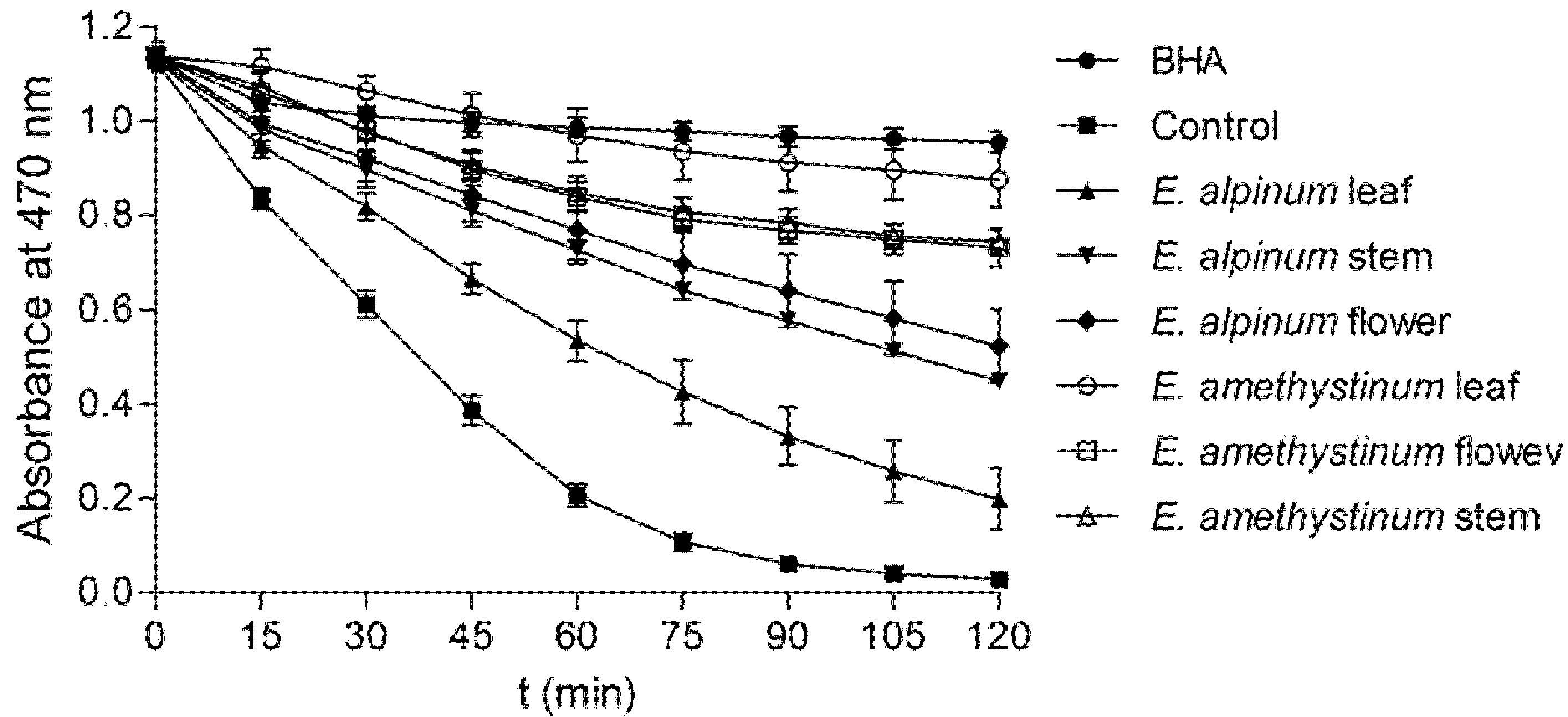
2.6.2. β-Carotene-Linoleic Acid Assay

The β-carotene-linoleic acid test described by Amarowicz et al. [24] was applied to investigate the antioxidant activity of prepared plant extracts. An emulsion of β-carotene-linoleic acid mixture was prepared in a few steps. At first, 0.2 mg of β-carotene was dissolved in 10 mL of chloroform. After that, 1 mL of the solution was added to a mixture made from 200 mg of Tween 40 and 20 mg of linoleic acid. Chloroform as an undesirable component was eliminated using reduced pressure. Then, distilled water saturated with oxygen (50 mL) was added with energetic shaking. The 5 mL of the prepared emulsion was transferred into tubes with 2 mg of extract or 0.5 mg of BHA. An emulsion without added antioxidant was used as a control. After that, each tube was put into a water bath for 2 h at a temperature of 50 °C. During the next 2 h, the absorbance degree of each plant extract and control was measured at a wavelength of 470 nm at intervals of 15 min. The measurement was begun immediately after plant sample preparation (t = 0 min) and finished after 2 h (t = 120 min). The first-order kinetics were used to calculate R (rate of β-carotene bleaching) for the extracts, BHA and water. The antioxidant activity degree or ANT was expressed as a percentage this was calculated according to the equation ANT = (Rcontrol − Rsample/Rcontrol) × 100. In this equation, Rcontrol and Rsample denote the average bleaching rates of control (water) and antioxidant (extract made from herbal material and BHA), respectively.

2.6.3. Fe2+ Chelating Activity (ChA)

The method of Decker and Welch [25] was used to estimate the chelation of iron ions (II). At first, the methanol extract (1.3 mL) was mixed with 2 mM FeCl2 (100 μL). Following this, the reaction was initiated after 5 min by addition of 5 mM ferrozine (200 μL). The obtained solution was left at room temperature for a period of 10 min. Methanol (1.3 mL) was used as a control. The absorbance degree of the mixture and control was estimated at a wavelength of 562 nm. The ChA was calculated using the equation: ChA = (1 − Asample/Acontrol) × 100. In this equation, Asample is the absorbance value of the extract, while Acontrol is the absorbance value of the control. The ChEC50 (the concentration which chelates 50 percent of the Fe2+ ions) was calculated at the end. Quercetin and EDTA (ethylenediaminetetraacetic acid) were used for comparison.

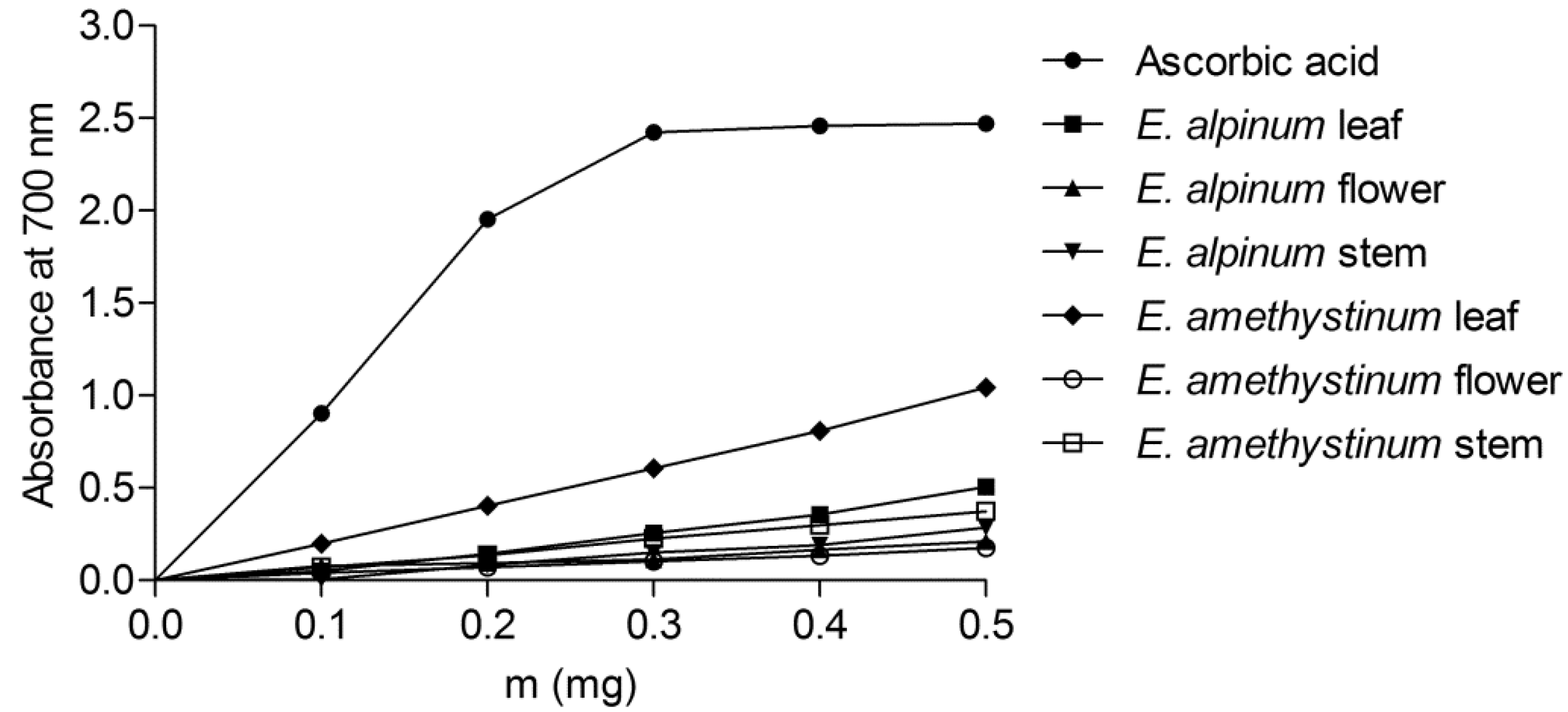
2.6.4. The Reducing Power of the Extracts

The method described by Yen et al. [26], with some minor variations, was used for estimating the reducing power of the extracts. At first, extracts in concentrations from 0.1 to −0.5 mg/mL were dissolved in 0.5 mL of distilled water. Following this, each solution was mixed with 200 mM of sodium phosphate buffer with a pH value of 6.6 (1.25 mL) and 1% (m/v) of potassium ferricyanide (1.25 mL). The prepared mixture was left to stand at 50 °C for a period of 20 min. After incubation, 10% (m/v) trichloroacetic acid (1.25 mL) was added to each sample. The obtained mixture was then centrifuged at 2795× g; only the 1.25 mL of the upper layer was removed. The obtained solution was mixed with 0.1% (m/v) ferric chloride (0.25 mL) and deionized water (1.25 mL). At the end of the procedure, the absorbance value of the mixture solution was measured at a wavelength of 700 nm against water as a blank. For the comparison, ascorbic acid was used.

2.7. Antimicrobial Susceptibility Assay

2.7.1. Micro-Dilution Assay

Prior to analysis, plant extracts were diluted. The dilution was made with 70% (v/v) ethanol and a final concentration of 20 mg/mL was obtained. A series of two-fold diluted extracts ranged from 10 mg/mL to 4.89 μg/mL was used for implementation of the micro-dilution assay. Fresh cultures of microbial strains cultured on tryptic-soy agar at a temperature of 37 °C for a period of 18 h (for bacteria) and of 48 h (for fungi) were used for preparation of inoculums. Yeasts and dermatophyte were cultivated with the addition of 50 mg/L of chloramphenicol on Sabouraud 2% (w/v) glucose agar at a temperature of 37 °C for 48 h. A nephelometer was used to set the density of the inoculums to 0.5 McFarland units. The final concentrations were 1.5 × 10⁸ CFU (colony forming units) per mL for tested bacteria and 5 × 10⁵ CFU/mL for tested fungi. The MIC, that is, the minimal inhibitory concentration, was estimated using the twofold micro-dilution method. The assay was conducted using Müller–Hinton broth for bacteria and RPMI (Roswell Park Memorial Institute) 1640 medium (pH value of 7.0) for yeasts, adhering to the Clinical and Laboratory Standards Institute (formerly known as NCCLS) recommendation M07-A8 and recommendation M-27A [27,28]. The MIC value was determined as the lowest concentration of the extract which allowed microbial growth up to 20 percent. This is observed as a reduced number of microbial colonies after removal of a loop with 10 μL of each rarefaction on a substrate made from tryptic-soy agar or Sabouraud agar, with addition of 2% glucose agar and incubation for 18–48 h at a temperature of 37 °C. The growth of microbes was determined by the use of a microbial culture medium. All analyses were performed in triplicate. The obtained values were calculated as mean ± SD (standard deviation).

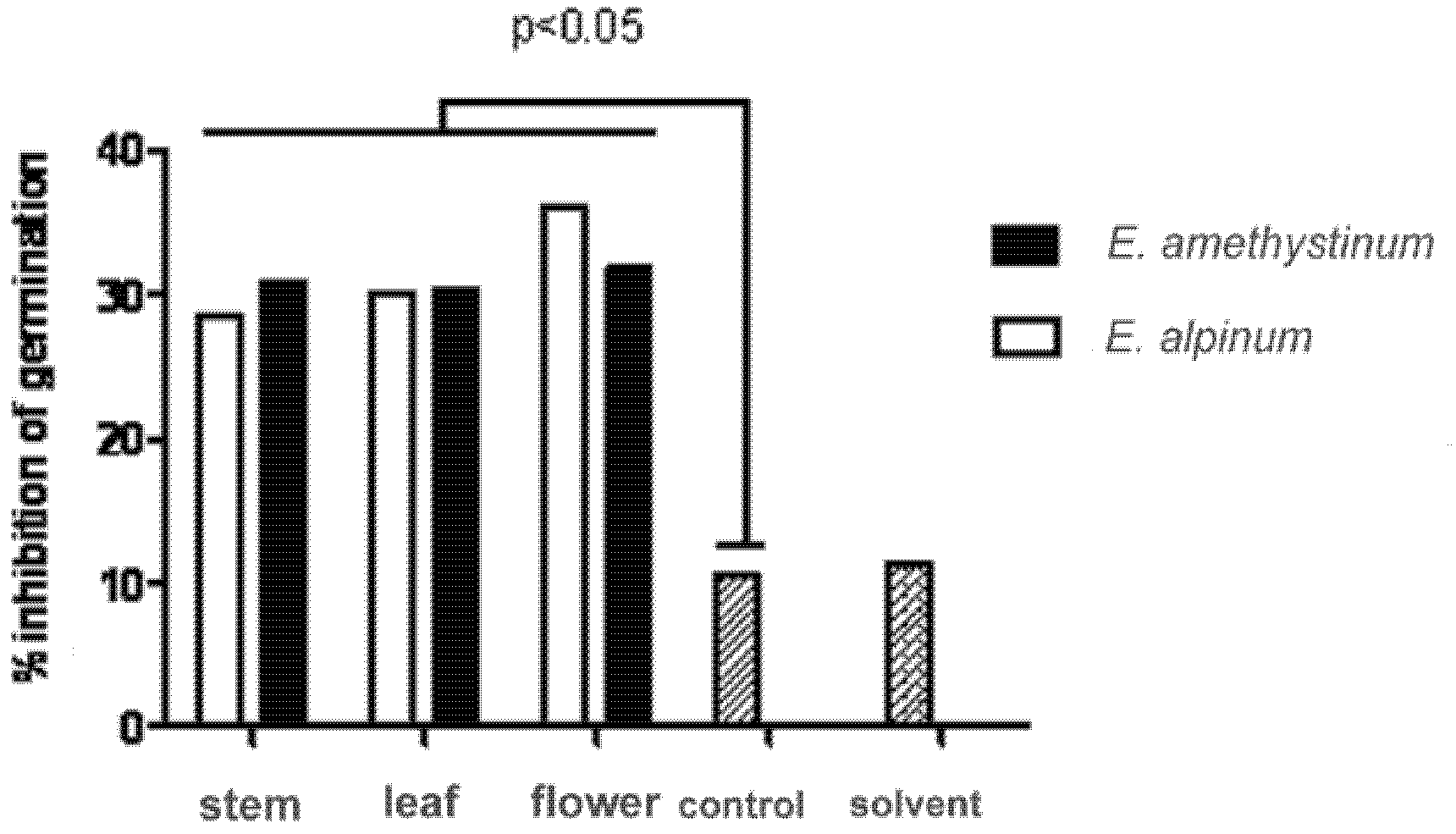
2.7.2. Inhibition of Germination of Candida Albicans Blastospores

Inhibition of creation of germ-tube as a virulence factor of C. albicans was done using the method of Ishida et al. [29]. Briefly, the suspensions of C. albicans blastospores were cultivated in Sabouraud 2% (w/v) glucose broth at a temperature of 37 °C for 24 h. After that, suspensions were centrifuged at 2500× g and washed twice using phosphate buffer (pH 7.4). Approximately 1–5 × 10⁴ of blastospores were treated with 100 mg and 300 mg of both Eryngium L. spp. in fetal bovine serum (FBS; Sigma-Aldrich, USA) for 3 h at 37 °C. Blastospores in FBS without extracts were served as a negative control in this investigation. After the incubation period, the ratios between germinated and non-germinated blastospores were calculated using a haemocytometer under 1000× magnification by phase-contrast microscopy. Sixty corner squares were observed for each test tube. During observation and counting, the tubes were kept at +4 °C.

2.8. Statistical Analysis

Comparison of the content of investigated phenolic constituents in different plant parts, as well as antioxidant activity among plant parts were estimated using ANOVA (one-way analysis of variance) and a Dunnett’s post-hoc test. Student’s t-test was used to compare different extracts while Welch’s t-test was used for comparisons between species. p values < 0.05 were taken as statistically significant. These analyses were carried out using SAS software (JMP V6 procedure) [30]. Statistical comparisons of antimicrobial activity among species were performed using a one-way ANOVA and a Scheffe’s post-hoc test. p values < 0.05 were again taken as significant. Analysis was carried out with help of Statistica 7 software package [31]. Logarithmic transformation was used for data transformation before the analysis.

3. Results and Discussion

3.1. Gas Chromatography and Mass Spectrometry (GC and GC–MS)

Thirty-nine (E. amethystinum) and thirty-six components (E. alpinum) were determined in the EO, accounting for 91.3% and 90.4% of the total oil composition, respectively (see Table 1). The oil of E. amethystinum was mainly composed of sesquiterpenes (54.9%), especially sesquiterpene hydrocarbons (35.2%). Eleven sesquiterpene hydrocarbons were determined in this oil, and among them β-caryophyllene (15.2%) and germacrene D (5.9%) predominated. Dunkić et al. [32] also found that β-caryophyllene (19.7%) was the major compound in E. amethystinum from Croatia [32], while germacrene D (31.3%) was identified as the main compound in E. amethystinum growing in Italy [12,33]. Germacrene D was also the main compound (19.7%) in E. serbicum Pančić from Serbia [11] and one of the main compounds (0.4–53.4%) in Eryngium campestre from Western Algeria [34]. Oxygenated sesquiterpenes (athulenol, ledol and α-bisabolol) were also the dominant compound in E. dichotomum C. Presl, E. ilicifolium Lam. and E. triquetrum Vahl growing in Tunisia [35]; a higher concentration of carbonylic compound 2,3,6-trimethylbenzaldehyde (9.3%) was also determined, which was also identified in high concentrations in the oils of Italian E. amethystinum [12] and in oils of E. foetidum from São Tomé and Principe [5]. However, in the oil of E. triquetrum Vahl [36], the isomers of germacrene D and trimethylbenzaldehyde are completely absent.
In EOs of E. alpinum, the major fraction consisted of sesquiterpenes (70.3%). Among these, oxygenated sesquiterpenes (41.4%) prevailed on sesquiterpene hydrocarbons (28.9%). The main components were caryophyllene oxide (27.9%) and bicyclogermacrene (13.2%). Caryophyllene oxide was also found in E. alpinum from Croatia (21.6%) [32] and in E. palmatum Pančić et Vis. (16.0%) from Serbia [11]. On the other hand, bicyclogermacrene (12.5%) was the main compound in E. rosulatum P. W. Michael from Australia [10]. Kikowska et al. [37] found that the main compounds in leaves of ground (intact) plants of E. alpinum growing in a botanical garden in Poznań (Poland) were elemenone (10.3%), selina-4(15),7(11)-diene (7.1%), selina-3,7(11)-diene (6.7%) germacrone (5.8%) and 1,8-cineole (5.3).

3.2. HPLC Analysis

The concentrations of the phenolic compounds are shown in Table 2. Quercitrin, protocatehuic and rosmarinic acid were identified in E. amethystinum, while coumaric acid was identified in E. alpinum. Only rutin was identified in both species. In both the shoots of the ground plant of E. alpinum and shoots cultured in vitro (developed from axillary buds and regenerated from callus tissue) Kikowska et al. [38] found chlorogenic, isochlorogenic, rosmarinic and 3,4-dihydroxyphenylacetic acid. On the other hand, caftaric, neochlorogenic and caffeic acid were found only in shoots cultured in vitro. Moreover, isoquercitin was identified only in shoots of ground plants, while quercitrin was found only in shoots cultured in vitro [38].
Rutin was also identified as one of the flavonoids in E. campestre and E. octophyllum Korovin, while quercitrin was found in E. campestre [39,40] and E. creticum [41]. In addition, isoquercitrin was found in E. campestre, E. maritimum L. and E. giganteum M. Bieb. [39,42,43]. According to Vukic et al. [44], the most abundant phenolic compounds in E. serbicum were chlorogenic and rosmarinic acid. Le Claire et al. [45] identified rosmarinic acid in E. alpinum, E. amethystinum, E. maritimum and E. campestre. However, rosmarinic acid was not found in our analysed sample of E. amethystinum. This difference could be ascribed to some ecological conditions and genetic variations within the species.

3.3. Total Phenol and Total Flavonoid Content

The results indicated that the quantity of total polyphenols in E. alpinum was almost twice as high as the content of phenols in E. amethystinum (Table 3). On the other hand, the extracts from the leaves and stems of E. amethystinum contained significantly greater amounts of total flavonoids than corresponding extracts of E. alpinum. The total phenolic contents in aqueous, ethyl acetate and n-hexan extracts of E. caucasicum Trautv were 214.18, 140.57, and 29.06 mg of gallic acid equivalent per gram of investigated extract, respectively. On the other hand, the total flavonoid contents in E. caucasicum were 75.36, 31.51, and 97.37 mg of quercetin equivalent per gram of extract powder, respectively [46]. According to Daneshzadeh et al. [47] the total phenolic content in E. billardieri F. Delaroche ranged from 10.71 to 33.38 mg gallic acid equivalent per gram of dry extract, while the total flavonoids ranged between 15.04 and 27.13 mg quercetin equivalent per gram of dry extract. Finally, the content of total phenols in the methanolic extract of E. pyramidale Boiss. & Hausskn. was 0.36 ± 0.01 mg of gallic acid equivalent per gram of dry plant material, while the content of total flavonoid in the methanolic extract was 0.90 ± 0.05 mg of quercetin equivalent per gram of dry plant material [48].

3.4. Antioxidant Capacity

3.4.1. DPPH Radical-Scavenging Activity

The radical-scavenging activity of the extracts varied between 30.73 μg/mL (E. amethystinum leaf) and 1510.17 μg/mL (E. alpinum flower; Table 3). In addition, the activity of the E. amethystinum leaf was statistically equal to the activity of BHA (butylated hydroxyanisol). In general, the extracts of E. amethystinum were more potent radical scavengers than the extracts of E. alpinum. For comparison, the radical-scavenging activity (DPPH IC50) of methanol, acetone, ethyl acetate, and butanol extracts of E. maritimum root were 0.2350, 0.0818, 0.1200, and 0.0104 mg/mL [49]. According to Meot-Duros et al. [50], the ABTS IC50 for E. maritimum was 0.28 mg/mL. Additionally, the concentration of the investigated extracts that reduced 50 percent of DPPH radicals was 0.6 ± 0.0 mg/mL for the methanol extract prepared from the above-ground parts of E. palmatum Pančić and Vis. and 0.7 ± 0.0 mg/mL for the methanol extract obtained from the root [51].

3.4.2. β-Carotene-Linoleic Acid Assay

The extracts of E. amethystinum were found to be more efficient antioxidants than the extracts of E. alpinum in this test (Table 3; Figure 1). The extract which showed the most antioxidant activity in this assay was extract made from E. amethystinum leaves. Its activity was statistically the same as the activity of synthetic antioxidant BHA.

3.4.3. Chelating Activity

The extract with the highest activity in this assay was extract made from E. alpinum flower (Table 3). However, the chelating properties of all the extracts were somewhat lower than the activity of quercetin and EDTA. It was also noticed that the chelating effect of the extracts did not differ between species.

3.4.4. The Reducing Power of the Extracts

All the investigated extracts showed some degree of reducing activity (Figure 2). The reducing power of the extracts was increased linearly with higher concentration (r2 ≥ 0.98). On the other hand, the absorbance of the reaction solution with ascorbic acid remains relatively constant at higher concentrations, probably due to the restrictions of the Beer–Lambert law. For comparison, slopes of the trend lines were obtained for the lowest three concentrations where r2 > 0.97 for all investigated samples (Table 3). It was also noticed that the activity of all investigated extracts was somewhat lower in comparison with ascorbic acid activity. However, the extracts also differed in their activity; the most active were the leaf extracts of the two species.
These studies have shown that the extracts prepared from both Eryngium species have significant antioxidant activity, especially the leaves of E. amethystinum. The antioxidant activity of the leaves of E. amethystinum studied via DPPH radical-scavenging activity and the β-carotene-linoleic acid assay was statistically equal to the activity of synthetic antioxidant BHA. This significant antioxidant activity can be attributed primarily to the phenol content of the species studied. Phenolics contain one or more aromatic rings with added hydroxyl groups in their structure and their antioxidant activity is directly related to the phenolic rings and hydroxyl groups [52]. The relationshiop between phenol content and antioxidant activity has been studied by many authors and their results showed a statistically significant relationship between total phenol content and antioxidant activity [53,54,55,56]. Moreover, some authors suggested that phenolic compound content could be used as an indicator of antioxidant properties [54]. Additionally, the antioxidant activity of EO cannot be ignored either. For example, the studies conducted by Guo et al. [55] have shown a synergistic activity between γ-terpinene and polyphenols.

3.5. Antimicrobial Activity

Screening of antimicrobial activity showed that extracts exhibited antimicrobial activity with MIC values below or equal 1.94 ± 0.48 mg/mL (Table 4). For comparison, Thiem et al. [57] found that ethanolic extracts (70%) from Eryngium planum, E. campestre and E. maritimum showed antibacterial activity against Staphylococcus aureus with MIC values ranging from 400 (E. planum leaves) to 1900 (E. campestre leaves) mg/mL. Hołderna-Kędzia and Kędzia [58] reported that ethanolic extract (50%) from the roots of E. maritimum showed antibacterial activity against S. aureus (MIC = 2500 mg/mL). Chloroformic fractions of the methanolic extract of E. maritimum leaf inhibited the growth of several microorganisms, mainly S. aureus (MIC = 10 mg/mL) [49]. Moreover, aqueous and chloroformic fractions possess a strong antimicrobial activity against tested Pseudomonas aeruginosa and P. fluorescens (MIC = 1 and 2 mg/L, respectively) [49]. Ethanolic extract obtained from above-ground parts of E. caeruleum M. Bieb. and E. thyrsoideum Boiss. showed antimicrobial activity against tested Staphylococcus epidermidis with an MIC value of 3.125 mg/mL [59].
Fungal strains tested for antimicrobial activity were most sensitive to both the extracts with an MIC below 0.39 ± 0.20 mg/mL. Due to the lower MIC values against tested C. albicans ATCC 10231, the antifungal testing was expanded to clinical isolates of Candida spp.
The results of the determination of MIC values in Table 5 show differences in fungal susceptibility to E. amethystinum and E. alpinum. C. albicans, C. glabrata, C. krusei were the most sensitive to the extracts of both species. MIC values ranged from 0.12 to 1.11 mg/mL. Below MIC values of 0.37 ± 0.05 mg/mL, the inhibition of germination of clinical isolates of C. albicans was determined for both extracts at a concentration of 0.30 mg/mL (Figure 3), but not at a concentration of 0.100 mg/mL (data not shown). This is the first data on antifungal activity and inhibition of germ-tube formation of blastospores of C. albicans by extracts of E. amethystinum and E. alpinum. According to Thiem et al. [57], the ethanolic extracts (70%) of Eryngium planum, E. maritimum and E. campestre exhibited antifungal activity against Candida albicans with MIC values from 90 (E. planum leaves) to 7500 (E. campestre leaves) mg/mL. In addition, antifungal activity against C. glabrata ranged from 40 (E. planum leaves) to 700 (E. maritimum leaves) mg/mL.
Both studied Eryngium species showed antimicrobial activity, which can be attributed to the content of phenols and EOs. The antimicrobial activity of polyphenols is explained by changes in the permeability of the microbial cell membrane, changes in cell wall rigidity caused by the interaction of phenols and cell membranes, and some changes in intracellular activity due hydrogen binding of phenols to enzymes [60]. In this study, the antimicrobial activity of Eryngium species can be attributed to the effect of phenolic compounds present (Table 2). Rutin, quercitrin and rosmarinic acid have a proven activity against S. aureus, E. coli as well as against some other microbes [61,62]. The effect of EO on microbes is manifested in several ways. Damage to the cell membrane and degradation of the cell wall, structural changes in the membrane protein, condensation of the cytoplasm, leakage of cytolymph, and alteration of nuclear activity [63,64,65]. Among the compounds detected in EOs in higher quantities, α- and β-pinene have proven activity against S. aureus, E. coli, and C. albicans [66], α-bisabolol against S. aureus and various other microbes [67,68], β-caryophyllene against S. aureus [69], and caryophyllene oxide against S. aureus and C. albicans [70].
In conclusion, the results on the antimicrobial activity of essential oils of Eryngium campestre, E. thorifolium, and E. creticum [71] against MRSA provide direction for future studies of antimicrobial activity of E. amethystinum and E. alpinum.

4. Conclusions

E. amethystinum is a wild vegetable consumed in Croatia. Considering its use as food, the chemical composition of E. amethystinum was studied and compared with E. alpinum. The EOs of E. amethystinum and E. alpinum show that the oil of both species was characterized by a higher content of sesquiterpenes. Several in vitro assays showed that both species are significant source of polyphenols and other antioxidant substances with chelating and radical-scavenging properties. Screening of antibacterial and antifungal activity revealed that both E. amethystinum and E. alpinum exhibited antimicrobial activity against tested microbes. The present results indicate that E. alpinum and E. amethystinum could be useful as antioxidant and antimicrobial agents.

Author Contributions

Conceptualization, D.K. and I.K.; methodology, V.D., I.J.K., M.Z.K., A.Č. and I.K.; validation, V.D., I.J.K., M.Z.K. and I.K.; formal analysis, V.D., E.V., T.P., M.Z.K., A.Č. and I.K.; investigation, V.D., E.V., I.J.K., T.P., M.Z.K., A.Č. and I.K.; resources, V.D., I.J.K. and M.Z.K.; data curation, V.D., I.J.K., M.Z.K. and I.K.; writing—original draft preparation, D.K., M.Z.K., V.D. and I.K.; writing—D.K., M.Z.K., I.K. and V.D.; visualization, D.K.; supervision, V.D., E.V. and S.S.; project administration, S.S.; funding acquisition, V.D. and E.V. All authors have read and agreed to the published version of the manuscript.

Funding

This research received no external funding.

Conflicts of Interest

The authors declare no conflict of interest.

References

  1. Wielgorskaya, T. Dictionary of Generic Names of Seed Plants; Columbia University Press: New York, NY, USA, 1995; p. 254. [Google Scholar]
  2. Chater, A.O. Eryngium L. In Flora Europaea; Tutin, T.G., Heywood, V.H., Burges, N.A., Moore, D.M., Valentine, D.H., Walters, S.M., Webb, D.A., Eds.; Cambridge University Press: Cambridge, UK, 1978; Volume 2, pp. 320–324. [Google Scholar]
  3. Domac, R. Croatian Flora; Školska knjiga: Zagreb, Croatia, 1994; pp. 231–232. [Google Scholar]
  4. Abu-Rabia, A. Herbs as a food and medicine source in Palestine. Asian Pac. J. Cancer Prev. 2005, 6, 404–407. [Google Scholar] [PubMed]
  5. Martinis, A.P.; Salgueiro, L.G.; de Proenca Cunha, A.; Vila, R.; Casigueral, F.; Casanova, J. Essential oil composition of Eryngium foetidum from S. Tome e Principe. J. Essent. Oil Res. 2003, 15, 93–95. [Google Scholar] [CrossRef]
  6. Yesilada, E.; Tanaka, S.; Tabata, M.; Sezik, E. The antiinflammatory activity of the fractions from Eryngium billardieri in mice. Phytother. Res. 1989, 3, 38–40. [Google Scholar] [CrossRef]
  7. Pahlow, M. Great Book of Medicinal Plants; Cankarjeva založba: Ljubljana, Slovenia; Zagreb, Croatia, 1989; pp. 153–154. [Google Scholar]
  8. Redžić, S.S. The ecological aspect of ethnobotany and ethnopharmacology of population in Bosnia and Herzegovina. Coll. Antropol. 2007, 31, 869–890. [Google Scholar]
  9. Grlić, L.J. Encyclopaedia of Wild Edible Plants; August Cesarec: Zagreb, Croatia, 1986; pp. 245–246. [Google Scholar]
  10. Palá-Paúl, J.; Copeland, L.M.; Brophy, J.J.; Goldsack, R.J. Essential oil of Eryngium rosulatum P. W. Michael: A new undescribed species from eastern Australia. Biochem. Syst. Ecol. 2006, 34, 796–801. [Google Scholar] [CrossRef]
  11. Capetanos, C.; Saroglou, V.; Marin, P.D.; Simic, A.; Skaltsa, H.D. Essential oil analysis of two endemic Eryngium species from Serbia. J. Serb. Chem. Soc. 2007, 72, 961–965. [Google Scholar] [CrossRef]
  12. Flamini, G.; Tebano, M.; Cioni, L. Composition of the essential oils from leafy parts of the shoots, flowers and fruits of Eryngium amethystinum from Amiata Mount (Tuscany, Italy). Food Chem. 2008, 107, 671–674. [Google Scholar] [CrossRef]
  13. Zhang, Z.; Li, S.; Ownby, S.; Wang, P.; Yuan, W.; Zhang, W.; Beasley, R.S. Phenolic compounds and rare polyhydroxylated triterpenoid saponins from Eryngium yuccifolium. Phytochemistry 2008, 69, 2070–2080. [Google Scholar] [CrossRef] [PubMed]
  14. Valko, M.; Leibfritz, D.; Moncol, J.; Cronin, M.T.D.; Mazur, M.; Telser, J. Free radicals and antioxidants in normal physiological functions and human disease. Int. J. Biochem. Cell Biol. 2007, 39, 44–84. [Google Scholar] [CrossRef] [PubMed]
  15. Leopoldini, M.; Russo, N.; Toscano, M. The molecular basis of working mechanism of natural polyphenolic antioxidants. Food Chem. 2011, 125, 288–306. [Google Scholar] [CrossRef]
  16. Damianaki, A.; Bakogeorgou, E.; Kampa, M.; Notas, G.; Hatzoglou, A.; Panagiotou, S.; Gemetzi, C.; Kouroumalis, E.; Martin, P.M.; Castanas, E. Potent inhibitory action of red wine polyphenols on human breast cancer cells. J. Cell. Biochem. 2000, 78, 429–441. [Google Scholar] [CrossRef]
  17. Adams, R.P. Identification of Essential Oil Components by Gas Chromatography/Mass Spectroscopy, 4th ed.; Allured Publishing Corp.: Carol Stream, IL, USA, 2007. [Google Scholar]
  18. NIST WebBook. Available online: https://webbook.nist.gov/ (accessed on 14 August 2021).
  19. Kremer, D.; Košir, I.J.; Kosalec, I.; Zovko Končić, M.; Potočnik, T.; Čerenak, A.; Bezić, N.; Srečec, S.; Dunkić, V. Investigation of chemical compounds, antioxidant and antimicrobial properties of Teucrium arduini L. (Lamiaceae). Curr. Drug Targets 2013, 14, 1006–1014. [Google Scholar] [CrossRef]
  20. Čeh, B.; Kač, M.; Košir, I.J.; Abram, V. Relationships between xanthohumol and polyphenol content in hop leaves and hop cones with regard to water supply and cultivar. Int. J. Mol. Sci. 2007, 8, 989–1000. [Google Scholar] [CrossRef] [Green Version]
  21. Singleton, V.L.; Orthofer, R.; Lamuela-Raventos, R.M. Analysis of total phenols and other oxidation substrates and antioxidants by means of Folin–Ciocalteau reagent. Methods Enzymol. 1999, 299, 152–178. [Google Scholar] [CrossRef]
  22. Kumazawa, S.; Hamasaka, T.; Nakayama, T. Antioxidant activity of propolis of various geographic origins. Food Chem. 2004, 84, 329–339. [Google Scholar] [CrossRef]
  23. Zovko Končić, M.; Kremer, D.; Karlović, K.; Kosalec, I. Evaluation of antioxidant activities and phenolic content of Berberis vulgaris L. and Berberis croatica Horvat. Food Chem. Toxicol. 2010, 48, 2176–2180. [Google Scholar] [CrossRef] [PubMed]
  24. Amarowicz, R.; Pegg, R.B.; Rahimi-Moghaddam, P.; Barl, B.; Weil, J.A. Free-radical scavenging capacity and antioxidant activity of selected plant species from the Canadian prairies. Food Chem. 2004, 84, 551–562. [Google Scholar] [CrossRef]
  25. Decker, E.A.; Welch, B. Role of ferritin as lipid oxidation catalyst in muscle food. J. Agric. Food Chem. 1990, 38, 674–677. [Google Scholar] [CrossRef]
  26. Yen, G.-C.; Duh, P.-D.; Chuang, D.-Y. Antioxidant activity of anthraquinones and anthrone. Food Chem. 2000, 70, 437–441. [Google Scholar] [CrossRef]
  27. National Committee for Laboratory Standards (NCCLS). Reference Method for Broth Dilution Antifungal Susceptibility of Yeasts, Approved Standards M27-A; National Committee for Laboratory Standards: Wayne, PA, USA, 1997. [Google Scholar]
  28. National Committee for Laboratory Standards (NCCLS). Reference Method for Dilution Antimicrobial Susceptibility Testing of Bacteria that Growth Aerobically, Approved Standards M07-A8; National Committee for Laboratory Standards: Wayne, PA, USA, 2009. [Google Scholar]
  29. Ishida, K.; de Mello, J.C.; Cortez, D.A.; Filho, B.P.; Ueda-Nakamura, T.; Nakamura, C.V. Influence of tannins from Stryphnodendron adstringens on growth and virulence factors of Candida albicans. J. Antimicrob. Chemother. 2006, 58, 942–949. [Google Scholar] [CrossRef] [PubMed] [Green Version]
  30. JMP; Version 6; SAS Institute Inc.: Cary, NC, USA, 2005–2021.
  31. STATISTICA; Version 7; StatSoft Inc.: Tulsa, OK, USA, 2005.
  32. Dunkić, V.; Vuko, E.; Bezić, N.; Kremer, D.; Ruščić, M. Composition and antiviral activity of the essential oils of Eryngium alpinum and E. amethystinum. Chem. Biodivers. 2013, 10, 1894–1902. [Google Scholar] [CrossRef]
  33. Cianfaglione, K.; Blomme, E.; Quassinti, L.; Bramucci, M.; Lupidi, G.; Dall’Acqua, S.; Maggi, F. Cytotoxic essential oils from Eryngium campestre and Eryngium amethystinum (Apiaceae) growing in central Italy. Chem. Biodivers. 2017, 14. [Google Scholar] [CrossRef] [PubMed]
  34. Medbouhi, A.; Benbelaïd, F.; Djabou, N.; Beaufay, C.; Bendahou, M.; Quetin-Leclercq, J.; Tintaru, A.; Costa, J.; Muselli, A. Essential oil of Algerian Eryngium campestre: Chemical variability and evaluation of biological activities. Molecules 2019, 24, 2575. [Google Scholar] [CrossRef] [PubMed] [Green Version]
  35. Landoulsi, A.; Hennebelle, T.; Bero, J.; Rivière, C.; Sahpaz, S.; Quetin-Leclercq, J.; Neut, C.; Benhamida, J.; Roumy, V. Antimicrobial and light-enhanced antimicrobial activities, cytotoxicity and chemical variability of all Tunisian Eryngium species. Chem. Biodivers. 2020, 14. [Google Scholar] [CrossRef]
  36. Casiglia, S.; Brunoa, M.; Rossellia, S.; Senatore, F. Chemical composition and antimicrobial activity of the essential oil from flowers of Eryngium triquetrum (Apiaceae) collected wild in Sicily. Nat. Prod. Commun. 2016, 11, 1019–1024. [Google Scholar] [CrossRef] [PubMed] [Green Version]
  37. Kikowska, M.; Kalemba, D.; Dlugaszewska, J.; Thiem, B. Chemical composition of essential oils from rare and endangered species—Eryngium maritimum L. and E. alpinum L. Plants 2020, 9, 417. [Google Scholar] [CrossRef] [Green Version]
  38. Kikowska, M.; Thiem, B.; Szopa, A.; Klimek-Szczykutowicz, M.; Rewers, M.; Sliwinska, E.; Ekiert, H. Comparative analysis of phenolic acids and flavonoids in shoot cultures of Eryngium alpinum L.: An endangered and protected species with medicinal value. Plant Cell Essent. Organ Cult. 2019, 139, 167–175. [Google Scholar] [CrossRef] [Green Version]
  39. Ikramov, M.T.; Bandyukova, V.A.; Khalmatov, K.K. Flavonoids of some Eryngium species. Khim. Prir. Soedin. 1971, 7, 117–118. [Google Scholar] [CrossRef]
  40. Kartnig, T.; Wolf, J. Flavonoids from the aerial parts of Eryngium campestre. Planta Med. 1993, 59, 285–286. [Google Scholar] [CrossRef]
  41. Suleiman, A.K. Phytochemistry of Eryngium creticum. Alexandria J. Pharm. Sci. 1994, 8, 73–75. [Google Scholar]
  42. Zarnack, J.; Hildebrandt, B.; Hiller, K.; Otto, A. To the knowledge of the compounds contained in some Saniculoideae. part 33. isolation of flavonol glycosides from Eryngium giganteum M.B. Z. Chem. 1979, 19, 214–215. [Google Scholar] [CrossRef]
  43. Hiller, K.; Pohl, B.; Franke, P. Flavonoid spectrum of Eryngium maritimum L. Part 35. Components of some Saniculoideae. Pharmazie 1981, 36, 451–452. [Google Scholar]
  44. Vukic, M.D.; Vukovic, N.L.; Djelic, G.T.; Obradovic, A.; Kacaniova, M.M.; Markovic, S.; Popović, S.; Baskić, D. Phytochemical analysis, antioxidant, antibacterial and cytotoxic activity of different plant organs of Eryngium serbicum L. Ind. Crops Prod. 2018, 115, 88–97. [Google Scholar] [CrossRef]
  45. Le Claire, E.; Schwaiger, S.; Banaigs, B.; Stuppner, H.; Gafner, F. Distribution of a new rosmarinic acid derivative in Eryngium alpinum L. and other Apiaceae. J. Agric. Food Chem. 2005, 53, 4367–4372. [Google Scholar] [CrossRef]
  46. Nabavi, S.M.; Nabavi, S.F.; Alinezhad, H.; Zare, M.; Azimi, R. Biological activities of flavonoid-rich fraction of Eryngium caucasicum Trautv. Riv. Eur. Sci. Med. Farmacol. 2012, 16 (Suppl. 3), 81–87. [Google Scholar]
  47. Daneshzadeh, S.M.; Abbaspour, H.; Amjad, L.; Nafchi, A.M. An investigation on phytochemical, antioxidant and antibacterial properties of extract from Eryngium billardieri F. Delaroche. J. Food Meas. Charact. 2020, 14, 708–715. [Google Scholar] [CrossRef]
  48. Nejati, M.; Masoudi, S.; Dastan, D.; Masnabadi, N. Phytochemical analysis and antibacterial activity of E. pyramidale Boiss. & Hausskn. J. Chil. Chem. Soc. 2021, 66, 5230–5236. [Google Scholar] [CrossRef]
  49. Kholkhal, W.; Ilias, F.; Bekhechi, C.; Bekkara, F.A. Eryngium maritimum: A rich medicinal plant of polyphenols and flavonoids compounds with antioxidant, antibacterial and antifungal activities. Curr. Res. J. Biol. Sci. 2012, 4, 437–443. [Google Scholar]
  50. Meot-Duros, L.; Le Floch, G.; Magné, C. Radical scavenging, antioxidant and antimicrobial activities of halophytic species. J. Ethnopharmacol. 2008, 116, 258–262. [Google Scholar] [CrossRef] [PubMed] [Green Version]
  51. Marčetić, M.D.; Petrović, S.D.; Milenković, M.T.; Niketić, M.S. Composition, antimicrobial and antioxidant activity of the extracts of Eryngium palmatum Pančić and Vis. (Apiaceae). Cent. Eur. J. Biol. 2014, 9, 149–155. [Google Scholar] [CrossRef]
  52. Minatel, I.O.; Vanz Borges, C.; Ferreira, M.I.; Gomez Gomez, H.A.; Chen, C.-Y.O.; Pace Pereira Lima, G. Phenolic compounds: Functional properties, impact of processing and dioavailability. In Phenolic Compunds—Biological Activit; Soto Hernández, M., Ed.; Intech: London, UK, 2017; pp. 1–24. [Google Scholar] [CrossRef] [Green Version]
  53. Dudonné, S.; Vitrac, X.; Coutière, P.; Woillez, M.; Mérillon, J.-M. Comparative study of antioxidant properties and total phenolic content of 30 plant extracts of industrial interest using DPPH, ABTS, FRAP, SOD, and ORAC assays. J. Agric. Food. Chem. 2009, 57, 1768–1774. [Google Scholar] [CrossRef] [PubMed]
  54. Piluzza, G.; Bullitta, S. Correlations between phenolic content and antioxidant properties in twenty-four plant species of traditional ethnoveterinary use in the Mediterranean area. Pharm. Biol. 2011, 49, 240–247. [Google Scholar] [CrossRef]
  55. Guo, Y.; Baschieri, A.; Amorati, R.; Valgimigli, L. Synergic antioxidant activity of γ-terpinene with phenols and polyphenols enabled by hydroperoxyl radicals. Food Chem. 2021, 345, 128468. [Google Scholar] [CrossRef] [PubMed]
  56. Johari, M.A.; Khong, H.Y. Total phenolic content and antioxidant and antibacterial activities of Pereskia bleo. Adv. Pharmacol. Pharm. Sci. 2019, 428593. [Google Scholar] [CrossRef] [Green Version]
  57. Thiem, B.; Goślińska, O.; Kikowska, M.; Budzianowski, J. Antimicrobial activity of three Eryngium L. species (Apiaceae). Herba Pol. 2010, 56, 52–59. [Google Scholar]
  58. Hołderna-Kędzia, E.; Kędzia, B. Estimation of antibiotic activity of plant extracts. Postępy Fitoter. 2010, 2, 59–70. [Google Scholar]
  59. Karimi, S.; Lotfipour, L.; Asnaashari, S.; Asgharian, P.; Sarvari, Y.; Hazrati, S. Phytochemical analysis and anti-microbial activity of some Important medicinal plants from north-west of Iran. Iran. J. Pharm. Res. 2019, 18, 1871–1883. [Google Scholar] [CrossRef] [PubMed]
  60. Bouarab-Chibane, L.; Valérian Forquet, V.; Lantéri, P.; Clément, Y.; Léonard-Akkari, L.; Oulahal, N.; Degraeve, P.; Bordes, C. Antibacterial properties of polyphenols: Characterization and QSAR (quantitative structure–activity relationship) models. Front. Microbiol. 2019, 10, 829. [Google Scholar] [CrossRef] [PubMed]
  61. Arima, H.; Ashida, H.; Danno, G. Rutin-enhanced antibacterial activities of flavonoids against Bacillus cereus and Salmonella enteritidis. Biosci. Biotechnol. Biochem. 2002, 66, 1009–1014. [Google Scholar] [CrossRef] [Green Version]
  62. Adamczak, A.; Ożarowski, M.; Karpiński, T.M. Antibacterial activity of some flavonoids and organic acids widely distributed in plants. J. Clin. Med. 2019, 9, 109. [Google Scholar] [CrossRef] [Green Version]
  63. Bouhdid, S.; Abrini, J.; Amensour, M.; Zhiri, A.; Espuny, M.J.; Manresa, A. Functional and ultrastructural changes in Pseudomonas aeruginosa and Staphylococcus aureus cells induced by Cinnamomum verum essential oil. J. Appl. Microbiol. 2010, 109, 1139–1149. [Google Scholar] [CrossRef] [PubMed]
  64. Devi, K.P.; Nisha, S.A.; Sakthivel, R.; Pandian, S.K. Eugenol (an essential oil of clove) acts as an antibacterial agent against Salmonella typhi by disrupting the cellular membrane. J. Ethnopharmacol. 2010, 130, 107–115. [Google Scholar] [CrossRef] [PubMed]
  65. Wu, K.; Lin, Y.; Chai, X.; Duan, X.; Zhao, X.; Chun, C. Mechanisms of vapor-phase antibacterial action of essential oil from Cinnamomum camphora var. linaloofera Fujita against Escherichia coli. Food Sci. Nutr. 2019, 7, 2546–2555. [Google Scholar] [CrossRef] [PubMed] [Green Version]
  66. Salehi, B.; Upadhyay, S.; Orhan, I.E.; Jugran, A.K.; Jayaweera, S.L.D.; Dias, D.A.; Sharopov, F.; Taheri, Y.; Martins, N.; Baghalpour, N.; et al. Therapeutic potential of α- and β-Pinene: A miracle gift of nature. Biomolecules 2019, 9, 738. [Google Scholar] [CrossRef] [Green Version]
  67. De Lucca, A.J.; Pauli, A.; Schilcher, H.; Sien, T.; Bhatnagar, D.; Walsh, T.J. Fungicidal and bactericidal properties of bisabolol and dragosantol. J. Essent. Oil Res. 2011, 23, 47–54. [Google Scholar] [CrossRef]
  68. Marcel Forrer, F.; Kulika, E.M.; Filippi, A.; Waltimo, T. The antimicrobial activity of alpha-bisabolol and tea tree oil against Solobacterium moorei, a Gram-positive bacterium associated with halitosis. Arch. Oral Biol. 2013, 58, 10–16. [Google Scholar] [CrossRef]
  69. Dahham, S.S.; Tabana, Y.M.; Iqbal, M.A.; Ahamed, M.B.K.; Ezzat, M.O.; Majid, A.S.A.; Majid, A.M.S.A. The anticancer, antioxidant and antimicrobial properties of the sesquiterpene β-caryophyllene from the essential oil of Aquilaria crassna. Molecules 2015, 20, 11808–11829. [Google Scholar] [CrossRef]
  70. Schmidt, E.; Bail, S.; Friedl, S.M.; Jirovetz, L.; Gerhard Buchbauer, G.; Wanner, J.; Denkova, Z.; Slavchev, A.; Stoyanova, A.; Geissler, M. Antimicrobial activities of single aroma compounds. Nat. Prod. Commun. 2009, 5, 1365–1368. [Google Scholar] [CrossRef] [Green Version]
  71. Celik, A.; Aydınlık, N.; Arslan, I. Phytochemical constituents and inhibitory activity towards methicillin-resistant Staphylococcus aureus strains of Eryngium species (Apiaceae). Chem. Biodivers. 2011, 8, 454–459. [Google Scholar] [CrossRef]
Figure 1. The reduction of absorbance of β-carotene-linoleic acid emulsion caused by Eryngium spp. extracts. Each value represents the mean of three independent measurements with SD error bar.
Horticulturae 07 00364 g001
Figure 2. Reductive capacities of the Eryngium spp. extracts. Each value shown represents the mean of three independent measurements with SD error bar (SD are not visible due to their small size relative to the mean).
Horticulturae 07 00364 g002
Figure 3. The inhibition of germination of Candida albicans due to extracts of Eryngium spp. at 300 μg/mL. Control: FBS (foetal bovine serum) without extracts; solvent: 10% (v/v) 70% ethanol in FBS. N = 3.
Horticulturae 07 00364 g003
Table 1. Composition (expressed in %) of the essential oils of both E. amethystinum and E. alpinum.
ComponentRE. amethystinumE. alpinumIdentificationCAS No.
Monoterpene hydrocarbons 14.512.0
α-Pinene93810.2 ± 0.011.9 ± 0.01RI, MS, Co-GC80-56-8
Camphene9620.8 ± 0.01RI, MS79-92-5
β-Pinene9820.3 ± 0.13.3 ± 0.07RI, MS, Co-GC127-91-3
Myrcene9922.3 ± 0.015.1 ± 0.01RI, MS123-35-3.
Limonene10320.9 ± 0.05RI, MS, Co-GC5989-27-5
(Z)-β-Ocimene10520.6 ± 0.07RI, MS3338-55-4
Terpinolene10890.8 ± 0.070.3 ± 0.02RI, MS586-62-9
Oxygenated monoterpenes 8.94.7
β-Thujone11210.3 ± 0.01-RI, MS1125-12-8
trans-Pinocarveol11470.7 ± 0.01RI, MS547-61-5
Camphor11514.1 ± 0.032.8 ± 0.01RI, MS, Co-GC76-22-2
Borneol11760.1 ± 0.02RI, MS507-70-0
Terpinen-4-ol11840.7 ± 0.010.3 ± 0.01RI, MS562-74-3
β-Thujone11210.3 ± 0.01-RI, MS471-15-8
Myrtenol11970.9 ± 0.010.6 ± 0.01RI, MS515-00-4
Linalyl acetate12520.8 ± 0.020.3 ± 0.02RI, MS115-95-7
Bornyl acetate12850.4 ± 0.07RI, MS76-49-3
α-Terpenyl acetate13490.1 ± 0.020.4 ± 0.03RI, MS80-26-2
Sesquiterpene hydrocarbons 35.228.9
α-Copaene13770.9 ± 0.01RI, MS3856-25-5
β-Bourbonene13831.7 ± 0.010.6 ± 0.07RI, MS5208-59-3
α-Gurjunene14073.6 ± 0.01trRI, MS489-40-7
β-Caryophyllene142415.2 ± 0.012.1 ± 0.01RI, MS, Co-GC87-44-5
β -Copaene14290.8 ± 0.020.5 ± 0.07RI, MS18252-44-3
trans-α-Bergamotene14330.4 ± 0.01RI, MS13474-59-4
(Z)-β-Farnesene14540.3 ± 0.010.2 ± 0.03RI, MS28973-97-9
α-Humulene14561.1 ± 0.01RI, MS6753-98-6
allo-Aromadendrene14650.6 ± 0.01RI, MS25246-27-9
Germacrene D14815.9 ± 0.018.2 ± 0.01RI, MS23986-74-5
β-Bisabolene14941.3 ± 0.01RI, MS495-61-4
Bicyclogermacrene15004.7 ± 0.0113.2 ± 0.01RI, MS24703-35-3
δ-Cadinene15172.8 ± 0.01RI, MS483-76-1
Oxygenated sesquiterpenes 19.741.4
Spathulenol15770.3 ± 0.02RI, MS6750-60-3
Caryophyllene oxide15814.2 ± 0.0127.9 ± 0.01RI, MS, Co-GC1139-30-6
γ-Eudesmol16326.4 ± 0.015.7 ± 0.01RI, MS1209-71-8
α-Cadinol16550.5 ± 0.01RI, MS481-34-5
α-Bisabolol16888.3 ± 0.017.8 ± 0.01RI, MS515-69-5
Phenolic compounds 2.01.7
Thymol12901.2 ± 0.010.9 ± 0.01RI, MS, Co-GC89-83-8
Carvacrol12990.5 ± 0.010.6 ± 0.01RI, MS, Co-GC499-75-2
Eugenol13700.3 ± 0.070.2 ± 0.01RI, MS, Co-GC97-53-0
Carbonylic compounds 10.00.2
3-Octanol acetate11250.4 ± 0.020.2 ± 0.01RI, MS4864-61-3
Butylhexanoate11930.3 ± 0.01RI, MS626-82-4
2,3,6-Trimethylbenzaldehyde13409.3 ± 0.01RI, MS34341-29-2
Hydrocarbons 1.01.5
Eicosane20000.2 ± 0.03RI, MS, Co-GC112-95-8
Docosane22000.5 ± 0.010.3 ± 0.01RI, MS, Co-GC629-97-0
Tricosane23000.2 ± 0.01RI, MS, Co-GC638-67-5
Tetracosane24000.2 ± 0.01RI, MS, Co-GC646-31-1
Pentacosane25000.3 ± 0.01RI, MS, Co-GC629-99-2
Hexacosane26000.1 ± 0.02RI, MS, Co-GC630-01-3
Octacosane28000.3 ± 0.02RI, MS, Co-GC630-02-4
Nonacosane29000.3 ± 0.020.1 ± 0.1RI, MS, Co-GC630-03-5
Total identified (%) 91.390.4
Yield (%) 0.10.1
Note: R = retention indices were defined relative to a series of n-alkanes (C8–C40) on capillary column VF5-MS; RI = identification made using the literature [17]; MS = identification made with help of database NIST02, Wiley 7 and homemade library; Co-GC = identification using reference compounds; – = component is not determined; tr = traces (mean value below 0.1%); SD = standard deviation (N = 3); CAS No. = CAS Registry Number [18].
Table 2. Contents of investigated phenolic compounds (%) determined in methanolic solvent.
CompoundE. amethystinumE. alpinum
Chrysin
Rutin0.002 ± 0.0000.001 ± 0.000
Quercetin
Quercitrin0.026 ± 0.005
Cichoric acid
Coumaric acidtr
Ferulic acid
Protocatehuic acid0.015 ± 0.003
Rosmarinic acid0.005 ± 0.001
Syringic acid
Tannic acid
Extracts of E. amethystinum and E. alpinum. Tr = traces (mean < 0.001%); N = 3.
Table 3. Content of total phenols (TP) and flavonoids (TF), radical scavenging (EC50), and antioxidant activity obtained in β-carotene-linoleate assay (ANT), metal chelating activity (ChEC50) and slope of trendline in a reducing power assay (SRP) of E. amethystinum and E. alpinum extracts. Values are calculated as means ± SD. N = 3.
SpeciesPlant PartTP (mg/g)TF (mg/g)EC50 (μg/mL)ANT (%)ChEC50 (μg/mL)SRP (mg−1)
leaf34.48 ± 0.33 A17.24 ± 1.16 A30.73 ± 0.29 A90.94 ± 1.58 A682.58 ± 4.56 A1.05 ± 0.02 A
E. amethystinumflower11.77 ± 0.45 B9.65 ± 0.52 B169.78 ± 2.86 B84.71 ± 0.19 B484.12 ± 17.44 B0.17 ± 0.01 B
stem23.8 ± 0.71 C16.21 ± 0.97 A100.25 ± 2.24 C85.26 ± 1.42 B684.14 ± 39.95 A0.38 ± 0.00 C
leaf73.42 ± 1.05 D1.78 ± 0.24 C268.47 ± 17.19 D48.81 ± 5.24 C1049.68 ± 21.59 C1.12 ± 0.08 A
E. alpinumflower29.39 ± 2.92 E11.34 ± 0.30 D1510.17 ± 29.74 E75.89 ± 3.09 D331.2 ± 6.88 D0.33 ± 0.04 C
stem33.77 ± 1.93 A13.50 ± 0.27 E241.08 ± 4.66 F71.59 ± 1.45 D951.14 ± 6.20 E0.67 ± 0.06 D
Standard --a 2.83 ± 0.02 Aa 95.39 ± 0.21 Ab 219.16 ± 4.70 Fc 5.49 ± 0.25 Gd 7.59 ± 0.08 E
Note: samples connected by the same capital letter (A–F) are statistically different at p < 0.05. Inside column a = BHA; b = quercetin; c = EDTA; d = ascorbic acid.
Table 4. Antimicrobial activity of methanolic extracts obtained from Eryngium alpinum and E. amethystinum. Presented values denote means ± SD. N = 3.
SpeciesPlantMIC ± SD (mg/mL)
PartS. aureus
ATCC 6538
E. coli
ATCC 10536
C. albicans
ATCC 10231
M. gypseum
MFBF S2
stem0.39 ± 0.021.94 ± 0.480.16 ± 0.120.24 ± 0.07
E. amethystinumleaf0.39 ± 0.0201.53 ± 0.240.06 ± 0.018 *0.16 ± 0.12
flower1.32 ± 0.601.53 ± 0.240.43 ± 0.3340.06 ± 0.02 *
stem0.24 ± 0.071.94 ± 0.480.39 ± 0.200.21 ± 0.14
E. alpinumleaf0.29 ± 0.021.94 ± 0.480.39 ± 0.200.08 ± 0.06 *
flower0.24 ± 0.07 *1.53 ± 0.240.32 ± 0.290.13 ± 0.13
Note: * = significantly lower MIC value than the MIC of the same organ of another Eryngium species tested (p < 0.05).
Table 5. Antimicrobial activity of prepared methanolic extracts of Eryngium amethystinum and E. alpinum.
SpeciesPlantMIC ± SD (mg/mL)
PartC. albicans MFBF 40630/2C. parapsilosis MFBF 4800C. krusei
MFBF 429
C. glabrata MFBF 3309
stem0.37 ± 0.050.37 ± 0.050.37 ± 0.050.37 ± 0.05
E. amethystinumleaf0.37 ± 0.050.37 ± 0.050.37 ± 0.050.37 ± 0.05
flower0.37 ± 0.050.37 ± 0.050.37 ± 0.050.37 ± 0.05
stem0.37 ± 0.051.11 ± 0.050.37 ± 0.050.37 ± 0.05
E. alpinumleaf0.37 ± 0.051.11 ± 0.050.37 ± 0.050.37 ± 0.05
flower0.37 ± 0.051.11 ± 0.050.12 ± 0.010.37 ± 0.05
On clinical isolates of Candida spp. Presented values denote means ± SD. N = 3.
Publisher’s Note: MDPI stays neutral with regard to jurisdictional claims in published maps and institutional affiliations.

