Review

Emerging Roles of Polyamines and Autophagy in Plant In Vitro Regeneration

by
Mahboubeh Davoudi Pahnekolayi
1,*,
Majid Babouyeh Darabi
1 and
Negin Samadi
2
1
Research Group of Phytochemistry and Biochemistry of Natural Compounds, Institute of Chemical, Environmental and Bioscience Engineering, Technische Universität Wien (TU Wien), 1040 Vienna, Austria
2
Department of Horticultural Science and Landscape, Faculty of Agriculture, College of Agriculture and Natural Resources, University of Tehran, Karaj 31587-77871, Iran
*
Author to whom correspondence should be addressed.
Horticulturae 2026, 12(8), 923; https://doi.org/10.3390/horticulturae12080923
Submission received: 26 June 2026 / Revised: 21 July 2026 / Accepted: 24 July 2026 / Published: 27 July 2026
(This article belongs to the Special Issue Plant Tissue Culture: Advances and Perspectives)

Highlights

What are the main findings?
Polyamines and autophagy have emerged as important regulators of plant cell reprogramming and in vitro regeneration alongside classical hormonal and developmental pathways.
Recent studies demonstrate distinct roles for polyamines and autophagy in stress adaptation, cellular homeostasis, somatic embryogenesis, and organogenesis during plant regeneration.
This review summarizes current knowledge on polyamines and autophagy in plant regeneration and discusses common physiological processes that may connect these pathways, highlighting important gaps for future research.
What are the implications of the main findings?
A better understanding of the individual contributions of polyamines and autophagy may facilitate the development of improved regeneration protocols for horticultural and crop species.
Identifying potential links between these regulatory pathways represents a promising direction for enhancing plant propagation, genetic transformation, and other biotechnology applications.

Abstract

Plant regeneration demonstrates the remarkable developmental plasticity of plants, enabling tissue repair, organ formation, and adaptation to environmental challenges through cellular reprogramming and redifferentiation. This capacity underpins numerous horticultural and biotechnological applications, including grafting, micropropagation, somatic embryogenesis, organogenesis, and genetic transformation. While regeneration has traditionally been explained by the coordinated actions of auxin and cytokinin together with key developmental regulators such as WUSCHEL, BABY BOOM, and WUSCHEL-related homeobox genes, recent studies indicate that regeneration is also influenced by stress signaling, metabolic reprogramming, reactive oxygen species, and epigenetic regulation. Among these regulatory components, polyamines have emerged as important modulators of cell division, differentiation, stress responses, and morphogenic competence during in vitro regeneration. Likewise, autophagy, a conserved intracellular recycling pathway, has gained increasing attention for its role in maintaining cellular homeostasis, facilitating metabolic adaptation, and supporting developmental transitions under tissue culture conditions. This review summarizes current knowledge on the independent roles of polyamines and autophagy in plant cell reprogramming and in vitro regeneration, with particular emphasis on wound responses, somatic embryogenesis, and organogenesis. In addition, it highlights common physiological processes through which these pathways may influence regeneration and identifies the limited understanding of their potential relationship as an important direction for future research.

1. Introduction

Plant regeneration is a remarkable manifestation of developmental plasticity that enables plants to repair damaged tissues, restore lost organs, and adapt to changing environmental conditions through cellular reprogramming and redifferentiation. This regenerative capacity underpins numerous agricultural and biotechnological applications, including grafting, micropropagation, somatic embryogenesis (SE), organogenesis, and genetic transformation. Among these approaches, SE and de novo organogenesis are the most widely used in vitro regeneration systems for plant propagation, germplasm conservation, genetic improvement, and genome editing. However, the efficiency of these techniques remains highly genotype-dependent. It is strongly influenced by the physiological status of the explant and the stresses imposed during tissue culture, including wounding, surface sterilization, osmotic stress, and oxidative stress [1,2,3,4,5,6].
The molecular basis of plant regeneration has traditionally been explained by the coordinated actions of plant growth regulators (PGRs), particularly auxin and cytokinin, which establish new meristematic centers by activating developmental regulators such as WUSCHEL (WUS), BABY BOOM (BBM), and members of the WUSCHEL-related homeobox (WOX) gene family. Although these hormonal and genetic networks are central to regeneration, accumulating evidence indicates that successful regeneration depends on a much broader regulatory framework involving stress perception, reactive oxygen species (ROS) signaling, metabolic reprogramming, epigenetic modifications, and cellular quality-control mechanisms [7,8,9]. These findings suggest that regeneration is not governed solely by hormonal balance but results from the integration of developmental and stress-responsive pathways.
Among the emerging regulators of plant regeneration, polyamines (PAs) have attracted increasing attention because of their diverse roles in cell proliferation, differentiation, morphogenesis, and stress adaptation. Putrescine (Put), spermidine (Spd), and spermine (Spm) are ubiquitous polycationic molecules that interact with nucleic acids, proteins, membranes, and phenolic compounds, thereby influencing chromatin organization, gene expression, redox homeostasis, and signaling pathways associated with developmental transitions [1,7,10]. Numerous studies have demonstrated that endogenous and exogenous PAs improve regeneration efficiency by promoting embryogenic competence, shoot organogenesis, and cellular tolerance to tissue culture-induced stress.
Another process receiving increasing attention is autophagy, a highly conserved intracellular degradation and recycling pathway that maintains cellular homeostasis by removing damaged organelles, protein aggregates, and other unnecessary cellular constituents. Through the recycling of nutrients and metabolic intermediates, autophagy supports growth, developmental transitions, and adaptation to environmental stress. During regeneration, extensive cellular remodeling accompanies the transition from differentiated to pluripotent or totipotent cell states, highlighting the importance of efficient intracellular recycling and energy remobilization. Recent studies suggest that autophagy contributes to these processes by facilitating cellular quality control, metabolic adaptation, and stress tolerance during developmental reprogramming [11,12,13].
Despite substantial progress in understanding the hormonal and molecular regulation of plant regeneration, the contributions of PAs and autophagy to cellular reprogramming during in vitro regeneration remain incompletely understood. Although both have been independently implicated in regulating stress responses, cellular homeostasis, metabolic adaptation, and developmental transitions, direct evidence linking PA metabolism to autophagic processes during plant regeneration remains limited. Consequently, this review summarizes current knowledge of the individual roles of PAs and autophagy in plant in vitro regeneration, with particular emphasis on stress responses, cellular reprogramming, SE, and organogenesis. Furthermore, it discusses common physiological processes through which these pathways may influence regeneration and identifies important knowledge gaps and future research directions.

2. Polyamine Biosynthesis and Its Roles in Plant Regeneration

2.1. Definition and Biological Functions of Polyamines

Polyamines (PAs) are low-molecular-weight aliphatic polycations that are ubiquitously distributed in living organisms. In plants, the major naturally occurring PAs are Put, Spd, and Spm, although cadaverine is also present in some species. Their multiple amino groups confer a positive charge at physiological pH, enabling interactions with negatively charged macromolecules such as nucleic acids, proteins, phospholipids, and cell wall components. In plant cells, PAs occur in free, conjugated, and bound forms, allowing them to participate in diverse physiological processes. Through these interactions, they regulate cell division, differentiation, morphogenesis, stress responses, redox homeostasis, and developmental plasticity, highlighting their importance in plant growth and regeneration [7,14].
They act as key signaling and regulatory molecules in plant cells. They influence growth and development pathways and improve tolerance to various biotic and abiotic stresses [7]. Besides PAs, plants encounter different biostimulants such as biopolymers, inorganic compounds, and microbial metabolites. However, PAs stand out because of their specific functions and unique impact on the production of secondary metabolites [15,16]. Intracellular distribution of PAs varies according to their biological functions. Spd and Spm are commonly associated with cell walls, where they bind to pectic polysaccharides and help regulate lignification, cell wall pH, and structural integrity. In contrast, Put is predominantly found in cytoplasmic extracts. The occurrence and abundance of PAs also differ among taxonomic groups. In prokaryotes, Put is generally present at high concentrations, Spd at lower levels, and Spm is often absent. Conversely, eukaryotic organisms typically contain relatively low levels of Put and higher concentrations of Spd and Spm [7]. These compounds help regulate a wide range of processes, including DNA replication, cell division, organ development, and embryonic development. Additionally, PAs help stabilize proteins by preventing denaturation and promoting proper folding. They also adjust stress-responsive pathways, maintain intracellular cation and pH balance, boost defense mechanisms, and control light-dependent growth and developmental responses. Furthermore, they serve as precursors and/or regulators in the production of secondary metabolites [17,18].

2.2. Polymine Biosynthesis in Plants

PA biosynthesis in plants proceeds through a series of enzymatic reactions that generate the diamine Put, which serves as the central precursor for the synthesis of higher PAs. Put can be produced via two distinct metabolic pathways: the arginine decarboxylase (ADC) pathway and the ornithine decarboxylase (ODC) pathway. In the ADC pathway, arginine is first decarboxylated by arginine decarboxylase to form agmatine. Agmatine is subsequently converted into N-carbamoylputrescine, which is then hydrolyzed to produce Put. Alternatively, in the ODC pathway, ornithine is directly decarboxylated by ornithine decarboxylase, yielding Put as the final product. The synthesis of higher PAs, including Spd and Spm, occurs through the sequential addition of aminopropyl groups to Put. Methionine serves as a common precursor for both ethylene and PA biosynthesis. Through a series of metabolic reactions, methionine is converted into S-adenosylmethionine (SAM), which is subsequently decarboxylated by S-adenosylmethionine decarboxylase (SAMDC) to generate decarboxylated SAM (dcSAM). This molecule functions as the principal aminopropyl donor during PA biosynthesis [3,5,8]. Spd is synthesized when spermidine synthase (SPDS) transfers an aminopropyl group from dcSAM to Put. Subsequently, Spm is produced through the transfer of an additional aminopropyl group to Spd, a reaction catalyzed by spermine synthase (SPMS). In addition to these major PAs, plants also contain smaller quantities of thermospermine (tSpm), an isomer of Spm synthesized from Spd through the activity of thermospermine synthase [7]. Several specific inhibitors have been widely employed to investigate PA biosynthesis and function. These include α-difluoromethylornithine (DFMO), an inhibitor of ODC; α-difluoromethylarginine (DFMA), which inhibits ADC; methylglyoxal bis(guanylhydrazone) (MGBG), an inhibitor of SAMDC; and cyclohexylamine, which inhibits SPDS activity. Another biologically important diamine, cadaverine, is synthesized from L-lysine through a decarboxylation reaction catalyzed by lysine decarboxylase [7,16,19].

2.3. Polyamines’ Roles in In Vitro Plant Regeneration

Plant tissue culture has become an indispensable tool for the propagation, conservation, genetic improvement, and genetic transformation of horticultural, medicinal, and economically important plant species. Direct and indirect in vitro regeneration systems, including SE and organogenesis, are widely used for large-scale propagation, the production of elite genotypes, phytochemical enhancement, and crop improvement [20,21,22,23,24,25,26,27,28]. However, the efficiency of these regeneration systems remains highly dependent on species and genotype and is frequently limited by low morphogenic competence, oxidative damage, tissue browning, poor explant responsiveness, and the physiological stress associated with in vitro culture conditions [29,30,31]. Successful regeneration therefore requires precise optimization of culture media composition, PGRs, environmental conditions, and stress management to promote controlled cell division, cellular reprogramming, and differentiation [25,26,27,28]. These limitations have stimulated increasing interest in identifying additional regulatory mechanisms beyond classical hormone signaling, including PA metabolism and autophagy, which may contribute to improved regeneration efficiency under tissue culture conditions.
Increasing evidence demonstrates that the effects of PAs on in vitro regeneration are species- and protocol-dependent and are strongly influenced by their interaction with PGRs (Table 1). In several horticultural and medicinal species, supplementation of culture media with Put, Spd, or Spm in combination with auxins and/or cytokinins has been reported to improve morphogenic responses, including shoot induction, callus formation, rooting, and SE. For example, the addition of PAs enhanced shoot regeneration and secondary metabolite accumulation in Echinacea species when combined with optimized cytokinin treatments, while Spd significantly increased shoot induction and regeneration efficiency in Mucuna pruriens cultured from cotyledonary node explants. Likewise, PA supplementation improved shoot multiplication in Psoralea corylifolia, enhanced organ development and root phenolic accumulation in Verbascum bugulifolium, and promoted efficient liquid-culture regeneration of Stevia rebaudiana when used together with appropriate growth regulators [32,33,34,35,36]. In addition to their effects on morphogenesis, PAs influence endogenous PA homeostasis and interact with cytokinin signaling during shoot development, as demonstrated in Dalbergia nigra, while also stimulating the accumulation of phenolic and other secondary metabolites in species such as caper (Capparis spinosa) [37,38]. These responses have been associated with the ability of PAs to regulate cell division, redox homeostasis, stress tolerance, and primary and secondary metabolism, thereby improving cellular competence for regeneration under in vitro culture conditions [15,35,36,37,38,39].

3. Autophagy and Its Involvement in Plant Cell Reprogramming

3.1. Autophagy Machinery and Associated Gene Networks

Plants possess three major forms of autophagy—macroautophagy, microautophagy, and mega-autophagy—with macroautophagy representing the best-characterized pathway involved in intracellular recycling. During macroautophagy, cytoplasmic components are sequestered within double-membrane autophagosomes that subsequently fuse with the vacuole, where their cargo is degraded and recycled. This process is mediated by a conserved set of Autophagy-Related Genes (ATGs), particularly the ATG8 conjugation system (ATG3, ATG4, ATG5, ATG7, ATG10, ATG12, and ATG16), which is essential for autophagosome formation [40,41,42,43,44]. Beyond its established role in nutrient recycling, accumulating evidence indicates that autophagy contributes to the extensive cellular remodeling required during developmental transitions. In plants, several ATGs are rapidly induced by wounding, nutrient limitation, and oxidative stress, conditions that are also characteristic of in vitro culture. Furthermore, studies have shown that autophagy participates in wound healing, callus formation, and tissue regeneration by maintaining cellular homeostasis, removing damaged organelles and proteins, and providing metabolic resources necessary for cell reprogramming. Although the molecular functions of individual ATG proteins during in vitro regeneration remain only partially understood, available evidence suggests that ATG-mediated autophagy plays an important role in supporting cellular viability and morphogenic competence under tissue culture conditions [42,43,44,45,46,47] (Table 2).
A key molecular marker of autophagy is the lipidation of ATG8 through conjugation with phosphatidylethanolamine (PE), which facilitates its association with autophagic membranes. Consequently, the abundance of ATG8–PE conjugates is widely used as an indicator of autophagic activity, while fluorescently tagged ATG8 proteins serve as valuable tools for visualizing autophagic structures in vivo. Additional components, including ATG2, ATG9, and ATG18, form functional complexes involved in lipid transport and membrane expansion during autophagosome formation [40].

3.2. Autophagy and Its Induction in Wound Healing and Tissue Repair

Genetic and physiological studies have demonstrated the indispensable role of autophagy in plant growth and survival. Under nutrient-limiting conditions, ATG-mediated conjugation pathways contribute to efficient nutrient remobilization and recycling, thereby supporting plant adaptation to environmental stress. Conversely, disruption of autophagic pathways in atg mutants results in accelerated senescence, reduced lifespan, impaired nutrient utilization, and diminished tolerance to fluctuating environmental conditions. These findings underscore the critical importance of autophagy as a central regulatory mechanism coordinating nutrient homeostasis, stress adaptation, growth, and developmental processes in plants [45,46,47,48].
Multicellular organisms possess a remarkable capacity for growth and tissue repair following injury, processes that are fundamentally dependent on regenerative cellular programs and, in many cases, stem cell activity. In animals, wound healing often involves the migration of specialized differentiated cells or stem/progenitor cells toward the damaged area, where they contribute directly to tissue restoration and functional recovery. In contrast, plant systems are constrained by rigid cell walls, which prevent cell migration and necessitate alternative regenerative strategies. Instead of mobilizing cells to the injury site, plants predominantly rely on the reprogramming of local, adjacent cells to restore damaged tissues. This cellular plasticity enables plants to activate developmental programs in situ, thereby compensating for the absence of directed cell movement [1,8,49].
Plant regenerative responses are diverse and can generally be categorized into two major types: (i) regeneration processes that require in vitro or artificial culture conditions supplemented with exogenous plant growth regulators (PGRs) such as auxins and cytokinins, and (ii) wound-induced regenerative responses that are directly triggered by mechanical injury under natural conditions. The magnitude and nature of these responses are influenced by multiple factors, including the type and severity of the injury, the developmental stage, and the physiological age of the plant [50,51,52]. Following damage, plants may restore tissue integrity through localized cell proliferation and callus formation, which functions to seal wound sites and re-establish structural continuity. Alternatively, regenerative programs may lead to the replacement of lost tissues or even the de novo formation of new organs with altered developmental identity. Collectively, these mechanisms highlight the extraordinary developmental plasticity of plants and their ability to re-establish growth after injury through context-dependent regenerative pathways [52].
Detached leaves represent an important experimental system for investigating plant regenerative capacity because they exhibit diverse regenerative responses depending on the species, genotype, and developmental stage. For example, succulent species such as Kalanchoë are capable of regenerating complete plantlets directly from leaf tissues, whereas in Arabidopsis thaliana, regeneration is generally restricted to localized cell proliferation, leading to callus formation or adventitious root development [50,51,52,53]. These contrasting responses illustrate the considerable variation in developmental plasticity and wound-induced regenerative competence among plant species [50,51,52,53].
Wounding rapidly activates a complex network of stress responses, including the production of ROS, calcium signaling, and the accumulation of stress-related phytohormones such as abscisic acid (ABA), jasmonates, ethylene, and salicylic acid, all of which contribute to the initiation of tissue repair and regenerative reprogramming [54,55,56,57,58]. These signaling pathways interact with auxin biosynthesis and transport to establish localized auxin maxima near the wound site, a key requirement for regeneration competence [54,55,56,57,58,59]. In Arabidopsis, localized auxin accumulation is mediated in part by the auxin biosynthetic genes YUCCA4 (YUC4) and ANTHRANILATE SYNTHASE ALPHA SUBUNIT 1 (ASA1), thereby creating a microenvironment that promotes cellular reprogramming within mesophyll and vascular tissues [55,58,59,60].
Within this regeneration-competent region, transcription factors such as WUSCHEL-related homeobox11 (WOX11); PAs, polyamines; PGRs, plant growth regulators. ★ = limited evidence or minor effect; ★★ = moderate evidence or moderate effect; ★★★ = strong evidence or consistently reported major effect. — = not reported, not investigated, or not applicable in the referenced study and WOX12 specify root founder cell identity, which subsequently gives rise to root stem cells and adventitious roots through the activation of downstream developmental regulators [56,57,58,59,60]. In addition, several wound-responsive transcription factors, including members of the PLETHORA (PLT) family, LATERAL ORGAN BOUNDARIES DOMAIN16 (LBD16), ENHANCER OF SHOOT REGENERATION1 (ESR1), NAC DOMAIN CONTAINING PROTEIN1 (NAC1), ETHYLENE RESPONSE FACTOR109 (ERF109), and ETHYLENE INSENSITIVE3 (EIN3), have been shown to coordinate wound signaling, hormone responses, and stem cell reprogramming during regeneration by regulating downstream targets such as WOX5 and PLT1/PLT2 [56,57,58,59,60,61].
Plant wound perception is initiated by rapid Ca2+ influx and the immediate production of ROS at the site of injury, which act as early signaling events during tissue damage [13,58,59,60]. These primary signals activate a multilayered signaling network involving reactive nitrogen species (RNS), damage-associated molecular patterns (DAMPs), pathogen-associated molecular patterns (PAMPs), and mitogen-activated protein kinase (MAPK) cascades, which collectively coordinate stress perception, defense activation, and cellular reprogramming following wounding [58,59,60,61,62]. Depending on the developmental context and the severity of cellular damage, these signaling pathways regulate diverse responses, including selective degradation of damaged cellular components, maintenance of cellular homeostasis, or, under severe stress conditions, programmed cell death-like processes [60,61,62,63]. Autophagy is considered a key component of these adaptive responses because it contributes to cellular quality control and stress tolerance while influencing the balance between cell survival and cell death [13,58,61,62,63]. Although autophagy has been extensively studied in plant responses to pathogen infection, herbivory, viral infection, nutrient starvation, and senescence, its role in regenerative processes remains comparatively less understood. To date, most evidence has been derived from studies on graft healing, wound repair, and tissue culture systems, highlighting the need for further investigation into the mechanisms by which autophagy contributes to plant regeneration [13,58,64].
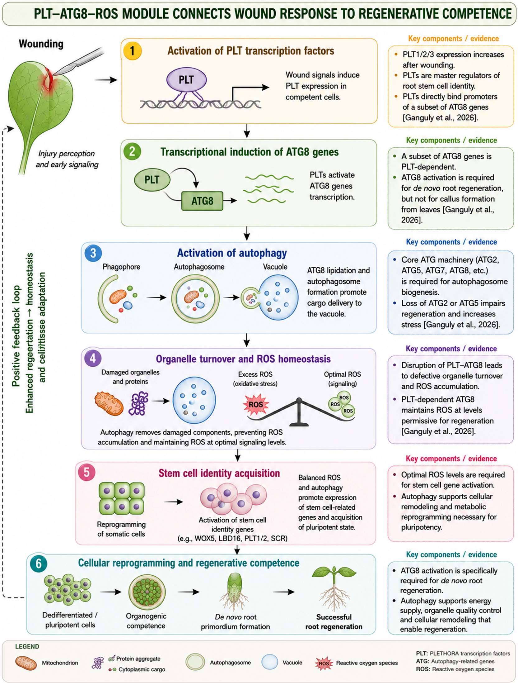
Evidence from loss-of-function mutants of ATGs, including ATG2 and ATG5, demonstrates that autophagy is an important regulator of cellular responses to tissue injury. Although the precise contribution of autophagy to in vitro regeneration remains incompletely understood, recent studies indicate that it plays a central role in coordinating stress adaptation with developmental reprogramming. Ganguly et al. [59] provided compelling evidence that transcriptional activation of ATG8 genes is specifically required for de novo root regeneration but is dispensable for wound-induced callus formation from excised leaves, indicating that distinct regeneration pathways have different autophagy requirements. Their study further demonstrated that a subset of ATG8 genes is directly regulated by PLT transcription factors, establishing a developmental link between stem cell specification and the autophagy machinery. Disruption of the PLT–ATG8 regulatory module impaired organelle turnover, resulting in excessive ROS accumulation and compromised regenerative capacity. Conversely, PLT-mediated activation of ATG8 maintained ROS homeostasis at levels permissive for the induction of stem cell-associated genes and successful root regeneration. These findings identify the PLT–ATG8–ROS regulatory module as a key mechanism linking wound perception, intracellular quality control, stem cell identity acquisition, and regenerative competence during organ regeneration [59].
These observations are consistent with the broader concept that successful regeneration requires coordinated cellular reprogramming rather than transcriptional regulation alone. Petersen et al. [60] emphasized that the transition of differentiated cells to pluripotent or stem cell-like states involves extensive remodeling of gene expression, metabolism, organelle composition, and protein turnover. In this context, autophagy provides an efficient mechanism for removing damaged or unnecessary cellular components while recycling metabolic resources needed for developmental transitions. Together, these studies support the emerging view that autophagy functions not only as a stress-response pathway but also as an integral component of the cellular remodeling processes that underlie regenerative competence (Figure 1).

4. Polyamines, Autophagy, and In Vitro Wounding Stress Modulation

4.1. Polyamines and Wounding Stress in Plants

Plants are constantly exposed to various abiotic stresses, including natural or artificial mechanical injury, which trigger complex physiological and biochemical defense responses. Among the different signaling molecules linked to phytohormones that help plants adapt to stress, PAs are vital for regulating growth, development, and stress tolerance. Evidence shows that PA metabolism is closely linked to wound healing processes, affecting cellular defense mechanisms, reinforcing cell walls, and activating stress signaling pathways [61,62].
PA catabolism has also been implicated in the adaptation of plant tissues to wounding stress, although much of the available evidence has been obtained from specific experimental systems. In wounded potato (Solanum tuberosum) tissues, exogenous Spd has been reported to enhance the activities of key PA biosynthetic enzymes, including ADC, ODC, and S-adenosylmethionine decarboxylase (SAMDC), leading to increased endogenous levels of Put, Spd, and Spm. Elevated PA levels were associated with increased activities of phenylalanine ammonia-lyase (PAL), 4-coumarate-CoA ligase (4CL), and cinnamyl alcohol dehydrogenase (CAD), thereby stimulating the phenylpropanoid pathway and promoting the accumulation of phenolic compounds and lignin precursors involved in wound healing [61,62,63]. In the same system, the conversion of Put into higher PAs followed by their oxidation through polyamine oxidase (PAO) and diamine oxidase (DAO) generated hydrogen peroxide (H2O2), which functions as an important signaling molecule regulating cell wall reinforcement, defense responses, and oxidative metabolism. The H2O2 produced through PA catabolism acts together with peroxidases (PODs) to promote lignin and suberin polyphenolic deposition, thereby strengthening protective barriers at wound sites [15,61]. Although these mechanisms have been characterized primarily in potato and other wound-healing models, they suggest that PA metabolism may contribute to stress adaptation and cellular competence during in vitro regeneration by regulating redox homeostasis and cell wall remodeling. However, further studies are required to determine the extent to which these mechanisms operate across different plant species and regeneration systems.
In addition to responding to stress, PAs are vital regulators of essential cellular processes. Inhibiting ADC-mediated PA synthesis slows down DNA replication, cell division, and cell cycle progression, but these effects can be reversed by adding Put externally. The need for Spd and Spm in eukaryotic translation shows their crucial role in protein synthesis, especially when rapid enzyme production for defense and tissue regeneration is necessary [61]. Research has shown changes in PA metabolism under abiotic stress conditions like low temperatures, droughts, salinity, and mechanical injury. While stress exposure often triggers the transcriptional activation of PA biosynthetic genes, the associated changes in PA levels are not always proportional. This suggests that complex regulatory mechanisms control PA levels. Potato tubers are a useful model for studying wound healing because they develop specialized protective tissues, like the closing layer and wound periderm, after injury. Although there has been significant research on wound-induced suberization and lignification, limited information exists about the timing of PA accumulation and the related molecular responses during the healing process. Previous studies have noted increased SAMDC expression after wounding and suggested a possible link between PA metabolism and ethylene production due to their common precursor, S-adenosylmethionine (SAM) [61,64].
Early in vitro studies suggested that PAs and ethylene are metabolically linked through their common precursor, SAM. Exogenous application of Put and Spd reduced the conversion of methionine to 1-aminocyclopropane-1-carboxylate (ACC), the immediate precursor of ethylene, leading to the hypothesis that PA biosynthesis competes with ethylene production for the available SAM pool [64,65]. However, subsequent studies demonstrated that this relationship is more complex than simple substrate competition. Although short-term in vitro experiments showed that Spd and Spm can transiently suppress ACC synthase expression and ethylene biosynthesis, long-term studies using transgenic plants and in vivo systems revealed that PA and ethylene biosynthesis can occur simultaneously without significant depletion of cellular SAM levels [64,65,66,67]. These findings indicate that the interaction between the two pathways is regulated through dynamic metabolic and transcriptional mechanisms rather than by competition for a limiting substrate alone.
The balance between PA and ethylene metabolism has important implications for in vitro regeneration because both pathways influence cellular responses to stress and developmental reprogramming. Ethylene is required at appropriate levels during regeneration; however, excessive ethylene accumulation under tissue culture conditions is frequently associated with tissue senescence, oxidative stress, explant browning, and reduced morphogenic competence. Consequently, ethylene inhibitors such as silver nitrate or aminoethoxyvinylglycine (AVG) have been widely used to improve shoot regeneration and SE in several plant species by enhancing explant survival and regeneration efficiency. Conversely, exogenous PA supplementation has been shown to promote organogenesis and SE while alleviating tissue culture-induced stress. These observations suggest that successful regeneration depends not on suppressing ethylene per se, but on maintaining a balanced coordination between PA and ethylene metabolism that supports cellular homeostasis, developmental competence, and efficient regeneration under in vitro conditions [32,33,34,35,64,65,66,67].
Collectively, current evidence suggests that PAs function as central regulators linking stress perception, ROS production, phenylpropanoid metabolism, protein synthesis, and structural barrier formation. Understanding the coordinated regulation of these processes may provide valuable insights into the molecular mechanisms that govern wound healing and stress resilience across various crop species.

4.2. Autophagy and Wounding Stress in Plants

As it is described, wound healing and tissue regeneration are essential processes for plants. They help plants survive mechanical injuries and recover from environmental stress [61,62,63,64,65,66,67]. During tissue injuries such as grafting, the physical disruption of plant tissues creates a wound interface. This interface requires significant cellular changes, metabolic adjustments, and reconnection of vascular tissues to restore integrity. Recent research has shown that autophagy, a process for breaking down and recycling cellular components, is crucial for the plant’s response to wounds. Studies with Arabidopsis thaliana and Nicotiana benthamiana have shown that tissue separation is a primary trigger of autophagy. After an injury, autophagic structures and GFP-ATG8-labeled autophagosomes quickly build up in nearby cells. This indicates a fast cellular response to damage. Autophagy remains active even when the separated cut surfaces cannot reconnect. This suggests that tissue injury drives the start of autophagy rather than the later joining of tissues [13,68].
When mechanical wounding occurs, it interrupts the vascular system, affecting both xylem and phloem tissues. This disruption limits the transport of water, carbohydrates, and other necessary resources. Consequently, the cells surrounding the injury face local nutrient shortages and metabolic stress. This triggers pathways that respond to nutrient deprivation and activate autophagic recycling processes. These processes mobilize resources inside the cell to maintain balance. Through breaking down and reusing cellular components, autophagy provides amino acids and other substrates that support alternative ways for mitochondria to produce energy, ensuring enough ATP production when resources are limited [59,69,70]. The importance of this process is clear during callus formation, a critical step in repairing wounds. After an injury, nearby differentiated cells change back to less specialized cells and multiply to form a callus layer that covers the damaged area. However, plants with mutations in key autophagy-related genes like ATG2 and ATG5 show significant drops in both callus size and occurrence, especially when carbon resources are limited. These observations suggest that autophagy is vital for meeting the energy and biosynthetic needs of healing growth. Overall, current studies indicate that autophagy activated by wounds helps reduce local nutrient shortages, encourages callus growth, and aids in tissue repair. By supporting cellular rebuilding and the eventual reconnection of vascular tissues, autophagy plays a key role in the successful healing of wounds in plants [42,58,59,71,72].
In addition to in vivo regeneration, plant tissue culture and micropropagation rely on the remarkable developmental plasticity of plant cells, enabling dedifferentiation, cellular reprogramming, and the formation of new organs. The initiation of in vitro culture invariably involves explant excision, which imposes severe mechanical injury and disrupts vascular continuity, resulting in restricted transport of water, nutrients, and signaling molecules together with rapid accumulation of ROS at the wound site [59]. To survive these conditions, explant cells rapidly activate multiple stress-response pathways, including macroautophagy. Triggered by ROS signaling and local nutrient deprivation, autophagy induces the expression of ATGs and the formation of autophagosomes that sequester damaged organelles, oxidized proteins, and other cellular components for degradation and recycling [73,74,75]. Beyond its role in cellular quality control, autophagy supports metabolic reprogramming by recycling amino acids, lipids, and other metabolites required to sustain energy production, cell cycle re-entry, callus formation, and subsequent organ development. These observations suggest that autophagy may contribute to maintaining explant viability and morphogenic competence during the early stages of in vitro regeneration. However, direct experimental evidence demonstrating that targeted manipulation of autophagy consistently improves regeneration efficiency or micropropagation outcomes across diverse plant species remains limited. Future studies integrating genetic approaches, autophagic flux analyses, and optimized tissue culture protocols will be essential to determine whether modulation of autophagy could be translated into practical strategies for enhancing explant survival, regeneration efficiency, and large-scale micropropagation [76,77,78,79,80,81].

5. Polyamines, Autophagy, and In Vitro Organogenesis

5.1. Role of Polyamines in Direct In Vitro Organogenesis

Direct organogenesis refers to the regeneration of shoots or roots directly from explant tissues without an intervening callus phase. Because this pathway enables rapid regeneration while minimizing somaclonal variation, it is widely used for plant propagation and genetic transformation. Organ formation is primarily regulated by the balance between auxins and cytokinins, but increasing evidence indicates that PAs act as important modulators of these hormonal responses. Put, Spd, and Spm participate in cellular processes essential for morphogenesis, including DNA stabilization, maintenance of membrane integrity, regulation of gene expression, and control of cell division and differentiation. Across diverse plant species, alterations in PA metabolism influence organogenic competence, as inhibition of PA biosynthesis generally suppresses regeneration, whereas increased endogenous PA levels or exogenous PA supplementation often enhance morphogenic responses. These findings support a regulatory role for PAs during direct organogenesis, although their effectiveness depends on species, explant type, hormonal environment, and culture conditions [82,83,84].
Among the three major PAs, Put shows the most consistent promotive effect on direct organogenesis across different species. Most studies report that Put enhances shoot induction when combined with cytokinin-rich media, suggesting a synergistic interaction with conventional PGRs. For example, in tomato (Solanum lycopersicum), Put combined with benzylaminopurine (BAP) and indole-3-acetic acid (IAA) significantly increased shoot regeneration from cotyledon explants compared with Spd [85]. Similar responses were reported in Cedrela fissilis, where increased shoot production and elongation following BA treatment were accompanied by elevated endogenous Put levels [86]. Likewise, in Mucuna pruriens, supplementation of MS medium with Put and BA produced the highest shoot multiplication rate, while Put also enhanced root induction, acclimatization success, and genetic stability of regenerated plants [33]. In Psoralea corylifolia, Put promoted shoot proliferation, elongation, spontaneous rooting, and increased endogenous Put accumulation, demonstrating that it can complement or partially substitute conventional auxin–cytokinin combinations [35]. Collectively, these studies indicate that Put consistently promotes organogenic competence by interacting with cytokinin signaling while maintaining endogenous PA homeostasis.
In contrast, the effects of Spd and Spm appear to be more species-dependent. In lemon (Citrus limon), both Spd and Spm stimulated adventitious shoot regeneration more effectively than Put, and these responses were associated with changes in cytokinin metabolism rather than ethylene production, suggesting that PA–cytokinin interactions may differ among species [87]. These findings contrast with those reported in tomato and several medicinal plants, where Put was consistently the most effective PA. Together, the available evidence suggests that although higher PAs can promote organogenesis, their relative effectiveness depends on the physiological characteristics of the explant and the endogenous hormonal balance.
Several studies further demonstrate that successful organogenesis depends not only on exogenous PA application but also on maintaining endogenous PA homeostasis. In Passiflora cincinnata and P. edulis, inhibition of PA biosynthesis markedly reduced shoot bud differentiation despite limited effects of exogenous PA supplementation, indicating that endogenous PA metabolism is required for morphogenic competence [88]. Similar conclusions were reached in Bixa orellana, where inhibition of PA biosynthesis using difluoromethylornithine (DFMO) significantly reduced shoot multiplication and disrupted endogenous PA levels, whereas Put supplementation restored regeneration and improved shoot elongation [89]. Proteomic analysis in Cedrela fissilis further showed that Put treatment modified proteins involved in primary metabolism, energy production, stress responses, and cell division, suggesting that PAs regulate organogenesis through coordinated metabolic reprogramming rather than acting solely as growth-promoting compounds [82].
Altogether, the available evidence indicates several consistent trends. Put is the PA most frequently associated with enhanced shoot organogenesis, particularly in combination with cytokinins, whereas Spd and Spm exhibit species-dependent effects that appear to involve modulation of cytokinin metabolism. In addition, regeneration responses vary according to explant type and endogenous PA status, indicating that maintaining PA homeostasis is at least as important as exogenous supplementation. Despite these common patterns, considerable variation among plant species and culture systems suggests that optimal PA treatments must be determined empirically rather than applied universally. Future comparative studies integrating PA metabolism with hormonal signaling and molecular analyses will be essential for establishing generalized strategies to improve direct organogenesis (Table 3).

5.2. Roles of Polyamines in Indirect in Vitro Organogenesis

Indirect organogenesis is a widely utilized regeneration pathway in plant tissue culture in which new organs are formed through an intermediate callus phase. This developmental process involves the dedifferentiation of specialized cells into an unorganized mass of totipotent cells, followed by their redifferentiation into shoots, roots, or other organs. Consequently, callus induction represents a critical initial stage, providing the cellular foundation for subsequent organogenic development and serving as an important platform for tissue culture and cell suspension systems. The efficiency of indirect organogenesis is influenced by numerous factors, including plant genotype, explant developmental stage, culture conditions, and the composition of the culture medium [90,91].
The promotive effects of PAs on plant regeneration have been attributed to their ability to stimulate cell division, enhance cellular growth, and facilitate the transition of differentiated cells into a competent organogenic state. However, the effectiveness of PA treatments depends largely on the type and concentration of the PA applied, as well as the genetic background of the plant species. Consequently, optimization of PA supplementation must be performed on a genotype-specific basis, since inappropriate concentrations may suppress rather than enhance regeneration. Furthermore, the physiological functions of PAs are closely associated with endogenous hormonal balance and stress-response pathways. Through extensive interactions with phytohormones and stress-signaling networks, PAs contribute to the regulation of plant growth, developmental processes, and responses to environmental stimuli. Changes in PA metabolism can influence the biosynthesis, signaling, and activity of major phytohormones, thereby affecting organogenic competence and regeneration efficiency [18,92,93].
Ajithan et al. [94] reported on Indian pea (Pisum sativum L.), where the effects of exogenous PAs on chloroplast development and callus-mediated organogenesis were investigated. The combined application of PAs and conventional PGRs, including 2,4-dichlorophenoxyacetic acid (2,4-D), benzyladenine (BA), and naphthaleneacetic acid (NAA), significantly enhanced all stages of indirect organogenesis. Put in combination with 2,4-D was particularly effective for callus induction, producing the highest percentage of friable callus. For shoot regeneration, the combination of Spd and BA yielded the greatest number of shoots per callus explant, whereas Put combined with NAA produced the highest rooting efficiency and root growth. Overall, PA supplementation substantially improved callus proliferation, shoot multiplication, and root formation, increasing regeneration efficiency by two- to three-fold compared with conventional PGR-based protocols. Additionally, regenerated plants exhibited increased chloroplast numbers, suggesting that PAs may also contribute to the enhancement of photosynthetic capacity and physiological vigor during regeneration.
Viu et al. [95] evaluated the influence of exogenous PAs on callus induction and subsequent indirect organogenesis on Curcuma longa L. using leaf base, root tip, and rhizome-derived axillary bud explants. Among the explant types tested, axillary buds exhibited the highest callogenic potential. Supplementation of the culture medium with a PA mixture (Put, Spd, and Spm in a 1:1:1 ratio), in combination with NAA, markedly improved callus proliferation and root development compared with PA-free treatments. Subsequent transfer of callus tissues to a regeneration medium containing the same PA mixture together with BAP enhanced shoot differentiation and overall plant regeneration. The most pronounced regenerative response was observed under treatment with 10 mM PA mixture combined with BAP, which produced vigorous plantlets with improved shoot formation. Their results indicate that PAs are key modulators of indirect organogenesis, promoting callus growth, root initiation, and shoot regeneration in a coordinated manner. In addition, elevated Put levels and increased peroxidase activity were associated with the differentiation process, suggesting their potential utility as biochemical indicators of organogenic competence in C. longa. Collectively, their findings demonstrate a synergistic interaction between exogenous PAs and PGRs in enhancing regeneration efficiency and facilitating the transition from callus formation to organ development during indirect organogenesis.
Jafari et al. [96] developed an indirect organogenesis protocol for Salvia officinalis L. using leaf and internode explants derived from in vitro-germinated plantlets. Explants were cultured on MS medium supplemented with various combinations of auxins (2,4-D or NAA) and the cytokinin BAP to induce callus formation. The most effective callus induction was obtained from internode explants cultured with NAA (1 mg L−1) combined with BAP (0.5 mg L−1). Subsequent shoot regeneration was significantly enhanced when internode-derived calli were transferred to MS medium containing Put (50 mg L−1) and thidiazuron (TDZ, 0.5 mg L−1), yielding an average of 5.66 shoots per explant. Shoot elongation and rooting were successfully achieved using MS medium supplemented with IBA (1 mg L−1). Regenerated plantlets showed efficient acclimatization under greenhouse conditions and exhibited no detectable morphological differences compared with the donor plants, indicating genetic and phenotypic stability. Their findings demonstrate that PA supplementation, particularly Put when combined with TDZ, enhances shoot regeneration efficiency during indirect organogenesis in S. officinalis, and the established protocol may also be applicable for producing stable transformants free of chimeric variation.
Kiełkowska et al. [97] investigated the effects of exogenous PAs on protoplast culture development, cell viability, and subsequent regeneration of Brassica oleracea L. var. capitata. Protoplasts isolated from seedling hypocotyls were cultured in media supplemented with Put, Spd, or Spm at different concentrations. PA-treated cultures, particularly those receiving Put and Spd, exhibited reduced accumulation of ROS during the early stages of culture, indicating an alleviation of oxidative stress. These treatments also enhanced protoplast viability and stimulated mitotic activity, suggesting that PAs promote cell division and support the maintenance of cellular competence during regeneration. Among the tested treatments, supplementation with 10 μM Put resulted in the highest frequency of shoot organogenesis, achieving a regeneration rate of 21% from protoplast-derived microcalli. Furthermore, ploidy analysis revealed that most regenerated plants were diploid, indicating the genetic stability of the regeneration process. Thus, their findings demonstrate that exogenous PAs, particularly Put, enhance indirect organogenesis by reducing oxidative damage, promoting cell proliferation, and improving regeneration efficiency, highlighting their important role in protoplast-based plant regeneration systems.
Collectively, available evidence indicates that PAs are key regulators of indirect organogenesis, influencing both callus formation and subsequent organ differentiation. Their regulatory functions are mediated through interactions with phytohormones, modulation of stress-response pathways, promotion of cell division and differentiation, and enhancement of cellular totipotency. The positive effects of PA supplementation on callus induction, shoot regeneration, root formation, and overall regeneration efficiency highlight their potential as valuable additives for improving indirect organogenesis protocols across a wide range of plant species.

5.3. Roles of Autophagy in Direct/Indirect In Vitro Organogenesis

Autophagy has been increasingly recognized as an important regulator of cellular remodeling during plant development and stress responses. During in vitro organogenesis, differentiated somatic cells undergo dedifferentiation, acquire morphogenic competence, and subsequently redifferentiate into new organs. These developmental transitions require extensive metabolic and cellular reorganization, including the selective removal of damaged or unnecessary cellular components. Direct evidence from plant regeneration studies indicates that autophagy contributes to wound-induced tissue repair, callus formation, and maintenance of cellular homeostasis during regenerative processes [58]. In addition, studies investigating developmental reprogramming suggest that autophagy-mediated recycling may facilitate the metabolic adjustments required for changes in cell fate by promoting proteome remodeling and intracellular quality control [98]. However, direct evidence demonstrating that autophagy regulates proteome turnover specifically during the dedifferentiation and redifferentiation phases of direct or indirect in vitro organogenesis remains limited. Therefore, while current findings support a role for autophagy in creating favorable conditions for cellular reprogramming, the precise molecular mechanisms linking autophagy to pluripotency acquisition and organ formation in plant tissue culture require further investigation.
Ganguly et al. [59] reported that autophagy plays a pivotal role in wound-induced organogenesis by preserving cellular homeostasis during the extensive reprogramming of somatic cells. Following tissue injury, differentiated cells experience substantial metabolic and oxidative stress that must be resolved before regenerative programs can be successfully initiated. They have demonstrated that, during de novo root organogenesis, plant-specific PLETHORA (PLT) transcription factors directly activate a subset of ATG8 genes, thereby stimulating autophagy. This regulatory pathway promotes the removal of damaged organelles and maintains intracellular redox balance, creating a cellular environment that is conducive to stem cell fate acquisition. Loss of PLT-dependent activation of autophagy disrupts organelle quality control, leading to excessive accumulation of ROS and elevated cellular stress, which ultimately suppresses the expression of key stem cell regulators required for root regeneration. Interestingly, this PLT–ATG8 signaling module is specifically required for de novo organ formation but is not essential for wound-induced callus development, indicating that autophagy fulfills distinct functions depending on the regenerative context. Their findings reveal that plants integrate lineage-specific developmental regulators with the evolutionarily conserved autophagy machinery to coordinate stress mitigation, proteome and organelle remodeling, and redox homeostasis during organogenesis. By facilitating the transition from differentiated somatic cells to regenerative stem cell-like states, autophagy emerges as a critical determinant of developmental plasticity and successful organogenesis following injury.
Wojciechowska et al. [99] recognized autophagy as an important regulator of plant developmental processes, including the coordinated growth and differentiation of roots and stems. During both primary and secondary development, autophagic activity is dynamically regulated in association with the formation of vascular tissues. Their study in Populus trichocarpa has shown that the expression of ATGs and the accumulation of ATG8 proteins vary throughout root and stem development, reflecting stage-specific activation of the autophagic machinery. Structural analyses have revealed characteristic autophagic features, including tonoplast invagination, the formation of autophagic bodies, and the accumulation of autophagosomes in differentiating vascular cells. Notably, these events occur during the early stages of xylem and phloem differentiation, well before the onset of developmental programmed cell death (PCD). This temporal pattern suggests that autophagy initially functions as a cytoprotective mechanism, facilitating intracellular remodeling, organelle turnover, and cellular differentiation required for vascular tissue development. At later developmental stages, however, autophagy contributes to the execution of developmental PCD, particularly through vacuole-mediated degradation associated with mega-autophagy, culminating in irreversible protoplast elimination during xylem maturation. Their findings highlight the dual role of autophagy as both a homeostatic mechanism supporting cellular differentiation and a component of the PCD machinery required for the completion of vascular organ development.
Recent advances in single-cell transcriptomics and other high-resolution single-cell technologies have substantially improved our understanding of the cellular and molecular events underlying de novo organogenesis by enabling the identification of distinct cell populations and reconstruction of developmental trajectories during regeneration [100]. These approaches have identified key regulators of cell fate transitions, including C-repeat Binding Factor 3 (CBF3) during lateral root initiation, SHOOTBORNE ROOTLESS (SBRL) during adventitious root formation, and transcription factors such as SHORT ROOT (SHR), SCARECROW (SCR), WUSCHEL-RELATED HOMEOBOX 5 (WOX5), PLETHORA 1/2 (PLT1/2), and JACKDAW (JKD) that contribute to shoot regeneration [100,101,102,103,104]. Although these studies have not directly examined autophagy, they provide valuable methodological frameworks for investigating when and where autophagy-related genes (ATGs) are activated during cellular reprogramming. Integrating single-cell transcriptomics with analyses of autophagic activity and autophagic flux could help identify cell-type-specific roles of autophagy during direct and indirect organogenesis and clarify its interaction with developmental regulatory networks.
Beyond identifying regulatory genes, single-cell approaches have provided important insights into the cellular origins of regenerating organs. Distinct transitional cell populations have been characterized during lateral root formation, including quiescent center (QC)-transitioning cells that contribute to meristem establishment [103]. Adventitious roots have been shown to originate from reprogrammed phloem-associated cells, whereas shoot regeneration in tissue culture involves QC-like cell populations within the callus that subsequently establish new shoot apical meristems. These findings demonstrate that organ regeneration is initiated by specialized subsets of competent cells rather than occurring uniformly throughout the tissue [104].
Despite these advances, several important questions regarding the molecular regulation of plant regeneration remain unresolved. Although single-cell transcriptomic and spatial transcriptomic approaches have substantially improved the characterization of cell fate transitions, they primarily provide correlative information and do not directly establish the functional roles of candidate regulators during organogenesis. In particular, the spatial and temporal dynamics of autophagy activation, its relationship with polyamine metabolism, and its interactions with hormonal and reactive oxygen species (ROS) signaling during cellular reprogramming remain poorly understood. Furthermore, most current studies have been conducted in a limited number of model species, making it difficult to determine whether the identified regulatory networks are broadly conserved across horticultural crops with contrasting regeneration capacities. Future research integrating single-cell and spatial transcriptomics with functional genetic analyses, autophagic flux monitoring, metabolomics, and targeted manipulation of polyamine metabolism will be essential to distinguish causal regulatory mechanisms from transcriptional associations and to identify strategies that can be translated into more efficient and reproducible regeneration systems for economically important crop species.

6. Polyamines, Autophagy, and In Vitro Somatic Embryogenesis

6.1. Roles of Polyamines in In Vitro Somatic Embryogenesis

The efficiency of in vitro morphogenesis remains a major limitation for the propagation, genetic transformation, and improvement of many economically important crop species because regeneration capacity is frequently genotype- and explant-dependent, with numerous species exhibiting partial or complete recalcitrance to in vitro regeneration [1,2,3,4,5]. Although PGRs, particularly auxins and cytokinins, are recognized as the primary regulators of morphogenic responses, accumulating evidence indicates that successful SE also depends on the coordinated regulation of stress signaling, cellular metabolism, ROS, and epigenetic reprogramming [6,7,8,9,10]. Among these additional regulatory factors, PAs have emerged as important modulators of embryogenic competence. Studies in several plant species have consistently reported a positive association between endogenous PA accumulation and SE induction, whereas inhibition of PA biosynthesis suppresses embryogenic responses and, in some systems, these effects can be partially reversed by exogenous Put supplementation [90,91,92,93]. Nevertheless, despite growing evidence supporting their involvement in SE, the molecular and physiological mechanisms by which PAs regulate embryogenic induction, developmental progression, and interactions with other signaling pathways remain incompletely understood.
Investigations in Momordica charantia, an economically and medicinally valuable species with limited reports of in vitro regeneration, have provided further insight into PA-associated regulation of embryogenesis [105,106]. During the culture of leaf explants on embryogenic media, dynamic changes in endogenous PA metabolism were observed over 42 days, suggesting that PA levels are tightly linked to developmental progression during SE. Elevated concentrations of free PAs were detected in young, actively dividing tissues, consistent with their known enrichment in proliferative and meristematic cells. This pattern supports previous findings that PA biosynthesis correlates positively with cell division activity and suggests that increased PA abundance is associated with the early induction phase of SE, when embryogenic cell clusters are established. As callus cultures progressed, a decline in PA levels was observed during callogenesis, likely reflecting the reduced metabolic activity of more differentiated callus cells compared with embryogenically competent meristematic cells. At later stages, further decreases in PA content coincided with embryo maturation, a phase characterized by cell expansion rather than intensive proliferation, indicating that PA demand is developmentally regulated and stage-specific. Notably, higher levels of Put were detected during early embryogenic induction, which may be attributed to increased activity of ornithine decarboxylase (ODC), an enzyme typically associated with cell proliferation, whereas arginine decarboxylase (ADC) activity is more commonly linked to stress responses. These observations are consistent with reports in other plant systems where ODC activity correlates positively with embryogenic competence, whereas ADC activity shows a more variable association depending on developmental stage and species context [105,106].
El-Dawayati et al. [107] developed an efficient in vitro regeneration system in date palm (Phoenix dactylifera L.) due to its limited natural propagation capacity through offshoots in a tissue culture-based propagation system, which offers clear advantages, including rapid clonal multiplication, genetic uniformity, and the production of pathogen-free planting material. In this context, optimization of SE in date palm cv. Sewi has been explored through the combined application of PAs and amino acid supplements in both solid and liquid culture systems. PAs such as Put and Spd, together with glutamine supplementation, were evaluated for their effects on embryogenic callus development and embryo induction. Distinct responses were observed depending on the culture medium: solid media primarily supported robust embryonic callus formation, whereas liquid media were more effective in promoting somatic embryo induction. These differences highlight the importance of physical culture conditions in shaping developmental outcomes during in vitro morphogenesis. Quantitative analyses demonstrated that the highest number of somatic embryos was obtained in liquid culture supplemented with Put and an elevated glutamine concentration, indicating a synergistic effect between nitrogen metabolism and PA-mediated developmental regulation. In contrast, increased embryonic callus biomass was observed in solid media, particularly under treatments involving Spd alone or in combination with glutamine, suggesting that Spd may preferentially support callus proliferation rather than embryo conversion under these conditions. Thus, Put combined with glutamine supplementation, as well as Spd in the presence of enhanced glutamine levels, were identified as the most effective treatments for promoting embryogenic progression in date palm.
Sundararajan et al. [108] reported that exogenous PAs enhanced embryogenic competence and regeneration capacity in indica rice cultivars, which often display variable and sometimes limited responsiveness to in vitro regeneration systems. Comparative analyses across cultivars such as ASD16, IR64, and ADT43 indicate that PA treatments can differentially modulate somatic embryo formation, shoot regeneration, and overall culture growth, reflecting strong genotype-dependent responses. Among the tested compounds, Put at moderate concentrations consistently produced the most pronounced stimulatory effects, leading to increased regeneration frequency, enhanced somatic embryo production, and elevated shoot formation. In contrast, Spd exhibited a more variable influence, including inhibitory effects on embryogenic response in certain genotypes such as ADT43, suggesting that PA function is highly context- and genotype-specific. Spm and Put were generally more effective in improving regeneration performance during extended culture periods, particularly in highly responsive cultivars such as ASD16 and IR64, where sustained increases in regenerative potential were observed. In addition to influencing embryogenesis and shoot formation, PAs also affected the biomass accumulation of suspension cultures, indicating broader roles in cellular growth regulation. The differential responses among cultivars highlight the importance of genetic background in determining sensitivity to PA-mediated regulation of developmental processes.
Aydin et al. [109] identified PAs as important modulators of SE and plant regeneration in wheat. They evaluated the effects of different PAs on embryogenic induction and regeneration efficiency. While PA treatments did not significantly affect the initial formation of embryogenic callus, they markedly enhanced the proportion of calli that acquired regenerative competence as well as the overall efficiency of regeneration. Among the tested compounds, Put exerted the strongest positive effect, significantly increasing both the frequency of responsive embryogenic calli and the number of regenerated plants. Moreover, a dose-dependent response was observed, with 1 mM Put producing the highest regeneration efficiency and the greatest improvement in developmental outcomes. Importantly, increased Put levels consistently promoted higher regeneration rates compared with other PAs, underscoring its central role in facilitating somatic embryo development and plantlet formation. Their findings highlight the functional significance of PAs, particularly Put, in enhancing SE-based regeneration systems in wheat and support their application as effective additives for improving transformation and regeneration efficiency in cereal crop biotechnology.
Ben Ali et al. [110] reported an important but still insufficiently explored pathway for secondary SE, particularly in woody species such as Quercus suber L., a dominant forest tree in the Mediterranean region. In this context, the effects of exogenous PAs on secondary SE were evaluated using primary mature embryos derived from rejuvenated leaf tissues of Moroccan cork oak. Explants were cultured on a basal medium composed of N30K macronutrients supplemented with glucose and agar, and amended with varying concentrations of Put, Spd, and Spm. Among the tested treatments, Spd at 0.4 mg L−1 produced the most pronounced stimulatory effect on secondary embryo formation, resulting in a highly significant increase in embryogenic output. This optimal concentration led to a substantial enhancement in the number of directly formed secondary embryos, as well as a marked increase in both embryo cluster formation and the frequency of embryogenic structures. In addition to promoting embryo quantity, Spd also positively influenced embryo quality, as evidenced by a significant increase in embryo size compared with control conditions lacking PA supplementation.
Bollati et al. [111] investigated the influence of multiple culture parameters on SE in Olea europaea L. using radicle-derived embryogenic tissues. The study simultaneously evaluated basal medium composition, incubation temperature, organic nitrogen sources, and exogenous PAs to determine their relative contributions to callus growth and embryo production. A modified basal medium (ECOm), developed by adjusting ionic strength, inorganic nitrogen balance, and calcium availability, significantly increased both callus growth and somatic embryo production compared with the original ECO formulation. Likewise, incubation temperature proved to be a major determinant of embryogenic success, with the highest embryo yield obtained at 24 °C. In contrast, organic nitrogen supplements such as glutamine and casein hydrolysate had little effect under the experimental conditions. Exogenous PAs exerted more specific and comparatively modest effects. Put at 5.7 µM enhanced embryo number and embryo elongation, whereas higher concentrations provided no additional benefit, indicating a narrow optimal concentration range. Spd showed limited or non-significant effects. Importantly, model-based analyses identified temperature and basal medium composition as the strongest predictors of regeneration efficiency, with PA supplementation contributing a secondary, fine-tuning role. These findings demonstrate that, although PAs can enhance specific aspects of embryogenic development under appropriate conditions, their effectiveness depends on the culture environment and should be considered complementary to, rather than a substitute for, optimization of basal medium composition and physical culture conditions.
Di et al. [112] reported the role of PAs in the Ginkgo biloba SE process, particularly during early developmental transitions from initial callus to embryogenic callus and globular embryo formation. Genome-wide analyses identified 34 genes involved in PA metabolism in G. biloba, which were classified into six gene families and distributed unevenly across most of the plant’s chromosomes. These gene families contain numerous cis-regulatory elements associated with phytohormone signaling, abiotic stress responses, and meristem-associated expression patterns, suggesting broad regulatory integration. Transcriptomic profiling revealed stage-specific expression patterns of PA metabolism genes during embryogenic progression. In particular, genes such as GbADC2, GbSAMDC2, GbSPMS1, and several amine oxidase-related genes (GbCuAO and GbPAO family members) exhibited elevated expression during the globular embryo stage compared with the initial callus stage, indicating enhanced PA metabolic activity during embryogenic advancement. Functional analyses further demonstrated that exogenous application of specific PAs differentially influenced developmental transitions: Spm at 1.0 mg L−1 promoted the conversion from initial callus to embryogenic callus, whereas low concentrations of Put (0.01 mg L−1) facilitated both embryogenic induction and subsequent progression to globular embryos.
Wang et al. [113] investigated the roles of Put and D-arginine in regulating embryogenic callus proliferation and SE in litchi (Litchi chinensis Sonn.). The authors examined histomorphological changes, endogenous PA levels, and the activities of key enzymes involved in PA metabolism in embryogenic calli of the cultivar ‘Feizixiao’. Embryogenic calli were cultured on a standard proliferation medium or media supplemented with either Put or D-arginine. Although both treatments promoted callus proliferation, they exerted contrasting effects on subsequent embryogenic development. Put reduced the number of somatic embryos and regenerated plantlets, whereas D-arginine enhanced embryogenic differentiation and regeneration efficiency. Histological analyses further showed that both compounds altered the normal pattern of proembryo development. Under control conditions, embryogenic cells progressed through the typical sequence of multicellular proembryos followed by globular, heart-shaped, and cotyledonary embryos. In contrast, treated cultures exhibited enhanced cell enlargement and repeated cell division, resulting in the formation of meristematic cell masses with modified developmental trajectories. These findings demonstrate that modulation of PA metabolism can influence multiple stages of SE, but the outcomes are not uniformly positive. Instead, the effects of exogenous PAs depend on the specific compound applied, its concentration, the developmental stage, and the plant species. Therefore, while manipulation of PA metabolism represents a promising approach for improving regeneration, optimization must be tailored to individual regeneration systems because certain treatments, such as exogenous Put in this study, may promote callus proliferation while simultaneously reducing embryogenic competence.
Noceda et al. [114] compared embryogenic and non-embryogenic cell suspensions from two commercial banana cultivars (Musa spp. cvs. ‘Grande Naine’ and ‘FHIA-18’) to identify potential biochemical markers of embryogenic competence. Two major classes of putative indicators were analyzed: intracellular PA profiles and extracellular endochitinase EP3-like proteins. PAs, including 1,3-diaminopropane, Put, Spd, and Spm, were quantified and evaluated in terms of their free and conjugated forms using high-performance liquid chromatography. In parallel, EP3-like protein accumulation and release were assessed using immunological approaches. The results demonstrated that PA composition and relative abundance provide clear discrimination between embryogenic and non-embryogenic cultures. Embryogenic cell suspensions were characterized by the absence of Spm in the insoluble conjugated fraction, reduced Put content (below 1000 nmol g−1 fresh weight), and a low Put-to-Spm ratio, particularly values below unity. In contrast, more proliferative and non-embryogenic cultures exhibited elevated Put levels and altered PA balance, suggesting that shifts in PA homeostasis reflect differences in developmental competence and cellular organization. In addition, EP3-like endochitinase proteins were predominantly detected in highly embryogenic cultures, where they were actively released into the extracellular environment.
Domínguez et al. [115] reported that the regulation of PA metabolism is closely associated with somatic embryo maturation and developmental progression in grapevine (Vitis vinifera L. ‘Mencía’), particularly during in vitro culture conditions designed to optimize embryo differentiation. In this context, the effect of a semipermeable membrane on somatic embryo aggregates cultured in differentiation medium was investigated by analyzing endogenous PA levels and the expression of key biosynthetic and catabolic genes. Put was the dominant free PA, accounting for more than 95% of the total free pool, and showed distinct temporal peaks during culture. The first peak occurred during the second week, coinciding with elevated expression of the VvADC gene, suggesting enhanced arginine-dependent biosynthesis and a strong contribution of de novo Put production. This accumulation was accompanied by low levels of free Spd, indicating a metabolic bias toward Put maintenance during early maturation. A second increase in free Put was observed toward the end of the culture period. Interestingly, this occurred despite upregulation of the VvSPDS2 gene and persistently low Spd levels, suggesting that Spd was not accumulated but instead likely underwent back-conversion to Put, potentially mediated by polyamine oxidase (VvPAO) activity. This dynamic indicates that PA homeostasis during somatic embryo maturation is tightly regulated through coordinated biosynthesis and catabolic interconversion pathways.

6.2. Roles of Autophagy in In Vitro Embryogenesis

SE is initiated when differentiated somatic cells undergo extensive cellular reprogramming in response to developmental cues or stress signals, leading to the acquisition of embryogenic competence and the formation of bipolar embryos [116]. During this transition, cells experience profound metabolic and structural remodeling, processes in which autophagy has recently emerged as a potential contributor. Although the molecular mechanisms remain incompletely understood, accumulating evidence suggests that autophagy supports cellular homeostasis during SE by facilitating the recycling of damaged organelles and macromolecules, maintaining energy balance, and promoting adaptation to stress conditions associated with embryogenic induction [58,116].
Functional studies have begun to provide direct evidence for the involvement of autophagy in SE. In Dimocarpus longan, transcriptomic and single-cell RNA sequencing (scRNA-seq) analyses revealed activation of autophagy-related pathways during the transition from embryogenic callus to somatic embryos [116,117,118]. Importantly, pharmacological modulation of autophagy demonstrated that intermediate levels of autophagic activity were required for efficient embryo formation, whereas excessive inhibition or activation reduced embryogenic efficiency. These findings indicate that precise regulation of autophagic flux, rather than maximal autophagy, is necessary to support successful cellular reprogramming. Consistent with this interpretation, increased expression of ATGs and enrichment of autophagy-associated biological processes were observed during embryogenic development, suggesting that autophagy contributes to the metabolic adjustments accompanying embryo initiation.
Additional insights into the regulatory networks controlling SE have been obtained through high-resolution single-cell technologies. In D. longan, scRNA-seq identified multiple cell populations representing distinct developmental states and revealed stress-responsive regulators, including ETHYLENE-RESPONSIVE FACTOR 6 (ERF6), that may coordinate embryogenic induction under heat stress conditions [116,117,118]. Likewise, comparative scRNA-seq analyses in cotton (Gossypium hirsutum) reconstructed the developmental trajectories leading to embryogenic competence and identified SE-ASSOCIATED LIPID TRANSFER PROTEIN (SELTP) as a negative regulator of SE [118,119,120,121]. Although these studies primarily focused on developmental regulators rather than autophagy itself, they provide valuable frameworks for identifying the spatial and temporal activation of autophagy-related genes and for integrating autophagic pathways with the gene regulatory networks governing SE.
Despite these advances, direct functional evidence linking autophagy to SE remains limited. Few studies have examined autophagic flux using ATG8-based markers or genetically characterized the roles of specific ATGs during embryogenic induction and embryo development. Likewise, mutant analyses and systematic evaluation of autophagy activators or inhibitors under tissue culture conditions remain scarce. Future studies combining genetic approaches, live-cell imaging of autophagic flux, and single-cell transcriptomics will be essential for clarifying the precise contribution of autophagy to both direct and indirect in vitro embryogenesis.

7. Literature Search Strategy

This review is a narrative review that synthesizes current knowledge on the roles of PAs and autophagy in plant in vitro regeneration. The literature was primarily retrieved from the Web of Science, Scopus, PubMed, and Google Scholar databases. Searches were conducted using combinations of keywords including PAs, autophagy, plant regeneration, in vitro regeneration, SE, organogenesis, cellular reprogramming, plant tissue culture, and plant growth regulators. Publications from peer-reviewed journals published primarily between 2000 and 2026 were considered, while earlier landmark studies were included when essential for historical context. Priority was given to original research articles and comprehensive review papers directly related to plant regeneration, PA metabolism, and autophagy. Studies outside the scope of plant biology or lacking relevance to regeneration processes were excluded. The selected literature was critically evaluated to provide an integrated overview of current knowledge and identify remaining research gaps.

8. Conclusions and Future Perspectives

Plant in vitro regeneration relies on the remarkable developmental plasticity of differentiated cells, which undergo dedifferentiation, cellular reprogramming, and redifferentiation to generate new organs or somatic embryos. Although the central roles of auxins, cytokinins, and developmental regulators such as WUS, PLT, and BBM have been extensively characterized, increasing evidence indicates that PAs and autophagy represent additional regulatory components that contribute to regeneration competence through complementary mechanisms.
Current evidence consistently supports a positive role for PAs in in vitro regeneration, particularly during direct organogenesis and SE. Put is the PA most frequently associated with enhanced shoot induction, whereas Spd and Spm exhibit more species- and developmental stage-dependent effects. Collectively, published studies indicate that PA-mediated regulation involves interactions with endogenous hormone metabolism, modulation of ROS homeostasis, maintenance of cellular redox balance, stabilization of nucleic acids and membranes, and promotion of cell division and differentiation. However, regeneration responses vary considerably among plant species, explant types, developmental stages, and culture conditions, demonstrating that PA supplementation cannot be considered a universal strategy. Instead, optimization of PA treatments should be tailored to individual regeneration systems while considering endogenous PA homeostasis and interactions with conventional PGRs.
Compared with PAs, the role of autophagy during in vitro regeneration remains less well characterized. Available evidence demonstrates that autophagy contributes to wound-induced tissue repair, nutrient recycling, organelle quality control, and metabolic adaptation during regeneration. Recent studies further suggest that autophagy may facilitate callus formation, cellular remodeling, and the maintenance of morphogenic competence, particularly under stress conditions commonly encountered during tissue culture. Nevertheless, direct functional evidence remains limited. Most current knowledge is derived from wound-healing studies, transcriptomic analyses, or indirect observations, whereas relatively few investigations have employed genetic approaches involving ATG mutants, ATG8-based autophagic flux markers, or pharmacological modulation of autophagy under in vitro regeneration conditions.
Although both PAs and autophagy have emerged as important regulators of plant regeneration, direct mechanistic links between these two pathways remain largely unexplored. Existing evidence suggests that they converge on several common physiological processes, including ROS regulation, stress adaptation, energy metabolism, and cellular remodeling. However, whether PAs directly regulate autophagy, whether autophagy influences PA metabolism, or whether both pathways operate independently under the control of shared upstream signals remains unknown. Addressing these questions represents an important opportunity for future research.
Several testable hypotheses arise from the current literature. First, PAs may influence regenerative competence by modulating autophagic activity through changes in cellular redox status and nutrient signaling. Second, autophagy may regulate endogenous PA homeostasis by controlling the turnover of enzymes involved in PA biosynthesis and catabolism. Third, coordinated regulation of PA metabolism, autophagy, hormone signaling, and ROS homeostasis may determine the developmental transitions required for successful organogenesis and SE. These hypotheses should be examined using integrated approaches combining genetics, genome editing, live-cell imaging of autophagic flux, metabolomics, single-cell and spatial transcriptomics, proteomics, and quantitative phenotyping across diverse horticultural species.
From an applied perspective, improved understanding of PA metabolism and autophagy has the potential to enhance regeneration systems used in horticulture, crop improvement, and plant biotechnology. However, current evidence does not yet support routine manipulation of either pathway as a general strategy for micropropagation or genetic transformation. Future studies should therefore focus on validating candidate regulators across multiple species and culture systems, identifying optimal combinations of PAs with PGRs, and determining how controlled modulation of autophagy influences regeneration efficiency without compromising developmental stability. Such advances will provide a stronger mechanistic foundation for developing more robust, reproducible, and genotype-independent regeneration protocols for economically important horticultural crops.

Author Contributions

Conceptualization, M.D.P.; writing—original draft preparation, M.D.P., M.B.D. and N.S.; visualization, M.D.P.; writing—review and editing, M.D.P. All authors have read and agreed to the published version of the manuscript.

Funding

This research received no external funding.

Data Availability Statement

No new data were created or analyzed in this study. Data sharing is not applicable to this article.

Acknowledgments

Open Access Funding by TU Wien. The authors gratefully acknowledge TU Wien for the Open Access Funding Programme of TU Wien Bibliothek.

Conflicts of Interest

The authors declare no conflicts of interest.

References

  1. Ikeuchi, M.; Favero, D.S.; Sakamoto, Y.; Iwase, A.; Coleman, D.; Rymen, B.; Sugimoto, K. Molecular mechanisms of plant regeneration. Annu. Rev. Plant Biol. 2019, 70, 377–406. [Google Scholar] [CrossRef] [Scilit] [PubMed]
  2. Long, Y.; Yang, Y.; Pan, G.; Shen, Y. New insights into tissue culture plant-regeneration mechanisms. Front. Plant Sci. 2022, 13, 926752. [Google Scholar] [CrossRef] [Scilit] [PubMed]
  3. Chen, Z.; Debernardi, J.M.; Dubcovsky, J.; Gallavotti, A. Recent advances in crop transformation technologies. Nat. Plants 2022, 8, 1343–1351. [Google Scholar] [CrossRef] [Scilit] [PubMed]
  4. Pasternak, T.P.; Steinmacher, D. Plant growth regulation in cell and tissue culture in vitro. Plants 2024, 13, 327. [Google Scholar] [CrossRef] [Scilit] [PubMed]
  5. He, Z.; Webster, S.; He, S.Y. Growth–defense trade-offs in plants. Curr. Biol. 2022, 32, R634–R639. [Google Scholar] [CrossRef] [Scilit] [PubMed]
  6. Anh, T.T.L.; Mai, N.T.N.; Tung, H.T.; Khai, H.D.; Cuong, D.M.; Luan, V.Q.; Phuong, H.T.N.; Van Binh, N.; Vinh, B.V.T.; Thuy, N.T.T.; et al. Effect of spermidine, glutamine, and proline on somatic embryogenesis and silver nanoparticles supplied culture improved rhizome formation of Panax vietnamensis var. langbianensis. S. Afr. J. Bot. 2023, 163, 226–236. [Google Scholar] [CrossRef] [Scilit]
  7. Rakesh, B.; Sudheer, W.N.; Nagella, P. Role of polyamines in plant tissue culture: An overview. Plant Cell Tissue Organ Cult. 2021, 145, 487–506. [Google Scholar] [CrossRef] [Scilit]
  8. Sugimoto, K.; Temman, H.; Kadokura, S.; Matsunaga, S. To regenerate or not to regenerate: Factors that drive plant regeneration. Curr. Opin. Plant Biol. 2019, 47, 138–150. [Google Scholar] [CrossRef] [Scilit] [PubMed]
  9. Jha, P.; Ochatt, S.J.; Kumar, V. WUSCHEL: A master regulator in plant growth signaling. Plant Cell Rep. 2020, 39, 431–444. [Google Scholar] [CrossRef] [Scilit] [PubMed]
  10. Fazilati, M.; Forghani, A.H. The role of polyamine to increasing growth of plant: As a key factor in health crisis. Int. J. Health Syst. Disaster Manag. 2015, 3, 89–94. [Google Scholar]
  11. Qi, H.; Wang, Y.; Bao, Y.; Bassham, D.C.; Chen, L.; Chen, Q.F.; Hou, S.; Hwang, I.; Huang, L.; Lai, Z.; et al. Studying plant autophagy: Challenges and recommended methodologies. Adv. Biotechnol. 2023, 1, 2. [Google Scholar] [CrossRef] [Scilit] [PubMed]
  12. Yagyu, M.; Yoshimoto, K. New insights into plant autophagy: Molecular mechanisms and roles in development and stress responses. J. Exp. Bot. 2024, 75, 1234–1251. [Google Scholar] [CrossRef] [Scilit] [PubMed]
  13. Kurotani, K.I.; Shinozaki, D.; Okada, K.; Tabata, R.; Kawakatsu, Y.; Sugita, R.; Utsugi, Y.; Okayasu, K.; Mori, M.; Tanoi, K.; et al. Autophagy is induced during plant grafting to promote wound healing. Nat. Commun. 2025, 16, 3483. [Google Scholar] [CrossRef] [Scilit] [PubMed]
  14. Kusano, T.; Berberich, T.; Tateda, C.; Takahashi, Y. Polyamines: Essential factors for growth and survival. Planta 2008, 228, 367–381. [Google Scholar] [CrossRef] [Scilit] [PubMed]
  15. Nandy, S.; Das, T.; Tudu, C.K.; Mishra, T.; Ghorai, M.; Gadekar, V.S.; Anand, U.; Kumar, M.; Behl, T.; Shaikh, N.K.; et al. Unravelling the multi-faceted regulatory role of polyamines in plant biotechnology, transgenics and secondary metabolomics. Appl. Microbiol. Biotechnol. 2022, 106, 905–929. [Google Scholar] [CrossRef] [Scilit] [PubMed]
  16. Kaur-Sawhney, R.; Tiburcio, A.F.; Altabella, T.; Galston, A.W. Polyamines in plants: An overview. J. Cell Mol. Biol. 2003, 2, 1–12. [Google Scholar]
  17. Galston, A.W.; Flores, H.E. Polyamines and plant morphogenesis. In Biochemistry and Physiology of Polyamines in Plants; Slocum, R.D., Flores, H.E., Eds.; CRC Press: Boca Raton, FL, USA, 1991; pp. 175–186. [Google Scholar]
  18. Jangra, A.; Chaturvedi, S.; Kumar, N.; Singh, H.; Sharma, V.; Thakur, M.; Tiwari, S.; Chhokar, V. Polyamines: The gleam of next-generation plant growth regulators for growth, development, stress mitigation, and hormonal crosstalk in plants—A systematic review. J. Plant Growth Regul. 2023, 42, 5167–5191. [Google Scholar] [CrossRef] [Scilit]
  19. Slocum, R.D.; Flores, H.E. Biochemistry and Physiology of Polyamines in Plants; CRC Press: Boca Raton, FL, USA, 1991. [Google Scholar]
  20. Todorova, D.; Katerova, Z.; Sergiev, I.; Alexieva, V. Polyamines—Involvement in plant stress tolerance and adaptation. In Plant Adaptation to Environmental Change: Significance of Amino Acids and Their Derivatives; Anjum, N.A., Gill, S.S., Gill, R., Eds.; CABI: Wallingford, UK, 2014; pp. 194–221. [Google Scholar] [CrossRef] [Scilit]
  21. Pasternak, T.; Steinmacher, D. Plant Tissue Culture In Vitro: A Long Journey with Lingering Challenges. Int. J. Plant Biol. 2025, 16, 97. [Google Scholar] [CrossRef] [Scilit]
  22. Abdalla, N.; El-Ramady, H.; Seliem, M.K.; El-Mahrouk, M.E.; Taha, N.; Bayoumi, Y.; Shalaby, T.A.; Dobránszki, J. An academic and technical overview on plant micropropagation challenges. Horticulturae 2022, 8, 677. [Google Scholar] [CrossRef] [Scilit]
  23. Arora, K.; Rai, M.K.; Sharma, A.K. Tissue culture mediated biotechnological interventions in medicinal trees: Recent progress. Plant Cell Tissue Organ Cult. 2022, 150, 267–287. [Google Scholar] [CrossRef] [Scilit]
  24. Bhardwaj, S.; Kumari, M.; Chethan, T.; Hosamani, M.; Paul, M.; Moirangthem, A.; Dash, A.P.; Verma, S. Advances in Micropropagation and Tissue Culture for Horticultural Crops: A Review. Plant Cell Biotechnol. Mol. Biol. 2025, 26, 166–182. [Google Scholar] [CrossRef] [Scilit]
  25. Gupta, S.; Chaturvedi, P. (Eds.) Commercial Scale Tissue Culture for Horticulture and Plantation Crops; Springer: Singapore, 2022; pp. 1–21. [Google Scholar] [CrossRef] [Scilit]
  26. Nhut, D.T.; Tung, H.T.; Yeung, E.C.T. (Eds.) Plant Tissue Culture: New Techniques and Application in Horticultural Species of Tropical Region; Springer: Singapore, 2022; pp. 383–397. [Google Scholar] [CrossRef] [Scilit]
  27. Koti, P.; Bill, T. Plant tissue culture and genetic transformation in crop improvement. J. Bacteriol. Mycol. Open Access 2025, 13, 61–69. [Google Scholar] [CrossRef] [Scilit]
  28. Wang, P.; Si, H.; Li, C.; Xu, Z.; Guo, H.; Jin, S.; Cheng, H. Plant genetic transformation: Achievements, current status and future prospects. Plant Biotechnol. J. 2025, 23, 2034–2058. [Google Scholar] [CrossRef] [Scilit] [PubMed]
  29. Malabadi, R.B.; Chalannavar, R.K.; Kolkar, K.P. Plant cell totipotency: Plant tissue culture applications-An updated review. World J. Adv. Eng. Technol. Sci. 2025, 16, 112–135. [Google Scholar] [CrossRef] [Scilit]
  30. Zhang, D.; Song, X.Y.; Zheng, D.R.; Tian, J.; Li, Y.H.; Wang, Y.; Wu, H.; Wang, W. Advances in the molecular mechanisms of plant tissue culture and regeneration regulated by totipotency-related transcription factors. Biotechnol. Bull. 2024, 40, 23–33. [Google Scholar]
  31. Desai, P.; Desai, S.; Rafaliya, R.; Patil, G. Plant tissue culture: Somatic embryogenesis and organogenesis. In Advances in Plant Tissue Culture; Academic Press: Cambridge, MA, USA, 2022; pp. 109–130. [Google Scholar] [CrossRef] [Scilit]
  32. Tanur Erkoyuncu, M. Interactive Effects of Polyamines and Plant Growth Regulators on Shoot Induction and Secondary Metabolism in In Vitro Shoot Cultures of Echinacea Species. Molecules 2026, 31, 686. [Google Scholar] [CrossRef] [Scilit] [PubMed]
  33. Alam, N.; Ahmad, A.; Ahmad, N.; Anis, M. Polyamines mediated in vitro morphogenesis in cotyledonary node explants of Mucuna pruriens (L.) DC.: A natural source of L-Dopa. J. Plant Growth Regul. 2023, 42, 5203–5215. [Google Scholar] [CrossRef] [Scilit]
  34. Kıymaz, G.; Acemi, A. Effects of polyamines and optimized culture medium on in vitro organ development and accumulation of root phenolics in endangered Verbascum bugulifolium. Plant Cell Tissue Organ Cult. 2025, 162, 33. [Google Scholar] [CrossRef] [Scilit]
  35. Gajula, H.; Kumar, V.; Vijendra, P.D.; Rajashekar, J.; Sannabommaji, T.; Basappa, G.; Anuradha, C.M. In vitro regeneration of Psoralea corylifolia Linn.: Influence of polyamines during in vitro shoot development. Vitr. Cell. Dev. Biol. Plant 2022, 58, 103–113. [Google Scholar] [CrossRef] [Scilit]
  36. Subrahmanyeswari, T.; Gantait, S.; Sarkar, R.; Kamble, S.N.; Singh, S.; Bhattacharyya, S. Polyamines-and growth inducers-mediated enhanced mono-phasic in vitro regeneration of sugar leaf plant (Stevia rebaudiana Bert.) in liquid medium. S. Afr. J. Bot. 2024, 173, 34–45. [Google Scholar] [CrossRef] [Scilit]
  37. Pessanha, L.D.S.; Aragão, V.P.M.; de Oliveira, T.D.R.; de Sousa, K.R.; Silveira, V.; Santa-Catarina, C. Benzyladenine affects polyamine contents and proteomic profiles during in vitro shoot development and ex vitro rooting in Dalbergia nigra (Vell.) Allemão ex Benth. (Fabaceae). Plant Cell Tissue Organ Cult. 2022, 151, 75–92. [Google Scholar] [CrossRef] [Scilit]
  38. Coskun, Y.; Yaman, V. Enhancing phenolic compounds in caper through the incorporation of polyamines in In Vitro cultures from various explant sources. S. Afr. J. Bot. 2024, 165, 43–48. [Google Scholar] [CrossRef] [Scilit]
  39. Flores, H.E.; Martin-Tanguy, J. Polyamines and plant secondary metabolites. In Biochemistry and Physiology of Polyamines in Plants; Slocum, R.D., Flores, H.E., Eds.; CRC Press: Boca Raton, FL, USA, 1991; pp. 57–76. [Google Scholar]
  40. Wu, Y.; Xu, R.; Zhuang, X. Multifaceted roles of the ATG8 protein family in plant autophagy: From autophagosome biogenesis to cargo recognition. J. Mol. Biol. 2025, 437, 168981. [Google Scholar] [CrossRef] [Scilit] [PubMed]
  41. Bassham, D.C. Plant autophagy—More than a starvation response. Curr. Opin. Plant Biol. 2007, 10, 587–593. [Google Scholar] [CrossRef] [Scilit] [PubMed]
  42. Bozhkov, P.V. Plant autophagy: Mechanisms and functions. J. Exp. Bot. 2018, 69, 1281–1285. [Google Scholar] [CrossRef] [Scilit] [PubMed]
  43. Yoshimoto, K.; Ohsumi, Y. Unveiling the molecular mechanisms of plant autophagy—From autophagosomes to vacuoles in plants. Plant Cell Physiol. 2018, 59, 1337–1344. [Google Scholar] [CrossRef] [Scilit] [PubMed]
  44. Liu, Y.; Bassham, D.C. Autophagy: Pathways for self-eating in plant cells. Annu. Rev. Plant Biol. 2012, 63, 215–237. [Google Scholar] [CrossRef] [Scilit] [PubMed]
  45. Wei, S.; Chen, Z.; Xu, C.; Qian, X.; Tang, Q. Autophagy and stress tolerance in plants: The central role of ATG18—A review. Crit. Rev. Biotechnol. 2026, 46, 562–581. [Google Scholar] [CrossRef] [Scilit] [PubMed]
  46. Li, Y.; Xu, X.; Qi, G.; Cui, D.; Huang, C.; Sui, X.; Li, G.; Fan, Q. Mechanisms of autophagy function and regulation in plant growth, development, and response to abiotic stress. Crop J. 2023, 11, 1611–1625. [Google Scholar] [CrossRef] [Scilit]
  47. Petersen, M.; Avin-Wittenberg, T.; Bassham, D.C.; Dagdas, Y.; Fan, C.; Fernie, A.R.; Jiang, L.; Mishra, D.; Otegui, M.S.; Rodriguez, E.; et al. Autophagy in plants. Autophagy Rep. 2024, 3, 2395731. [Google Scholar] [CrossRef] [Scilit] [PubMed]
  48. Agbemafle, W.; Jayasinghe, V.; Bassham, D.C. Can autophagy enhance crop resilience to environmental stress? Philos. Trans. R. Soc. B 2025, 380, 20240245. [Google Scholar] [CrossRef] [Scilit] [PubMed]
  49. Ikeuchi, M.; Ogawa, Y.; Iwase, A.; Sugimoto, K. Plant regeneration: Cellular origins and molecular mechanisms. Development 2016, 143, 1442–1451. [Google Scholar] [CrossRef] [Scilit] [PubMed]
  50. Bidabadi, S.S.; Jain, S.M. Cellular, molecular, and physiological aspects of in vitro plant regeneration. Plants 2020, 9, 702. [Google Scholar] [CrossRef] [Scilit] [PubMed]
  51. Kumar, N.; Reddy, M.P. In vitro plant propagation: A review. J. For. Environ. Sci. 2011, 27, 61–72. [Google Scholar]
  52. Perez-Garcia, P.; Moreno-Risueno, M.A. Stem cells and plant regeneration. Dev. Biol. 2018, 442, 3–12. [Google Scholar] [CrossRef] [Scilit] [PubMed]
  53. Eshed Williams, L. Genetics of shoot meristem and shoot regeneration. Annu. Rev. Genet. 2021, 55, 661–681. [Google Scholar] [CrossRef] [Scilit] [PubMed]
  54. Mostafa, S.; Wang, Y.; Zeng, W.; Jin, B. Plant responses to herbivory, wounding, and infection. Int. J. Mol. Sci. 2022, 23, 7031. [Google Scholar] [CrossRef] [Scilit] [PubMed]
  55. Moore, B.M.; Lee, Y.S.; Wang, P.; Azodi, C.; Grotewold, E.; Shiu, S.H. Modeling temporal and hormonal regulation of plant transcriptional response to wounding. Plant Cell 2022, 34, 867–888. [Google Scholar] [CrossRef] [Scilit] [PubMed]
  56. Chen, C.; Hu, Y.; Ikeuchi, M.; Jiao, Y.; Prasad, K.; Su, Y.H.; Xiao, J.; Xu, L.; Yang, W.; Zhao, Z.; et al. Plant regeneration in the new era: From molecular mechanisms to biotechnology applications. Sci. China Life Sci. 2024, 67, 1338–1367. [Google Scholar] [CrossRef] [Scilit] [PubMed]
  57. Wang, F.X.; Shang, G.D.; Wang, J.W. Towards a hierarchical gene regulatory network underlying somatic embryogenesis. Trends Plant Sci. 2022, 27, 1209–1217. [Google Scholar] [CrossRef] [Scilit]
  58. Rodriguez, E.; Chevalier, J.; Olsen, J.; Ansbøl, J.; Kapousidou, V.; Zuo, Z.; Svenning, S.; Loefke, C.; Koemeda, S.; Drozdowskyj, P.S.; et al. Autophagy mediates temporary reprogramming and dedifferentiation in plant somatic cells. EMBO J. 2020, 39, e103315. [Google Scholar] [CrossRef] [Scilit] [PubMed]
  59. Ganguly, A.; Humnabadkar, A.; Gautam, K.; Willemsen, V.; Xu, L.; Dagdas, Y.; Prasad, K. PLETHORA–autophagy axis activates organ regeneration through ROS modulation. Proc. Natl. Acad. Sci. USA 2026, 123, e2513954123. [Google Scholar] [CrossRef] [Scilit] [PubMed]
  60. Petersen, M.; Ebstrup, E.; Rodriguez, E. Going through changes—The role of autophagy during reprogramming and differentiation. J. Cell Sci. 2024, 137, jcs261655. [Google Scholar] [CrossRef] [Scilit] [PubMed]
  61. Wang, Y.; Yang, R.; Wang, Q.; Xie, P.; Wang, X.; Li, Y.; Prusky, D.; Han, Y.; Bi, Y. Exogenous spermidine promotes the formation of the closing layer at potato tuber wounds by inducing polyamine synthesis and phenylpropanoid metabolism. Postharvest Biol. Technol. 2025, 226, 113572. [Google Scholar] [CrossRef] [Scilit]
  62. Lulai, E.C.; Neubauer, J.D.; Olson, L.L.; Suttle, J.C. Wounding induces changes in tuber polyamine content, polyamine metabolic gene expression, and enzyme activity during closing layer formation and initiation of wound periderm formation. J. Plant Physiol. 2015, 176, 89–95. [Google Scholar] [CrossRef] [Scilit] [PubMed]
  63. Angelini, R.; Tisi, A.; Rea, G.; Chen, M.M.; Botta, M.; Federico, R.; Cona, A. Involvement of polyamine oxidase in wound healing. Plant Physiol. 2008, 146, 162–177. [Google Scholar] [CrossRef] [Scilit] [PubMed]
  64. Harpaz-Saad, S.; Yoon, G.M.; Mattoo, A.K.; Kieber, J.J. The formation of ACC and competition between polyamines and ethylene for SAM. Annu. Plant Rev. 2012, 44, 53–81. [Google Scholar] [CrossRef] [Scilit]
  65. Kushad, M.M.; Dumbroff, E.B. Metabolic and physiological relationships between the polyamine and ethylene biosynthetic pathways. In Biochemistry and Physiology of Polyamines in Plants; Slocum, R.D., Flores, H.E., Eds.; CRC Press: Boca Raton, FL, USA, 1991; pp. 77–92. [Google Scholar]
  66. Torres-Contreras, A.M.; Nair, V.; Senés-Guerrero, C.; Pacheco, A.; González-Agüero, M.; Ramos-Parra, P.A.; Cisneros-Zevallos, L.; Jacobo-Velázquez, D.A. Cross-talk and physiological role of jasmonic acid, ethylene, and reactive oxygen species in wound-induced phenolic biosynthesis in broccoli. Plants 2023, 12, 1434. [Google Scholar] [CrossRef] [Scilit] [PubMed]
  67. Baharudin, N.F.; Osman, N.I. Plant development, stress responses, and secondary metabolism under ethylene regulation. Plant Stress 2023, 7, 100146. [Google Scholar] [CrossRef] [Scilit]
  68. Kurotani, K.I.; Tabata, R.; Kawakatsu, Y.; Sugita, R.; Okayasu, K.; Tanoi, K.; Notaguchi, M. Autophagy is induced during plant grafting for wound healing. bioRxiv 2020. [Google Scholar] [CrossRef] [Scilit]
  69. Signorelli, S.; Tarkowski, Ł.P.; Van den Ende, W.; Bassham, D.C. Linking autophagy to abiotic and biotic stress responses. Trends Plant Sci. 2019, 24, 413–430. [Google Scholar] [CrossRef] [Scilit] [PubMed]
  70. Moreno-Blas, D.; Adell, T.; González-Estévez, C. Autophagy in tissue repair and regeneration. Cells 2025, 14, 282. [Google Scholar] [CrossRef] [Scilit] [PubMed]
  71. Tyutereva, E.V.; Murtuzova, A.V.; Voitsekhovskaja, O.V. Autophagy and the energy status of plant cells. Russ. J. Plant Physiol. 2022, 69, 19. [Google Scholar] [CrossRef] [Scilit]
  72. Gross, A.S.; Raffeiner, M.; Zeng, Y.; Üstün, S.; Dagdas, Y. Autophagy in plant health and disease. Annu. Rev. Plant Biol. 2025, 76, 197–227. [Google Scholar] [CrossRef] [Scilit] [PubMed]
  73. Avin-Wittenberg, T. Autophagy and its role in plant abiotic stress management. Plant Cell Environ. 2019, 42, 1045–1053. [Google Scholar] [CrossRef] [Scilit] [PubMed]
  74. Katz, E.; Chamovitz, D.A. Wounding of Arabidopsis leaves induces indole-3-carbinol-dependent autophagy in roots of Arabidopsis thaliana. Plant J. 2017, 91, 779–787. [Google Scholar] [CrossRef] [Scilit] [PubMed]
  75. Xiong, Y.; Contento, A.L.; Nguyen, P.Q.; Bassham, D.C. Degradation of oxidized proteins by autophagy during oxidative stress in Arabidopsis. Plant Physiol. 2007, 143, 291–299. [Google Scholar] [CrossRef] [Scilit] [PubMed]
  76. Feng, Y.; Chen, Y.; Wu, X.; Chen, J.; Zhou, Q.; Liu, B.; Zhang, L.; Yi, C. Interplay of energy metabolism and autophagy. Autophagy 2024, 20, 4–14. [Google Scholar] [CrossRef] [Scilit] [PubMed]
  77. Masclaux-Daubresse, C.; Chen, Q.; Havé, M. Regulation of nutrient recycling via autophagy. Curr. Opin. Plant Biol. 2017, 39, 8–17. [Google Scholar] [CrossRef] [Scilit] [PubMed]
  78. Avin-Wittenberg, T.; Baluška, F.; Bozhkov, P.V.; Elander, P.H.; Fernie, A.R.; Galili, G.; Hassan, A.; Hofius, D.; Isono, E.; Le Bars, R.; et al. Autophagy-related approaches for improving nutrient use efficiency and crop yield protection. J. Exp. Bot. 2018, 69, 1335–1353. [Google Scholar] [CrossRef] [Scilit] [PubMed]
  79. Soto-Burgos, J.; Bassham, D.C. SnRK1 activates autophagy via the TOR signaling pathway in Arabidopsis thaliana. PLoS ONE 2017, 12, e0182591. [Google Scholar] [CrossRef] [Scilit] [PubMed]
  80. Janse van Rensburg, H.C.; Van den Ende, W.; Signorelli, S. Autophagy in plants: Both a puppet and a puppet master of sugars. Front. Plant Sci. 2019, 10, 14. [Google Scholar] [CrossRef] [Scilit] [PubMed]
  81. Chen, Q.; Shinozaki, D.; Luo, J.; Pottier, M.; Havé, M.; Marmagne, A.; Reisdorf-Cren, M.; Chardon, F.; Thomine, S.; Yoshimoto, K.; et al. Autophagy and nutrients management in plants. Cells 2019, 8, 1426. [Google Scholar] [CrossRef] [Scilit] [PubMed]
  82. Aragão, V.P.M.; Reis, R.S.; Silveira, V.; Santa-Catarina, C. Putrescine promotes changes in the endogenous polyamine levels and proteomic profiles to regulate organogenesis in Cedrela fissilis Vellozo (Meliaceae). Plant Cell Tissue Organ Cult. 2017, 130, 495–505. [Google Scholar] [CrossRef] [Scilit]
  83. Kaszler, N.; Benkő, P.; Bernula, D.; Szepesi, Á.; Fehér, A.; Gémes, K. Polyamine metabolism is involved in the direct regeneration of shoots from Arabidopsis lateral root primordia. Plants 2021, 10, 305. [Google Scholar] [CrossRef] [Scilit] [PubMed]
  84. Baron, K.; Stasolla, C. The role of polyamines during in vivo and in vitro development. Vitr. Cell. Dev. Biol. Plant 2008, 44, 384–395. [Google Scholar] [CrossRef] [Scilit]
  85. Kaur, Y.; Das, N. Roles of polyamines in growth and development of the Solanaceous crops under normal and stressful conditions. J. Plant Growth Regul. 2023, 42, 4989–5010. [Google Scholar] [CrossRef] [Scilit]
  86. Aragão, V.P.M.; de Souza Ribeiro, Y.R.; Reis, R.S.; Macedo, A.F.; Floh, E.I.S.; Silveira, V.; Santa-Catarina, C. In vitro organogenesis of Cedrela fissilis Vell. (Meliaceae): The involvement of endogenous polyamines and carbohydrates on shoot development. Plant Cell Tissue Organ Cult. 2016, 124, 611–620. [Google Scholar] [CrossRef] [Scilit]
  87. Pérez-Jiménez, M.; Celdrán-Sánchez, V.; Martínez-Romero, D.; Pérez-Tornero, O. Assessment of the polyamines modulation on cytokinins and ethylene and its effect in lemon (Citrus limon) de novo regeneration. Plant Cell Tissue Organ Cult. 2022, 150, 165–175. [Google Scholar] [CrossRef] [Scilit]
  88. Dias, L.L.; Santa-Catarina, C.; Ribeiro, D.M.; Barros, R.S.; Floh, E.I.; Otoni, W.C. Ethylene and polyamine production patterns during in vitro shoot organogenesis of two passion fruit species as affected by polyamines and their inhibitor. Plant Cell Tissue Organ Cult. 2009, 99, 199–208. [Google Scholar] [CrossRef] [Scilit]
  89. Parimalan, R.; Giridhar, P.; Ravishankar, G.A. Enhanced shoot organogenesis in Bixa orellana L. in the presence of putrescine and silver nitrate. Plant Cell Tissue Organ Cult. 2011, 105, 285–290. [Google Scholar] [CrossRef] [Scilit]
  90. Chen, D.; Shao, Q.; Yin, L.; Younis, A.; Zheng, B. Polyamine function in plants: Metabolism, regulation on development, and roles in abiotic stress responses. Front. Plant Sci. 2019, 9, 1945. [Google Scholar] [CrossRef] [Scilit] [PubMed]
  91. Tiburcio, A.F.; Altabella, T.; Bitrián, M.; Alcázar, R. The roles of polyamines during the lifespan of plants: From development to stress. Planta 2014, 240, 1–18. [Google Scholar] [CrossRef] [Scilit] [PubMed]
  92. Blázquez, M.A. Polyamines: Their role in plant development and stress. Annu. Rev. Plant Biol. 2024, 75, 95–117. [Google Scholar] [CrossRef] [Scilit] [PubMed]
  93. Tyagi, A.; Ali, S.; Ramakrishna, G.; Singh, A.; Park, S.; Mahmoudi, H.; Bae, H. Revisiting the role of polyamines in plant growth and abiotic stress resilience: Mechanisms, crosstalk, and future perspectives. J. Plant Growth Regul. 2023, 42, 5074–5098. [Google Scholar] [CrossRef] [Scilit]
  94. Ajithan, C.; Vasudevan, V.; Sathish, S.; Pavan, G.; Yamini, E.; Manickavasagam, M. Exogenous polyamines improved chloroplast count and indirect organogenesis of Indian pea (Pisum sativum L.) cv. Ageta 6. Plant Physiol. Rep. 2022, 27, 383–397. [Google Scholar] [CrossRef] [Scilit]
  95. Viu, A.F.; Viu, M.A.; Tavares, A.R.; Vianello, F.; Lima, G.P. Endogenous and exogenous polyamines in the organogenesis in Curcuma longa L. Sci. Hortic. 2009, 121, 501–504. [Google Scholar] [CrossRef] [Scilit]
  96. Jafari, S.; Daneshvar, M.H.; Salmi, M.R.S.; Jalal-Abadi, A.L. Influence of putrescine and thidiazuron on in vitro organogenesis in Salvia officinalis L. Iran. J. Field Crop Sci. 2017, 48, 523–532. [Google Scholar]
  97. Kiełkowska, A.; Adamus, A. Exogenously applied polyamines reduce reactive oxygen species, enhancing cell division and the shoot regeneration from Brassica oleracea L. var. capitata protoplasts. Agronomy 2021, 11, 735. [Google Scholar] [CrossRef] [Scilit]
  98. Boya, P.; Codogno, P.; Rodriguez-Muela, N. Autophagy in stem cells: Repair, remodelling and metabolic reprogramming. Development 2018, 145, dev146506. [Google Scholar] [CrossRef] [Scilit] [PubMed]
  99. Wojciechowska, N.; Smugarzewska, I.; Marzec-Schmidt, K.; Zarzyńska-Nowak, A.; Bagniewska-Zadworna, A. Occurrence of autophagy during pioneer root and stem development in Populus trichocarpa. Planta 2019, 250, 1789–1801. [Google Scholar] [CrossRef] [Scilit] [PubMed]
  100. Morinaka, H.; Chen, Y.; Sugimoto, K. Single-cell views of fate reprogramming in de novo organogenesis. J. Plant Res. 2025, 138, 915–920. [Google Scholar] [CrossRef] [Scilit] [PubMed]
  101. Giacomello, S. A new era for plant science: Spatial single-cell transcriptomics. Curr. Opin. Plant Biol. 2021, 60, 102041. [Google Scholar] [CrossRef] [Scilit] [PubMed]
  102. Yu, X.; Liu, Z.; Sun, X. Single-cell and spatial multi-omics in the plant sciences: Technical advances, applications, and perspectives. Plant Commun. 2023, 4, 100508. [Google Scholar] [CrossRef] [Scilit] [PubMed]
  103. Song, X.; Guo, P.; Xia, K.; Wang, M.; Liu, Y.; Chen, L.; Zhang, J.; Xu, M.; Liu, N.; Yue, Z.; et al. Spatial transcriptomics reveals light-induced chlorenchyma cells involved in promoting shoot regeneration in tomato callus. Proc. Natl. Acad. Sci. USA 2023, 120, e2310163120. [Google Scholar] [CrossRef] [Scilit] [PubMed]
  104. Yin, R.; Chen, R.; Xia, K.; Xu, X. A single-cell transcriptome atlas reveals the trajectory of early cell fate transition during callus induction in Arabidopsis. Plant Commun. 2024, 5, 100941. [Google Scholar] [CrossRef] [Scilit] [PubMed]
  105. Paul, A.; Mitter, K.; Raychaudhuri, S.S. Effect of polyamines on in vitro somatic embryogenesis in Momordica charantia L. Plant Cell Tissue Organ Cult. 2009, 97, 303–311. [Google Scholar] [CrossRef] [Scilit]
  106. Das, S.; Roy, A.; Chakraborty, P.; Raychaudhuri, S.S. Role of Additives and Nutrient Supplementation for Enhancement of Somatic Embryogenesis in Momordica charantia L. In The History and Philosophy of Science; Routledge: New Delhi, India, 2022; pp. 255–276. [Google Scholar]
  107. El-Dawayati, M.M.; Ghazzawy, H.S.; Munir, M. Somatic embryogenesis enhancement of date palm cultivar Sewi using different types of polyamines and glutamine amino acid concentration under in-vitro solid and liquid media conditions. Int. J. Biosci. 2018, 12, 149–159. [Google Scholar] [CrossRef] [Scilit]
  108. Sundararajan, S.; Sivakumar, H.P.; Nayeem, S.; Rajendran, V.; Subiramani, S.; Ramalingam, S. Influence of exogenous polyamines on somatic embryogenesis and regeneration of fresh and long-term cultures of three elite indica rice cultivars. Cereal Res. Commun. 2021, 49, 245–253. [Google Scholar] [CrossRef] [Scilit]
  109. Aydin, M.; Pour, A.H.; Haliloğlu, K.; Tosun, M. Effect of polyamines on somatic embryogenesis via mature embryo in wheat. Turk. J. Biol. 2016, 40, 1178–1184. [Google Scholar] [CrossRef] [Scilit]
  110. Ben Ali, N.; Benkaddour, R.; Rahmouni, S.; Hamdoun, O.; Boussaoudi, I.; Hassoun, M.; Azaroual, L.; Badoc, A.; Martin, P.; Lamarti, A. Influence of exogenous polyamines on the secondary somatic embryogenesis of cork oak (Quercus suber L.). Bioengineered 2023, 14, 2288354. [Google Scholar] [CrossRef] [Scilit] [PubMed]
  111. Bollati, S.A.; da Graça, J.P.; Conci, V.C.; Silvente, S.T. Somatic embryogenesis from radicles of mature zygotic embryos in olive (Olea europaea L.) cv. ‘Picual’: Effects of culture media, temperature, and polyamines revealed by random forest analysis. J. Plant Growth Regul. 2026, 45, 4542–4558. [Google Scholar] [CrossRef] [Scilit]
  112. Di, J.; Ge, W.; Chen, Y.; Hu, Y.; Lu, Y.; Cai, H. Polyamine-related gene families identification and regulatory effects on early somatic embryogenesis via modulating gene expressions and hormone levels in Ginkgo biloba. Plants 2026, 15, 1617. [Google Scholar] [CrossRef] [Scilit] [PubMed]
  113. Wang, G.; Liu, Y.; Gao, Z.; Wang, S.; Li, H.; Li, F.; Wang, J. Exogenous putrescine and D-arginine modulated polyamine metabolism to regulate callus proliferation and somatic embryogenesis in Litchi chinensis cv. ‘Feizixiao’. Vitr. Cell. Dev. Biol. Plant 2026, 62, 44–58. [Google Scholar] [CrossRef] [Scilit]
  114. Noceda, C.; Rodríguez, M.; Gómez Kosky, R.; Reyes Vega, M.; Hernández, R.; Jiménez González, E.; Rodríguez, R.; Cañal, M.J. Intracellular polyamines and released endochitinase EP3-like proteins as indicators of embryogenic potential of Musa spp. cvs. ‘Grande Naine’ (AAA) and ‘FHIA-18’ (AAAB) cell suspensions. Agronomy 2026, 16, 736. [Google Scholar] [CrossRef] [Scilit]
  115. Domínguez, C.; Martínez, Ó.; Nieto, Ó.; Ferradás, Y.; González, M.V.; Rey, M. Involvement of polyamines in the maturation of grapevine (Vitis vinifera L. ‘Mencía’) somatic embryos over a semipermeable membrane. Sci. Hortic. 2023, 308, 111537. [Google Scholar] [CrossRef] [Scilit]
  116. Méndez-Hernández, H.A.; Ledezma-Rodríguez, M.; Avilez-Montalvo, R.N.; Juárez-Gómez, Y.L.; Skeete, A.; Avilez-Montalvo, J.; De-la-Peña, C.; Loyola-Vargas, V.M. Signaling overview of plant somatic embryogenesis. Front. Plant Sci. 2019, 10, 77. [Google Scholar] [CrossRef] [Scilit] [PubMed]
  117. Zhang, S.; Zhu, C.; Zhang, X.; Liu, M.; Xue, X.; Lai, C.; Xuhan, X.; Chen, Y.; Zhang, Z.; Lai, Z.; et al. Single-cell RNA sequencing analysis of the embryogenic callus clarifies the spatiotemporal developmental trajectories of the early somatic embryo in Dimocarpus longan. Plant J. 2023, 115, 1277–1297. [Google Scholar] [CrossRef] [Scilit] [PubMed]
  118. Guo, H.; Zhang, L.; Guo, H.; Cui, X.; Fan, Y.; Li, T.; Qi, X.; Yan, T.; Chen, A.; Shi, F.; et al. Single-cell transcriptome atlas reveals somatic cell embryogenic differentiation features during regeneration. Plant Physiol. 2024, 195, 1414–1431. [Google Scholar] [CrossRef] [Scilit] [PubMed]
  119. Kao, P.; Schon, M.A.; Mosiolek, M.; Enugutti, B.; Nodine, M.D. Gene expression variation in Arabidopsis embryos at single-nucleus resolution. Development 2021, 148, dev199589. [Google Scholar] [CrossRef] [Scilit] [PubMed]
  120. Tian, R.; Paul, P.; Joshi, S.; Perry, S.E. Genetic activity during early plant embryogenesis. Biochem. J. 2020, 477, 3743–3767. [Google Scholar] [CrossRef] [Scilit] [PubMed]
  121. Verma, S.; Attuluri, V.P.S.; Robert, H.S. Transcriptional control of Arabidopsis seed development. Planta 2022, 255, 90. [Google Scholar] [CrossRef] [Scilit] [PubMed]
Figure 1. Proposed role of the PLT–ATG8–ROS regulatory module in wound-induced plant regeneration. Following tissue injury, wound signals activate PLETHORA (PLT) transcription factors, which induce the expression of a subset of ATG8 genes required for autophagy during de novo root regeneration. Activation of the autophagy machinery promotes organelle turnover and intracellular recycling, maintaining reactive oxygen species (ROS) homeostasis by preventing excessive ROS accumulation while preserving ROS signaling required for regeneration. Balanced ROS levels facilitate the activation of stem cell-related genes and cellular reprogramming, leading to the acquisition of regenerative competence and successful organ regeneration. The model is based primarily on the findings of Ganguly et al. [59] and integrates current knowledge of autophagy-mediated cellular remodeling during plant regeneration.
Horticulturae 12 00923 g001
Table 1. Comparative summary of published studies investigating the effects of polyamines (PAs) on in vitro plant regeneration. The table presents the plant species, explant type, PA treatment and concentration, associated plant growth regulators (PGRs), regeneration responses, and the proposed physiological or molecular mechanisms underlying PA-mediated improvements in regeneration.
Plant SpeciesExplantPolyamine (Concentration)Associated PGR(s)Regeneration ResponseProposed MechanismReference
Echinacea purpurea, E. angustifolia, E. pallidaIn vitro shoot culturesPutrescine, spermidine or spermine (0.5–2.0 mg L−1; optimum varied among species)BA + NAAEnhanced shoot induction, shoot proliferation, and increased accumulation of phenolic compounds and chicoric acidImproved morphogenesis and stimulation of secondary metabolism through interaction with cytokinin signaling[32]
Mucuna pruriensCotyledonary nodeSpermidine (100 μM, optimum)BAP + NAAHigher shoot induction frequency, greater number of shoots per explant, improved shoot elongationPromotion of cell division and enhanced morphogenic competence[33]
Verbascum bugulifoliumShoot/root culturesPutrescine or spermidine (0.5–2.0 mg L−1)Optimized BA + IBA mediumImproved shoot development and increased root phenolic accumulationEnhanced organ development and regulation of stress-related metabolism[34]
Psoralea corylifoliaNodal explantsPutrescine, spermidine or spermine (0.5–2.0 mg L−1)BAP + NAAIncreased shoot multiplication rate and shoot biomassEnhanced cell division, differentiation, and regeneration efficiency[35]
Stevia rebaudianaNodal explantsSpermidine (≈1 mg L−1)BA + kinetin + auxin combinationImproved regeneration efficiency in liquid culture and greater shoot proliferationEnhanced morphogenesis and culture responsiveness[36]
Dalbergia nigraIn vitro shootsEndogenous PA profiling (no exogenous PA application)BAChanges in endogenous Put, Spd, and Spm levels during shoot development and ex vitro rootingInteraction between cytokinin signaling and polyamine metabolism[37]
Capparis spinosaShoot cultures from different explantsPutrescine, spermidine or spermine (0.5–2.0 mg L−1)Standard regeneration mediumIncreased phenolic compound accumulation and improved shoot qualityRegulation of secondary metabolism and antioxidant capacity[38]
Table 2. Representative studies reporting the involvement of autophagy and autophagy-related (ATG) components in plant regeneration, wound responses, and cellular reprogramming. The table summarizes the autophagy-related component investigated, the plant species, biological context, reported function, and its relevance to in vitro regeneration.
ATG Component/PathwayPlant SpeciesBiological ContextEvidence Related to RegenerationProposed FunctionReference
ATG5Arabidopsis thalianaWound-induced tissue repairAutophagy-deficient mutants exhibit impaired wound healing and reduced cellular remodelingCellular quality control and recycling of damaged components[11]
ATG7Arabidopsis thalianaWound responses; nutrient stressLoss of ATG7 reduces autophagic activity and compromises stress adaptation required for tissue remodelingAutophagosome formation and nutrient remobilization[12]
ATG8Arabidopsis thalianaCellular reprogramming; developmental transitionsFrequently used as a marker of autophagic activity during developmental remodelingAutophagosome membrane expansion and cargo recruitment[41]
ATG18aArabidopsis thalianaCarbon starvation and stressRequired for efficient autophagosome formation under stress conditionsMembrane trafficking during autophagy[42]
TOR kinaseArabidopsis thalianaDevelopment and regenerationNegative regulator of autophagy; TOR inhibition activates autophagy during nutrient limitationCoordination of growth, metabolism, and autophagy[43]
NBR1Arabidopsis thalianaOxidative stressSelective autophagy receptor that removes ubiquitinated protein aggregatesProtein quality control under stress[44]
Core ATG machineryVarious plant speciesNutrient deprivation and oxidative stressExpression of ATGs is induced under conditions commonly encountered during tissue cultureMaintenance of cellular homeostasis and metabolic recycling[45,46]
Autophagy pathwayVarious speciesSomatic embryogenesis and organogenesis (indirect evidence)Increased autophagic activity has been associated with cellular remodeling during developmental transitions, although direct functional studies remain limitedEnergy remobilization, organelle turnover, maintenance of morphogenic competence[47]
Table 3. Summary of representative studies on the role of polyamines (PAs) in direct in vitro organogenesis. Studies are grouped according to the predominant polyamine involved (putrescine, spermidine, or spermine) and include the plant species, explant type, associated plant growth regulators (PGRs) or hormonal pathways, observed morphogenic responses, and the main conclusions. The table highlights the consistent association of putrescine with cytokinin-mediated shoot organogenesis, the species-dependent effects of spermidine and spermine, and the importance of endogenous PA homeostasis for successful regeneration.
SpeciesExplantPutSpdSpmHormonal InteractionMorphogenic Response
TomatoCotyledon★★★BAP + IAAShoot regeneration
CedrelaNodal★★★BAShoot elongation
LemonStem★★★★★★CytokininsAdventitious shoots
PassifloraHypocotylEndogenousEndogenousEthyleneBud differentiation
MucunaCotyledonary node★★★★★BAShoot multiplication
BixaShoot tip★★★BA + IBAShoot proliferation
PsoraleaInternode★★★★★BAP + NAAShoot + rooting
PAs, polyamines; PGRs, plant growth regulators. ★ = limited evidence or minor effect; ★★ = moderate evidence or moderate effect; ★★★ = strong evidence or consistently reported major effect. — = not reported, not investigated, or not applicable in the referenced study.
Disclaimer/Publisher’s Note: The statements, opinions and data contained in all publications are solely those of the individual author(s) and contributor(s) and not of MDPI and/or the editor(s). MDPI and/or the editor(s) disclaim responsibility for any injury to people or property resulting from any ideas, methods, instructions or products referred to in the content.