Article

Antifungal Efficacy of Strawberry Leaf Extract and Its Effects on Conidia Cell Integrity of Postharvest Citrus Pathogens

1
Instituto de Tecnología Agroindustrial del Noroeste Argentino (ITANOA), Estación Experimental Agroindustrial Obispo Colombres (EEAOC)−Consejo Nacional de Investigaciones Científicas y Técnicas (CONICET), Las Talitas T4101XAC, Tucumán, Argentina
2
Instituto Superior de Investigaciones Biológicas (INSIBIO), CONICET−UNT, e Instituto de Química Biológica “Dr Bernabé Bloj”, Facultad de Bioquímica, Química y Farmacia, UNT, Chacabuco 461, San Miguel de Tucumán T4000ILI, Tucumán, Argentina
3
Facultad de Ciencias Naturales e Instituto Miguel Lillo, UNT, Miguel Lillo 205, San Miguel de Tucumán T4000JFE, Tucumán, Argentina
*
Authors to whom correspondence should be addressed.
These authors contributed equally to this work.
Horticulturae 2026, 12(7), 782; https://doi.org/10.3390/horticulturae12070782
Submission received: 6 May 2026 / Revised: 15 June 2026 / Accepted: 16 June 2026 / Published: 26 June 2026
(This article belongs to the Section Postharvest Biology, Quality, Safety, and Technology)

Abstract

Postharvest diseases caused by fungal pathogens lead to significant economic losses in citrus production. The intensive use of synthetic fungicides has triggered the emergence of resistant strains and environmental contamination, emphasizing the need to search for sustainable alternatives. This study evaluated the antifungal efficacy of a strawberry leaf aqueous extract (SLE) against local isolates of citrus phytopathogens such as Penicillium digitatum and P. italicum (both fungicide−sensitive and fungicide−resistant) and Geotrichum citri-aurantii. In vitro assays showed complete inhibition of mycelial growth for all pathogens on potato dextrose agar plates supplemented with 0.05 g·mL−1 SLE after 5 days at 24 °C. Furthermore, total inhibition of conidial germination and loss of viability were achieved at 0.1 g·mL−1 following an 8 or 24 h exposure period respectively. To elucidate the underlying mode of action, membrane integrity and cellular ultrastructure were examined. SYTOX™ Green staining revealed increased membrane permeability, and transmission electron microscopy showed marked intracellular disorganization in SLE-treated conidia from all phytopathogens. These findings were further validated through in vivo assays using artificially inoculated lemons where a significant reduction in green mold incidence was demonstrated. Overall, SLE exhibited broad-spectrum efficacy against major citrus postharvest pathogens, effectively overcoming established fungicide resistance. Our results could position SLE as a promising biocontrol agent for sustainable fruit preservation.

1. Introduction

Globally, the citrus industry faces significant production losses with major economic impact due to postharvest fungal infections worldwide, such as green mold, blue mold, and sour rot, caused by Penicillium digitatum, P. italicum, and Geotrichum citri-aurantii, respectively [1,2,3]. Regions with warm, humid climates, such as Tucumán, a province in northwestern Argentina, offer optimal conditions for the proliferation of these fungi. This region generates approximately 80% of Argentina’s lemon production, positioned as a leading global producer [4]. Currently, only a few fungicides, such as thiabendazole (TBZ), imazalil (IMZ), and pyrimethanil (PYR), are authorized to mitigate fungal diseases in citrus. At the same time, its intensive use not only triggers the emergence of resistant pathogen populations [5,6] but also generates serious adverse impacts on human health and ecosystems [7,8]. Additionally, consumer demand for organic and safe compounds has increased. These concerns highlight interest in searching for new, effective, and more environmentally friendly alternatives for managing postharvest fungal diseases [9,10,11] in all fresh-fruit-producing regions. In this context, the exploitation of plant-derived bioactive compounds emerges as a promising strategy to ensure high-quality, safe produce while meeting sustainability standards [12].
Plant-derived extracts are particularly relevant by having strong antioxidants and antimicrobial properties related to the presence of secondary metabolites [13]. These components bring them a dual role in exerting direct antifungal activity or stimulating plant defenses, which are positioned as strategic candidates in the search for sustainable alternatives to synthetic fungicides to control postharvest fungal disease [14,15,16,17]. Moreover, recent studies suggest that plant extracts, particularly those rich in phenolic compounds and vitamins, can help maintain fruit quality during storage [18]. To obtain plant-derived bioactive compounds, the revalorization of agricultural residues and industrial by-products represents an attractive primary source [15,19]. In our region, strawberry cultivation is an important economic activity as the northwest of Argentina concentrates near 50% of national production (24,800 tn) [20]. Strawberry plants display organ-specific phenolic profiles dominated by ellagitannins, flavonoids, and phenolic acids, whose distribution is specific to each cultivar [21,22]. Therefore, investigation and development of new bioinputs based on residues derived from strawberry cultivation emerge as a highly relevant and promising strategy.
In this sense, our laboratory has demonstrated the capacity of different strawberry extracts and metabolites to protect plants against pathogen infections through enhanced plant immunity and direct antimicrobial activity [23,24,25,26]. For example, ellagitannin HeT showed ability to activate defense mechanisms in plants [25]. Subsequent studies using aqueous extracts obtained from leaves of different strawberry genotypes exhibited in vitro antimicrobial activity against bacteria and fungi [27]. More recently, a new family of strawberry acylglycosides (SAGs) displayed antimicrobial activity against bacteria and fungi, as well as plant growth-promoting properties [28,29]. However, the efficacy of aqueous extract of leaves of strawberry plants over postharvest fungal phytopathogens agents were still not well studied. Based on these considerations, the aim of this study was to evaluate in vitro and in vivo antifungal activity of a new bioinput, based on aqueous extract of leaves of strawberry plants (SLE) and their effect over local isolates G. citri-aurantii, and both fungicide-sensitive and resistant Penicillium spp.

2. Materials and Methods

2.1. SLE Preparation

SLE was prepared following the methodology described by Cerviño et al. [27], with minor modifications. Briefly, fully expanded leaves of strawberry plants (Fragaria × ananassa Duch., cv. San Andreas) were collected at the end of the crop cycle from a commercial organically managed farm located in Famaillá, Tucumán, Argentina (27°01′03.34″ S, 65°22′46.54″ W). The leaves were dried at 40 °C and stored until further processing. Extraction was carried out using distilled water as the solvent, with a solid-to-solvent ratio of 1:10 (w/v) under constant agitation at 50 rpm for 24 h at 25 °C. The resulting homogenate was filtered through Whatman filter paper to obtain an aqueous extract with a final concentration of 0.1 g dry weight mL−1. This extract contained 37 mg mL−1 of dry extract, 0.1 mg mL−1 of SAGs, which were indirectly determined by antifungal activity using the agar diffusion method according to Grellet Bournonville et al. [29], and 58 mg gallic acid equivalents mL−1 of total phenolic content [27]. Extracts were stored refrigerated at 4–10 °C until use. When required, this extract was further concentrated by lyophilization. Three independent extract preparations were performed from different production batches and showed consistent antifungal activity, with all batches displaying comparable activity at the equivalent concentration of 0.1 g dry weight mL−1.

2.2. Fungal Isolates and Conidial Suspensions Preparation

Previously characterized fungal isolates coming from the INSIBIO-CONICET-UNT collection were used. These included the fungicide-sensitive variants Penicillium digitatum F-Pd07-S (PDS) and P. italicum F-Pi09-S (PIS), as well as the triple fungicide-resistant variants P. digitatum F-Pd17-R31 (PDR) and P. italicum Pi15-R29 (PIR), which is resistant to IMZ, TBZ, and PYR, as previously described [30]. Additionally, a Geotrichum citri-aurantii (GC) isolate, recovered from decayed fruit and deposited in the same collection, was included. For conidial suspension preparation, the fungi were allowed to grow on PDA for 5 d at 24 ± 1 °C. Sterile distilled water containing 0.05% Tween 80 (Sigma-Aldrich, Chemical Co., St. Louis, MO, USA) was used to scrape the colony surface, and the conidia were collected and filtered through two layers of cheesecloth to remove hyphal fragments. The cellular concentration was adjusted to 1 × 10⁶ conidia mL−1 following counting in a Neubauer chamber. All isolates were conserved as conidial suspension in distilled water at 4 °C for brief periods of time or in 20% glycerol at −20 °C.

2.3. Evaluation of SLE Effect on Mycelial Growth, Conidia Germination and Minimum Killing Conidia Time

To evaluate the antifungal effect of SLE, mycelial growth was assessed by placing 5 µL drops of pathogens conidial suspensions of PDS, PDR, PIS, PIR and GC in the center of PDA plates containing 0.05 or 0.1 g·mL−1 of SLE. Controls consisting of PDA growth media at pH 5 without supplements were included. After 5 d at 24 ± 1 °C, colony diameter was measured and the inhibition growth percentage was calculated for each treatment following this mathematical expression: % Inhibition = (C − T/C) × 100, where C is the colony diameter in control conditions and T is colony diameter in the treatment. Conidia germination of all fungal isolates was assessed as reported by Olmedo et al. [31]. Briefly, conidial suspensions in potato dextrose broth (PDB) at pH 5, were exposed to 0.1 g mL−1 of SLE and incubated at 24 ± 1 °C for 24 h. After that, germinated and non-germinated conidia were observed under an inverted microscope (×40). Conidia were considered germinated when the germ tube length was equal to or greater than conidial diameter. To determine the minimum time necessary to kill all treated conidia, conidial suspensions of each pathogen were exposed for 4, 8, 24 or 48 h to 0.1 g mL−1 of SLE. At each time, a 5 μL aliquot of treated suspensions were spotted onto PDA medium without supplements and the mycelium growth (presence/absence of growth) was registered after 5 d of incubation at 24 ± 1 °C. Three replicates were performed for each condition, and the assay was done twice.

2.4. Evaluation of Conidial Membrane Integrity

The effect of SLE on fungal conidial membrane integrity was evaluated using the SYTOX™ Green nucleic acid stain (Thermo Fisher Scientific Inc., Waltham, MA, USA), a dye that undergoes a >500−fold emission enhancement upon nucleic acid binding, thereby identifying compromised membranes [32]. Briefly, conidial suspensions were treated with 0.1 g mL−1 SLE for 24 h, washed, and then incubated with 5 µM SYTOX™ Green in the dark for 30 min, following established protocols [31]. Negative control (untreated) and positive control (600 mM hydrogen peroxide-treated) conidia were included. Microscopic observations were performed on an Olympus IX51 fluorescence microscope (Olympus Corporation, Tokyo, Japan) coupled with a QColor5 digital camera (Q-Imaging, Olympus America Inc., Center Valley, PA, USA). Excitation and emission wavelengths were restricted to 450–490 nm and 515–565 nm, respectively. All experimental conditions were assayed in triplicate across two independent experiments.

2.5. Evaluation of Conidia Ultrastructure

For ultrastructural characterization through transmission electron microscopy, conidia were treated with 0.1 g mL−1 of SLE for 24 h and were processed as described by Cerioni et al. [33]. Observations were made using a Hitachi HT7800 transmission electron microscope, available at the Comprehensive Center for Electron Microscopy (CME, CONICET-UNC, Córdoba, Argentina). Controls of conidia water-treated were performed in parallel. The assay was done twice.

2.6. Application of SLE on Lemons in Situ

To evaluate the potential application of SLE in controlling postharvest diseases, an in situ trial was performed on lemon fruits (Citrus limon (L.) Burm. f., cv. Eureka) harvested from a commercial orchard in Tucumán province, Argentina. The fruits had not received any prior postharvest treatment or coating. Before treatment, each lemon was superficially disinfected with 70% ethanol, rinsed with tap water, and air-dried at room temperature. A wound (approximately 2 mm deep and 3 mm wide) was made in the equatorial zone of each fruit using a stainless-steel rod. Subsequently, 10 µL of SLE (0.1 g mL−1) was applied directly into each wound. Distilled water or a commercial fungicide (1000 mg L−1 imazalil Fungaflor® 500 EC, 44.6% a.i.) (IMZ) were used as negative and positive controls, respectively. After drying for 2 h at room temperature, the treated wounds were inoculated with 10 µL of conidial suspension of 105 conidia mL−1 of PDS. The fruit was incubated for 5 d in a chamber under controlled conditions (24 °C and 95% relative humidity). Disease incidence (DI) was evaluated at the end of incubation period and calculated as follows: DI (%) = (number of decayed fruit/numbers of total fruit) × 100 [34].

2.7. Statistic Analysis

For the in vitro assays, two complete sets of experiments were conducted, encompassing three repetitions for each condition. The in situ assays comprised three repetitions involving 10 lemons (each with two wounds) for each condition and the assay was done twice. In all cases, the data underwent variance analysis, followed by Tukey’s test using Infostat software v 2020I. Differences in p-value ≤ 0.05 were considered significant.

3. Results

3.1. Antifungal Action of SLE on Mycelial Growth, Conidia Germination and Time of Death Conidia

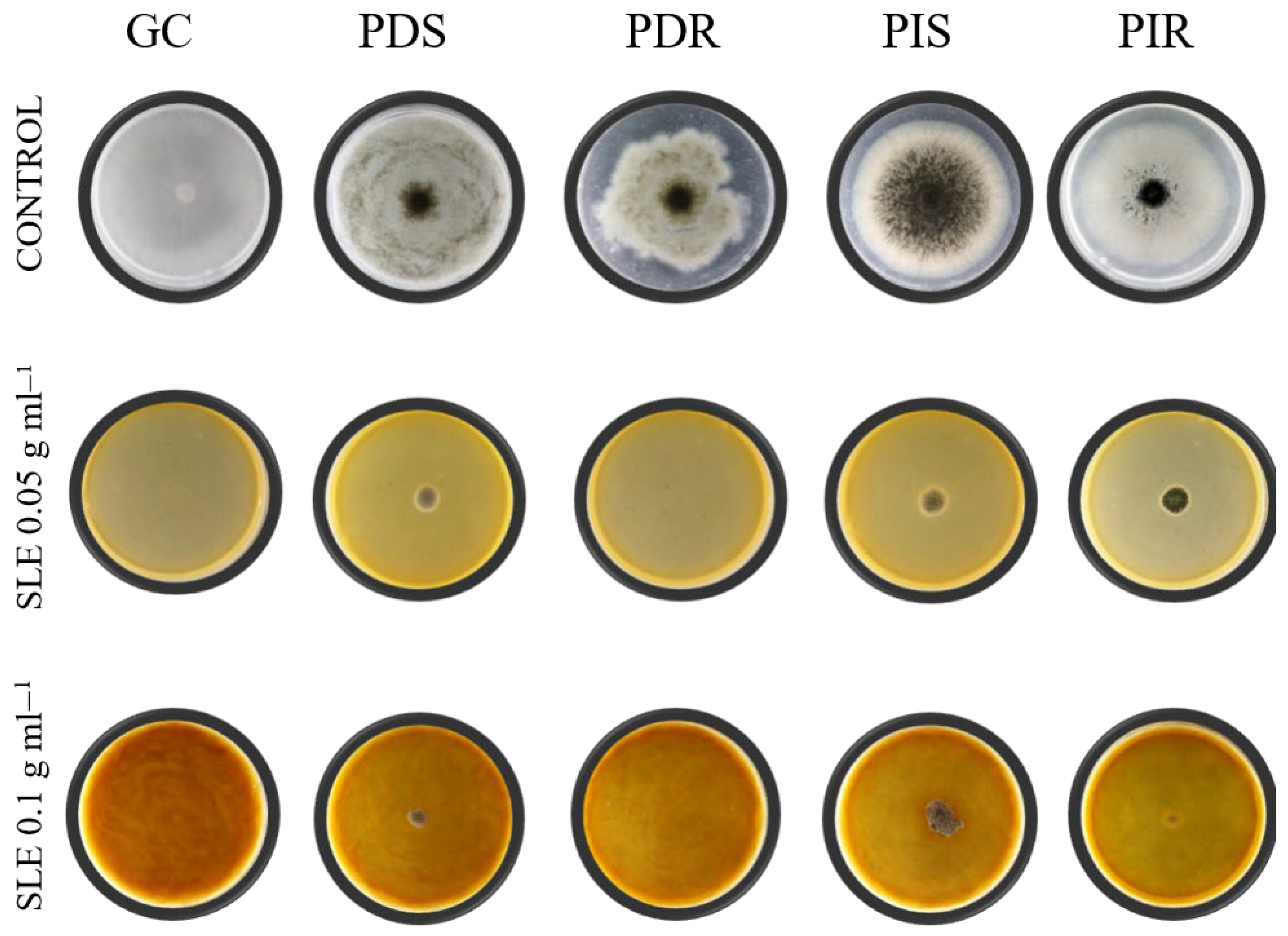
The antifungal activity of SLE against the fungicide-sensitive or fungicide-resistant phytopathogens (PDS, PDR, PIS and PIR) and GC were evaluated. Figure 1 shows that 0.05 g mL−1 of SLE incorporated in the PDA plate was enough to inhibit the mycelial growth of all fungal pathogens evaluated. Controls were able to grow and complete PDA plate without SLE at 5d. As was expected, the mycelial growth on PDA plate amended with 0.1 g mL−1 SLE was totally inhibited under the experimental conditions tested here (Table 1).
Mycelial growth was evaluated as a measure of colony diameter after 5 d incubation at 24 ± 1 °C, for each condition. Controls consisted of PDA plates without supplements. Different letters indicate significant differences comparing each pathogen to their respective control according to Tukey’s analysis with a p-value ≤ 0.05. % Inhibition = (C − T/C) × 100, where C is the colony diameter in control conditions and T is colony diameter in the treatment. GC, G. citri-aurantii; PDS, P. digitatum; PIS, P. italicum; triples fungicide-resistant variants PDR, P. digitatum; PIR, P. italicum.
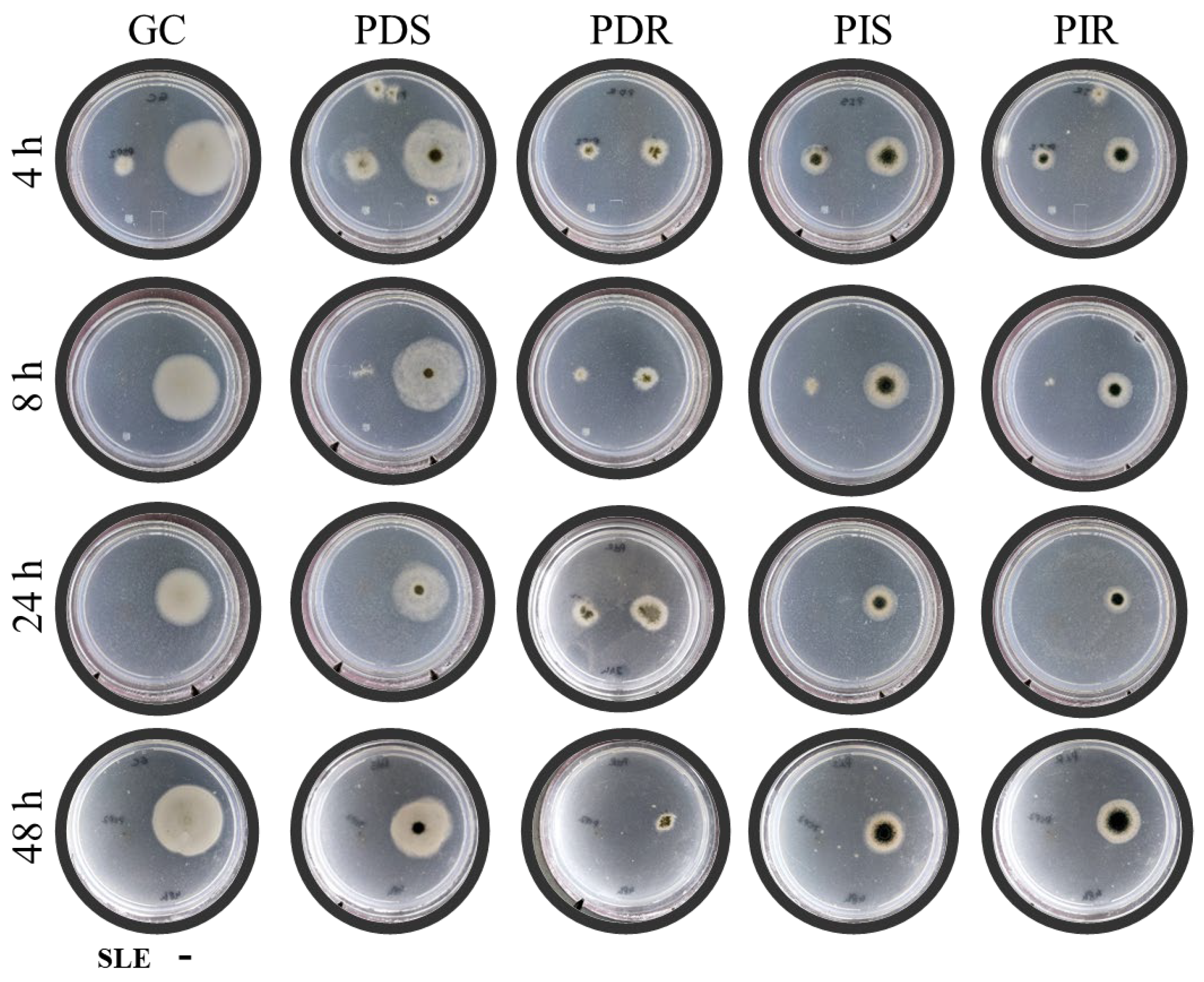
In the minimum conidia killing time assays, when GC and PIR were incubated with 0.1 g mL−1 SLE for 8 h, a complete growth inhibition was observed. In respect to PDS and PIS, a 24 h of treatment with SLE were necessary to achieve the same effect. Finally, PDR appears as the most resistant isolates assayed since it required a 48 h SLE treatment to complete the growth inhibition (Figure 2) Additionally, it is worth noting that the incubation of all pathogens conidia with SLE for 4 h resulted in only a partial growth inhibition.
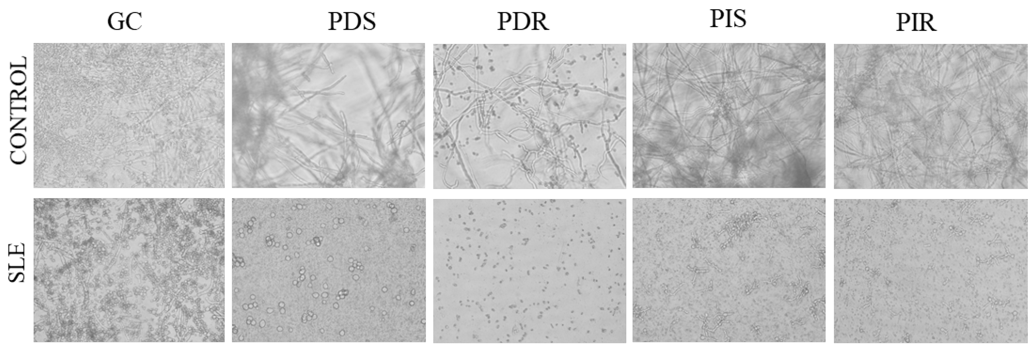
In respect to conidia germination, the results showed that 0.1 g mL−1 SLE was able to inhibit conidia germination of all phytopathogens evaluated (Figure 3), while the control condition was completely germinated after 24 h.

3.2. Effect of SLE on Conidial Membrane Integrity

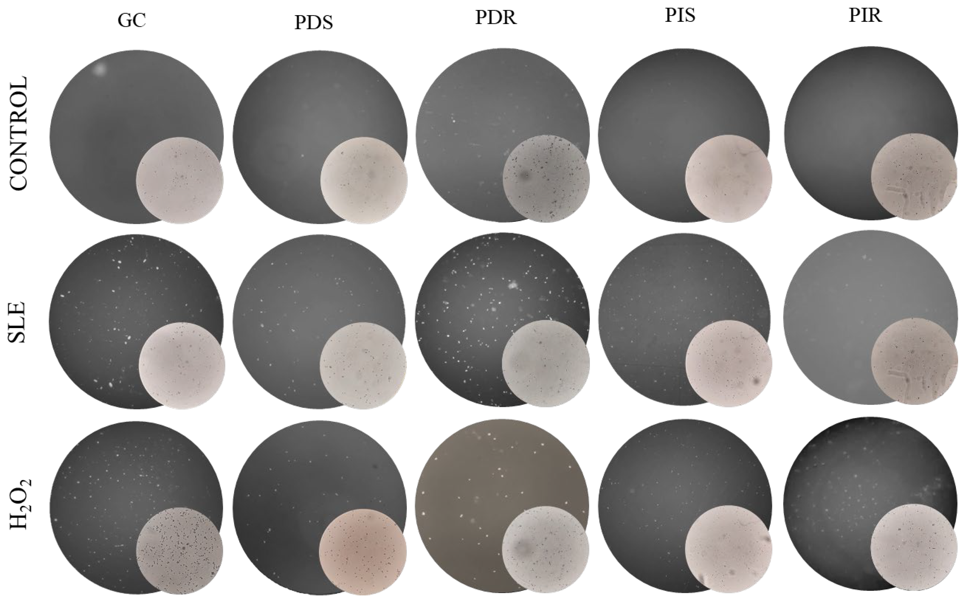
In order to analyze the effect of SLE over fungal conidia, a study of the integrity cell membrane was conducted using the SYTOXTM Green probe (Thermo Fisher Scientific, Waltham, MA, USA) (Figure 4). In the dark field, fluorescent conidia were observed for all fungal isolates treated with 0.1 g mL−1 of SLE, indicating membrane permeability as evidence of cellular damage associated with SLE treatment. As positive control of membrane damages, an incubation with 600 mM H2O2 was included, which shows fluorescence for all the phytopathogens, similar to SLE treatments. No fluorescence emission was registered in samples incubated with water, as negative control.

3.3. Internal Structure Conidial After SLE Treatment

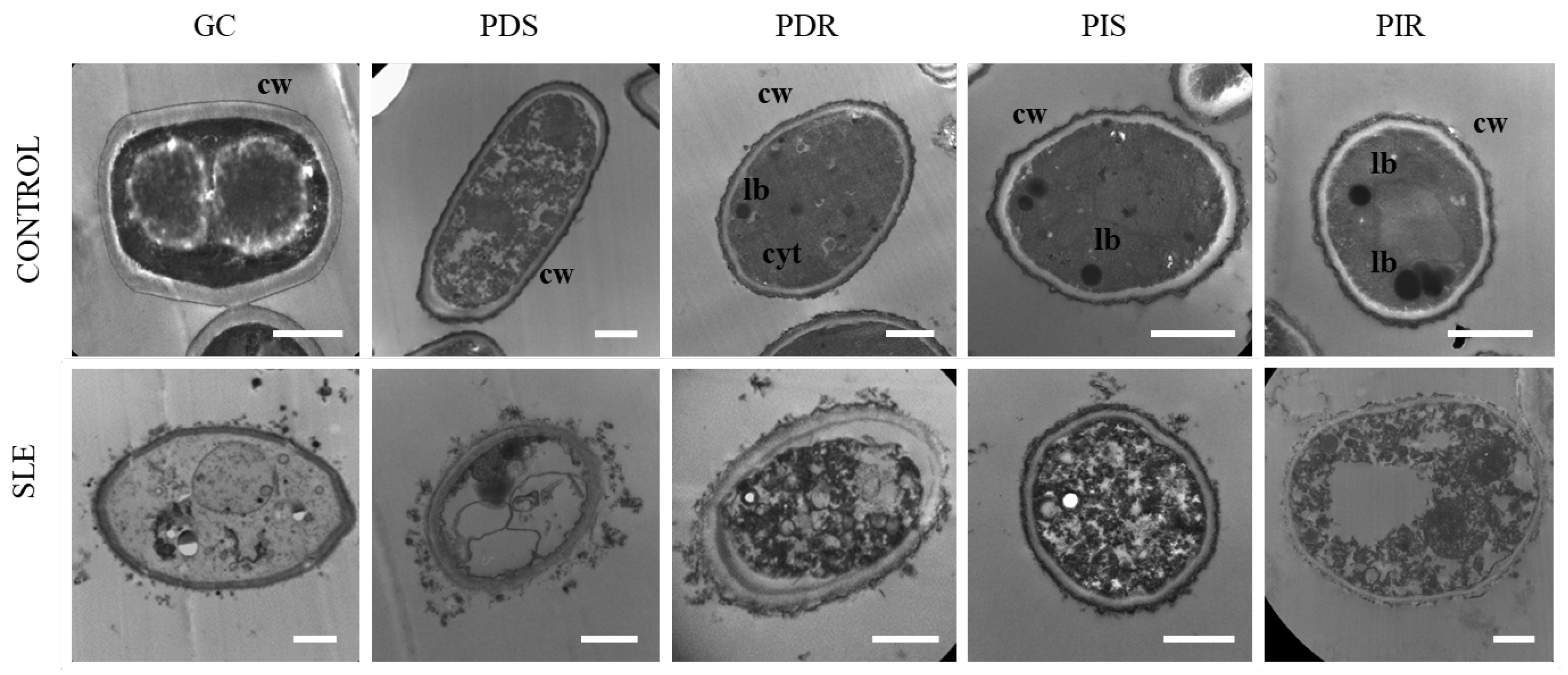
Ultrastructural characterization of conidia via transmission electron microscopy (TEM, Thermo Fisher Scientific, Waltham, MA, USA) (Figure 5) revealed that fungi in the control condition exhibited a well-preserved cellular architecture, characterized by organized cell walls and defined cytoplasmic components, such as mitochondria and lipid bodies. In contrast, exposure to 0.1 g mL−1 of SLE for 24 h induced considerable morphological alterations in all evaluated isolates. Significant observations included marked cell deformation, generalized cytoplasmic disorganization, and the proliferation of multiple intracellular vesicles. Furthermore, severe modifications in plasma membrane integrity and cell wall structure were evident, confirming that SLE treatment results in irreversible and lethal cellular damage to the conidia.

3.4. Effect of SLE In Situ Application over Green Mold Incidence on Lemons

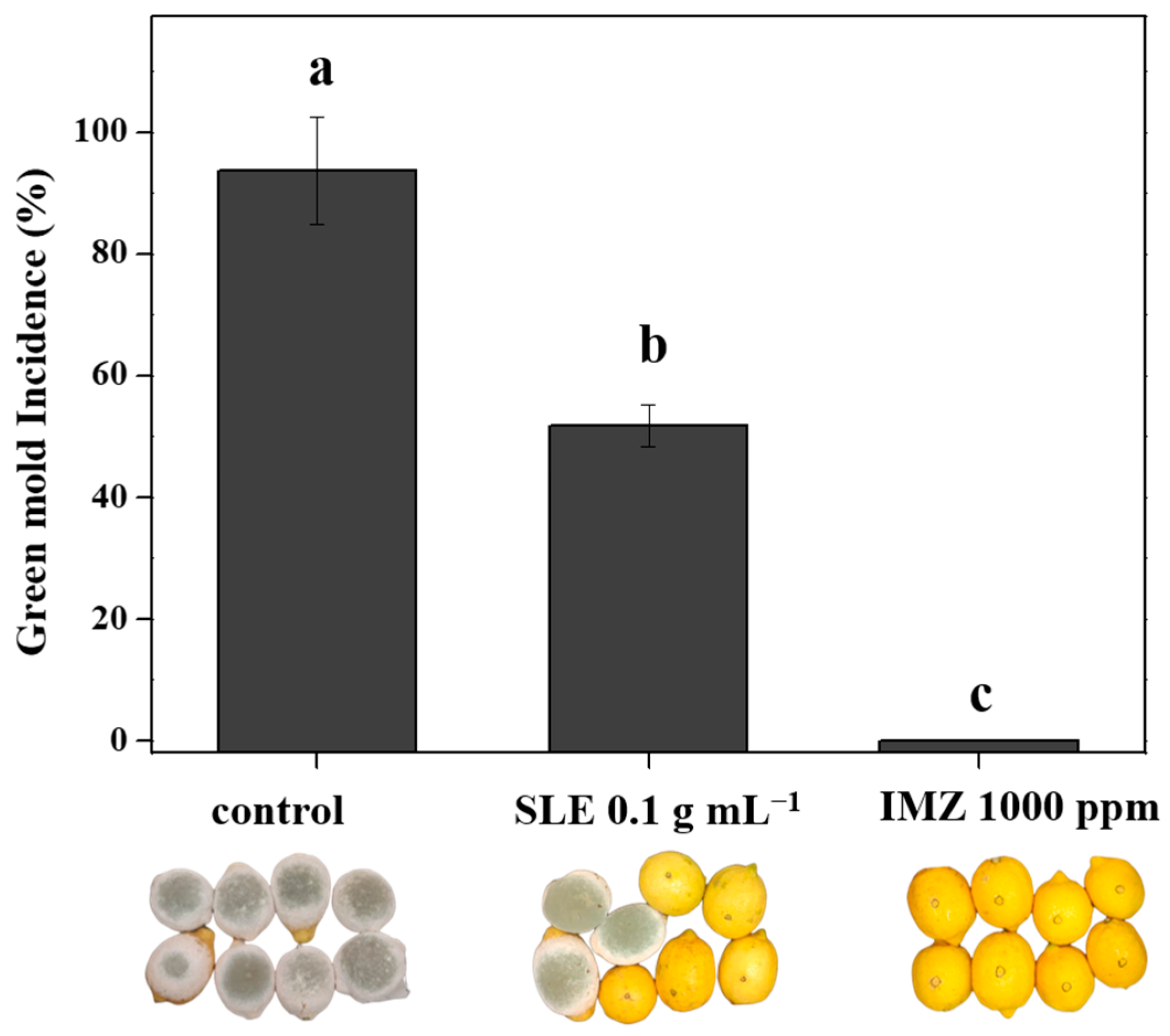
The antifungal efficacy of SLE was evaluated by in situ application on artificial wounded and inoculated lemons. In control conditions, a high disease incidence to 100% was observed (Figure 6). The application of SLE demonstrated significant green mold control, achieving 52 ± 3.5% disease incidence in respect to the control without treatment with 92% ± 8 of decay incidence. As was expected, the IMZ application was able to totally control green mold caused by PDS.

4. Discussion

In this study, the effectiveness of SLE, an aqueous extract obtained from strawberry leaves, was demonstrated to inhibit in vitro and in vivo the major citrus fungal pathogens. Therefore, our contribution not only identifies a new product to contribute to postharvest diseases, but also to waste plant utilization. Strawberry production in Tucumán covers approximately 350 hectares, with the cultivar San Andreas representing around 34% of the cultivated area [35]. Considering a planting density of approximately 60,000 plants per hectare and an estimated yield of 100 g of fresh leaves per plant, a substantial amount of vegetative biomass is generated during the crop cycle. This corresponds to roughly 6000 kg of fresh leaf material per hectare, which is typically discarded. These figures highlight the significant potential of this agricultural by-product as a readily available and sustainable raw material to produce bioinputs as SLE. Moreover, the production process involves relatively low costs, as it mainly requires logistics, appropriate storage of the leaves, and basic extraction equipment, making it an economically feasible strategy that adds value to the production system while contributing to waste reduction.
Our results shown that SLE inhibited mycelial growth at 0.05 g·mL−1 and completely suppresses conidia germination at 0.10 g·mL−1 for P. digitatum, P. italicum, and G. citri-aurantii, regardless of their sensitivity to conventional fungicides. These inhibitory concentrations are comparable to those reported for other plant-derived extracts active against postharvest citrus pathogens. For instance, aqueous extracts of Cistus spp. inhibited 80–100% of G. citri-aurantii growth and germination at concentrations from 0.1 and 0.05 g·mL−1, respectively [17]. Similarly, powders, solvent extracts, and essential oils from species such as Thymus leptobotrys and Peganum harmala have shown broad-spectrum antifungal activity against P. digitatum, P. italicum, and Geotrichum spp., achieving growth reductions close to complete inhibition [36,37]. Comparable antifungal effects have also been reported for extracts from extremophile plant species, which inhibited P. digitatum and affected G. citri-aurantii growth at phenolic-adjusted concentrations [38,39].
To elucidate the antifungal mode(s) of action of the SLE on phytopathogenic fungi, membrane integrity assays with fluorescent probes were performed, together with ultrastructural analysis by TEM. These analyses confirmed plasma membrane permeabilization, cytoplasmic disorganization, and contraction of the cell wall, evidence of irreversible cellular damage. It is known that fungal viability relies primarily on the integrity of the plasma membrane [40,41] which would explain the loss of viability observed with SLE treatment. Moreover, the pattern of structural injury is consistent with mechanisms reported for other natural metabolites, such as cinnamaldehyde, the principal component of cinnamon essential oil, which disrupts cell wall integrity [14], and natamycin, obtained from Streptomyces natalensis fermentation, whose action centers on plasma membrane damage [42,43]. Plant-derived antifungal compounds acting on multiple cellular targets are associated with a reduced risk of resistance development, as they limit adaptation to single molecular targets [14,44,45]. The antifungal activity of SLE suggests a general structural disruption due to multi-target mode of action, conferring an advantage over single-site synthetic fungicides for resistance management. According to the criteria of the Fungicide Resistance Action Committee, multi-site fungicides are classified as low-risk tools for resistance development [46].
The effect observed over membrane and internal structure in fungal conidia exposed to SLE (0.1 g mL−1) for 24 h is in agreement with previous reports. OuYang et al. [14] observed a similar effect after 2 h of exposure of G. citri-aurantii conidia to cinnamaldehyde. The effect of SLE is particularly critical for controlling GC, causal agent of sour rot, a pathogen for which commercial fungicidal options remain limited. Since total inhibition of viability in GC and fungicide-resistant PIR was achieved after only 8 h of incubation with SLE—and for other pathogens between 24 and 48 h—future studies could focus on optimizing the minimum contact time required to induce irreversible fungitoxicity, in order to determine the operational and economic feasibility of the extract in postharvest applications.
In vivo disease control trials are critical for validating the commercial potential of biofungicides. In this work, inoculation assays on fruit under control conditions were conducted. In the in situ application of SLE on artificially wounded lemons, reduced green mold (P. digitatum) incidence was nearly 50% with respect to the control without treatment, whereas IMZ achieved total disease suppression. Compared with other plant extracts, the efficacy obtained with the SLE surpassed that reported for aqueous, methanolic, and ethanolic extracts of Cynara cardunculus in citrus fruit against P. digitatum [15]; only the ethanolic extract produced a reduction of approximately 10% in green mold incidence relative to the control. Additionally, aqueous extracts rich in phenolic compounds derived from Cistus spp. reduced sour rot incidence in mandarins caused by G. citri-aurantii by approximately 85% [17]. Similarly, Alvarez et al. [39] evaluated the methanolic extract of the extremophile plant Solanum pilcomayense (3000 ppm), applied by immersion to oranges inoculated with P. digitatum, and demonstrated significant curative activity, reducing the sporulation index to levels comparable to those achieved with the fungicide IMZ. Likewise, Fernández et al. [47] achieved control efficacy similar to SLE using a commercial product formulated from Allium extract, reducing G. citri-aurantii incidence by about 50% compared with the control under controlled conditions. However, this product requires co-application with a wax coating to prevent peel phytotoxicity. It is worth to note the SLE did not cause phytotoxicity in treated fruit, representing an advantage in terms of safety and technological feasibility. Therefore, its efficacy could be further improved through formulation strategies aimed at enhancing extract concentration and retention at the wound site without compromising fruit integrity. It is also important to highlight the efficacy of the SLE against strains with multiple resistances to fungicides such as IMZ, TBZ and PYR. This behavior suggests that incorporating SLE into resistance management programs could provide strategic benefits, although further optimization and validation are required under commercial conditions to confirm this hypothesis. Its ability to control both sensitive and resistant strains positions SLE as a potentially valuable candidate for diversifying and strengthening the tools available for postharvest citrus disease management.

5. Conclusions

In conclusion, strawberry leaf extract (SLE) represents a potent and novel alternative for the postharvest management of citrus fungal diseases. This study demonstrated that the antifungal activity of SLE is mediated by plasma membrane permeabilization and irreversible intracellular damage, leading to a complete loss of conidial viability. The efficacy of SLE was further validated through in vivo trials, which confirmed a significant reduction in green mold incidence on lemons. Beyond its biological activity, the valorization of strawberry crop residues as a feedstock for SLE production offers sustainable and economically viable bioinput. The integration of SLE into postharvest protocols could substantially decrease reliance on synthetic fungicides, facilitating a transition toward safer, more efficient systems that align with global food security standards and environmental sustainability regulations.

Author Contributions

Conceptualization, L.C. and S.I.V.; methodology, G.M., P.D.P., M.A.D., L.C. and S.I.V.; validation, G.M. and P.D.P.; formal analysis, N.R.C., L.C. and S.I.V.; investigation, G.M. and P.D.P.; data curation, M.A.D., G.M. and P.D.P.; writing—original draft preparation, P.D.P., L.C. and S.I.V.; writing—review and editing, G.M., P.D.P., M.A.D., N.R.C., B.V.W., L.C. and S.I.V.; supervision, L.C. and S.I.V.; funding acquisition, B.V.W., N.R.C., L.C. and S.I.V. All authors have read and agreed to the published version of the manuscript.

Funding

This work was supported by grants from Agencia Nacional de Promoción Científica y Tecnológica (PICT 2019-1380 and PICT 2021-I-A-00850), CONICET (PIP 2547) and Universidad Nacional de Tucumán (PIUNT D768).

Data Availability Statement

The original contributions presented in the study are included in the article, further inquiries can be directed to the corresponding authors.

Acknowledgments

We extend our special thanks to Las Tipas farm for generously providing the lemons and to Ing. Gabriel Rodriguez for supplying lemon fruit. Additionally, we thank Agustin Padilla, Andrea Peña and Fernanda Trejo for their technical contributions. We also acknowledge ANNUIT S.A. for their support.

Conflicts of Interest

The authors declare no conflicts of interest.

References

  1. Eckert, J.W.; Eaks, I.L. Postharvest disorders and diseases of citrus fruits. In The Citrus Industry; Reuther, W., Calavan, E.C., Carman, G.E., Eds.; University of California Division of Agricultural Natural Resources: Oakland, CA, USA, 1989; Volume 5, pp. 179–260. [Google Scholar]
  2. Pitt, J.I.; Hocking, A.D. Primary keys and miscellaneous fungi. In Fungi and Food Spoilage; Pitt, J.I., Hocking, A.D., Eds.; Springer: Boston, MA, USA, 2009; pp. 53–143. [Google Scholar]
  3. Wang, W.; Liu, S.; Deng, L.; Ming, J.; Yao, S.; Zeng, K. Control of citrus post-harvest green molds, blue molds, and sour rot by the cecropin A-melittin hybrid peptide BP21. Front. Microbiol. 2018, 9, 2455. [Google Scholar] [CrossRef] [PubMed]
  4. Federcitrus. Actividad Citrícola. 2025. Available online: https://www.federcitrus.org/wp-content/uploads/2025/10/Federcitrus-Actividad-citricola-2025-02102025.pdf (accessed on 27 February 2026).
  5. Kinay, P.; Mansour, M.F.; Mlikota Gabler, F.; Margosan, D.A.; Smilanick, J.L. Characterization of fungicide-resistant isolates of Penicillium digitatum collected in California. Crop Prot. 2007, 26, 647–656. [Google Scholar] [CrossRef]
  6. Sánchez-Torres, P.; Tuset, J.J. Molecular insights into fungicide resistance in sensitive and resistant Penicillium digitatum strains infecting citrus. Postharvest Biol. Technol. 2011, 59, 159–165. [Google Scholar] [CrossRef]
  7. Schneider, K.; Barreiro-Hurle, J.; Rodriguez-Cerezo, E. Pesticide reduction amidst food and feed security concerns in Europe. Nat. Food 2023, 6, 746–750. [Google Scholar] [CrossRef]
  8. Yin, Y.; Miao, J.; Shao, W.; Liu, X.; Zhao, Y.; Ma, Z. Fungicide resistance: Progress in understanding mechanisms, monitoring, and management. Phytopathology 2023, 113, 707–718. [Google Scholar] [CrossRef] [PubMed]
  9. Tilman, D.; Cassman, K.G.; Matson, P.A.; Naylor, R.; Polasky, S. Agricultural sustainability and intensive production practices. Nature 2002, 418, 671–677. [Google Scholar] [CrossRef] [PubMed]
  10. Palou, L.; Ali, A.; Fallik, E.; Romanazzi, G. GRAS, plant- and animal-derived compounds as alternatives to conventional fungicides for the control of postharvest diseases of fresh horticultural produce. Postharvest Biol. Technol. 2016, 122, 41–52. [Google Scholar] [CrossRef]
  11. Usall, J.; Ippolito, A.; Sisquella, M.; Neri, F. Physical treatments to control postharvest diseases of fresh fruits and vegetables. Postharvest Biol. Technol. 2016, 122, 30–40. [Google Scholar] [CrossRef]
  12. Devi, M.J.; Gidado, S. Sustainable valorization of postharvest waste for disease control and quality preservation in fruits and vegetables. Waste Manag. 2026, 192, 25–38. [Google Scholar]
  13. Bajaj, K.; Adhikary, T.; Gill, P.P.S.; Kumar, A. Edible coatings enriched with plant-based extracts preserve postharvest quality of fruits: A review. Prog. Org. Coat. 2023, 182, 107669. [Google Scholar] [CrossRef]
  14. OuYang, Q.; Duan, X.; Li, L.; Tao, N. Cinnamaldehyde exerts its antifungal activity by disrupting the cell wall integrity of Geotrichum citri-aurantii. Front. Microbiol. 2019, 10, 55. [Google Scholar] [CrossRef] [PubMed]
  15. Restuccia, C.; Lombardo, M.; Scavo, A.; Mauromicale, G.; Cirvilleri, G. Combined application of antagonistic Wickerhamomyces anomalus BS91 strain and Cynara cardunculus L. leaf extracts for the control of postharvest decay of citrus fruit. Food Microbiol. 2020, 92, 103583. [Google Scholar] [CrossRef] [PubMed]
  16. Hao, W.; Li, H.; Hu, M.; Yang, L.; Rizwan-ul-Haq, M. Integrated control of citrus green and blue mold and sour rot by Bacillus amyloliquefaciens in combination with tea saponin. Postharvest Biol. Technol. 2011, 59, 316–323. [Google Scholar] [CrossRef]
  17. Karim, H.; Boubaker, H.; Askarne, L.; Cherifi, K.; Lakhtar, H.; Msanda, F.; Boudyach, E.H.; Ait Ben Aoumar, A. Use of Cistus aqueous extracts as botanical fungicides in the control of citrus sour rot. Microb. Pathog. 2017, 104, 139–143. [Google Scholar] [CrossRef]
  18. Bai, T.; Wang, X.; Du, W.; Cheng, J.; Zhang, J.; Zhang, Y.; Klinjapo, R.; Asavasanti, S.; Yasurin, P. Recent advances, challenges, and functional applications of natural phenolic compounds in the meat products industry. Antioxidants 2025, 14, 138. [Google Scholar] [CrossRef] [PubMed]
  19. Villamil-Galindo, E.; Van de Velde, F.; Piagentini, A.M. Strawberry agro-industrial by-products as a source of bioactive compounds: Effect of cultivar on the phenolic profile and the antioxidant capacity. Bioresour. Bioprocess. 2021, 8, 61. [Google Scholar] [CrossRef] [PubMed]
  20. Secretaría de Agricultura; Ganadería y Pesca de la Nación. Producción de frutilla en Argentina (Informe de Enero 2023). Ministerio de Economía, Presidencia de la Nación. 2023. Available online: https://alimentosargentinos.magyp.gob.ar/HomeAlimentos/difusion-y-publicaciones/Revistas/AA_83.pdf (accessed on 27 February 2026).
  21. Buendía, B.; Gil, M.I.; Tudela, J.A.; Gady, A.L.; Medina, J.J.; Soria, C.; Tomás-Barberán, F.A. HPLC-MS analysis of proanthocyanidin oligomers and other phenolics in 15 strawberry cultivars. J. Agric. Food Chem. 2010, 58, 3916–3926. [Google Scholar] [PubMed]
  22. Hernanz, D.; Recamales, A.F.; Meléndez-Martínez, A.J.; González-Miret, M.L.; Heredia, F.J. Assessment of the differences in the phenolic composition of five strawberry cultivars (Fragaria × ananassa Duch.) grown in two different soilless systems. J. Agric. Food Chem. 2007, 55, 1846–1852. [Google Scholar] [CrossRef] [PubMed]
  23. Filippone, M.P.; Diaz Ricci, J.M.; Mamaní de Marchese, A.; Castagnaro, A.; Farías, R.N. Effect of fragarin on the cytoplasmic membrane of the phytopathogen Clavibacter michiganensis. Mol. Plant-Microbe Interact. 2001, 14, 925–928. [Google Scholar] [CrossRef] [PubMed][Green Version]
  24. Filippone, M.P.; Diaz Ricci, J.C.; Mamani de Marchese, A.; Farías, R.N.; Castagnaro, A.P. Isolation and purification of a 316 Da pre-formed compound from strawberry (Fragaria ananassa) leaves active against plant pathogens. FEBS Lett. 1999, 459, 115–118. [Google Scholar] [CrossRef] [PubMed]
  25. Mamaní, A.; Filippone, M.P.; Grellet, C.; Björn, W.; Castagnaro, A.P.; Díaz Ricci, J.C. Pathogen-induced accumulation of an ellagitannin elicits plant defense response. Mol. Plant-Microbe Interact. 2012, 25, 1430–1439. [Google Scholar] [CrossRef] [PubMed]
  26. Martos, G.G.; Mamaní, A.; Filippone, M.P.; Abate, P.O.; Katz, N.E.; Castagnaro, A.P.; Díaz Ricci, J.C. Ellagitannin HeT obtained from strawberry leaves is oxidized by bacterial membranes and inhibits the respiratory chain. FEBS Open Bio 2018, 8, 211–218. [Google Scholar] [CrossRef] [PubMed]
  27. Cerviño, A.; Grellet, C.F.; Di Peto, P.A.; Rodríguez, L.C.; Castagnaro, A.P.; Filippone, M.P.; Mamani de Marchese, A.I. Evaluación de la actividad antimicrobiana in vitro de extractos de hojas de diferentes cultivares de Fragaria ananassa Duch. para la formulación de bioinsumos fitosanitarios. Rev. Agron. Noroeste Argent. 2019, 39, 83–87. [Google Scholar] [CrossRef]
  28. Welin, B.; Castagnaro, A.P.; Filippone, M.P.; Grellet-Bournonville, C.F.; Mamani, A.I.; Di Peto, P. Glycoside Compound of Fatty Acids, Composition Comprising It, Process for Its Obtention and Methods to Apply It on Plants or Fruits or Both at the Same Time. International Patent Application No. PCT/IB2018/056778, 14 March 2019. [Google Scholar]
  29. Grellet Bournonville, C.; Filippone, M.P.; Di Peto, P.A.; Trejo, M.F.; Couto, A.S.; Mamani de Marchese, A.I.; Díaz Ricci, J.C.; Björn, W.; Castagnaro, A.P. Strawberry fatty acyl glycosides enhance disease protection, have antibiotic activity and stimulate plant growth. Sci. Rep. 2020, 10, 8196. [Google Scholar] [CrossRef] [PubMed]
  30. Olmedo, G.M.; Debes, M.A.; Sepúlveda, M.; Ramallo, J.; Rapisarda, V.A.; Cerioni, L.; Volentini, S.I. Overcoming lemon postharvest molds caused by Penicillium spp. multiresistant isolates by the application of potassium sorbate in aqueous and wax treatments. J. Food Sci. 2023, 88, 1234–1245. [Google Scholar] [CrossRef]
  31. Olmedo, G.M.; Cerioni, L.; González, M.M.; Cabrerizo, F.M.; Rapisarda, V.A.; Volentini, S.I. Antifungal activity of β-carbolines on Penicillium digitatum and Botrytis cinerea. Food Microbiol. 2017, 62, 9–15. [Google Scholar] [CrossRef] [PubMed]
  32. Roth, B.L.; Poot, M.; Yue, S.T.; Millard, P.J. Bacterial viability and antibiotic susceptibility testing with SYTOX green nucleic acid stain. Appl. Environ. Microbiol. 1997, 63, 2421–2431. [Google Scholar] [CrossRef] [PubMed]
  33. Cerioni, L.; Volentini, S.I.; Prado, F.E.; Rapisarda, V.A.; Rodríguez-Montelongo, L. Cellular damage induced by a sequential oxidative treatment on Penicillium digitatum. J. Appl. Microbiol. 2010, 109, 123–130. [Google Scholar] [CrossRef]
  34. Liu, Y.; Wang, W.H.; Zhou, Y.H.; Yao, S.X.; Deng, L.L.; Zeng, K.F. Isolation, identification and in vitro screening of Chongqing orangery yeasts for the biocontrol of Penicillium digitatum on citrus fruit. Biol. Control 2017, 110, 18–24. [Google Scholar] [CrossRef]
  35. Frutillas—Economía Regional—Alimentos Argentinos. 2024. Available online: https://www.bcr.com.ar/es/mercados/investigacion-y-desarrollo/informativo-semanal/noticias-informativo-semanal/economias (accessed on 6 April 2026).
  36. Ameziane, N.; Boubaker, H.; Boudyach, H.; Msanda, F.; Jilal, A.; Ait Benaoumar, A. Antifungal activity of Moroccan plants against citrus fruit pathogens. Agron. Sustain. Dev. 2007, 27, 273–277. [Google Scholar] [CrossRef]
  37. Boubaker, H.; Karim, H.; El Hamdaoui, A.; Msanda, F.; Leach, D.; Bombarda, I.; Vanloot, P.; Abbad, A.; Boudyach, E.H.; Ait Ben Aoumar, A. Chemical characterization and antifungal activities of four Thymus species essential oils against postharvest fungal pathogens of citrus. Ind. Crops Prod. 2024, 86, 95–101. [Google Scholar]
  38. Sayago, J.E.; Ordoñez, R.M.; Kovacevich, L.N.; Torres, S.; Isla, M.I. Antifungal activity of extracts of extremophile plants from the Argentine Puna to control citrus postharvest pathogens and green mold. Postharvest Biol. Technol. 2012, 67, 19–24. [Google Scholar] [CrossRef]
  39. Alvarez, N.H.; Stegmayer, M.I.; Seimandi, G.M.; Pensiero, J.F.; Zabala, J.M.; Favaro, M.A.; Derita, M.G. Natural products obtained from Argentinean native plants are fungicidal against citrus postharvest diseases. Horticulturae 2023, 9, 562. [Google Scholar] [CrossRef]
  40. Shao, X.; Cheng, S.; Wang, H.; Yu, D.; Mungai, C. The possible mechanism of antifungal action of tea tree oil on Botrytis cinerea. J. Appl. Microbiol. 2013, 114, 1642–1649. [Google Scholar] [CrossRef] [PubMed]
  41. Tao, N.G.; OuYang, Q.L.; Jia, L. Citral inhibits mycelial growth of Penicillium italicum by a membrane damage mechanism. Food Control 2014, 41, 116–121. [Google Scholar] [CrossRef]
  42. He, C.; Zhang, Z.; Li, B.; Xu, Y.; Tian, S. Effect of natamycin on Botrytis cinerea and Penicillium expansum postharvest pathogens of grape berries and jujube fruit. Postharvest Biol. Technol. 2019, 151, 134–141. [Google Scholar] [CrossRef]
  43. te Welscher, Y.M.; ten Napel, H.H.; Balagué, M.M.; Souza, C.M.; Riezman, H.; de Kruijff, B.; Breukink, E. Natamycin blocks fungal growth by binding specifically to ergosterol without permeabilizing the membrane. J. Biol. Chem. 2008, 283, 6393–6401. [Google Scholar] [CrossRef] [PubMed]
  44. Regnier, T.; Combrinck, S.; Veldman, W.; Du Plooy, W. Application of essential oils as multi-target fungicides for the control of Geotrichum citri-aurantii and other postharvest pathogens of citrus. Ind. Crops Prod. 2014, 61, 151–159. [Google Scholar] [CrossRef]
  45. Leiva-Mora, M.; Bustillos, D.; Arteaga, C.; Hidalgo, K.; Guevara-Freire, D.; López-Hernández, O.; Saa, L.R.; Padilla, P.S.; Bustillos, A. Antifungal mechanisms of plant essential oils: A comprehensive literature review for biofungicide development. Agriculture 2025, 15, 2303. [Google Scholar] [CrossRef]
  46. FRAC. Importance of Multisite Fungicides in Managing Pathogen Resistance. Fungicide Resistance Action Committee. 2018. Available online: https://www.frac.info/media/ztqosoch/frac-statement-on-multisite-fungicides-2018.pdf (accessed on 20 January 2026).
  47. Fernández, G.; Sbres, M.; Lado, J.; Pérez-Faggiani, E. Postharvest sour rot control in lemon fruit by natamycin and an Allium extract. Int. J. Food Microbiol. 2022, 368, 109605. [Google Scholar] [CrossRef] [PubMed]
Figure 1. Antifungal activity of SLE. Mycelial growth was assessed by placing 5 µL drops of conidial suspensions (106 CFU mL−1) of each fungal pathogen—G. citri-aurantii (GC), P. digitatum (PDS), P. italicum (PIS) and triples fungicide-resistant variants P. digitatum (PDR) and P. italicum (PIR)—on PDA supplemented with the indicated concentration of SLE. Controls consisted of PDA plates without supplements. The photograph is a representative image from three independent experiments.
Horticulturae 12 00782 g001
Figure 2. Minimum killing conidia time. Conidial suspensions (106 CFU mL−1) of each pathogen—G. citri-aurantii (GC), P. digitatum (PDS), P. italicum (PIS) and triples fungicide-resistant variants P. digitatum (PDR) and P. italicum (PIR)—were exposed to SLE (0.1 g mL−1). At each indicated time, an aliquot of treated suspensions was spotted onto PDA medium without supplements and was incubated 5 d at 24 ± 1 °C. Controls consisting of conidial suspensions incubated with water. The photograph is a representative image from three independent experiments.
Horticulturae 12 00782 g002
Figure 3. Conidia germination. Conidial suspensions (106 CFU mL−1) in PDB at pH 5 of each fungal pathogen—G. citri-aurantii (GC), P. digitatum (PDS), P. italicum (PIS) and triples fungicide-resistant variants P. digitatum (PDR) and P. italicum (PIR)—were exposed to 0.1 g mL−1 SLE and incubated at 24 ± 1 °C for 24 h. After that, germinated and non-germinated conidia were observed under an inverted microscope (×40). The photograph is a representative image from three independent experiments of germination and general aspect to control and treated conidia (magnification = 400×).
Horticulturae 12 00782 g003
Figure 4. Effect of SLE on membrane integrity. Conidial suspensions (106 CFU mL−1) of each fungal pathogen—G. citri-aurantii (GC), P. digitatum (PDS), P. italicum (PIS) and triples fungicide-resistant variants P. digitatum (PDR) and P. italicum (PIR)—were treated with 0.1 g mL−1 SLE for 24 h, before their incubation with SYTOXTM Green. Fluorescent field images at a 40× magnification accompanied by a bright field (insets) are shown. The upper panels display shown controls with water and, in the lower panels display, positive controls corresponding to conidia treated with H2O2. The images are representative of three independent experiments.
Horticulturae 12 00782 g004
Figure 5. Intracellular damage induced by SLE. Conidial suspensions of each fungal pathogen—G. citri-aurantii (GC), P. digitatum (PDS), P. italicum (PIS) and triples fungicide-resistant variants P. digitatum (PDR) and P. italicum (PIR)—were treated at 0.1 g mL−1 of SLE for 24 h and then were washed and visualized by TEM. The tests were repeated three times for each treatment, and at least two replicates were examined. In micrographs, a scale graphic = 1 µm is represented. cw, cell wall; cyt, cytoplasm; lb, lipid body.
Horticulturae 12 00782 g005
Figure 6. In situ action of SLE against P. digitatum (PDS) on lemons. SLE, IMZ or water (control) were added on lemon wounds before PDS inoculation. Fruits were stored at 23 °C and 95% relative humidity, and disease incidence was evaluated at 5 d post infection. Different letters indicate significant differences with respect to the control group according to Tukey’s analysis, with a p-value ≤ 0.05. Photographs show fruit aspect after incubation and are representative of three different experiments.
Horticulturae 12 00782 g006
Table 1. Effect of SLE on phytopathogens mycelial growth.
PathogenDiameter Radial
Grow (mm)
% Inhibition
ControlSLE
0.05 g mL−1
0.1 g mL−1SLE
0.05 g mL−1
0.1 g mL−1
GC49 ± 1 a0 b0 b100 b100 b
PDS50 ± 1 a7 ± 1 b4 ± 1 b86 ± 2 b94 ± 2 b
PDR40 ± 1 a0 b0 b100 b100 b
PIS44 ± 2 a11 ± 1 b6 ± 2 b75 ± 2 b86 ± 3 b
PIR48 ± 1 a6 ± 2 b0 b86 ± 4 b100 b
Different letters indicate significant differences p-value ≤ 0.05.
Disclaimer/Publisher’s Note: The statements, opinions and data contained in all publications are solely those of the individual author(s) and contributor(s) and not of MDPI and/or the editor(s). MDPI and/or the editor(s) disclaim responsibility for any injury to people or property resulting from any ideas, methods, instructions or products referred to in the content.