Article

Organ-Dependent Comparative Metabolomic Profiling of Actinidia arguta Using LC–QTOF–MS Reveals Enrichment of Condensed Tannins in Roots

1
College of Pharmacy, Chungbuk National University, Cheongju 28160, Republic of Korea
2
College of Pharmacy, Korea University, Sejong 30019, Republic of Korea
3
Food and Pharmacy College, Xuchang University, Xuchang 461000, China
4
Interdisciplinary Major Program in Innovative Pharmaceutical Sciences, Korea University, Sejong 30019, Republic of Korea
*
Authors to whom correspondence should be addressed.
These authors contributed equally to this work.
Horticulturae 2026, 12(4), 454; https://doi.org/10.3390/horticulturae12040454
Submission received: 16 February 2026 / Revised: 1 April 2026 / Accepted: 3 April 2026 / Published: 7 April 2026

Abstract

Actinidia arguta is a valuable plant resource known for its diverse bioactive constituents. However, organ-dependent metabolic variation remains insufficiently explored. In this study, an integrated approach combining LC–QTOF–MS-based metabolomic profiling, multivariate analysis, and phytochemical isolation was employed to investigate metabolic differences among fruits, leaves, and roots of A. arguta. Comparative LC–QTOF–MS profiling and principal component analysis (PCA) revealed clear organ-specific metabolic differentiation. The root extract formed a distinct cluster, primarily characterized by flavan-3-ol oligomers, including procyanidin dimers and a trimer. Targeted isolation and spectroscopic analysis identified these compounds as major constituents of the root. Quantitative analysis showed that the root exhibited the highest antioxidant activity (60.8 ± 6.2%) and total phenolic content (10.8 ± 0.7 mg GAE/g dried weight), followed by leaves and fruits, indicating significant organ-dependent variation. The enhanced antioxidant activity observed in the root extract was consistent with the enrichment of oligomeric procyanidins, which are known for their strong radical-scavenging capacity. These findings demonstrate pronounced organ-specific metabolic specialization in A. arguta, with the root characterized by a condensed tannin–dominant chemical profile. This study highlights the potential of root-derived procyanidins as bioactive natural products and provides a basis for their utilization in functional and phytochemical applications, as well as insights into plant defense-related metabolism.

1. Introduction

The genus Actinidia (Actinidiaceae) comprises deciduous climbing plants widely distributed in East Asia and is best known for its edible fruits and diverse biological properties [1,2]. Among them, Actinidia arguta, commonly known as hardy kiwifruit or kiwiberry, has attracted increasing attention due to its cold resistance, rich phytochemical composition, and a range of reported biological activities, including antioxidant, anti-inflammatory, antidiabetic, and antimicrobial effects [3,4].
Previous phytochemical studies on A. arguta have predominantly focused on its aerial parts. In our earlier investigation of the fruits, a series of phenolic conjugates were identified as characteristic constituents, revealing a metabolic profile dominated by polar phenolic derivatives associated with antioxidant and anti-inflammatory activities [4,5,6,7]. In addition to its fruits, a phytochemical study of the leaves led to the isolation of triterpenoids, which were structurally distinct from the metabolites observed in the fruits [8,9,10].
The roots of A. arguta have also attracted attention, and several studies have reported the presence of triterpenoids and phenolic compounds [11,12]. However, most previous investigations have focused primarily on the detection of constituents or the biological activities of crude extracts [13,14], while detailed phytochemical studies on individual compounds remain limited. Although few comparative studies on different organs of A. arguta have been conducted [14], systematic metabolomic investigations integrating chemical profiling and compound-level characterization across multiple organs, particularly roots, remain scarce. In addition, condensed tannins in root tissues are known to play important roles in plant defense and rhizosphere interactions, including protection against microbial pathogens and environmental stress [15,16]. Despite their potential ecological and functional significance, the chemical characteristics and biological relevance of root-derived tannins in A. arguta have not been fully elucidated.
Therefore, the objective of this study was to investigate organ-dependent metabolic differences in A. arguta and to identify key metabolites associated with antioxidant activity using an integrated approach combining liquid chromatography–mass spectrometry (LC–MS)-based metabolomic profiling, multivariate analysis, and phytochemical isolation. Untargeted LC–MS-based metabolite profiling combined with principal component analysis (PCA) was employed to compare the chemical compositions of fruits, leaves, and roots. The PCA results revealed that root samples formed a clearly separated cluster from the aerial parts, suggesting the presence of distinct metabolites responsible for this differentiation. Guided by these findings, a targeted isolation strategy was applied to identify the compounds contributing to the root-specific metabolic profile.
As a result, seven compounds were isolated and structurally elucidated from the roots of A. arguta. In contrast to metabolites dominating the fruits and the leaves, the roots were found to be enriched in oligomeric flavan-3-ols, indicating a condensed tannin dominant chemical phenotype. Taken together with our previous studies on the fruits and leaves, the present work provides a comprehensive view of organ-dependent metabolic specialization in A. arguta and highlights condensed tannins as characteristic constituents of the root. This organ-specific chemical differentiation expands the understanding of phytochemical diversity in A. arguta and provides a basis for further investigation of the biological and ecological roles of root-derived procyanidins.

2. Materials and Methods

2.1. Plant Materials and Sample Preparation

The fruits, leaves, and roots of A. arguta were obtained from the National Institute of Forest Science (Suwon, Republic of Korea) in April 2016. The plant materials were authenticated, and voucher specimens of the roots (CBNU2016-AAR), fruits (CBNU2016-AAF), and leaves (CBNU2016-AAL) were deposited in the Herbarium of the College of Pharmacy, Chungbuk National University (Cheongju, Republic of Korea). Upon collection, the samples were immediately freeze-dried, ground into fine powders, and stored at −20 °C until analysis to preserve metabolite stability. The extracts used for phytochemical investigation were prepared at the time of initial sample processing, and LC–QTOF–MS analysis was subsequently performed using these well-preserved samples.
Powdered samples (0.5 g each) of the fruits, leaves, and roots were extracted with methanol (5 mL) by sonication at room temperature. The extracts were centrifuged, and the supernatants were filtered through a 0.45 μm membrane filter prior to LC–QTOF–MS analysis. All extractions were performed in triplicate.

2.2. LC–QTOF–MS/MS Analysis and Multivariate Data Analysis

LC–QTOF–MS/MS analysis was performed using an Agilent 1260 series HPLC system (Agilent Technologies, Santa Clara, CA, USA) coupled to an Agilent 6530 quadrupole time-of-flight (QTOF) mass spectrometer (Agilent Technologies, Santa Clara, CA, USA). The HPLC system was equipped with an autosampler, binary pump, degasser, and diode array detector (DAD). Chromatographic separation was achieved on a Shiseido CapCell PAK C18 column (5 μm, 4.6 mm × 150 mm) equipped with a C18 guard column (4.0 × 3.0 mm; Phenomenex, Torrance, CA, USA). The mobile phase consisted of water containing 0.1% formic acid (solvent A) and acetonitrile containing 0.1% formic acid (solvent B). The gradient elution program was as follows: 0–5 min, 5% B; 5–30 min, linear increase from 5% to 95% B. The flow rate was set at 0.6 mL/min, and the injection volume was 5 μL. UV spectra were monitored at 254 nm.
Mass spectrometric detection was carried out using an electrospray ionization (ESI) source operated in the negative ion mode. Full-scan MS and MS/MS spectra were acquired over the m/z range of 50–1700. Collision energies for MS/MS fragmentation were set at 10, 20, 30, and 40 V. All data acquisition parameters were controlled using MassHunter Workstation LC/MS Data Acquisition software for the 6530 QTOF series (version B.05.00, Agilent Technologies, Santa Clara, CA, USA). LC–QTOF–MS data obtained from the fruits, leaves, and roots of A. arguta were processed using Mass Profiler Professional (MPP, Agilent Technologies, Santa Clara, CA, USA).
Peak detection was performed using a defined minimum intensity threshold, followed by retention time alignment with a tolerance of 0.2 min. Detected features were manually inspected, and representative peaks contributing to organ-dependent differences were selected for further analysis. Normalization was not applied, as the analysis was intended for relative comparison of major metabolites rather than comprehensive untargeted metabolomics. Principal component analysis (PCA) was performed to evaluate organ-dependent metabolic differences among the samples. Score and loading plots were used to visualize sample clustering and to identify metabolites contributing to the observed separation. Each sample was analyzed in triplicate.

2.3. Isolation of Compounds

Dried roots of A. arguta (2.0 kg) was pulverized and extracted twice with 80% methanol (30 L each) at room temperature. The combined extracts were filtered and concentrated under reduced pressure to remove the solvent, yielding a crude root extract (190 g).
The crude extract was suspended in distilled water and successively partitioned with n-hexane, CH2Cl2, EtOAc and n-BuOH. The EtOAc-soluble fraction (22.6 g) was subjected to silica gel column chromatography using a stepwise gradient of CH2Cl2–MeOH to yield six subfractions (E1–E6).
Subfraction E3 (6.7 g) was further separated by MPLC on RP-silica gel using a MeOH–H2O gradient to afford four subfractions (E3A–E3D). Subfraction E3A was purified by semi-preparative HPLC using MeCN–H2O (15:85, v/v) as the mobile phase to yield compounds 1 and 2. Subfraction E3B was subjected to Sephadex LH-20 column chromatography eluted with 100% MeOH to afford three fractions, E3B1–E3B3. Purification of E3B1 and E3B3 by semi-preparative HPLC using MeCN–H2O (12:88, v/v) yielded compounds 7 and 4, respectively. E4 was separated by MPLC on RP-silica gel using a MeOH–H2O gradient to obtain five subfractions (E4A–E4E). Subfraction E4A was purified by Sephadex LH-20 column chromatography eluted with 100% MeOH to yield compounds 3 and 5. In addition, subfraction E4C was subjected to Sephadex LH-20 column chromatography using 100% MeOH to afford compound 6.

2.4. Measurement of Antioxidant Activity

The antioxidant activity was evaluated using the DPPH radical scavenging assay [17]. Briefly, a freshly prepared DPPH solution (0.1 mM in methanol, Signa-Aldrich, St. Louis, MO, USA) was mixed with samples at various concentrations ranging from 1.0 to 30 μM. The reaction mixtures were incubated at room temperature for 10 min in the dark. The absorbance was measured at 550 nm using a microplate reader following instrument-specific optimization. Ascorbic acid (Signa-Aldrich, St. Louis, MO, USA) was used as a positive control. Radical scavenging activity was calculated, and IC50 values were determined from dose–response curves. All experiments were performed in triplicate, and the results are expressed as mean ± standard deviation (SD).

2.5. Measurement of Total Phenolic Content

Total phenolic content (TPC) was determined using the Folin–Ciocalteu method [18]. Briefly, the sample solution was mixed with Folin–Ciocalteu reagent (Signa-Aldrich, St. Louis, MO, USA) and sodium carbonate solution, and the mixture was incubated before measuring the absorbance at 630 nm. The total phenolic content of each sample was expressed as gallic acid equivalents (GAE) using gallic acid (Signa-Aldrich, St. Louis, MO, USA) as a standard. All experiments were performed in triplicate.

3. Results

3.1. Comparison of Total Phenolic Content and Antioxidant Activity of Fruits, Leaves and Roots of A. arguta

To compare chemical and functional differences among organs of A. arguta, the total phenolic content and antioxidant activity of the fruits, leaves, and roots were evaluated. The total phenolic content of the leaves and roots was markedly higher than that of the fruits (Table 1). A similar trend was observed for antioxidant activity as measured by DPPH radical scavenging activity, which followed the order root > leaf > fruit, with the root exhibiting the highest activity among the three organs.
The observed increase in total phenolic content from fruits to leaves and roots was consistent with the corresponding increase in antioxidant activity, suggesting a strong association between phenolic abundance and antioxidant capacity. These results indicate that the root may serve as a rich source of phenolic antioxidants in A. arguta.

3.2. LC–MS Profiles and Multivariate Analysis of Different Organs of A. arguta

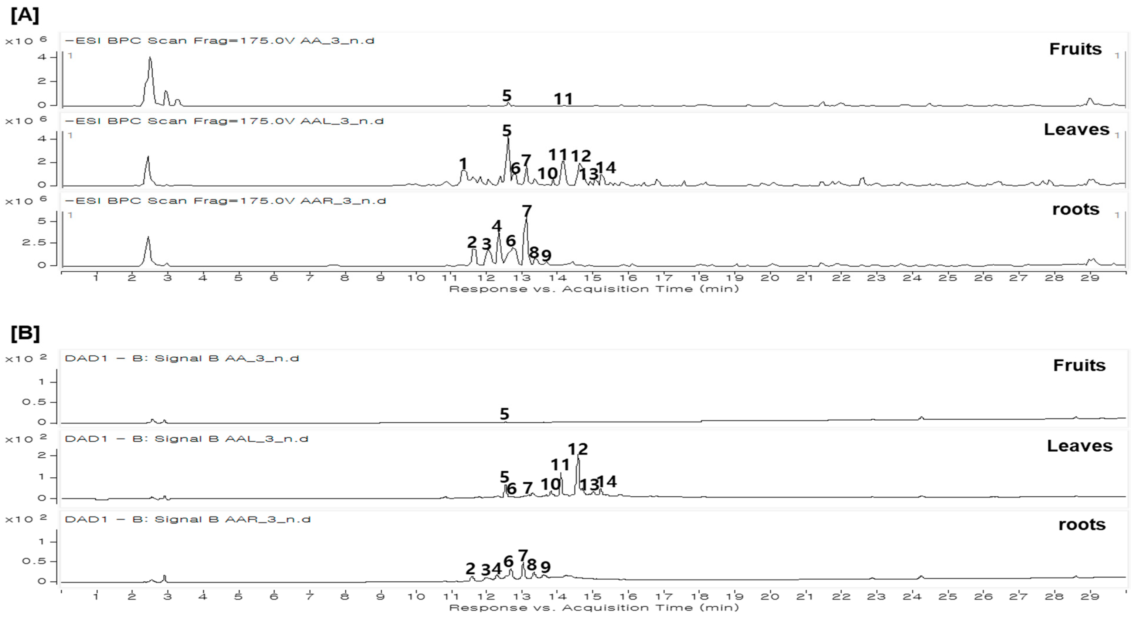
Based on the pronounced organ-dependent differences in phenolic content and antioxidant activity, LC–QTOF–MS-based metabolite profiling was performed to elucidate the underlying chemical basis. The chemical compositions of extracts obtained from the fruits, leaves, and roots of A. arguta were analyzed using UV detection and LC–QTOF–MS to evaluate organ-dependent differences in metabolite profiles. As shown in Figure 1, the three organs exhibited clearly distinct chromatographic patterns, with the root displaying a markedly different profile from those of the fruits and leaves, suggesting the presence of characteristic root-enriched metabolites.
A total of fourteen major peaks were detected across the samples, and peak profiling was conducted using LC–MS/MS analysis based on retention times, UV absorption maxima (λmax), and diagnostic fragmentation patterns. The detected metabolites were tentatively classified as flavan-3-ols and their oligomeric forms, including dimers and trimers (peaks 1, 2, 3, 4, 6, 7, 8, and 9), as well as flavonol glycosides (peaks 10, 11, 12, and 13) (Table 2). Distinct organ-specific differences were observed in the distribution of these peaks. The fruit extract contained only minor peaks with no clearly dominant constituents, whereas the leaf extract was characterized by prominent flavonoid glycosides. In contrast, the root extract exhibited a distinct profile dominated by peaks 2, 3, 4, 6, and 7, which were assigned to catechin and its oligomeric derivatives, including procyanidin dimers and trimers, indicating that flavan-3-ol–based condensed tannins represent the major constituents of the root.
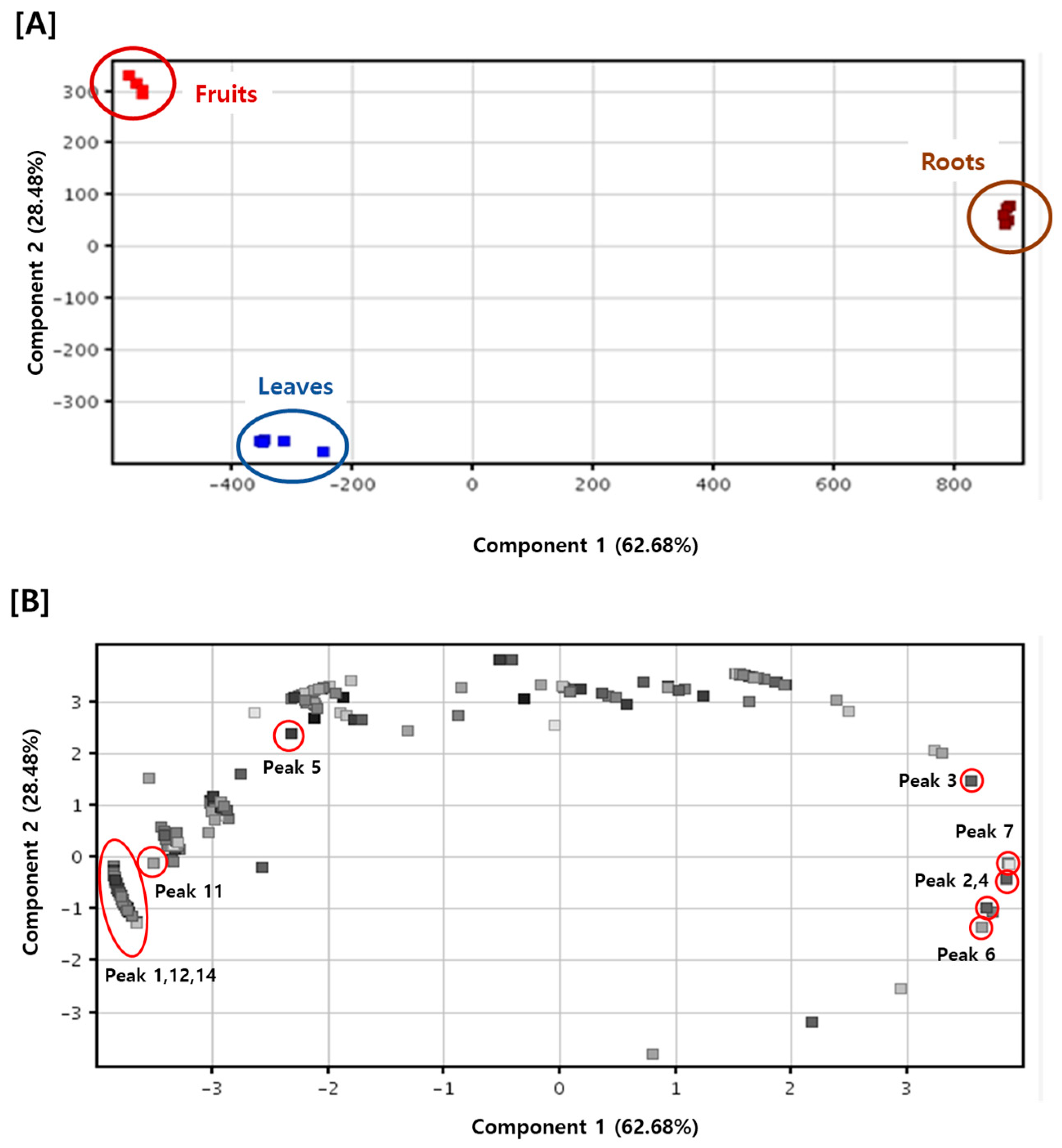
To further visualize and statistically evaluate these differences, PCA was performed using the LC–QTOF–MS dataset (Figure 2). PCA was performed using the full LC–MS dataset, including all detected features, to capture the overall metabolic variation among the samples (Tables S1 and S2). Among these, representative metabolites contributing to organ-dependent differentiation were selected for further identification and are summarized in Table 2. Among the detected peaks, representative metabolites contributing to organ-dependent differentiation were selected for isolation and structural identification. The PCA score plot showed clear separation of the fruit, leaf, and root samples. The first two principal components, PC1 and PC2, accounted for 62.68% and 28.48% of the total variance, respectively, with a cumulative explained variance of 91.16%. Biological replicates were consistently clustered according to tissue type in the PCA score plot, supporting the reproducibility of the organ-dependent metabolic differences. Loading plot analysis indicated that the grouping of the root samples was primarily driven by peaks 2, 3, 4, 6, and 7, whereas the leaf samples were associated with peaks 1, 5, 11, 12, and 14, and the fruit samples were influenced mainly by peak 5 and other minor components. The loading plot includes all detected variables, whereas only selected major metabolites relevant to organ-specific differences are discussed in this study. PCA effectively visualized organ-dependent metabolic differences among the samples and enabled the identification of metabolites contributing to the observed separation.
Taken together, the combined LC–QTOF–MS profiling and PCA results demonstrate organ-specific metabolic differentiation in A. arguta, with the root characterized by a condensed tannin–dominant chemical profile distinct from the flavonoid glycoside-rich leaves and the chemically less defined fruit extracts.

3.3. Isolation and Structural Elucidation of Compounds from the Roots of A. arguta

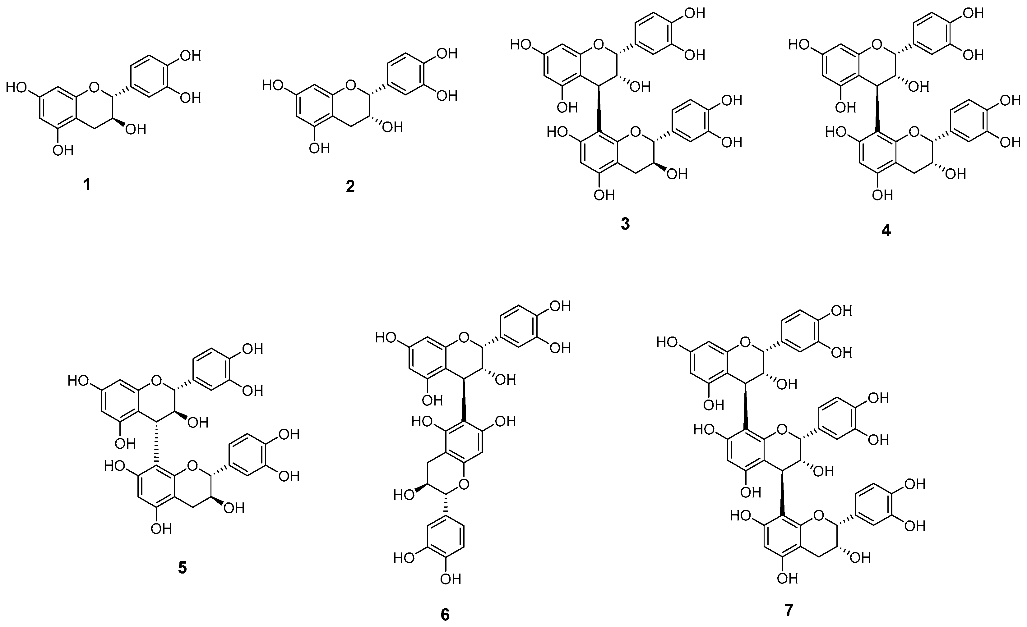
LC–MS/MS-based chemical profiling revealed that the fourteen detected metabolites shared similar flavonoid-related structural features, including flavan-3-ols, their oligomers, and glycosylated derivatives (Table 2). Several peaks observed predominantly in the root extract (peaks 2, 3, 6, and 9) exhibited identical molecular weights and were tentatively assigned as procyanidin dimers. However, their exact structures could not be unambiguously determined based solely on MS data. In addition, peak 8, presumed to be a procyanidin trimer, also required definitive structural confirmation. Therefore, targeted chromatographic separation of the root extract of A. arguta was conducted using a combination of column chromatography and preparative HPLC, resulting in the isolation of seven compounds (17) (Figure 3).
Compounds 1 and 2 were identified as catechin and epicatechin, respectively, by comparison of their spectroscopic data with reported values including chemical shifts and coupling constants [19,20]. Compounds 37 were obtained as reddish amorphous solids and were identified as procyanidin-type condensed tannins based on comprehensive spectroscopic analyses.
Compounds 3, 4, 5 and 6 exhibited molecular ion peaks at m/z 577 [M–H], indicative of procyanidin dimers. The 1H and 13C NMR spectra of Compound 3 displayed signals attributable to two 1,3,4-trisubstituted aromatic rings, multiple oxymethine and methylene groups characteristic of flavan-3-ol units, and diagnostic coupling patterns indicative of catechin and epicatechin subunits. Key HMBC correlations supported a C4–C8 interflavan linkage, allowing compound 3 to be identified as procyanidin B1 by comparison with literature data [20]. The NMR spectra of compound 4 displayed characteristic singlet oxymethine signals consistent with an epicatechin–epicatechin composition, whereas compound 5 exhibited coupling constants indicative of a catechin–catechin unit. Accordingly, compounds 4 and 5 were identified as procyanidin B2 and procyanidin B3, respectively, by comparison with published data [20,21]. Compound 6 showed NMR features corresponding to a heterodimeric structure composed of catechin and epicatechin units and HMBC correlations from H-4 to C-6. Therefore, compound 6 was identified as procyanidin B7 based on comparison with reported data [22].
Compound 7 showed a deprotonated molecular ion at m/z 865 [M–H] in the ESI–MS spectrum, corresponding to a procyanidin trimer. Its NMR spectra exhibited signal patterns characteristic of higher oligomeric flavan-3-ols, and compound 7 was identified as procyanidin C1 based on its molecular weight and spectroscopic features [23].
Collectively, the isolated compounds comprised catechin- and epicatechin-based procyanidin dimers and a trimer, demonstrating that oligomeric flavan-3-ols represent the major phenolic constituents of the root of A. arguta. These results enabled clear differentiation of compounds with identical molecular weights in LC–MS/MS-based chemical profiling.

3.4. Antioxidant Effect of Compounds 17

Because the root extract of A. arguta exhibited strong antioxidant activity, the antioxidant activities of the isolated compounds were evaluated (Table 3). All isolated compounds showed measurable radical-scavenging activity. Overall, the procyanidin dimers (36) exhibited stronger antioxidant activity than the monomeric flavan-3-ols catechin (1) and epicatechin (2), while the procyanidin trimer (7) showed the strongest activity among the tested compounds, with an IC50 value of 4.5 μM. Notably, the activity of procyanidin C1 (7) (IC50 = 4.5 μM) was comparable to that of ascorbic acid under similar experimental conditions, indicating strong radical-scavenging potency. This trend suggests that antioxidant activity is associated with the degree of polymerization of flavan-3-ol units. The antioxidant activities of the isolated compounds were comparable to or stronger than that of the crude root extract, suggesting that procyanidin oligomers are major contributors to the overall antioxidant capacity of the extract. Although these compounds are well known for their antioxidant activity, their enrichment in the root of A. arguta and their contribution to the overall extract activity provide important insight into the chemical basis of organ-specific functional properties.

4. Discussion

4.1. Root-Enriched Condensed Tannin Phenotype in A. arguta

This study identified the major metabolites contributing to organ-specific differences and demonstrated that the root of A. arguta is characterized by a condensed tannin–dominant chemical profile distinct from those of the fruits and leaves. Untargeted LC–MS profiling combined with PCA showed that the root samples formed an independent cluster, primarily driven by flavan-3-ol oligomers, including procyanidin dimers and a trimer.
Because this work was designed as a comparative phytochemical study focusing on major organ-dependent differences and targeted compound isolation, the LC–MS dataset was used for exploratory profiling rather than comprehensive untargeted metabolomics requiring extensive QC-based validation. Organ-specific accumulation of secondary metabolites has been widely reported and reflects tissue-dependent regulation of biosynthetic pathways.
Structural elucidation confirmed that the root is enriched in catechin- and epicatechin-based procyanidins, whereas these oligomeric flavan-3-ols were not major constituents in the fruits or leaves in our previous studies. Instead, the fruits were characterized by organic acid–phenolic conjugates [4], while the leaves accumulated phenylpropanoid-conjugated triterpenoids [10], highlighting pronounced organ-dependent specialization of secondary metabolism in A. arguta.
Condensed tannins, including procyanidins, are widely recognized as defense-related metabolites in plants. Their ability to form stable complexes with proteins, polysaccharides, and microbial enzymes contributes to protection against pathogens and herbivores, particularly in subterranean tissues exposed to soil-borne microorganisms [24,25]. In addition to their ecological role, oligomeric procyanidins exhibit strong antioxidant activity, consistent with the high activity observed for both the root extract and the isolated compounds in this study [26,27]. The stronger activity of oligomeric procyanidins compared with monomeric flavan-3-ols further supports their functional relevance as major root constituents.
From a metabolic perspective, the predominance of procyanidin oligomers in the root contrasts with the accumulation of flavonoid glycosides in the leaves and phenolic conjugates in the fruits. This pattern suggests that flavan-3-ol biosynthesis in the root is preferentially directed toward oligomerization, rather than glycosylation or esterification, reflecting organ-specific regulation of the phenylpropanoid pathway. This interpretation is consistent with previous studies describing the regulation of flavan-3-ol biosynthesis and oligomerization within the phenylpropanoid and flavonoid pathways [28,29,30].
Although procyanidins have been reported in the roots of several Actinidia species, most studies have focused on individual compounds rather than systematic organ-level comparisons [31,32]. Similar organ-dependent phenolic distributions have been observed in other Actinidia species and fruit crops, suggesting that tissue-specific tannin enrichment represents a conserved metabolic strategy rather than a unique feature.
By integrating LC–MS-based metabolite profiling, multivariate analysis, structural elucidation, and antioxidant evaluation, this study demonstrates that condensed tannins represent a defining metabolic and functional phenotype of A. arguta roots rather than incidental constituents. However, this study is limited by the absence of transcriptomic or enzymatic data to directly support the regulatory mechanisms underlying the observed metabolic differences. Future multi-omics approaches will provide deeper insight into the molecular basis of organ-specific metabolite accumulation.
Taken together, these findings highlight a clear root-specific condensed tannin phenotype in A. arguta and underscore the value of organ-resolved metabolomic approaches in elucidating the chemical and functional diversity of plant secondary metabolism.

4.2. Broader Biological Relevance and Future Perspectives

Beyond defining a root-specific condensed tannin phenotype, these findings provide broader insight into the biological and functional significance of procyanidin accumulation in A. arguta. The strong antioxidant activity observed in the root extract supports the role of procyanidin oligomers as effective redox-active compounds. Given the well-established relationship between the degree of polymerization and radical-scavenging capacity [26,27,33], the enrichment of procyanidin dimers and a trimer in the root likely contributes to enhanced oxidative stress tolerance. Such properties may be particularly relevant for subterranean tissues, which are continuously exposed to oxidative challenges arising from interactions with soil microorganisms, metal ions, and fluctuating environmental conditions [26,27].
Ecologically, the accumulation of condensed tannins in root tissues may confer adaptive advantages by enhancing resistance to soil-borne pathogens and environmental stress. Similar organ-dependent phenolic distributions have been reported in other Actinidia species, where roots often exhibit higher levels of condensed tannins than aerial parts, supporting the role of roots as reservoirs of defense-related metabolites. From a biosynthetic perspective, the preferential accumulation of procyanidins in the root suggests organ-specific regulation of the phenylpropanoid and flavonoid pathways. Flavan-3-ol monomers such as catechin and epicatechin are synthesized through these pathways and subsequently undergo oligomerization to form procyanidins [30,33]. Although regulatory mechanisms were not directly investigated, differential expression of key biosynthetic enzymes may underlie the observed tissue-specific metabolic patterns.
From an application standpoint, procyanidin-rich extracts have been reported to exhibit antimicrobial, enzyme inhibitory, and anti-inflammatory activities [16,34]. Although this study focused primarily on antioxidant activity, the identified procyanidin profile in A. arguta roots suggests potential relevance to broader biofunctional applications.
This study has several limitations. Although the DPPH assay was considered sufficient for comparative evaluation of antioxidant capacity [35,36], additional methods such as ABTS or FRAP would provide a more comprehensive assessment. In addition, multivariate analysis was applied in an exploratory manner to identify key metabolites contributing to organ-specific differentiation. Future studies incorporating expanded datasets and advanced statistical approaches, including correlation or regression analyses, would enable a more detailed understanding of metabolite–activity relationships.
Further investigation is also required to elucidate the biological activities of individual root-derived procyanidins and their potential synergistic effects, as well as to clarify the molecular mechanisms governing flavan-3-ol oligomerization in root tissues. In addition, expanding the analysis to include a broader range of cultivars and tissue types may further improve the understanding of metabolic variability and its relationship to biological activity
Overall, these findings highlight the biological relevance of root-derived procyanidins and support the potential of A. arguta roots as a valuable source of bioactive natural products.

5. Conclusions

This study provides a comprehensive characterization of organ-dependent metabolic variation in Actinidia arguta by integrating LC–MS-based metabolite profiling, multivariate analysis, and targeted phytochemical isolation.
This study demonstrates organ-dependent metabolic variation in Actinidia arguta through an integrated approach combining LC–MS-based profiling, multivariate analysis, and targeted phytochemical isolation.
The root was found to be enriched in oligomeric flavan-3-ols, particularly procyanidin dimers and a trimer, representing a condensed tannin–dominant chemical phenotype distinct from those of the fruits and leaves. These results highlight pronounced metabolic specialization among different plant organs and demonstrate the value of integrating metabolomics with conventional phytochemical analysis to identify key metabolites.
The strong antioxidant activity of the root extract and isolated compounds supports the functional relevance of procyanidin oligomers. However, their broader ecological roles require further experimental validation. From an applied perspective, the enrichment of bioactive procyanidins in the root suggests potential for utilization in nutraceutical and pharmaceutical applications.
Overall, this work provides a chemical and functional framework for understanding organ-specific metabolite distribution in A. arguta and highlights root-derived procyanidins as promising bioactive natural products.

Supplementary Materials

The following supporting information can be downloaded at: https://www.mdpi.com/article/10.3390/horticulturae12040454/s1, Table S1: PCA score values for all samples used in the analysis; Table S2: PCA loading values and associated information for the major metabolites included in the analysis.

Author Contributions

Conceptualization, K.Y.L. and M.K.L.; methodology, H.H.L., J.H.A., S.J.K. and Y.K.H.; software, Y.K.H. and K.Y.L.; validation, H.H.L. and M.K.L.; investigation, H.H.L., Y.K.H., J.H.A., S.J.K., Q.L., B.Y.H., K.Y.L. and M.K.L.; writing—original draft preparation, H.H.L., Y.K.H., K.Y.L. and M.K.L.; writing—review and editing, K.Y.L. and M.K.L.; visualization, H.H.L. and Y.K.H.; supervision, M.K.L.; project administration, M.K.L.; funding acquisition, M.K.L. All authors have read and agreed to the published version of the manuscript.

Funding

This work was supported by the National Research Foundation of Korea (NRF) grant funded by the Korean government (RS-2022-NR069422 and RS-2026-25481077).

Data Availability Statement

The original contributions presented in this study are included in the article/Supplementary Materials. Further inquiries can be directed to the corresponding authors.

Conflicts of Interest

The authors declare no conflicts of interest.

Abbreviations

The following abbreviations are used in this manuscript:
PCAPrincipal component analysis
HPLC–MS/MSHigh-performance liquid chromatography–tandem mass spectrometry

References

  1. Wang, S.; Qiu, Y.; Zhu, F. Kiwifruit (Actinidia spp.): A review of chemical diversity and biological activities. Food Chem. 2021, 350, 128469. [Google Scholar] [CrossRef]
  2. Waswa, E.N.; Ding, S.X.; Wambua, F.M.; Mkala, E.M.; Mutinda, E.S.; Odago, W.O.; Amenu, S.G.; Muthui, S.W.; Linda, E.L.; Katumo, D.M.; et al. The genus Actinidia Lindl. (Actinidiaceae): A comprehensive review on its ethnobotany, phytochemistry, and pharmacological properties. J. Ethnopharmacol. 2024, 319, 117222. [Google Scholar] [CrossRef]
  3. Pinto, D.; Delerue-Matos, C.; Rodrigues, F. Bioactivity, phytochemical profile and pro-healthy properties of Actinidia arguta: A review. Food Res. Int. 2020, 136, 109449. [Google Scholar] [CrossRef]
  4. Ahn, J.H.; Park, Y.; Jo, Y.H.; Kim, S.B.; Yeon, S.W.; Kim, J.G.; Turk, A.; Song, J.Y.; Kim, Y.; Hwang, B.Y.; et al. Organic acid conjugated phenolic compounds of hardy kiwifruit (Actinidia arguta) and their NF-κB inhibitory activity. Food Chem. 2020, 308, 125666. [Google Scholar] [PubMed]
  5. Ahn, J.H.; Yeon, S.W.; Ryu, S.H.; Lee, S.; Turk, A.; Hwang, B.Y.; Lee, M.K. Three new succinate-phenolic conjugates from the fruits of Actinidia arguta. Phytochem. Lett. 2022, 48, 128–131. [Google Scholar] [CrossRef]
  6. Ryu, S.H.; Ahn, J.H.; Turk, A.; Kim, S.B.; Hwang, B.Y.; Lee, M.K. Argutinic acid, a new triterpenoid from the fruits of Actinidia arguta. Nat. Prod. Sci. 2024, 30, 208–211. [Google Scholar] [CrossRef]
  7. Ryu, S.H.; Lee, H.H.; Ahn, J.H.; Turk, A.; Park, Y.; Kim, S.B.; Hwang, B.Y.; Lee, M.K. α-Glucosidase inhibitory flavonoids of Actinidia arguta fruits: Comparison of different cultivars. Folia Hortic. 2025, 37, 25–31. [Google Scholar] [CrossRef]
  8. Ahn, J.H.; Ryu, S.H.; Yeon, S.W.; Lee, S.; Kim, S.B.; Hwang, B.Y.; Lee, M.K. Phenyldilactones from the leaves of hardy kiwifruit (Actinidia arguta). Biochem. System. Ecol. 2023, 108, 103636. [Google Scholar] [CrossRef]
  9. Almeida, D.; Pinto, D.; Santos, J.; Vinha, A.F.; Palmeira, J.; Ferreira, H.N.; Rodrigues, F.; Oliveira, M.B.P.P. Hardy kiwifruit leaves (Actinidia arguta): An extraordinary source of value-added compounds for food industry. Food Chem. 2018, 259, 113–121. [Google Scholar] [CrossRef] [PubMed]
  10. Ahn, J.H.; Park, Y.; Yeon, S.W.; Jo, Y.H.; Han, Y.K.; Turk, A.; Ryu, S.H.; Hwang, B.Y.; Lee, K.Y.; Lee, M.K. Phenylpropanoid-conjugated triterpenoids from the leaves of Actinidia arguta and their inhibitory activity on α-glucosidase. J. Nat. Prod. 2020, 83, 1416–1423. [Google Scholar] [CrossRef] [PubMed]
  11. Jang, D.S.; Lee, G.Y.; Kim, J.; Lee, Y.M.; Kim, J.M.; Kim, Y.S.; Kim, J.S. A new pancreatic lipase inhibitor isolated from the roots of Actinidia arguta. Arch. Pharm. Res. 2008, 31, 666–670. [Google Scholar] [CrossRef]
  12. Jang, D.S.; Lee, G.Y.; Lee, Y.M.; Kim, Y.S.; Sun, H.; Kim, D.H.; Kim, J.S. Flavan-3-ols having a gamma-lactam from the roots of Actinidia arguta inhibit the formation of advanced glycation end products in vitro. Chem. Pharm. Bull. 2009, 57, 397–400. [Google Scholar] [CrossRef]
  13. Zhao, X.; Wen, F.; Wang, W.; Lu, Z.; Guo, Q. Actinidia arguta (Hardy Kiwi) root extract exerts anti-cancer effects via Mcl-1-mediated apoptosis in cholangiocarcinoma. Nutr. Cancer 2019, 71, 246–256. [Google Scholar]
  14. Hou, Y.F.; Bai, L.; Guo, S.; Hu, J.B.; Zhang, S.S.; Liu, S.J.; Zhang, Y.; Li, S.; Ho, C.T.; Bai, N.S. Nontargeted metabolomic analysis of four different parts of Actinidia arguta by UPLC-Q-TOF-MSE. Food Res. Int. 2023, 163, 112228. [Google Scholar]
  15. Barbehenn, R.V.; Constabel, C.P. Tannins in plant–herbivore interactions. Phytochemistry 2011, 72, 1551–1565. [Google Scholar] [CrossRef] [PubMed]
  16. Scalbert, A. Antimicrobial properties of tannins. Phytochemistry 1991, 30, 3875–3883. [Google Scholar] [CrossRef]
  17. Kang, D.-M.; Kwon, J.-M.; Jeong, W.-J.; Neupane, B.D.; Ahn, M.-J. Antioxidant compounds of Sambucus pendula stem. Nat. Prod. Sci. 2024, 30, 275–281. [Google Scholar] [CrossRef]
  18. Woo, S.-I.; Oh, M.; Lee, H.H.; Song, I.; Kim, S.J.; Oh, Y.L.; Im, J.-H.; Lee, E.-J.; Lee, M.K. Development of hybrid Pleurotus cystidiosus strains with enhanced functional properties. Foods 2025, 14, 4329. [Google Scholar] [CrossRef] [PubMed]
  19. Le, T.T.; Ha, M.T.; Hoang, L.M.; Vu, N.K.; Kim, J.A.; Min, B.S. α-Glucosidase inhibitory activity of phenolic compounds isolated from the stems of Caesalpinia decapetala var. japonica. Nat. Prod. Sci. 2022, 28, 143–152. [Google Scholar] [CrossRef]
  20. Shoji, T.; Mutsuga, M.; Nakamura, T.; Kanda, T.; Akiyama, H.; Goda, Y. Isolation and structural elucidation of some procyanidins from apple by low-temperature nuclear magnetic resonance. J. Agric. Food Chem. 2003, 51, 3806–3813. [Google Scholar] [CrossRef] [PubMed]
  21. Stark, T.; Bareuther, S.; Hofmann, T. Sensory-guided decomposition of roasted cocoa nibs (Theobroma cacao) and sturucture determination of taste-active polyphenols. J. Agric. Food Chem. 2005, 53, 5407–5418. [Google Scholar] [CrossRef] [PubMed]
  22. Park, S.Y.; Han, Y.K.; Vinh, L.B.; Lee, K.Y. Analysis of α-glucosidase inhibitory constituents from Acer tegmentosum using LC-QTOF MS/MS and molecular networking. Nat. Prod. Sci. 2023, 29, 242–250. [Google Scholar] [CrossRef]
  23. Esatbeyoglu, T.; Jaschok-Kentner, B.; Wray, V.; Winterhalter, P. Structure elucidation of procyanidin oligomers by low-temperature 1H NMR spectroscopy. J. Agric. Food Chem. 2011, 59, 62–69. [Google Scholar] [CrossRef]
  24. Ha, M.T.; Tran, T.H.; Huh, J.; Min, S.E.; Han, K.-H.; Kim, Y.-B.; Kim, J.A.; Min, B.S. Areca catechu L.: A comprehensive and updated review of its phytochemical and pharmacological properties. Nat. Prod. Sci. 2025, 31, 236–250. [Google Scholar] [CrossRef]
  25. Lattanzio, V.; Lattanzio, V.M.T.; Cardinali, A. Role of phenolics in the resistance mechanisms of plants against fungal pathogens and insects. Phytochem. Adv. Res. 2006, 661, 23–67. [Google Scholar]
  26. Rice-Evans, C.A.; Miller, N.J.; Paganga, G. Structure-antioxidant activity relationships of flavonoids and phenolic acids. Free Radic. Biol. Med. 1996, 20, 933–956. [Google Scholar] [CrossRef]
  27. Nie, F.; Liu, L.; Cui, J.; Zhao, Y.; Zhang, D.; Zhou, D.; Wu, J.; Li, B.; Wang, T.; Li, M.; et al. Oligomeric proanthocyanidins: An updated review of their natural sources, synthesis, and potentials. Antioxidants 2023, 12, 1004. [Google Scholar] [CrossRef] [PubMed]
  28. Dixon, R.A.; Xie, D.Y.; Sharma, S.B. Proanthocyanidins—A final frontier in flavonoid research? New Phytol. 2005, 165, 9–28. [Google Scholar] [CrossRef] [PubMed]
  29. Lepiniec, L.; Debeaujon, I.; Routaboul, J.M.; Baudry, A.; Pourcel, L.; Nesi, N.; Cabocheet, M. Genetics and biochemistry of seed flavonoids. Annu. Rev. Plant Biol. 2006, 57, 405–430. [Google Scholar] [CrossRef]
  30. Cheynier, V.; Comte, G.; Davies, K.M.; Lattanzio, V.; Martens, S. Plant phenolics: Recent advances on their biosynthesis, genetics, and ecophysiology. Plant Physiol. Biochem. 2013, 72, 1–20. [Google Scholar] [CrossRef] [PubMed]
  31. Latocha, P. The nutritional and health benefits of kiwiberry (Actinidia arguta)—A review. Plant Foods Hum. Nutr. 2017, 72, 325–334. [Google Scholar]
  32. Jia, D.; Liao, G.; Ye, B.; Zhong, M.; Huang, C.; Xu, X. Changes in fruit quality, phenolic compounds, and antioxidant activity of kiwifruit (Actinidia eriantha) during on-vine ripening. LWT 2024, 206, 116564. [Google Scholar] [CrossRef]
  33. Plumb, G.W.; De Pascual-Teresa, S.; Santos-Buelga, C.; Cheynier, V.; Williamson, G. Antioxidant properties of catechins and proanthocyanidins: Effect of polymerisation, galloylation and glycosylation. Free Radic. Res. 1998, 29, 351–358. [Google Scholar] [CrossRef] [PubMed]
  34. Gu, L.; Kelm, M.A.; Hammerstone, J.F.; Beecher, G.; Holden, J.; Haytowitz, D.; Gebhardt, S.; Prior, R.L. Concentrations of proanthocyanidins in common foods and estimations of normal consumption. J. Nutr. 2004, 134, 613–617. [Google Scholar] [CrossRef] [PubMed]
  35. Prior, R.L.; Wu, X.; Schaich, K. Standardized methods for the determination of antioxidant capacity and phenolics in foods and dietary supplements. J. Agric. Food Chem. 2005, 53, 4290–4302. [Google Scholar] [CrossRef]
  36. Apak, R.; Güçlü, K.; Özyürek, M.; Karademir, S.E. Novel total antioxidant capacity index for dietary polyphenols and vitamins C and E, using their cupric ion reducing capability in the presence of neocuproine: CUPRAC method. J. Agric. Food Chem. 2004, 52, 7970–7981. [Google Scholar] [CrossRef]
Figure 1. (A) MS chromatograms (negative ionization mode) and (B) UV chromatograms (254 nm) of each part of A. arguta.
Horticulturae 12 00454 g001
Figure 2. (A) Score plot for fruits, leaves and roots of A. arguta and (B) loading plot of peaks contributing to PC1 and PC2. Annotated peaks represent major or representative metabolites contributing to organ-specific differences.
Horticulturae 12 00454 g002
Figure 3. Chemical structures of compounds 17 from the roots of A. arguta.
Horticulturae 12 00454 g003
Table 1. DPPH radical scavenging activity and total phenolic content of fruits, leaves and roots of A. arguta.
FruitsLeavesRoots
DPPH radical scavenging activity (% of control) *11.2 ± 3.1 b52.9 ± 3.9 a60.8 ± 6.2 a
Total phenolic content (mg GAE/g dried weight)1.9 ± 0.2 c6.8 ± 0.5 b10.8 ± 0.7 a
* 100 μg/mL. Values are expressed as mean ± SD (n = 3). Different letters indicate significant differences among organs at p < 0.05 by one-way ANOVA followed by Tukey’s test.
Table 2. Peak profiling of major compounds of each part of A. argute using LC-QTOF MS/MS.
Peak NoCompounds IdentificationtR (min)Observed m/zCalculated m/zMolecular Formula
[M-H]-
MS/MS Fragments
(m/z)
UV
(λmax, nm)
1(epi)catechin-O-glucoside11.353451.2429451.1246C21H23O11289 [M-C6H10O5-H]-
2Procyanidin dimer11.663577.1641577.1351C30H25O12289 [M-C15H12O6-H]-280
3Procyanidin dimer12.038577.1639577.1351C30H25O12289 [M-C15H12O6-H]-280
4Catechin12.350289.0874289.0718C15H13O6109 [M-C9H8O4-H]-230 (sh), 280
5Roseoside12.602431.2153431.1923C20H31O10385 [M-H]-203
6Procyanidin dimer12.725577.1643577.1351C30H25O12289 [M-C15H12O6-H]-280
7Epicatechin13.100289.0873289.0718C15H13O6109 [M-C9H8O4-H]-230 (sh), 280
8Procyanidin trimer13.412865.2405865.1985C45H37O18577 [M-C15H12O6-H]-280
9Procyanidin dimer13.662577.1640577.1351C30H25O12289 [M-C15H12O6-H]-280
10Quercetin-3-O-sambubioside13.851595.1610595.1305C26H27O16300 [M-C11H19O9-H]-285
11Quercetin-3-O-rutinoside14.164609.1764609.1461C27H29O16300 [M-C12H21O9-H]-260, 355
12Quercetin-3-O-glucoside14.601463.1123463.0882C21H19O12300 [M-C6H11O5-H]-260, 355
13Kaempferol-3-O-galactoside15.038447.1173447.0933C21H19O11284 [M-C6H11O5-H]-280
14Kaempferol-3-O-glucoside15.226447.1165447.0933C21H19O11284 [M-C6H11O5-H]-280
Table 3. IC50 values for DPPH radical scavenging activity of compounds isolated from the roots of A. arguta.
CompoundsIC50 (μM)Peak NoCompoundsIC50 (μM)Peak No
Catechin (1)12.24Procyanidin B3 (5)5.93
Epicatechin (2)11.37Procyanidin B7 (6)7.26
Procyanidin B1 (3)6.82Procyanidin C1 (7)4.58
Procyanidin B2 (4)5.69Ascorbic acid *8.1
* Ascorbic acid was used as a positive control.
Disclaimer/Publisher’s Note: The statements, opinions and data contained in all publications are solely those of the individual author(s) and contributor(s) and not of MDPI and/or the editor(s). MDPI and/or the editor(s) disclaim responsibility for any injury to people or property resulting from any ideas, methods, instructions or products referred to in the content.