Article

Pseudomonas aeruginosa CAKS2: A Multifaceted Endophyte Enhancing Growth and Combating Anthracnose in Sweet Orange (Citrus sinensis L.)


1
Institute of Biology, Vietnam Academy of Science and Technology, 18 Hoang Quoc Viet, Nghia Do, Hanoi 100000, Vietnam
2
Department of Biology, Graduate University of Science and Technology, Vietnam Academy of Science and Technology, 18 Hoang Quoc Viet, Nghia Do, Hanoi 100000, Vietnam
3
Plant Protection Research Institute, Vietnam Academy of Agricultural Sciences, Duc Thang, Bac Tu Liem, Hanoi 100000, Vietnam
4
Institute of Tropical Biology, Vietnam Academy of Science and Technology, 9/621 Hanoi Highway, Thu Duc, Ho Chi Minh 700000, Vietnam
*
Author to whom correspondence should be addressed.
These authors contributed equally to this work.
Horticulturae 2026, 12(4), 442; https://doi.org/10.3390/horticulturae12040442
Submission received: 4 March 2026 / Revised: 29 March 2026 / Accepted: 31 March 2026 / Published: 3 April 2026
(This article belongs to the Section Plant Pathology and Disease Management (PPDM))

Abstract

Plant growth-promoting rhizobacteria (PGPR) have been collected and used to promote plant growth and enhance disease tolerance of various crops. In the current work, Pseudomonas aeruginosa CAKS2, an endophytic strain isolated from the rhizosphere of sweet orange, exhibited both growth promotion and antimicrobial activities. Under the in vitro condition, the CAKS2 showed multiple plant growth-promoting properties such as phosphate, potassium, and calcium solubilization, nitrogen fixation as well as production of siderophores, IAA, ammonia, exopolysaccharides, hydrogen cyanide, and biofilm formation. This P. aeruginosa strain inhibited the growth of different tested fungal and bacterial pathogens. Under the in vivo condition, the CAKS2 enhanced sweet orange plant growth, indicated by increases in the root and shoot lengths, the leaf number, and the total biomass. The biochemical components and the transcription levels of genes related to plant hormone biosynthesis were altered in the CAKS2-inoculated sweet orange. Under the in vivo infection of C. gloeosporioides, the CAKS2 reduced the diameter of lesions on orange leaves and harvested fruits and decreased disease severity and incidence at the whole plant level. The whole genome sequence of CAKS2 showed the presence of candidate genes involved in different molecular pathways contributing to plant-promoting and biocontrol properties. Importantly, certain changes in the expression of gene response for plant growth promotion and biocontrol were observed when the CAKS2 was exposed to sweet orange root exudates. This study highlights P. aeruginosa CAKS2 as a potential PGPR strain for enhancing plant growth and C. gloeosporioides tolerance in sweet orange and other citrus plants.

1. Introduction

Plant growth-promoting rhizobacteria (PGPR) have been applied to control and reduce the negative effects of pathogens in many crops, including citrus plants [1,2,3]. Previously, PGPR were successfully applied to control anthracnose disease caused by Collectotrichum spp., in various plant species [4,5,6,7,8]. Particularly, Bacillus subtilis ZSH-1 could effectively reduce anthracnose disease in poplar plants under greenhouse conditions [7]. In addition, Pseudomonas PT11 was shown to protect coffee plants against anthracnose caused by Colletotrichum gloeosporioides [5]. More recently, different strains of Pseudomonas aeruginosa have been selected and utilized to improve plant growth and disease tolerance of various crops [9,10,11,12,13,14]. Moreover, P. aeruginosa has been reported to inhibit various species of Colletotrichum spp. in chili and sugarcane [15,16]. These reports all indicate the potential of PGPR in controlling plant diseases caused by Colletotrichum spp.
Regarding the mechanisms by which PGPRs protect plants from abiotic stress and pathogen infection, they have been shown to increase the accumulation of several non-enzymatic compounds, including phenolics, flavonoids, lignins, and antioxidant enzymes, including superoxide dismutase (SOD), catalase (CAT), peroxidases (POD), and glutathione reductase (GR) [17,18,19]. Moreover, PGPR could alter hormonal signaling pathways related to induced systemic resistance (ISR) and systemic acquired resistance (SAR) of plants to eliminate the negative impact of pathogens. In which Jasmonic acid (JA) and Ethylene (ETH), which belong to the ISR pathway, are known to be associated with resistance against necrotrophic pathogens. Whereas, SA, which belongs to the SAR pathway, is primarily linked to defense against biotrophic pathogens. Furthermore, the abscisic acid (ABA) pathway modulates both stress responses and disease resistance [20,21,22]. Additionally, PGPR exhibited direct and indirect mechanisms to boost plant growth by releasing beneficial substances (phosphates, potassium, silicon, and zinc), fixing nitrogen, chelating iron, enhancing nutrient uptake, and synthesizing phytohormones (gibberellins, indoleacetic acid, and cytokinins) [23].
Citrus is one of the most important commercial crops, widely cultivated across tropical and subtropical regions [24]. The sweet orange (Citrus sinensis L.), the foremost citrus species, constitutes more than half of global citrus production [25]. However, citrus cultivation has faced significant challenges from numerous fungal and bacterial diseases, including gummosis, greasy spots, neck rot, huanglongbing, citrus canker, citrus blast, anthracnose, and root rot [24,26,27]. Among these, anthracnose disease caused by C. gloeosporioides has recently been recognized as among the top 10 most dangerous plant pathogenic fungi affecting citrus fruits after flowering and postharvest [28]. C. gloeosporioides poses a serious threat to citrus fruits after harvest, with different symptoms including sunken, dark lesions, and fruit decay. This fungus could spread quickly and remain latent until symptoms appear, contributing to considerable post-harvest losses. Different approaches, including chemical, physical, and biological controls, have been developed to manage and reduce the effects of C. gloeosporioides on plants [29].
Despite increasing reports on the beneficial roles of endophytic bacteria in plant growth promotion and disease suppression, systematic studies on native citrus endophytes remain limited. In particular, their dual role in enhancing plant growth and suppressing anthracnose disease at the physiological and molecular levels has not been well characterized. Therefore, this study is the first to assess the effects of P. aeruginosa CAKS2, a multifunctional PGPR isolated from a native sweet orange, on citrus growth and antifungal improvement at both in vitro and in planta conditions. In addition, the whole genome sequencing of the selected strain was generated and analyzed to find expected mechanisms related to these characteristics of the selected strain at the molecular level. Our results have confirmed the PGP and antagonistic properties of the P. aeruginosa CAKS2 and shown the potential of this strain for further application in biofertilizer, biocontrol of citrus, as well as other crops.

2. Materials and Methods

2.1. Endophytic Bacteria Isolation

Orange root samples were collected in Cao Phong district, Hoa Binh province (20°43′42.4″ N, 105°18′30.9″ E) in December 2022. Root samples were collected at a depth of 20 cm below the soil surface from five sites of each five-year-old sweet orange tree. The collected roots from at least three plants were pooled together and used for endophytic microbial isolation following the procedure by Reinhold-Hurek and Hurek, with some modifications [30]. In brief, orange roots were sterilized in 1% NaOCl solution for 5 min, then 1 g of root was crushed in a mortar and added to 10 mL of phosphate-buffered saline (PBS, pH 7.2) solution to get a 10−1 dilution. Afterward, a series of dilutions to extinction up to 10−6 was performed using R2A minimal media, and 100 µL of this solution was transferred into each well of the 96-well U-bottom plates and incubated at 28 °C for 7 days. The endophytic isolates were repeatedly streaked on R2A agar plates, and the pure isolates were obtained and stored at −80 °C for further experiments.

2.2. In Vitro Antimicrobial Assays

The preliminary screening was performed in vitro to identify the potential isolates that could inhibit the growth of C. gloeosporioides [31]. Then, the selected isolate (CAKS2) was used to assess antagonistic activity with other fungal pathogens using the method described by Zhou and colleagues [32]. Different fungal isolates, including Fusarium oxysporum, Phytophthora parasitica var. nicotianae, and Rhizoctonia solani, were provided by the Plant Protection Research Institute (Vietnam Academy of Agricultural Science, Hanoi, Vietnam). In which the inhibition ratio was calculated as r = (1 − R/Rc) × 100%, where Rc and R are the radial growth (cm) of the fungal mycelium on the control plate and bacterial propagation plate, respectively. The antibacterial assays were conducted on dual culture plates as described by Zhou and colleagues, with modifications [32]. Different bacterial strains provided by the Plant Protection Research Institute (Vietnam Academy of Agricultural Science, Hanoi, Vietnam), including Pseudomonas syringae, Xanthomonas campestris, and Ralstonia solanacearum, were used for the assay. The inhibition rate was measured via the diameter of the clear halo (mm) on the inoculated plate.

2.3. PGP Characteristic Screening

The Salkowski colorimetric method was utilized to analyze indole-3-acetic acid (IAA) production of the tested isolates [33], while siderophore production was evaluated using a Chrome Azurol S (CAS) agar plate as described by Louden and co-workers [34]. Nitrogen (N) fixation ability and hydrogen cyanide (HCN) production were assessed following previous protocols [35,36]. The phosphate (P), potassium (K), and calcium (Ca) solubilizing were tested on Pikovskaya (PVK), Aleksandrov agar, or calcite agar medium according to the previous reports [37,38,39]. Moreover, biofilm formation was assessed on a microtiter plate at OD550nm [40]. The EPS production was screened on the EPS medium, while ammonia production was measured using Nessler’s reagent [41]. Different enzymatic activities, including amylase, cellulase, and chitinase, were analyzed following the previous descriptions [42,43].

2.4. Identification of CAKS2

Two primers, 27F (5′-AGAGTTTGATCMTGGCTCAG-3′) and 1492R (5′-GGTTACCTTGTTACGACTT-3′), were used to amplify the DNA fragment (1450 bp) of the 16S rRNA following the procedure by Tuong and his team [44]. The PCR product was purified by GeneJET PCR Purification Kit (ThermoScientific, Singapore) and used for Sanger sequencing by Lobi Company (Hanoi, Vietnam). The genus classification of the CAKS2 isolate was identified using 16S-based ID (EZ BioCloud, Seoul, Republic of Korea).

2.5. PGP Testing on Sweet Orange

2.5.1. Plant Growth Promotion

The growth promotion experiments were conducted using sweet orange (Xa Doai cultivar), following the method described by Giassi with some modifications [1]. Particularly, mature seeds of orange were sterilized with 1% NaOCl solution for 5 min, then washed three times with sterilized distilled water. Subsequently, seeds were placed on ½ MS medium for 15 days for germination at 28 °C in the dark. Then, 15-day-old sweet orange seedlings were transferred to a pot (15 × 13 cm) in the greenhouse for the next 15 days with ten pots per tray (54 × 28 × 5 cm). The 30-day-old seedlings were inoculated twice (with a seven-day interval between treatments) using 100 mL of bacterial suspension (OD600 = 0.05, 3 × 10⁷ CFU/mL), while sterilized water served as a control treatment (mock). The seedlings were watered once per week. Different plant growth parameters, including root and shoot lengths, leaf number, and root and shoot biomass, were measured three months post-inoculation (Supplementary Figure S1A).

2.5.2. In Planta Antagonism

The in planta inhibition assay against C. gloeosporioides sp. was conducted following a modified method [2,45]. To prepare C. gloeosporioides conidial suspension, 5 mL of sterile NaCl solution (0.85% w/v) was added to a fungal plate at 15 days of cultivation, and the plate surface was gently scraped with an inoculating loop to collect spores. The suspension was then passed through double-layer sterile cheesecloth, and the conidial density was counted using a hemocytometer chamber and adjusted to 1 × 10⁵ conidia/mL.
For infection, 30-day-old sweet orange seedlings were treated twice with bacterial suspension or sterilized water and were grown under greenhouse conditions for three months as described above. Substrates and soil debris were removed from the orange root systems for fungal infection. Subsequently, the roots were dipped into the fungal spore suspension (1 × 10⁵ conidia/mL) for 30 min. The inoculated plants were transplanted into new pots containing sterilized soil mix added with 10 mL of the fungal spore suspension. Then, 100 mL of the CAKS2 isolate suspension (OD600 = 0.05) was supplemented to the pots. The sweet orange was maintained under greenhouse conditions and watered once weekly. The experiment was performed in a randomized design with three replicates. Disease incidence and severity were evaluated at 14 days after fungal treatment by visually assessing symptoms with a scale ranging from 0 to 4 (0: no symptom; 1: 1–20% foliage affected; 2: 21–50% foliage affected; 3: 51–80% foliage affected; 4: 81–100% foliage affected) [2,45] (Supplementary Figure S1B).

2.5.3. Leaf In Vivo Assay

Leaf in vivo assays were performed following Patricia et al. (2021) [28]. Orange leaves (~10 cm long) were surface-sterilized with 1% NaOCl for 5 min and rinsed three times with sterile water. Six wounds (5 mm diameter) were created per leaf using sterile needles. Each wound was inoculated with 15 µL of fungal conidial suspension (1 × 10⁵ spores/mL). After drying, 15 µL of CAKS2 suspension (OD600 = 0.05) was applied to the same sites. Leaves were incubated on moist filter paper in Petri dishes under dark conditions at 25 °C. The experiment was conducted with three replicates, each of which included at least 3 leaves. The infection rate and the diameter of infected sites were assessed 7 days after infection.

2.5.4. Fruit In Vivo Assay

Fruit assays were conducted following procedures from Guarnaccia et al. (2017) and Vu et al. (2023) [46,47]. Orange fruits were surface-sterilized with 70% ethanol for 10 min and rinsed twice with sterile water. Wounding, fungal inoculation, and bacterial treatment were performed as described for leaf assays. Fruits were incubated in plastic bags at 25 °C and 100% relative humidity. The infection rate and the diameter of infected sites were calculated at 10 days post-infection.

2.6. Biochemical Analysis

Leaf samples were collected from sweet orange at 90 days after bacterial inoculation of the growth promotion tests and at 14 days after fungal treatment of the in planta antagonistic experiments (Figure 1A,B). The chlorophyll (Chl) was extracted and measured using a procedure described by Senthilkumar et al. (2021) [48]. Total protein was determined as described by Chen and Zhang (2016) [49]. The total soluble sugar content was estimated using a modified protocol from Khan and Bano (2019) [50]. Total phenolic and flavonoid contents were determined using methanolic extracts according to Aryal et al. (2019) [51]. Hydrogen peroxide accumulation was visualized using 3,3′-diaminobenzidine (DAB) staining [52], while the peroxidase (POD) activity was measured following Chen and Zhang (2016) [49].

2.7. Genome Sequencing, Phylogenetic, and BGC (Biosynthetic Gene Clusters) Analysis

CAKS2 genomic DNA was extracted using a Wizard® Genomic DNA Purification Kit (Promega, Madison, WI, USA) with the manufacturer’s protocol. The libraries were prepared using the NEBNext® UltraTM DNA Library Prep Kit (NEB, Ipswich, MA, USA) for Illumina, and the CAKS2 genome was sequenced using Illumina NovaSeq PE150 (LOBI Co., Ltd., Hanoi, Vietnam). The phylogenetic tree was created using 107 essential core genes from 33 available genomes of the Pseudomonas genus on the NCBI database “https://www.ncbi.nlm.nih.gov/ (accessed on 16 April 2025)” using bcgtTree v1.1.0.
The de novo genome of CAKS2 was assembled based on SPAdes v3.15.5, and the assembly quality was assessed using QUAST v5.2.0 and BUSCO v5.4.6 tools [53,54,55]. Genome annotation was conducted via Prokka v1.14.6 [56]. Putative genes involved in plant growth promotion and pathogen-tolerance mechanisms were determined using the RAST v2.0 and KAAS [57,58]. Secondary metabolite synthesis clusters were identified with the antiSMASH v 7.1.0, while antibiotic resistance, antimicrobial, and virulence genes were predicted using the ABRicate v1.0.1 tool with the CARD and VFDB databases.

2.8. Real-Time qPCR Assay

2.8.1. Plant Gene Expression Analysis

The total RNA (Supplementary Figure S1A,B) was extracted from orange leaf tissue using the Trizol method [59]. cDNA was synthesized by the RevertAid First Strand cDNA Synthesis Kit (ThermoScientific, Singapore) with 1 µg of RNA as a template. Quantitative real-time PCR was performed with three replicates in the Rotor-Gene Q Real-Time PCR System (Qiagen, Hilden, Germany) [60]. The primer list for qPCR of sweet orange is shown in B in Supplementary Table S1. Of which, CsEF1 (Elongation factor-1 alpha) and CsF-box (F-box/kelch-repeat protein) were used as reference genes.

2.8.2. Bacterial Gene Expression Analysis

The expression of related genes in the CAKS2 isolate was performed following the method of Mwita et al. (2016) [61] with some modifications. Briefly, the CAKS2 was grown overnight on LB at 28 °C at 190 rpm, and five mL of this culture was transferred to flasks containing 35 mL of LB supplemented with ten mL of control (mock) or root exudate solution (5 mL of sterilized water and 5 mL of orange root exudate) (RE) [61]. Cultures were incubated for 6 h at 28 °C and 190 rpm. Cells were harvested by centrifugation at 6000 rpm for 10 min (Supplementary Figure S1C). RNA extraction, cDNA synthesis, and qPCR were performed as described above. Primer sequences are provided in A in Supplementary Table S1. Of which, algD (GDP-mannose 6-dehydrogenase) and gyrA (DNA gyrase subunit A) were used as reference genes.

2.9. Statistical Analysis

All experiments were conducted in a completely randomized design with at least three biological replicates. All datasets were displayed as mean ± standard error. Statistical analysis was performed using Student’s t-test with significant differences at p ≤ 0.01. Additionally, the remaining data were tested using one-way ANOVA, followed by post hoc analysis with the Duncan test on SPSS statistical software version 20 (IBM, NY, USA) at a significance level of α = 0.05.

3. Results

3.1. Antimicrobial Activities of the CAKS2 Isolate

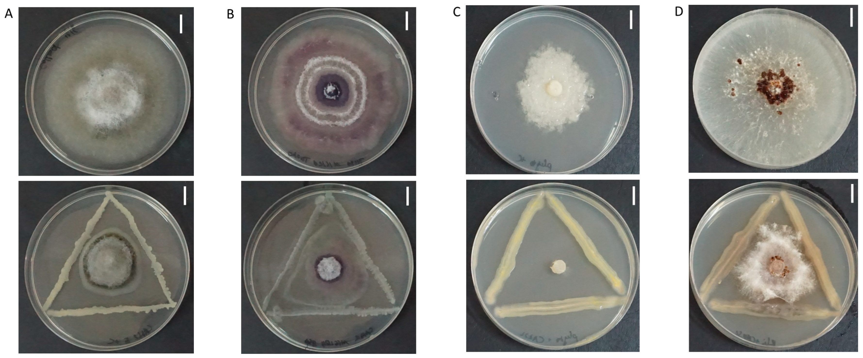
A total of 10 endophytic isolates collected from the roots of healthy sweet oranges grown in Cao Phong, Hoa Binh (Vietnam), were first screened for in vitro antagonistic activity against C. gloeosporioides (Supplementary Table S1). The result showed that six isolates (CAKS2, 3, 4, 5, 6, and 9) could inhibit the growth of C. gloeosporioides. Among them, CAKS2 provided the highest inhibition against the fungal growth with a ratio of 50.13% (Figure 1A, Supplementary Table S2). Then, the CAKS2 isolate was continuously screened for antagonistic activity against other fungal pathogens, including Fusarium oxysporum, Phytophthora parasitica var. nicotianae, and Rhizoctonia solani Kuhn. The assay showed that CAKS2 could inhibit the growth of these fungal pathogens to different degrees (Figure 1B–D, Table 1). Particularly, the inhibition ratios were 39.10% against F. oxysporum, 79.37% against P. parasitica, and 66.65% against R. solani.
In addition, the CAKS2 isolate also showed the ability to inhibit the growth of Pseudomonas syringae, Xanthomonas campestris, and Ralstonia solanacearum (Supplementary Figure S2, B in Table S1). In which, the mean of inhibition halo zones were 12.45, 15.80, and 14.45 mm against P. syringae, X. campestris, and R. solanacearum, respectively. Thus, CAKS2 exhibited notable in vitro antifungal and antibacterial activities against various plant pathogens.

3.2. Plant Growth-Promoting Characteristics of the CAKS2 Isolate

In addition to antimicrobial activities, the CAKS2 isolate also exhibited different plant growth-promotion characteristics, including IAA, ammonia, exopolysaccharide (EPS), HCN, and siderophore production; nitrogen fixation; N, P, K, and Ca solubilization; and biofilm formation (Table 2). Particularly, IAA, ammonia, and EPS were produced by the CAKS2 isolate at 1.28 µg/mL, 2.47 µM, and 0.51 g/L, respectively. The biofilm formation of the CAKS2 was recorded at 0.51 using OD550. Additionally, the solubilization capacity of the CAKS2 isolate for P, K, and Ca was 1.25, 1.41, and 1.16 SI, respectively. However, the CAKS2 isolate did not exhibit the capacity for degradation of CMC, chitin, or starch (Table 2).

3.3. 16S rRNA Identification of CAKS2

To facilitate further application of the potential CAKS2 isolate, the 16S rRNA fragment (1450 bp) was subjected to Sanger sequencing. The BLASTN version 2.7.0 analysis using the NCBI database indicated the 16S rRNA sequence of CAKS2 showed 100% similarity as compared to other Pseudomonas aeruginosa strains (Supplementary Figure S3A). Moreover, the outcome of 16S-based ID analysis on EzBioCloud confirmed the highest similarity (100%) between the 16S rRNA sequence of CAKS2 and two of the top-hit strains, P. aeruginosa JCM 5962 and PA7 (Supplementary Figure S3B). Therefore, CAKS2 could be classified as a P. aeruginosa strain (named P. aeruginosa CAKS2), and its 16S rRNA sequence was submitted to the NCBI GenBank under accession number PQ225979.

3.4. CAKS2 Affected Plant Growth, Biochemical Profiles, and Gene Expression of Inoculated Oranges

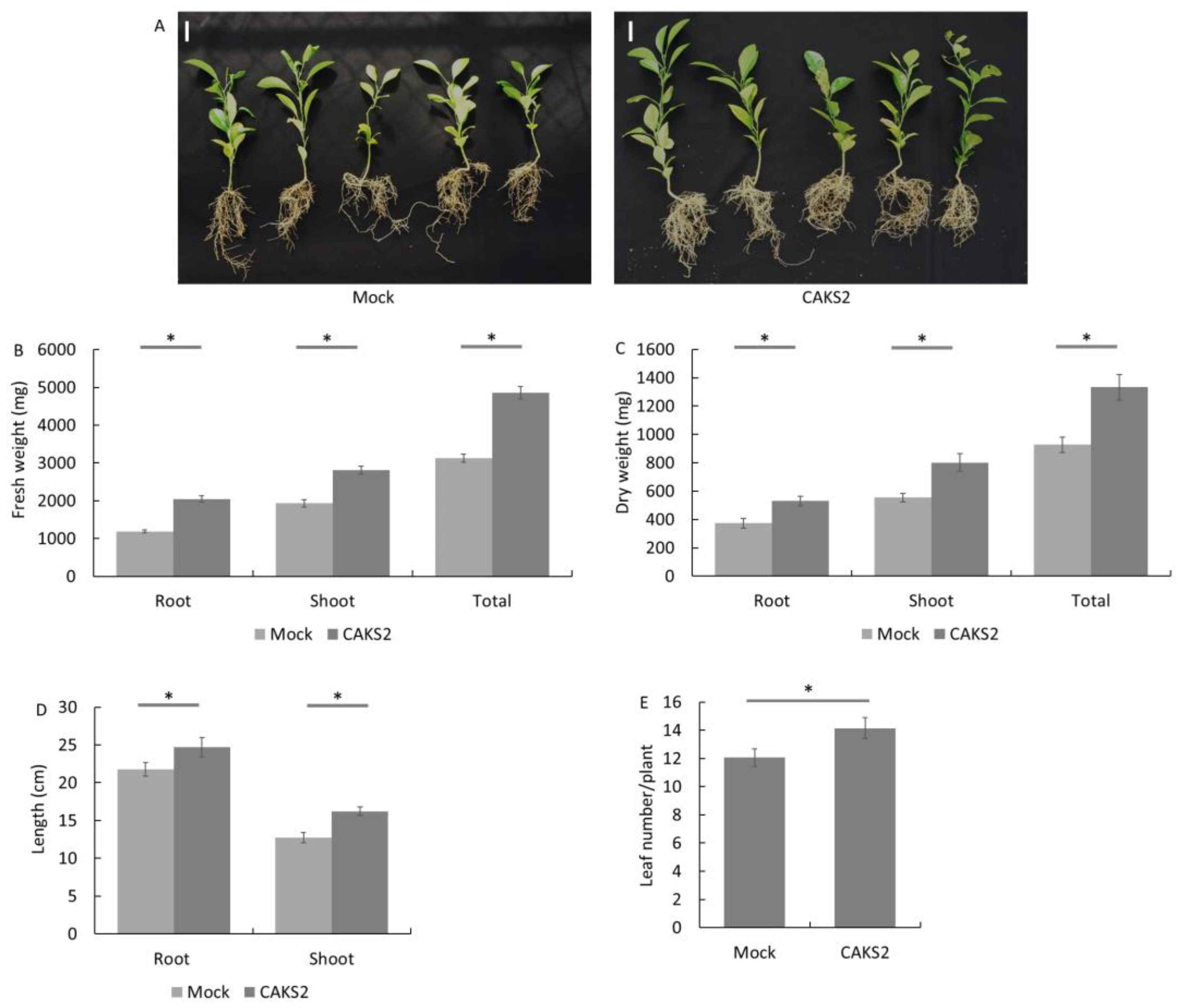
The CAKS2 isolate significantly altered the root, shoot, and biomass of treated sweet orange (Figure 2A). Particularly, the fresh weights of roots, shoots, and whole plants of the CAKS2-inoculated orange increased by 71.85, 45.19, and 55.36% as compared to the mock plants, while those for dry weights increased by 42.36, 44.76, and 43.80%, respectively (Figure 2B,C). Additionally, the longer roots and shoots, as well as the higher leaf number, were also found in the CAKS2-inoculated sweet orange (Figure 2D,E). These results confirmed the plant growth promotion characteristics of the CAKS2 isolate.
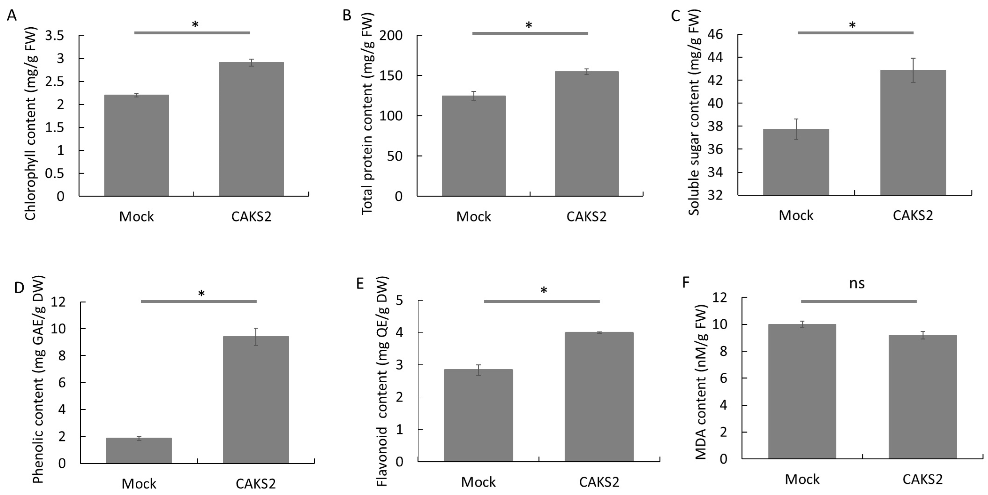
The biochemical analysis showed alterations in all tested biochemical components except the MDA content (Figure 3). In which, chlorophyll, total protein, soluble sugar, phenolic, and flavonoid contents in the CAKS2-inoculated plants increased by 32.35, 24.06, 13.63, 403.41, and 40.89%, respectively, as compared to the mock plants (Figure 3A–E). In contrast, no significant difference in the MDA level was observed between the CAKS2-inoculated oranges and the mock plants (Figure 3F). Additionally, DAB staining showed stronger H2O2 accumulation, indicated by the higher density of brown spots in the mock leaves as compared to the CAKS2-inoculated leaves (Supplementary Figure S4A). Indeed, the POD activity of the CAKS2-inoculated plants was higher by 140.36% than in mock plants (Supplementary Figure S4C). Thus, the CAKS2 inoculation altered different biochemical contents, reduced H2O2 accumulation as well as toxic elements via POD activity, and consequently promoted the growth of the sweet orange.
The transcription abundance of different genes related to plant hormone metabolism was assessed in the CAKS2-inoculated sweet orange. The qPCR result showed higher expression of all tested genes involved in gibberellin (GA) and auxin biosynthesis pathways in the CAKS2-inoculated plants than in the mock sweet orange (Supplementary Figure S5). Notably, the expression of CsGA20ox2 (GA 20-oxidase) was upregulated to 2.90-fold in the CAKS2-inoculated sweet orange. In addition, the transcription levels of CsTSB (Tryptophan synthase beta chain 1) and CsYUC8 (Indole-3-pyruvate monooxygenase YUCCA8) increased by 1.69- and 1.63-fold, respectively. These results again explain the plant growth promotion parameters of the CAKS2 isolate.

3.5. CAKS2 Reduced the Effects of C. gloeosporioides on Orange Leaves and Harvested Fruits

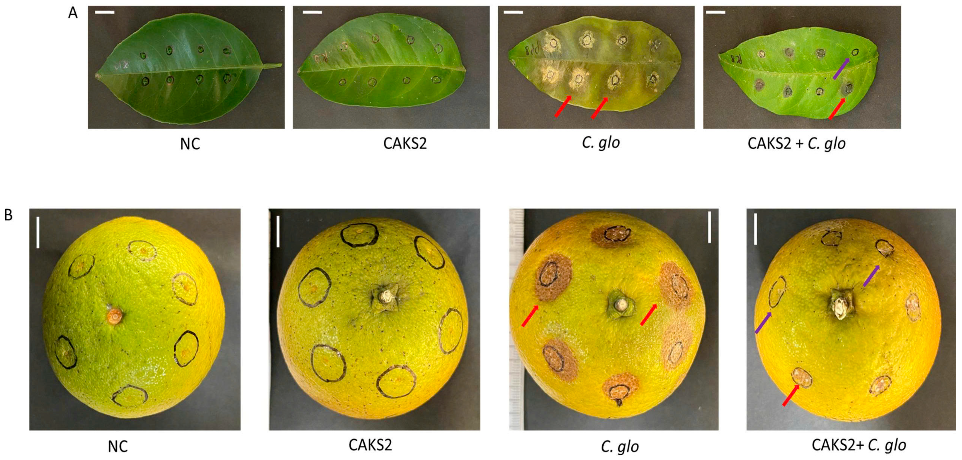
In the leaf infection assay of C. gloeosporioides, no change in leaf color around the wounded sites (purple arrow) was observed in the orange leaves treated with sterilized water (NC) or CAKS2. However, critical leaf damage and disease symptoms, including necrotic lesions, leaf spots, and blight symptoms (red arrow), were observed in the leaves infected with C. gloeosporioides (C. glo and CAKS22 + C. glo treatments) (Figure 4A). Importantly, both leaf necrosis and disease symptoms were significantly decreased in the leaves treated with CAKS2. Compared to leaves inoculated only with C. gloeosporioides (C. glo), which had a 100% infection rate, the infection rate of CAKS2-inoculated leaves was reduced to 74.30% (CAKS2 + C. glo). In addition, the infection diameter decreased from 0.71 cm in C. glo treatment to 0.58 cm in CAKS2 + C. glo treatment (Supplementary Figure S6A,B).
In line with the results of the leaf assay, no symptom was observed in fruits treated with NC or CAKS2, whereas fruits infected with C. gloeosporioides exhibited clear symptoms (red and purple arrows), including sunken lesions, dark spots, and fruit rot (C. glo and CAKS22 + C. glo treatments) (Figure 4B). In particular, CAKS2 significantly decreased the infection ratio to 45.37% in the treated fruits compared to 100% in the non-treated ones. The inoculation of CAKS2 also reduced the infection diameter of C. gloeosporioides from 1.37 cm (treated fruits) to 0.65 cm (non-treated fruits) (Supplementary Figure S6C,D).

3.6. CAKS2 Reduced the Effects of C. gloeosporioides on Sweet Orange

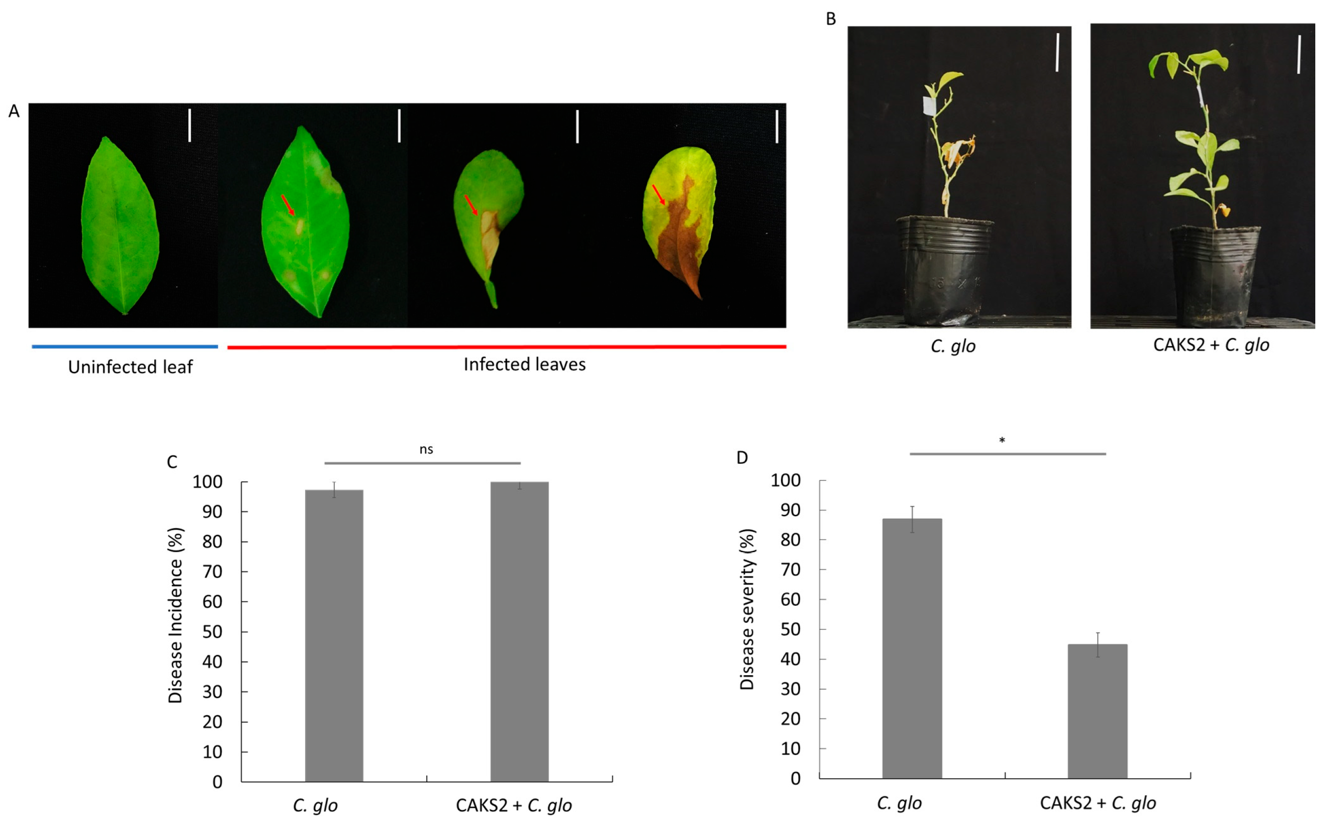
C. gloeosporioides infection was performed using 4-month-old sweet orange plants. Fourteen days post-infection, brown spots were observed on the leaf surface of the C. glo-infected plants, and the leaves turned dry and fell down (Figure 5A,B). However, the disease severity and incidence were much decreased in the treatment of CAKS2 + C. glo (Figure 5C). In particular, the disease severity was significantly reduced from 86.84% in the C. glo-infected plants to 44.73% in the CAKS2 + C. glo-treated sweet orange (Figure 5D). This result indicated that the damage of C. gloeosporioides on sweet orange could be reduced by the application of the CAKS2 isolate.
Subsequently, various biochemical contents of sweet orange were assessed in C. glo and CAKS2 + C. glo treatments (Supplementary Figure S6). The results indicated that all tested components (except MDA) were higher in the CAKS2 + C. glo plants as compared to C. glo-infected plants. Of which, total chlorophyll, total protein, soluble sugar, phenolic, and flavonoid contents increased by 10.93, 18.06, 13.05, 133.89, and 16.13% in the CAKS2 + C. glo plants as compared to those in C. glo plants, respectively (Supplementary Figure S7A–E). In contrast, the MDA content in CAKS2 + C. glo plants was 46.35% lower than that in C. glo plants (Supplementary Figure S7F). Additionally, the leaves of C. glo plants showed more prominent brown spots than mock plants, indicating higher H2O2 accumulation in pathogen-infected plants. Importantly, the CAKS2 reduced the intensity of brown spots in the treated plants infected with C. gloeosporioides (Supplementary Figure S4B). CAKS2 further enhanced 51.04% of POD activity in inoculated plants compared to those in C. glo plants (Supplementary Figure S4D). In summary, the reduced MDA content in inoculated plants demonstrated that CAKS2 could affect biochemical properties of sweet orange to alleviate the adverse effects of C. gloeosporioides.
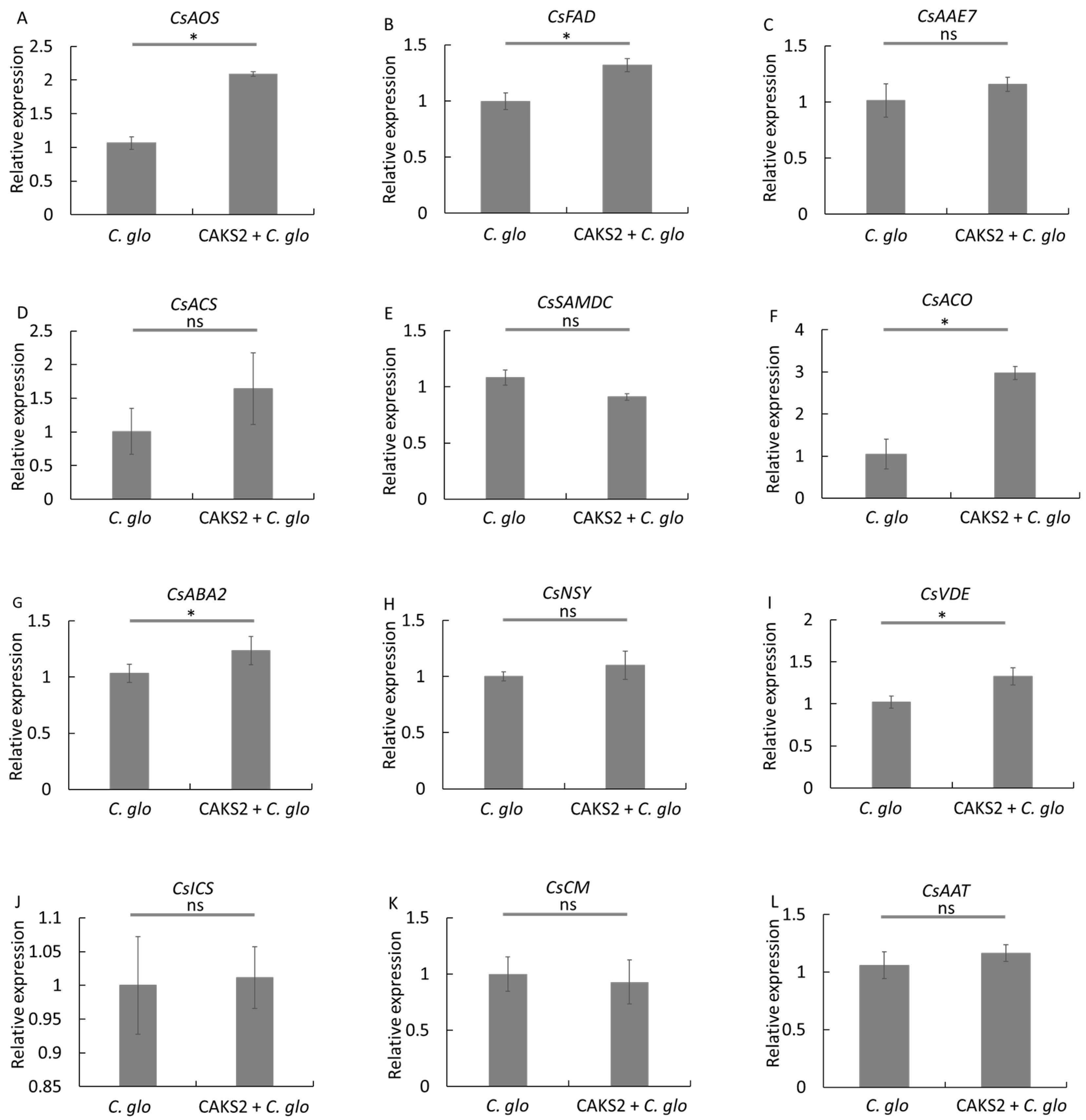
In this analysis, the expression of different genes related to plant pathogen tolerance (03 genes for JA, 03 genes for ETH, and 03 genes for ABA) was assessed in the C. gloeosporioides-infected plants and the CAKS2 + C. glo plants. For the JA biosynthesis pathway, CAKS2 inoculation increased the expression of CsAOS (Allene oxide synthase) and CsFAD (ω-3-fatty acid desaturase) by 1.96- and 1.32-fold, respectively, compared to C. glo plants (Figure 6A,B). However, the expression of CsAAE7 (Acetate/butyrate CoA ligase AAE7) was not different between CAKS2 + C. glo and C. glo plants (Figure 6C). Furthermore, ETH pathway genes, including CsACS (ACC synthase-like), CsACO (ACC oxidase), and CsSAMDC (S-adenosylmethionine decarboxylase), exhibited distinct expression patterns. In which CsACS and CsSAMDC genes showed no significant difference in transcript levels in both treatments (Figure 6D,E). In contrast, the CsACO expression was 2.84-fold higher in the CAKS2 + C. glo plants compared to the C. glo plants (Figure 6F). Additionally, the transcript levels of most ABA pathway genes were higher in C. glo plants, except for CsNSY (Neoxanthin synthase) (Figure 6G–I). In the CAKS2 + C. glo plants, CsABA2 (short-chain alcohol dehydrogenase) and CsVDE (Violaxanthin de-epoxidase) expression levels increased by 1.19- and 1.29-fold, respectively, compared to C. glo plants (Figure 6G,H). However, all genes in the SA pathway, including CsICS (Isochorismate synthase), CsCM (Chorismate mutase), and CsAAT (Alcohol acyl transferase), exhibited similar expression patterns across the treatments (Figure 6J–L). In summary, CAKS2 enhanced the expression of various genes associated with ABA, JA, and ETH pathways in sweet orange under C. gloeosporioides infection.

3.7. CAKS2 Whole Genome Sequencing

CAKS2 sequencing data (NCBI accession number JBNIDF000000000.1) generated from 77 contigs showed that the CAKS2 genome is about 6.3 Mbp with 66.44% GC. In which, a total of 5820 CDS were identified, as well as 68 tRNAs, 3 rRNAs, and 1 tmRNA (Supplementary Table S3). The circular genome visualization for the P. aeruginosa CAKS2 was established and showed the protein-coding sequences, forward and reverse CDS strands, GC content, and skew (Supplementary Figure S8A). Additionally, the phylogenetic tree was established using 107 core genes of P. aeruginosa CAKS2 and 33 other Pseudomonas sp. (Supplementary Figure S8B). In which the P. aeruginosa CAKS2 was highly close to the P. aeruginosa strains Y010, CI00795, and P2550, with ANI values of 99.24, 99.28, and 99.28%, respectively. The high ANI values (above 99%) indicated that P. aeruginosa CAKS2 could be classified as the P. aeruginosa species.
Utilizing different programs such as GO (Gene Ontology), COG (Clusters of Orthologous Groups of proteins), KEGG (Kyoto Encyclopedia of Genes and Genomes), and antiSMASH, several genes and gene clusters related to plant growth promotion, such as nitrogen fixation (norBCR), ammonia assimilation (gltABCDIJKPRSX), IAA production (trpABCDEFGI), as well as phosphate metabolism (pstBS and phoABDUR), were found in the CAKS2 genome. In addition, other genes involved in antifungal and antibacterial activities were also identified (Supplementary Table S4, Supplementary Dataset S1). Different genes related to the metabolisms of putative natural products, including siderophores (entSH and fepG), hydrogen cyanide (hcnABC), phenazines (phzABFGMS), salicylate (pchABR), EPS (algADEFGJKLX8), bicyclomycin (bcr), chorismate (ubiC), volatile metabolism (acoABCR), and 2,3-butanediol (ilvACDEGHI), were also found in the CAKS2 genome. Moreover, several genes related to oxidoreductase (sodB, osmCEVXYW, and katABE) and hydrolase (gdhAB and folBCDEPKMX) were detected from the CAKS2 genome sequence (Supplementary Table S4, Supplementary Dataset S1).

3.8. Root Exudate Altered the Expression of Related Genes in the CAKS2

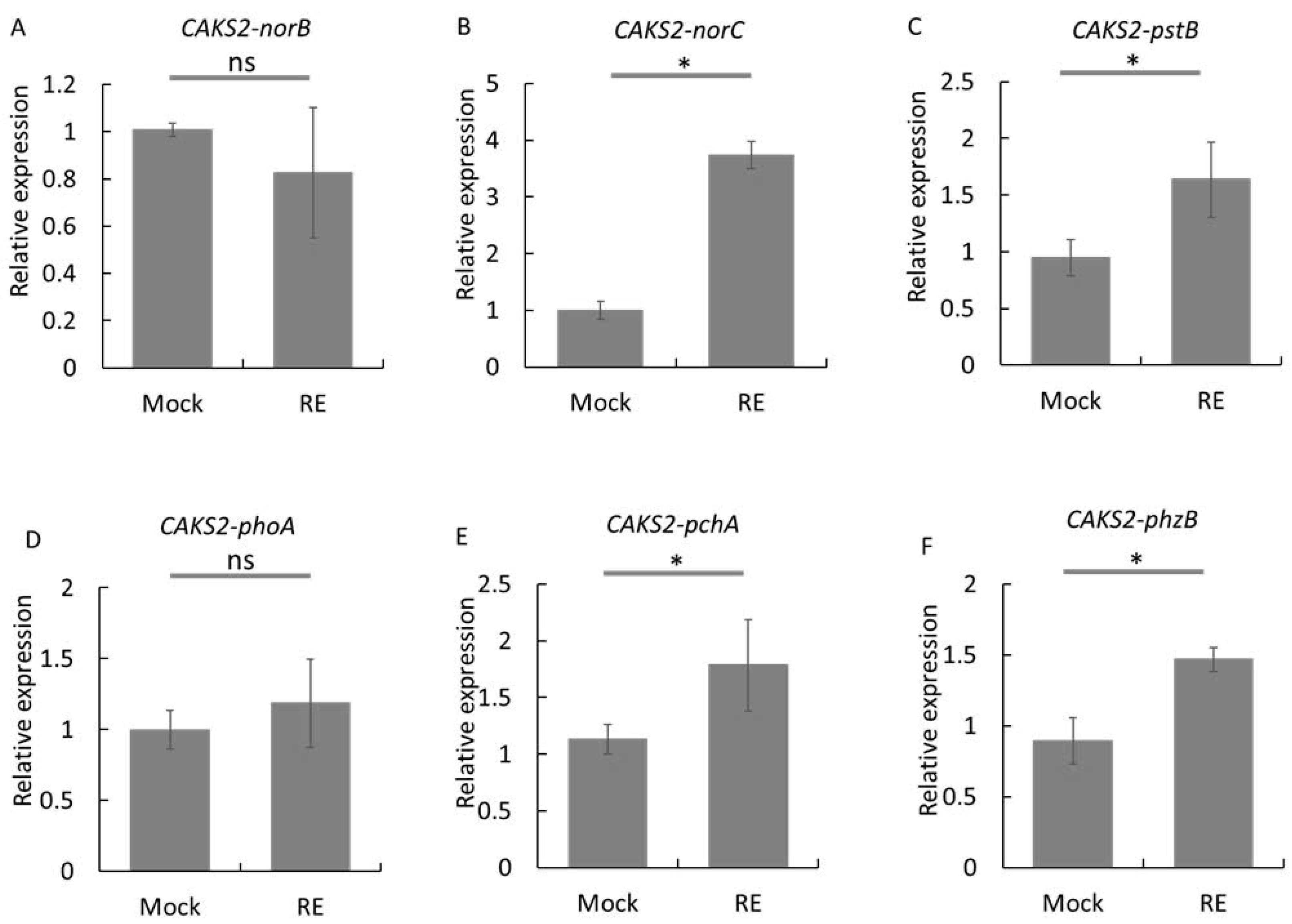
Different genes associated with nitrogen metabolism (Nitric oxide reductase subunit C, CAKS2-norC), phosphate metabolism (Phosphate import ATP-binding protein PstB, CAKS2-pstB), salicylate (Salicylate biosynthesis isochorismate synthase, CAKS2-pchA), and phenazine biosynthesis (Phenazine biosynthesis protein PhzB1, CAKS2-phzB) were upregulated when CAKS2 was exposed to orange root exudates. Notably, under the RE treatment, the expression of CAKS2-norC, CAKS2-pstB, CAKS2-pchA, and CAKS2-phzB was increased by 3.73-, 1.72-, 1.57-, and 1.63-fold compared to the mock treatment, respectively (Figure 7B,C,E,F). However, the expression of CAKS2-norB (Nitric oxide reductase subunit B) and CAKS2-phoA (Alkaline phosphatase H) remained unchanged across the mock and RE treatments (Figure 7A,D). These findings suggest that several genes associated with plant growth promotion and biocontrol in CAKS2 exhibited higher transcript levels in response to RE treatment.

4. Discussion

Anthracnose, caused by Colletotrichum spp., is a major disease that significantly reduces citrus production, particularly in oranges, worldwide. It negatively impacts both yield and fruit quality during preharvest and postharvest periods, hindering export potential and marketability [62]. Recently, the use of plant growth-promoting rhizobacteria (PGPR) has shown potential in controlling citrus anthracnose for sustainable agriculture [1,2,3]. Notably, P. aeruginosa species have been demonstrated to enhance plant growth and improve tolerance to both biotic and abiotic stresses in various crops [9,10,11,12,13,14]. In this study, we investigated and demonstrated the potential of P. aeruginosa strain CAKS2 for enhancing plant growth and reducing the effects of C. gloeosporioides on sweet orange.

4.1. CAKS2 Shows Potential for Enhancing Sweet Orange Plant Growth and Reducing the Effect of C. gloeosporioides

PGPR with significant benefits to plants has been used in biocontrol and biofertilizer for sustainable crop production and protection [63]. Previous studies showed that P. aeruginosa strains enhanced the growth and pathogen tolerance of various crops [9,10,11,12,13,14]. Particularly, these bacterial species enhanced various growth parameters, including root and shoot lengths, fresh and dry weights, and leaf number of inoculated plants [10,12]. Moreover, different P. aeruginosa strains also decreased disease severity on leaves, fruits, and whole plants of various crops, including tomato and banana, under the infection of fungal and bacterial diseases [10,12,13,64]. In the current study, the P. aeruginosa CAKS2 increased the fresh and dry weights, root and shoot lengths, and leaf number of inoculated sweet orange under greenhouse conditions. In addition, the CAKS2 reduced disease severity, infection ratio, diameter of lesions on orange leaves, harvested fruits, and whole sweet orange infected by C. gloeosporioides sp. Although different PGPR species have been identified and utilized in various plant species [2,3,10,12,47], our result is the first success in using P. aeruginosa to enhance growth and reduce the effects of C. gloeosporioides sp on sweet orange. This strain will be added to the list of PGPR that have the potential for further application in sustainable crop improvement.

4.2. CAKS2 Changes Biochemical Properties and Gene Expression, and Consequently Promotes Orange Growth

Previously, the potential PGPR for crop growth promotion often had main characteristics including nutrient solubilization, phytohormone production, nitrogen fixation, as well as secretion of secondary compounds [23,44]. In this study, the P. aeruginosa CAKS2 exhibited important parameters for plant growth promotion, such as fixing nitrogen, solubilizing P, K, and Ca, and producing IAA as well as ammonia. These parameters were also observed in the other P. aeruginosa strain, showing plant growth promotion in a previous report [12].
Whole genome sequencing has been used to assess molecular mechanisms under the plant growth promotion characteristics of several PGPRs, recently [14,65,66]. From the whole genome sequencing data, different genes related to plant growth promotion properties have been found and confirmed in several P. aeruginosa strains [14,67,68,69,70]. In our study, the whole genome sequencing of P. aeruginosa CAKS2 showed certain genes involved in nitrogen metabolism, IAA, GA, siderophore production, and phosphate metabolism that were revealed by the PGP test in vitro mentioned above. The presence of these genes in the CAKS2 genome supports the production of certain compounds observed in in vitro tests (Table 2). In addition, the alteration in transcription abundance of these genes was confirmed as the P. aeruginosa CAKS2 was exposed to the treatments of orange root exudates. We also found increases in the accumulation of chlorophyll, total protein, soluble sugar, phenolic, and flavonoid contents, and a reduction in the MDA component of the CAKS2-inoculated sweet orange. These changes in biochemical properties were also observed in different bacterial strains showing plant growth promotion in other crops, such as Arabidopsis, cucumber, mung bean, and tomato [44,71,72,73]. In agreement with previous studies, all these data again indicated the potential of the CAKS2 for enhancing plant growth and development.

4.3. CAKS2 Protects Sweet Orange from C. gloeosporioides via Different Biochemical and Molecular Properties

Under pathogen infection, plants trigger various physio-biochemical changes related to stress defense pathways, including non-enzymatic compounds (flavonoid and phenolic compounds), and antioxidant enzymes (SOD, CAT, POD, GR) to neutralize ROS production [74]. Moreover, plants must minimize the damage of photosynthesis and reduce the energy loss caused by pathogen invasion to ensure plant growth [17]. In previous reports, PGPR strains have been shown to enhance the accumulation of non-enzymatic compounds and antioxidant enzymes, as well as maintain photosynthesis components to protect plants from disease infection [17]. In our current research, under the infection of C. gloeosporioides, the CAKS2 increased the phenolic and flavonoid contents as well as POD activity and reduced the MDA content of the inoculated sweet orange. Phenolic compounds and peroxidase (POD) are important components in a plant’s defense system, and often act synergistically. Phenolic compounds with antimicrobial and antioxidant activity can help plants resist pathogen attacks. While POD is known to be widely involved in strengthening plant cell walls, transmitting hormone signals, and responding to biotic and abiotic stressors. In contrast, MDA is a widely used marker for lipid peroxidation and oxidative damage to cellular membranes. The lower MDA levels under stress often indicate higher resistance or a more effective defense system.
Furthermore, PGPR were demonstrated to affect the expression of genes involved in plant hormone signaling pathways in response to pathogen invasion [20,21,22,75]. In this study, the expression of certain genes in the JA, ETH, and ABA pathways was also altered in the CAKS2-inoculated sweet orange. Furthermore, various genes related to the production of siderophores, hydrogen cyanide, phenazines, salicylate, bicyclomycin, chorismate, hydrolases, etc., have been found in the P. aeruginosa CAKS2 genome. The transcription abundance of genes related to biocontrol was also upregulated when the P. aeruginosa CAKS2 was treated with orange root exudates. These results indicate that CAKS2 has PGPR parameters to protect the sweet orange from C. gloeosporioides and should be potentially tested with other plant diseases.

5. Conclusions

This study demonstrates the potential for plant growth promotion and antifungal activity of the Pseudomonas aeruginosa CAKS2 isolated from local sweet orange. The PGP characteristics of the selected isolate were indicated by enhanced plant growth parameters, including root and shoot length, number of leaves, and both fresh and dry biomass of inoculated oranges as compared to the mock plants. The antifungal activity of the CKS2 was validated by a reduction in anthracnose disease severity caused by Colletotrichum gloeosporioides in sweet orange and harvested fruits. The whole-genome analysis and qPCR confirmed changes in the expression of genes expectedly related to plant growth promotion and biocontrol activity in both sweet orange and the bacterial isolate. This selected bacterial strain is potentially useful for further research on plant-microbe interactions as well as for agricultural applications.

Supplementary Materials

The following supporting information can be downloaded at https://www.mdpi.com/article/10.3390/horticulturae12040442/s1, Figure S1: Experimental designs for testing different potential parameters of the P. aeruginosa CAKS2 isolate; Figure S2: Antibacterial activities of the P. aeruginosa CAKS2 isolate; Figure S3: Nucleotide BLAST of CAKS2 on NCBI and 16S-Based ID of CAKS2 on EzBioCloud; Figure S4: The hydrogen peroxide accumulation and POD activities of P. aeruginosa CAKS2 inoculated sweet orange under the treatment of C. gloeosporioides; Figure S5: The transcription abundance of related genes in the mock and CAKS2 inoculated sweet orange; Figure S6: The Disease severity of C. gloeosporioides in orange leaves and fruits; Figure S7: Changes in biochemical properties of the mock and the CAKS2 inoculated sweet orange under the C. gloeosporioides infection; Figure S8: Whole genome sequencing and phylogenetic analysis of the CAKS2 strain. Table S1. qPCR primers for bacteria and orange analysis. Table S2. Assessment of in vitro antifungal of endophytic isolates against C. gloeosporioides. Table S3. Genome characteristics of CAKS2. Table S4. Presence of PGP characteristics in the CAKS2 genome related to plant growth promotion and pathogen inhibition.

Author Contributions

Conceptualization, T.M.H., H.Q.P., and P.T.D.; methodology, T.M.H., H.Q.P., M.V.L., H.H.T.N., H.T.T., T.T.N., C.H.N., T.D.N., and Q.P.; validation, T.M.H., H.Q.P., and P.T.D.; investigation, H.Q.P., M.V.L., H.H.T.N., and H.T.T.; data curation, T.M.H.; writing—original draft, T.M.H. and H.Q.P.; writing—review and editing, P.T.D. and H.H.C.; supervision, H.H.C., S.N.H., and P.T.D.; funding acquisition, H.Q.P.; project administration, H.H.C., S.N.H., and P.T.D. All authors have read and agreed to the published version of the manuscript.

Funding

This research was funded by the Vietnam Academy of Science and Technology (VAST) through Grant No. TĐCPVS.02/23-25.

Data Availability Statement

The original contributions presented in this study are included in the article/Supplementary Material. Further inquiries can be directed to the corresponding author.

Acknowledgments

We thank the Vietnam Academy of Science and Technology (VAST) for supporting the project grant. We appreciate the Plant Cell Biotechnology Laboratory, Institute of Biology (IB), where the experiments are performed.

Conflicts of Interest

The authors declare no conflict of interest.

Abbreviations

The following abbreviations are used in this manuscript:
PGPRPlant growth promotion rhizobacteria
SODSuperoxide dismutase
CATCatalase
PODPeroxidase
GRGlutathione reductase
ISRInduced systemic resistance
SARSystemic acquired resistance
CASChrome Azurol S
HCNHydrogen cyanide
PKVPikovskaya
EPSExopolysaccharide
ChlChlorophyll content
DAB3′-diaminobenzidine
BGCBiosynthetic gene clusters
CsEF1Elongation factor-1 alpha
CsF-boxF-box/kelch-repeat protein
algDGDP-mannose 6-dehydrogenase
gyrADNA gyrase subunit A
IAAIndole-3-acetic acid
CMCCarboxymethyl cellulose
MDAMalondialdehype
GAGibberellin
GA20ox2GA 20-oxidase
TSBTryptophan synthase beta chain 1
YUC8Indole-3-pyruvate mono oxygenase YUCCA8
JAJasmonic acid
ETHEthylene
ABAAbscisic acid
CsAOSAllene oxide synthase
CsFADω-3-fatty acid desaturase
CsAAE7Acetate/butyrate CoA ligase AAE7
CsACSACC synthase-like
CsACOACC oxidase
CsSAMDCS-adenosylmethionine decarboxylase
CsVDEViolaxanthin de-epoxidase
CsCMChorismate mutase
CsAATAlcohol acyl transferase
ANIAverage Nucleotide Identity
GOGene Ontology
COGClusters of Orthologous Groups of proteins
KEGGKyoto Encyclopedia of Genes and Genomes

References

  1. Giassi, V.; Kiritani, C.; Kupper, K.C. Bacteria as growth-promoting agents for citrus rootstocks. Microbiol. Res. 2016, 190, 46–54. [Google Scholar] [CrossRef] [Scilit]
  2. Ezrari, S.; Mhidra, O.; Radouane, N.; Tahiri, A.; Polizzi, G.; Lazraq, A.; Lahlali, R. Potential role of Rhizobacteria isolated from citrus rhizosphere for biological control of citrus dry root rot. Plants 2021, 10, 872. [Google Scholar] [CrossRef] [Scilit]
  3. Adhyaningtyas, R.F.I.; Rahmawati, R.; Mukarlina, M. Effect of phosphate solubilizing and nitrogen-fixing bacteria on Pontianak siam citrus (Citrus × nobilis var. microcarpa) seed germination. Nusant. Biosci. 2023, 15, 245–250. [Google Scholar] [CrossRef] [Scilit]
  4. Kejela, T.; Thakkar, V.R.; Thakor, P. Bacillus species (BT42) isolated from Coffea arabica L. rhizosphere antagonizes Colletotrichum gloeosporioides and Fusarium oxysporum and also exhibits multiple plant growth promoting activity. BMC Microbiol. 2016, 16, 277. [Google Scholar] [CrossRef] [Scilit]
  5. Kejela, T.; Thakkar, V.R.; Patel, R.R. A novel strain of Pseudomonas inhibits Colletotrichum gloeosporioides and Fusarium oxysporum infections and promotes germination of coffee. Rhizosphere 2017, 4, 9–15. [Google Scholar] [CrossRef] [Scilit]
  6. Guerrero-Barajas, C.; Constantino-Salinas, E.A.; Amora-Lazcano, E.; Tlalapango-Ángeles, D.; Mendoza-Figueroa, J.S.; Cruz-Maya, J.A.; Jan-Roblero, J. Bacillus mycoides A1 and Bacillus tequilensis A3 inhibit the growth of a member of the phytopathogen Colletotrichum gloeosporioides species complex in avocado. J. Sci. Food Agric. 2020, 100, 4049–4056. [Google Scholar] [CrossRef] [Scilit] [PubMed]
  7. Huang, H.; Tian, C.; Huang, Y.; Huang, H. Biological control of poplar anthracnose caused by Colletotrichum gloeosporioides (Penz.) Penz. & Sacc. Egypt. J. Biol. Pest. Control 2020, 30, 104. [Google Scholar] [CrossRef] [Scilit]
  8. Choub, V.; Ajuna, H.B.; Won, S.-J.; Moon, J.-H.; Choi, S.-I.; Maung, C.E.H.; Kim, C.-W.; Ahn, Y.S. Antifungal activity of Bacillus velezensis CE 100 against Anthracnose disease (Colletotrichum gloeosporioides) and growth promotion of walnut (Juglans regia L.) trees. Int. J. Mol. Sci. 2021, 22, 10438. [Google Scholar] [CrossRef] [Scilit] [PubMed]
  9. Chopra, A.; Bobate, S.; Rahi, P.; Banpurkar, A.; Mazumder, P.B.; Satpute, S. Pseudomonas aeruginosa RTE4: A tea rhizobacterium with potential for plant growth promotion and biosurfactant production. Front. Bioeng. Biotechnol. 2020, 8, 861. [Google Scholar] [CrossRef] [Scilit]
  10. Wang, X.; Zhou, X.; Cai, Z.; Guo, L.; Chen, X.; Chen, X.; Liu, J.; Feng, M.; Qiu, Y.; Zhang, Y.; et al. A biocontrol strain of Pseudomonas aeruginosa CQ-40 promote growth and control botrytis cinerea in tomato. Pathogens 2020, 10, 22. [Google Scholar] [CrossRef] [Scilit]
  11. Kauppinen, A.; Siponen, S.; Pitkänen, T.; Holmfeldt, K.; Pursiainen, A.; Torvinen, E.; Miettinen, I.T. Phage biocontrol of Pseudomonas aeruginosa in water. Viruses 2021, 13, 928. [Google Scholar] [CrossRef] [Scilit] [PubMed]
  12. Ghadamgahi, F.; Tarighi, S.; Taheri, P.; Saripella, G.V.; Anzalone, A.; Kalyandurg, P.B.; Catara, V.; Ortiz, R.; Vetukuri, R.R. Plant growth-promoting activity of Pseudomonas aeruginosa FG106 and its ability to act as a biocontrol agent against potato, tomato and taro pathogens. Biology 2022, 11, 140. [Google Scholar] [CrossRef] [Scilit]
  13. Xie, J.; Singh, P.; Qi, Y.; Singh, R.K.; Qin, Q.; Jin, C.; Wang, B.; Fang, W. Pseudomonas aeruginosa strain 91: A multifaceted biocontrol agent against banana fusarium wilt. J. Fungi 2023, 9, 1047. [Google Scholar] [CrossRef] [Scilit] [PubMed]
  14. Meng, J.; Zan, F.; Liu, Z.; Zhang, Y.; Qin, C.; Hao, L.; Wang, Z.; Wang, L.; Liu, D.; Liang, S.; et al. Genomics analysis reveals the potential biocontrol mechanism of Pseudomonas aeruginosa QY43 against Fusarium pseudograminearum. J. Fungi 2024, 10, 298. [Google Scholar] [CrossRef] [Scilit] [PubMed]
  15. Goswami, D.; Borah, S.N.; Lahkar, J.; Handique, P.J.; Deka, S. Antifungal properties of rhamnolipid produced by Pseudomonas aeruginosa DS9 against Colletotrichum falcatum. J. Basic Microbiol. 2015, 55, 1265–1274. [Google Scholar] [CrossRef] [Scilit]
  16. Jisha, M.S.; Linu, M.S.; Sreekumar, J. Induction of systemic resistance in chilli (Capsicum annuum L.) by Pseudomonas aeruginosa against anthracnose pathogen Colletotrichum capsici. J. Trop. Agric. 2018, 56, 153–166. [Google Scholar]
  17. Su, F.; Zhao, B.; Dhondt-Cordelier, S.; Vaillant-Gaveau, N. Plant-growth-promoting rhizobacteria modulate carbohydrate metabolism in connection with host plant defense mechanism. Int. J. Mol. Sci. 2024, 25, 1465. [Google Scholar] [CrossRef] [Scilit]
  18. Tanveer, S.; Akhtar, N.; Ilyas, N.; Sayyed, R.Z.; Fitriatin, B.N.; Perveen, K.; Bukhari, N.A. Interactive effects of Pseudomonas putida and salicylic acid for mitigating drought tolerance in canola (Brassica napus L.). Heliyon 2023, 9, e14193. [Google Scholar] [CrossRef] [Scilit]
  19. Nakkeeran, S.; Kavitha, K.; Chandrasekar, G.; Renukadevi, P.; Fernando, W.G.D. Induction of plant defence compounds by Pseudomonas chlororaphis PA23 and Bacillus subtilis BSCBE4 in controlling damping-off of hot pepper caused by Pythium aphanidermatum. Biocontrol Sci. Technol. 2006, 16, 403–416. [Google Scholar] [CrossRef] [Scilit]
  20. Fan, D.; Smith, D.L. Characterization of selected plant growth-promoting rhizobacteria and their non-host growth promotion effects. Microbiol. Spectr. 2021, 9, e0027921. [Google Scholar] [CrossRef] [Scilit]
  21. Yu, Y.; Gui, Y.; Li, Z.; Jiang, C.; Guo, J.; Niu, D. Induced systemic resistance for improving plant immunity by beneficial microbes. Plants 2022, 11, 386. [Google Scholar] [CrossRef] [Scilit]
  22. Salwan, R.; Sharma, M.; Sharma, A.; Sharma, V. Insights into plant beneficial microorganism-triggered induced systemic resistance. Plant Stress 2023, 7, 100140. [Google Scholar] [CrossRef] [Scilit]
  23. Singh, A.; Yadav, V.K.; Chundawat, R.S.; Soltane, R.; Awwad, N.S.; Ibrahium, H.A.; Yadav, K.K.; Vicas, S.I. Enhancing plant growth promoting rhizobacterial activities through consortium exposure: A review. Front. Bioeng. Biotechnol. 2023, 11, 1099999. [Google Scholar] [CrossRef] [Scilit] [PubMed]
  24. Sáenz Pérez, C.A.; Hernández, E.O.; Estrada Drouaillet, B.; Poot Poot, W.A.; Delgado Martínez, R.; Herrera, R.R.; Sáenz Pérez, C.A.; Hernández, E.O.; Estrada Drouaillet, B.; Poot Poot, W.A.; et al. Main diseases in citrus. Rev. Mex. Cienc. Agrícolas 2019, 10, 1653–1665. [Google Scholar] [CrossRef] [Scilit]
  25. Rouseff, R.L.; Ruiz Perez-Cacho, P.; Jabalpurwala, F. Historical review of citrus flavor research during the past 100 years. J. Agric. Food Chem. 2009, 57, 8115–8124. [Google Scholar] [CrossRef] [Scilit]
  26. Poveda, J.; Roeschlin, R.A.; Marano, M.R.; Favaro, M.A. Microorganisms as biocontrol agents against bacterial citrus diseases. Biol. Control 2021, 158, 104602. [Google Scholar] [CrossRef] [Scilit]
  27. Shahbaz, E.; Ali, M.; Shafiq, M.; Atiq, M.; Hussain, M.; Balal, R.M.; Sarkhosh, A.; Alferez, F.; Sadiq, S.; Shahid, M.A. Citrus canker pathogen, its mechanism of infection, eradication, and impacts. Plants 2022, 12, 123. [Google Scholar] [CrossRef] [Scilit]
  28. Patricia, L.-C.C.; María del Socorro, R.-G.; Iván, R.-H.; Erika, D.l.C.-A.; Carolina, D.S.; Keiko, S.; José Alberto, N.-Z. Occurrence and infective potential of Colletotrichum gloeosporioides isolates associated to Citrus limon var Eureka. Biotechnol. Rep. 2021, 31, e00651. [Google Scholar] [CrossRef] [Scilit] [PubMed]
  29. Salotti, I.; Liang, Y.-J.; Ji, T.; Rossi, V. Development of a model for Colletotrichum diseases with calibration for phylogenetic clades on different host plants. Front. Plant Sci. 2023, 14, 1069092. [Google Scholar] [CrossRef] [Scilit]
  30. Reinhold-Hurek, B.; Hurek, T. Reassessment of the taxonomic structure of the diazotrophic genus Azoarcus sensu lato and description of three new genera and new species, Azovibrio restrictus gen. nov., sp. nov., Azospira oryzae gen. nov., sp. nov. and Azonexus fungiphilus gen. nov., sp. nov. Int. J. Syst. Evol. Microbiol. 2000, 50, 649–659. [Google Scholar] [CrossRef] [Scilit]
  31. Pham, Q.H.; Ho, M.T.; Tran, T.H.; Nguyen, T.H.H.; Le, V.M.; Chu, H.H.; Do, T.P. Isolation and characterization of Colletotrichum gloeosporioides causing anthracnose on sweet orange (Citrus sinensis) at Cao Phong, Hoa Binh. In Proceedings of the 22nd National Conference of Phytopathological Society of Vietnam, Hanoi, Vietnam, 25–26 October 2023; pp. 163–170. [Google Scholar]
  32. Zhou, L.; Song, C.; Li, Z.; Kuipers, O.P. Antimicrobial activity screening of rhizosphere soil bacteria from tomato and genome-based analysis of their antimicrobial biosynthetic potential. BMC Genom. 2021, 22, 29. [Google Scholar] [CrossRef] [Scilit]
  33. Gordon, S.A.; Weber, R.P. Colorimetric estimation of indoleacetic Acid. Plant Physiol. 1951, 26, 192–195. [Google Scholar] [CrossRef] [Scilit]
  34. Louden, B.C.; Haarmann, D.; Lynne, A.M. Use of blue agar CAS assay for siderophore detection. J. Microbiol. Biol. Educ. 2011, 12, 51–53. [Google Scholar] [CrossRef] [Scilit]
  35. Lotfi, N.; Soleimani, A.; Çakmakçı, R.; Vahdati, K.; Mohammadi, P. Characterization of plant growth-promoting rhizobacteria (PGPR) in Persian walnut associated with drought stress tolerance. Sci. Rep. 2022, 12, 12725. [Google Scholar] [CrossRef] [Scilit]
  36. AlAli, H.A.; Khalifa, A.; Almalki, M. Plant growth-promoting rhizobacteria from Ocimum basilicum improve growth of Phaseolus vulgaris and Abelmoschus esculentus. S. Afr. J. Bot. 2021, 139, 200–209. [Google Scholar] [CrossRef] [Scilit]
  37. Sun, F.; Ou, Q.; Wang, N.; Guo, Z.X.; Ou, Y.; Li, N.; Peng, C. Isolation and identification of potassium-solubilizing bacteria from Mikania micrantha rhizospheric soil and their effect on M. micrantha plants. Glob. Ecol. Conserv. 2020, 23, e01141. [Google Scholar] [CrossRef] [Scilit]
  38. Amri, M.; Rjeibi, M.R.; Gatrouni, M.; Mateus, D.M.R.; Asses, N.; Pinho, H.J.O.; Abbes, C. Isolation, identification, and characterization of phosphate-solubilizing bacteria from tunisian soils. Microorganisms 2023, 11, 783. [Google Scholar] [CrossRef] [Scilit] [PubMed]
  39. Saheewala, H.; Sanadhya, S.; Upadhyay, S.K.; Mohanty, S.R.; Jain, D. Polyphasic characterization of indigenous potassium-solubilizing bacteria and its efficacy studies on maize. Agronomy 2023, 13, 1919. [Google Scholar] [CrossRef] [Scilit]
  40. Mir, M.I.; Hameeda, B.; Quadriya, H.; Kumar, B.K.; Ilyas, N.; Kee Zuan, A.T.; El Enshasy, H.A.; Dailin, D.J.; Kassem, H.S.; Gafur, A.; et al. Multifarious indigenous diazotrophic rhizobacteria of rice (Oryza sativa L.) rhizosphere and their effect on plant growth promotion. Front. Nutr. 2022, 8, 781764. [Google Scholar] [CrossRef] [Scilit]
  41. Abdelwahed, S.; Trabelsi, E.; Saadouli, I.; Kouidhi, S.; Masmoudi, A.S.; Cherif, A.; Mnif, W.; Mosbah, A. A new pioneer colorimetric micro-plate method for the estimation of ammonia production by plant growth promoting rhizobacteria (PGPR). Main Group Chem. 2022, 21, 55–68. [Google Scholar] [CrossRef] [Scilit]
  42. Kaur, K.; Dattajirao, V.; Shrivastava, V.; Bhardwaj, U. Isolation and characterization of chitosan-producing bacteria from beaches of chennai, India. Enzyme Res. 2012, 2012, 421683. [Google Scholar] [CrossRef] [Scilit] [PubMed]
  43. Jabiri, S.; Legrifi, I.; Benhammou, M.; Laasli, S.-E.; Mokrini, F.; Amraoui, M.B.; Lahlali, R. Screening of rhizobacterial isolates from apple rhizosphere for their biocontrol and plant growth promotion activity. Appl. Microbiol. 2023, 3, 948–967. [Google Scholar] [CrossRef] [Scilit]
  44. Manh Tuong, H.; Garcia Mendez, S.; Vandecasteele, M.; Willems, A.; Luo, D.; Beirinckx, S.; Goormachtig, S. Stenotrophomonas sp. SRS1 promotes growth of Arabidopsis and tomato plants under salt stress conditions. Plant Soil. 2022, 473, 547–571. [Google Scholar] [CrossRef] [Scilit]
  45. Thilagam, R.; Hemalatha, N. Plant growth promotion and chilli anthracnose disease suppression ability of rhizosphere soil actinobacteria. J. Appl. Microbiol. 2019, 126, 1835–1849. [Google Scholar] [CrossRef] [Scilit]
  46. Guarnaccia, V.; Groenewald, J.Z.; Polizzi, G.; Crous, P.W. High species diversity in Colletotrichum associated with citrus diseases in Europe. Persoonia 2017, 39, 32–50. [Google Scholar] [CrossRef] [Scilit]
  47. Vu, T.X.; Tran, T.B.; Tran, M.B.; Do, T.T.K.; Do, L.M.; Dinh, M.T.; Thai, H.-D.; Pham, D.-N.; Tran, V.-T. Efficient control of the fungal pathogens Colletotrichum gloeosporioides and Penicillium digitatum infecting citrus fruits by native soilborne Bacillus velezensis strains. Heliyon 2023, 9, e13663. [Google Scholar] [CrossRef] [Scilit]
  48. Senthilkumar, M.; Amaresan, N.; Sankaranarayanan, A. Determination of Chlorophyll. In Plant-Microbe Interactions: Laboratory Techniques; Senthilkumar, M., Amaresan, N., Sankaranarayanan, A., Eds.; Springer: New York, NY, USA, 2021; pp. 145–146. [Google Scholar]
  49. Chen, T.; Zhang, B. Measurements of proline and malondialdehyde content and antioxidant enzyme activities in leaves of drought stressed cotton. Bio-Protocol 2016, 6, e1913. [Google Scholar] [CrossRef] [Scilit]
  50. Khan, N.; Bano, A. Exopolysaccharide producing rhizobacteria and their impact on growth and drought tolerance of wheat grown under rainfed conditions. PLoS ONE 2019, 14, e0222302. [Google Scholar] [CrossRef] [Scilit] [PubMed]
  51. Aryal, S.; Baniya, M.K.; Danekhu, K.; Kunwar, P.; Gurung, R.; Koirala, N. Total Phenolic content, Flavonoid content and antioxidant potential of wild vegetables from Western Nepal. Plants 2019, 8, 96. [Google Scholar] [CrossRef] [Scilit]
  52. Daudi, A.; O’Brien, J.A. Detection of Hydrogen Peroxide by DAB Staining in Arabidopsis Leaves. Bio-Protocol 2012, 2, e263. [Google Scholar] [CrossRef] [Scilit]
  53. Simão, F.A.; Waterhouse, R.M.; Ioannidis, P.; Kriventseva, E.V.; Zdobnov, E.M. BUSCO: Assessing genome assembly and annotation completeness with single-copy orthologs. Bioinformatics 2015, 31, 3210–3212. [Google Scholar] [CrossRef] [Scilit]
  54. Bankevich, A.; Nurk, S.; Antipov, D.; Gurevich, A.A.; Dvorkin, M.; Kulikov, A.S.; Lesin, V.M.; Nikolenko, S.I.; Pham, S.; Prjibelski, A.D.; et al. SPAdes: A New Genome Assembly Algorithm and Its Applications to Single-Cell Sequencing. J. Comput. Biol. 2012, 19, 455–477. [Google Scholar] [CrossRef] [Scilit]
  55. Gurevich, A.; Saveliev, V.; Vyahhi, N.; Tesler, G. QUAST: Quality assessment tool for genome assemblies. Bioinformatics 2013, 29, 1072–1075. [Google Scholar] [CrossRef] [Scilit]
  56. Seemann, T. Prokka: Rapid prokaryotic genome annotation. Bioinformatics 2014, 30, 2068–2069. [Google Scholar] [CrossRef] [Scilit]
  57. Aziz, R.K.; Bartels, D.; Best, A.A.; DeJongh, M.; Disz, T.; Edwards, R.A.; Formsma, K.; Gerdes, S.; Glass, E.M.; Kubal, M.; et al. The RAST Server: Rapid Annotations Using Subsystems Technology. BMC Genom. 2008, 9, 75. [Google Scholar] [CrossRef] [Scilit] [PubMed]
  58. Moriya, Y.; Itoh, M.; Okuda, S.; Yoshizawa, A.C.; Kanehisa, M. KAAS: An automatic genome annotation and pathway reconstruction server. Nucleic Acids Res. 2007, 35, W182–W185. [Google Scholar] [CrossRef] [Scilit] [PubMed]
  59. Chomczynski, P.; Mackey, K. Short technical reports. Modification of the TRI reagent procedure for isolation of RNA from polysaccharide- and proteoglycan-rich sources. Biotechniques 1995, 19, 942–945. [Google Scholar] [PubMed]
  60. Ly, L.K.; Ho, T.M.; Bui, T.P.; Nguyen, L.T.; Phan, Q.; Le, N.T.; Khuat, L.T.M.; Le, L.H.; Chu, H.H.; Pham, N.B.; et al. CRISPR/Cas9 targeted mutations of OsDSG1 gene enhanced salt tolerance in rice. Funct. Integr. Genom. 2024, 24, 70. [Google Scholar] [CrossRef] [Scilit]
  61. Mwita, L.; Chan, W.Y.; Pretorius, T.; Lyantagaye, S.L.; Lapa, S.V.; Avdeeva, L.V.; Reva, O.N. Gene expression regulation in the plant growth promoting Bacillus atrophaeus UCMB-5137 stimulated by maize root exudates. Gene 2016, 590, 18–28. [Google Scholar] [CrossRef] [Scilit]
  62. Wang, W.; de Silva, D.D.; Moslemi, A.; Edwards, J.; Ades, P.K.; Crous, P.W.; Taylor, P.W.J. Colletotrichum species causing anthracnose of Citrus in Australia. J. Fungi 2021, 7, 47. [Google Scholar] [CrossRef] [Scilit]
  63. de Andrade, L.A.; Santos, C.H.B.; Frezarin, E.T.; Sales, L.R.; Rigobelo, E.C. Plant growth-promoting rhizobacteria for sustainable agricultural production. Microorganisms 2023, 11, 1088. [Google Scholar] [CrossRef] [Scilit]
  64. Li, C.; Gao, X.; Huo, Y.; Asseri, T.A.Y.; Tian, X.; Luo, K. Evaluation of biocontrol efficacy of rhizosphere Pseudomonas aeruginosa for management of Phytophthora capsici of pepper. PLoS ONE 2024, 19, e0309705. [Google Scholar] [CrossRef] [Scilit]
  65. Wang, Y.; Sun, Z.; Zhao, Q.; Yang, X.; Li, Y.; Zhou, H.; Zhao, M.; Zheng, H. Whole-genome analysis revealed the growth-promoting and biological control mechanism of the endophytic bacterial strain Bacillus halotolerans Q2H2, with strong antagonistic activity in potato plants. Front. Microbiol. 2024, 14, 1287921. [Google Scholar] [CrossRef] [Scilit] [PubMed]
  66. Wang, Y.; Zhao, Q.; Sun, Z.; Li, Y.; He, H.; Zhang, Y.; Yang, X.; Wang, D.; Dong, B.; Zhou, H.; et al. Whole-genome analysis revealed the growth-promoting mechanism of endophytic bacterial strain Q2H1 in potato plants. Front. Microbiol. 2022, 13, 1035901. [Google Scholar] [CrossRef] [Scilit] [PubMed]
  67. Illakkiam, D.; Shankar, M.; Ponraj, P.; Rajendhran, J.; Gunasekaran, P. Genome sequencing of a Mung Bean plant growth promoting strain of P. aeruginosa with biocontrol ability. Int. J. Genom. 2014, 2014, 123058. [Google Scholar] [CrossRef] [Scilit]
  68. Singh, P.; Singh, R.K.; Guo, D.-J.; Sharma, A.; Singh, R.N.; Li, D.-P.; Malviya, M.K.; Song, X.-P.; Lakshmanan, P.; Yang, L.-T.; et al. Whole genome analysis of sugarcane root-associated endophyte Pseudomonas aeruginosa B18—A plant growth-promoting bacterium with antagonistic potential against Sporisorium scitamineum. Front. Microbiol. 2021, 12, 628376. [Google Scholar] [CrossRef] [Scilit] [PubMed]
  69. Rikame, T.; Borde, M. Whole genome, functional annotation and comparative genomics of plant growth-promoting bacteria Pseudomonas aeruginosa (NG61) with potential application in Agro-industry. Curr. Microbiol. 2022, 79, 169. [Google Scholar] [CrossRef] [Scilit] [PubMed]
  70. Guo, D.-J.; Singh, P.; Yang, B.; Singh, R.K.; Verma, K.K.; Sharma, A.; Khan, Q.; Qin, Y.; Chen, T.-S.; Song, X.-P.; et al. Complete genome analysis of sugarcane root associated endophytic diazotroph Pseudomonas aeruginosa DJ06 revealing versatile molecular mechanism involved in sugarcane development. Front. Microbiol. 2023, 14, 1096754. [Google Scholar] [CrossRef] [Scilit]
  71. Tuong, H.M.; Méndez, S.G.; Vandecasteele, M.; Willems, A.; Iancheva, A.; Ngoc, P.B.; Phat, D.T.; Ha, C.H.; Goormachtig, S. A novel microbacterium strain SRS2 promotes the growth of Arabidopsis and MicroTom (S. lycopersicum) under normal and salt stress conditions. Planta 2024, 260, 79. [Google Scholar] [CrossRef] [Scilit]
  72. Khan, A.; Bano, A.; Khan, R.A.; Khan, N. Role of PGPR in suppressing the growth of Macrophomina phaseolina by regulating antioxidant enzymes and secondary metabolites in Vigna radiata (L.) R. Wilczek. S. Afr. J. Bot. 2023, 158, 443–451. [Google Scholar] [CrossRef] [Scilit]
  73. Zapata-Sifuentes, G.; Fortis-Hernández, M.; Sáenz-Mata, J.; Silva-Martínez, C.; Lara-Capistran, L.; Preciado-Rangel, P.; Hernández-Montiel, L.G. Effect of plant growth promoting rhizobacteria on the development and biochemical composition of Cucumber under different substrate moisture levels. Microbiol. Res. 2024, 15, 1505–1515. [Google Scholar] [CrossRef] [Scilit]
  74. Kaur, S.; Samota, M.K.; Choudhary, M.; Choudhary, M.; Pandey, A.K.; Sharma, A.; Thakur, J. How do plants defend themselves against pathogens-Biochemical mechanisms and genetic interventions. Physiol. Mol. Biol. Plants 2022, 28, 485–504. [Google Scholar] [CrossRef] [Scilit] [PubMed]
  75. Shan, Y.; Wang, D.; Zhao, F.-H.; Song, J.; Zhu, H.; Li, Y.; Zhang, X.-J.; Dai, X.-F.; Han, D.; Chen, J.-Y. Insights into the biocontrol and plant growth promotion functions of Bacillus altitudinis strain KRS010 against Verticillium dahliae. BMC Biol. 2024, 22, 116. [Google Scholar] [CrossRef] [Scilit]
Figure 1. Antifungal activities of the P. aeruginosa CAKS2 isolate. (A) CAKS2 against Colletotrichum gloeosporioides; (B) CAKS2 against Fusarium oxysporum; (C) CAKS2 against Phytophthora parasitica var. nicotianae; (D) CAKS2 against Rhizoctonia solani Kuhn. Top plates: fungal pathogens on PDA medium; bottom plates: antimicrobial dual assay. The observation was conducted at 7 days of incubation. Scale bars: 1 cm.
Horticulturae 12 00442 g001
Figure 2. The P. aeruginosa CAKS2 isolate promoted sweet growth. (A) The sweet orange at three months post-inoculation with the P. aeruginosa CAKS2 isolate compared to the mock (un-inoculated) plants. (B) Root, shoot, and total fresh weights. (C) Root, shoot, and total dry weights. (D) Root and shoot lengths. (E) The total leaf number. Values represent mean values ± SD (n = 3), and asterisks (*) indicate significant differences at p ≤ 0.01 using Student’s t-test. Scale bars: 3 cm.
Horticulturae 12 00442 g002
Figure 3. Changes in biochemical properties of the P. aeruginosa CAKS2-inoculated sweet orange. (A) The total chlorophyll. (B) The total protein. (C) The soluble sugar. (D) The phenolic component. (E) The total flavonoid. (F) The MDA levels. Values represent mean values ± SD (n = 3). Asterisks (*) indicate significant differences at p ≤ 0.01, and (ns) indicates no significance using Student’s t-test.
Horticulturae 12 00442 g003
Figure 4. Disease symptoms in sweet orange leaves and fruits. (A) Leaves and (B) fruits inoculated with water (NC), CAKS2 (CAKS2), C. gloeosporioides (C. glo), and CAKS2 + C. gloeosporioides (CAKS2 + C. glo). The observation was performed at 7 days for the leaf and 10 days for the fruit of infection. Scale bars: 1 cm.
Horticulturae 12 00442 g004
Figure 5. P. aeruginosa CAKS2 isolate reduced the effect of C. gloeosporioides on sweet orange. (A) The fungal uninfected and infected leaves. Scale bars: 1 cm. (B) The mock (C. glo) and CAKS2 (CAKS2 + C. glo) inoculated sweet orange under the C. gloeosporioides infection. Scale bars: 3 cm. (C) The disease incidence at 2 weeks of C. gloeosporioides treatment. (D) The disease severity at 2 weeks of C. gloeosporioides treatment. Values represent mean values ± SD (n = 3), asterisks (*) indicate significant differences at p ≤ 0.01, and (ns) indicates no significance using Student’s t-test.
Horticulturae 12 00442 g005
Figure 6. The transcription abundance of related genes in the mock- and CAKS2-inoculated sweet orange under the C. gloeosporioides infection. (A) CsAOS: Allene oxide synthase; (B) CsFAD: ω-3-fatty acid desaturase; (C) CsAAE7: Acetate/butyrate CoA ligase AAE7; (D) CsACS: ACC synthase-like; (E) CsSAMDC: S-adenosylmethionine decarboxylase; (F) CsACO: ACC oxidase; (G) CsABA2: short-chain alcohol dehydrogenase; (H) CsNSY: Neoxanthin synthase; (I) CsVDE: Violaxanthin de-epoxidase; (J) CsICS: Isochorismate synthase; (K) CsCM: Chorismate mutase; (L) CsAAT: Alcohol acyl transferase. Values represent mean values ± SD (n = 3). Asterisks (*) indicate significant differences at p ≤ 0.01, and (ns) indicates no significance using Student’s t-test.
Horticulturae 12 00442 g006
Figure 7. The transcription abundance of related genes in the P. aeruginosa CAKS2. (A) CAKS2-norB: Nitric oxide reductase subunit B; (B) CAKS2-norC: Nitric oxide reductase subunit C; (C) CAKS2-pstB: Phosphate import ATP-binding protein PstB; (D) CAKS2-phoA: Alkaline phosphatase H; (E) CAKS2-pchA: Salicylate biosynthesis isochorismate synthase; (F) CAKS2-phzBA: Phenazine biosynthesis protein PhzB1; algD: GDP-mannose 6-dehydrogenase gyrA (DNA gyrase subunit A) were used as reference genes. Mock: Bacteria treated with sterilized water; RE: Bacteria treated with root exudate. Values represent mean values ± SD (n = 3). Asterisks (*) indicate significant differences at p ≤ 0.01, and (ns) indicates no significance using Student’s t-test.
Horticulturae 12 00442 g007
Table 1. Antimicrobial activities of CAKS2 against various plant-pathogen fungal strains in vitro.
Antifungal Activity
No.Fungal StrainsOriginInhibition Ratio (%)
1C. gloeosporioidesCitrus sinensis50.53 ± 0.43 b
2F. oxysporumCoffee canephora39.10 ± 0.86 a
3P. parasitica var. nicotianaeNicotiana tabacum79.37 ± 0.15 d
4R. solani KuhnZea mays66.65 ± 0.15 c
Antibacterial activity
No.Bacterial strainsOriginInhibition zone (mm)
1P. syringaeCoffee canephora12.45 ± 0.24 a
2X. campestrisCitrus sinensis15.80 ± 0.28 c
3R. solanacearumNicotiana tabacum14.45 ± 0.54 b
Values represent the mean ± SE (n = 20), and different letters (a–d) indicate significant differences at α = 0.05 using ANOVA and DMRT tests.
Table 2. In vitro screening of several PGP characteristics of CAKS2.
No.PGP TraitsValuesNo.PGP TraitsActivities
1IAA (µg/mL)1.28 ± 0.038CMC
2Ammonia (µM)2.47 ± 0.019Chitin
3P solubilization (SI)1.25 ± 0.0410Starch
4K solubilization (SI)1.41 ± 0.0311Siderophore (SI)+
5Ca solubilization (SI)1.16 ± 0.0112Nitrogen fixation+
6Biofilm (OD550)0.51 ± 0.0113HCN+
7Exopolysaccharide (g/L)0.51 ± 0.09
Values indicate the mean ± SE for three replications. +: activity, −: no activity, SI: solubilization index.
Disclaimer/Publisher’s Note: The statements, opinions and data contained in all publications are solely those of the individual author(s) and contributor(s) and not of MDPI and/or the editor(s). MDPI and/or the editor(s) disclaim responsibility for any injury to people or property resulting from any ideas, methods, instructions or products referred to in the content.