Article

High Selenate Doses Suppress Selenomethionine Formation in Chicory, Rocket, and Dandelion Leaves


1
Department of Agriculture and Nutrition, Institute of Agriculture and Tourism, Karla Huguesa 8, 52440 Poreč, Croatia
2
Biotechnical Centre Naklo, Strahinj 99, 4202 Naklo, Slovenia
3
Faculty of Health Studies, University of Rijeka, Viktora Cara Emina 5, 51000 Rijeka, Croatia
*
Author to whom correspondence should be addressed.
Horticulturae 2026, 12(2), 256; https://doi.org/10.3390/horticulturae12020256
Submission received: 12 January 2026 / Revised: 19 February 2026 / Accepted: 20 February 2026 / Published: 23 February 2026

Abstract

Selenium (Se) biofortification of vegetables can improve dietary Se intake; however, the dose-dependent balance between inorganic Se retention and organic Se assimilation following foliar selenate application remains insufficiently resolved across species. Five leafy vegetable species (garden rocket, wild rocket, dandelion, and two chicory cultivars) were grown under controlled greenhouse conditions and treated twice with foliar sodium selenate at increasing application rates (1 + 1, 2 + 2, 5 + 5, 10 + 0, 10 + 10, and 10 + 50 mg Se L−1) across two experiments. Total Se and Se species were determined by HPLC-UV-HG-AFS following enzymatic extraction and cross-checked on selected extracts by HPLC-ICP-MS. Foliar selenate induced substantial Se accumulation in all species, reaching up to 102 µg g−1 DW in garden rocket. At moderate application rates (notably 2 + 2 and 5 + 5 mg Se L−1), a considerable proportion of extracted Se was converted into organic forms, with selenomethionine (SeMet) accounting for up to ~40% of total extracted Se. In contrast, at the highest application rate (10 + 50 mg Se L−1), inorganic Se(VI) became predominant (often >40%), while SeMet proportion declined sharply to ~2–4%, indicating a saturation of metabolic assimilation capacity under high Se exposure. Leaf biomass was promoted at intermediate treatments (e.g., 5 + 5 and 10 + 0/10 + 10 mg Se L−1), whereas the highest rate reduced growth. Overall, foliar selenate effectively biofortifies chicory, rocket, and dandelion leaves, but excessive application rates shift Se speciation toward inorganic storage and markedly suppress SeMet formation. These findings highlight the importance of dose optimization to maximize nutritional quality while avoiding metabolic overload.

1. Introduction

Selenium (Se) is a trace element that can be found in various geological compartments, including the atmosphere, lithosphere, hydrosphere, and biosphere. Its distribution in the Earth’s crust is broad yet heterogeneous, depending largely on the type of parent rock. Soil selenium concentrations typically range from 0.01 to 2 mg kg−1, with the global average estimated between 0.09 and 0.4 mg/kg [1,2]. Such low background levels contribute to widespread dietary selenium insufficiency. As plants are the primary source of Se in human diets and animal feed, inadequate soil Se availability represents a significant global nutritional problem that can lead to serious health consequences. It is estimated that insufficient selenium intake affects up to one billion people worldwide [3].
In human nutrition, selenium occupies a unique position as both an essential micronutrient and a potential toxin. Its essentiality derives from its incorporation into numerous selenoproteins in animals and humans, including antioxidant enzymes such as glutathione peroxidase, which protect cells from oxidative damage. In humans, selenium deficiency can lead to disorders affecting the endocrine, musculoskeletal, cardiovascular, immune, reproductive, and nervous systems. Recent studies indicate that adequate selenium levels in the body may reduce the risk of certain cancers, male infertility, viral infections, mood disorders, and cardiovascular diseases, thereby enhancing its dietary value [4,5,6]. The recommended selenium intake for adults differs between women and men and varies worldwide. For men, it ranges from 40 µg/day according to the World Health Organization (WHO) to 85 µg/day in Australia. For women, it ranges from 30 µg/day according to WHO recommendations to 70 µg/day in Australia. WHO has defined the basal requirement for selenium intake (the amount needed to prevent pathologically and clinically significant signs of dietary inadequacy), establishing a minimum recommended intake of 21 µg/day for men and 16 µg/day for women, while 400 µg/day is considered a toxic intake level for both sexes [7]. Short-term intake of high selenium levels may cause nausea, vomiting, and diarrhea. Chronic excessive intake, however, can lead to a specific condition known as selenosis and may also damage the cardiovascular, gastrointestinal, neurological, and hematological systems [8]. Thus, selenium has a narrow safety margin, and its nutritional value cannot be equated simply with higher concentration; rather, it must fall within a defined beneficial range.
Plants absorb selenium based on soil levels, bioavailability, farming methods, and species, entering the food chain accordingly. Europe’s low soil selenium leads to widespread deficiency, positioning Se-biofortified leafy vegetables as effective supplements [9,10,11]. Although selenium (Se) is not formally designated as an essential element for plant growth [12], numerous plant species possess the capacity to uptake inorganic selenium and metabolically convert it into bioactive organic derivatives without exhibiting phytotoxic effects. This metabolic flexibility is of considerable relevance for human health and nutrition [13]. Increasing the selenium content of plants not only enhances the nutritional profile of plant-derived foods during both cultivation and post-harvest processing, but also exerts biostimulatory effects that can modulate and improve various aspects of plant metabolism [11]. Selenium biofortification of crops can be achieved through several agronomic strategies, including soil application, supplementation of nutrient solutions in hydroponic and aeroponic systems, seed priming with selenium-containing solutions, and foliar application [8,14,15]. Among these, foliar application is generally regarded as the most efficient approach due to superior uptake efficiency, reduced requirements for selenium salts, and the absence of residual accumulation in the soil matrix. Numerous experimental studies have validated the effectiveness of foliar Se application in significantly increasing total selenium concentrations across diverse crop species [14]. Since the 1960s, Finland has implemented a national selenium biofortification program as a preventive measure to mitigate the effects of inherently low soil Se levels, thereby improving the selenium content of both animal feed and human diets [16]. Following two decades of agronomic research on selenium supplementation in Finnish cropping systems, Aspila [17] identified foliar application of sodium selenate as the most effective and reliable method for achieving consistent and safe Se biofortification.
Vegetable intake (g per day) from the EFSA database of different European countries varies from 90 (Austria) to 236 (Italy), with a European average of 164 g per day. Croatia and Slovenia are below the EU average (of 24 countries) with average intakes of 136 and 158 g per day, respectively [18]. A growing body of research has investigated the biostimulant effects of selenium (Se) application and the associated plant responses, particularly in leaves across various crop types, including fruits (Prunus persica, Pyrus communis, Olea europaea), grains (Hordeum vulgare, Triticum aestivum), and leafy vegetables (Cichorium intybus, Spinacia oleracea, Lactuca sativa), reporting positive effects in terms of Se accumulation and metabolic activity [15,19,20,21,22,23,24,25].
The quantification of total selenium is frequently employed as an indicator of biofortification efficacy. Nevertheless, this metric alone is insufficient, as the nutritional value of selenium for humans is strongly influenced by the specific chemical species in which it occurs. Selenium speciation—encompassing compounds such as selenoproteins and various selenometabolites—has predominantly been performed using extraction protocols that commonly rely on the non-selective enzyme protease. These extraction steps are typically followed by separation and detection using hyphenated analytical techniques. High-performance liquid chromatography (HPLC) coupled with inductively coupled plasma mass spectrometry (ICP-MS) represents the most widely applied approach, although hydride generation atomic absorption spectrometry (HG-AAS) and hydride generation atomic fluorescence spectrometry (HG-AFS) are also routinely employed for this purpose [26,27].

Objectives of the Study

The objective of this study was to assess the effectiveness of foliar selenium (Se) biofortification and to characterize the resulting Se speciation in five leafy vegetable species of notable dietary importance. The plant material included dandelion (Taraxacum officinale L.), garden rocket (Eruca sativa Mill.), wild rocket (Diplotaxis tenuifolia DC.), and two chicory (Cichorium intybus L.) cultivars (‘Monivip’ and ‘Anivip’). These species were chosen based on their rapid growth rates, substantial leaf biomass production, and recognized nutritional value.

2. Materials and Methods

2.1. Vegetative Development

Seeds of all species were obtained from a commercial supplier on the Slovenian market. The greenhouse trials were conducted at the experimental field of the Biotechnical Faculty, Ljubljana, Slovenia (46°04′ N, 14°31′ E; 320 m a.s.l.). The experiment was arranged in five blocks. Within each block, selenium treatments (four levels in each experiment) were applied separately to each leafy vegetable species (five species). Each replicate consisted of one styrofoam tray containing 40 cells, with one plant per cell maintained after thinning. Each species × treatment combination was replicated five times, resulting in 800 plants per species per experiment and a total of 8000 plants across both experiments. Each tray contained 40 circular cells (5.5 cm deep × 5.5 cm wide; volume 90 mL) manually filled with a peat-based growing substrate (Klasmann Tray Substrate, Klasmann-Deilmann GmbH, Geeste, Germany; pH 6–6.5; N 180 mg L−1; P2O5 210 mg L−1; K2O 250 mg L−1; MgO 85 mg L−1, plus microelements). Two to three seeds per cell were sown in March (Experiment 1) or April (Experiment 2). Thinning was performed at the second true leaf stage, leaving one plant per cell. Trays were initially covered with a 10% shade cloth to ensure uniform germination; the shading was removed two weeks after sowing, after which plants continued their vegetative development under natural light.
Greenhouse conditions were maintained as close to optimal as possible. Ventilation was triggered 2 °C above the heating setpoint. Air temperature fluctuated between 10 °C (minimum) and 20–30 °C (maximum). Relative humidity was maintained at 75 ± 10% throughout the vegetative growth period using automated ventilation. Environmental parameters were monitored and regulated by a DGT-Volmatic (Volmatic A/S, Kolding, Denmark) control system. Irrigation was applied as required—typically four times per week—using tap water containing <0.5 µg Se L−1 (below the detection limit). Once per week, all plants received a water-soluble fertilizer (Peters Professional, Scotts Company, Marysville, OH, USA), supplying 0.75 g N, 0.55 g P2O5, and 1.45 g K2O L−1.
Foliar Se treatments were applied twice, at a 5-day interval, using Se(VI) (selenate, Na2SeO4, Sigma-Aldrich, SigmaUltra grade, St. Louis, MO, USA) solutions prepared at the concentrations listed in Table 1. The first application occurred when plants developed four fully expanded leaves. The foliar application volume was approximately 60 mL per tray, corresponding to ~1.5 mL solution per plant (40 plants per tray). This volume was determined in preliminary trials as the minimum required to achieve uniform leaf coverage with minimal excessive runoff and was kept constant across all treatments to ensure comparability of foliar selenium exposure. Absorption efficiency was estimated by comparing the amount of selenium accumulated in plant tissues with the nominal amount of selenium applied per plant, calculated from the spray volume (~1.5 mL per plant) and the selenium concentration of the foliar solution, according to the following formula: absorption efficiency (%) = (Se accumulated in plant tissue/Se applied per plant) × 100. Control plants were grown under identical conditions but physically isolated to prevent spray drift by placing control trays on a separate greenhouse bench approximately 5 m away from the treatment area and shielding them during foliar application. The second experiment was conducted under the same cultivation conditions but employed higher foliar Se concentrations to assess potential phytotoxicity and evaluate plant tolerance limits.

2.2. Sample Collection and Preparation

During the first experiment (May) and the second experiment (June), two independent sampling schemes were applied for morphological and chemical analyses. For morphological measurements, twenty plants per treatment (four per tray) were randomly selected to ensure representative sampling. Plant height, leaf number, fresh leaf mass, and root mass were recorded on these individual plants (n = 20). For chemical analyses (total selenium determination), five independent biological replicates were prepared per treatment (n = 5). Each biological replicate consisted of a composite leaf sample obtained from 35 randomly selected plants, collected across trays to minimize within-treatment variability. Selenium speciation analysis was performed on pooled composite leaf samples prepared per treatment and was therefore presented descriptively.
Morphological traits, including plant height, leaf number per plant, and fresh biomass of leaves and roots, were recorded according to standard protocols. To remove potential surface residues, plants were gently rinsed with distilled water. Following air-drying, the fresh weights of leaves and roots were determined. Sub-samples were subsequently oven-dried at 60 ± 5° until constant weight was achieved, thereby enabling calculation of dry matter yield.
For chemical analyses, leaves from 35 plants per biological replicate were immediately frozen in liquid nitrogen to preserve biochemical integrity and stored at −20 °C until lyophilization (−50 °C, 0.05 mbar; Freeze-dryer CHRIST ALPHA 1-4, LOC-1 (Martin Christ Gefriertrocknungsanlagen GmbH, Osterode am Harz, Germany)). Lyophilization was performed to prevent metabolite degradation and to facilitate reproducibility of subsequent analyses. After freeze-drying, leaf material was ground into a fine, homogeneous powder using a planetary micro mill (Pulverisette 7, (Fritsch GmbH, Idar-Oberstein, Germany)), ensuring consistency across all samples for downstream chemical determinations.

2.3. Plant Analysis

2.3.1. Total Se Determination

For the determination of total Se, lyophilized samples (0.2 g each) were subjected to digestion with a mixture of HNO3, H2SO4, H2O2, V2O5 (Suprapur, Merck, Darmstadt, Germany), to ensure complete oxidation of organic matter and release of selenium. The digests were subsequently reduced with HCl (Suprapur, Merck, Darmstadt, Germany) to stabilize selenium species prior to quantification. Detection was performed using hydride generation atomic fluorescence spectrometry (HG-AFS; PS Analytical, Orpington, UK), a method selected for its high sensitivity and reproducibility. Detailed mineralization procedures and instrumental operating conditions have been reported elsewhere [28], ensuring methodological transparency.
To verify the accuracy and precision of the analytical protocol and to minimize matrix effects, a certified reference material with a comparable composition (NIST SRM 1570a, Trace Elements in Spinach Leaves) was analyzed in parallel. The inclusion of this standard provided an internal quality control, thereby confirming the reliability of the results obtained.

2.3.2. Extraction and Speciation

Selenium was extracted from lyophilized leaf material (600 mg per sample) by adding 8 mL of water containing 60 mg of protease XIV from Streptomyces griseus (type XIV: bacterial, 4.4 units per mg solid; Sigma-Aldrich, St. Louis, MO, USA). To enhance the efficiency of protein and cell wall degradation, protease was applied either alone or in combination with 60 mg of cellulase from Aspergillus niger (0.57 units per mg solid; Sigma C-1184; Sigma-Aldrich, St. Louis, MO, USA) or 60 mg of detergent (CellLytic P, Cell Lysis, Sigma C-2360; Sigma-Aldrich, St. Louis, MO, USA). These treatments were selected to target different structural components of the leaf matrix, thereby facilitating selenium release.
To maximize Se solubility and ensure reproducibility of extraction, a sequential three-step protocol was employed. This approach allowed progressive disruption of cellular structures and minimized potential losses of soluble selenium fractions, thereby improving overall recovery:
(a)
An initial water extraction (8 mL) to remove inorganic Se;
(b)
Enzymatic extraction with protease XIV (60 mg in 8 mL water);
(c)
A second application of protease XIV under identical conditions.
All incubations were conducted at 37 °C for 24 h to ensure optimal enzymatic activity. Following extraction, mixtures were centrifuged at 11,000 rpm for 60 min at 4 °C (5804R, Eppendorf, Hamburg, Germany) to achieve efficient separation of soluble and insoluble fractions. The supernatant was carefully decanted from the sediment and successively filtered through 0.45 μm and 0.22 μm Millex GV filters (Millipore Corporation, Billerica, MA, USA) to remove particulate matter and prepare samples for selenium speciation analysis. Speciation was performed by high-performance liquid chromatography coupled either with ultraviolet detection and hydride generation atomic fluorescence spectrometry (HPLC-UV-HG-AFS) or with inductively coupled plasma mass spectrometry (HPLC-ICP-MS), thereby ensuring both sensitivity and selectivity of detection.
Several enzymatic extraction approaches were initially evaluated during method optimization. However, all selenium speciation data reported in this study were generated exclusively using a standardized sequential extraction protocol based on protease XIV, consisting of (i) water extraction followed by (ii–iii) two consecutive protease treatments. Cellulase and detergent-assisted extractions were not included in the final analytical workflow.
For Se speciation analysis, standards were prepared at concentrations of approximately 100 ng Se g−1 for each species in the supernatants of control plant groups. These standards were included to monitor potential matrix-induced shifts in retention times and to validate the accuracy of chromatographic separation.
Supernatants and sediments were stored at −20 °C until subsequent determination of total Se by HG-AFS. Sediments were digested and analyzed for Se content as previously described (determination of total Se concentration). For quantification of total Se in supernatants, digestion was performed with HNO3 and H2O2, followed by reduction with HCl, and detection was carried out by HG-AFS according to the protocol outlined in [29].

2.3.3. Separation and Detection of Selenium Species

The concentrations of extracted selenium species were determined using either an ion exchange HPLC system (Varian ProStar 210, Varian Inc., Palo Alto, CA, USA) directly coupled to UV-HG-AFS (PS Analytical, Orpington, UK) or an HPLC system (Agilent 1100, Waldbronn, Germany) coupled to an ICP-MS detector (Agilent 7500ce, Tokyo, Japan). The ICP-MS detection system was employed to validate and control the results obtained by UV-HG-AFS, as it provides a lower detection limit [29,30] and is widely recognized as the method of choice for selenium speciation in plant matrices.
The selenium species analyzed included Na2SeO3 (Se(IV), Sigma-Aldrich, St. Louis, MO, USA >98%), Na2SeO4 (Se(VI), Sigma-Aldrich, SigmaUltra grade, St. Louis, MO, USA), selenomethionine (SeMet, Fluka Chemie, Buchs, Switzerland >99%), selenocystine (SeCys2, Fluka Chemie, Buchs, Switzerland, >98%), and selenomethylselenocysteine (SeMeSeCys, Fluka Chemie, Buchs, Switzerland >98%). Separation was achieved on an anion exchange column (Hamilton PRP-X 100, Hamilton Company, Reno, NV, USA) using an aqueous mobile phase containing 40 mM phosphate buffer (Fluka Chemie, puriss, Buchs, Switzerland, pH 6) for detection by UV-HG-AFS, or a gradient elution of 3 and 10 mM citrate buffer (Fluka Chemie, puriss p.a.) in 2% methanol (Primar, Fisher Scientific, Loughborough, UK; trace analysis grade, v/v, pH 4.8) for detection by ICP-MS. Methanol was incorporated into the mobile phase to enhance sensitivity of the selenium signal.
Since SeCys2 elutes with the void volume of the anion exchange column, an alternative cation exchange column (Zorbax 300-SCX, Agilent Technologies, Santa Clara, CA, USA) was employed. For this system, an aqueous mobile phase containing 3 mM pyridine solution (Fluka Chemie, puriss p.a., pH 2.1) supplemented with 2% methanol was used for ICP-MS detection. The detailed operating conditions for both HPLC-UV-HG-AFS and HPLC-ICP-MS systems have been reported elsewhere [29,30], ensuring reproducibility and methodological transparency. Method performance characteristics (LOD/LOQ) for individual Se species are reported in Section 3.3 for both detection systems (HPLC-UV-HG-AFS and HPLC-ICP-MS). Selenium speciation analysis was performed on pooled composite leaf samples prepared per treatment. This approach was adopted due to the analytical complexity, long extraction times, and material requirements associated with multi-step enzymatic extraction and hyphenated HPLC-based speciation techniques. Consequently, selenium speciation results are presented descriptively and are intended to reflect treatment-dependent trends rather than statistically tested effects. Extraction recovery (extraction efficiency) was calculated as the ratio between total Se in the supernatant and total Se in the corresponding sample. Extraction efficiency and mass balance calculations for all species and treatments are provided in Table S1.

2.4. Statistical Analysis

The two greenhouse experiments (sown in March and April, respectively) were analyzed separately because they differed in the ranges of selenium treatments and were conducted at different times. Statistical analyses were performed only within each experiment, with no direct comparisons made between experiments. Also, treatment effects were tested separately for each species. Morphological and chemical datasets were obtained from independent plant samples, as described in Section 2.2. All data were subjected to one-way analysis of variance (ANOVA) to assess the significance of treatment effects, and differences among treatments were evaluated using Tukey’s multiple comparison test at a confidence level of p < 0.05. Statistical analyses were performed using the MSTAT-C (Michigan State University, East Lansing, MI, USA) software package.

3. Results and Discussion

3.1. Plant Response to Selenium Application

During plant growth, no visible toxic symptoms (e.g., garlic-like odor, root reddening, leaf necrosis, or plant mortality) were detected across the applied Se concentrations, indicating that the tested ranges did not induce acute phytotoxicity. Plant height and leaf number per plant remained unaffected, suggesting that morphological development was relatively stable under Se supplementation. Nevertheless, foliar application of 5 + 5, 10 + 0, and 10 + 10 mg Se L−1 promoted fresh leaf biomass accumulation, whereas treatment with 10 + 50 mg Se L−1 Na2SeO4 exerted a negative impact on growth (Table 2), highlighting a concentration-dependent effect.
It should be noted that all species were cultivated under identical greenhouse conditions to ensure comparability. While this approach minimizes environmental variability, it does not necessarily reflect species-specific optimal growth conditions and may influence the magnitude of physiological responses. Accordingly, species differences observed in this study should be interpreted within the context of a shared growth environment.
The average dry matter content of leaves ranged from 10.82–13.32% in chicory cv. ‘Anivip’, 11.60–14.80% in chicory cv. ‘Monivip’, 11.82–13.82% in dandelion, 9.51–12.98% in rocket, and 9.05–10.82% in wild rocket (Table 2). The observed decrease in leaf dry matter content at higher Se doses is therefore interpreted as a physiological stress response, likely reflecting altered water relations and biomass allocation under elevated selenium exposure. These values reflect species-specific differences in biomass composition, which may influence nutritional quality and storage potential. Because parameters related to oxidative stress, osmotic regulation, or sulfur metabolism were not directly measured, these interpretations remain mechanistic hypotheses based on established plant physiological responses to elevated selenium exposure.
Root biomass was consistently higher in plants without Se supplementation compared to Se-enriched plants, irrespective of concentration. This reduction in root growth, coupled with increased leaf biomass, resulted in elevated leaf-to-root ratios in most treatments (Table 3). Such shifts in biomass allocation suggest that Se enrichment may alter carbon partitioning, favoring above-ground productivity at the expense of root development.
Substrate pH and electrical conductivity (EC) were not monitored during the experiment, which represents a limitation, particularly at higher selenium application rates. Although selenium was applied foliarly and substrate composition and irrigation were standardized across treatments, indirect effects on the root-zone environment cannot be fully excluded. Although Se is not considered essential for higher plants, accumulating evidence points to potential beneficial roles. Consistent with previous reports, low Se concentrations acted as antioxidants and stimulated growth, whereas higher concentrations exerted pro-oxidant effects, reducing yield [30,31,32]. In the present study, plants exposed to low Se concentrations absorbed approximately 80% of the applied Se, while uptake decreased to ~40% under high concentration treatments. This reduced efficiency at elevated concentrations may reflect saturation of uptake pathways or physiological mechanisms limiting Se accumulation to avoid toxicity.
From an agronomic perspective, these findings indicate that moderate Se supplementation can enhance leaf yield and potentially improve nutritional value, while excessive concentrations compromise growth and uptake efficiency. The observed increase in leaf-to-root ratio under Se enrichment may be advantageous for crops cultivated primarily for foliage, though it could negatively affect root crops. Overall, the results support the hypothesis that Se, while non-essential, can exert biologically relevant functions in higher plants, with concentration-dependent outcomes that warrant careful optimization in biofortification strategies.

3.2. Selenium in Leafy Vegetables

All the plants used in the study (two cultivars of chicory, dandelion, garden rocket, and wild rocket) contained a low amount of Se, ranging from 0.03 to 0.06 µg g−1 on a dry matter basis, when cultivated under the usual conditions, which served as control treatments. The accumulation of Se increased with increasing Se concentration in the spraying solution (Table 4). The dependence between Se(VI) concentration and Se content obtained was almost linear (Y = a + bX) to the concentration of 10 + 0 mg Se L−1, and onward an exponential (Y = ae ^{bX}) increase was observed. A similar linear increase over the 50 mg L−1 Se(VI) application timeline was previously observed in our olive field experiment [20]. When comparing the ability of leafy vegetables to absorb Se(VI) from the spraying solution containing various concentrations, small but significant differences were observed at the lowest concentrations used, 1 + 1 mg Se L−1 and 2 + 2 mg Se L−1. The contents were between 0.34 and 0.51 µg Se g−1 for 1 + 1 mg Se L−1 and 0.93–1.26 µg Se g−1 for leafy vegetables sprayed with 2 + 2 mg Se L−1. At higher concentrations, greater differences in selenium content were observed in the plants. Garden rocket and wild rocket showed the ability to absorb twice as much (3.72 µg Se g−1; 2.59 µg Se g−1) as other plants (1.51–1.96 µg Se g−1) at the spraying concentration of 5 + 5 mg Se L−1. At the highest foliar concentration used (10 + 50 mg Se L−1), garden rocket had the ability to absorb the most Se, 102.38 µg Se g−1. The content was comparable with that obtained for dandelion (97.42 µg Se g−1), twice as high as the Se content in chicory (cv. ‘Monivip’ 61.42 µg Se g−1; cv. ‘Anivip’ 63.15 µg Se g−1) and ten times higher than that obtained for wild rocket (13.04 µg Se g−1). Although the highest foliar treatment (10 + 50 mg Se L−1) resulted in leaf Se concentrations exceeding nutritionally relevant levels, this treatment was included to define the upper physiological limits of Se(VI) assimilation and to identify the threshold at which organic Se formation becomes saturated. It does not represent a recommended biofortification level for human consumption.
The clear interspecies differences in selenium accumulation observed in this study, particularly between garden rocket and wild rocket, are likely related to species-specific physiological traits governing foliar uptake and selenium metabolism. Differences in leaf surface characteristics, transpiration dynamics, and sulfur assimilation capacity may influence the efficiency of Se(VI) absorption and its subsequent metabolic conversion. Although both species belong to the Brassicaceae family, inherent differences in their capacity to assimilate inorganic selenium into organic forms may contribute to the markedly higher selenium accumulation observed in garden rocket. Because these physiological traits were not directly measured, this interpretation should be regarded as a mechanistic hypothesis based on established principles of selenium and sulfur metabolism in higher plants [12].
Schiavon et al. [33] previously observed similar patterns in the differentiation of Se leaf concentrations between garden rocket and wild rocket at higher foliar application rates, concluding that foliar Se treatment was more effective in increasing Se accumulation in garden rocket leaves. Thus, our experimental results are in agreement with crop-dependent behavior of leafy vegetables and their responsiveness to biofortification previously reported by different authors [30,34]. Furthermore, compared to this study, the accumulated Se values were higher in the aeroponic cultivations conducted by Mazej et al. [30], except for dandelion, where double the highest spraying solution of 10 + 50 mg Se L−1 was used, indicating that foliar application may serve a more effective method for most such vegetables, with a reduced risk of Se overload.
Chicory cv. ‘Anivip’ and ‘Monivip’ took up comparable amounts of Se, regardless of the Se(VI) concentration in the spraying solution. Previously, Stibilj et al. [35] reported 139 and 370 μg Se g−1 in ‘Anivip’ and 205 or 460 μg Se g−1 in ‘Monivip’ leaves after 10 and 40 days of aeroponics exposure. Also, Mazej et al. [36] cultivated the chicory cv. ‘Anivip’ aeroponically for 5, 10 and 41 days. The Se content in leaves was 88 µg Se g−1 after 5 days, 131 µg Se g−1 after 10 days and up to 480 µg Se g−1 after 41 days. The Se content in chicory leaves of the cv. ‘Monivip’ after 41 days of aeroponic cultivation was 460 µg Se g−1. Germ et al. [23] observed double the Se concentrations in the same young chicory cultivar leaves following foliar Se(VI) (1 mg L−1) application, as well as differences in Se distribution within leaves of selected cultivars. Interestingly, these concentrations in chicory leaves were significantly lower, ranging from 21–24 to 43–46 ng g−1 dry mass. The observed patterns of selenium accumulation were closely reflected in plant growth responses. Treatments that promoted higher leaf biomass and increased leaf-to-root ratios at low to moderate Se doses were also associated with a higher proportion of organically bound selenium, particularly selenomethionine. In contrast, the highest Se dose (10 + 50 mg Se L−1), which reduced biomass and altered biomass allocation, coincided with predominant inorganic Se(VI) accumulation, indicating saturation of metabolic assimilation pathways and a shift toward inorganic Se storage under elevated selenium exposure.

3.3. Extraction of Se Species from Se Enriched Leafy Vegetables

In the present study, several extraction procedures were tested to identify optimal conditions for selenium (Se) speciation analysis in leafy vegetables. For optimization, plants foliarly sprayed with 2 + 2 mg Se(VI) L−1 solution were used. Initial water extractions, followed by enzymatic hydrolysis with protease alone or in combination with cellulase or detergent, yielded comparable efficiencies: 48% for chicory cv. ‘Anivip’, 51% for chicory cv. ‘Monivip’, 47% for dandelion, 60% for rocket, and 64% for wild rocket (Table S1). To further improve recovery, a sequential three-step extraction was employed, consisting of water extraction to remove inorganic Se, followed by duplicate protease treatments. This approach increased efficiency by ~20%, achieving 70–80% recovery across species. However, the procedure required 72 h, was not fully quantitative, and carried a high risk of Se species interconversion. Based on these limitations and literature evidence [36,37,38], protease XIV was selected as the optimal enzyme for Se extraction. Previous studies reported efficiencies ranging from 45–85% in leafy vegetables [36], 75% in carrot leaves [38], and up to 83% in sesame seeds [37], confirming its suitability.
Further analyses with 0.6 g leaf samples sprayed with 5 + 5 and 10 + 50 mg Se L−1, treated with 0.06 g protease in 8 g water, showed consistent extraction efficiencies regardless of Se content (Table S1). Extracts from plants sprayed with 2 + 2 mg Se L−1 were also examined for differences in Se species under various extraction procedures. Plants treated with 1 + 1 mg Se L−1 contained insufficient Se for reliable speciation. Insoluble fractions were analyzed after each extraction to enable mass balance calculations, yielding quantitative results across all species (Table S1).
Speciation analysis was performed using HPLC-UV-HG-AFS, with detection limits of 27 ng g−1 for Se(IV), 66 ng g−1 for Se(VI), 105 ng g−1 for SeMet, and 70 ng g−1 for SeMeSeCys. Since lower detection limits were achievable with HPLC-ICP-MS, selected extracts were re-analyzed, yielding limits of 15 ng g−1 for Se(IV), 1 ng g−1 for Se(VI), 10 ng g−1 for SeMet, and 10 ng g−1 for SeMeSeCys [29]. Stability tests confirmed that Se species in control extracts remained below detection limits, and spiked standards (~100 ng g−1) were stable over four days, with no evidence of degradation or transformation. Because selenium speciation was determined on pooled composite samples per treatment, differences in relative selenium species proportions are interpreted strictly as observational, treatment-dependent trends rather than statistically tested effects. Accordingly, speciation results are used to support the mechanistic interpretation of selenium assimilation and detoxification processes, while statistical inference is restricted to total selenium concentrations determined on independent biological replicates.
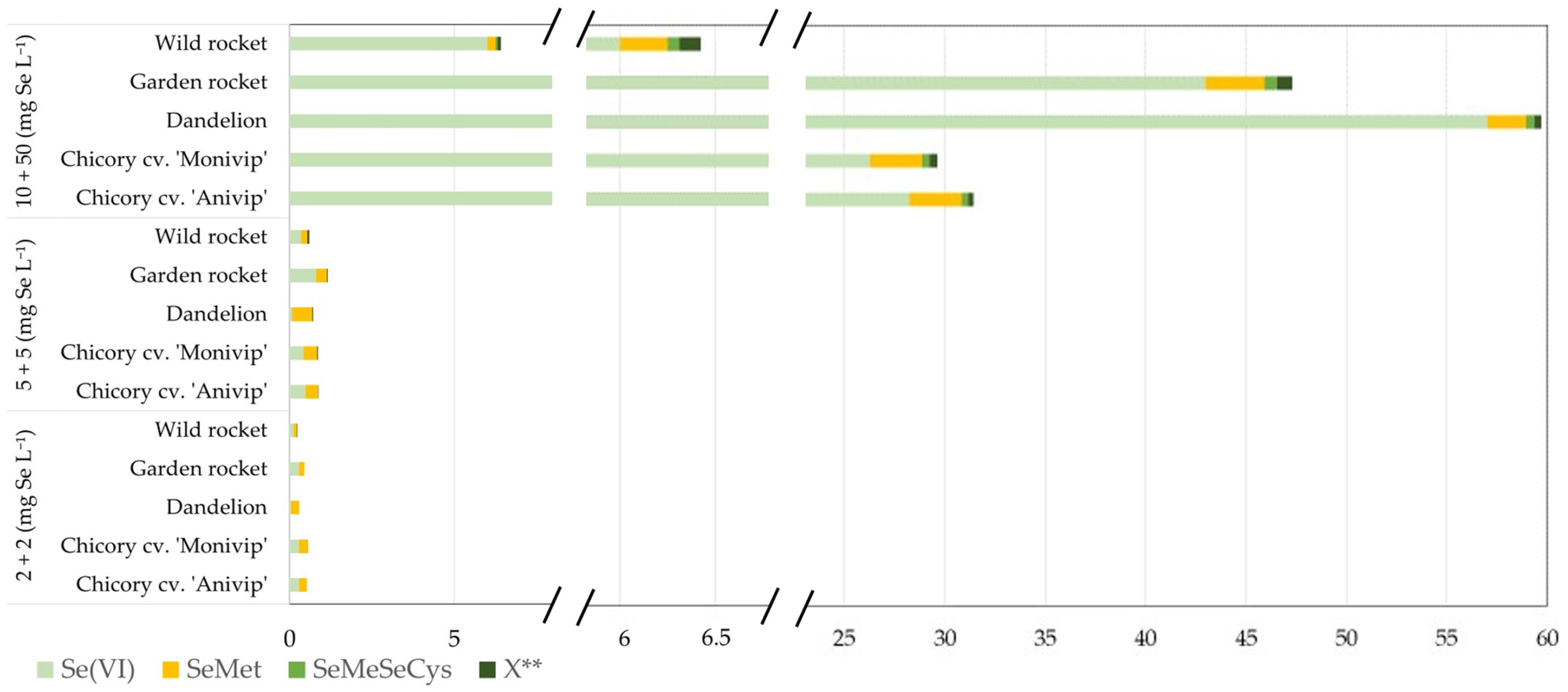
Figure 1 illustrates Se species detected in green tissues of exposed plants. Extracts from vegetables sprayed with 2 + 2 and 5 + 5 mg Se(VI) L−1 contained Se(VI), SeMet, and a minor unknown compound eluting with the void volume of the anion exchange column (<3% of total Se). No SeCys2 was detected on the cation exchange column. Se(VI) accounted for 21–27% of total Se in chicory (‘Anivip’, ‘Monivip’) and rocket, 12–13% in wild rocket, and 5% in dandelion. SeMet represented ~20% in chicory and ~10% in rocket and wild rocket, with dandelion showing a 15% increase at 5 + 5 mg Se L−1. At the highest concentration (10 + 50 mg Se(VI) L−1), Se(VI) dominated, representing 42% in rocket and 58% in dandelion, while SeMet formation was reduced (2–4% across species). At the highest Se(VI) dose, the predominance of inorganic Se together with reduced biomass and suppressed SeMet formation suggests activation of detoxification mechanisms. One likely mechanism is vacuolar sequestration of Se(VI), which limits its metabolic conversion and protects cytosolic processes under excessive selenium exposure. Such sequestration has been reported as a common response in plants exposed to supra-optimal Se supply. Proportion of selenium remained unidentified in some species, most notably garden rocket. Although this fraction was tentatively attributed to peptide-bound or intermediate organic selenium forms based on chromatographic behavior, no further structural identification was attempted in the present study. Advanced analytical approaches such as alternative enzymatic digestions, spiking with additional candidate standards, or coupling HPLC to high-resolution mass spectrometry would be required for definitive identification but were beyond the scope of this work. The presence of this unidentified fraction therefore represents a limitation of the speciation resolution rather than of selenium uptake assessment, and does not affect the main conclusions regarding dose-dependent shifts between inorganic and organic selenium forms.
These findings demonstrate that extraction efficiency and speciation outcomes are strongly influenced by both methodological choices and Se concentration. Protease XIV provided consistent recovery across species, while speciation analysis revealed concentration-dependent shifts in Se(VI) and SeMet proportions. The predominance of Se(VI) at high concentrations, coupled with reduced SeMet transformation, suggests limited metabolic conversion capacity under elevated Se exposure.
Only limited data are available on dose-dependent changes in SeMet proportion across multiple leafy species. Recent studies have reported that SeMet accumulation patterns vary considerably depending on the species with respect to Se concentration. For example, while green algae Scenedesmus quadricauda showed increased SeMet levels at higher Se concentrations in the culturing media [39], Hu et al. [40] reported decreased SeMet concentration in winter wheat roots, which was probably correlated with higher Se (IV) root concentration due to hydroponic Se(VI) and phosphorus application. Furthermore, the foliar application of Se(VI) led to an increase in total selenium, predominantly as SeMet, the main organic Se species in the lettuce trial [41]. Likewise, as our results showed traces of SeMeSeCys (<1% of total Se) (Table S1, Figure 1), Cubadda et al. [42] reported similar findings, identifying it as a minor compound (0.2−0.5%) in high-Se wheat. Additionally, traces of SeMeSeCys were observed (<1% of total Se) (Table 4, Figure 2 and Figure 3). Identified Se species represented quantitative recovery of soluble Se, excepting rocket and wild rocket foliar sprayed with 2 + 2 and 5 + 5 mg Se L−1. The unidentified fraction was particularly pronounced in garden rocket and, at lower doses, in both rocket species. This could be explained by formation of the dipeptide γ-glutamyl-SeMeSeCys as a protective mechanism, similar to that in Se accumulating plants [12]. By increasing the Se content (10 + 50 mg Se L−1), plants transformed less inorganic Se than those treated with lower concentrations, possibly reflecting reduced metabolic conversion and enhanced inorganic Se retention.
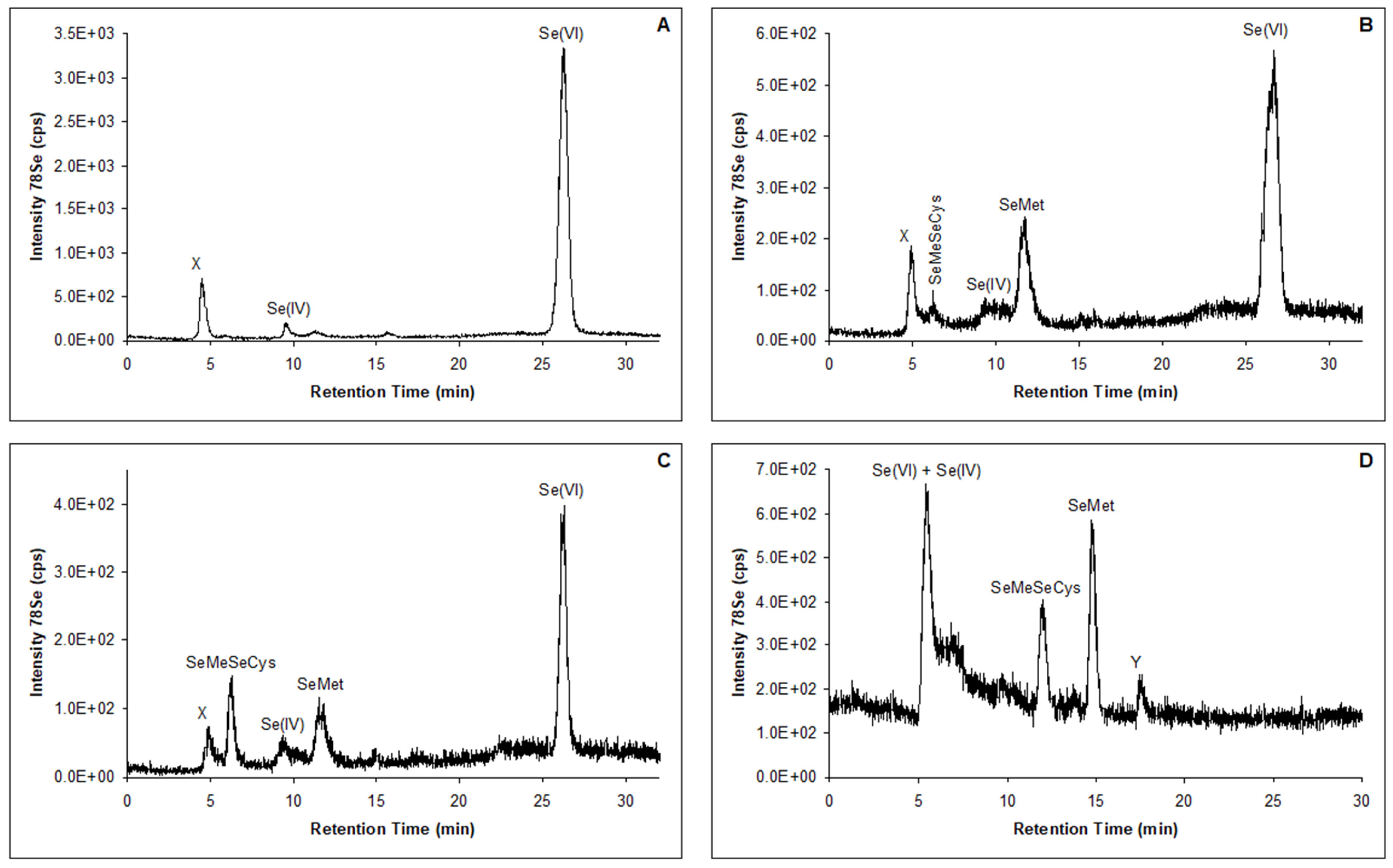
By performing analysis of the extracts obtained after different types of hydrolysis by HPLC-ICP-MS, the same Se species and concentrations were observed as when performing analysis by HPLC-UV-HG-AFS. Since lower detection limits could be achieved with the ICP-MS detection system, traces of SeMeSeCys and Se(IV) were observed after three-step hydrolysis in plants foliarly sprayed with 2 + 2 mg Se L−1 (Figure 3). The presence of SeMeSeCys was confirmed on the cation exchange column and was also determined in leafy vegetables. In selected vegetables, Mazej et al. [36] determined SeMeSeCys, SeMet, inorganic Se and SeCys2.
The data presented in Figure 1 highlight notable differences in the metabolic capacity of chicory and dandelion compared to rocket, particularly in their ability to transform inorganic selenium (Se) into selenomethionine (SeMet). These differences can be attributed to species-specific metabolic pathways, reflecting the distinct phylogenetic origins of the plants: chicory and dandelion belong to the Asteraceae family, whereas rocket is a member of the Brassicaceae family. A finer dose spacing between 5 + 5 and 10 + 50 mg Se L−1 would be required to precisely identify the onset of SeMet decline and the point at which inorganic Se(VI) becomes dominant. The chemical form of Se supplied strongly influenced speciation outcomes. Across all tested species (chicory, rocket, and dandelion), foliar application of Se(VI) resulted in a broadly similar metabolic profile, with Se detected as selenoamino acids (primarily SeMet and SeMeSeCys) alongside residual inorganic Se. At higher concentrations (10 + 50 mg Se L−1), conversion of Se(VI) was markedly reduced, indicating saturation of metabolic assimilation pathways. The concomitant presence of SeMeSeCys under these conditions suggests activation of plant defense mechanisms, as previously reported [12].
Comparable findings have been documented in other species. Pedrero et al. [43] identified SeMeSeCys in radish following 40 days of hydroponic exposure to Na2SeO3 (1 mg L−1). Smrkolj et al. [44] reported that 30% of total Se in bean seeds foliarly sprayed with 10 mg Se L−1 was present as SeMeSeCys. Similarly, Sugihara et al. [45] demonstrated that sprouts of 28 species from 10 plant families cultivated hydroponically in high-selenium environments (10 mg L−1 selenite) accumulated SeMeSeCys as the predominant Se species. In addition to SeMeSeCys, SeMet, Se(IV), γ-glutamyl-Se-methylselenocysteine, and an unidentified Se compound were detected in several high-Se sprouts.
Taken together, these results indicate that the fraction of SeMeSeCys in enriched plants is highly variable and depends on multiple factors, including plant species, plant organ, and uptake efficiency. The observed accumulation of SeMeSeCys in chicory and dandelion, coupled with reduced conversion of Se(VI) at high concentrations, underscores the role of Se speciation as both a metabolic adaptation and a defense mechanism in higher plants.

4. Conclusions

To conclude, dandelion, garden rocket, wild rocket, and two cultivars of chicory were foliarly sprayed twice at five-day intervals with Na2SeO4 solutions of varying concentrations (1 + 1, 2 + 2, 5 + 5, 10 + 0, 10 + 10, and 10 + 50 mg Se L−1) to evaluate both the transformation of Se(VI) and plant biomass responses. Selenium accumulated efficiently in leaves, reaching up to 102 µg Se g−1 dry mass in garden rocket. In addition to inorganic Se (Se(VI), the form supplied in the solution), selenomethionine (SeMet) and selenomethylselenocysteine (SeMeSeCys) were identified in enzymatic extracts.
No acute toxic effects were observed even at the highest Se concentrations, suggesting that plants tolerated foliar Se application without visible stress symptoms. However, the ability to convert inorganic Se into SeMet decreased with increasing Se concentration, irrespective of species, indicating a concentration-dependent limitation of metabolic conversion. This pattern points to the activation of protective mechanisms similar to those described in Se-accumulating plants. One plausible mechanism is vacuolar storage of Se(VI), consistent with its predominance in extracts. The detection of SeMeSeCys further suggests the possible formation of the dipeptide γ-glutamyl-SeMeSeCys, which may represent an additional detoxification or storage pathway. Among the tested treatments, the 5 + 5 mg Se L−1 application provided the most favorable balance between efficient selenium accumulation, high SeMet proportion, and preserved biomass, indicating this range as optimal for foliar selenium biofortification of leafy vegetables.
Overall, these findings indicate that foliar Se application can substantially increase Se accumulation in leafy vegetables while eliciting species-specific metabolic responses. The balance between Se(VI) storage and conversion to organic forms, such as SeMet and SeMeSeCys, highlights the dual role of Se in plant physiology: as a potentially beneficial micronutrient at low concentrations and as a compound that requires detoxification at higher concentrations.

Supplementary Materials

The following supporting information can be downloaded at: https://www.mdpi.com/article/10.3390/horticulturae12020256/s1, Table S1: Selenium (Se) content in water-soluble extracts and mass balance after enzymatic hydrolysis of lyophilised green parts of foliarly sprayed plants (expressed as an average of at least two determinations) and Se species in extracts after enzymatic hydrolysis determined by HPLC-UV-HG-AFS (mg Se g−1 sample and % of Se with respect to the total content in the sample; average of at least two determinations); 2 + 2; 5 + 5 mg Se L−1 - First experiment; 10 + 50 mg Se L−1 - Second experiment.

Author Contributions

Conceptualization, D.Ž., M.P. and I.G.; methodology, D.Ž. and M.P.; software, D.Ž. and M.P.; validation, M.P.; formal analysis M.P.; investigation, D.Ž.; resources, D.Ž. and M.P.; data curation, D.Ž. and M.P.; writing—original draft preparation, D.Ž. and M.P.; writing—review and editing, M.P.P., I.G., D.B. and I.P.; visualization, M.P.P., D.B. and I.P.; supervision, D.Ž. and M.P.; project administration, D.Ž.; funding acquisition, D.Ž. All authors have read and agreed to the published version of the manuscript.

Funding

This work was financially supported by the Slovenian Research Agency through the programme P1-0143, contract 1000-05-310030 and project J7-9805.

Data Availability Statement

The original contributions presented in this study are included in the article/Supplementary Material. Further inquiries can be directed to the corresponding author.

Acknowledgments

During the preparation of this manuscript, the authors used OpenAI ChatGPT (GPT-5) for language refinement and improvement of textual clarity. The authors have reviewed and edited the output and take full responsibility for the content of this publication.

Conflicts of Interest

The authors declare no conflicts of interest.

References

  1. Mehdi, Y.; Hornick, J.-L.; Istasse, L.; Dufrasne, I. Selenium in the Environment, Metabolism and Involvement in Body Functions. Molecules 2013, 18, 3292–3311. [Google Scholar] [CrossRef]
  2. Sharma, V.K.; McDonald, T.J.; Sohn, M.; Anquandah, G.A.; Pettine, M.; Zboril, R. Biogeochemistry of selenium: A review. Environ. Chem. Lett. 2015, 13, 49–58. [Google Scholar] [CrossRef]
  3. Jones, G.D.; Droz, B.; Greve, P.; Gottschalk, P.; Poffet, D.; McGrath, S.P.; Seneviratne, S.I.; Smith, P.; Winkel, L.H.E. Selenium deficiency risk predicted to increase under future climate change. Proc. Natl. Acad. Sci. USA 2017, 114, 2848–2853. [Google Scholar] [CrossRef]
  4. Clark, L.C.; Combs, G.F., Jr.; Turnbull, B.W.; Slate, E.H.; Chalker, D.K.; Chow, J.; Davis, L.S.; Glover, R.A.; Graham, G.F.; Gross, E.G.; et al. Effects of selenium supplementation for cancer prevention in patients with carcinoma of the skin: A randomized controlled trial. J. Am. Med. Assoc. 1996, 276, 1957–1963. [Google Scholar] [CrossRef]
  5. Rayman, M.P. Selenium in cancer prevention: A review of the evidence and mechanism of action. Proc. Nutr. Soc. 2005, 64, 527–542. [Google Scholar] [CrossRef] [PubMed]
  6. Martens, I.B.; Cardoso, B.R.; Hare, D.J.; Niedzwiecki, M.M.; Lajolo, F.M.; Martens, A.; Cozzolino, S.M. Selenium status in preschool children receiving a Brazil nut–enriched diet. Nutrition 2015, 31, 1339–1343. [Google Scholar] [CrossRef]
  7. Thomson, C.D. Assessment of requirements for selenium and adequacy of selenium status: A review. Eur. J. Clin. Nutr. 2004, 58, 391–402. [Google Scholar] [CrossRef]
  8. Lintschinger, J.; Fuchs, N.; Moser, J.; Kuehnelt, D.; Goessler, W. Selenium-enriched sprouts: A raw material for fortified cereal-based diets. J. Agric. Food Chem. 2000, 48, 5362–5368. [Google Scholar] [CrossRef]
  9. Pirc, S.; Šajn, R. The Influence of Geochemistry in Determination of Chemical Loading of Environment; Slovenian Ecological Society: Ljubljana, Slovenia, 1997; p. 185. [Google Scholar]
  10. De Bruno, A.; Piscopo, A.; Cordopatri, F.; Poiana, M.; Mafrica, R. Effect of Agronomical and Technological Treatments to Obtain Selenium-Fortified Table Olives. Agriculture 2020, 10, 284. [Google Scholar] [CrossRef]
  11. Terry, N.; Zayed, A.M.; de Souza, M.P.; Tarun, A.S. Selenium in higher plants. Annu. Rev. Plant Physiol. Plant Mol. Biol. 2000, 51, 401–432. [Google Scholar] [CrossRef]
  12. Pyrzynska, K. Selenium speciation in enriched vegetables. Food Chem. 2009, 114, 1183–1191. [Google Scholar] [CrossRef]
  13. Puccinelli, M.; Malorgio, F.; Pezzarossa, B. Selenium enrichment of horticultural crops. Molecules 2017, 22, 933. [Google Scholar] [CrossRef] [PubMed]
  14. Kapoor, B.; Kumar, P.; Gill, N.S.; Sharma, R.; Thakur, N.; Irfan, M. Molecular mechanisms underpinning the silicon-selenium (Si-Se) interactome and cross-talk in stress-induced plant responses. Plant Soil 2023, 486, 45–68. [Google Scholar] [CrossRef]
  15. Ferrarese, M.; Sourestani, M.; Quattrini, E.; Schiavi, M.; Ferrante, A. Biofortification of spinach plants applying selenium in the nutrient solution of floating system. Veg. Crops Res. Bull. 2012, 76, 127–136. [Google Scholar] [CrossRef]
  16. Rengel, Z.; Batten, G.D.; Crowley, D.D. Agronomic approaches for improving the micronutrient density in edible portions of field crops. Field Crops Res. 1999, 60, 27–40. [Google Scholar] [CrossRef]
  17. Aspila, P. History of selenium supplemented fertilization in Finland. In Proceedings: Twenty Years of Selenium Fertilization; Eurola, M., Ed.; Agricultural Research Centre of Finland: Jokioinen, Finland, 2005; pp. 19–24. [Google Scholar]
  18. Bohn, T.; Despotovic, M.; Vahid, F.; Rühl, R. Estimated Dietary Intakes of Vitamin A. Nutrients 2024, 16, 4004. [Google Scholar] [CrossRef]
  19. Pezzarossa, B.; Remorini, D.; Gentile, M.L.; Massai, R. Effects of foliar and fruit addition of sodium selenate on selenium accumulation and fruit quality. J. Sci. Food Agric. 2012, 92, 781–786. [Google Scholar] [CrossRef]
  20. Polić Pasković, M.; Herak Ćustić, M.; Lukić, I.; Marcelić, Š.; Žurga, P.; Vidović, N.; Major, N.; Goreta Ban, S.; Pecina, M.; Ražov, J.; et al. Foliar Nutrition Strategies for Enhancing Phenolic and Amino Acid Content in Olive Leaves. Plants 2024, 13, 3514. [Google Scholar] [CrossRef]
  21. MacLeod, J.A.; Gupta, U.C.; Milburn, P.; Sanderson, J.B. Selenium concentration in plant material, drainage and surface water as influenced by Se applied to barley foliage in a barley–red clover–potato rotation. Can. J. Soil Sci. 1998, 78, 685–688. [Google Scholar] [CrossRef]
  22. Lara, T.S.; Correia, T.S.; de Oliveira, C.; Lessa, J.H.L.; de Souza, K.R.D.; Corguinha, A.P.B.; da Silva, E.C., Jr.; Martins, F.A.D.; Lopes, G.; Guilherme, L.R.G. Selenium application provides nutritional and metabolic benefits to wheat plants. Agronomy 2024, 14, 462. [Google Scholar] [CrossRef]
  23. Germ, M.; Stibilj, V.; Osvald, J.; Kreft, I. Effect of selenium foliar application on chicory (Cichorium intybus L.). J. Agric. Food Chem. 2007, 55, 795–798. [Google Scholar] [CrossRef] [PubMed]
  24. Skrypnik, L.; Styran, T.; Savina, T.; Golubkina, N. Effect of selenium application and growth stage at harvest on hydrophilic and lipophilic antioxidants in lamb’s lettuce (Valerianella locusta L. Laterr.). Plants 2021, 10, 2733. [Google Scholar] [CrossRef] [PubMed]
  25. da Silva, E.; Cidade, M.; Heerdt, G.; Ribessi, R.; Morgon, N.; Cadore, S. Effect of selenite and selenate application on mineral composition of lettuce plants cultivated under hydroponic conditions: Nutritional balance overview using a multifaceted study. J. Braz. Chem. Soc. 2018, 29, 371–379. [Google Scholar] [CrossRef]
  26. Montes-Bayon, M.; Diaz Molet, M.J.; Blanco Gonzalez, E.; Sanz-Medel, A. Evaluation of different sample extraction strategies for selenium determination in selenium-enriched plants (Allium sativum and Brassica juncea) and Se speciation by HPLC-ICP-MS. Talanta 2006, 68, 1287–1293. [Google Scholar] [CrossRef]
  27. Polatajko, A.; Jakubowski, N.; Szpunar, J. State of the art report of selenium speciation in biological samples. J. Anal. Atom. Spectrom. 2006, 21, 639–654. [Google Scholar] [CrossRef]
  28. Smrkolj, P.; Osvald, M.; Osvald, J.; Stibilj, V. Selenium uptake and species distribution in selenium-enriched bean (Phaseolus vulgaris L.) seeds obtained by two different cultivations. Eur. Food Res. Technol. 2007, 225, 233–237. [Google Scholar] [CrossRef]
  29. Cuderman, P.; Kreft, I.; Germ, M.; Kovačevič, M.; Stibilj, V. Selenium species in selenium-enriched and drought-exposed potatoes. J. Agric. Food Chem. 2008, 56, 9114–9120. [Google Scholar] [CrossRef]
  30. Mazej, D.; Falnoga, I.; Veber, M.; Stibilj, V. Determination of selenium species in plant leaves by HPLC-UV-HG-AFS. Talanta 2006, 68, 558–568. [Google Scholar] [CrossRef]
  31. Hartikainen, H.; Xue, T.L.; Piironen, V. Selenium as an anti-oxidant and pro-oxidant in ryegrass. Plant Soil 2000, 225, 193–200. [Google Scholar] [CrossRef]
  32. Xue, T.; Hartikainen, H.; Piironen, V. Antioxidation and growth-promoting effect of selenium on senescing lettuce. Plant Soil 2001, 237, 55–61. [Google Scholar] [CrossRef]
  33. Schiavon, M.; Nardi, S.; Pilon-Smits, E.A.; Dall’Acqua, S. Foliar selenium fertilization alters the content of dietary phytochemicals in two rocket species. Front. Plant Sci. 2022, 13, 987935. [Google Scholar] [CrossRef]
  34. Francini, A.; Quattrini, E.; Giuffrida, F.; Ferrante, A. Biofortification of baby leafy vegetables using nutrient solution containing selenium. J. Sci. Food Agric. 2023, 103, 5472–5480. [Google Scholar] [CrossRef] [PubMed]
  35. Stibilj, V.; Smrkolj, P.; Jaćimović, R.; Osvald, J. Selenium uptake and distribution in chicory (Cichorium intybus L.) grown in an aeroponic system. Acta Agric. Slov. 2011, 97, 189–196. [Google Scholar] [CrossRef]
  36. Mazej, D.; Osvald, J.; Stibilj, V. Selenium species in leaves of chicory, dandelion, lamb’s lettuce and parsley. Food Chem. 2008, 107, 75–83. [Google Scholar] [CrossRef]
  37. Kapolna, E.; Gergely, V.; Dernovics, M.; Illes, A.; Fodor, P. Fate of selenium species in sesame seeds during simulated bakery process. J. Food Eng. 2007, 79, 494–501. [Google Scholar] [CrossRef]
  38. Kapolna, E.; Hilleström, P.R.; Laursen, K.H.; Husted, S.; Larsen, E.H. Effect of foliar application of selenium on its uptake and speciation in carrot. Food Chem. 2009, 115, 1357–1363. [Google Scholar] [CrossRef]
  39. Umysová, D.; Vítová, M.; Doucha, J.; Lívanský, K.; Hlavová, M.; Šková, M.; Macht, J.; Zachleder, V. Bioaccumulation and toxicity of selenium compounds in the green alga Scenedesmus quadricauda. BMC Plant Biol. 2009, 9, 58. [Google Scholar] [CrossRef]
  40. Hu, C.; Nie, Z.; Shi, H.; Peng, H.; Li, G.; Liu, H.; Li, C.; Liu, H. Selenium uptake, translocation, subcellular distribution and speciation in winter wheat in response to phosphorus application combined with three types of selenium fertilizer. BMC Plant Biol. 2023, 23, 227. [Google Scholar] [CrossRef]
  41. Cheng, H.; Shi, X.; Li, L. The effect of exogenous selenium supplementation on the nutritional value and shelf life of lettuce. Agronomy 2024, 14, 1380. [Google Scholar] [CrossRef]
  42. Cubadda, F.; Aureli, F.; Ciardullo, S.; D’Amato, M.; Raggi, A.; Acharya, R.; Reddy, R.A.V.; Prakash, N.T. Changes in selenium speciation associated with increasing tissue concentrations of selenium in wheat grain. J. Agric. Food Chem. 2010, 58, 2295–2301. [Google Scholar] [CrossRef]
  43. Pedrero, Z.; Madrid, Y.; Camara, C. Selenium species bioaccessibility in enriched radish (Raphanus sativus): A potential dietary source of selenium. J. Agric. Food Chem. 2006, 54, 2412–2417. [Google Scholar] [CrossRef]
  44. Smrkolj, P.; Stibilj, V. Determination of selenium in vegetables by hydride generation atomic fluorescence spectrometry. Anal. Chim. Acta 2004, 512, 11–17. [Google Scholar] [CrossRef]
  45. Sugihara, S.; Kondo, M.; Chihara, Y.; Yuji, M.; Hattori, H.; Yoshida, M. Preparation of selenium-enriched sprouts and identification of their selenium species by high-performance liquid chromatography-inductively coupled plasma mass spectrometry. Biosci. Biotechnol. Biochem. 2004, 68, 193–199. [Google Scholar] [CrossRef]
Figure 1. Relative proportions of Se species in extracts after enzymatic hydrolysis of chicory cv. ‘Anivip’, chicory cv. ‘Monivip’, dandelion, garden rocket, and wild rocket leaves, following foliar application of Na2SeO4 solutions (2 + 2; 5 + 5 mg Se L−1, first experiment; 10 + 50 mg Se L−1, second experiment), determined by HPLC-UV-HG-AFS. Speciation data are derived from pooled composite samples per treatment and are presented descriptively. Values are expressed as percentages of total Se in the extract. SeMet—selenomethionine; Se(VI)—selenate; SeMeSeCys—Se-methylselenocysteine; ** X—unidentified Se species.
Horticulturae 12 00256 g001
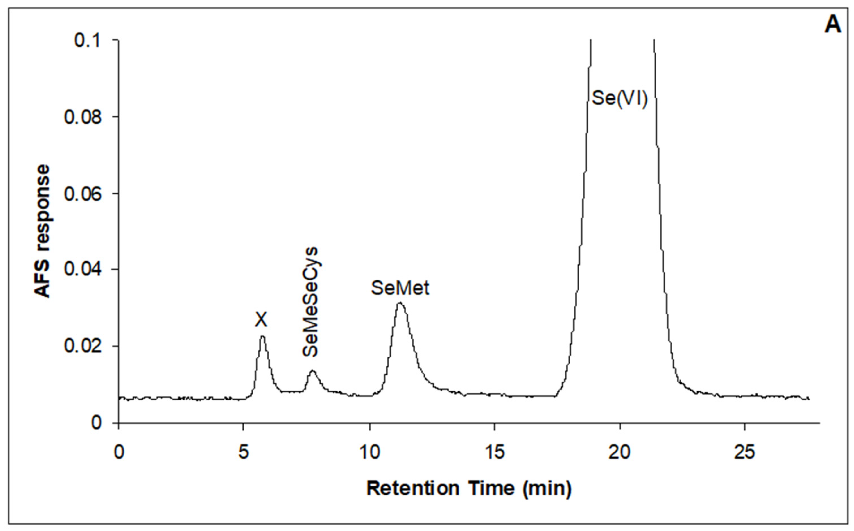
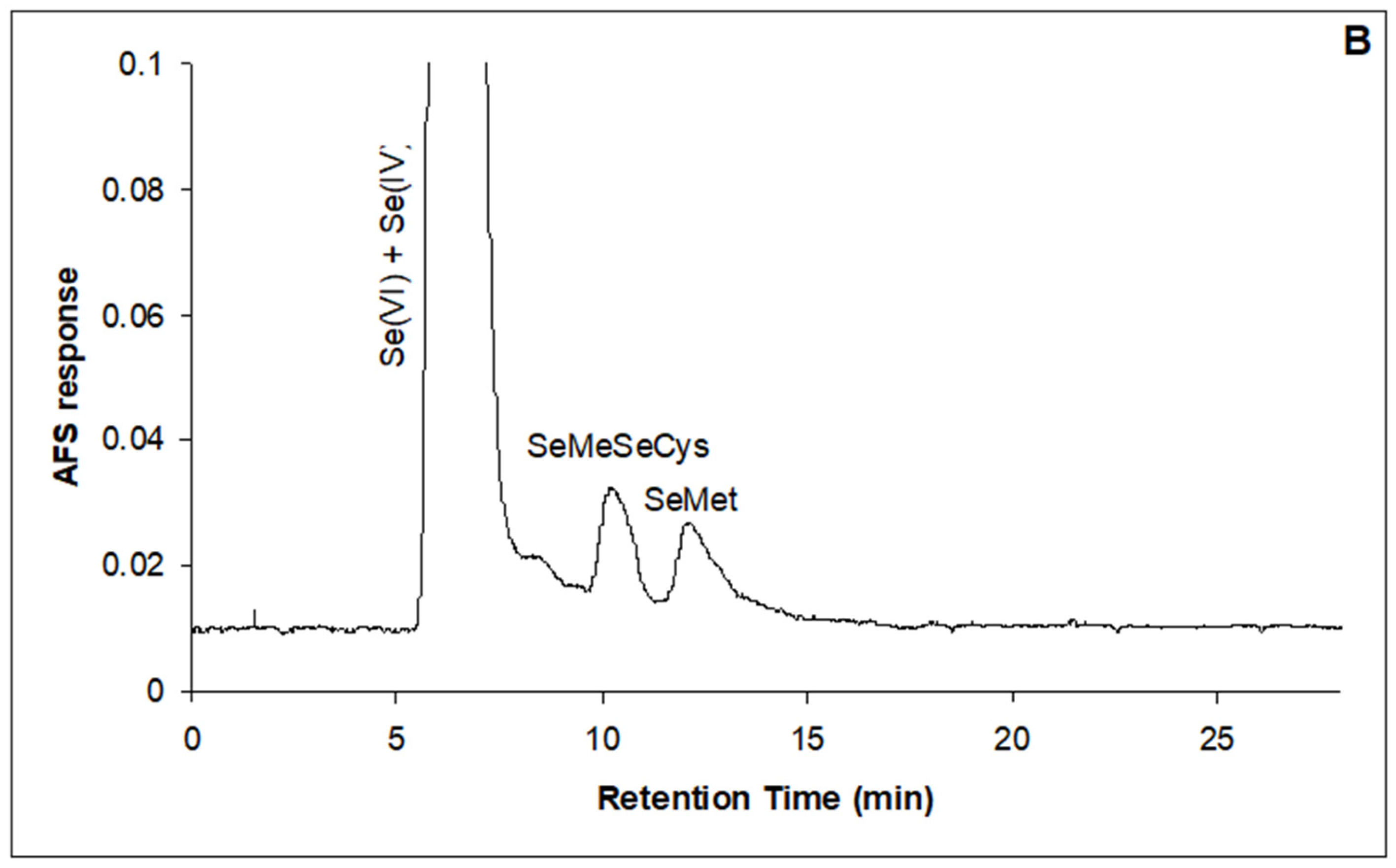
Figure 2. Anion (A) and cation (B) exchange chromatograms of chicory cv. ‘Anivip’ leaf extracts (10 + 50 mg Se L−1) after enzymatic hydrolysis, obtained by HPLC-UV-HG-AFS.
Horticulturae 12 00256 g002aHorticulturae 12 00256 g002b
Figure 3. Three-step extraction of Se from wild rocket (2 + 2 mg Se L−1). Se species in the first extract (A) after water hydrolysis and second (B) and the third extract (C) after enzymatic (protease) hydrolysis obtained by separation on anion exchange column and ICP-MS detection. Se species (SeMeSeCys, SeMet) after the third extraction were confirmed on the cation exchange column (D).
Horticulturae 12 00256 g003
Table 1. Selenium foliar application concentrations applied in the first and second greenhouse experiments. Treatments were applied twice, with a 5-day interval.
TreatmentFirst Treatment
Foliar Solution
(mg Se L−1)
Second Treatment
Foliar Solution
(mg Se L−1)
First
experiment
Control I00
1 + 111
2 + 222
5 + 555
Second
experiment
Control II00
10 + 0100
10 + 101010
10 + 501050
Table 2. Dry matter and average mass of fresh leaves (g plant−1) [Dry matter (%)] of chicory cv. ‘Anivip’, chicory cv. ‘Monivip’, dandelion, garden rocket, and wild rocket leaves, following two foliar applications of Na2SeO4 solution.
Mass of Fresh Leaves (g per Plant)
[Dry Matter of Leaves (%)]
Foliar Solution (mg Se L−1)Chicory cv. ‘Anivip’Chicory cv. ‘Monivip’DandelionGarden RocketWild Rocket
Control I6.35 ± 0.21 a4.66 ± 0.23 a3.11 ± 0.06 a2.60 ± 0.06 a2.41 ± 0.04 a
[12.60 ± 0.56 b][14.80 ± 0.58 b][13.82 ± 0.46 b][12.98 ± 0.42 b][10.80 ± 0.45 b]
1 + 16.59 ± 0.24 a4.98 ± 0.19 a3.16 ± 0.04 a2.61 ± 0.08 a2.53 ± 0.05 b
[12.50 ± 0.45 b][14.10 ± 0.52 b][13.80 ± 0.34 b][12.90 ± 0.46 b][10.76 ± 0.42 b]
2 + 26.76 ± 0.34 a5.06 ± 0.23 a3.17 ± 0.05 a2.65 ± 0.07 a2.83 ± 0.03 c
[12.12 ± 0.35 b][13.90 ± 0.45 b][13.00 ± 0.28 a][12.82 ± 0.54 b][10.23 ± 0.36 ab]
5 + 57.58 ± 0.42 b6.76 ± 0.24 b3.56 ± 0.05 b2.94 ± 0.04 b3.20 ± 0.12 d
[10.82 ± 0.46 a][11.60 ± 0.41 a][12.90 ± 0.36 a][10.70 ± 0.45 a][9.40 ± 0.35 a]
Control II6.45 ± 0.22 a4.83 ± 0.25 a3.08 ± 0.06 a2.51 ± 0.06 b2.37 ± 0.04 b
[13.32 ± 0.53 b][13.67 ± 0.62 b][13.52 ± 0.38 b][11.68 ± 0.42 b][10.82 ± 0.32 c]
10 + 07.58 ± 0.33 b6.38 ± 0.27 b3.50 ± 0.04 b2.94 ± 0.03 c3.16 ± 0.02 c
[11.58 ± 0.42 a][12.05 ± 0.43 a][13.26 ± 0.54 b][11.24 ± 0.45 b][10.06 ± 0.41 b]
10 + 107.68 ± 0.34 b7.36 ± 0.34 c3.51 ± 0.03 b2.95 ± 0.07 c3.32 ± 0.03 d
[11.46 ± 0.36 a][12.14 ± 0.72 a][13.24 ± 0.46 b][11.16 ± 0.47 b][9.82 ± 0.34 b]
10 + 506.66 ± 0.31 a5.34 ± 0.36 a3.53 ± 0.02 b2.09 ± 0.04 a2.28 ± 0.03 a
[11.12 ± 0.55 a][11.92 ± 0.64 a][11.82 ± 0.36 a][9.51 ± 0.42 a][9.05 ± 0.34 a]
Mean ± SD (n = 20). Within each column, values followed by the same letter are not significantly different according to Tukey’s HSD test (p < 0.05). Values in brackets represent dry matter content (%). Control I–5 + 5 mg Se L−1 belong to the first experiment; Control II–10 + 50 mg Se L−1 belong to the second experiment.
Table 3. Leaf–root ratio (fresh mass basis) of chicory cv. ‘Anivip’, chicory cv. ‘Monivip’, dandelion, garden rocket, and wild rocket leaves, after two foliar applications of Na2SeO4 solution.
Leaf–Root Ratio
Foliar Solution
(mg Se L−1)
Chicory cv. ‘Anivip’Chicory cv. ‘Monivip’DandelionGarden RocketWild Rocket
Control I2.12 ± 0.24 a1.87 ± 0.17 a1.95 ± 0.06 a1.58 ± 0.08 b1.46 ± 0.11 a
1 + 12.45 ± 0.21 ab2.21 ± 0.16 b1.96 ± 0.04 a1.60 ± 0.09 b1.56 ± 0.07 a
2 + 22.64 ± 0.25 b2.58 ± 0.18 c1.88 ± 0.06 a1.74 ± 0.08 bc1.73 ± 0.04 b
5 + 53.40 ± 0.32 c3.48 ± 0.23 d1.97 ± 0.04 b1.87 ± 0.07 c2.28 ± 0.12 c
Control II2.29 ± 0.21 a1.83 ± 0.18 a1.70 ± 0.10 a1.43 ± 0.06 ab1.91 ± 0.07 a
10 + 02.91 ± 0.28 b2.86 ± 0.24 b2.09 ± 0.08 a1.51 ± 0.04 b3.06 ± 0.05 c
10 + 103.00 ± 0.26 b3.41 ± 0.27 c2.18 ± 0.14 b1.87 ± 0.08 c3.14 ± 0.04 c
10 + 503.02 ± 0.23 b2.78 ± 0.26 b2.10 ± 0.11 b1.37 ± 0.07 a2.71 ± 0.08 b
Mean ± SD (n = 20 plants per treatment). Within each column, values followed by the same letter are not significantly different according to Tukey’s HSD test (p < 0.05). Control I–5 + 5 mg Se L−1 belong to the first experiment; Control II–10 + 50 mg Se L−1 belong to the second experiment.
Table 4. Total Se content of chicory cv. ‘Anivip’, chicory cv. ‘Monivip’, dandelion, garden rocket, and wild rocket leaves, following foliar applications of Na2SeO4 solutions in lyophilized green parts on a dry matter basis.
Total Se Content (µg Se g−1)
Foliar Solution
(mg Se L−1)
Chicory cv. ‘Anivip’Chicory cv. ‘Monivip’DandelionGarden RocketWild Rocket
Control I0.06 ± 0.01 a0.03 ± 0.01 a0.05 ± 0.01 a0.05 ± 0.01 a0.04 ± 0.01 a
1 + 10.35 ± 0.01 b0.41 ± 0.02 b0.34 ± 0.01 b0.48 ± 0.01 b0.51 ± 0.01 b
2 + 21.14 ± 0.01 c1.22 ± 0.01 c0.93 ± 0.01 c1.26 ± 0.01 c1.08 ± 0.02 c
5 + 51.96 ± 0.04 d1.58 ± 0.06 d1.51 ± 0.09 d3.72 ± 0.08 d2.59 ± 0.08 d
Control II0.05 ± 0.02 a0.04 ± 0.02 a0.05 ± 0.01 a0.05 ± 0.01 a0.05 ± 0.01 a
10 + 02.22 ± 0.06 b2.20 ± 0.09 b6.71 ± 0.06 b14.31 ± 0.07b4.26 ± 0.04 b
10 + 1019.84 ± 0.28 c26.72 ± 0.31 c12.57 ± 0.06 c31.61 ± 0.37c9.67 ± 0.14 c
10 + 5063.15 ± 0.18 d61.42 ± 0.32 d97.42 ± 0.31 d102.38 ± 0.52d13.04 ± 0.17 d
Mean ± SD (n = 5). Within a column, values followed by the same letter are not significantly different according to Tukey’s HSD test (p < 0.05). Control I–5 + 5 mg Se L−1 and Control II–10 + 50 mg Se L−1 correspond to the first and second experiments, respectively.
Disclaimer/Publisher’s Note: The statements, opinions and data contained in all publications are solely those of the individual author(s) and contributor(s) and not of MDPI and/or the editor(s). MDPI and/or the editor(s) disclaim responsibility for any injury to people or property resulting from any ideas, methods, instructions or products referred to in the content.