Article

Effect of Explant Physiology and Media Composition on Callogenesis of Vitellaria paradoxa Leaf Explants

by
Moses Okao
1,2,
Rohit Bharati
3,4 and
Eloy Fernández-Cusimamani
1,*
1
Department of Crop Sciences and Agroforestry, Faculty of Tropical AgriSciences, Czech University of Life Sciences Prague, Kamýcká 129, Prague 6, 16500 Suchdol, Czech Republic
2
Ngetta Zonal Agricultural Research and Development Institute, National Agricultural Research Organisation, Lira P.O. Box 52, Uganda
3
Plant Virus and Vector Interactions, Czech Agrifood Research Center, Drnovská 507, Prague 6, Ruzyně, 16106 Prague, Czech Republic
4
Department of Economic Theories, Faculty of Economics and Management, Czech University of Life Sciences Prague, Kamýcká 129, Prague 6, 16500 Suchdol, Czech Republic
*
Author to whom correspondence should be addressed.
Horticulturae 2025, 11(9), 1127; https://doi.org/10.3390/horticulturae11091127
Submission received: 12 August 2025 / Revised: 5 September 2025 / Accepted: 12 September 2025 / Published: 17 September 2025
(This article belongs to the Collection Application of Tissue Culture to Horticulture)

Abstract

Vitellaria paradoxa (shea tree) is an economically and medicinally important species indigenous to sub-Saharan Africa. Although the species holds substantial value, domestication efforts have been constrained, primarily due to the absence of efficient propagation alternatives, especially for the East African subspecies (V. paradoxa subsp. nilotica) which remains understudied in tissue culture research. This study investigated the influence of leaf explant developmental stage and media composition on callogenesis and embryogenic potential in V. paradoxa subsp. nilotica. Thus, leaf explants from six distinct growth stages were cultured on Murashige and Skoog (MS) media supplemented with various concentrations of 2,4-D, TDZ, NAA, and BAP. Callogenesis was significantly influenced by explant age, media strength, and specific PGR combinations. Results revealed that explants from Stage III (11–15 days) and Stage IV (16–20 days) exhibited the highest callus induction rates (up to 100%), particularly on half-strength MS media containing 2.0 mg/L 2,4-D and 0.5–1.0 mg/L TDZ. Histological analysis suggests that varying responses at the different stages relate to chloroplast distribution, trichome density/orientation, and vascular tissue maturity. Pro-embryogenic structures were successfully induced, representing a developmental milestone with strong prospects for advanced stages of differentiation. The findings also emphasize the importance of explant physiology and media formulation in developing regeneration protocols for V. paradoxa from leaf explants.

Graphical Abstract

1. Introduction

Vitellaria paradoxa C.F. Gaertn., commonly known as the shea tree, is a perennial species indigenous to sub-Saharan Africa. The tree is primarily valued for its seeds, which produce shea butter—a globally traded commodity with nutritional, cosmetic, and medicinal applications [1]. Despite its economic and cultural significance, propagation of V. paradoxa remains largely dependent on seed germination, a process that is inherently slow as its cultivation is mainly through semi-domestication [2]. Therefore, the development of reliable in vitro regeneration systems has become crucial for enhancing conservation, genetic improvement, and large-scale propagation of this species.
Recent advances in plant tissue culture have enabled the use of leaf explants as a viable approach for achieving callogenesis and regeneration in several woody plant species [3,4,5]. However, in the case of the shea tree, previous studies have primarily relied on nodal segments, shoot tips, and immature cotyledons as explant sources [6,7,8,9,10] and offered limited insight into the physiological and biochemical factors that influence responses in vitro. Moreover, these studies have predominantly focused on the West African subspecies (V. paradoxa subsp. paradoxa), while the East African subspecies (V. paradoxa subsp. nilotica) has, to our knowledge, remained largely uninvestigated.
As demonstrated in other woody species, successful callogenesis and subsequent plant regeneration is highly dependent on several factors, including explant physiological age, culture medium composition, and the type and concentration of plant growth regulators [11,12,13]. In particular, this study highlights the significant influence of leaf developmental stage on in vitro success rate—an area that has been largely overlooked in previous shea tree research.
Thus, the study aims to evaluate the effect of leaf developmental stage on callus induction in V. paradoxa subsp. Nilotica, determine optimal media composition(s) for callogenesis, and confer the potential for further differentiation of induced calli. The findings not only contribute to the development of a regeneration protocol for this underexplored subspecies but also highlight the broader potential of leaf-derived calli in biotechnological applications such as metabolite enhancement and conservation strategies.

2. Materials and Methods

2.1. Establishment of Mother Plants

Ripened V. paradoxa fruits were collected from Agago District in Northern Uganda for the establishment of mother plants. The fruits were sourced from trees exhibiting diverse phenotypic and presumably, genetic traits. Firstly, the fruits were depulped by hand and washed with running tap water at the Faculta Tropikeho green house, Czech University of Life Sciences, Prague. Immediately, the seeds were sown in black pots containing composted soil and perlite in a 3:1 ratio. The seeds were sown by placing their longitudinal axes parallel to the soil surface to prevent the emergent tap root from coiling within the seed shell at the dorsal end. Sowing was concluded by partially covering the seeds with soil and thereafter watering was carried out thrice a week. Although germination occurred within a week of sowing, shoots emerged from the soil after 6–10 weeks. Hence, the shoots bore leaves that were used as explants for the study.

2.2. Assessment of Explant Physiology

Leaf explants were excised at different stages of growth to ascertain the effect of plant physiology on micro-propagation success. According to ref. [14], leaf development takes place in five stages, namely primordium initiation, polarity establishment, expansion, leaf shape formation, and senescence. The current study focused on leaves that had expanded following primordium initiation and polarity establishment. Thus, six different growth phases (within thirty days) were used for the study (Table 1). The first growth phase is preceded by a dormancy period, during which swollen buds remain inactive for 3 to 40 days (observation). This phase was excluded from the study due to its irregularity both within and among phenotypes.
Hence, the start of the first growth period (Stage I) was characterized by leaf buds that had unfurled and measured approximately 1.0–1.5 cm in length (Figure 1a). The leaves were morphologically diverse with respect to leaf shape and appearance of the veins; thus, eight phenotypes were examined for this physiological study.

2.3. Histological Studies

Leaf histology was studied to understand the physiological changes taking place at each stage of growth. Freshly excised samples were embedded in paraffin at a paraffin embedding station (HistoCore Arcadia H). Successive sections of 10 μm in thickness were prepared using a rotary microtome (Histocore MULTICUT) and immediately stained with 1% safranin for 1 min. The sections were prepared further by floating in a warm water bath set at 40 °C for three to five minutes. Afterwards, the flattened sections were carefully mounted on glass slides and covered using cover slips with a drop of glycerin. The tissues were examined using a light microscope (Nikon® Eclipse E100, Tokyo, Japan) and the images were photographed using a microscope digital camera (Levenhuk® C-Series, New York, NY, USA).

2.4. Explant Preparation

Freshly excised leaves were subjected to preparation for initiation as depicted in the schematic representation (Figure 2).

2.5. Culture Conditions and Callus Induction

Callus induction media (CIM) at full strength comprised 0.8% (w/v) agar, 3% sucrose, and an MS [15] (mineral solution), whereas half-strength media contained 2% sucrose and MS nutrients at half concentration, but the amount of agar remained unchanged. Media were prepared by enrichment with various combinations of plant growth regulators (PGRs) and then setting the pH to 5.8 ± 0.01 using 1M KOH or 1M HCl, followed by pouring into 75 mL Erlenmeyer flasks. Thereafter, respective media were autoclaved for 25 min at 121 °C and 15 psi.
To evaluate the effect of PGRs on callogenesis, 1 cm2 leaf segments were cultured on MS media having different concentrations. The segments were prepared without the midrib or leaf margins. Preliminary experiments showed 2,4-D as the only phytohormone that gave response when used singly, with 2 mg/L as its optimum concentration. In addition, higher concentrations of BAP (>0.5 mg/L) produced no response. Thus, experiments were conducted by initiating leaf explants on media containing 2,4-D (2.0 mg/L) along with TDZ (0.5–1.0 mg/L), NAA (0.05–1.0 mg/L), and BAP (0.5 mg/L). The PGRs used in this study were sourced from Sigma-Adrich (Steinheim, Germany). Callogenesis was assessed four weeks after initiation by recording the fresh weight of calli and calculating the percentage of callusing. The resulting calli were subcultured every 14 days for 8 weeks. The subculture media comprised several combinations of TDZ (0.5–1.0 mg/L), NAA (0.05–1.0 mg/L), BAP (0.5–3.0 mg/L) and 0.5 mg/L GA3. Calli that were induced using media formulations containing TDZ and NAA were grouped as T1, whereas T2 referred to calli generated from 2,4-D and NAA CIM. Accordingly, subculturing involved transferring 0.5 g of callus to the respective growth media. Calli were ranked based on the percentage of explant transformation after initiation and the degree of weight increase following transfer.

2.6. Experimental Design and Data Analysis

A completely randomized design with 10 explants per treatment and three replications was applied to study callogenesis and performance of transferred calli. Analysis of variance, t-tests, and mean separation (Tukey’s HSD) were performed at p ≤ 0.05 to examine distinctions between treatments using R software (version 4.4.2). Percentage callusing was angular-transformed for ANOVA analysis. Graphs were generated using Microsoft® Excel® software. (Microsoft Corporation, Redmond, WA, USA; Microsoft 365, 2025 version)

3. Results

3.1. Growth Trend of V. paradoxa Leaf Explants: A 30-Day Assessment

Generally, leaf growth was slow in the first seven days, but it increased sharply afterwards for approximately a fortnight. The various phenotypes assessed exhibited an identical growth pattern where shoot growth stagnated after 18–22 days and no further development was recorded thereafter (Figure 3).
The process of leaf development from primordium expansion up to its final size and shape is regulated by plant hormones, transcriptional regulators, and mechanical properties of the tissue [14]. For instance, ref. [16] found the transcriptional factors prominent in young rapidly dividing leaves to be different from those expressed in mature leaves of Arabidopsis. This means that leaf expansion is intricately controlled by multitudinous genes being expressed or being suppressed at different stages of development. It also points to the fact that expansion of leaf lamina follows a synchronized pattern of further cell division (of the primordia) and cell enlargement under genetic control, but can be modified by the environment, especially light [17]. Thus, the growth trend reported in this study can be correlated with the assertion of the latter authors that initial leaf expansion is steered by exponential cell division, which continues until the leaf is fully grown (Stages I to V, Figure 3), and that complete cell division occurs when the leaf has reached the period of linear rate of expansion (Stage VI, Figure 3). During this final phase of growth, maximum leaf expansion occurs due to enlargement of pre-formed cells [17].

3.2. Effect of Media Composition on Callogenesis
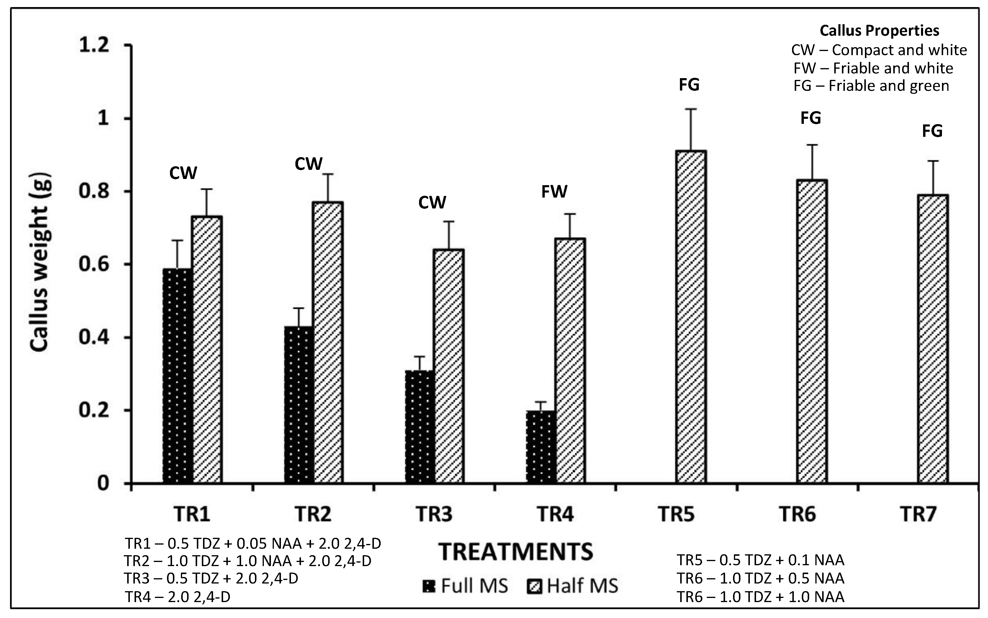
Generally, calli began to form two weeks after initiation. Swollen whitish structures first appeared on the cut edges of the leaf explants, then the main veins started to bulge, and the entire leaf vasculature followed suit. Variations were obtained depending on media composition and strength. Leaf explants cultured on half-strength media completely transformed into calli within 4–5 weeks, but it required eight weeks to obtain complete explant transformation with full-strength MS media. The latter calli were durable and remained fresh up to 12 weeks without needing transfer, but the former deteriorated fast and required transfer to fresh media instantly.
In addition, only media formulations containing either 2,4-D or TDZ or both were able to produce a response. Thus, watery and loose-textured calli were induced when 2,4-D was used singly. Such calli were considered non-embryogenic. Other formulations containing 2,4-D induced compact white calli irrespective of media strength (Figure 4 and Figure 5).
Findings from this study showed that leaf explants produced extremely responsive calli in 2,4-D + TDZ media combination (Figure 5), as also reported earlier from leaf explant of Primula vulgaris by ref. [18]. A combination of 2, 4-D and TDZ has also been reported to induce embryogenic calli in plants like Coffea arabica [19] and Fragaria vesca [20].
Media composition is one of the most critical factors influencing plant growth [21]. In the present study, callogenesis was enhanced when media strength was reduced by half (Figure 5). Indeed, leaf explants were non-responsive when initiated on media containing TDZ combined with NAA at full strength but produced friable green calli when the media strength was halved (Figure 5). This finding is supported by the assertion of ref. [22] that ½ MS contains less macronutrient salts, which does not seem to induce ammonium toxicity, a condition which is favorable for woody plants. Although many studies have yielded optimum callusing with full MS [20,23], ½ MS is largely successful with recalcitrant plant species that respond poorly to the higher salt concentrations of the full-strength MS medium due to osmotic stress [22]. Therefore, improved callogenesis in half-strength MS is attributable to reduced osmotic and ammonium stress, which, according to ref. [22], is known to impair morphogenic competence in woody and recalcitrant species.
Regarding callus weight, the treatment with 0.5 mg/L TDZ + 0.1 mg/L NAA at ½ concentration of MS salts exhibited the highest mean fresh weight. In fact, calli that were induced on half-strength media were significantly heavier (p ≤ 0.05) than those produced on full-strength MS media (Figure 5).
Figure 5. Effect of media composition on callus properties. Data represents mean ± standard deviation. Between media strengths, average callus weights differed significantly according to t-test (p < 0.001). Treatments (TR1–TR7) contain PGR values in mg/L.
Horticulturae 11 01127 g005

3.3. Effect of Media Composition on Callus Growth at Various Phases of Development

Callus induction was greatest where 2,4-D (2.0 mg/L) was used singly or in combination with 0.5 mg/L TDZ. But the incorporation of BAP either diminished the callus rate or completely failed to induce calli (Table 2). Furthermore, the use of BAP together with 2,4-D only did not result in any response in this study. On the contrary, ref. [24] reported up to 87.3% callogenesis from leaf disks of V. paradoxa using media formulations containing BAP in combination with 2,4-D. A similar trend was also observed in a study by ref. [25], where a media concoction of 0.5 mg/L 2,4-D and 1.5 mg/L BAP optimally induced callus in young leaf explants of V. paradoxa. More recently, calli have also been induced from nodal explants of V. paradoxa cultured in media containing BAP, Kin, and NAA [7].
This contradiction underscores the need to understand the inherent variation between the genotypes investigated. Thus, it is important to point out that previous studies [24,25] were conducted on the West African subspecies (paradoxa), whereas the current study utilized the East African subspecies (nilotica). According to ref. [21], media composition for successful culture may vary between different plant species or even among genotypes of the same species. This notion supports the results from this study in which media formulations different from those reported in previous investigations gave optimum callus induction. It also explains why media formulations deployed in previous studies were ineffective in the current investigation.
Furthermore, this negation could also be a result of auxin–cytokinin antagonism between BAP and 2,4-D or NAA. It is well documented that cell division and growth in plant tissue cultures occur under the synergistic regulation of cytokinin and auxin. Cytokinin coordinates the distribution of auxin, whereas auxin supports cytokinin biosynthesis—all occurring under complex but coordinated molecular mechanisms [26]. However, antagonism affects the following: biosynthesis, where one hormone affects the production of the other; signaling, a phenomenon characterized by one hormone negatively regulating the signaling pathways of another; and transport, where interactions influence the distribution of hormones, especially auxin [27,28]. Hence, with reference to this study, it is plausible to assume that BAP may have distorted the distribution and or signaling pathways of NAA or 2,4-D during callogenesis.
The findings of this study also indicate that callogenesis varied significantly (p ≤ 0.05) across different growth stages of the leaf explants (Table 2). The highest callus induction rate (100%) was obtained when either Stage III or IV explants were initiated on half-strength CIM that contained 2.0 mg/L 2,4-D + 0.5–1.0 mg/L TDZ. Very poor rates of callogenesis were recorded for Stage VI explants, while Stage I explants did not give any response at all (Table 2). A possible explanation for these variations can be drawn from the authors of [29], who opined that uptake of nutrients in vitro can be affected by the biochemical or physiological status of plant tissues. Examination of leaf histology showed structural changes at the different stages of leaf development (Figure 6). As noted by ref. [30], leaf development is characterized by three continuous and overlapping growth stages, namely initiation, morphogenesis, and differentiation. The initiation phase is marked by the emergence of the leaf from the flanks of the shoot apical meristem. It follows that, through morphogenesis, the lamina is initiated, and leaf marginal structures such as leaflets, lobes, and serrations are formed. During differentiation, the leaf area grows substantially, leaf tissues mature and differentiate, and the final leaf form is determined. With reference to this study, Stage I (Figure 1 and Figure 2) marks the last stage of morphogenesis and the beginning of leaf differentiation. At this stage, leaf explants do not give any response (Table 2) and become necrotic within a week of placement in CIM. A possible explanation could be that at this stage, the leaf cells (especially vascular ones) are not yet fully differentiated (Figure 4). This phenomenon hinders their ability to initiate calli, and the authors of [31] believe that calli are usually induced from pericycle cells adjacent to the xylem through asymmetric or formative cell divisions.
Further, Stages III and IV gave the best results for callogenesis overall (Table 2). During this phase, the explants exhibited exponential growth (Figure 3), which, according to ref. [17], is the period when cell division is at its highest. This instant cell proliferation could be one of the factors responsible for optimum callus induction at this stage of growth. Additionally, an examination of leaf cross sections showed that chloroplasts were evenly distributed throughout the leaf mesophyll between Stages I and IV (Figure 6). Chloroplasts are the sites of photosynthesis, converting light energy into chemical energy in the form of ATP and NADPH. These products are crucial for the metabolic processes that drive cell growth and differentiation [32], including those involved in callogenesis. Hence, the presence of chloroplasts all over the mesophylls could have also contributed to enhanced callogenesis for the growth periods from II to IV (Figure 6; Table 2). However, in the latter stages, chloroplasts accumulated mostly in the palisade mesophyll towards the adaxial side of the leaf (Figure 6).
Figure 6. Leaf cross sections at different stages of growth. (A) Stage I; (B) Stage II showing trichomes denoted by ‘T’; (C) Stage III; (D) Stage IV; (E) Stage V, leaf mesophyll with minor vasculature; (F) Stage VI, major vasculature and part of leaf mesophyll. ‘ad’ represents the adaxial side of the leaf; cl, chloroplast.
Horticulturae 11 01127 g006
This change in physiology coupled with the cessation of cell division and expansion [17] implies that the leaf at this point is committed to mobilizing nutrients mainly for relocation to other developing organs [33]. It also suggests that the withdrawal of chloroplasts from the vicinity of other cell types (including pericycle cells responsible for callus initiation) reduces the callogenesis potential of older leaf explants. This line of argument aligns with the authors of [31,34], who believe that photosynthetic activity usually interferes with callus formation.
Despite the abundance of chloroplasts in the spongy mesophyll of Stage II explants (Figure 6), their callus induction rates were substantially lower than those of Stage III and IV explants (Table 2). This difference is likely due to the presence and orientation of trichomes at this stage of leaf development (Figure 6). Plant trichomes are epidermal appendages normally found on the surfaces of stems, leaves, petals, and fruits. They are structurally designed to protect plants against abiotic and biotic stressors such as UV rays, temperature extremities, and herbivores [35]. An examination of leaf sections in this study showed Stage II explants with densely packed trichomes oriented perpendicularly to the leaf surface (Figure 6). But leaf explants at later stages bore trichomes observed to be positioned parallel to the leaf surface, rendering them not visible in the cross sections (Figure 6). Thus, the bulk and alignment of trichomes in Stage II explants may have provided a significant physical barrier between the leaf surface and CIM, which led to a lower callogenesis rate.

3.4. Effect of Media Composition on Growth of T1 and T2 Calli

Further growth of calli was observed following their transfer to various media. The combination of TDZ with NAA had a substantial effect on callus proliferation, suggesting their joint effectiveness (Table 3). However, the interaction of GA3 with BAP was not as effective as its combination with TDZ, NAA, and BAP (Table 3). Such an occurrence where a growth regulator shows different responses in several combinations underscores the need for manipulation of phytohormone ratios to accomplish optimum results [36].
This is because plant growth regulators exert their effects after binding to specific receptors of plant tissue. Moreover, plant tissues possess different binding affinities for various versions of the same phytohormone [22].
Generally, T1 calli had a higher mean weight compared to T2 but it was not statistically significant at 5% alpha level (Table 3). The latter callus type appeared embryogenic and yellowish white with nodular surfaces, whereas the latter were irregularly shaped but maintained a green color. Although the two callus types were alike in weight, subsequent transfer into media devoid of phytohormones gave different results. While T1 calli retained green, irregular shapes, T2 calli formed green, globular structures that were round or oval. Neither of the callus types differentiated into embryos or organs. In fact, the globular structures from T2 calli raptured into reddish brown friable structures, which eventually became necrotic. But the T1 calli remained unchanged despite numerous transfers.
According to ref. [37], globular structures are pro-embryogenic masses that are fundamental for somatic embryogenesis. Previous studies have shown that globular structures can give rise to somatic embryos. For example, ref. [5] reported the differentiation of pro-embryogenic masses into various stages of somatic embryos that eventually germinated into plantlets with Sapindus trifoliatus. A similar study by ref. [3] also demonstrated plant regeneration of Quercus alba leaf explants through somatic embryogenesis facilitated by intermediate nodular structures. Such findings from previous studies highlight the necessity of media modification in the present work. Thus, our future studies will aim to promote the differentiation of globular structures and achieve regeneration. Strategies such as modifying auxin–cytokinin ratios, introducing stress preconditioning, or supplementing with morphogenic elicitors will be employed.
Explants from other Sapotaceae species, including Chrysophyllum caimito, Synsepalum dulcificum, Argania spinosa, Manilkara zapota, and Palaquium gutta, often exhibit high phenolic content and sap exudation similar to V. paradoxa. Such traits, as noted by ref. [38], contribute to contamination and tissue browning, ultimately limiting in vitro culture success. Another hinderance to in vitro propagation within this family is the recalcitrance of explants to rooting [39]. However, the attainment of somatic embryogenesis and in vitro organogenesis by refs. [40] and [39], respectively, not only stress the challenges with the Sapoteceae family but also the need for media optimization and (or) explant manipulation if regeneration is to be achieved.
Although the current study establishes a foundation for future induction of somatic embryos, the significance of plant calli cannot be overlooked. Besides plant regeneration, callus cultures have been found useful in enhancing the quantity and quality of secondary metabolites in leaf explants. For example, ref. [41] reported elevated levels of aromatic compounds like rosmarinic acid and caffeic acid in leaf calli of Rosmarinus officinalis. Generally, secondary metabolites have numerous pharmacological properties such as regular antioxidants, antimicrobial, anti-inflammatory, anti-diabetic, antispasmodic, and, lately, anticarcinogenic [42,43]. In relation, V. paradoxa leaves are traditionally used to make medicinal concoctions for the treatment of abdominal pain, headaches, and fever. This is due to the leaves containing bioactive compounds such as tannins, saponins, steroids, flavonoids, alkaloids, and triterpenes, which exhibit antibacterial properties against numerous microorganisms [44,45]. Thus, the quantity of secondary metabolites in V. paradoxa leaves can also be amplified in vitro through callogenesis.

4. Conclusions

This study demonstrates that physiological age, media strength, and precise PGR combinations are critical for optimizing callogenesis in Vitellaria paradoxa (subsp. nilotica). The combination of 2,4-D + TDZ consistently led to high callus induction, with peak responsiveness from leaves at Stages III and IV, where up to 100% callus induction was achieved. Differential stage responses are attributable to histological traits, including chloroplast localization, trichome arrangement, and the degree of vascular tissue maturation. Even without achieving somatic embryogenesis, this maiden comprehension of physiological age can be exploited further to attain regeneration V. paradoxa using leaf explants. This may necessitate modification of the CIM, growth media, or other in vitro culture conditions. In addition, callus cultures of V. paradoxa are promising for enhancing the production of secondary metabolites (e.g., flavonoids, tannins, triterpenes). These compounds have pharmacological relevance, aligning with the plant’s traditional medicinal uses.

Author Contributions

Conceptualization, M.O. and E.F.-C.; methodology, M.O. and R.B.; validation, R.B.; formal analysis, M.O. and E.F.-C.; investigation, M.O.; resources, R.B. and E.F.-C.; data curation, M.O.; writing—original draft preparation, M.O.; writing—review and editing, M.O., R.B. and E.F.-C.; visualization, M.O. and E.F.-C.; supervision, R.B. and E.F.-C.; project administration, E.F.-C.; funding acquisition, E.F.-C. All authors have read and agreed to the published version of the manuscript.

Funding

This research was funded by the Internal Grant Agency, grant number 20253126. Faculty of Tropical AgriSciences, Czech University of Life Sciences, Prague.

Data Availability Statement

Data will be made available upon request.

Acknowledgments

The authors would like to express their sincere gratitude to Ngetta Zonal Agricultural Research and Development Institute of the National Agricultural Research Organization of Uganda for providing logistical support towards obtaining the plant materials used in this study.

Conflicts of Interest

The authors confirm that there are no financial or personal relationships that could be perceived as influencing the research reported in this paper.

Abbreviations

The following abbreviations are used in this manuscript:
2,4-D2,4-Dichlorophenoxyacetic acid
BAP6-Benzylaminopurine
CIMCallus induction medium
HClHydrochloric acid
HSDHonestly significant difference
KOHPotassium hydroxide
NAANaphthalene acetic acid
TDZThidiazuron

References

  1. Choungo Nguekeng, P.B.; Hendre, P.; Tchoundjeu, Z.; Kalousová, M.; Tchanou Tchapda, A.V.; Kyereh, D.; Masters, E.; Lojka, B. The current state of knowledge of Shea butter tree (Vitellaria paradoxa cf gaertner.) for nutritional value and tree improvement in West and Central Africa. Forests 2021, 12, 1740. [Google Scholar] [CrossRef]
  2. Jepsen, T.; Stopponi, G.; Jørgensen, N.O. Shea tree (Vitellaria paradoxa CF Gaertn.) agroforestry systems in Northern Ghana: Population structure, management of trees and impact of below canopy microclimate. Agrofor. Syst. 2024, 98, 1493–1506. [Google Scholar] [CrossRef]
  3. Martínez, M.T.; Vieitez, A.M.; Corredoira, E. Improved secondary embryo production in Quercus alba and Q. rubra by activated charcoal, silver thiosulphate and sucrose: Influence of embryogenic explant used for subculture. Plant Cell Tissue Organ Cult. (PCTOC) 2015, 121, 531–546. [Google Scholar] [CrossRef]
  4. Xiong, Y.; Chen, S.; Wu, T.; Wu, K.; Li, Y.; Zhang, X.; da Silva, J.A.T.; Zeng, S.; Ma, G. Shoot organogenesis and somatic embryogenesis from leaf and petiole explants of endangered Euryodendron excelsum. Sci. Rep. 2022, 12, 20506. [Google Scholar] [CrossRef] [PubMed]
  5. Singh, M.; Asthana, P.; Rai, M.K.; Jaiswal, U. Somatic embryogenesis and plant regeneration from suspension cultures of Sapindus trifoliatus. Plant Cell Tissue Organ Cult. (PCTOC) 2024, 157, 36. [Google Scholar] [CrossRef]
  6. Afful, N.T.; Abdulai, I.; Azu, E.; Elegba, W.; Annor, C.; Akama, C.; Asare, K.; Dentey, J.; Amoatey, H.M. In vitro regeneration of Vitellaria paradoxa from shoot tip explants. BioTechnologia 2022, 103, 71. [Google Scholar] [CrossRef]
  7. Attikora, A.J.P.; Silue, S.; Kone, M.; Silue, N.; Kwibuka, Y.; Yao, S.D.M.; De Clerck, C.; Him, S.L.; Diarrassouba, N.; Alabi, T.; et al. Efficient in vitro regeneration and genetic fidelity analysis of shea tree (Vitellaria paradoxa Gaertn) using ISSR markers. Electron. J. Biotechnol. 2025, 75, 28–38. [Google Scholar] [CrossRef]
  8. Afolabi, J.O.; Olorode, E.M.; Olomola, D.B.; Fasakin, Y.O.; Adekunle, E.A. Effects of different media strengths and hormone concentrations on in-vitro regeneration of Vitellaria paradoxa CF Gaertn. Niger. J. Biotechnol. 2020, 37, 150–158. [Google Scholar] [CrossRef]
  9. Lovett, P.N.; Haq, N. Progress in developing in vitro systems for shea tree (Vitellaria paradoxa CF Gaertn.) propagation. For. Trees Livelihoods 2013, 22, 60–69. [Google Scholar] [CrossRef]
  10. Adu-Gyamfi, P.K.K.; Barnor, M.; Dadzie, A.M.; Lowor, S.; Opoku, S.Y.; Opoku-Ameyaw, K.; Bissah, M.; Padi, F.K. Preliminary investigation on somatic embryogenesis from immature cotyledon explants of shea (Vitellaria paradoxa G.). J. Agric. Sci. Technol. B 2012, 2, 1171. [Google Scholar]
  11. George, E.F.; Hall, M.A.; De Klerk, G.J. Plant Propagation by Tissue Culture, 3rd ed.; The Back Ground; Springer: Dordrecht, The Netherlands, 2008; pp. 65–175. [Google Scholar]
  12. Read, P.E.; Bavougian, C.M. In Vitro Rejuvenation of Woody Species. In Protocols for Micropropagation of Selected Economically-Important Horticultural Plants; Lambardi, M., Ozudogru, E., Jain, S., Eds.; Methods in Molecular Biology; Humana Press: Totowa, NJ, USA, 2012; Volume 994. [Google Scholar] [CrossRef]
  13. Grzelak, M.; Pacholczak, A.; Nowakowska, K. Challenges and insights in the acclimatization step of micropropagated woody plants. Plant Cell Tiss. Organ. Cult. 2024, 159, 72. [Google Scholar] [CrossRef]
  14. Lv, Z.; Zhao, W.; Kong, S.; Li, L.; Lin, S. Overview of molecular mechanisms of plant leaf development: A systematic review. Front. Plant Sci. 2023, 14, 1293424. [Google Scholar] [CrossRef] [PubMed]
  15. Murashige, T.; Skoog, F.A. Revised medium for rapid growth and bioassays with tobacco tissue cultures. Physiol. Plant 1962, 15, 473–497. [Google Scholar] [CrossRef]
  16. Ding, A.M.; Xu, C.T.; Xie, Q.; Zhang, M.J.; Yan, N.; Dai, C.B.; Lv, J.; Cui, M.; Wang, W.; Sun, Y. ERF4 interacts with antagonizes TCP15 in regulating endoreduplication cell growth in Arabidopsis. J. Integr. Plant Biol. 2022, 64, 1673–1689. [Google Scholar] [CrossRef] [PubMed]
  17. Munns, R.; Schmidt, S.; Beveridge, C. Plants in Action A Resource for Teachers and Students of Plant Science (II); Austrlaian and New Zealand Societes of Plant Sciences: 2010. Available online: https://rseco.org/ (accessed on 1 February 2025).
  18. Hayta, S.; Smedley, M.A.; Li, J.; Harwood, W.A.; Gilmartin, P.M. Plant regeneration from leaf-derived callus cultures of primrose (Primula vulgaris). HortScience 2016, 51, 558–562. [Google Scholar] [CrossRef]
  19. Wang, Y.C.; Lin, M.Z.; Huang, B.; Chung, H.H.; Chen, J.T. Thidiazuron enhanced somatic embryogenesis is from callus lines of Arabica coffee and subsequent plant regeneration. Acta Biol. Cracoviensia s. Bot. 2018, 60, 35–44. [Google Scholar]
  20. Chung, H.H.; Ouyang, H.Y. Use of thidiazuron for high-frequency callus induction and organogenesis of wild strawberry (Fragaria vesca). Plants 2020, 10, 67. [Google Scholar] [CrossRef]
  21. Rezali, N.I.; Sidik, N.J.; Saleh, A.; Osman, N.I.; Adam, N.A.M. The effects of different strength of MS media in solid and liquid media on in vitro growth of Typhonium flagelliforme. Asian Pac. J. Trop. Biomed. 2017, 7, 151–156. [Google Scholar] [CrossRef]
  22. Phillips, G.C.; Garda, M. Plant tissue culture media and practices: An overview. Vitr. Cell. Dev. Biol.-Plant 2019, 55, 242–257. [Google Scholar] [CrossRef]
  23. Mosoh, D.A.; Khandel, A.K.; Verma, S.K.; Vendrame, W.A. Optimizing callus induction and indirect organogenesis in non-dormant corm explants of Gloriosa superba (L.) via media priming. Front. Hortic. 2024, 3, 1378098. [Google Scholar] [CrossRef]
  24. Ndoumou, D.O.; Tchinda, N.D. Comparaison des Premières Étapes de L’embryogenèse Somatique chez Baillonella toxisperma et Vitellaria paradoxa (Sapotacées). Biotechnol. Agron. Soc. Environ. 2008, 12, 131–138. [Google Scholar]
  25. Aisagbonhi, E.P.; Isalar, C.E.; Odenore, V.D.; Ogbebor, J.U.; Eke, C.R.; Asemota, O.; Shittu, H.O. The interplay between explant developmental stages and phytohormone type in callogenesis of shea tree (Vitellaria paradoxa CF Gaertn). Euro. Int. J. Sci. Tech. 2015, 4, 50–57. [Google Scholar]
  26. Hurný, A.; Cuesta, C.; Cavallari, N.; Ötvös, K.; Duclercq, J.; Dokládal, L.; Montesinos, J.C.; Gallemí, M.; Semerádová, H.; Rauter, T.; et al. SYNERGISTIC ON AUXIN AND CYTOKININ 1 positively regulates growth and attenuates soil pathogen resistance. Nat. Commun. 2020, 11, 2170. [Google Scholar] [CrossRef] [PubMed]
  27. Jing, H.; Strader, L.C. Interplay of auxin and cytokinin in lateral root development. Int. J. Mol. Sci. 2019, 20, 486. [Google Scholar] [CrossRef] [PubMed]
  28. Romanov, G.A. Perception, transduction and crosstalk of auxin and cytokinin signals. Int. J. Mol. Sci. 2022, 23, 13150. [Google Scholar] [CrossRef]
  29. George, E.F.; Hall, M.A.; De Klerk, G.J. The components of plant tissue culture media I: Macro-and micro-nutrients. In Plant Propagation by Tissue Culture: Volume 1. The Background; Springer: Dordrecht, The Netherlands, 2008; pp. 65–113. [Google Scholar]
  30. Bar, M.; Ori, N. Leaf development and morphogenesis. Development 2014, 141, 4219–4230. [Google Scholar] [CrossRef] [PubMed]
  31. Shin, J.; Seo, P.J. Varying auxin levels induce distinct pluripotent states in callus cells. Front. Plant Sci. 2018, 9, 1653. [Google Scholar] [CrossRef]
  32. Alberts, B.; Johnson, A.; Lewis, J.; Raff, M.; Roberts, K.; Walter, P. Molecular Biology of the Cell, 4th ed.; Garland Science: New York, NY, USA, 2002. Available online: https://www.ncbi.nlm.nih.gov/books/NBK21054/ (accessed on 1 February 2025).
  33. Lee, J.; Kang, M.H.; Kim, J.Y.; Lim, P.O. The role of light and circadian clock in regulation of leaf senescence. Front. Plant Sci. 2021, 12, 669170. [Google Scholar] [CrossRef]
  34. Fehér, A. Callus, dedifferentiation, totipotency, somatic embryogenesis: What these terms mean in the era of molecular plant biology? Front. Plant Sci. 2019, 10, 536. [Google Scholar] [CrossRef]
  35. Watts, S.; Kariyat, R. Morphological characterization of trichomes shows enormous variation in shape, density and dimensions across the leaves of 14 Solanum species. AoB Plants 2021, 13, plab071. [Google Scholar] [CrossRef]
  36. Herman, E.B. Recent Advances in Plant Tissue Culture, X.X.I. Media and Techniques for Growth, Regeneration and Storage: 2011–2015; Agritech Consultants Inc.: Shrub Oak, NY, USA, 2015; 148p. [Google Scholar]
  37. Corredoira, E.; San-José, M.C.; Vieitez, A.M. Induction of somatic embryogenesis from different explants of shoot cultures derived from young Quercus alba trees. Trees Struct. Funct. 2012, 26, 881–891. [Google Scholar] [CrossRef]
  38. Kaur, S.; Boora, R.S.; Singh, D. Propagation studies in Sapodilla [Manilkara zapota (L.) P. Royen]: A Review. Agric. Rev. 2020, 41, 356–363. [Google Scholar] [CrossRef]
  39. Amghar, I.; Ibriz, M.; Ibrahimi, M.; Boudra, A.; Gaboun, F.; Meziani, R.; Iraqi, D.; Mazri, M.A.; Diria, G.; Abdelwahd, R. In Vitro Root Induction from Argan (Argania spinosa (L.) Skeels) Adventitious Shoots: Influence of Ammonium Nitrate, Auxins, Silver Nitrate and Putrescine, and Evaluation of Plantlet Acclimatization. Plants 2021, 10, 1062. [Google Scholar] [CrossRef]
  40. Akkenapally, S.; Mudalkar, S.; Bodiga, S.; Sundaram, R.; Bokam, A.K. In vitro callus generation somatic embryogenesis from leaf explant of Madhuca longifolia var latifolia (Roxb), A. Chev. Vegetos 2024, 1–7. [Google Scholar] [CrossRef]
  41. Coskun, Y.; Duran, R.E.; Kilic, S. Striking effects of melatonin on secondary metabolites produced by callus culture of rosemary (Rosmarinus officinalis L.). Plant Cell Tissue Organ Cult. (PCTOC) 2019, 138, 89–95. [Google Scholar] [CrossRef]
  42. Conrado, R.; Gomes, T.C.; Roque, G.S.C.; De Souza, A.O. Overview of bioactive fungal secondary metabolites: Cytotoxic and antimicrobial compounds. Antibiotics 2022, 11, 1604. [Google Scholar] [CrossRef] [PubMed]
  43. Moore, J.; Yousef, M.; Tsiani, E. Anticancer effects of rosemary (Rosmarinus officinalis L.) extract and rosemary extract polyphenols. Nutrients 2016, 8, 731. [Google Scholar] [CrossRef] [PubMed]
  44. Adetunde, L.A.; Awo, O.; Imoro, A.W.M. Antimicrobial activities of the extract of shea tree (Vitellaria paradoxa Gaertn. F.) leaf and bark on some selected clinical pathogens. J. Med. Plants Res. 2023, 17, 338–344. [Google Scholar] [CrossRef]
  45. Mohammed, I.H.; Halilu, M.E.; Atinga, V.; Sabo, I.; Aliyu, W.A.; Yusuf, A.A.; Suleiman, A.U.; Zainab, M.; Garba, I.; Avosuahi, O.R. Comparative phytochemistry of Vitellaria paradoxa: Towards establishing a chemotaxonomic marker. Malays. J. Pharm. Sci. 2024, 22, 61–76. [Google Scholar] [CrossRef]
Figure 1. Growth phases: (a) Stage I; (b) Stage II; (c) Stage III; (d) Stage IV; (e) Stage V. Bar = 1 cm.
Horticulturae 11 01127 g001
Figure 2. Explant surface sterilization.
Horticulturae 11 01127 g002
Figure 3. Growth trend of leaves (leaf length) for thirty days: (a) Assessment of growth for eight selected phenotypes; (b) abridged growth trend.
Horticulturae 11 01127 g003
Figure 4. Appearance of calli after 4–6 weeks after initiation of leaf lamina on various induction media: (a) 1.0 mg/L TDZ + 0.5 mg/L NAA; (b) 1.0 mg/L TDZ + 1.0 mg/L NAA; (c) 2.0 mg/L 2,4-D; (d) 2.0 mg/L 2,4-D + 0.5 mg/L TDZ. Bar = 1 cm.
Horticulturae 11 01127 g004
Table 1. Phases of leaf growth.
Stage of GrowthAge (Days)Leaf Color and Appearance
I0–5Pink/brown
II6–10Brown with green patches
III11–15Green with dark patches
IV16–20Dark green
V21–25Light green
VI26–30Light green
Table 2. Effect of media composition on callus induction at different growth stages.
MS Media
Strength
Media Formulations (mg/L)Average Callusing at Six Stages of Growth (%)
TDZNAA2,4-DBAPIIIIIIIVVVI
Full strength0.50.052.0 041.290.054.748.835.3
0.50.052.00.5031.131.150.833.20.0
1.01.02.0 035.363.452.741.226.6
0.5 2.0 050.875.063.445.028.9
2.0 054.761.171.650.837.3
0.50.1 00.00.00.00.00.0
1.00.5 00.00.00.00.00.0
1.01.0 00.00.00.00.00.0
Half strength0.50.052.0 048.890.090.052.735.3
1.01.02.0 045.079.590.054.731.1
0.5 2.0 058.990.090.054.726.6
2.0 063.490.061.150.843.1
0.50.1 00.035.328.90.00.0
1.00.5 090.046.943.128.90.0
1.01.0 061.156.839.224.118.4
0.50.1 0.500.00.00.00.00.0
Grand average callusing (%)0.0 c36.3 ab50.6 a46.0 a30.3 ab17.7 bc
Rank of callus formation++++++++++++++
Legend: (−) No callusing; (+) Less than 10% of explant callused; (++) 10–30% explant transformation; (+++) 31–50% of explant callused; (++++) 51–79% explant transformation; (+++++) More than 80% of the leaf callused. Different letters indicate significantly different means at (p ≤ 0.05). Values shown are angularly transformed percentages. Colors were used to represent percentage ranges of callus induction for ease of visual comparison across treatments and growth stages.
Color code (percentage range)0–2021–4041–6061–8081–100
Table 3. Effect of growth media on performance of calli from two categories of CIM.
Source of Callus (CIM)Compositions of Transfer Media
(Half-Strength MS)
Mean Callus Weight (g)Callus
Ranking
TDZNAABAPGA3
T1 Calli1.01.0 2.30 ± 0.19+++++
0.50.12.00.53.28 ± 0.26+++++
0.50.50.40 ± 0.05
2.00.50.53 ± 0.06+
0.52.00.51.90 ± 0.20++++
0.52.0 3.05 ± 0.29+++++
0.53.0 1.08 ± 0.11+++
T2 Calli1.01.0 1.38 ± 0.17+++
0.50.12.00.51.74 ± 0.16++++
0.50.51.05 ± 0.12+++
2.00.50.92 ± 0.11++
0.52.00.51.53 ± 0.16+++
0.52.0 1.97 ± 0.17++++
0.53.0 1.82 ± 0.16++++
T1 = Calli induced on CIM containing 1.0 mg/L TDZ +0.5 mg/L NAA or 1.0 mg/L TDZ +1.0 mg/L NAA. T2 = Calli induced on CIM containing 2.0 mg/L 2,4-D+0.5 mg/L TDZ or 2.0 mg/L 2,4-D+1.0 mg/L TDZ+1.0 mg/L NAA or 2.0 mg/L 2,4-D+0.5 mg/L TDZ+0.05 mg/NAA. Ranking legend: (−) No callus growth observed; (+) 0.1–0.5 g callus increment; (++) 0.6–1.0 g callus growth; (+++) 1.1–1.5 g increment; (++++) 1.6–2.0 g callus growth; (+++++) more than 2.1 g of callus increment. Data represents mean ± standard deviation. No significant difference was observed between T1 and T2 calli (t-test, p = 0.52).
Disclaimer/Publisher’s Note: The statements, opinions and data contained in all publications are solely those of the individual author(s) and contributor(s) and not of MDPI and/or the editor(s). MDPI and/or the editor(s) disclaim responsibility for any injury to people or property resulting from any ideas, methods, instructions or products referred to in the content.