Article

Expression of the Protein Phosphatase Gene SlPP2C28 Confers Enhanced Tolerance to Bacterial Wilt in Tobacco

1
College of Horticulture and Landscape Architecture, Southwest University, Chongqing 400715, China
2
Key Laboratory of Agricultural Biosafety and Green Production of Upper Yangtze River (Ministry of Education), Southwest University, Chongqing 400715, China
3
Academy of Agricultural Sciences, Southwest University, Beibei, Chongqing 400715, China
*
Author to whom correspondence should be addressed.
Horticulturae 2025, 11(8), 937; https://doi.org/10.3390/horticulturae11080937
Submission received: 17 June 2025 / Revised: 28 July 2025 / Accepted: 6 August 2025 / Published: 8 August 2025
(This article belongs to the Section Biotic and Abiotic Stress)

Abstract

Plant protein phosphatase 2C (PP2C) is recognized as one of the most critical protein family in plants and plays a pivotal role in disease resistance responses. However, the involvement of tomato PP2C family members in resistance to bacterial wilt caused by Ralstonia solanacearum remains poorly understood. In this study, we found that silencing SlPP2C28 increased tomato susceptibility to R. solanacearum; then, we introduced the tomato gene SlPP2C28, which exhibits a strong response to R. solanacearum, into the Nicotiana benthamiana genome via Agrobacterium-mediated transformation, generating high-expression transgenic lines OE-2 and OE-3. Following inoculation with R. solanacearum, the transgenic tobacco plants displayed reduced wilting symptoms, delayed disease onset, lower disease index, reduced stem cross-section damage, decreased internal bacterial colonization, diminished accumulation of reactive oxygen species in leaves, enhanced expression of SlPP2C28, up-regulated expression of defense-related genes NbSOD, NbPOD, and NbPAL along with an increase in the activities of their corresponding enzymes, and elevated expression levels of pathogenesis-related genes NbPR1a, NbPR2, NbPR4, and NbPR10. Collectively, these findings demonstrate that the SlPP2C28 gene has the function of enhancing resistance to bacterial wilt disease.

1. Introduction

Bacterial wilt, caused by the soil-borne pathogen Ralstonia solanacearum, is one of the most destructive plant diseases globally. As a common root and vascular pathogen, this bacterium was among the first plant pathogens to have its genome fully sequenced [1]. Due to its widespread distribution and strain diversity, which can be classified into four phylotypes (genetic clusters)—I, II, III, and IV—R. solanacearum poses a significant threat to agricultural production [2]. Its host range includes economically important crops such as tomatoes, tobacco, eggplants, and potatoes. Despite extensive research, managing bacterial wilt remains a formidable challenge [3,4]. The pathogen infects plants via wounds on the roots and stems and spreads systemically through the xylem vascular system, leading to severe blockage of water transport and eventual plant death [5]. Given the lack of effective control measures, further research into the prevention and management of bacterial wilt is critically needed [6].
Under pathogen infection, plants rapidly produce excessive reactive oxygen species (ROS), including superoxide (O2) and hydrogen peroxide (H2O2) [7]. While these ROS exhibit antibacterial activity that can suppress pathogen vitality, they may also exacerbate plant damage [8]. ROS are primarily generated through two mechanisms: First, mitochondrial oxidative metabolism, which produces ROS as byproducts of essential biochemical reactions. Second, during the cellular response to bacterial invasion, specific processes deliberately generate ROS for involvement in signal transduction or cellular defense mechanisms [9,10]. The initial products of the mitochondrial respiratory chain, such as O2−, are rapidly converted to H2O2 by mitochondrial superoxide dismutase (SOD) [11,12]. Several antioxidant proteins have been identified as contributing to the excessive accumulation of intracellular ROS. The balance between ROS production and detoxification determines the extent of cell death in infected plants [13].
Plants have developed sophisticated antioxidant systems to mitigate ROS toxicity [14]. Among these systems, antioxidant enzymes are the most effective in combating ROS, with SOD being the primary enzyme responsible for removing ROS from plant cells [15]. SOD is a metallic enzyme found in various cellular compartments and catalyzes O2 and H2O2. There are three types of SOD, each containing Mn, Fe, or Cu+Zn as cofactors [7]. MnSOD is localized in mitochondria and peroxisomes [16], while FeSOD is found in chloroplasts [15]. Cu/ZnSOD, on the other hand, is present in the cytosol and chloroplasts and may also be found in the apoplast [16,17]. Additionally, peroxidase (POD) plays a crucial role in ROS detoxification within cells. POD represents a family of enzymes widely distributed across organisms [18,19]. To date, genome-wide analyses of POD family members have been conducted in several plants, including Arabidopsis [20], rice [21], and maize [22].
In addition to antioxidant enzymes, phenylalanine ammonia-lyase (PAL) serves as a key defense enzyme in plants, participating in the biosynthesis of phenolic compounds and antifungal agents and thereby inhibiting pathogen infection establishment within plant systems [23]. Lignin and certain antifungal compounds are synthesized through the phenylpropanoid metabolic pathway. The initial step of this pathway is catalyzed by PAL, which converts phenylalanine into cinnamic acid, a precursor to the synthesis of phenylpropanoid-derived compounds [24]. The production of antifungal compounds in plants can be induced by enhancing the expression of the gene encoding PAL, which plays a critical role in plant disease resistance [25]. Moreover, the expression of pathogenesis-related genes (PRs) represents a hallmark feature of plant defense mechanisms activation [26]. Enhanced pathogen resistance is closely associated with the expression of PRs and the stimulation of multiple defense pathways [27]. To date, eleven PRs families have been officially identified [28].
Plant protein phosphatase 2C genes (PP2Cs), the largest phosphatase gene family in plants [29,30], play critical regulatory roles in various aspects of plant growth and development. These include plant hormone signal transduction, developmental processes, and responses to both abiotic and biotic stresses [29,30,31]. PP2Cs are implicated in conferring resistance to pathogenic bacteria. For instance, the overexpression of OsBIPP2C1 and OsBIPP2C2 has been shown to enhance disease resistance in transgenic tobacco following infection with Magnaporthe grisea [32,33]. Additionally, AtPP2C26 and AtPP2C62 have been associated with resistance to bacterial pathogens [34]. Studies also indicate that the PP2C gene family plays a significant role in immune regulation in tomatoes, and PP2C proteins possess the potential to strengthen disease resistance in plants [35,36]. Nevertheless, genetic investigations into the tomato PP2C family in relation to R. solanacearum remain limited.
Through a preliminary bioinformatic analysis of the tomato PP2C genes family, we identified a SlPP2C28 gene that shows a strong responsive to R. solanacearum [37]. To clarify whether the SlPP2C28 gene has the function of increasing resistance to bacterial wilt, in this study we conducted an association analysis of the SlPP2C28 gene and tomato cultivars with different resistance to bacterial wilt, determined whether silencing this gene reduces tomato resistance to bacterial wilt using virus-induced gene silencing (VIGS) technology, and overexpressed it in tobacco to determine whether it increases the resistance of tobacco to bacterial wilt. The results lay the foundation for further analysis of the molecular mechanism of the SlPP2C28 gene, which is involved in regulating the resistance of tomato to bacterial wilt.

2. Materials and Methods

2.1. Bacterial Strains and Preparation of Pathogenic Inoculum

The Escherichia coli strain DH5α and the Agrobacterium tumefaciens strain LBA4404 are both preserved in the laboratory, and their growth conditions are described in a study by Zhang et al. [38]. R. solanacearum tomato bacterial wilt strain GMI1000 and tobacco bacterial wilt strain CQPS-1 were provided by Professor Wei Ding from Southwest University of China. The pathogen culture methods have been described by Tang et al. [39]. Once the concentration of the bacterial suspension reached 1 × 10⁸ CFU/mL, it was used for the inoculation experiments.

2.2. Plant Materials and Inoculation Methods

The plant materials used in this study included Nicotiana benthamiana Domin (propagated in our laboratory), as well as Solanum lycopersicum L. cultivars: Ailsa Craig (AC, a moderately resistant cultivar to bacterial wilt), M82 (a susceptible cultivar), Hongchuan (a commercial resistant cultivar), and Hongguifei (a commercial resistant cultivar). Tomato plants were inoculated with the bacterial strain GMI1000 at the four-leaf stage by stem injection [37]. Similarly, tobacco plants were inoculated with the tobacco wilt strain CQPS-1 at the four-leaf stage using the same stem inoculation method.

2.3. Preliminary Analysis of the Response of SlPP2C28 to R. solanacearum in Tomato

Stems of four tomato varieties, AC, M82, Hongchuan, and Hongguifei, were inoculated with R. solanacearum. The leaves were removed from the stems, and scissors were disinfected with 75% ethanol before cutting the stem segment above the injection site. Stem samples were collected at 0, 3, 6, 12, 24, and 48 h post-inoculation, placed into properly labeled enzyme-free tubes, immediately frozen in liquid nitrogen, and stored temporarily at −80 °C for subsequent measurement of the expression level of SlPP2C28 upon treatment [38]. Three biological replicates and three technical replicates were conducted for each time-point (control and treated plants included three biological replicates). Disease progression of the four surviving tomato varieties was photographed and documented 7 d post-inoculation (control and treated plants included three biological replicates). Primer sequences are shown in Table S1.

2.4. Virus-Induced Gene Silencing in Tomato

VIGS was used for silencing the SlPP2C28 gene with the tobacco rattle virus-based vectors in tomato AC. The cDNA fragment of SlPP2C28 was amplified using the gene-specific primers listed in Table S1 and were inserted into the pTRV2 vector (Novopro, Shanghai, China). The resulting plasmid was confirmed by sequencing and subsequently transformed into A. tumefaciens strain GV3101 (WEIDI, Shanghai, China). VIGS was performed as described by Chen et al. [40]. Control and treated plants included three biological replicates.

2.5. Construction of Plant Overexpression Vector

SlPP2C28 (Solyc03g096670.3.1) was amplified from tomato AC-cDNA using the PrimStar high-fidelity enzyme (Takara, Dalian, China). The Polymerase Chain Reaction (PCR) products, generated with primers listed in Table S1 containing the target gene SlPP2C28 and the pVCT2345 vector (preserved in the laboratory) were digested with Xba I and Sac I restriction enzymes (Takara, Dalian, China), respectively [38]. This digestion yielded two fragments: an 8862 bp fragment from the pVCT2345 vector and a 736 bp fragment corresponding to the SlPP2C28 gene. These two fragments were ligated overnight at 37 °C using T4 DNA ligase (Takara, Dalian, China) to construct the recombinant vector pVCT2470-SlPP2C28. The detection and validation of positive clones, and their preservation, were carried out following the methods described by Zhang et al. [38]. Structural diagrams of the vectors and gene sequences are presented in Figures S1 and S2, respectively.

2.6. Acquisition of Tobacco Transgenic Lines

Healthy N. benthamiana wild-type (WT) plants were selected and genetically transformed using the Agrobacterium-mediated transformation method (Tang et al. [39]). To identify positive transformants, PCR was performed using leaf DNA as the template, which was extracted via a modified cetyltrimethylammonium bromide (CTAB (Coolaber, Beijing, China)) protocol. The reactions of PCR were carried out in the following conditions: 94 °C for 5 min, followed by 35 cycles of denaturation at 94 °C for 30 s, annealing for 30 s, extension at 72 °C for 45 s (calculated as 1 KB/min), and the cycle ended at 72 °C for 10 min. Subsequently, RNA was isolated from the confirmed positive transformants for quantitative real-time PCR analysis to determine the expression levels of the target genes in the positive plants, and high-expression lines were screened. The reaction conditions were as follows: denaturation at 95 °C for 10 min, 40 cycles of 15 s at 95 °C, 30 s at the primer annealing temperature, 5 s at 65 °C, and data acquisition at 95 °C [38]. The experiment set up three technical replicates (control plants included three biological replicates). Following further propagation, T1 generation-positive plants were utilized for subsequent disease resistance assays. The primer sequences employed in this study are listed in Table S1.

2.7. Expression Analysis of SlPP2C28, Defense Enzyme-Related Genes, and Pathogenesis-Related Genes Following Inoculation with R. solanacearum

The bacterial solution was inoculated at the base of the stem. Subsequently, the tobacco leaves were harvested at 0, 24, and 48 h post-inoculation for analyzing the relative expression levels of SlPP2C28, defense enzyme-related genes (NbSOD, NbPOD, and NbPAL), and pathogenesis-related genes (NbPR1a, NbPR2, NbPR4, and NbPR10). The tomato leaves were harvested at 7 d post-inoculation for analyzing the relative expression levels of defense enzyme-related genes (SlSOD, SlPOD, and SlPAL), and pathogenesis-related genes (SlPR5, SlPR10, and SlPR-NP24) [39]. Total RNA was extracted using TRIzol reagent (Takara, Dalian, China). cDNAs were synthesized from 1 μg total RNA was reverse-transcribed using the HiFiScript gDNA Removal cDNA Synthesis Kit (Kangwei, Taizhou, China). Quantitative real-time PCR was conducted by SYBR Green Real-Time PCR system (Bao Guang, Chongqing, China); the β-Actin was used as the internal control. PCR products were monitored using a CFX96 (Bio-Rad, CA, USA) PCR system [38]. The experiment included three biological replicates, each with three technical replicates (control and treated plants included three biological replicates). Data were statistically analyzed using GraphPad Prism software 9.4.1. Primer sequences are provided in Table S1.

2.8. Determination of Defense-Related Enzyme Activity in N. benthamiana Leaves

After inoculation, leaves were excised from the same position on different plants using scissors disinfected with 75% ethanol, with each sample weighing ≥ 2.0 g. Leaves were collected at 0, 24, and 48 h post-inoculation and immediately transferred to enzyme-free tubes. Subsequently, the samples were frozen in liquid nitrogen and stored temporarily at −80 °C for the determination of defense enzyme activities, including SOD, POD, and PAL. Three biological replicates were performed, each with three technical replicates. The SOD, POD, and PAL assay kits (Beyotime, Shanghai, China) were used according to the manufacturer’s instructions, and the test results were analyzed using GraphPad Prism software 9.4.1.

2.9. Determination of the Tobacco Plant Disease Index

Using WT N. benthamiana as a control, we monitored the disease progression in plants following inoculation and documented characteristics by capturing images on days 0, 5, and 7 post-inoculation. Each group consisted of ten seedlings, and the experiment was independently repeated three times to ensure reproducibility (control and treated plants included ten biological replicates). The disease index of the inoculated plants was statistically analyzed according to the method described by Tang et al. [39]. The experimental results were analyzed using GraphPad Prism software 9.4.1.

2.10. Detection of the Number of R. solanacearum Infections in N. benthamiana Stems

Five days post-inoculation, 0.5 g of stem tissue was surface-sterilized by soaking in 75% ethanol for 1 min on a super-clean workbench, followed by rinsing three to five times with sterile water. The sterilized tissue was then transferred into a sterile test tube containing 5 mL of sterile water and incubated for 30 min. After shaking the suspension, it was serially diluted to gradient concentrations (10−1, 10−2, 10−3, 10−4, 10−5, and 10−6). Subsequently, 0.1 mL of the appropriate diluted solution was evenly spread onto triphenyl tetrazolium chloride (TTC (Coolaber, Beijing, China)) solid medium. Petri dishes were inverted and incubated at 28 °C in a bacterial incubator, and colony counts were performed after 2–3 days (Tang et al. [39]). Each treatment included three biological replicates (control and treated plants included three biological replicates), and statistical analyses of the results were conducted using GraphPad Prism software 9.4.1.

2.11. Tissue Staining Analysis of Leaves

In tobacco, on the abaxial surface of leaves, two or three circular spots were injected on either side of the midrib. The left side of the midrib was injected with water as a control, while the right side was injected with an R. solanacearum suspension. Each treatment group consisted of three replicates. Five days post-inoculation, the disease status of the plant leaves was assessed visually, and the leaves were photographed for documentation. Twenty-four hours after inoculation, selected leaves were stained overnight using freshly prepared 3,3′-diaminobenzidine (DAB (Coolaber, Beijing, China)) and nitrotetrazolium blue chloride (NBT (Coolaber, Beijing, China)) staining solutions. The following day, the leaves were decolorized in a boiling water bath containing 95% ethanol until all chlorophyll was completely removed (Liu et al. [41]). After cooling, the leaves were transferred to 60% glycerol for preservation and photographed. In tomato, DAB staining was performed on the leaves of tomato plants injected with R. solanacearum for 0 and 5 days, respectively, and the subsequent experimental operation process was the same as above. Control and treated plants included three biological replicates.

2.12. Anatomical Observation of the N. benthamiana Stems

The control group was inoculated with sterile water, whereas the treatment group was inoculated with R. solanacearum. Five days post-inoculation, tobacco stems were excised using sterilized scissors. Stem fragments from the upper 2–3 cm of the inoculation sites were collected and promptly immersed in freshly prepared formaldehyde–acetic acid–alcohol (FAA (Coolaber, Beijing, China)) fixative for a minimum of 48 h. Subsequently, the samples were sent to ServiceBio (Wuhan, China, http://www.servicebio.cn (accessed on 17 June 2025)) for paraffin-embedding and sectioning. Cross-sections of the tobacco samples were stained for analysis. Control and treated plants in the experiment included three biological replicates.

2.13. Statistical Analysis

The GraphPad Prism version 9.4.1 software was used for statistical analyses and graphing. The data between two treatments were compared and statistically analyzed through the t-test. When the dependent variable across multiple treatments was a single variable, data comparisons and statistical analyses were performed using one-way ANOVA analysis of variance. When the analysis across multiple treatments involved two factors, data comparisons and statistical analyses were performed using two-way ANOVA analysis of variance.

3. Results

3.1. SlPP2C28 Was Highly Expressed in R. solanacearum-Resistant Tomato Plants

Based on our previous findings, we observed that the expression of SlPP2C28 gene was significantly upregulated in tomato plants following inoculation with R. solanacearum, suggesting its strong response to R. solanacearum reaction [37]. Therefore, we further validated its role in tomatoes of different resistant varieties, as illustrated in Figure 1. Seven days post-inoculation with R. solanacearum, the susceptible variety M82 exhibited the most severe wilting symptoms, followed by the moderately resistant variety AC. In contrast, the resistant varieties Hongchuan and Hongguifei displayed relatively mild wilting (Figure 1a). The expression of SlPP2C28 gene remained relatively stable in M82, was significantly upregulated in AC at 48 h post-inoculation, and showed a consistent significant upregulation in the resistant varieties Hongchuan and Hongguifei overall (Figure 1b). The results demonstrated that SlPP2C28 was significantly upregulated in varieties resistant to R. solanacearum compared to those susceptible to the pathogen. Collectively, these findings strengthen the established link between SlPP2C28 and tomato resistance to R. solanacearum.

3.2. SlPP2C28-Silencing Reduced the Resistance of Tomato to Bacterial Wilt

To further investigate whether SlPP2C28 plays a role in tomato resistance against R. solanacearum, we generated SlPP2C28-silenced (V-SlPP2C28) plants using VIGS technology. SlPP2C28-silenced (V-SlPP2C28) plants with reduced transcript levels (c. 77.0%) of SlPP2C28 relative to the control (CK) were used for further experiments (Figure S3a). After inoculation with R. solanacearum for seven days, SlPP2C28-silenced plants showed decreased disease resistance (Figure S3b), as reflected by the increased ROS product accumulation (Figure S3c), and significantly lower expression of defense enzyme-related genes and pathogenesis-related genes (Figure S3d,e), indicating that SlPP2C28 positively regulates tomato resistance to R. solanacearum.

3.3. SlPP2C28-Overexpression Tobacco Lines Were Obtained

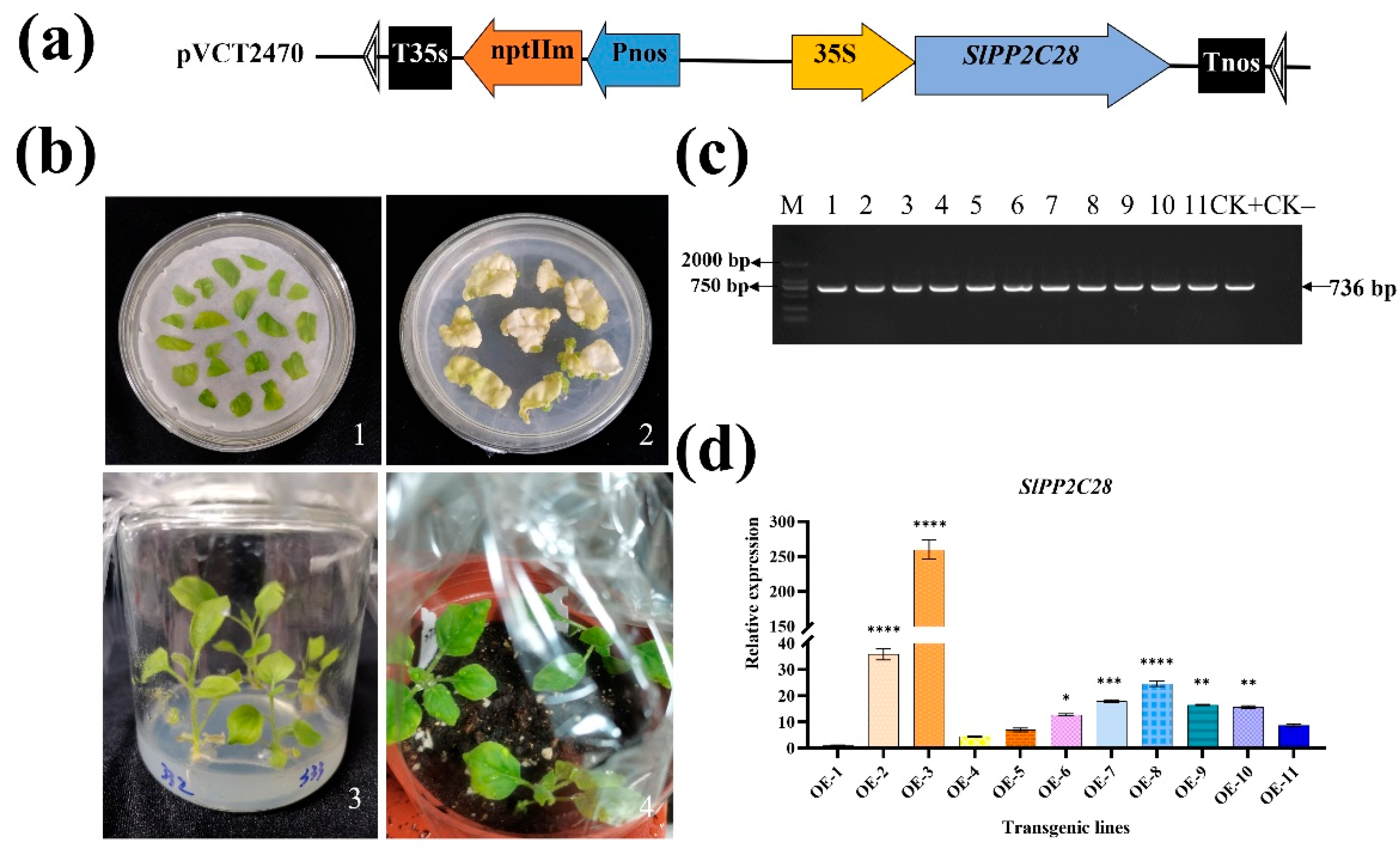
To further investigate the function of the SlPP2C28 gene, we cloned and analyzed this gene, constructed its overexpression vector, and performed genetic transformation experiments. Sequence analysis confirmed that the coding region of the modified SlPP2C28 gene spans 1221 bp and encodes a protein of 406 amino acids with a molecular weight of 44.68 kDa. Among these amino acids, 54 are strongly basic, 59 are strongly acidic, 123 are hydrophobic, and 108 are polar. The primary structure of the pVCT2470-SlPP2C28 vector is presented in Figure 2a. Plasmids extracted from vector-positive bacterial colonies were verified by restriction enzyme digestion. Furthermore, the sequencing results provided by Wuhan Jinkairui Bioengineering Co., Ltd. (Wuhan, China, https://www.genecreate.cn/ (accessed on 17 June 2025)) corroborated the accuracy of the pVCT2470-SlPP2C28 vector construction.
In total, 30 rooted plants were successfully obtained via Agrobacterium-mediated transformation (Figure 2b). PCR amplification detected a fragment of approximately 736 bp in 11 plants (Figure 2c), confirming the successful acquisition of 11 SlPP2C28 overexpression transgenic lines. Quantitative real-time PCR analysis was performed on the T0 generation of these transgenic lines, with the transgenic line OE-1 (exhibiting the lowest expression level) serving as the control. The relative expression levels in each transgenic line are presented in Figure 2d. Ultimately, the two lines with the highest expression levels (OE-2 and OE-3, exhibiting expression levels 35.79- and 259.71-fold higher than that of OE-1, respectively) were chosen for subsequent disease resistance assays.

3.4. SlPP2C28-Overexpressing Improved Transgenic Tobacco Resistance to R. solanacearum Inoculation

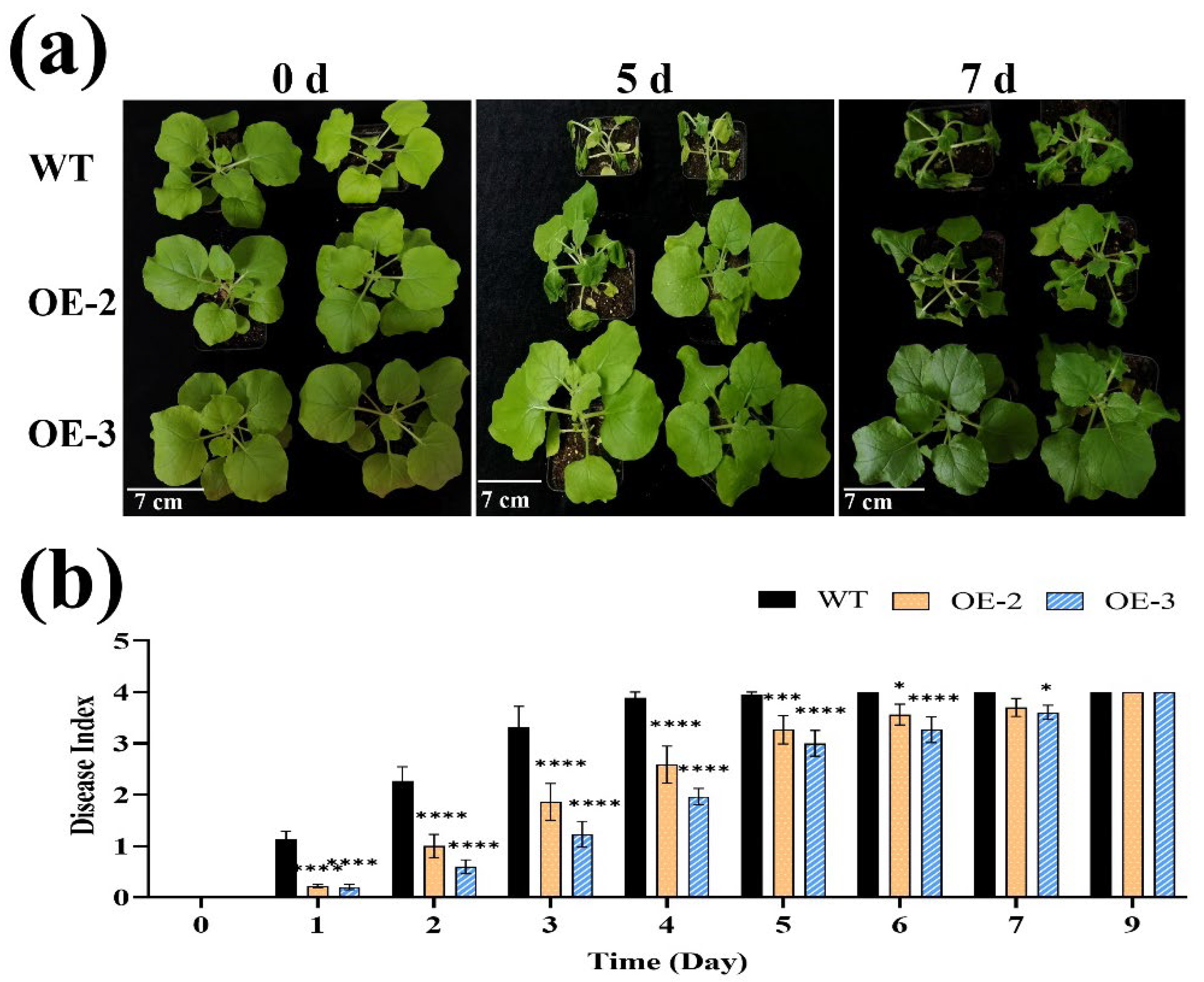
To investigate the function of genes, we inoculated WT (control) and transgenic tobacco plants with R. solanacearum. Following inoculation, all plants exhibited wilted leaves, with the degree of wilting progressively increasing over time. Among the transgenic lines, OE-3 demonstrated the best growth status, showing slightly reduced disease severity compared to OE-2 (Figure 3a). On the sixth day post-inoculation, the disease index of WT plants reached 4.00, whereas the transgenic lines OE-2 and OE-3 remained partially healthy, with disease index of 3.5 and 3.27, respectively, both significantly lower than that of the control. On the ninth day, the disease index of all plants converged to 4.00 (Figure 3b). Notably, the disease onset time in the transgenic lines was delayed relative to the WT, and the disease severity was milder. These findings suggest that overexpression of the SlPP2C28 gene in transgenic plants enhances their tolerance to bacterial wilt.

3.5. Overexpression of SlPP2C28 Enhanced Transgenic Stem Tolerance to R. solanacearum Infection

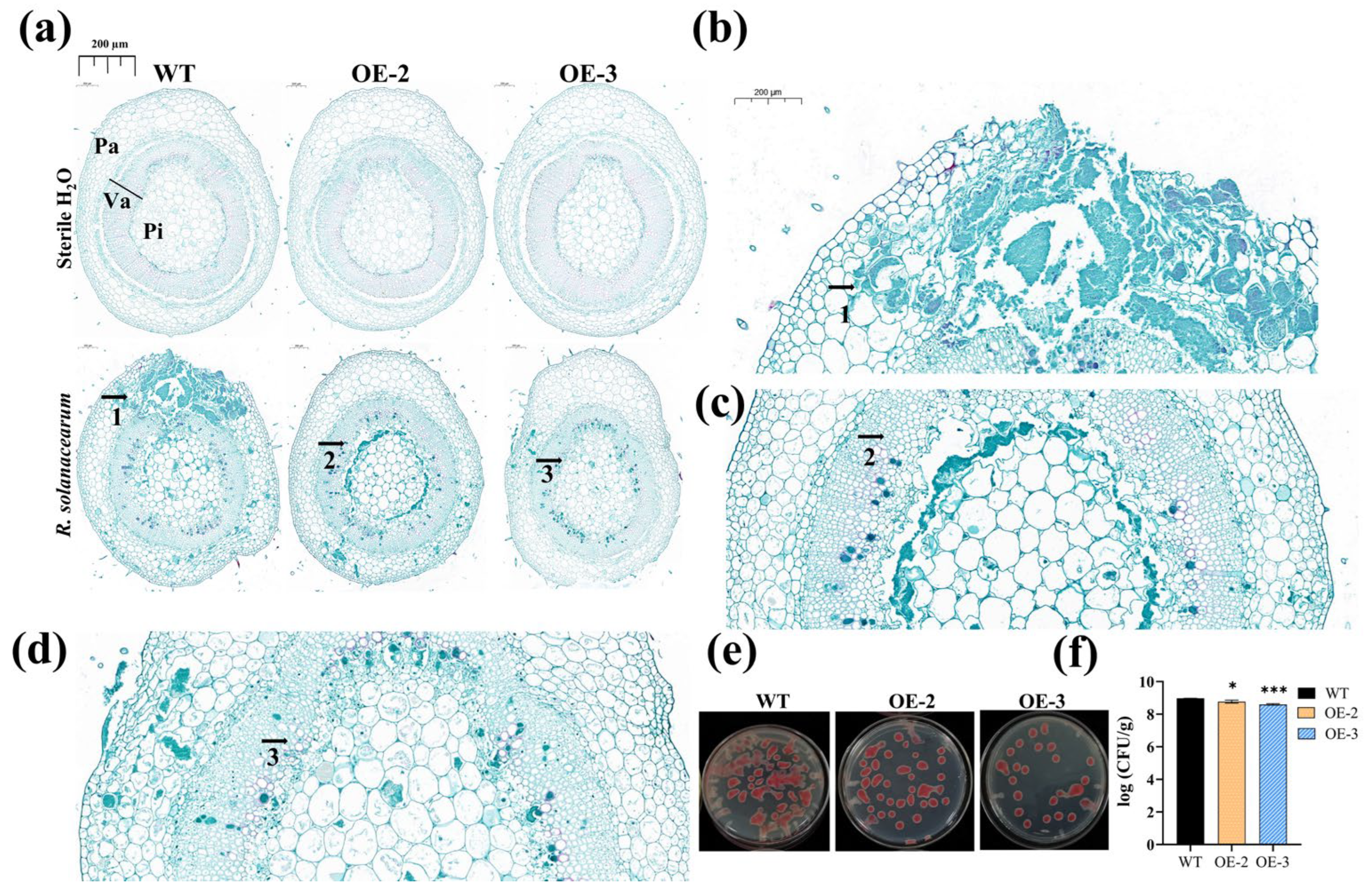
Bacterial wilt is a plant disease caused by bacteria that primarily affects the vascular systems of plants, leading to severe wilting and tissue damage. To further investigate the presence of R. solanacearum in the stems of both WT plants and those overexpressing SlPP2C28, we conducted a detailed analysis. Using sterile water as a negative control, the cross-section of a WT stem inoculated with R. solanacearum exhibited extensive destruction of vascular tissues, with significant blockage of xylem vessels. In contrast, the transgenic stems showed markedly reduced damage (Figure 4a). Additionally, the amount of R. solanacearum in stem tissues were statistically analyzed post-inoculation. Compared to WT plants, the colony-forming units (CFUs) on the medium from SlPP2C28 OE-2 and OE-3 transgenic lines were significantly lower (Figure 4b,c). Specifically, on Day 5, the bacterial load in WT stem was 9.42 × 10⁸ CFU/g, whereas the CFU counts for OE-2 and OE-3 were 5.99 × 10⁸ and 3.98 × 10⁸ CFU/g, respectively, both showing statistically significant reductions (Figure 4c). Therefore, the overexpression of SlPP2C28 in transgenic tobacco enhances the tolerance of stems to R. solanacearum.

3.6. Overexpression of SlPP2C28 Enhanced the Disease Tolerance of Transgenic Leaves by ROS Scavenging

After inoculating the leaves of N. benthamiana with the pathogen, the transgenic lines OE-2 and OE-3 exhibited significantly reduced leaf damage compared to WT (Figure 5a). Following a 24 h inoculation of tobacco plant leaves with R. solanacearum, DAB and NBT histochemical staining were conducted to detect the accumulation of H2O2 and O2 within the leaves (Figure 5b,c). The deeper the color intensity at the injection site on the blade, the higher the accumulation of H2O2 and O2, which correlates with greater damage caused by R. solanacearum. The experimental results demonstrated that there was no significant difference in the accumulation of H2O2 and O2 in the leaves of all tobacco plants in the control group after inoculation with sterile water. In comparison to WT, the injection sites on the blade of OE-2 and OE-3 displayed lighter coloration (Figure 5b,c), indicating that the overexpression of SlPP2C28 effectively reduced the accumulation of H2O2 and O2 in the transgenic leaves. At 0, 24, and 48 h post-inoculation, the expression levels of SlPP2C28 in OE-3 leaves reached highly significant values of 34,878.78, 24,479.12, and 10,566.59, respectively. Notably, the SlPP2C28 expression level in OE-2 peaked at 24 h, reaching 15,339.08 (Figure 5d). Therefore, overexpression of SlPP2C28 in transgenic tobacco enhances the tolerance of leaves to R. solanacearum.

3.7. Overexpression of SlPP2C28 Increased the Expression of Defense Enzyme-Related Genes and Enhanced the Activity of Defense-Related Enzymes in Transgenic Tobacco

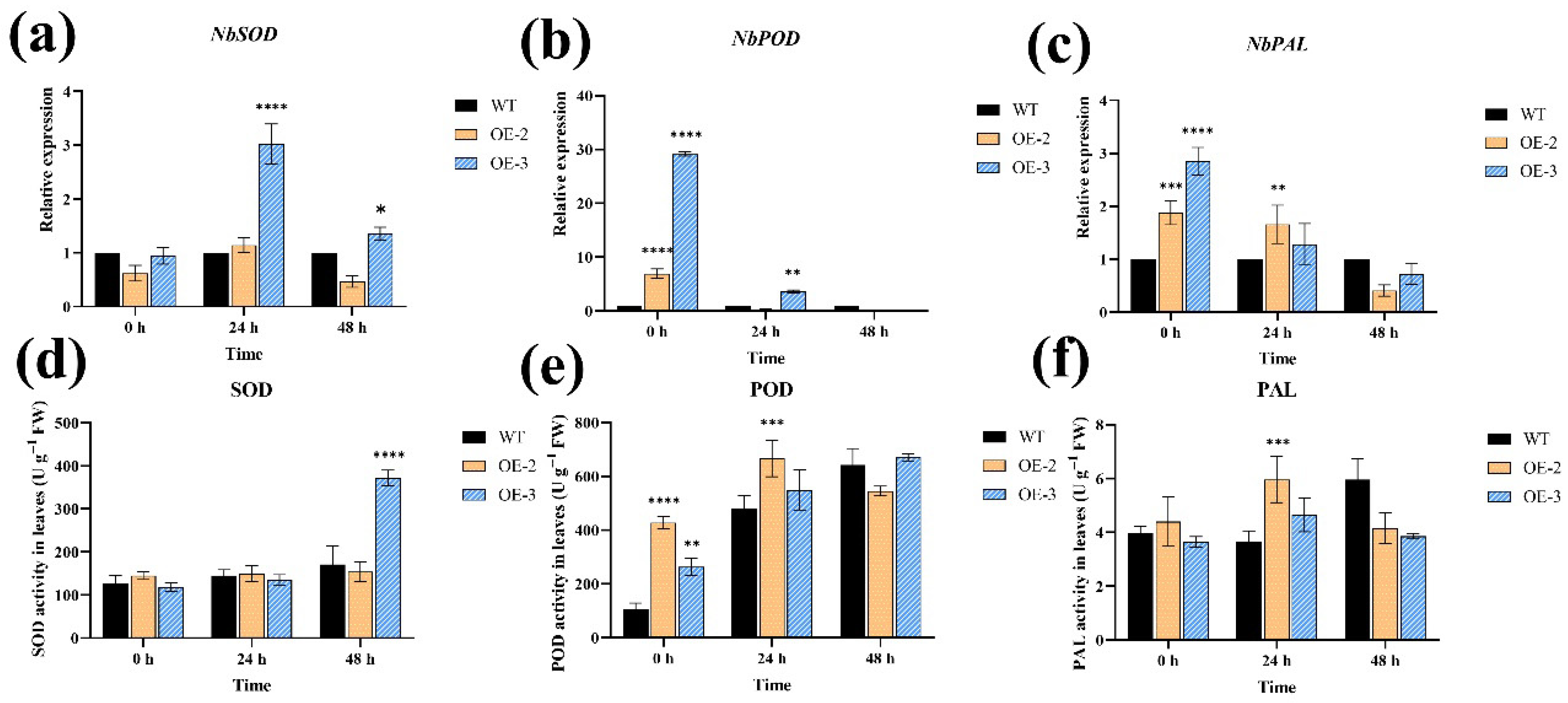
Defense-related enzymes, including SOD, POD, and PAL, play a critical role in plant resistance to biotic stress [19,23,42]. By using the expression levels of defense enzyme-related genes in the WT as a control, at 24 and 48 h post-inoculation, the relative expression levels of NbSOD in OE-3 were significantly elevated, reaching 3.02-fold and 1.36-fold compared to the WT, respectively. This suggests that NbSOD exhibited an initial increase followed by a decrease in expression (Figure 6a). For NbPOD, the expression levels in OE-2 and OE-3 were markedly higher at 0 h, being 29.15-fold and 6.96-fold greater than those in the WT, respectively, with significant differences observed. At 24 h, the NbPOD expression level in OE-3 remained significantly different from the WT, and between 0 and 48 h, it demonstrated a consistent decreasing trend (Figure 6b). Regarding NbPAL, its expression in tobacco plants decreased over time. Compared to the WT, the expression of NbPAL in OE-2 was significantly higher at 0 and 24 h, while in OE-3, it was only significantly elevated at 0 h, reaching 2.52-fold of the WT level (Figure 6c). These experimental results indicate that, upon inoculation with R. solanacearum, the genes responsible for synthesizing defense-related enzymes are activated and expressed.
In terms of enzyme activity, compared to 0 h, SOD levels in both the WT and transgenic line OE-3 exhibited an increasing trend. Notably, OE-3 reached the highest SOD value of 371.82 U/g at 48 h, which was significantly different from that of WT (Figure 6d). The POD activity in WT and the transgenic line OE-3 increased over time, whereas the POD activity in OE-2 initially increased and then decreased. At 0 h, the differences between OE-2 and OE-3 were significant, with POD values reaching 428.02 and 264.42 U/g, respectively. At 24 h, the POD value of OE-3 peaked at 666.60 U/g, which was significantly higher than that of WT (Figure 6e). During the 0–48 h period, the PAL activity in the transgenic lines OE-2 and OE-3 first increased and then decreased. At 24 h, the PAL value of OE-2 reached its maximum 5.96 U/g, which was significantly different from that of WT (Figure 6f). In summary, the changes in defense-related enzymes in tobacco plants were consistent with the tissue-staining results. At 24 h, the overall content of defense-related enzymes was higher in the transgenic lines than that of the WT, indicating that overexpression of SlPP2C28 enhances the protective function of transgenic plants against leaf damage.

3.8. Overexpression of SlPP2C28 Increased Expression of Pathogenesis-Related Genes in Transgenic Tobacco

We further investigated the expression of pathogenesis-related genes. Compared to the WT, the relative expression level of NbPR1a in OE-3 was significantly higher at 0 h, reaching 12.96 times that of the WT (Figure 7a). The relative expression level of NbPR2 in OE-2 was significantly elevated at 0 h, whereas in OE-3, significant increases were observed at 0, 24, and 48 h, with the highest level at 0 h being 8.34 times that of the WT (Figure 7b). For NbPR4, both OE-2 and OE-3 showed significant increases at 0 and 24 h, with the highest expression level in OE-3 reaching 4.70 times that of the WT (Figure 7c). Regarding NbPR10, OE-3 exhibited significant increases at 0, 24, and 48 h, while OE-2 showed significant increases only at 0 h and 24 h. The relative expression levels of this gene in the transgenic lines were the highest at 0 h, being 1.97 times (OE-2) and 2.66 times (OE-3) that of the WT, respectively (Figure 7d). In summary, the trend of pathogenesis-related gene expression changes in the transgenic lines was generally consistent, with overall levels higher than those in the WT. These results indicate that the overexpression of SlPP2C28 promotes the expression of pathogenesis-related genes, thereby enhancing the tolerance of transgenic tobacco to R. solanacearum.

4. Discussion

Previous studies have demonstrated that members of plant protein phosphatase PP2C family play essential roles in the growth and development of various plants, including Arabidopsis, rice, tomato, and tobacco [37]. The PP2C gene has been reported to play a significant role in the immune response of tomato [35]. Tomato PP2C is upregulated in tomato plants infected with pathogens [43,44]. Following inoculation with R. solanacearum, the expression of the PP2C gene is significantly increased, potentially contributing to resistance against R. solanacearum [40]. In this study, we further determined that SlPP2C28 was significantly upregulated in the commercial tomato species resistant to bacterial wilt (Figure 1). Firstly, we found that silencing of SlPP2C28 reduced tomato resistance to R. solanacearum. Additionally, the gene was overexpressed in tobacco under the control of the CaMV 35S promoter. This confirmed that SlPP2C28 confers tolerance to bacterial wilt (Figure 3a,b), thereby suggesting that this gene could serve as a key candidate for enhancing bacterial wilt resistance in future tomato breeding programs.
ROS are one of the earliest cellular responses to pathogen infection [45], and are often activated during plant defense responses [46]. Excessive accumulation of ROS can cause oxidative damage in plants, which can be visualized using DAB and NBT staining [47]. Our results demonstrate that overexpression of the SlPP2C28 gene in N. benthamiana enhances the ability of transgenic plant leaves to mitigate ROS-induced damage (Figure 5b,c) and improves their capacity to scavenge ROS, thereby reducing the detrimental effects of R. solanacearum on transgenic tobacco leaves (Figure 5a). This phenomenon may be attributed to the increased content of SOD and POD in transgenic plants (Figure 6d–f), findings that align with previous studies [48]. To counteract ROS-induced damage, plants often enhance their resistance to pathogens by increasing the activity of defense-related enzymes and upregulating the expression of defense enzyme-related genes. For instance, resistant plants exhibit elevated SOD activity and SOD expression levels [49,50,51]. POD is involved in a wide range of physiological processes [52,53,54,55,56,57], and plays a critical role in stress response [57,58]. Overexpression of the POD gene not only enhances stress tolerance in transgenic plants [59] but also increases resistance to bacterial diseases [60]. PAL can serve as a marker for inducing plant disease resistance [61] and plays a crucial role in plant defense against biotic stress. Studies have shown that PAL genes in transgenic plants are upregulated upon pathogen induction [62,63], which enhances PAL enzyme activity and significantly improves pathogens resistance [64]. In this study, the expression levels of NbSOD, NbPOD, and NbPAL in the transgenic lines were higher than those of WT (Figure 6a–c) following the overexpression of SlPP2C28 in N. benthamiana. This increase in defense enzyme-related genes expression led to elevated levels of SOD, POD, and PAL enzymes (Figure 6d–f), suggesting that SlPP2C28 promotes the protective effect of defense-related enzymes in SlPP2C28-overexpressing transgenic tobacco, thereby mitigating ROS-induced damage and reducing sensitivity to R. solanacearum. These findings align with previous reports [48]. In addition, we found that increased ROS damage and decreased expression of defense enzyme-related genes in SlPP2C28-silenced plants indicate that their resistance to bacterial wilt was reduced (Figure S3c,d).
PP2C is emerging as a critical component in plant stress-signal transduction [65], regulating defense responses linked to the expression of pathogenesis-related proteins PR1, PR2, and PR3 [66]. Signal transduction pathways activated by defense-related proteins are frequently associated with increased expression of disease resistance-related genes, including those encoding PR proteins [67]. In our study, we observed that overexpression of the SlPP2C28 gene significantly enhanced the expression levels of NbPR1a, NbPR2, NbPR4, and NbPR10 genes in transgenic plants (Figure 7a–d), whereas SlPP2C28-silenced plants showed significantly lower expression of pathogenesis-related genes (Figure S3e). The accumulation of PR-1 contributes to reducing the plant disease index and bacterial load, thereby improving plants’ tolerance to pathogenic bacteria or inhibitory effects on pathogens [68,69,70,71]. PR10 also plays a crucial role in plant resistance to biological stresses [72], and reduced expression of the PR10 gene markedly compromises the antimicrobial capacity of plants [73]. Similarly, the overexpression of SlPP2C28 in transgenic tobacco enhances tolerance to R. solanacearum by upregulating the expression of pathogenesis-related genes (Figure 7a–d).
In summary, silencing SlPP2C28 reduced tomato resistance to R. solanacearum (Figure S3a); however, the overexpression of SlPP2C28 in transgenic tobacco plants mitigated ROS-induced damage (Figure 5b,c), decreased the disease index (Figure 3b), postponed the onset of symptoms (Figure 3b), reduced wilting severity (Figure 3a), and lowered bacterial colonization levels in plants (Figure 4b,c). These effects were achieved by activating the defense-related enzyme system (Figure 6a–f) and promoting the upregulation of pathogenesis-related genes (Figure 7a–d). Additionally, the overexpression alleviated structural damage to stems and leaves (Figure 4a and Figure 5a), thereby enhancing the tolerance of transgenic plants to R. solanacearum infection. These findings elucidate some of the resistance mechanisms mediated by SIPP2C28 in combating plant bacterial wilt, providing a foundation for further theoretical studies on the bacterial wilt resistance of this gene. Nevertheless, the precise molecular mechanisms underlying its disease resistance require additional in-depth investigation.

5. Conclusions

This study demonstrates that silencing the SIPP2C28 gene reduces plant resistance to bacterial wilt through VIGS technology, while overexpression of SIPP2C28 enhances disease resistance in transgenic plants, indicating that this gene could improve disease resistance in plants.

Supplementary Materials

The following supporting information can be downloaded at: https://www.mdpi.com/article/10.3390/horticulturae11080937/s1, Figure S1: SlPP2C28 sequence in the pVCT2470-SlPP2C28 vector; Figure S2: Structural diagram of the tobacco overexpression vector pVCT2470-SlPP2C28; Figure S3: Effect of SlPP2C28 silencing on the resistance of tomato to bacterial wilt; Table S1: Primers used in this study.

Author Contributions

Experimental design and conception, L.N. and X.Z.; experiment execution, L.N. and Y.Q.; data analysis, L.N., M.W., J.Q., D.T. and L.C.; writing—original draft, L.N.; writing—review and editing, X.Z.; funding acquisition, Y.Q., J.L., Y.P., L.W. and X.Z. All authors have read and agreed to the published version of the manuscript.

Funding

This research was funded by grants from the Chongqing Graduate Student Research Innovation Project (No. CYB22135), the National Natural Science Foundation of China (No. 31872119), and the Fundamental Research Funds for the Central Universities (No. SWU-KQ22041/7110100866).

Data Availability Statement

The data that support the results are included in this article and its Supplementary Materials.

Acknowledgments

We thank Wei Ding (Southwest University) for providing R. solanacearum strain GMI1000 and CQPS-1.

Conflicts of Interest

The authors declare no conflicts of interest.

References

  1. Salanoubat, M.; Genin, S.; Artiguenave, F.; Gouzy, J.; Mangenot, S.; Arlat, M.; Billault, A.; Brottier, P.; Camus, J.C.; Cattolico, L.; et al. Genome sequence of the plant pathogen Ralstonia solanacearum. Nature 2002, 415, 497–502. [Google Scholar] [CrossRef]
  2. Genin, S. Molecular traits controlling host range and adaptation to plants in Ralstonia solanacearum. New Phytol. 2010, 187, 920–928. [Google Scholar] [CrossRef] [PubMed]
  3. Peeters, N.; Guidot, A.; Vailleau, F.; Valls, M. Ralstonia solanacearum, a widespread bacterial plant pathogen in the post-genomic era. Mol. Plant Pathol. 2013, 14, 651–662. [Google Scholar] [CrossRef] [PubMed]
  4. Mansfield, J.; Genin, S.; Magori, S.; Citovsky, V.; Sriariyanum, M.; Ronald, P.; Dow, M.; Verdier, V.; Beer, S.V.; Machado, M.A.; et al. Top 10 plant pathogenic bacteria in molecular plant pathology. Mol. Plant Pathol. 2012, 13, 614–629. [Google Scholar] [CrossRef] [PubMed]
  5. Lowe-Power, T.M.; Khokhani, D.; Allen, C. How Ralstonia solanacearum exploits and thrives in the flowing plant xylem environment. Trends Microbiol. 2018, 26, 929–942. [Google Scholar] [CrossRef]
  6. Jiang, G.; Wei, Z.; Xu, J.; Chen, H.; Zhang, Y.; She, X.; Macho, A.P.; Ding, W.; Liao, B. Bacterial Wilt in China: History, Current Status, and Future Perspectives. Front. Plant Sci. 2017, 8, 1549. [Google Scholar] [CrossRef]
  7. Halliwell, B.; Gutteridge, J.M. Free Radicals in Biology and Medicine; Oxford University Press: Oxford, UK, 2015. [Google Scholar]
  8. Lamb, C.; Dixon, R.A. The oxidative burst in plant disease resistance. Annu. Rev. Plant Physiol. Plant Mol. Biol. 1997, 48, 251–275. [Google Scholar] [CrossRef]
  9. Finkel, T. Signal transduction by reactive oxygen species. J. Cell Biol. 2011, 194, 7–15. [Google Scholar] [CrossRef]
  10. Grivennikova, V.G.; Vinogradov, A.D. Mitochondrial production of reactive oxygen species. Biochemistry 2013, 78, 1490–1511. [Google Scholar] [CrossRef]
  11. Galluzzi, L.; Morselli, E.; Kepp, O.; Vitale, I.; Rigoni, A.; Vacchelli, E.; Michaud, M.; Zischka, H.; Castedo, M.; Kroemer, G. Mitochondrial gateways to cancer. Mol. Asp. Med. 2010, 31, 1–20. [Google Scholar] [CrossRef]
  12. Turrens, J.F. Mitochondrial formation of reactive oxygen species. J. Physiol. 2003, 552, 335–344. [Google Scholar] [CrossRef] [PubMed]
  13. De Gara, L.; de Pinto, M.C.; Tommasi, F. The antioxidant systems vis-à-vis reactive oxygen species during plant–pathogen interaction. Plant Physiol. Biochem. 2003, 41, 863–870. [Google Scholar] [CrossRef]
  14. Allen, R.D. Dissection of oxidative stress tolerance using transgenic plants. Plant Physiol. 1995, 107, 1049–1054. [Google Scholar] [CrossRef] [PubMed]
  15. Asada, K. The water-water cycle in chloroplasts: Scavenging of active oxygens and dissipation of excess photons. Annu. Rev. Plant Physiol. Plant Mol. Biol. 1999, 50, 601–639. [Google Scholar] [CrossRef] [PubMed]
  16. Palma, J.M.; Sandauo, L.M.; Del Río, L.A. Manganese superoxide dismutase and higher plant chloroplasts: A reappraisal of a controverted cellular localization. J. Plant Physiol. 1986, 125, 427–439. [Google Scholar] [CrossRef]
  17. Hernaīndez, J.; Ferrer, M.; Jimeīnez, A.; Ros-Barceloī, A.; Sevilla, F. Antioxidant systems and O2·/H2O2 production in the apoplast of Pea leaves. Its relation with salt-induced necrotic lesions in minor veins. Plant Physiol. 2001, 127, 817–831. [Google Scholar] [CrossRef]
  18. Passardi, F.; Cosio, C.; Penel, C.; Dunand, C. Peroxidases have more functions than a Swiss army knife. Plant Cell Rep. 2005, 24, 255–265. [Google Scholar] [CrossRef]
  19. Almagro, L.; Gómez Ros, L.V.; Belchi-Navarro, S.; Bru, R.; Ros Barceló, A.; Pedreño, M.A. Class III peroxidases in plant defence reactions. J. Exp. Bot. 2009, 60, 377–390. [Google Scholar] [CrossRef]
  20. Tognolli, M.; Penel, C.; Greppin, H.; Simon, P. Analysis and expression of the class III peroxidase large gene family in Arabidopsis thaliana. Gene 2002, 288, 129–138. [Google Scholar] [CrossRef]
  21. Passardi, F.; Longet, D.; Penel, C.; Dunand, C. The class III peroxidase multigenic family in rice and its evolution in land plants. Phytochemistry 2004, 65, 1879–1893. [Google Scholar] [CrossRef]
  22. Wang, Y.; Wang, Q.; Zhao, Y.; Han, G.; Zhu, S. Systematic analysis of maize class III peroxidase gene family reveals a conserved subfamily involved in abiotic stress response. Gene 2015, 566, 95–108. [Google Scholar] [CrossRef]
  23. Mariutto, M.; Duby, F.; Adam, A.; Bureau, C.; Fauconnier, M.-L.; Ongena, M.; Thonart, P.; Dommes, J. The elicitation of a systemic resistance by Pseudomonas putida BTP1 in tomato involves the stimulation of two lipoxygenase isoforms. BMC Plant Biol. 2011, 11, 29. [Google Scholar] [CrossRef]
  24. Dixon, R.A.; Paiva, N.L. Stress-induced phenylpropanoid metabolism. Plant Cell 1995, 7, 1085. [Google Scholar] [CrossRef] [PubMed]
  25. Yedidia, I.; Shoresh, M.; Kerem, Z.; Benhamou, N.; Kapulnik, Y.; Chet, I. Concomitant induction of systemic resistance to Pseudomonas syringae pv. lachrymans in cucumber by Trichoderma asperellum (T-203) and accumulation of phytoalexins. Appl. Environ. Microbiol. 2003, 69, 7343–7353. [Google Scholar] [CrossRef] [PubMed]
  26. Fritig, B.; Heitz, T.; Legrand, M. Antimicrobial proteins in induced plant defense. Curr. Opin. Immunol. 1998, 10, 16–22. [Google Scholar] [CrossRef] [PubMed]
  27. Durrant, W.E.; Dong, X. Systemic acquired resistance. J. Phytopathol. 2004, 42, 185–209. [Google Scholar] [CrossRef]
  28. van Loon, L.C.; Pierpoint, W.S.; Boller, T.; Conejero, V. Recommendations for naming plant pathogenesis-related proteins. Plant Mol. Biol. Rep. 1994, 12, 245–264. [Google Scholar] [CrossRef]
  29. Kerk, D.; Templeton, G.; Moorhead, G.B. Evolutionary radiation pattern of novel protein phosphatases revealed by analysis of protein data from the completely sequenced genomes of humans, green algae, and higher plants. Plant Physiol. 2008, 146, 323–324. [Google Scholar] [CrossRef]
  30. Singh, A.; Giri, J.; Kapoor, S.; Tyagi, A.K.; Pandey, G.K. Protein phosphatase complement in rice: Genome-wide identification and transcriptional analysis under abiotic stress conditions and reproductive development. BMC Genomics 2010, 11, 435. [Google Scholar] [CrossRef]
  31. Singh, A.; Pandey, A.; Srivastava, A.K.; Tran, L.-S.P.; Pandey, G.K. Plant protein phosphatases 2C: From genomic diversity to functional multiplicity and importance in stress management. Crit. Rev. Biotechnol. 2016, 36, 1023–1035. [Google Scholar] [CrossRef]
  32. Hu, X.; Song, F.; Zheng, Z. Molecular characterization and expression analysis of a rice protein phosphatase 2C gene, OsBIPP2C1, and overexpression in transgenic tobacco conferred enhanced disease resistance and abiotic tolerance. Physiol. Plant. 2006, 127, 225–236. [Google Scholar] [CrossRef]
  33. Hu, X.; Zhang, H.; Li, G.; Yang, Y.; Zheng, Z.; Song, F. Ectopic expression of a rice protein phosphatase 2C gene OsBIPP2C2 in tobacco improves disease resistance. Plant Cell Rep. 2009, 28, 985–995. [Google Scholar] [CrossRef]
  34. Akimoto-Tomiyama, C.; Tanabe, S.; Kajiwara, H.; Minami, E.; Ochiai, H. Loss of chloroplast-localized protein phosphatase 2Cs in Arabidopsis thaliana leads to enhancement of plant immunity and resistance to Xanthomonas campestris pv. campestris infection. Mol. Plant Pathol. 2018, 19, 1184–1195. [Google Scholar] [CrossRef] [PubMed]
  35. Sobol, G.; Chakraborty, J.; Martin, G.B.; Sessa, G. The emerging role of PP2C phosphatases in tomato immunity. Mol. Plant-Microbe Interact. 2022, 35, 737–747. [Google Scholar] [CrossRef] [PubMed]
  36. Shi, J.; Shui, D.; Su, S.; Xiong, Z.; Zai, W. Gene enrichment and co-expression analysis shed light on transcriptional responses to Ralstonia solanacearum in tomato. BMC Genomics 2023, 24, 159. [Google Scholar] [CrossRef] [PubMed]
  37. Qiu, J.; Ni, L.; Xia, X.; Chen, S.; Zhang, Y.; Lang, M.; Li, M.; Liu, B.; Pan, Y.; Li, J.; et al. Genome-wide analysis of the protein phosphatase 2C genes in Tomato. Genes 2022, 13, 604. [Google Scholar] [CrossRef]
  38. Zhang, Y.; Ni, L.; Chen, S.; Qin, Y.; Ding, X.; Li, J.; Pan, Y.; Zhang, X. Pterostilbene production of tomato transformed with resveratrol synthase and resveratrol O-methyltransferase genes. Plant Sci. 2022, 322, 111343. [Google Scholar] [CrossRef]
  39. Tang, Y.; Liu, Q.; Liu, Y.; Zhang, L.; Ding, W. Overexpression of NtPR-Q up-regulates multiple defense-related genes in Nicotiana tabacum and enhances plant resistance to Ralstonia solanacearum. Front. Plant Sci. 2017, 8, 1963. [Google Scholar] [CrossRef]
  40. Chen, N.; Shao, Q.; Lu, Q.; Li, X.; Gao, Y. Transcriptome analysis reveals differential transcription in tomato (Solanum lycopersicum) following inoculation with Ralstonia solanacearum. Sci. Rep. 2022, 12, 22137. [Google Scholar] [CrossRef]
  41. Liu, Q.; Liu, Y.; Tang, Y.; Chen, J.; Ding, W. Overexpression of NtWRKY50 Increases Resistance to Ralstonia solanacearum and Alters Salicylic Acid and Jasmonic Acid Production in Tobacco. Front. Plant Sci. 2017, 8, 1710. [Google Scholar] [CrossRef]
  42. Miller, A.-F. Superoxide dismutases: Ancient enzymes and new insights. FEBS Lett. 2012, 586, 585–595. [Google Scholar] [CrossRef]
  43. Rosli, H.G.; Zheng, Y.; Pombo, M.A.; Zhong, S.; Bombarely, A.; Fei, Z.; Collmer, A.; Martin, G.B. Transcriptomics-based screen for genes induced by flagellin and repressed by pathogen effectors identifies a cell wall-associated kinase involved in plant immunity. Genome Biol. 2013, 14, R139. [Google Scholar] [CrossRef]
  44. Zipfel, C.; Robatzek, S.; Navarro, L.; Oakeley, E.J.; Jones, J.D.G.; Felix, G.; Boller, T. Bacterial disease resistance in Arabidopsis through flagellin perception. Nature 2004, 428, 764–767. [Google Scholar] [CrossRef]
  45. Torres, M.A.; Jones, J.D.G.; Dangl, J.L. Reactive oxygen species signaling in response to pathogens. Plant Physiol. 2006, 141, 373–378. [Google Scholar] [CrossRef]
  46. Bellincampi, D.; Dipierro, N.; Salvi, G.; Cervone, F.; De Lorenzo, G. Extracellular H2O2 induced by oligogalacturonides is not involved in the inhibition of the auxin-regulated rolB gene expression in tobacco leaf explants. Plant Physiol. 2000, 122, 1379–1386. [Google Scholar] [CrossRef]
  47. Zheng, Y.; Li, M.; Liu, B.; Qin, Y.; Li, J.; Pan, Y.; Zhang, X. Effects of Phytochelatin-like Gene on the Resistance and Enrichment of Cd2+ in Tobacco. Int. J. Mol. Sci. 2022, 23, 16167. [Google Scholar] [CrossRef] [PubMed]
  48. Chen, C.; Wang, Y.; Wu, K.; Ding, Y.; Tang, M.; Zhang, X.; Pan, Y.; Wu, L.; Su, C.; Hong, Z.; et al. The DnaJ1 heat shock protein interacts with the flavanone 3-hydroxylase-like protein F3HL to synergistically enhance drought tolerance by scavenging reactive oxygen species in tomato. Plant J. 2025, 121, e70097. [Google Scholar] [CrossRef] [PubMed]
  49. Montalbini, P.; Buonaurio, R. Effect of tobacco mosaic virus infection of levels of soluble superoxide dismutase (SOD) in nicotiana tabacum and nicotiana glutinosa leaves. Plant Sci. 1986, 47, 135–143. [Google Scholar] [CrossRef]
  50. Buonaurio, R.; Della Torre, G.; Montalbini, P. Soluble superoxide dismutase (SOD) in susceptible and resistant host-parasite complexes of Phaseolus vulgaris and Uromyces phaseoli. Physiol. Mol. Plant Pathol. 1987, 31, 173–184. [Google Scholar] [CrossRef]
  51. Lu, W.; Duanmu, H.; Qiao, Y.; Jin, X.; Yu, Y.; Yu, L.; Chen, C. Genome-wide identification and characterization of the soybean SOD family during alkaline stress. PeerJ 2020, 8, e8457. [Google Scholar] [CrossRef]
  52. Welinder, K.G. Superfamily of plant, fungal and bacterial peroxidases. Curr. Opin. Struct. Biol. 1992, 2, 388–393. [Google Scholar] [CrossRef]
  53. Hiraga, S.; Yamamoto, K.; Ito, H.; Sasaki, K.; Matsui, H.; Honma, M.; Nagamura, Y.; Sasaki, T.; Ohashi, Y. Diverse expression profiles of 21 rice peroxidase genes. FEBS Lett. 2000, 471, 245–250. [Google Scholar] [CrossRef]
  54. González, A.M.; Marcel, T.C.; Kohutova, Z.; Stam, P.; van der Linden, C.G.; Niks, R.E.; Grebe, M. Peroxidase profiling reveals genetic linkage between peroxidase gene clusters and basal host and non-host resistance to rusts and mildew in barley. PLoS ONE 2010, 5, e10495. [Google Scholar] [CrossRef] [PubMed]
  55. Jin, J.; Hewezi, T.; Baum, T.J. Arabidopsis peroxidase AtPRX53 influences cell elongation and susceptibility to Heterodera schachtii. Plant Signal. Behav. 2011, 6, 1778–1786. [Google Scholar] [CrossRef] [PubMed]
  56. Shigeto, J.; Tsutsumi, Y. Diverse functions and reactions of class III peroxidases. New Phytol. 2016, 209, 1395–1402. [Google Scholar] [CrossRef] [PubMed]
  57. de Azevedo Neto, A.D.; Prisco, J.T.; Enéas-Filho, J.; de Abreu, C.E.B.; Gomes-Filho, E. Effect of salt stress on antioxidative enzymes and lipid peroxidation in leaves and roots of salt-tolerant and salt-sensitive maize genotypes. Environ. Exp. Bot. 2006, 56, 87–94. [Google Scholar] [CrossRef]
  58. Wei-Feng, X.; Wei-Ming, S.; Ueda, A.; Takabe, T. Mechanisms of salt tolerance in transgenic Arabidopsis thaliana carrying a peroxisomal ascorbate peroxidase gene from barley. Pedosphere 2008, 18, 486–495. [Google Scholar] [CrossRef]
  59. Coego, A.; Ramirez, V.; Ellul, P.; Mayda, E.; Vera, P. The H2O2-regulated Ep5C gene encodes a peroxidase required for bacterial speck susceptibility in tomato. Plant J. 2005, 42, 283–293. [Google Scholar] [CrossRef]
  60. Choi, H.W.; Kim, Y.J.; Lee, S.C.; Hong, J.K.; Hwang, B.K. Hydrogen peroxide generation by the pepper extracellular peroxidase CaPO2 activates local and systemic cell death and defense response to bacterial pathogens. Plant Physiol. 2007, 145, 890–904. [Google Scholar] [CrossRef]
  61. Zhang, S.; Yang, X.; Sun, M.; Sun, F.; Deng, S.; Dong, H. Riboflavin-induced priming for pathogen defense in Arabidopsis thaliana. J. Integr. Plant Biol. 2009, 51, 167–174. [Google Scholar] [CrossRef]
  62. Pant, S.; Huang, Y. Genome-wide studies of PAL genes in sorghum and their responses to aphid infestation. Sci. Rep. 2022, 12, 22537. [Google Scholar] [CrossRef] [PubMed]
  63. Blanco, F.A.; Zanetti, M.E.; Daleo, G.R. Calcium-dependent Protein Kinases are Involved in Potato Signal Transduction in Response to Elicitors from the Oomycete Phytophthora infestans. J. Phytopathol. 2008, 156, 53–61. [Google Scholar] [CrossRef]
  64. Kim, D.S.; Hwang, B.K. An important role of the pepper phenylalanine ammonia-lyase gene (PAL1) in salicylic acid-dependent signalling of the defence response to microbial pathogens. J. Exp. Bot. 2014, 65, 2295–2306. [Google Scholar] [CrossRef] [PubMed]
  65. Schweighofer, A.; Hirt, H.; Meskiene, I. Plant PP2C phosphatases: Emerging functions in stress signaling. Trends Plant Sci. 2004, 9, 236–243. [Google Scholar] [CrossRef]
  66. Widjaja, I.; Lassowskat, I.; Bethke, G.; Eschen-Lippold, L.; Long, H.-H.; Naumann, K.; Dangl, J.L.; Scheel, D.; Lee, J. A protein phosphatase 2C, responsive to the bacterial effector AvrRpm1 but not to the AvrB effector, regulates defense responses in Arabidopsis. Plant J. 2010, 61, 249–258. [Google Scholar] [CrossRef]
  67. van Loon, L.; Rep, M.; Pieterse, C. Significance of inducible defense-related proteins in infected plants. Annu. Rev. Phytopathol. 2006, 44, 135–162. [Google Scholar] [CrossRef]
  68. Alexander, D.; Goodman, R.M.; Gut-Rella, M.; Glascock, C.; Weymann, K.; Friedrich, L.; Maddox, D.; Ahl-Goy, P.; Luntz, T.; Ward, E. Increased tolerance to two oomycete pathogens in transgenic tobacco expressing pathogenesis-related protein 1a. Proc. Natl. Acad. Sci. USA 1993, 90, 7327–7331. [Google Scholar] [CrossRef]
  69. Hugot, K.; Aimé, S.; Conrod, S.; Poupet, A.; Galiana, E. Developmental regulated mechanisms affect the ability of a fungal pathogen to infect and colonize tobacco leaves. Plant J. 1999, 20, 163–170. [Google Scholar] [CrossRef]
  70. Liu, F.; Wei, F.; Wang, L.; Liu, H.; Zhu, X.; Liang, Y. Riboflavin activates defense responses in tobacco and induces resistance against Phytophthora parasitica and Ralstonia solanacearum. Physiol. Mol. Plant Pathol. 2010, 74, 330–336. [Google Scholar] [CrossRef]
  71. Niderman, T.; Genetet, I.; Bruyere, T.; Gees, R.; Stintzi, A.; Legrand, M.; Fritig, B.; Mosinger, E. Pathogenesis-related PR-1 proteins are antifungal (isolation and characterization of three 14-kilodalton proteins of tomato and of a basic PR-1 of tobacco with inhibitory activity against Phytophthora infestans). Plant Physiol. 1995, 108, 17–27. [Google Scholar] [CrossRef]
  72. Suhaimi, A.H.; Rajendram, A.; Khaidizar, F.D.; Mir, P.; Pulido-Lucas, E.; Quirce, S.; Pedrosa, M.; Rodriguez-Perez, R.; Al-Idrus, A. Occurrences of allergenicity to banana pathogenesis-related-10 (PR10) protein variants. Food Funct. 2024, 15, 11715–11725. [Google Scholar] [CrossRef]
  73. Chen, Z.; Brown, R.L.; Damann, K.E.; Cleveland, T.E. PR10 expression in maize and its effect on host resistance against Aspergillus flavus infection and aflatoxin production. Mol. Plant Pathol. 2010, 11, 69–81. [Google Scholar] [CrossRef]
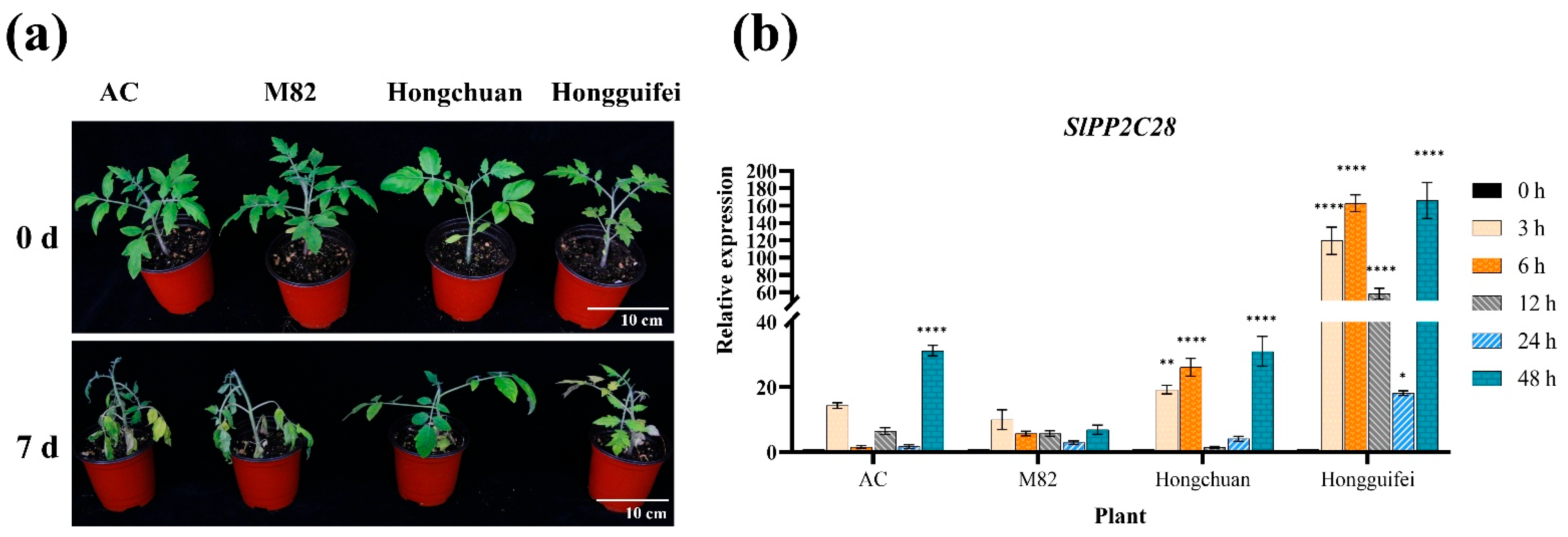
Figure 1. Expression of SlPP2C28 at different times under Ralstonia solanacearum infection treatment in different tomato varieties. (a) Disease status of four tomato varieties seven days after inoculation with R. solanacearum. 0 d: before treatment; 7 d: treatment of seven days. AC is a moderately resistant tomato cultivar, M82 is a susceptible tomato cultivar, and Hongchuan and Hongguifei are resistant tomato cultivars. Scale bars = 10 cm. (b) Expression levels of SlPP2C28 in the four tomato varieties at 0, 3, 6, 12, 24, and 48 h after inoculation with R. solanacearum. Three biological replicates were set with three technical replicates. Quantitative PCR results are expressed as mean ± standard deviation, and statistical analysis was performed using two-way ANOVA; α = 0.05, * p < 0.05, ** p < 0.01, **** p < 0.0001.
Horticulturae 11 00937 g001
Figure 2. Acquisition of transgenic lines expressing the SlPP2C28 gene. (a) Structure of the pVCT2470-SlPP2C28 vector. (b) Genetic transformation of Nicotiana benthamiana (1: precultured explants; 2: selected explants; 3: rooting cultivation; and 4: transplanting seedling of transgenic plants). (c) PCR of transgenic lines (M: 2000 bp DNA Marker; 1–11: transgenic lines; CK+: plasmid positive control; CK–: negative control). (d) Relative expression levels of transgenic lines (OE-1 to OE-11 represent 11 transgenic lines positive for SlPP2C28 expression). Three biological replicates were set with three technical replicates. The results are expressed as the mean ± standard deviation, and statistical analysis was conducted using one-way ANOVA; α = 0.05, * p < 0.05, ** p < 0.01, *** p < 0.001, **** p < 0.0001.
Horticulturae 11 00937 g002
Figure 3. Phenotypic and disease index of N. benthamiana after inoculation with R. solanacearum. (a) Comparison of seedling growth before and five and seven days after inoculation. WT: wild-type; OE-3 and OE-3: T1 generation transgenic lines; 0 d: before treatment; 5 d: treatment of five days; 7 d: treatment of seven days. Scale bars = 7 cm. (b) Disease indices for different growing days. Results are expressed as the mean ± standard deviation, and statistical analysis was performed using two-way ANOVA; α = 0.05, * p < 0.05, *** p < 0.001, **** p < 0.0001.
Horticulturae 11 00937 g003
Figure 4. Effect of SlPP2C28 overexpression on the stems of N. benthamiana infected with R. solanacearum. (a) Paraffin-embedded cross-sections of stems after five days of R. solanacearum infection. The first column shows the effects of sterile water (control) and the second column shows the inoculation treatment effects. The small black arrow and number indicates the damage caused by R. solanacearum. 1: WT inoculated with R. solanacearum; 2–3: transgenic lines inoculated with R. solanacearum. The pictures in (a) show the whole stem cross-sections, and the pictures in (bd) show the same sites after magnification. Pa: parenchyma, Va: vascular bundle, Pi: pith. Scale bars = 200 µm. (e) Bacterial growth in the stems of WT and transgenic lines after five days of R. solanacearum infection. (f) Bacterial content in the stems of WT and transgenic lines after five days of R. solanacearum infection. Results are expressed as the mean ± standard deviation, and statistical analysis was performed using one-way ANOVA; α = 0.05, * p < 0.05, *** p < 0.001. Three biological replicate were used in the experiment.
Horticulturae 11 00937 g004
Figure 5. Effect of SlPP2C28 overexpression on the leaves of N. benthamiana. (a) Bacterial growth in leaves infected with R. solanacearum after five days. (b) DAB staining (sterile water was injected into the left side of the leaves, while R. solanacearum was injected to the right side. The black arrow indicates the staining result at the injection site on the leaf blade). (c) NBT staining (same as described in panel (b)). Scale bars = 1 cm. (d) Expression levels of the SlPP2C28 gene in the leaves of different transgenic lines at various time-points. Results are expressed as the mean ± standard deviation, and the statistical analysis was performed using two-way ANOVA; α = 0.05, **** p < 0.0001. The experimental parts of (ac) used three biological replicates, and the experimental parts of (d) used three biological replicates; experiments were repeated three times.
Horticulturae 11 00937 g005
Figure 6. Effect of SlPP2C28 overexpression on defense-related enzyme of N. benthamiana. (a) Relative expression levels of NbSOD at different time-points after infection. (b) Relative expression levels of NbPOD at different time-points after infection. (c) Relative expression levels of NbPAL at different time-points after infection. (d) SOD enzyme activity at different time-points after infection. (e) POD enzyme activity at different time-points after infection. (f) PAL enzyme activity at different time-points after infection. The results are expressed as the mean ± standard deviation, and statistical analysis was performed using two-way ANOVA; α = 0.05, * p < 0.05, ** p < 0.01, *** p < 0.001, **** p < 0.0001.
Horticulturae 11 00937 g006
Figure 7. Effect of SlPP2C28 overexpression on the expression of pathogenesis-related genes of N. benthamiana. (a) Relative expression levels of NbPR1a gene at different time-points after infection. (b) Relative expression levels of NbPR2 gene at different time-points after infection. (c) Relative expression levels of NbPR4 gene at different time-points after infection. (d) Relative expression levels of NbPR10 gene at different time-points after infection. Three biological replicates were set with three technical replicates. The results are expressed as the mean ± standard deviation, and statistical analysis was performed using two-way ANOVA; α = 0.05, * p < 0.05, ** p < 0.01, **** p < 0.0001.
Horticulturae 11 00937 g007
Disclaimer/Publisher’s Note: The statements, opinions and data contained in all publications are solely those of the individual author(s) and contributor(s) and not of MDPI and/or the editor(s). MDPI and/or the editor(s) disclaim responsibility for any injury to people or property resulting from any ideas, methods, instructions or products referred to in the content.