Article

Comparative Metabolomic Analysis Reveals Tissue- and Species-Specific Differences in the Abundance of Dammarane-Type Ginsenosides in Three Panax Species

1
Yunnan Provincial Key Laboratory for Conservation and Utilization of In-Forest Resource, Southwest Forestry University, Kunming 650224, China
2
Key Laboratory for Forest Resources Conservation and Utilization in the Southwest Mountains of China, Ministry of Education, International Ecological Forestry Research Center of Kunming, Southwest Forestry University, Kunming 650224, China
3
Research Institute of Forest Policy and Information, Chinese Academy of Forestry, Beijing 100091, China
4
Forest Plant Department, Vietnam National University of Forestry, Xuan Mai, Ha Noi 13400, Vietnam
*
Authors to whom correspondence should be addressed.
These authors contributed equally to this work.
Horticulturae 2025, 11(8), 916; https://doi.org/10.3390/horticulturae11080916 (registering DOI)
Submission received: 16 April 2025 / Revised: 6 June 2025 / Accepted: 15 July 2025 / Published: 5 August 2025
(This article belongs to the Section Medicinals, Herbs, and Specialty Crops)

Abstract

The genus Panax contains traditional herbs that have been widely used in traditional medicine. The active constituents, collectively known as ginsenosides, are well characterized in the most representative species, P. notoginseng. However, the major bioactive chemical constituents of P. stipuleanatus together with P. vietnamensis are relatively less studied. In this study, an untargeted metabolomic analysis was performed in P. notoginseng, P. stipuleanatus, and P. vietnamensis using root and leaf organs. Further metabolomic differences in P. stipuleanatus were compared with those of the two most prevalent species. The analysis results revealed tissue-specific qualitative and quantitative metabolic differences in each species. Several differentially accumulated metabolites were enriched in the biosynthesis of secondary metabolites, including the biosynthesis of ginsenosides I. The ginsenosides Rb1, Rf, Rg1, Rh1, Rh8, and notoginsenosides E, M, and N had a higher abundance level in the roots of both P. notoginseng and P. vietnamensis. In P. stipuleanatus, the accumulation of potentially important ginsenosides is mainly found in the leaf. In particular, the dammarane-type ginsenosides Rb3, Rb1, Mx, and F2 as well as the notoginsenosides A, Fe, Fa, Fd, L, and N were identified to have a higher accumulation in the leaf. The strong positive correlation network of different ginsenosides probably enhanced secondary metabolism in each species. The comparative analysis revealed a significant differential accumulation of metabolites in the leaves of both species. The various compounds of dammarane-type ginsenoside, such as Rb1, Rg1, Rg6, Rh8, Rh10, Rh14, and majoroside F2, had a significantly higher concentration level in the leaves of P. stipuleanatus. In addition, several notoginsenoside compounds such as A, R1, Fe, Fd, and Ft1 showed a higher abundance in the leaf. These results show that the abundance level of major ginsenosides is significant in P. stipuleanatus and provides an important platform to improve the ginsenoside quality of Panax species.

1. Introduction

The Panax genus of the Araliaceae family is a publicly recognized valuable traditional medicine resource and includes more than 15 species in different countries worldwide [1]. Three major Panax species are P. ginseng (Korean ginseng), P. quinquefolius (American ginseng), and P. notoginseng (Sanchi ginseng) which are popularly known and have a very high pharmaceutical and economic value [2,3]. Many other species of this genus especially P. japonicus, P. zingiberensis, and P. stipuleanatus have shown wider phytochemical and pharmacological activities [4]. It has been widely cultivated in China, Korea, Japan, and Western countries. However, Korea and China are the largest importers and exporters in the world. Most diploid species of Panax are cultivated in warm regions from southern China to southern Vietnam. In contrast, the tetraploid species are widely distributed in northeastern Asia and northeastern America [5]. The Panax plant, commonly known as ginseng, contains active constituents called ginsenosides. Ginsenosides are regarded as the “king” of herbal groups used for human health and have medicinal potential for anti-inflammatory, antioxidant, antimicrobial, antibacterial, anti-obesity, antiviral, and antifungal activities [6,7,8]. The various species of Panax have been widely used in traditional Chinese medicine to treat hypertension, stress, and thrombosis [9], as well as to cure various neurological disorders such as Alzheimer’s disease, Parkinson’s disease, and Huntington’s disease [10,11,12,13]. In addition, various in vivo and clinical studies have shown that ginseng can inhibit reactive oxygen species production, improve blood circulation, and help regulate total cholesterol, triglycerides, and low-density lipoprotein cholesterol l [14,15]. Ginsenosides and their derivatives have significant inhibitory effects on skin, prostate, and colon cancer [16,17]. In China, six native varieties and one introduced variety of P. quinquefolius have been widely cultivated. Five are officially registered as medicinal plants. Among them, P. ginseng C.A. Meyer (Chinese ginseng, Renshen) has the largest market value, followed by P. quinquefolius [18,19]. With the contribution of many research groups, phytochemical purification, and structural identification techniques, P. notoginseng constituents, their genetic mechanism, and pharmacological activities have been thoroughly investigated by many researchers [20,21]. However, the characterization of the chemical components of other species such as P. japonicus, P. zingiberensis, and P. stipuleanatus has not been studied in detail. Such results can only provide a theoretical basis for correct use and will open up new horizons for the pharmacological and food industries in the future.
Phytochemically, different species of Panax have diverse properties, all essentially comprising ginsenosides, polysaccharides, flavonoids, proteins, amino acids, and organic acids. However, various ginsenoside compounds, also known as saponins, are the most important active ingredients of Panax plants [22]. Saponins are glycoside compounds consisting of steroid or triterpenoid glycoside ligands and sugar chains which are mainly synthesized from the precursor 2,3-oxi-dosqualene, which is generated via the mevalonic acid pathway [23,24] and has been widely used in folk medicine since ancient times [25]. To date, nearly 289 ginsenosides have been described in the literature. Some of these, including Rb1, Rb2, Rc, Rd, Re, and Rg1, are well-known ginsenosides [8]. Previous studies reported that triterpenoid saponin accumulation depends on the species, growth conditions, season, location, climate, and plant organ [26,27,28]. In particular, the composition of ginsenosides, mainly in the form of aglycones, such as protopanaxadiol (PPD)-, protopanaxatriol (PPT)-, ocotillol (OCT)-, and oleanane (OA)- related saponins, may differ among different Panax species. For example, many PPD and PPT saponins were higher in P. notginseng taproots than in shoots [29], and the total saponin content of 3-year-old roots was higher than in 2-year-old plants [30]. Ginsenoside Rb2 is abundant in P. ginseng, notoginsenoside R1 in P. notoginseng, and pseudoginsenoside F11 in P. quinquefolius [31]. The chemical composition, differences and characterization of numerous saponins have been extensively studied in elite species [32,33]. The metabolic profiling of the chemical constituents of less-studied Panax species is not only important for the discovery of differential saponins but also may have great pharmacological importance.
Given the importance of metabolite research, the identification and quantification of specific endogenous metabolites have been effective in understanding the complete landscape of the biochemical pathway [34]. Metabolic profiling and interpreting the endogenous low-molecular-weight metabolites improves our knowledge of the inherent biological network of the targeted traits [35]. In particular, liquid chromatography–mass spectrometry (LC-MS)-based untargeted metabolomics can detect the total abundance of different metabolite species, while downstream analysis can quantify differential metabolites, and provide useful insights into the biochemical network of important metabolites [36,37]. Many of the previous studies on Panax species have mainly focused on the changes in the target metabolites at different root stages [32,38]. The dynamic changes in metabolite composition and concentration have only been studied in well-known and high-quality Panax species [20,39,40]. At present, the in-depth quantitative and qualitative metabolic differences between different Panax species lack sufficient research data. Additionally, biosynthesis could differ in one organ because one organ may serve as a factory while another serves as a storage unit. In particular, the differences between the root and leaf require highly efficient discrimination. These findings may improve our knowledge of the metabolic diversity present in Panax species and may accelerate the commercial production of novel herbal medicines. Therefore, in this study, an untargeted metabolomic analysis was conducted in the elite species P. notoginseng and two other less-researched species including P. stipuleanatus and P. vietnamensis. The metabolic analysis was performed by using root and leaf organs in these three species with the following aims: (1) to explore the composition and concentration of the metabolite in the three species; (2) the determination of dynamic metabolic changes in the root and leaf of each species; (3) to compare the metabolic differences in P. stipuleanatus with P. notoginseng and P. vietnamensis; (4) the identification of a triterpenoid saponins correlated network related to ginsenosides in each species. These results provide valuable resources for distinguishing the accumulation patterns of potential phytochemicals and elucidating storage patterns in the source–sink organ. They also offer a theoretical basis for improving the pharmacological quality of Panax species. Our results demonstrate the potential of bioactive compounds to improve the quality of ginsenosides, especially in Panax stipuleanatus.

2. Materials and Methods

2.1. Plant Materials

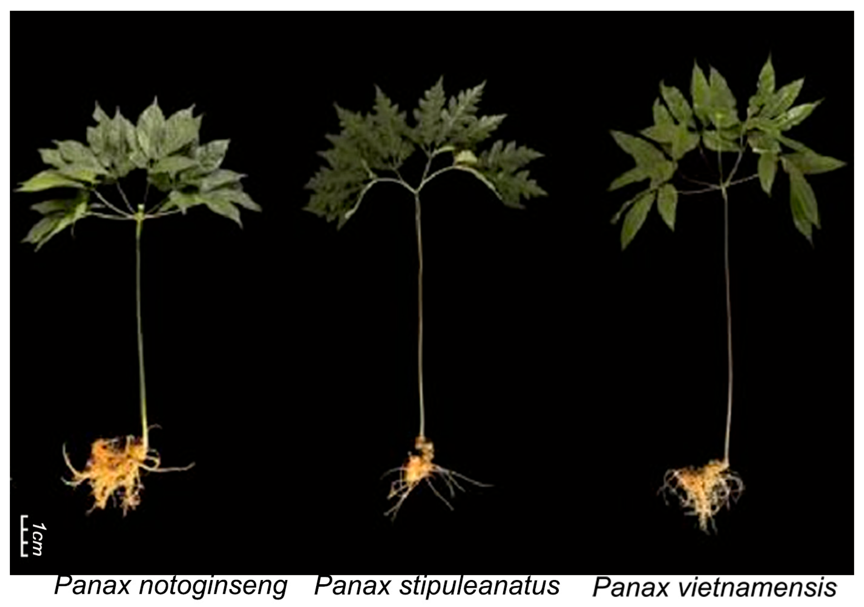
Three different species of Panax, including P. stipuleanatus, P. vietnamensis, and P. notoginseng, were mainly used as plant material in this study. Their phenotype can be observed in Figure 1. These materials were cultivated in the field experiments of the Yigudi Group, Dafei Village Committee, Yuping Town, Pingbian County, Honghe Prefecture, Yunnan Province (longitude 22°59′2″ N, latitude 103°37′24″ E, altitude 1474.7 m). This area has a subtropical monsoon climate similar to that found in South Asia. The average annual temperature is about 19 °C, and the annual precipitation ranges from 1300 to 1500 mm. The relative humidity is high and there is abundant sunlight, which is ideal for growing various medicinal plants. The soil is mainly acidic red soil with a loose texture, good drainage, and high organic and mineral content, providing a solid foundation for cultivating P. notoginseng. To ensure consistent environmental conditions for the experimental materials, P. notoginseng (four years old), P. stipuleanatus (three years old), and P. vietnamensis (four years old), were planted in the same plot. The plants were grown under the same soil and climate conditions with synchronized sampling times. The plants were randomly harvested from the field plots for the sampling of the roots and leaves. Sampling was performed on 6 June 2024, with three biological replicates. Each replicate included at least 10 subsamples from different plants. The whole Panax plants, including the entire primary roots, were collected and washed, followed by the removal of surface water. The leaves were separated from the roots. After the separation of the plant organs, all samples were immediately placed in liquid nitrogen and stored in an ultra-low temperature refrigerator at −80 °C before metabolic analysis.

2.2. Untargeted LC-MS Profiling of Metabolites

Next, 100 mg of liquid nitrogen-frozen root and leaf samples were placed in an Eppendorf tube and then ground (30 Hz, 1.5 min) to a powder using a mill (MM 400, Retsch) to investigate metabolite profiles. Then, the powder was dissolved with 500 µL of 80% aqueous methanol, vortexed for 30 min, and finally kept on ice for 5 min. The supernatant was diluted with mass spectrometry-grade water until the methanol content was 53%, followed by further centrifugation at 15,000 rpm for 20 min. The liquid was filtered through a 0.22 pore size to obtain an upper clear liquid for metabolic analysis studies using tandem mass spectrometry (MS/MS) (Applied Biosystems 4500 QTRAP, Manchester, UK) combined with ultra-performance liquid chromatography (UPLC) (Shimadzu Nexera X2, Milford, MA, USA). Standards for UPLC-MS/MS analytical conditions were prepared as previously reported for plants by Metware Biotechnology Co., Ltd. (Wuhan, China) [41]. The qualitative and quantitative aspects of the metabolites were checked using various methods such as peak area integration, and further corrections were made to the raw data. In particular, metabolite quantification was performed using multiple reaction monitoring (MRM) with triple quadrupole mass spectrometry. Five parameters (declustering potential, collision energy, retention time, Q1, and Q3) were used to detect the relative content of species in different samples and to obtain qualitative and quantitative data on metabolites [42]. After obtaining metabolite profiling data from different samples, the peak areas of all chromatographic peaks were integrated for each species. Then, the mass spectrometry peaks of the same metabolite in different samples were integrated and corrected [43]. After obtaining the processed metabolic data, an unsupervised principal component analysis was performed using the R statistical package 4.1.2 (www.r-project.org (accessed on 30 August 2024)). The data comprised unit variance scaled prior to unsupervised principal component analysis. The R package ComplexHeatmap 2.9.4 was used to perform a hierarchical cluster analysis and Pearson’s correlation coefficients were calculated with the R corrplot package 0.92 [44]. The results were then presented as heat maps with dendrograms of normalized metabolite signal intensities (unit variance scaling) visualized as a color spectrum. Unit variance scaling, also known as z-score normalization, normalizes data based on its mean and standard deviation. This process results in data that conforms to a standard normal distribution. For paired group analysis, variable importance in projection (VIP) values ≥ 1 and fold change (FC) ≤ 0.5 or ≥0.5 were used to identify differentially accumulated metabolites (DAMs) [36]. Orthogonal partial least squares discriminant analysis (OPLS-DA) analyses of R MetaboAnalystR 1.0.1 were used to extract VIP values [45]. A permutation test (200 permutations) was performed to avoid overfitting the OPLS results. Correlation analysis can help measure metabolic proximity between significantly different metabolites. The correlation analysis of the differential metabolites identified according to the screening criteria was carried out using the Pearson correlation method. A metabolite-to-metabolite correlation network diagram was obtained with R igraph software, version 1.2.11. The functional enrichment of DAMs was annotated from the KEGG Compound database (http://www.kegg.jp/kegg/compound/ (accessed on 30 August 2024)), and enriched metabolites were mapped to the KEGG Pathway database (http://www.kegg.jp/kegg/pathway.html (accessed on 30 August 2024)).

3. Results

3.1. Metabolite Profiling of Potential Saponin-Related Metabolites in the Root and Leaf of P. notoginseng

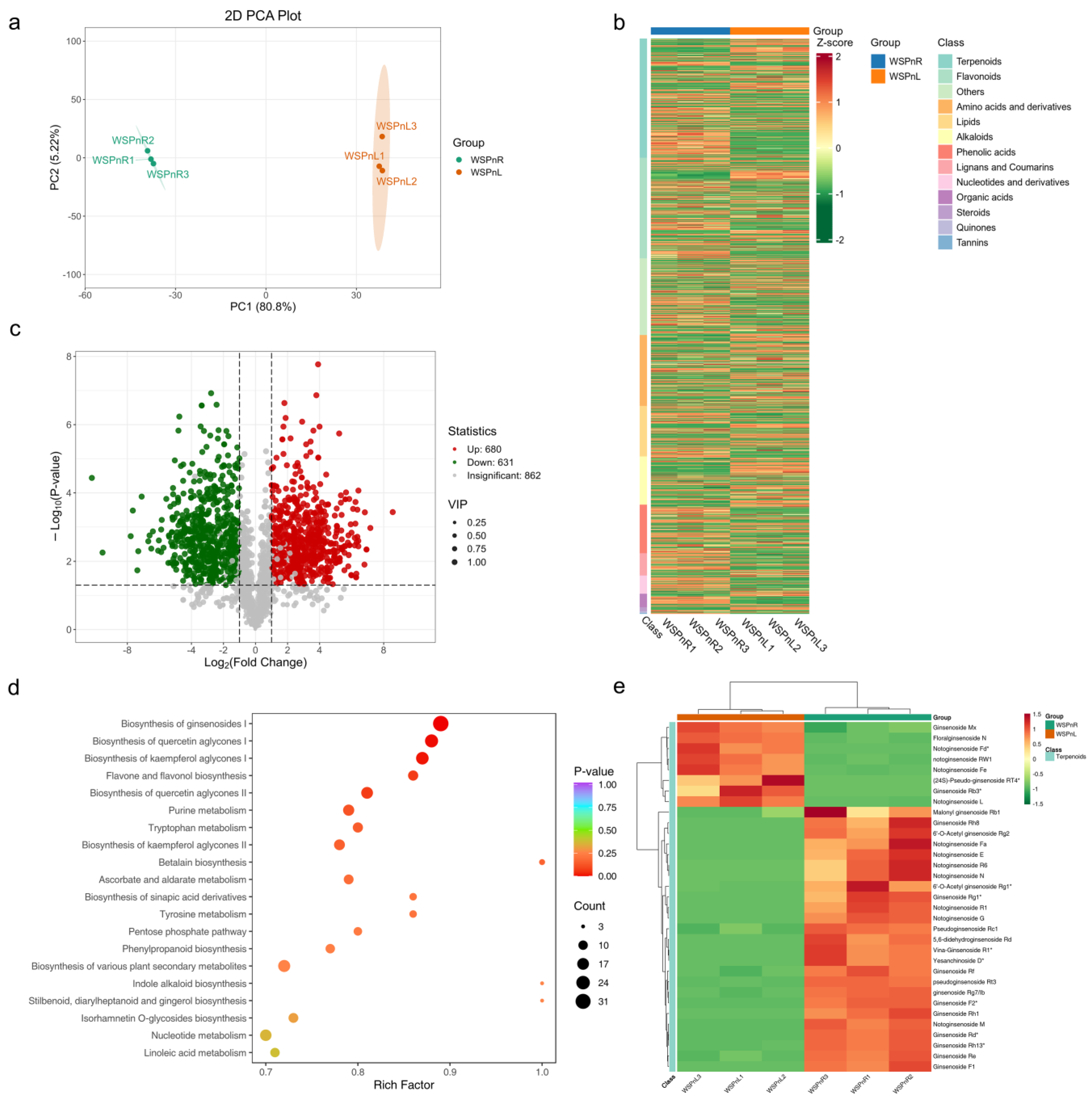
To investigate the distribution of certain active ingredients, this study analyzed the difference in composition and abundance of various metabolites in the root and leaf of P. notoginseng. Liquid chromatography–mass spectrometry (LC-MS)-based untargeted metabolomics revealed a comprehensive landscape of metabolites from a biochemical perspective in both organs. Among them, 2173 metabolites were enriched with major classes including alkaloids, flavonoids, organic acids, nucleic acids, phenolic acids, lipids, amino acids, and terpenoids (Table S1). Terpenoids (16.01%) followed by amino acids (15.28%) and flavonoids (14.68%) had the highest number of identified substances (Figure S1a). The principal component analysis showed that three biological replicates were clustered together for each root and leaf, indicating significant differences in their metabolite profiling (Figure 2a). Furthermore, the identified metabolites showed quantitative differences in both organs, and terpenoids as well as flavonoids were relatively abundant in the root, whereas amino acids were higher in the leaf than in the root (Figure 2b). To further understand the differences in metabolic accumulation, all detected metabolites were compared with OPLS-DA, which identified 1311 differentially accumulated metabolites (DAMs) between the root and the leaf. Among the DAMs, 680 were up-regulated in the root and 631 in the leaf (Figure 2c). The K-means cluster analysis revealed two main subclasses, including 717 DAMs of subclass 2 with higher accumulation in the root and 662 DAMs of subclass 1 with higher accumulation in the leaf (Figure S1b). According to the screening results of the functional pathway enrichment analysis of the DAMs, most of them were enriched in pathways related to secondary metabolites. Among these, the biosynthesis of ginsenosides I, the biosynthesis of quercetin aglycones I/II, flavone and flavonol biosynthesis, and the biosynthesis of kaempferol aglycones I were predominantly the most significantly altered pathways (Figure 2d). Many putative key metabolites annotated with the biosynthesis of ginsenosides I, which are most likely imperative for triterpene saponin biosynthesis, had a significant difference between the root and the leaf. In particular, most dammarane-type ginsenosides Rb1, Rd, Re, Rf, F, Rg1, and Rh, as well as notoginsenosides E, Fa, M, N, and R1 had a higher accumulation level in the root than in the leaf, whereas ginsenosides Mx and Rb3 as well as notoginsenosides Fd, Fe, and L had a higher abundance level in the leaf than in the root (Figure 2e). Our results further confirmed that ginsenosides F2 and notoginsenosides M exhibited a strong positive correlation network with each other along with many other secondary metabolite pathway enriched compounds, phenolic acids, and amino acids (Figure S1c). Our results indicated that the metabolic composition and enrichment pattern of P. notoginseng differed in roots and leaves. In particular, the biosynthesis and accumulation of potentially important saponin-related substances are higher in the root.

3.2. Metabolite Profiling of Potential Saponin-Related Metabolites in the Root and Leaf of P. stipuleanatus

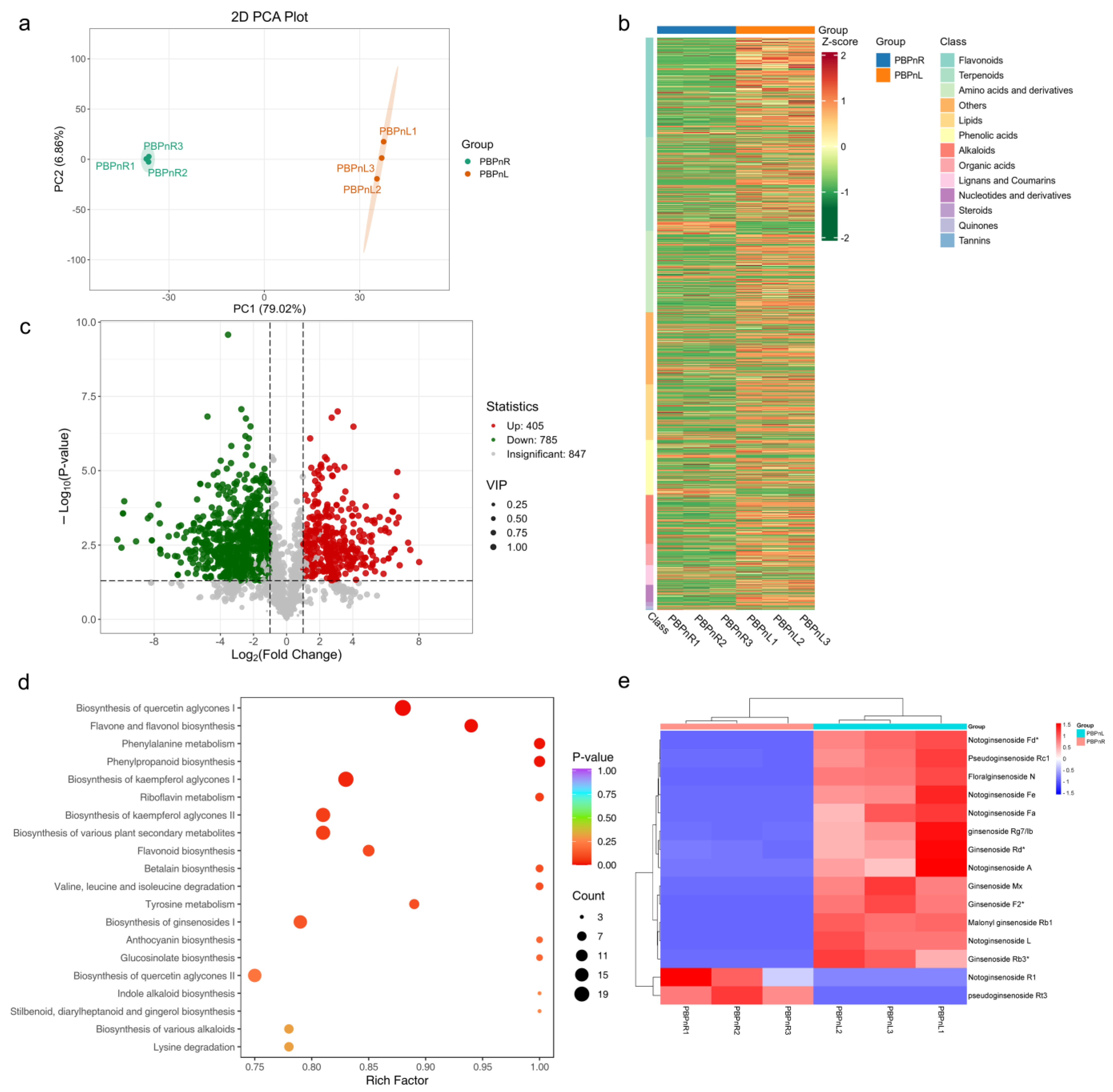
This study further analyzed the composition and abundance of metabolites in P. stipuleanatus roots and leaves. A total of 2037 metabolites were detected, which were associated with different types of alkaloids, flavonoids, phenolic acids, organic acids, lipids, amino acids, tannins, and terpenoids (Table S2). Among these major classes, amino acids (16.2%), followed by terpenoids (14.19%) and flavonoids (13.5%) had the highest number of metabolites (Figure S2a). Three biological replicates were clustered together in the principal component analysis for each root and leaf, indicating significant differences in the abundance of their metabolites (Figure 3a). Furthermore, the identified metabolites had different abundance levels in the root and the leaf. Amino acids, terpenoids, flavonoids, and other secondary metabolites showed a higher accumulation in the leaf compared with the root (Figure 3b). The comparative analysis identified 1326 metabolites that differed between the root and the leaf. Of these, 449 and 877 metabolites showed higher and lower levels of accumulation in the root than in the leaf, respectively (Figure 3c). The cluster analysis divided the DAMs into two main subclasses: subclass 2 DAMs had higher accumulation levels in the root and subclass 1 DAMs had higher accumulation levels in the leaf (Figure S2b). Most of these DAMs were annotated with the biosynthesis of quercetin aglycones I, flavone and flavonol biosynthesis, the biosynthesis of kaempferol aglycones I/II, and the biosynthesis of ginsenosides I (Figure 3d). Furthermore, triterpene saponin biosynthesis-related compounds from the ginsenosides I biosynthesis pathway showed significantly different abundance levels between the root and the leaf. In particular, 13 out of 15 major dammarane-type substances such as ginsenosides Rb3, Rb1, Mx, and F2 along with notoginsenosides A, Fe, Fa, Fd, L, and N were detected to have higher levels of accumulation in the leaf compared with the root (Figure 3e). In addition, the results of the correlation network analysis showed that dammarane-type ginsenosides Rb1 exhibited a significantly strong positive network relationship with many DAMs including alkaloids, flavonoids, phenolic acids, organic acids, lipids, and amino acids (Figure S2c). Metabolic profiling revealed that P. stipuleanatus has tissue-specific qualitative and quantitative metabolic accumulation. The biosynthesis and accumulation of potentially important saponin-related substances are mainly generated in the leaf compared with P. notoginseng.

3.3. Metabolite Profiling of Potential Saponin-Related Metabolites in the Root and Leaf of P. vietnamensis

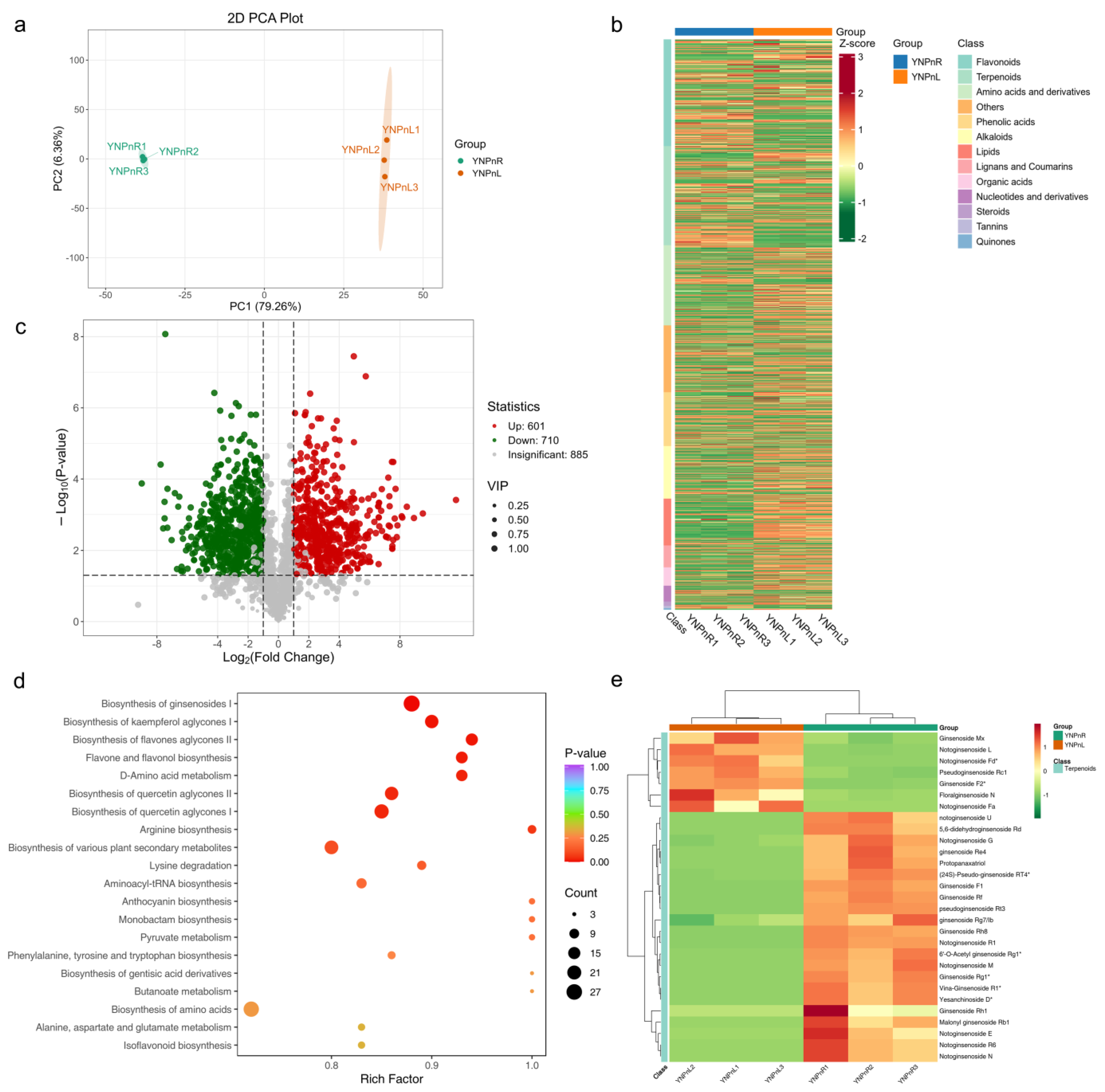
To identify the reasons for the differences in secondary metabolites between the root and the leaf, our metabolic analysis quantitatively determined a total of 2196 metabolic compounds in P. vietnamensis (Table S3). A higher percentage of these compounds belonged to terpenoids (15.8%), amino acids (15.21%), flavonoids (14.68), and phenolic acids (8.29%) (Figure S3a). There was a clear difference between the root and leaf samples, which were distinctly clustered, and 85% of the variance was covered by both principal components 1 and 2 (Figure 4a). In addition, the heatmap-based quantification results revealed differences in the concentration of different classes of metabolites in the root and the leaf. For example, the majority of terpenoid- and flavonoid-related compounds were highly abundant in the root, whereas compounds related to other classes were highly abundant in the leaf (Figure 4b). Our comparative analysis identified 1311 metabolites with dynamic differences in the root compared with the leaf. Of these, 601 showed a higher accumulation in the root, while 710 showed a lower accumulation in the root compared with the leaf (Figure 4c). Furthermore, these were clustered into only two subclasses and each class showed an opposite trend in both the root and the leaf (Figure S3b). The functional classification of DAMs showed that the biosynthesis of ginsenosides I, the biosynthesis of quercetin aglycones I/II, the biosynthesis of flavonol aglycones II, and the biosynthesis of kaempferol aglycones I had a higher significance, and these pathway-annotated substances had a larger number of metabolites with differential accumulation between both organs (Figure 4d). A total of 22 significant triterpene saponins showed a higher concentration in the root, while only 7 saponins had a higher concentration in the leaf. Dammarane-type ginsenosides Rb1, Re4, Rf, F1, Rg1, Rh1, and Rh8, and notoginsenosides E, G, M, N, and U were mainly accumulated in the root. Only the ginsenosides Mx, F2, and the notoginsenosides Fa, Fd, and L accumulated more in the leaf (Figure 4e). The metabolite-to-metabolite correlation analysis identified that dammarane-type ginsenosides Rc, Rh1, and Rh8 showed a strong positive correlation network relationship with each other along with many key DAMs enriched with the biosynthesis of secondary metabolites important for the biosynthesis of saponins (Figure S3c). Our main findings show that the metabolic profile of P. vietnamensis roots differs from that of the leaves. The accumulation of potentially important saponins is mainly high in the roots, similar to P. notoginseng.

3.4. Comparison of Differential Metabolites in the Root of Three Panax Species

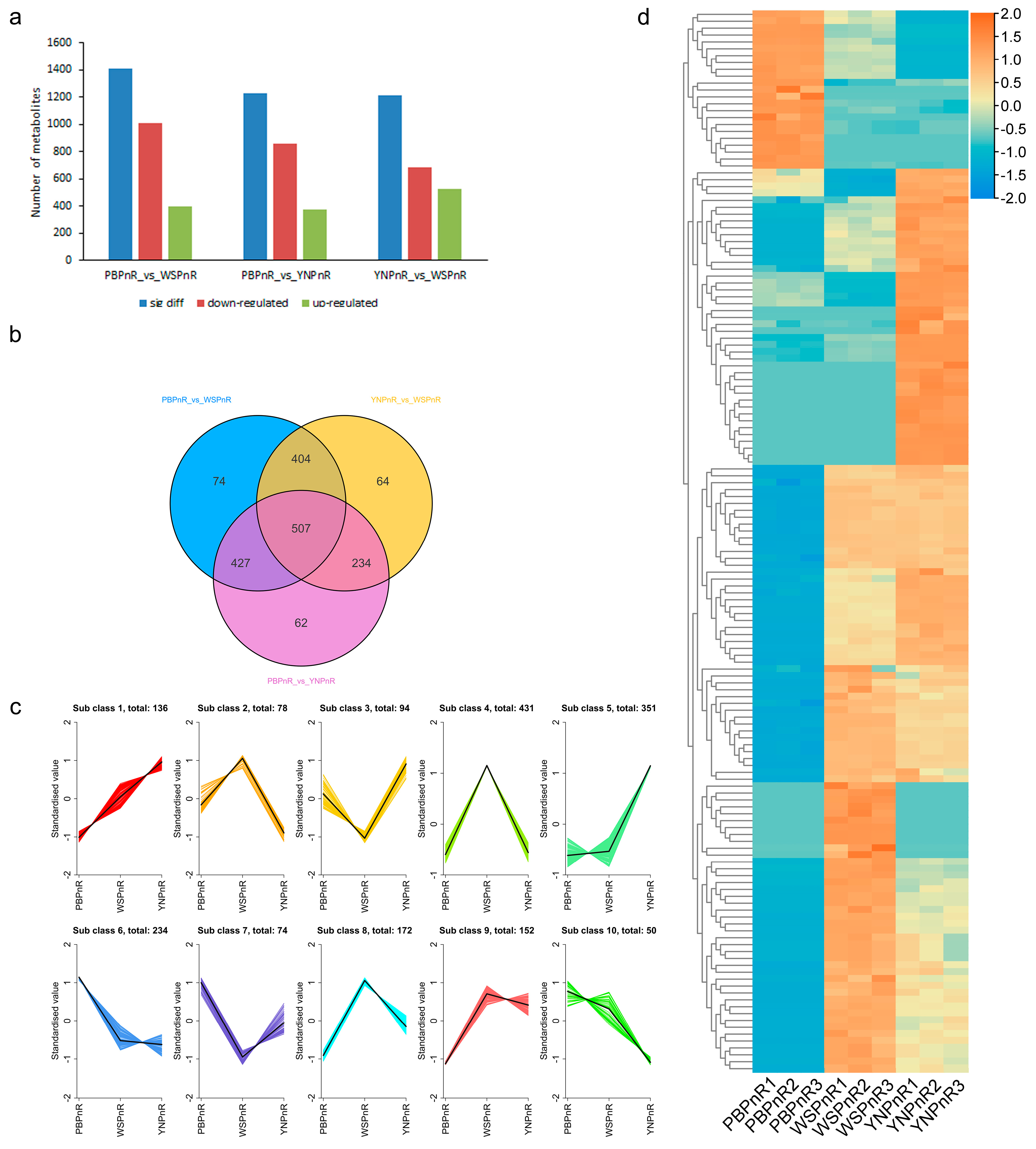
To uncover the potential metabolic differences in the root and leaf of three Panax species, our study further performed a pairwise comparison of identified metabolites according to their relative concentrations in each Panax species. In the root, the pairwise comparison of P. stipuleanatus with P. vietnamensis and P. notoginseng revealed significant differences. For example, a total of 1412 metabolites showed a significant differential accumulation when comparing P. stipuleanatus with P. notoginseng (PBPnR_vs_WSPnR) (Figure 5a). A total of 400 metabolites showed a higher abundance, while 1012 had a lower abundance level in P. stipuleanatus root. The comparison of P. stipuleanatus with P. vietnamensis (PBPnR_vs_YNPnR) had 1230 DAMs, of which 854 showed a lower accumulation and 376 were detected with a higher accumulation in P. stipuleanatus root (Figure 5a). However, in the comparison between P. vietnamensis and P. notoginseng (YNPnR_vs_WSPnR) a lower abundance level was exhibited by 681 metabolites, while a higher abundance level was exhibited by 528 metabolites in the P. vietnamensis root (Figure 5a). All of these comparisons showed that P. notoginseng root had a higher abundance level for most of the identified metabolites. In addition, there was a relatively higher number of less specific differential metabolites in each comparison group, rather than overlapping in each comparison (Figure 5b). Only 74 metabolites specifically showed differential regulation in the PBPnR_vs_WSPnR comparison, whereas 62 differential metabolites were specifically identified in the PBPnR_vs_YNPnR comparison (Figure 5b). There were 427 common DAMs in both comparison groups. K-means clustering divided the accumulation patterns of metabolites into 10 distinct subclasses, with each subclass having a different profile of metabolites compared with other subclasses (Figure 5c). In subclass 1, the abundance of 138 metabolites was highest in P. vietnamensis and lowest in P. stipuleanatus. In contrast, group 2 had 78 DAMs, which were relatively high in P. notoginseng. The highest number of metabolites were in subclass 6; overall, 234 metabolites in this subclass had a higher abundance level in P. stipuleanatus, but the other two species showed similar accumulation profiles (Figure 5c). The hierarchical clustering analysis further revealed that most DAMs showed less accumulation in P. stipuleanatus (Figure 5d). In particular, the major saponin-related compounds including dammarane-type ginsenosides Rb2, Rs2, Rt4, F3, Rh1, Rh8, and Rh13, as well as notoginsenosides A, E, Fa, Fd, R6, and Rw accumulated less in P. stipuleanatus compared with P. vietnamensis and P. notoginseng. Some compounds such as chikuselsusaponin 1b, V, IV, and 1bIV showed higher abundance levels in P. stipuleanatus than the other two species (Table S4). Overall, our comparative analysis among the roots of three Panax species indicated that most of the major saponins were highly abundant in the root of P. notoginseng compared with the other two species.

3.5. Comparison of Differential Metabolites in the Leaves of Three Panax Species

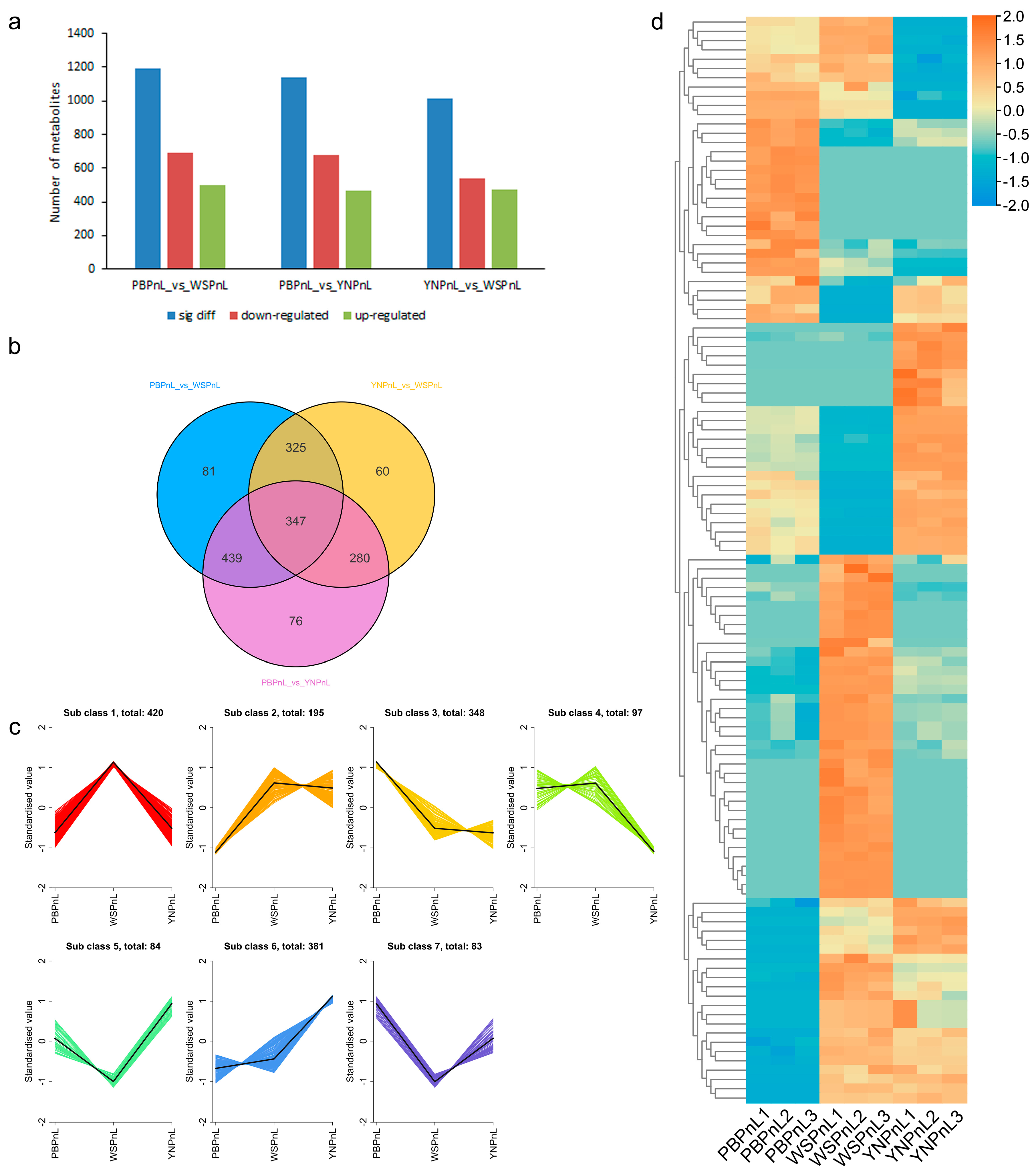
The paired comparison of P. stipuleanatus leaf with P. vietnamensis and P. notoginseng leaf showed significant differences in the composition and concentration of many metabolites. The comparison of P. stipuleanatus with P. notoginseng (PBPnL_vs_WSPnL) showed a total of 1192 metabolites with significant differential accumulation in the leaf (Figure 6a). Of these, 691 metabolites showed a lower abundance level, while 501 metabolites showed a higher abundance level in the leaf of P. stipuleanatus. A total of 1142 metabolites showed significant differences in accumulation when comparing P. stipuleanatus with P. vietnamensis (PBPnL_vs_YNPnL) (Figure 6a). A total of 679 metabolites were found to accumulate less, and 463 were observed to accumulate more, in the leaf of P. stipuleanatus. Furthermore, there were 1012 different metabolites when comparing P. vietnamensis and P. notoginseng (YNPnL_vs_WSPnL) (Figure 6a). The 540 with a lower abundance and 472 with a higher abundance were detected in the leaf of P. vietnamensis. These results mean that all these Panax species had rather inconsistent profiling of identified metabolites in leaf tissue. However, our results found relatively less specific and highly overlapping differential metabolites among all these comparisons (Figure 6b). A total of 439 metabolites showed a differential concentration in PBPnL_vs_WSPnL and PBPnL_vs_YNPnL. Only 81 DAMs were specifically identified in the PBPnL_vs_WSPnL group and 76 in the PBPnL_vs_YNPnL group (Figure 6b). Further quantification subdivided the DAMs into seven distinct subclasses, with each subclass showing a differential but specific accumulation of metabolites compared with other subclasses (Figure 6c). The maximum number of DAMs was in subclass 1; the abundance level of these 420 metabolites was highest in P. notoginseng, while almost very similar accumulation levels were found in the other two Panax species. Among other groups, group 3 with 348 DAMs showed the highest concentration in P. stipuleanatus. The lowest number of metabolites was in subclass 7; the 83 metabolites of this subclass had a higher abundance level in P. stipuleanatus, but P. notoginseng showed the lowest accumulation profiling (Figure 6c). The hierarchical clustering-based quantification further confirmed the differences in metabolic substances among the leaves of the three Panax species. However, P. stipuleanatus exhibited distinct patterns (Figure 6d). Some of the major dammarane-type compounds including ginsenosides Rb, Rg, Rh, mogroside III-E, and majoroside F2, and notoginsenosides R, F were most highly accumulated in P. stipuleanatus compared with P. vietnamensis and P. notoginseng. Meanwhile, some compounds such as ginsenosides F2, Re, Rs1, and Rg3 along with notoginsenosides ST-3, R9, Rw2, majoroside F6, and chikuselsusaponin Fk1 showed lower abundance levels in P. stipuleanatus compared with the other two species (Table S5). In the leaves, our comparative analysis among three Panax species specified that the majority of important saponins had a higher accumulation in the leaves of P. stipuleanatus compared with P. vietnamensis and P. notoginseng.

3.6. Regulatory Network of Metabolites of the Triterpene Saponins Biosynthesis Pathway in P. stipuleanatus Leaf

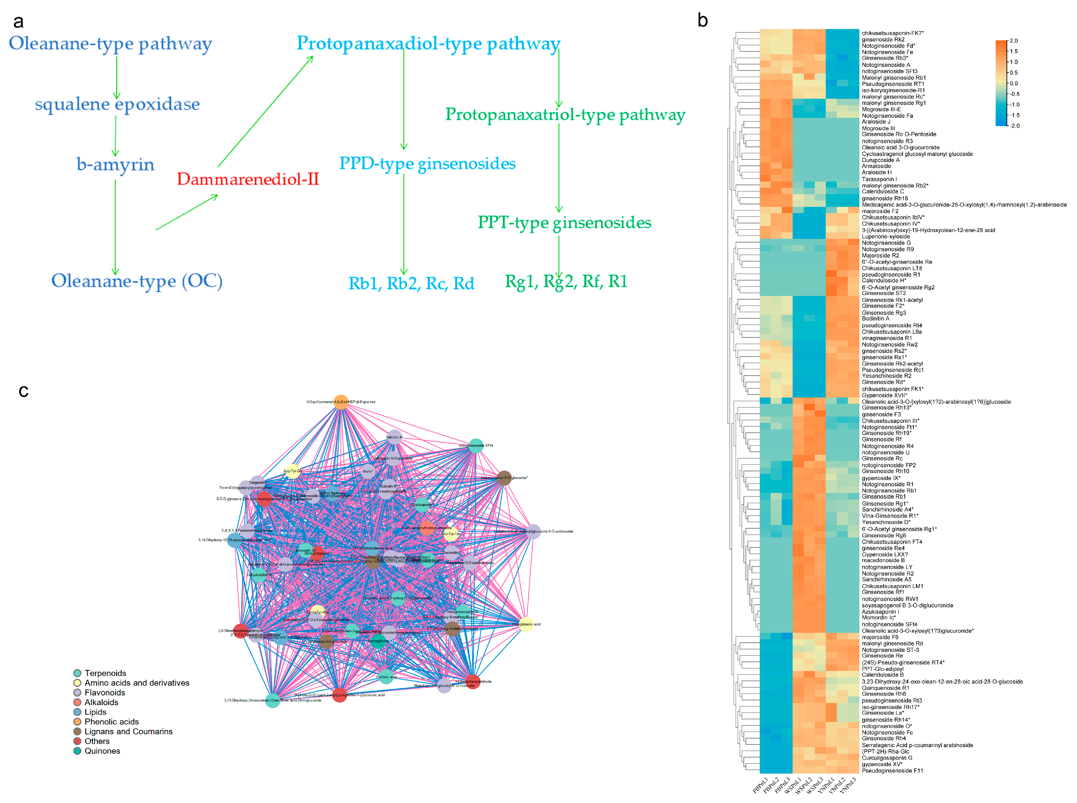
Triterpene saponins are considered to be the most useful medicinal constituents of the Panax species and their levels can vary depending on the species, harvest time, age of the plant, and other factors including organs. The core biosynthetic pathway for the various ginsenosides is triterpene glycosides, starting from 2,3-oxidosqualene, which is formed from isopentenyl pyrophosphate (IPP) via the mevalonate (MVA) pathway. The OA, PPD, and PPT are important backbone compounds and the first committed step in the biosynthesis of various saponins of the Panax species (Figure 7a). Our comparative metabolite profiling and differential analysis in three Panax species revealed the biosynthesis of many key triterpene saponin components in the leaf of P. stipuleanatus, and that their abundance level was relatively higher compared with the leaf of elite species P. notoginseng. However, the results for the root showed less significant changes in the composition of saponin content in both species. In the leaf of P. stipuleanatus, dammarene-type ginsenoside compounds such as Rb1, Rb3, Rg1, La, Rg6, Rh8, Rh10, and Rh14 had higher percentages of concentration levels (Figure 7b). Furthermore, the various compounds of notoginsenosides such as A, R1, Rb2, Fe, Fd, and Ft1 also showed a higher abundance. The metabolite-to-metabolite correlation analysis identified that most triterpenoid saponins-related differential metabolites show a strong correlation with other DAMs enriched by the biosynthesis of secondary metabolites. In particular, dammarane-type notoginsenoside R1 showed a strong positive correlation network relationship with other triterpenoid saponins and many other DAMs (Figure 7c). These findings showed that dammarane-type ginsenoside compounds are mainly synthesized in the leaf of P. stipuleanatus and their abundance level, especially in the leaf, is similar to the elite species P. notoginseng. Therefore, P. stipuleanatus can be a basic material that has great potential to be used on a wide scale in the herb industry.

4. Discussion

The genus Panax is one of the most renowned traditional Chinese medicinal plants. The accumulation of bioactive compounds in herbal plants influences the efficacy of quality characteristics and determines their economic value. The potential of the bioactive compounds of Panax as therapeutic agents varies greatly with species, plant organs, seasonal growth, and climate. The secondary metabolites of plants are significantly influenced by their physiological and developmental stages and change dynamically with age. As a plant ages, its metabolic networks, signal regulation, and defense mechanisms gradually improve, leading to changes in the types and quantities of secondary metabolites produced. For instance, research on medicinal plants like P. notoginseng has revealed significant variations in saponin and flavonoid content across different age stages [46,47]. These differences are usually related to the strength of metabolic pathway expression, growth resource allocation strategies, and environmental response mechanisms. Perennial plants adjust their metabolic strategies to cope with long-term environmental stress, such as accumulating antioxidants. Saponin components in P. notoginseng typically accumulate with age [48,49]. The accumulation of saponins is influenced by the season and induced tissues and increases over time after germination. Therefore, the characterization of the species- and tissue-specific metabolic patterns of less-explored Panax species could benefit the various new breeding programs focusing on ginseng products for the development of new herbal products. This study quantified the metabolic differences between P. notoginseng, P. stipuleanatus, and P. vietnamensis and further discussed the qualitative differences in potential ginsenosides.
Panax notoginseng is one of the earliest cultivated plants in the genus Panax and is rich in various saponins. Our metabolic profiling revealed that the highest number of metabolites identified in the root and the leaf were triterpenes. However, the biosynthesis of triterpene saponins had significant differences between the root and the leaf. Most of the saponins including ginsenoside Rb1, ginsenoside Rd, ginsenoside Re, ginsenoside Rf, ginsenoside F1, ginsenoside F2, ginsenoside Rg1, ginsenoside Rh1, ginsenoside Rh8, and ginsenoside Rh13 had a higher accumulation level in the root. Previous results showed that the chemical composition of various saponins in the root of P. notoginseng differed significantly from that in the seed. Another study found that the level of saponins in 2-year-old roots is lower than that of 3-year-old roots [36]. These results indicate that the accumulation of saponins is affected by season and inductive tissue and shows a time-dependent increase after germination. The biosynthesis of triterpene saponins in P. notoginseng takes place in the roots, leaves, flower buds, and fruit pedicels, but the greatest abundance was observed in the roots [18]. Our results showed a higher abundance of notoginsenosides E, notoginsenosides Fa, notoginsenosides M, notoginsenosides N, and notoginsenosides R1 in the root. Ginsenosides Mx and Rb3 together with notoginsenosides Fd, notoginsenosides Fe, and notoginsenosides L showed a higher level of abundance in the leaves. The growth conditions were significantly different for both the root and the leaf, which is most likely the main reason for the quantitative and qualitative differences in their metabolites. In China, the stem and leaves of P. notoginseng are mainly used as fodder, while new studies have identified leaves with an accumulation of ginsenosides Rg1, Rb1, Rb3, Rh2, and F2, notoginsenoside Fd, mogroside III-E, majoroside F2, ginsenoside Mx, and notoginsenoside R1 [50]. It has been found that protopanaxadiol saponins in particular are produced in abundance in P. notoginseng leaves [51]. A large number of clinical research studies provide incontrovertible evidence for the use of the major bioactive ginsenosides of P. notoginseng to mitigate the adverse effects of various diseases and disorders [52]. In addition to extensive research on the roots, studies on P. notoginseng leaves have become a hot research topic in recent years. Two new dammarane-type saponins, notoginsenoside SY1 and notoginsenoside SY2, were identified from P. notoginseng leaves [53]. New 20(S)-protopanaxadiol-type saponins with potential application in anti-inflammatory disorders were detected in P. notoginseng leaves [54]. These results highlight the need for further research on the leaves, which could lead to the quantification of rare ginsenoside variants in the future.
Panax vietnamensis is a species endemic to the central regions of Vietnam. It has a long history in the treatment of various disorders in high mountain areas and has good synergy with antibiotics and diabetes drugs [55]. Research on P. vietnamensis is relatively less extensive compared with P. notoginseng. A leaf-based metabolic analysis revealed that ginsenosides F1, Rg1, and Rh3 were quite significantly different in two genotypes, P. vietnamensis Ha et Grushv and P. vietnamensis var. langbianensis [56]. In our study, ginsenosides Rb1, Rd, Re, Rg1, and Rh1 had the highest level of accumulation in the root. Usually, roots are used for herbal medicine, and ginsenoside levels in 1-year-old P. vietnamensis were almost half that of 5-year-old plants, and the leaves were not used for saponin extraction [57]. Later research on aerial parts showed that ginsenosides such as ginsenoside Rb1, ginsenoside Rg1, ginsenoside Rd, majonoside R2, and pseudoginsenoside RS1 were present in the leaves of P. vietnamensis and had inhibitory effects on acetylcholinesterase [58]. In addition, P. vietnamensis contains PPD and PPT saponins such as ginsenoside Rb1, Rg1, Re, and Rd, but the leaves were rich in PPD saponins [59]. Our metabolic profiling identified 710 metabolites with a higher accumulation in the leaves compared with the roots. However, only the ginsenosides Mx and F2 and the notoginsenosides Fa, Fd, and L showed a higher abundance in the leaf. Furthermore, ginsenosides Rc, Rh1, and Rh8 showed a strong positive correlation network with other saponins. It was previously reported that the leaves of P. notoginseng contain significant amounts of ginsenosides Rb1, Rb2, Rb3, Rc, Rd, Re, Rg1, Rh1, Rh4, and Rh5; majonosides R1, R2, and F1; and notoginsenosides Fa, R1, R2, and R6. It demonstrates the potential application of the leaves as an alternative to the roots in the traditional medicine industry [60,61,62].
Panax stipuleanatus, also known as wild Sanchi and Pingbian Panax, is widely distributed in the tropical rainforests of Yunnan Province, China. It is a wild species that is an alternative source to P. notoginseng in Yunnan due to its similar environmental habitat, pharmaceutical functions, and morphological characteristics [63]. Its rhizome has been effectively used to disperse phlegm, relieve pain, and stop bleeding since ancient times and its broader medical function remains limited due to less research data [64]. Previously, it was reported that the panaxatriol and panaxadiol types of saponins were present at low levels, while two oleanolic saponins such as stipuleanoids R1 and R2 were also identified in the rhizome [65]. The complete metabolic landscape of P. stipuleanatus remained largely unclear compared with other elite species of the genus Panax. In this direction, our metabolic analysis revealed significant differences between the root and the leaf, with 877 higher abundance levels in the leaf. Furthermore, 13 out of 15 major saponins, including the ginsenosides Rb3, Rb1, Mx, and F2, were highly abundant in the leaves. Similarly, notoginsenosides A, Fe, Fa, Fd, L, and N showed higher percentages in leaves compared with the roots. Previously, ginsenoside Rb1 was reported to exert several pharmacological influences on metabolic disorders, including the inflammatory, vascular, and central nervous system [66]. Therefore, ginsenosides Rb3 and Rb1, which were specifically identified from P. stipuleanatus, may be developed as a potential therapeutic agent with multiple effects. Furthermore, it may provide a platform for future clinical trials. Our study further identified that several ginsenosides showed strong positive networking with various secondary metabolites annotated with alkaloids, flavonoids, phenolic acids, organic acids, lipids, and amino acids, which revealed a synergistic effect exerted by the vast mixture of secondary metabolites produced by plants. In addition, these may be correlated to the accumulation patterns along with the transformation of different ginsenosides in the leaf of P. stipuleanatus.
Of particular interest, our comparative results quantified the accumulation of key triterpene saponins in the leaf of P. stipuleanatus and its abundance level was relatively high compared with the leaf of elite species P. notoginseng as well as P. vietnamensis. At the same time, however, the root showed less significant changes in the arrangement of saponin than in both species. These results allowed us to propose that triterpene saponins are mainly produced in the leaf of P. stipuleanatus. Moreover, their abundance level, especially in the leaf, is similar to the elite species P. notoginseng. High levels of triterpenoid saponin accumulation are a primary goal of Panax plant breeding. All of these results are significant for improving future breeding work quality in the Panax genus. Furthermore, the results provide valuable resources for reference and can expedite in-depth research leading to the large-scale utilization of P. stipuleanatus in the pharmacological industry. Previous extensive clinical trials in P. notoginseng and P. quinquefolius provide evidence that various ginsenosides and notoginsenosides produce certain pharmacological effects, especially anti-diabetic, anti-aging, antioxidant, anti-cancer, and anti-tumor effects [67,68]. In a previous study, eleven oleanane-type triterpenoids from P. stipuleanatus were reported to have cytotoxic effects on human cancer cells [69]. In contrast, our study confirms that P. stipuleanatus leaves contain a higher abundance of dammarane-type ginsenoside compounds such as Rb1, Rb3, and Rg1. Meanwhile, various compounds of notoginsenosides, including R1 and Rb2, were found at a higher abundance in the leaf. These differences may be usually related to the strength of metabolic pathway expression, growth resource allocation strategies, and environmental response mechanisms. In addition, perennial plants adjust their metabolic strategies to cope with long-term environmental stress, such as accumulating antioxidants. These results show that accumulation could change in one organ because one organ can be the source and the other can be the storage site. In particular, the differences between the root and the leaf require effective insight to explore the growth resource allocation strategies in the genus Panax. This study will contribute to the scientific guidance of alternative ginsenoside resources from P. stipuleanatus.

5. Conclusions

Our metabolic profiling revealed high levels of potentially significant ginsenosides in three Panax species. It systematically compared the metabolome differences between roots and leaves. This highlights the intensity of the discrimination of key ginsenosides in the source and storage organs. The results further highlight that P. stipuleanatus has a rich profile of dammarane-type ginsenosides and notoginsenosides. In addition, the results provide metabolic links between ginsenoside synthesis and other secondary metabolisms at the whole plant level in different species. Further characterization of these compounds could improve the biochemical quality of the genus Panax. They also provide an important basis for the quality control of natural products derived from the Panax plant.

Supplementary Materials

The following supporting information can be downloaded at: https://www.mdpi.com/article/10.3390/horticulturae11080916/s1, Table S1: Total statistical values of metabolic profiling in root and leaf of P. notoginseng. Table S2: Total statistical values of metabolic profiling in root and leaf of P. stipuleanatus. Table S3: Total statistical values of metabolic profiling in root and leaf of P. vietnamensis. Table S4: Total statistical values of different metabolites identified in the roots of three Panax species. Table S5: Total statistical values of different metabolites identified in the leaves of three Panax species. Figure S1: Classification, clustering, and correlation analysis of differential metabolites of root and leaf of P. notoginseng. Figure S2: Classification, clustering, and correlation analysis of differential metabolites from root and leaf of P. stipuleanatus. Figure S3: Classification, clustering, and correlation analysis of differential metabolites of root and leaf of P. vietnamensis.

Author Contributions

Conceptualization, S.H., Y.G., S.D., X.C., X.H. and R.S.; Data curation, Y.G., H.V.S., X.C., X.H. and R.S.; Formal analysis, S.H., Y.G. and Y.D.; Funding acquisition, X.H. and R.S.; Investigation, S.H., Y.G., S.D., Y.D. and J.W.; Methodology, S.H., Y.G., S.D., Y.D. and X.H.; Project administration, R.S.; Resources, H.V.S. and X.C.; Software, J.W., H.V.S. and R.S.; Supervision, X.C., X.H. and R.S.; Validation, S.H., J.W., H.V.S. and X.H.; Writing—original draft, S.H., Y.G. and S.D.; Writing—review and editing, X.C., X.H. and R.S. All authors have read and agreed to the published version of the manuscript.

Funding

This work was supported by the National Natural Science Foundation of China (32260720), the China Agriculture Research System of MOF and MARA (CARS-21-05B), the Major Science and Technology Project of Yunnan (202402AE090026), the Yunnan Province international science and technology special correspondent (202303AK140011), the Yunnan Province rural revitalization science and technology innovation village project (202404BU090008), and the Major Science and Technology Project of Kunming (2021JH002).

Data Availability Statement

The datasets generated and/or analyzed during the current study are within the manuscript and its Supplementary Files.

Conflicts of Interest

The authors declare no conflict of interest.

References

  1. Yang, W.-Z.; Hu, Y.; Wu, W.-Y.; Ye, M.; Guo, D.-A. Saponins in the genus Panax L. (Araliaceae): A systematic review of their chemical diversity. Phytochemistry 2014, 106, 7–24. [Google Scholar] [CrossRef]
  2. Manzanilla, V.; Kool, A.; Nguyen Nhat, L.; Nong Van, H.; Le Thi Thu, H.; De Boer, H. Phylogenomics and barcoding of Panax: Toward the identification of ginseng species. BMC Evol. Biol. 2018, 18, 44. [Google Scholar] [CrossRef]
  3. Jee, H.-S.; Chang, K.-H.; Park, S.-H.; Kim, K.-T.; Paik, H.-D. Morphological characterization, chemical components, and biofunctional activities of Panax ginseng, Panax quinquefolium, and Panax notoginseng roots: A comparative study. Food Rev. Int. 2014, 30, 91–111. [Google Scholar] [CrossRef]
  4. Park, H.J.; Kim, D.H.; Park, S.J.; Kim, J.M.; Ryu, J.H. Ginseng in traditional herbal prescriptions. J. Ginseng Res. 2012, 36, 225–241. [Google Scholar] [CrossRef]
  5. Kim, N.H.; Jayakodi, M.; Lee, S.C.; Choi, B.S.; Jang, W.; Lee, J.; Kim, H.H.; Waminal, N.E.; Lakshmanan, M.; van Nguyen, B. Genome and evolution of the shade-requiring medicinal herb Panax ginseng. Plant Biotechnol. J. 2018, 16, 1904–1917. [Google Scholar] [CrossRef]
  6. Kim, J.K.; Tabassum, N.; Uddin, M.R.; Park, S.U. Ginseng: A miracle sources of herbal and pharmacological uses. Orient. Pharm. Exp. Med. 2016, 16, 243–250. [Google Scholar] [CrossRef]
  7. Gao, B.; Huang, L.; Liu, H.; Wu, H.; Zhang, E.; Yang, L.; Wu, X.; Wang, Z. Platelet P2Y12 receptors are involved in the haemostatic effect of notoginsenoside Ft1, a saponin isolated from P anax notoginseng. Br. J. Pharmacol. 2014, 171, 214–223. [Google Scholar] [CrossRef]
  8. Liu, L.; Xu, F.-R.; Wang, Y.-Z. Traditional uses, chemical diversity and biological activities of Panax L. (Araliaceae): A review. J. Ethnopharmacol. 2020, 263, 112792. [Google Scholar] [CrossRef]
  9. Leung, K.W.; Wong, A.S.-T. Pharmacology of ginsenosides: A literature review. Chin. Med. 2010, 5, 20. [Google Scholar] [CrossRef]
  10. Kim, K.H.; Lee, D.; Lee, H.L.; Kim, C.-E.; Jung, K.; Kang, K.S. Beneficial effects of Panax ginseng for the treatment and prevention of neurodegenerative diseases: Past findings and future directions. J. Ginseng Res. 2018, 42, 239–247. [Google Scholar] [CrossRef]
  11. Wahid, F.; Khan, T.; Subhan, F.; Khan, M.; Kim, Y. Ginseng pharmacology: Multiple molecular targets and recent clinical trials. Drugs Future 2010, 35, 399–407. [Google Scholar] [CrossRef]
  12. Shim, J.S.; Song, M.-Y.; Yim, S.-V.; Lee, S.-E.; Park, K.-S. Global analysis of ginsenoside Rg1 protective effects in β-amyloid-treated neuronal cells. J. Ginseng Res. 2017, 41, 566–571. [Google Scholar] [CrossRef]
  13. Wang, Y.; Yang, G.; Gong, J.; Lu, F.; Diao, Q.; Sun, J.; Zhang, K.; Tian, J.; Liu, J. Ginseng for Alzheimer’s disease: A systematic review and meta-analysis of randomized controlled trials. Curr. Top. Med. Chem. 2016, 16, 529–536. [Google Scholar] [CrossRef]
  14. Fan, S.; Zhang, Z.; Su, H.; Xu, P.; Qi, H.; Zhao, D.; Li, X. Panax ginseng clinical trials: Current status and future perspectives. Biomed. Pharmacother. 2020, 132, 110832. [Google Scholar] [CrossRef]
  15. Kim, J.-H. Cardiovascular diseases and Panax ginseng: A review on molecular mechanisms and medical applications. J. Ginseng Res. 2012, 36, 16–26. [Google Scholar] [CrossRef]
  16. Yang, Z.G.; Sun, H.X.; Ye, Y.P. Ginsenoside Rd from Panax notoginseng is cytotoxic towards HeLa cancer cells and induces apoptosis. Chem. Biodivers. 2006, 3, 187–197. [Google Scholar] [CrossRef]
  17. Baek, K.-S.; Yi, Y.-S.; Son, Y.-J.; Jeong, D.; Sung, N.Y.; Aravinthan, A.; Kim, J.-H.; Cho, J.Y. Comparison of anticancer activities of Korean Red Ginseng-derived fractions. J. Ginseng Res. 2017, 41, 386–391. [Google Scholar] [CrossRef]
  18. Qiao, Y.-J.; Shang, J.-H.; Wang, D.; Zhu, H.-T.; Yang, C.-R.; Zhang, Y.-J. Research of panax spp. in kunming institute of botany, CAS. Nat. Prod. Bioprospecting 2018, 8, 245–263. [Google Scholar] [CrossRef]
  19. Wang, D.; Koh, H.-L.; Hong, Y.; Zhu, H.-T.; Xu, M.; Zhang, Y.-J.; Yang, C.-R. Chemical and morphological variations of Panax notoginseng and their relationship. Phytochemistry 2013, 93, 88–95. [Google Scholar] [CrossRef]
  20. Wei, G.; Dong, L.; Yang, J.; Zhang, L.; Xu, J.; Yang, F.; Cheng, R.; Xu, R.; Chen, S. Integrated metabolomic and transcriptomic analyses revealed the distribution of saponins in Panax notoginseng. Acta Pharm. Sin. B 2018, 8, 458–465. [Google Scholar] [CrossRef]
  21. Niu, Y.; Luo, H.; Sun, C.; Yang, T.-J.; Dong, L.; Huang, L.; Chen, S. Expression profiling of the triterpene saponin biosynthesis genes FPS, SS, SE, and DS in the medicinal plant Panax notoginseng. Gene 2014, 533, 295–303. [Google Scholar] [CrossRef]
  22. Luo, H.; Sun, C.; Sun, Y.; Wu, Q.; Li, Y.; Song, J.; Niu, Y.; Cheng, X.; Xu, H.; Li, C. Analysis of the transcriptome of Panax notoginseng root uncovers putative triterpene saponin-biosynthetic genes and genetic markers. BMC Genom. 2011, 12, S5. [Google Scholar] [CrossRef]
  23. Haralampidis, K.; Trojanowska, M.; Osbourn, A.E.J.H. Biosynthesis of triterpenoid saponins in plants. Hist. Trends Bioprocess. Biotransform. 2002, 75, 31–49. [Google Scholar]
  24. Lichtenthaler, H.K.; Rohmer, M.; Schwender, J. Two independent biochemical pathways for isopentenyl diphosphate and isoprenoid biosynthesis in higher plants. Physiol. Plant. 1997, 101, 643–652. [Google Scholar] [CrossRef]
  25. Gao, S.; Basu, S.; Yang, Z.; Deb, A.; Hu, M. Bioavailability challenges associated with development of saponins as therapeutic and chemopreventive agents. Curr. Drug Targets 2012, 13, 1885–1899. [Google Scholar] [CrossRef]
  26. Liu, H.; Lu, X.; Hu, Y.; Fan, X. Chemical constituents of Panax ginseng and Panax notoginseng explain why they differ in therapeutic efficacy. Pharmacol. Res. 2020, 161, 105263. [Google Scholar] [CrossRef]
  27. Song, H.-H.; Kim, D.-Y.; Woo, S.; Lee, H.-K.; Oh, S.-R. An approach for simultaneous determination for geographical origins of Korean Panax ginseng by UPLC-QTOF/MS coupled with OPLS-DA models. J. Ginseng Res. 2013, 37, 341–348. [Google Scholar] [CrossRef]
  28. Chen, Y.; Zhao, Z.; Chen, H.; Yi, T.; Qin, M.; Liang, Z. Chemical differentiation and quality evaluation of commercial asian and american ginsengs based on a UHPLC–QTOF/MS/MS metabolomics approach. Phytochem. Anal. 2015, 26, 145–160. [Google Scholar] [CrossRef]
  29. Wei, G.; Wei, F.; Yuan, C.; Chen, Z.; Wang, Y.; Xu, J.; Zhang, Y.; Dong, L.; Chen, S. Integrated chemical and transcriptomic analysis reveals the distribution of protopanaxadiol-and protopanaxatriol-type saponins in Panax notoginseng. Molecules 2018, 23, 1773. [Google Scholar] [CrossRef]
  30. Li, J.; Ma, L.; Zhang, S.; Zuo, C.; Song, N.; Zhu, S.; Wu, J. Transcriptome analysis of 1-and 3-year-old Panax notoginseng roots and functional characterization of saponin biosynthetic genes DS and CYP716A47-like. Planta 2019, 249, 1229–1237. [Google Scholar] [CrossRef]
  31. Qi, L.-W.; Wang, C.-Z.; Yuan, C.-S. Isolation and analysis of ginseng: Advances and challenges. Nat. Prod. Rep. 2011, 28, 467–495. [Google Scholar] [CrossRef]
  32. Wan, J.; Yang, F.; Li, S.; Wang, Y.; Cui, X. Chemical characteristics for different parts of Panax notoginseng using pressurized liquid extraction and HPLC-ELSD. J. Pharm. Biomed. Anal. 2006, 41, 1596–1601. [Google Scholar] [CrossRef] [PubMed]
  33. Wan, J.B.; Li, S.p.; Chen, J.M.; Wang, Y.T. Chemical characteristics of three medicinal plants of the Panax genus determined by HPLC-ELSD. J. Sep. Sci. 2007, 30, 825–832. [Google Scholar] [CrossRef]
  34. Fan, M.; Fan, Y.; Rao, Z.; Li, Y.; Qian, H.; Zhang, H.; Wu, G.; Qi, X.; Wang, L. Comparative investigation on metabolite changes in ‘wu mi’production by Vaccinium bracteatum Thunb. leaves based on multivariate data analysis using UPLC–QToF–MS. Food Chem. 2019, 286, 146–153. [Google Scholar] [CrossRef] [PubMed]
  35. Fan, M.; Lian, W.; Li, T.; Fan, Y.; Rao, Z.; Li, Y.; Qian, H.; Zhang, H.; Wu, G.; Qi, X. Metabolomics approach reveals discriminatory metabolites associating with the blue pigments from Vaccinium bracteatum thunb. leaves at different growth stages. Ind. Crops Prod. 2020, 147, 112252. [Google Scholar] [CrossRef]
  36. Li, X.; Zhao, Y.; He, S.; Meng, J.; Lu, Y.; Shi, H.; Liu, C.; Hao, B.; Tang, Q.; Zhang, S. Integrated metabolome and transcriptome analyses reveal the molecular mechanism underlying dynamic metabolic processes during taproot development of Panax notoginseng. BMC Plant Biol. 2024, 24, 170. [Google Scholar] [CrossRef]
  37. Han, X.-T.; Yang, D.; Sun, M.-J.; Wei, F.-G.; Li, S.-Y.; Di, X.; Li, N.; Song, H.-P.; Zhang, H.; Kang, T.-G. Integrated transcriptomic and targeted-metabolomic analysis reveal the co-upregulation and mechanism of saponin and starch biosynthesis in the underground parts of Panax notoginseng induced by inflorescence removal. Ind. Crops Prod. 2025, 226, 120643. [Google Scholar] [CrossRef]
  38. Wang, H.; Zhang, C.; Zuo, T.; Li, W.; Jia, L.; Wang, X.; Qian, Y.; Guo, D.; Yang, W.J.A.; Chemistry, B. In-depth profiling, characterization, and comparison of the ginsenosides among three different parts (the root, stem leaf, and flower bud) of Panax quinquefolius L. by ultra-high performance liquid chromatography/quadrupole-Orbitrap mass spectrometry. Anal. Bioanal. Chem. 2019, 411, 7817–7829. [Google Scholar] [CrossRef]
  39. Ji, R.; Garran, T.A.; Luo, Y.; Cheng, M.; Ren, M.; Zhou, X. Untargeted Metabolomic Analysis and Chemometrics to Identify Potential Marker Compounds for the Chemical Differentiation of Panax ginseng, P. quinquefolius, P. notoginseng, P. japonicus, and P. japonicus var. major. Molecules 2023, 28, 2745. [Google Scholar] [CrossRef]
  40. Pu, R.; Cheng, Y.; Zeng, J.; Wang, H.; Li, N.; Gao, M.; Ma, J.; Cui, X. Metabolomic and transcriptomic analysis of Panax Notoginseng root: Mechanistic insights into the effects of flower bud removal on yield and phytochemical composition. Ind. Crops Prod. 2025, 223, 120138. [Google Scholar] [CrossRef]
  41. Shen, S.; Tang, Y.; Zhang, C.; Yin, N.; Mao, Y.; Sun, F.; Chen, S.; Hu, R.; Liu, X.; Shang, G. Metabolite profiling and transcriptome analysis provide insight into seed coat color in Brassica juncea. Int. J. Mol. Sci. 2021, 22, 7215. [Google Scholar] [CrossRef]
  42. Chen, W.; Gong, L.; Guo, Z.; Wang, W.; Zhang, H.; Liu, X.; Yu, S.; Xiong, L.; Luo, J. A novel integrated method for large-scale detection, identification, and quantification of widely targeted metabolites: Application in the study of rice metabolomics. Mol. Plant 2013, 6, 1769–1780. [Google Scholar] [CrossRef]
  43. Fraga, C.G.; Clowers, B.H.; Moore, R.J.; Zink, E.M. Signature-discovery approach for sample matching of a nerve-agent precursor using liquid chromatography-mass spectrometry, XCMS, and chemometrics. Anal. Chem. 2010, 82, 4165–4173. [Google Scholar] [CrossRef]
  44. Gu, Z.; Eils, R.; Schlesner, M. Complex heatmaps reveal patterns and correlations in multidimensional genomic data. Bioinformatics 2016, 32, 2847–2849. [Google Scholar] [CrossRef]
  45. Chong, J.; Xia, J. MetaboAnalystR: An R package for flexible and reproducible analysis of metabolomics data. Bioinformatics 2018, 34, 4313–4314. [Google Scholar] [CrossRef]
  46. Xue, L.; He, Z.; Bi, X.; Xu, W.; Wei, T.; Wu, S.; Hu, S. Transcriptomic profiling reveals MEP pathway contributing to ginsenoside biosynthesis in Panax ginseng. BMC Genom. 2019, 20, 383. [Google Scholar] [CrossRef]
  47. Raudone, L.; Motiekaityte, V.; Vainoriene, R.; Zymone, K.; Marksa, M.; Janulis, V. Phytochemical Profiles of Alpine Plant Horminum pyrenaicum L. during Phenological Growth Stages. Chem. Biodivers. 2018, 15, e1800190. [Google Scholar] [CrossRef]
  48. Xiao, D.; Yue, H.; Xiu, Y.; Sun, X.; Wang, Y.; Liu, S. Accumulation characteristics and correlation analysis of five ginsenosides with different cultivation ages from different regions. J. Ginseng Res. 2015, 39, 338–344. [Google Scholar] [CrossRef]
  49. Li, Q.; Yuan, M.; Li, X.; Li, J.; Xu, M.; Wei, D.; Wu, D.; Wan, J.; Mei, S.; Cui, T.; et al. New dammarane-type triterpenoid saponins from Panax notoginseng saponins. J. Ginseng Res. 2020, 44, 673–679. [Google Scholar] [CrossRef]
  50. Sun, X.; Deng, H.; Shu, T.; Xu, M.; Su, L.; Li, H. Study on chemical constituents of Panax notoginseng leaves. Molecules 2023, 28, 2194. [Google Scholar] [CrossRef]
  51. Cao, Y.; Li, Q.; Yang, Y.; Ke, Z.; Chen, S.; Li, M.; Fan, W.; Wu, H.; Yuan, J.; Wang, Z. Cardioprotective effect of stem-leaf saponins from panax notoginseng on mice with sleep deprivation by inhibiting abnormal autophagy through PI3K/Akt/mTOR pathway. Front. Cardiovasc. Med. 2021, 8, 694219. [Google Scholar] [CrossRef]
  52. Qu, Y.; Liu, H.-Y.; Guo, X.-X.; Luo, Y.; Wang, C.-X.; He, J.-H.; Xu, T.-R.; Yang, Y.; Cui, X.-M. Converting ginsenosides from stems and leaves of Panax notoginseng by microwave processing and improving their anticoagulant and anticancer activities. RSC Adv. 2018, 8, 40471–40482. [Google Scholar] [CrossRef]
  53. Yang, C.-K.; Xiong, J.; Shen, Y. Two new dammarane-type triterpenoids from the stems and leaves of Panax notoginseng. J. Asian Nat. Prod. Res. 2021, 23, 341–347. [Google Scholar] [CrossRef]
  54. Ruan, J.; Sun, F.; Zhang, Y.; Zheng, D.; Xiang, G.; Zhao, W.; Zhang, Y.; Wang, T. New 20 (S)-protopanaxadiol type saponins from the leaves of Panax notoginseng and their potential anti-inflammatory activities. Steroids 2020, 162, 108696. [Google Scholar] [CrossRef]
  55. Thanh, N.T.; Van, T.T.H.; Hung, L.V.; Van Khiem, N.; Mai, L.Q.; Xuyen, D.T.; Oanh, P.T.; Van Hai, D.; Dien, N.D.; Nhut, D.T. Bioactive compounds and biological activities of Vietnamese ginseng (Panax Vietnamensis Ha et Grushv.). In Bioactive Compounds in the Storage Organs of Plants; Springer: Berlin/Heidelberg, Germany, 2023; pp. 1–25. [Google Scholar]
  56. Cheng, R.; Yoon, Y.C.; Jung, C.W.; Kim, T.H.; Wang, Q.; Cho, W.; Yang, T.-J.; Van Le, T.H.; Cho, C.J.; Kim, J.H. Development of a leaf metabolite-based intact sample distinguishing algorithm for the three varieties of Panax Vietnamensis. Sci. Rep. 2025, 15, 7939. [Google Scholar] [CrossRef]
  57. Vo, H.T.; Ghimeray, A.K.; Vu, N.T.; Jeong, Y.-H. Quantitative estimation of ginsenosides in different ages of Panax vietnamensis and their anti-proliferation effects in hela cells. Afr. J. Tradit. Complement. Altern. Med. 2015, 12, 79–83. [Google Scholar] [CrossRef]
  58. Vu, V.T.; Hieu, N.N.; Hiep, D.H.; Anh, N.T.H.; Ngan, D.T.; Tung, P.H.T.; Thuong, P.T.; Tung, N.H. Dammarane-Type Saponins from the Leaves of Vietnamese Ginseng (Panax vietnamensis Ha & Grushv.) and Their Acetylcholinesterase Inhibition In Vitro and In Silico. Chem. Biodivers. 2025, 22, e202401329. [Google Scholar]
  59. Van Le, T.H.; Lee, G.J.; Vu, H.K.L.; Kwon, S.W.; Nguyen, N.K.; Park, J.H.; Nguyen, M.D. Ginseng saponins in different parts of Panax vietnamensis. Chem. Pharm. Bull. 2015, 63, 950–954. [Google Scholar] [CrossRef]
  60. Banskota, A.H.; Tezuka, Y.; Tran, Q.; Kadota, S. Chemical constituents and biological activities of Vietnamese medicinal plants. Curr. Top. Med. Chem. 2003, 3, 227–248. [Google Scholar] [CrossRef]
  61. Le Tran, Q.; Adnyana, I.K.; Tezuka, Y.; Harimaya, Y.; Saiki, I.; Kurashige, Y.; Tran, Q.K.; Kadota, S. Hepatoprotective effect of majonoside R2, the major saponin from Vietnamese ginseng (Panax vietnamensis). Planta Medica 2002, 68, 402–406. [Google Scholar] [CrossRef] [PubMed]
  62. Nguyen, T.H.; Phuong, T.T. Vietnamese Ginseng (Panax vietnamensis Ha and Grushv.): Phylogenetic, Phytochemical, and Pharmacological Profiles. Pharmacogn. Rev. 2019, 13, 59–62. [Google Scholar] [CrossRef]
  63. Xia, P.; Guo, H.; Zhang, Y.; Deyholos, M.K.; Peng, L.; Jia, Y.; Yan, X.; Liu, Y.; Liang, Z. Wild Panax vietnamensis and Panax stipuleanatus markedly increase the genetic diversity of Panax notoginseng (Araliaceae) revealed by start codon targeted (SCoT) markers and ITS DNA barcode. Biochem. Syst. Ecol. 2016, 66, 37–42. [Google Scholar] [CrossRef]
  64. Shu, P.-P.; Li, L.-X.; He, Q.-M.; Pan, J.; Li, X.-L.; Zhu, M.; Yang, Y.; Qu, Y. Identification and quantification of oleanane triterpenoid saponins and potential analgesic and anti-inflammatory activities from the roots and rhizomes of Panax stipuleanatus. J. Ginseng Res. 2021, 45, 305–315. [Google Scholar] [CrossRef]
  65. Yang, C. Two new saponins of oleanolic acid from the underground part of Panax stipuleanus Tsai et Feng. Acta Bot. Yunnanica 1985, 7, 103–108. [Google Scholar]
  66. Zhou, P.; Xie, W.; He, S.; Sun, Y.; Meng, X.; Sun, G.; Sun, X. Ginsenoside Rb1 as an anti-diabetic agent and its underlying mechanism analysis. Cells 2019, 8, 204. [Google Scholar] [CrossRef]
  67. Zhou, X.; Chen, L.-L.; Xie, R.F.; Lam, W.; Zhang, Z.-J.; Jiang, Z.-L.; Cheng, Y.-C. Chemosynthesis pathway and bioactivities comparison of saponins in radix and flower of Panax notoginseng (Burk.) FH Chen. J. Ethnopharmacol. 2017, 201, 56–72. [Google Scholar] [CrossRef]
  68. Mancuso, C.; Santangelo, R. Panax ginseng and Panax quinquefolius: From pharmacology to toxicology. Food Chem. Toxicol. 2017, 107, 362–372. [Google Scholar] [CrossRef]
  69. Liang, C.; Ding, Y.; Kim, J.-A.; Yang, S.-Y.; Boo, H.-J.; Kang, H.-K.; Nguyen, M.C.; Kim, Y.-H. Polyacetylenes from Panax stipuleanatus and their cytotoxic effects on human cancer cells. Bull. Korean Chem. Soc. 2011, 32, 3513–3516. [Google Scholar] [CrossRef]
Figure 1. Phenotypes of leaves and roots of P. notoginseng, P. stipuleanatus, and P. vietnamensis used for the profiling of the metabolome.
Horticulturae 11 00916 g001
Figure 2. Metabolomic analysis of root and leaf of P. notoginseng. (a) Principal component analysis of metabolites identified in root (WSPnR) and leaf (WSPnL). (b) Hierarchical clustering analysis of DAMs. (c) Statistics of differentially accumulated metabolites. (d) Significantly enriched KEGG pathways of differentially accumulated metabolites. (e) Relative abundance of differentially accumulated ginsenosides.
Horticulturae 11 00916 g002
Figure 3. Metabolomic analysis of root and leaf of P. stipuleanatus. (a) Principal component analysis of metabolites identified in root (PBPnR) and leaf (PBPnL). (b) Hierarchical clustering analysis of DAMs. (c) Statistics of differentially accumulated metabolites. (d) Significantly enriched KEGG pathways of the differentially accumulated metabolites. (e) Relative abundance of differentially accumulated ginsenosides.
Horticulturae 11 00916 g003
Figure 4. Metabolomic analysis of root and leaf of P. vietnamensis. (a) Principal component analysis of metabolites identified in root (YNPnR) and leaf (YNPnL). (b) Hierarchical clustering analysis of DAMs. (c) Statistics of differentially accumulated metabolites. (d) Significantly enriched KEGG pathways of differentially accumulated metabolites. (e) Relative abundance of differentially accumulated ginsenosides.
Horticulturae 11 00916 g004
Figure 5. Comparative metabolomics analysis in roots of P. stipuleanatus, P. notoginseng, and P. vietnamensis (a) Statistics of differentially accumulated metabolites among comparisons of roots of P. stipuleanatus (PBPnR), P. notoginseng (WSPnR), and P. vietnamensis (YNPnR). (b) Statistics of specific and overlapping differentially accumulated metabolites. (c) K-means cluster analysis of the differentially accumulated metabolites. (d) Relative abundance of differentially accumulated metabolites.
Horticulturae 11 00916 g005
Figure 6. Comparative metabolomics analysis in leaves of P. stipuleanatus, P. notoginseng, and P. vietnamensis (a) Statistics of differentially accumulated metabolites among comparisons of leaves of P. stipuleanatus (PBPnL), P. notoginseng (WSPnL), and P. vietnamensis (YNPnL). (b) Statistics of specific and overlapping differentially accumulated metabolites. (c) K-means cluster analysis of the differentially accumulated metabolites. (d) Relative abundance of differentially accumulated metabolites.
Horticulturae 11 00916 g006
Figure 7. Putative ginsenoside biosynthetic pathway with relative accumulation of major ginsenosides in leaves of three species. (a) Simplest flow diagram of different ginsenoside biosynthetic pathways. (b) Relative abundance level of major ginsenosides in the leaf of P. stipuleanatus (PBPnL), P. notoginseng (WSPnL), and P. vietnamensis (YNPnL), (c) Metabolism-to-metabolism correlation network in the leaf. The red line represents a positive correlation and the blue line represents a negative correlation. The thickness of the line indicates the absolute value of the Pearson correlation coefficient; the thicker the line, the stronger the correlation. All differential metabolites are plotted by default, only the top 50 with the largest VIP value are displayed.
Horticulturae 11 00916 g007
Disclaimer/Publisher’s Note: The statements, opinions and data contained in all publications are solely those of the individual author(s) and contributor(s) and not of MDPI and/or the editor(s). MDPI and/or the editor(s) disclaim responsibility for any injury to people or property resulting from any ideas, methods, instructions or products referred to in the content.