Article

Cloning and Functional Analysis of Flavonol Synthase Gene ZjFLS from Chinese Jujube (Ziziphus jujuba Mill.)

1
Research Institute of Pomology, Shanxi Agricultural University/Shanxi Key Laboratory of Germplasm Improvement and Utilization in Pomology, Taiyuan 030031, China
2
College of Horticulture, Shanxi Agricultural University, Jinzhong 030815, China
*
Author to whom correspondence should be addressed.
These authors contributed equally to this work.
Horticulturae 2025, 11(7), 729; https://doi.org/10.3390/horticulturae11070729
Submission received: 4 June 2025 / Revised: 20 June 2025 / Accepted: 21 June 2025 / Published: 23 June 2025
(This article belongs to the Special Issue Emerging Insights into Horticultural Crop Ecophysiology)

Abstract

Flavonoids are an important type of bioactive substance contained in jujubes. Flavonol synthase (FLS) is a key enzyme for the synthesis of flavonoids such as flavonols and anthocyanins. To study the biological functions of FLS in jujubes, we cloned the ZjFLS gene; analyzed its physicochemical properties and evolutionary relationships; and then conducted an expression characteristic analysis, subcellular localization, prokaryotic expression and heterologous overexpression in Arabidopsis thaliana. The results showed that the length of ZjFLS is 951 bp, and it encodes 316 amino acids. A sequence analysis revealed that ZjFLS exhibited a high degree of conservation in evolution. The results of a qRT-PCR analysis indicated that the ZjFLS gene could be expressed in different tissues of jujube: the expression level was the highest in the leaves, followed by the flowers, and the lowest was in the fruits. Within these expression levels, it was higher in young leaves than in mature leaves and higher in the white-ripe-stage fruits than in the semi-red-stage fruits. Subcellular localization indicated that the ZjFLS gene was located in the nucleus, cytoplasmic matrix, and cytoplasmic membrane. Our research findings show that the ZjFLS protein can be induced and obtained in the prokaryotic expression system and successfully purified. It mainly exists in the form of inclusion bodies and has a relatively low content in the soluble supernatant. The total flavonoid content of Arabidopsis thaliana strains with a heterologous overexpression of the ZjFLS gene was significantly higher than that of the wild type, confirming that the ZjFLS gene can promote the biosynthesis of flavonoid substances.

1. Introduction

Chinese jujube (Zizihpus jujuba Mill.) belongs to the Ziziphus genus of the Rhamnaceae family. It is the largest dry fruit native to China and an economically important forest tree species [1]. With a cultivation area of over 3.3 million acres and an annual output of over 8 million tons, it is the main economic source for over 20 million farmers. Therefore, the jujube industry plays a crucial role in the growth of the rural economy and the development of China’s economy. The cultivation history of the jujube is long and its germplasm resources are rich. So far, more than 1000 varieties or superior types of jujube have been discovered and recorded [2,3]. Jujube fruit is honored as a traditional ‘homology of medicine and food’-type nutritious food and has become an indispensable ingredient on the dining tables of ordinary families. In the field of traditional Chinese medicine, it is often used as a ‘an ingredient added to enhance the efficacy of a dose of medicine’ due to the rich bioactive substances it contains [4], such as flavonoids [5], polysaccharides [6], vitamins [7], cyclic nucleotides [8], triterpene acids [9], and other functional nutrients [10]. Flavonol substances such as rutin and quercetin are important flavonoid components of jujubes. They have attracted much attention due to their biological functions, such as antioxidation, anti-inflammation, lipid-lowering, blood coagulation, hypoglycemic, and anti-cancer properties [11,12,13]. Flavonols are important substances involved in the normal growth and development of plants and various metabolic regulatory pathways, and they also play an important role under abiotic stress. However, at present, there are few reports on the regulation of flavonol synthesis in jujubes.
The flavonol metabolic pathway is an important branch of the flavonoid metabolic pathway, and flavonol synthase (FLS) is a key enzyme in the flavonol synthesis pathway and belongs to the 2-oxoglutarate-dependent dioxygenase (2-ODD) group [14]. FLS can convert dihydroflavonols into flavonols and is a key rate-limiting enzyme that determines the biosynthesis reaction of flavonol substances [15,16]. FLS has been widely studied in many plants. Firstly, the full length of FLS was cloned in petunias [17]. Subsequently, FLSs from various plants such as apple [18], banana [19], Tartary buckwheat [20], rhododendrons [21], lily [22], and grape hyacinth [23] were cloned and identified. Many studies have confirmed that FLSs are highly correlated with the accumulation of flavonols and other flavonoids. In grape hyacinths, FLSs are mainly expressed in the early stage of flower development; hetero-overexpression in tobacco significantly upregulates the expression of NtFLS, resulting in a significant increase in the total flavonoid content [24]. The expression of the three FLS genes in Camellia sinensis leads to the accumulation of flavonols [25]. The two genes, FLS and DFR, compete for the common substrate dihydroflavonol, which may lead to a decrease in anthocyanin content by regulating the expression of FLS. In crabapples, when FLS is overexpressed or DFR is silenced, the flavonol content in the fruits significantly increases [26]. The FLS and DFR genes of raspberries (Rubus chingii Hu) are highly expressed in stems, leaves, and flowers. However, the accumulation of flavonols in these organs is significantly higher than that of proanthocyanidins. Low concentrations of flavonols could significantly inhibit the activity of DFR. Studies have shown that FLS is in an advantageous position in competition with DFR, and the competition between these two genes regulates the metabolic flux distribution of flavonols and proanthocyanidins [27]. Generally speaking, the FLS of angiosperms seems to be more inclined towards dihydrokathol as the substrate, while DFR is more inclined towards dihydroquercetin and dihydromyricetin [28]. The expression of FLS can be induced by salicylic acid, abscisic acid, ultraviolet rays, low temperatures, ethylene glycol, etc. In grapes, exogenous salicylic acid may regulate the biosynthesis of flavonols in the fruit by activating FLS activity at specific developmental stages, and the accumulation of flavonols may be involved in the formation of salicylic-acid-mediated acquired resistance [29].
Flavonoids play a significant role in the growth, development, and stress resistance of jujubes. FLS is located at a key hub in the flavonoid metabolic pathway, which has been extensively documented to be closely associated with flavonoid biosynthesis. However, the function and regulatory mechanisms of FLS in jujube flavonoid synthesis remain unelucidated. Therefore, cloning and functional research on the FLS gene in jujubes has a very important theoretical basis and application value for improving functional nutritional quality and stress resistance. Through the transcriptomal analysis of jujube fruits at different developmental stages, an FLS gene was screened and its expression level during fruit development was positively correlated with the contents of flavonol substances such as rutin and quercetin; hence, it was speculated that this gene may play a key role in the synthesis of flavonol substances. A bioinformatics analysis has become one of the core components of modern-life scientific research. Conducting a bioinformatics analysis on a gene not only allows for the quick location of its basic information but also helps in gaining deeper insights into gene functions or guiding experimental designs [30]. In this study, cloning and bioinformatics analyses of this gene were conducted and its function was preliminarily verified through an expression characteristic analysis, examining subcellular localization, prokaryotic expression, and heterologous transgenics; this could lay a theoretical foundation for the quality breeding of jujubes.

2. Materials and Methods

2.1. Plant Materials

The experiment was conducted at the Key Laboratory of Germplasm Improvement and Utilization in Pomology, Research Institute of Pomology, Shanxi Agricultural University of China, from 2022 to 2024. The test materials were collected from the National Jujube Germplasm Repository. The trees were 15 years old and the management conditions were consistent. The fruits of Ziziphus jujuba cv. Hupingzao in the white-ripe stage, semi-red stage and full-red stage, as well as the flowers, young leaves, and mature leaves, were selected as the experimental materials. Arabidopsis thaliana Col-0 and Nicotiana tabacum L. plants were individually cultivated in plant growth chambers under standardized conditions of a 14 h light and 10 h dark photoperiod, maintaining humidity levels between 55% and 65%. The growth temperature for plants was maintained at 22 ± 2 °C. Nicotiana tabacum L. plants aged 4 weeks were employed for the subcellular localization analysis.

2.2. ZjFLS Cloning and Sequence Analysis

The RNA of the semi-red-stage fruit of Ziziphus jujuba cv. Hupingzao was extracted using an RNA extraction kit (Rapid Universal Plant RNA Extraction Kit 3.0, Beijing Huayueyang Quick RNA Isolation Kit, Beijing, China), and the cDNA obtained by reverse transcription was used as the template. Amplification primers were designed using the CDS sequence of the FLS gene, and screened out by the transcriptome. The primer sequence is as follows: FLS—F: AGAACACGGGGGACGAGCTCATGGGTTGCTCCGGCATTCC and FLS—R: ACCATGGTGTCGACTCTAGAGGGGGTAATTGCATAGTGAGTAATATTG. The PCR reaction system was 40 μL: including 20 μL of 2 × Hieff Canace® Gold PCR Master Mix (Shanghai, China), 2 μL of forward primers, 2 μL of reverse primers, 1 μL of cDNA template, and 15 μL ddH2O. The PCR reaction procedure was as follows: pre-denaturation at 94 °C for 3 min; denaturation at 98 °C for 10 s, annealing at 68 °C for 1 min, and extension at 72 °C for 1 min for 35 cycles; and extension at 72 °C for another 5 min. The amplified fragments were subjected to 1.0 % agarose gel electrophoresis, and then the target fragments were recovered and purified using a DNA gel recovery kit (DP209, Tiangen Biology, Beijing, China). The target fragment was ligated with the clonal vector PCAMBIA2300-GFP (Beijing, China) and then transformed into Escherichia-coli-competent cells DH5α (DL1001, Weidi Biology, Shanghai, China). After plate inoculation, culture, plasmid DNA extraction, and PCR identification, the single colonies that tested positive with the PCR were activated and sent to Shanghai Bioengineering Company of China for sequencing. The BLAST function of NCBI was used to search for protein sequences, the SMART tool 1.9.7 was used for the domain analysis, and DNAMAN 8.0 was used for multiple sequence alignment. The physicochemical properties were analyzed using the online software ProtParam (https://web.expasy.org/protparam/ (accessed on 9 March 2024)). Secondary structure prediction was performed using SOPMA (https://npsa.lyon.inserm.fr/cgi-bin/secpred_sopma.pl (accessed on 9 March 2024)). The signal peptide analysis was conducted with SignalP-6.0 (https://services.healthtech.dtu.dk/services/SignalP-6.0/ (accessed on 9 March 2024)). The phosphorylation site analysis was carried out using NetPhos-3.1 software (https://services.healthtech.dtu.dk/services/NetPhos-3.1/ (accessed on 9 March 2024)). The phylogenetic tree visualization analysis was performed using CLC Sequence Viewer 8.0 with a bootstrap value of 1000 and all other parameters set as defaults.

2.3. qRT-PCR Expression Analysis of ZjFLS

The total RNA extraction and cDNA synthesis of samples such as fruits at different developmental stages and different tissues were carried out in accordance with the method in Section 2.2. Based on the cloned ZjFLS sequence, we designed and screened out specific primers using PrimerQuest Tool (http://sg.idtdna.com/Primerquest/Home/Index (accessed on 11 April 2024)), as follows: qZjFLS—F: GCATATTGAATGAGTGCTTGGG, qZjFLS—R: TCTGTGGCTGGAAAGTAACG. Taking the Actin gene of jujube as the internal reference, we designed the primers of the internal reference genes (Actin—F: AGCCTTCCTGCCAACGAGT, Actin—R: TTGCTTCTCACCCTTGATGC). The amplification reaction was carried out using an AB-7500 real-time quantitative PCR instrument, and fluorescence quantitative detection was performed using Kit Hieff® qPCR SYBR Green Master Mix (Low Rox Plus) (Shanghai, China). The reaction system was 20 μL, including 10.0 μL of the mix, 0.4 μL of forward and reverse primers with a final concentration of 0.2 μmol/L, 2.0 μL of cDNA template (200 ng/μL), and 7.2 μL ddH2O. Three biological repetitions were carried out for this experiment. The 2−△△CT method was applied to analyze the data in order to determine the gene relative expression level [31].

2.4. Subcellular Localization of ZjFLS

Using pCAMBIA2300-GFP as the skeletal vector, the linearized vector was prepared through the double enzyme digestion of SacI and XbaI. The specific steps were as follows. Firstly, we designed specific primers for PCR amplification (upstream primer: 5′-AGAACACGGGGGACGAGCTC-3′, underlined at the SacI site; downstream primer: 5′-ACCATGGTGTCGACTCTAGA-3′, underlined at the XbaI site). Secondly, using the ZjFLS clone plasmid as the template, the target fragment was amplified by high-fidelity DNA polymerase. Thirdly, the amplification products were linked to the linearized vector through a homologous recombination system and transformed into Escherichia coli DH5α-competent cells. Finally, through colony PCR screening and Sanger sequencing verification, the recombinant plasmid pZjFLS-GFP was successfully obtained.
The recombinant plasmid pZjFLS-GFP and the empty vector control pCAMBIA2300-GFP were transformed into Agrobacterium root-cancer GV3101 receptor cells using the thermal shock method, and then successfully transformed Agrobacterium was obtained after sequencing verification. Next, Nicotiana tabacum L. that had grown to the 4–6 true-leaf stage was selected, and a 1 mL sterile syringe was used to infiltrate and inject the Agrobacterium suspension, with OD 600 = 0.6 (containing 150 μmol/L acetyleugenone), onto the backs of the leaves. After inoculation, the plants were cultivated under conditions of 22 °C and 16 h of light/8 h of darkness for 48–72 h. The infected leaf tissues were taken to create temporary slides, and the GFP signals were observed using a Leica SP8 (Oberkochen, Germany) confocal laser microscope. DAPI (PI62247, Thermo Fisher Scientific, Waltham, MA, USA) nuclear staining was set up as a reference for nuclear localization.

2.5. Prokaryotic Expression Analysis of ZjFLS

Based on the sequence information of ZjFLS obtained by sequencing, we designed specific primers. The upstream primer was pET-ZjFLS-F: 5′-GGACAGCAAATGGGTCGCGGATCCATGGGTTGCTCCGGCATTC-3′ and the downstream primer was pET-ZjFLS-R: 5′-GTGGTGGTGGTGGTGGTGCTCGAGGGGGGTAATTGCATAGTGAGTAATATTG-3′. After obtaining the target gene fragment through PCR amplification, the target gene was directionally ligated to the pET-28a (+) expression vector and linearized by the double enzyme digestion of BamH I and Xho I using homologous recombination cloning technology. After verification by colony PCR and confirmation by sequencing, the recombinant expression vector pET—28a—ZjFLS was successfully constructed. The recombinant plasmid was transformed into competent Escherichia coli BL21(DE3) cells using the thermal shock method and then spread onto an LB solid medium containing 50 μg/mL kanamycin, which was inverted and cultured at 37 °C for 12–16 h. Single colonies were picked and inoculated into an LB liquid medium containing the corresponding antibiotics. After they had been shaken and cultured at 37 °C and 220 rpm until the logarithmic growth phase (OD 600 = 0.6–0.8), isopropyl-β-d-thiogalactoside (IPTG) was added until the final concentration was 0.5 mM for the induced expression. The induction culture was kept at 37 °C for 4 h. We centrifuged it at 4 °C and 5000× g for 10 min to collect the bacteria. The bacterial precipitates were resuspended with a pre-cooled Tris-NaCl buffer (20 mM Tris-HCl, 150 mM NaCl, and pH 8.0) and ultrasonically fragmented under ice-bath conditions. After centrifuging the crushed liquid at 12,000× g and 4 °C for 30 min, the supernatant (soluble protein) and the precipitate (inclusion body) were collected, respectively. The recombinant protein was purified by nickel ion affinity chromatography, the supernatant was loaded onto the pre-balanced HisTrap HP column (GE Healthcare, Chicago, IL, USA), and gradient elution was performed, successively using the binding buffer (20 mM Tris-HCl, 500 mM NaCl, 20 mM imidazole, and pH 8.0) and the elution buffer (20 mM Tris-HCl, 500 mM NaCl, 500 mM imidazole, and pH 8.0). The following samples were analyzed by 12% SDS-PAGE electrophoresis: pre-induction bacterial solution, post-induction bacterial cells, ultrasonic disruption supernatant, centrifugal precipitation, and purified protein samples. Then, the expression of the target protein was observed after Coomassie brilliant blue R-250 staining.

2.6. Acquisition of Arabidopsis thaliana Strains Overexpressed by ZjFLS

The constructed ZjFLS-pCAMBIA2300 recombinant plasmid was transformed into Arabidopsis thaliana by the Agrobacterium-mediated flower-soaking method, and the heterologous overexpressed positive strains were identified.
The harvested Arabidopsis thaliana seeds were added to sterile water to remove surface contaminants and disinfected with 70% ethanol and a 7% sodium hypochlorite solution (containing one drop of Twain) for 10 min and then spread evenly onto 1/2 MS culture plates (the screened antibiotic added was 30 μg/mL hygromycin) for germination to screen for resistant overexpressed strains. We observed after 8 to 15 days and selected the resistant seedlings that had grown true leaves and could take root for transplanting. We extracted DNA from the leaves of different transgenic seedlings and wild-type Arabidopsis thaliana, and conducted conventional PCR to amplify the target gene (the primers and procedures used were equivalent to those in Section 2.2) to confirm if heterologous overexpressed strains were obtained. The seeds of the positive seedlings were collected as individual plants. The T1 transgenic Arabidopsis thaliana underwent further resistance screening until the T3 generation homozygous strains were obtained.

2.7. qPCR Detection of ZjFLS Gene Expression Level and Determination of Total Flavonoid Content in Positive Arabidopsis thaliana Strains

We extracted total RNA from the leaves of wild-type Arabidopsis thaliana strains and some homozygous transgenic strains and reverse-transcribed them to obtain cDNA. Using the obtained cDNA as a template and the Arabidopsis thaliana EF1α gene as an internal reference, specific primers (F: CACCACTGGAGGTTTTGAGG; R: TGGAGTATTTGGGGGTGGT) were designed. The fluorescence quantitative primers for overexpressing the Arabidopsis thaliana gene ZjFLS were the same as those in Section 2.3. Each biological sample was repeated three times. The qRT-PCR test was conducted in accordance with the instructions for the Hieff® qPCR SYBR Green Master Mix (Low Rox Plus) kit, and the reaction system, reaction procedure, and calculation method of the relative expression level were the same as those in Section 2.3.
The determination of the total flavonoid content in Arabidopsis thaliana was carried out using the NaNO2-AI(NO3)3-NaOH colorimetric method, referring to the method of Zhao et al. [32], and optimized and improved on this basis. The specific procedure is as follows. Extract the total flavonoid using 80% methanol to obtain the total flavonoid extract. Transfer 0.5 mL of the extract to a 10 mL volumetric flask, add 80% methanol to bring the volume to 5 mL, and then add 0.3 mL of 10% NaNO2. Mix well and let stand for 5 min. After that, add 0.3 mL of 10% AI(NO3)3, mix well, and let stand for another 5 min. Then, add 2 mL of 8% NaOH, followed by dilution to the 10 mL mark with 80% methanol. Allow the mixture to stand for 15 min and measure the absorbance at 510 nm. Use the treatment without NaOH as the blank control. Draw the standard curve with rutin as the standard substance.

3. Results

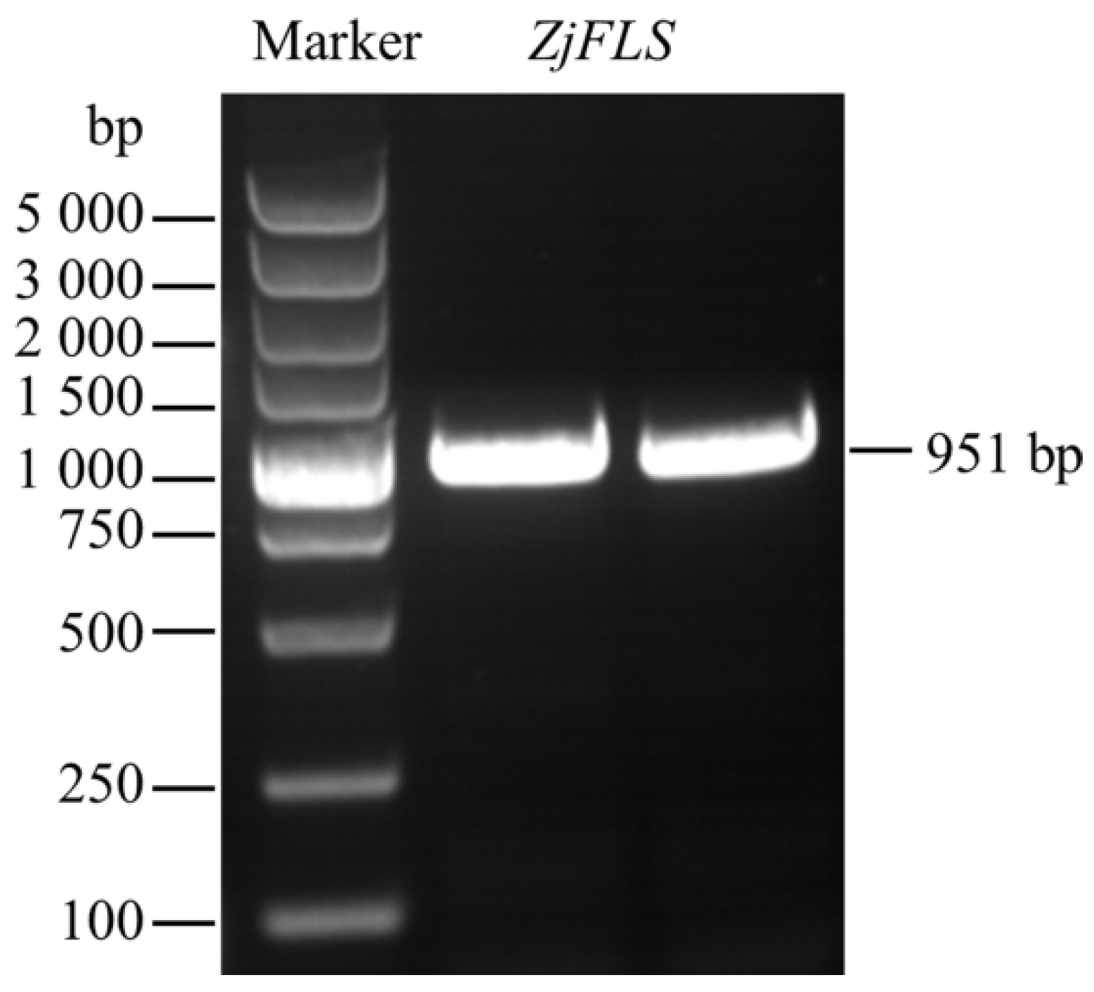
3.1. Cloning and Bioinformatics Analyses of the ZjFLS Gene

We amplified the FLS gene using the cDNA of the semi-red-stage fruit of Ziziphus jujuba cv. Hupingzao as a template and the designed specific primers. The results of the electrophoresis determination combined with a sequencing analysis indicated that the cloned sequence was consistent with the expected target (Figure 1). The length of the cloned FLS is 951 bp, with predicted encoding of 316 amino acids; the protein molecular weight is 36.20 kD; and the theoretical isoelectric point (pI) is 5.77. A protein secondary structure analysis revealed that ZjFLS was composed of 33.54% α-helix, 16.46% β-fold, and 50.00% random curl. This protein has no signal peptide and its phosphorylation site is mainly serine.

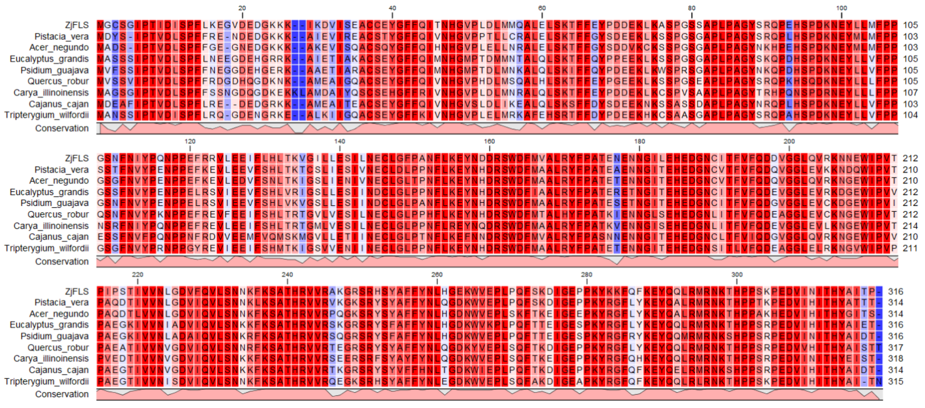
3.2. Analysis of ZjFLS Sequence Characteristics

We searched different plant FLS sequences within the NCBI database. Eight species, Pistacia vera, Eucalyptus grandis, Psidium guajava, Acer negundo, Cajanus cajan, Quercus robur, Carya illinoinensis, and Tripterygium wilfordii, were screened out based on multiple restrictive conditions such as coverage, sequence consistency, and required length, and then the sequences of these species were compared with ZjFLS in multiple sequences (Figure 2). It can be seen that the amino acid sequence encoded by the ZjFLS gene is highly similar to the FLS amino acid sequence of other species, indicating that the function of the ZjFLS protein in jujubes is relatively conformed.
Based on the protein sequence of ZjFLS, a BLAST comparison was conducted in NCBI, and we downloaded homologous genes of a total of eight species, including Pistacia vera, Eucalyptus grandis, Psidium guajava, Acer negundo, Cajanus cajan, Quercus robur, Carya Illinoinensis, and Tripterygium wilfordii. We conducted a phylogenetic analysis of these homologous genes and the ZjFLS gene and found that ZjFLS has a relatively close genetic relationship with Pistacia vera and Acer negundo (Figure 3). This may be because of a relatively recent common ancestor, less convergent evolution, or less hybridization interference.

3.3. Tissue and Organ Expression Characteristics of ZjFLS

The expression characteristics of ZjFLS in fruits at different developmental stages and different tissues of Ziziphus jujuba cv. Hupingzao were analyzed (Figure 4). The results showed that the ZjFLS gene could be expressed in fruits, leaves, and flowers, but the expression levels in different tissues greatly varied. For fruits at different developmental stages, the expression level of ZjFLS in the white-ripe stage was significantly higher than that in the half-red stage and full-red stage, while there was no significant difference between the half-red stage and full-red stage. For different tissues, the expression level of ZjFLS was the highest in leaves and was significantly higher in young leaves than in mature leaves; in mature leaves, it was significantly higher than in flowers. The expression level of ZjFLS in flowers was slightly higher than that in white-ripe fruits, but the difference was not significant. It can be found from this that the expression of ZjFLS has tissue specificity. The expression level is the highest in leaves, followed by flowers, and the lowest is in fruits.

3.4. Subcellular Localization of ZjFLS Protein

In order to understand the location of the ZjFLS protein in the cells, Agrobacterium containing the recombinant plasmid pZjFLS-GFP was injected into the leaves of Nicotiana tabacum L. and the empty plasmid pCAMBIA2300-GFP was used as the control. Then, the fluorescence position was observed by confocal microscopy. As can be seen from Figure 5, ZjFLS has no specific localization. In addition to being located in the cell nucleus, it can also be located in other parts of the cell, such as the cytoplasmic matrix and the cell membrane.

3.5. Prokaryotic Expression Analysis of ZjFLS

The protein expression analysis results based on SDS-PAGE indicated that the recombinant protein ZjFLS successfully achieved heterologous expression in the Escherichia coli BL21(DE3) system. The ZjFLS recombinant protein showed an obvious specific band four hours after being induced, and its apparent molecular weight was approximately 39 kDa (Figure 6), which was basically consistent with the theoretical molecular weight predicted based on amino acid sequences (39.46 kDa). The ZjFLS recombinant protein mainly exists in centrifugal precipitation, indicating that the protein is mainly expressed in the form of inclusion bodies. However, only trace amounts of ZjFLS were detected in the soluble supernatant. After purification by nickel column affinity chromatography, recombinant ZjFLS protein with good singularity could be obtained.

3.6. Identification of Arabidopsis thaliana Strains with Heterologous Overexpression of the ZjFLS Gene

The ZjFLS gene was transformed into Arabidopsis thaliana by the floral dip method, and the transgenic strains were detected by PCR. The agarose gel electrophoresis results indicated that bands of the same size as the target gene fragment were obtained (Figure 7), suggesting that the ZjFLS gene was successfully integrated into the Arabidopsis thaliana genome and, therefore, we had obtained positive overexpressed Arabidopsis thaliana strains.

3.7. Analysis of the Expression Level of ZjFLS and Total Flavonoid Content in Positive Arabidopsis thaliana Strains

By analyzing the relative expression levels and total flavonoid content levels of wild-type and positive Arabidopsis thaliana strains (Figure 8), it could be seen that the total flavonoid contents of the nine positive Arabidopsis thaliana strains were all higher than that of the wild type. Among them, the total flavonoid content of strain 8 was the highest (2.510 mg/g FW), 3.67 times that of the wild type, and strain 9 was the lowest (1.333 mg/g FW), 1.95 times that of the wild type. The expression level of the ZjFLS gene in different strains was significantly positively correlated with the total flavonoid content, while the expression level in the wild type was 0. In conclusion, the ZjFLS gene plays an important role in the synthesis of flavonoids in Chinese jujube.

4. Discussion

Chinese jujube (Ziziphus jujuba Mill.) is an economically important forest tree species with strong stress resistance. Its fruit is rich in nutrients and is an important ‘homology of medicine and food’ fruit. Flavonoids are an important type of secondary metabolite contained in jujubes. FLS, as a key gene influencing the formation of flavonoids, has been isolated from a variety of plants. Combined with the results of previous studies, we screened out an FLS gene from the transcriptome data that might play a key role in the biosynthesis of flavonoids. To further study its biological functions, we cloned an FLS homologous gene from jujube fruits and named it ZjFLS. A multiple sequence alignment analysis showed that the amino acids encoded by this gene had high homology with the FLS amino acids of several other plants, indicating that the ZjFLS gene was relatively conserved among different species, which was the same as in previous research results. The results of the evolutionary tree showed that ZjFLS has a relatively high homology and similarity with five other species, including Pistacia vera, Acer negundo, Quercus robur, Carya illinoinensis, and Tripterygium wilfordii. The subcellular localization results indicated that the ZjFLS protein had signals in the nucleus, cytoplasmic matrix, and cytoplasmic membrane, which was consistent with the research results of Li Wei et al. on wheat FLS [33]. These research results all indicate that the ZjFLS gene plays a role in multiple parts of the cell.
Gene expression related to flavonoid compound biosynthesis is affected by plant species, tissues, and environmental conditions, and there are different regulatory mechanisms at work in plants. The expression level of the FLS gene varies in different developmental stages of plants, different tissues, and different environments. Through the expression analysis results of ZjFLS in this study, it can be known that its expression exhibits certain tissue specificity. Overall, the order of expression levels of ZjFLS in different tissues was leaves > flowers > fruits, and the expression level in young leaves was significantly higher than that in mature leaves. During the fruit development process, the expression level of ZjFLS demonstrated a dynamic change trend, with the highest expression level during the white-ripe stage. Many scholars have conducted extensive studies on the expression characteristics of the FLS gene in different species. For example, the highest expression level of FLS in Yulania denudata was found in flowers, and Yulania liliiflora had the highest expression level in young leaves [34]. This might be caused by the genetic differences among different plants. Among the three different flower colors of Meconopsis species, FLS was expressed in different tissues and the expression levels were significantly different [35]. The SsFLS2 gene was expressed in the roots, stems, and leaves of Solanum sisymbriifolium Lam., and the relative expression levels in the roots and leaves were significantly higher than those in the stems [36]. The FLS gene was expressed in all tested tissues of blueberries and it could be seen that its expression level was particularly high in the petals and young leaves, while the expression level was relatively low in the fruits [37]. The above research results are basically consistent with those of this study. Therefore, the expression of FLS in different species shows tissue specificity.
Gene overexpression is widely used in the functional study of plant genes as the metabolic changes it causes can be manifested in the form of phenotypic changes or physiological data changes [38]. In apples, the overexpression of the proton pump gene MdMa12 increased the malic acid content in both tomato and apple callus tissues [39]. The overexpression of the MdmiR156n gene promoted the accumulation of flavonoids and the clearance of reactive oxygen species (ROS) in transgenic apple calluses under drought conditions and Arabidopsis thaliana [40]. In grapes, after the overexpression of the VvbZIP36 gene, the quercetin content increased due to the activation of the VvFLS promoter [41]. The overexpression of the CsPALs gene in citrus not only increases the phenolic substance content, but also causes changes in the contents of various sugars and increases most amino acid contents [42]. When the CHS gene was overexpressed, the contents of flavonoids such as glycyrrhizin and isoglycyrrhizin, as well as total flavonoid in the hairy roots of transgenic licorice, were significantly higher than those in the wild-type hairy roots [43]. The results in this study indicate that the overexpression of ZjFLS can promote an increase in the total flavonoid content in Arabidopsis thaliana, which is consistent with the research results in safflower and ferns [44,45]. In sweet potato, silencing the IbFLS gene suppresses the expression of IbFLS and upregulates the expression of DFR, ANS, and UFGT, which results in a significant increase in total anthocyanin content and a decrease in total flavonol content in the leaves of transgenic plants. Thus, IbFLS participates in the flavonol biosynthesis pathway and is a potential candidate gene for color modification in sweet potato [46]. The overexpression of a certain gene can also affect the expression changes of other related enzyme genes in the synthetic pathway. In this study, the overexpression of the ZjFLS gene increased the total flavonoid content in Arabidopsis thaliana. But which flavonoids increased? This doubt creates the need for further studies in the future. Transcription factors are a class of gene regulatory molecules that bind to DNA and either activate or suppress the transcription of specific genes. In Astraglus membranaceus, AmMYB35 promotes the expression of FLS by binding to its promoter. Under drought stress, flavonol accumulation is induced through the AmMYB35AmFLS module, thereby enhancing drought tolerance by reducing ROS accumulation [47]. Therefore, in the future, we should conduct in-depth research into the interaction mechanisms of different genes and analyze the transcriptional regulation of related genes from the perspective of transcription factors. The aim is to provide a solid theoretical basis for the in-depth revelation of the molecular mechanisms of flavonoid substance synthesis in Chinese jujube.

5. Conclusions

The ZjFLS gene of jujubes was successfully cloned, with a length of 951 bp and encoding 316 amino acids. We conducted a bioinformatics analysis of the amino acid sequence of ZjFLS and the results indicated that the sequence was conserved. The ZjFLS gene is expressed in the leaves, flowers, and fruits and is tissue-specific. The expression level was the highest in the leaves, followed by the flowers, and was relatively low in the fruits. Subcellular localization revealed that the ZjFLS protein was located in the nucleus, cytoplasmic matrix, and cell membrane. The recombinant protein ZjFLS was successfully heterologously expressed in Escherichia coli, and its purified protein was obtained, providing a basis for further verification of its catalytic activity. The ZjFLS gene was overexpressed in Arabidopsis thaliana. Compared with the wild type, the total flavonoid content of the positive transformed strains significantly increased, confirming that the ZjFLS gene can play an important role in the flavonoid accumulation of jujubes. Next, we will conduct research into the influence of the ZjFLS gene on the synthesis of flavonoid monomers and its transcriptional regulatory mechanism.

Author Contributions

Conceptualization, X.X., Y.W. and H.R.; methodology, A.Z. and L.F.; investigation, X.X. and D.L.; data curation, M.S., L.L. and W.S.; formal analysis, Y.L.; writing—original draft preparation, X.X.; writing—review and editing, X.X. and D.L.; funding acquisition, X.X. and A.Z. All authors have read and agreed to the published version of the manuscript.

Funding

This work was supported by the Shanxi Province Basic Research Program (202203021221176); The Doctoral Talent Introduction Research Start-up Project of Shanxi Agricultural University (2022BQ30); the China Agriculture System (CARS-30-1-07, CARS-30-ZZ-22); and the Major Science and Technology Special Project Plan of Shanxi Province (202201140601027).

Data Availability Statement

The original contributions presented in this study are included in this article and further inquiries can be directed to the corresponding author.

Acknowledgments

Thanks to the National Jujube Germplasm Repository of China for providing the experimental materials for this study.

Conflicts of Interest

The authors declare no conflicts of interest.

References

  1. Qu, Z.Z.; Wang, Y.H. Fruit records. In Chinese jujube (Ziziphus jujuba Mill.); China Forestry Publishing House: Beijing, China, 1993; pp. 1–2. [Google Scholar]
  2. Liu, M.J.; Wang, M. Germplasm Resources of Chinese jujube; China Forestry Publishing House: Beijing, China, 2009; pp. 23–24. [Google Scholar]
  3. Li, D.K.; Niu, X.W.; Tian, J.B. The Illustrated Germplasm Resources of Chinese jujube; China Agriculture Press: Beijing, China, 2013; pp. 523–526. [Google Scholar]
  4. Zhu, D.Q.; Jiang, N.; Wang, N.; Zhao, Y.F.; Liu, X.M. A literature review of the pharmacological effects of jujube. Foods 2024, 13, 193. [Google Scholar] [CrossRef] [PubMed]
  5. Xue, X.F.; Zhao, A.L.; Wang, Y.K.; Ren, H.Y.; Li, Y.; Li, D.K.; Du, J.J. Metabolomics-based analysis of flavonoid metabolites in Chinese jujube and sour jujube fruits from different harvest periods. J. Food Sci. 2022, 87, 3752–3765. [Google Scholar] [CrossRef] [PubMed]
  6. Ji, X.L.; Zhang, S.L.; Jin, X.Y.; Yin, C.X.; Zhang, Y.; Guo, X.D.; Lin, X.M. Systematic comparison of structural characterization of polysaccharides from Ziziphus jujuba cv. Muzao. Molecules 2023, 28, 562. [Google Scholar] [CrossRef]
  7. Yang, M.; Han, L.; Zhang, S.F.; Dai, L.; Li, B.; Han, S.K.; Zhao, J.; Liu, P.; Zhao, Z.H.; Liu, M.J. Insights into the evolution and spatial chromosome architecture of jujube from an updated gapless genome assembly. Plant Commun. 2023, 4, 100662. [Google Scholar] [CrossRef]
  8. Liu, Z.G.; Yuan, Y.; Wang, L.L.; Zhao, X.; Wang, L.X.; Wang, L.H.; Zhao, Z.H.; Zhao, X.; Chu, Y.T.; Gao, Y.N.; et al. Three novel adenylate cyclase genes show significant biological functions in plant. J. Agric. Food Chem. 2023, 71, 1149–1161. [Google Scholar] [CrossRef]
  9. Pan, F.X.; Zhao, Z.H.; Liu, F.W.; Liu, F.W.; Luo, Z.; Chen, S.J.; Liu, Z.G.; Zhao, Z.H.; Liu, M.J.; Wang, L.L. Triterpenoids in jujube: A review of composition, content diversity, pharmacological effects, synthetic pathway, and variation during domestication. Plants 2023, 12, 1501. [Google Scholar] [CrossRef]
  10. Cai, W.T.; Zhuang, H.N.; Wang, X.Y.; Fu, X.; Chen, S.; Yao, L.Y.; Sun, M.; Wang, H.T.; Yu, C.; Feng, T. Functional nutrients and jujube-based processed products in Ziziphus jujuba. Molecules 2024, 29, 3437. [Google Scholar] [CrossRef] [PubMed]
  11. Xue, X.F.; Zhao, A.L.; Wang, Y.K.; Ren, H.Y.; Du, J.J.; Li, D.K.; Li, Y. Composition and content of phenolic acids and flavonoids among the different varieties, development stages, and tissues of Chinese jujube (Ziziphus jujuba Mill.). PLoS ONE 2021, 16, e0254058. [Google Scholar] [CrossRef]
  12. Choi, S.S.; Park, S.R.; Lee, K.A. A comparative study of rutin and rutin glycoside: Antioxidant activity, anti-inflammatory effect, effect on platelet aggregation and blood coagulation. Antioxidants 2021, 10, 1696. [Google Scholar] [CrossRef]
  13. Wu, Q.; Needs, P.W.; Lu, Y.L.; Kroon, P.A.; Ren, D.Y.; Yang, X.B. Correlation: Different antitumor effects of quercetin, quercetin-3′-sulfate and quercetin-3-glucuronide in human breast cancer MCF-7 cells. Food Funct. 2019, 10, 4452. [Google Scholar] [CrossRef]
  14. Brenda, W.S. Flavonoid biosynthesis. A colorful model for genetics, biochemistry, cell biology and biotechnology. Plant Physiol. 2001, 126, 485–493. [Google Scholar]
  15. Xu, F.; Li, L.L.; Zhang, W.W.; Cheng, H.; Sun, N.N.; Cheng, S.Y.; Wang, Y. Isolation, characterization, and function of a flavonol synthase gene from Ginkgo biloba. Mol. Biol. Rep. 2012, 39, 2285–2296. [Google Scholar] [CrossRef] [PubMed]
  16. Stracke, R.; Vos, R.; Bartelniewoehner, L.; Ishihara, H.; Sagasser, M.; Martens, S.; Weisshaar, B. Metabolomic and genetic analyses of flavonol synthesis in Arabidopsis thaliana support the in vivo involvement of leucoanthocyanidin dioxygenase. Planta 2009, 229, 427–445. [Google Scholar] [CrossRef]
  17. Holton, T.A.; Brugllera, F.; Tanaka, Y. Cloning and expression of flavonol synthase from Petunia hybrida. Plant J. 1993, 4, 1003–1010. [Google Scholar] [CrossRef]
  18. Liu, X.; Lu, J.; Hao, Y.J.; You, C.X. Cloning, bioinformatics analysis and catalytic activity identification of MdFLS1 gene in apple. J. Fruit. Sci. 2018, 35, 905–916. [Google Scholar]
  19. Xiao, W.J.; Hu, Y.L.; Wang, Q.Y.; Duan, Y.J.; Hu, H.G. Cloning, bioinformatics and functional analysis of MaFLS1 in banana. J. Fruit. Sci. 2024, 41, 229–240. [Google Scholar]
  20. Ouyang, Y.N.; Liu, Y.; Peng, J.S.; Chen, Y.Y.; Zhao, H.; He, Y.T.; Zhao, X.Y.; Tang, X.K.; Zhou, M.L. Function and genetic diversity analysis of FtFLS1 from tartary buckwheat. J. Plant Genet. Resour. 2023, 24, 1401–1412. [Google Scholar]
  21. Wang, W.H.; Li, D.B.; He, F.; Wang, W.J.; Wu, Y.Y. Cloning and functional analysis of RhFLS gene of Rhododendron. Mol. Plant Breed. 2022, 20, 4243–4250. [Google Scholar]
  22. Liu, X.L.; Sun, T.T.; Yang, J.; He, H.B. Cloning and expression analysis of FLS gene of flavonol synthetase in Lilium auratum and L. speciosum var. gloriosoides. Acta Agric. Zhejiangensis 2024, 36, 344–357. [Google Scholar]
  23. Li, H.H.; Liu, Y.L.; Liu, H.L.; Lou, Q. Cloning and expression analysis of FLS gene in Muscari armeniacum. J. Northwest For. Univ. 2019, 34, 116–121. [Google Scholar]
  24. Liu, H.L.; Su, B.B.; Zhang, H.; Gong, J.X.; Zhang, B.X.; Liu, Y.L.; Du, L.J. Identification and functional analysis of a flavonol synthase gene from grape hyacinth. Molecules 2019, 24, 1579. [Google Scholar] [CrossRef] [PubMed]
  25. Jiang, X.L.; Shi, Y.F.; Fu, Z.P.; Li, W.W.; Lai, S.Y.; Wu, Y.H.; Wang, Y.S.; Liu, Y.J.; Gao, L.P.; Xia, T. Functional characterization of three flavonol synthase genes from Camellia sinensis: Roles in flavonol accumulation. Plant Sci. 2020, 300, 110632. [Google Scholar] [CrossRef]
  26. Tian, J.; Han, Z.Y.; Zhang, J.; Hu, Y.J.; Song, T.T.; Yao, Y.C. The balance of expression of dihydroflavonol 4-reductase and flavonol synthase regulates flavonoid biosynthesis and red foliage coloration in Crabapples. Sci. Rep. 2015, 5, 122228. [Google Scholar] [CrossRef]
  27. Lei, T.; Huang, J.; Ruan, H.X.; Qian, W.; Fang, Z.; Gu, C.Y.; Zhang, N.N.; Liang, Y.X.; Wang, Z.Y.; Gao, L.P.; et al. Competition between FLS and DFR regulates the distribution of flavonols and proanthocyanidins in Rubus chingii Hu. Front. Plant Sci. 2023, 14, 1134993. [Google Scholar] [CrossRef] [PubMed]
  28. Choudhary, N.; Pucker, B. Conserved amino acid residues and gene expression patterns associated with the substrate preferences of the competing enzymes FLS and DFR. PLoS ONE 2024, 19, e0305837. [Google Scholar] [CrossRef] [PubMed]
  29. Fang, F.; Huang, W.D. Salicylic acid modulated flavonol biosynthesis in three key phases during grape berry development. Eur. Food Res. Technol. 2013, 237, 441–448. [Google Scholar] [CrossRef]
  30. Yang, X.; Zhao, X.Y.; Miao, Y.N.; Wang, D.X.; Zhang, Z.H.; Liu, Y.X. Genome-wide identification and expression profile analysis of the WUSCHEL-related homeobox (WOX) genes in woodland strawberry (Fragaria vesca). Horticulturae 2022, 8, 1043. [Google Scholar] [CrossRef]
  31. Livak, K.J.; Schmittgen, T.D. Analysis of relative gene expression data using real-time quantitative PCR and the 2−△△CT method. Methods 2001, 25, 402–408. [Google Scholar] [CrossRef]
  32. Zhao, A.L.; Li, D.K.; Wang, Y.K.; Sui, C.L.; Du, X.M.; Cao, Y.Q. Study on the content of triterpene acids and flavones for jujubes of different varieties, different growing periods and different parts. Acta Hortic. 2009, 840, 533–540. [Google Scholar] [CrossRef]
  33. Li, W. Study on Mechanisms of TaFLS1 and TaANS1 of Triticum aestivum in Abiotic Stress Response. Ph.D. Thesis, Shandong University, Jinan, China, 2011. [Google Scholar]
  34. Wu, C.; Dai, M.Y.; Zhang, C.; Shi, C.G.; Ren, M.J.; Ma, J.J.; Shen, Y.M. Study on the mechanism of the FLS gene regulating flower color formation in Yulania denudata and Y. liliiflora. J. Nucl. Agric. Sci. 2023, 37, 1947–1956. [Google Scholar]
  35. Wang, H.J.; Li, T.J.; Luo, J.; Qu, Y. Cloning and expression analysis of FLS gene in three different color Meconopsis species. Plant Phys. J. 2023, 59, 2063–2073. [Google Scholar]
  36. Sun, M.; Wu, L.Y.; Gong, Y.J.; Bao, R.; Gui, M.; Li, Z.B.; Du, G.H. Gene cloning and its expression analysis of SsFLS2 in Solanum sisymbriifolium. Southwest China J. Agric. Sci. 2024, 37, 1669–1676. [Google Scholar]
  37. Zhang, C.Y.; Liu, H.C.; Jia, C.G.; Liu, Y.J.; Wang, F.T.; Wang, J.Y. Cloing, characterization and functional analysis of a flavonol synthase from Vaccinium corymbosum. Trees 2016, 30, 1595–1605. [Google Scholar] [CrossRef]
  38. Prelich, G. Gene overexpression: Uses, mechanisms, and interpretation. Genetics 2012, 190, 841–854. [Google Scholar] [CrossRef]
  39. Gao, M.; Zhao, H.Y.; Zheng, L.T.; Zhang, L.H.; Peng, Y.J.; Ma, W.F.; Tian, R.; Yuan, Y.Y.; Ma, F.W.; Li, M.J.; et al. Overexpression of apple Ma12, a mitochondrial pyrophosphatase pump gene, leads to malic acid accumulation and the upregulation of malate dehydrogenase in tomato and apple calli. Hortic. Res. 2022, 9, uhab053. [Google Scholar] [CrossRef]
  40. Chen, G.; Wang, Y.P.; Liu, X.L.; Duan, S.Y.; Jiang, S.H.; Zhu, J.; Zhang, Y.G.; Hou, H.M. The MdmiR156n regulates tolerance and flavonoid synthesis in apple calli and Arabidopsis. Int. J. Mol. Sci. 2023, 24, 6049. [Google Scholar] [CrossRef] [PubMed]
  41. Liu, G.C.; Li, M.F.; Zhang, Z.; Han, F.L.; Yan, W.; Liu, Y.L.; Li, M.R.; Xia, Y.N.; Yang, J.; Xu, X.F.; et al. Enhancement of grape tolerance through VvbZIP36-mediated quercetin production. Int. J. Bio. Macromol. 2025, 297, 139826. [Google Scholar] [CrossRef]
  42. Wei, L.L.; Wang, W.J.; Gao, X.Q.; Yao, S.X.; Deng, L.L.; Zeng, K.F. Alterations in glucose metabolism and shikimate pathway affected by transient overexpression of CsPAL gene in postharvest citrus fruit. Postharvest Biol. Technol. 2024, 213, 112936. [Google Scholar] [CrossRef]
  43. Yin, Y.C.; Hou, J.M.; Tian, S.K.; Yang, L.; Zhang, Z.X.; Li, W.D.; Liu, Y. Overexpressing chalcone synthase (CHS) gene enhanced flavonoids accumulation in Glycyrrhiza uralensis hairy roots. Bot. Lett. 2020, 167, 219–231. [Google Scholar] [CrossRef]
  44. Ma, X.T.; Hou, Y.Y.; Umar, A.W.; Wang, Y.H.; Yu, L.L.; Ahmad, N.; Yao, N.; Zhang, M.; Liu, X.M. Safflower CtFLS1-induced drought tolerance by stimulating the accumulation of flavonols and anthocyanins in Arabidopsis thaliana. Int. J. Mol. Sci. 2024, 25, 5546. [Google Scholar] [CrossRef]
  45. Fu, J.; Lu, Y.; Zhang, J.L.; Ni, R.; Liu, X.Y.; Hu, M.W.; Li, J.H.; Zhang, J.Z.; Jiang, N.; Xu, D.D.; et al. Functional characterization of promiscuous 2-ODD enzymes sheds light on the molecular basis for flavone and flavonol biosynthesis in ferns. Plant J. 2025, 122, e70189. [Google Scholar] [CrossRef] [PubMed]
  46. Kou, M.; Li, C.; Song, W.H.; Shen, Y.F.; Tang, W.; Zhang, Y.G.; Wang, X.; Yan, H.; Gao, R.F.; Ahmad, M.Q.; et al. Identification and functional characterization of a flavonol synthase gene from sweet potato [Ipomoea batatas (L.) Lam.]. Front. Plant Sci. 2023, 14, 1181173. [Google Scholar] [CrossRef] [PubMed]
  47. Qi, L.T.; Sun, H.G.; Tian, C.; Su, W.W.; Garcia-Caparros, P.; Wang, J.M.; Zhou, Y.J.; Liu, T.X.; Gao, F. The AmMYB35-AmFLS module mediates the accumulation of flavonol induced by drought stress in Astragalus membranaceus. Food Biosci. 2025, 68, 106541. [Google Scholar] [CrossRef]
Figure 1. The electrophoresis of PCR amplification of ZjFLS; 2 kb plus a DNA marker were utilized for agarose gel electrophoresis.
Horticulturae 11 00729 g001
Figure 2. Alignments of protein sequences of ZjFLS and other FLSs.
Horticulturae 11 00729 g002
Figure 3. Phylogenetic analysis of ZjFLS with proteins from other species. Cc: Cajanus cajan; Tw: Tripterygium wilfordii; Ci: Carya illinoinensis; Qr: Quercus robur; An: Acer negundo; Pv: Pistacia vera; Zj: Ziziphus jujuba; Pg: Psidium guajava; Eg: Eucalyptus grandis.
Horticulturae 11 00729 g003
Figure 4. Expression analyses of ZjFLS gene in different developmental stages of the fruits and tissues of Ziziphus jujuba cv. Hupingzao. Different letters above the bars indicate significant differences at the level of p < 0.05.
Horticulturae 11 00729 g004
Figure 5. Subcellular localization analysis of ZjFLS in tobacco. The GFP channel denotes ultraviolet excitation, DIC corresponds with bright field, and Merge represents the overlay image. DAPI signifies the nuclear dye. The excitation wavelengths are 488 nm for the GFP channel and 358 nm for the DAPI channel. he scale bar in the image is 10 μm.
Horticulturae 11 00729 g005
Figure 6. Expression and purification of ZjFLS in prokaryotes. Lane M denotes the protein marker. Lane 1 exhibits proteins from the lysed sample’s pellet, Lane 2 displays proteins from the lysed sample’s supernatant, Lane 3 shows proteins from the flow-through buffer, Lane 4 illustrates proteins from the wash buffer, and Lane 5 demonstrates proteins eluted from the purification bands. Red arrows point to the purified ZjFLS protein band.
Horticulturae 11 00729 g006
Figure 7. Identification of ZjFLS heterologously overexpressed Arabidopsis thaliana. M represents the marker and Lines 1 to 16 represent transgenic ZjFLS gene-positive lines. WT: wild type.
Horticulturae 11 00729 g007
Figure 8. Expression levels of ZjFLS gene and total flavonoid content in Arabidopsis thaliana with overexpressed ZjFLS. Different letters above the bar indicate significant differences at the level of p < 0.05.
Horticulturae 11 00729 g008
Disclaimer/Publisher’s Note: The statements, opinions and data contained in all publications are solely those of the individual author(s) and contributor(s) and not of MDPI and/or the editor(s). MDPI and/or the editor(s) disclaim responsibility for any injury to people or property resulting from any ideas, methods, instructions or products referred to in the content.

