Article

Characterization of Polyploid Embryoid Lines Induced via Unfertilized Ovule Culture of Loquat (Eriobotrya japonica Lindl.)

1
Key Laboratory of Agricultural Biosafety and Green Production of Upper Yangtze River (Ministry of Education), Chongqing Key Laboratory of Forest Ecological Restoration and Utilization in the Three Gorges Reservoir Area, College of Horticulture and Landscape Architecture, Southwest University, Beibei, Chongqing 400715, China
2
State Cultivation Base of Crop Stress Biology for Southern Mountainous Land, Academy of Agricultural Sciences, Southwest University, Beibei, Chongqing 400715, China
*
Authors to whom correspondence should be addressed.
These authors contributed equally to this work.
Horticulturae 2025, 11(7), 727; https://doi.org/10.3390/horticulturae11070727
Submission received: 14 May 2025 / Revised: 18 June 2025 / Accepted: 19 June 2025 / Published: 23 June 2025

Abstract

Polyploidy plays a significant role in loquat breeding, particularly in triploid breeding for seedless fruit production. Currently, loquat polyploid breeding primarily relies on natural seedling selection and sexual hybridization approaches. In this study, unfertilized ovules from four loquat varieties were in vitro cultured. Gynogenesis and embryoid regeneration were achieved in ‘Xingning 1’ and ‘Huabai 1’, with ‘Xingning 1’ demonstrating the highest gynogenesis efficiency (21.63%). Flow cytometry and chromosome counting revealed that the obtained embryoid lines included haploid, diploid, tetraploid, hexaploid, and chimeric ploidy types. Further characterization of ‘Xingning 1’-derived embryoid lines through SSR markers and whole-genome resequencing confirmed that the haploid, diploid, tetraploid, and hexaploidy embryoid originated from haploid–somatic chimeras, diploid, doubled diploid and tripled diploid, respectively. Metabolic analysis showed a positive correlation between ploidy level and the content of both soluble sugars and organic acids. This study explored a novel platform for polyploid induction in loquat and may provide methodological insights for improvement of other perennial fruit trees.

1. Introduction

Polyploidy plays a crucial role in speciation and evolutionary processes, particularly in higher plants [1,2]. Many important crops are classic polyploids, such as Brassica napus [3], wheat [4], sugarcane [5], potato [6], cotton [7], and peanut [8]. Polyploidy is also prevalent in fruit crops, including banana [9], grape [10], kiwifruit [11], jujube [12], and strawberry [13]. Compared with diploids, polyploid plants exhibit multiple agronomically valuable characteristics, such as strong plant morphology, vegetative organs gigantism, elevated nutritional quality, improved stress resistance, and increased genetic diversity [14,15,16,17]. Moreover, polyploidization can effectively break reproductive barriers (self-incompatibility and interspecific sterility) through genome duplication in some cases, thereby generating valuable new genetic resources for crop improvement [18].
Polyploid breeding in fruit crops can be achieved through various approaches, including seedling selection, physical induction, chemical induction, sexual hybridization, and tissue culture [19,20,21,22]. Among these methods, chemical induction and sexual hybridization represent the primary methods for obtaining polyploid fruit cultivars [23]. Additionally, tissue culture serves as an important pathway for polyploid generation. Through somatic hybridization techniques, a series of polyploid germplasms with improved horticultural traits have been obtained [24,25]. In vitro endosperm culture can directly produce triploid plants, effectively shortening the breeding cycle [22,26]. Generally, unfertilized ovule culture serves as an effective approach for inducing gynogenesis to obtain haploids or doubled haploids (DH) [27,28]. However, reports on polyploid induction through ovule culture remain limited.
Loquat (Eriobotrya japonica Lindl.), as an important subtropical evergreen fruit tree, possesses significant economic value. Renowned for its rich flavor and health-promoting properties, it is widely cultivated worldwide [29]. Triploid loquat varieties exhibit excellent traits, such as seedlessness, enhanced stress resistance, and elevated sugar content, demonstrating great potential in breeding [15,30,31,32]. However, due to the lack of a widely applicable regeneration system in loquat [33], most reported polyploids are obtained through seedling selection, which remains inefficient and labor consuming [34]. Sexual hybridization between diploid and tetraploid loquats is a more efficient process but is constrained by the limited genetic diversity of the tetraploid parent. Thus, the development of efficient polyploid induction approaches through biotechnological methods still requires further exploration.
In this study, unfertilized ovules from four loquat varieties were cultured in vitro. The ploidy levels of the obtained embryoids were analyzed by flow cytometry and chromosome counting. Furthermore, SSR markers and whole-genome resequencing were employed to investigate the genomic origins of embryoids with different ploidy levels. Finally, HPLC analysis revealed ploidy-dependent variations in soluble sugar and organic acid profiles. This research established a novel platform for polyploid induction in loquat and provided methodological insights for perennial fruit tree improvement.

2. Materials and Methods

2.1. Unfertilized Ovules Culture

Unfertilized mature floral buds collected 2–3 days prior to anthesis from five loquat varieties at Southwest University, Beibei, Chongqing, China (‘Senwei Zaosheng’ ‘Huabai 1’ ‘Xingning 1’ ‘Zaozhong 6’, and ‘Daduhe’), were selected as experimental material. Once collected, the floral buds were maintained at 4 °C for a duration of 48 h.
Subsequently, petals, anthers, and stigmas were removed from the floral buds, leaving only the ovaries. The remaining ovaries were surface-sterilized by immersion in 75% ethanol for 30–60 s, followed by treatment with a NaClO solution containing 4% effective chlorine for 15–20 min. After sterilization, the ovaries were rinsed 5 times with sterile water.
The ovaries were carefully dissected using a sterile scalpel, and unfertilized ovules were excised and transferred to the induction medium. The culture process was conducted under light (40 μmol m−2 s−1 illumination with a 16 h/8 h light/dark cycle) at 25 °C, and all steps were carried out within a laminar airflow hood to maintain sterility. The induction medium was prepared using a modified B5 basal medium [35], supplemented with 1 mg·L−1 2,4-D, 0.5 mg·L−1 6-BA, 0.1 mg·L−1 NAA, 30 g·L−1 sucrose, and 7.5 g·L−1 agar, with pH adjusted to 5.8. The formulated medium was sterilized via autoclaving at 121 °C for 20 min before use. For each genotype, more than 20 culture dishes were prepared, with nine ovules inoculated per dish, and the experiment was replicated three times.
Derived calluses and embryoids were sub-cultured monthly on a B5 basal medium containing 0.1 mg·L−1 2,4-D, 0.1 mg·L−1 6-BA, 0.1 mg·L−1 NAA, 30 g·L−1 sucrose, and 7.5 g·L−1 agar (pH 5.8). All cultures were maintained under continuous illumination at 25 °C.
The gynogenesis rate was calculated as GR (%) = (number of gynogenesis lines/total number of inoculated unfertilized ovules) × 100%; the embryoid induction rate was calculated as EIR (%) = (number of derived embryoid line/number of gynogenesis lines) × 100%.

2.2. Ploidy Analysis and Chromosomal Cytogenetic Analysis

The chromosome ploidy of regenerated callus and embryonic lines was identified using a ‘CyFlow Space’ flow cytometer (Sysmex-Partec, Munster, Germany) as described by Wang et al. [36]. Chromosome counting assays were performed on 15-day-old sub-cultured embryoids employing the methodologies described by Dang et al. [37]. Images were acquired using light microscopy (Olympus, Tokyo, Japan). Young leaves from the donor plants ‘Xingning 1’ served as the control, with their fluorescence intensity normalized to 100 (2x = 100).

2.3. Molecular and Genome Characterization

Genomic DNA was extracted from ovule-derived embryoid lines using an optimized CTAB method [38].
For SSR (Simple Sequence Repeat) marker analysis, young leaves from donor plants ‘Xingning 1’ served as the control. The concentration of extracted DNA was adjusted to 50 ng/μL using a NanoDrop 2000 UV-Vis spectrophotometer (Thermo, Wilmington, USA). PCR amplifications were performed in a C1000 Touch PCR System thermal cycler (Bio-Rad, Hercules, CA, USA) in a 20-μL final volume containing 7 μL ddH2O, 10 μL 2 × Mix taq (Bioground, Chongqing, China), 1 μL template DNA, and 1 μL forward/reserve primers. The PCR amplification conditions were set as follows: 94 °C for 4 min, followed by 30 cycles of 94 °C for 30 s, 56 °C for 30 s, and 72 °C for 20 s, with a final extension at 72 °C for 10 min. Polyacrylamide gel electrophoresis was performed according to Wen et al. [38], and DNA banding patterns were visualized through silver staining following the protocol of Ruiz et al. [39].
Genomic DNA samples from haploid (x), tetraploid (4x), and hexaploid (6x) embryoids were subsequently sent to Shanghai Personal Biotechnology Co., Ltd (Shanghai, China). for high-throughput sequencing. The genomic libraries were constructed using the standard Illumina TruSeq Nano DNA LT protocol (Illumina TruSeq DNA Sample Preparation Guide) and subsequently sequenced on an Illumina NovaSeq X Plus platform (Illumina, San Diego, CA, USA).
Sequence quality filtering was performed by sliding-window quality control using FastQC software (v0.23.0). High-quality reads were aligned to the loquat reference genome using a BWA-MEM algorithm (version 0.7.17-r1188) [40,41].
SNPs (single-nucleotide polymorphisms) and InDels (genome insertions and deletions variations) were identified using GATK software [42] and the Genome Analysis Toolkit software package (v3.8) [43], respectively. CNVs (copy number variations) were detected using CNVnator (version 0.2.7) software, and SVs (structural variations) were identified using BreakDancer (v1.1) software [44,45]. Gene Ontology (GO) enrichment analysis was performed using clusterProfiler [46].

2.4. Metabolic Substance Determination

Soluble sugar and organic acid compositions of embryoids with three ploidy levels (x, 4x, 6x; CK: 2x) were quantitatively analyzed using a Prominence Plus LC-20A high-performance liquid chromatography (HPLC) system (Shimadzu, Kyoto, Japan) following the optimized method of Liu et al. [15]. Briefly, fresh embryoid samples were cryogenically grinded into powder using liquid nitrogen. For each biological replicate, 0.1 g of powdered sample was precisely weighed and mixed with 1.5 mL of ultrapure water. The resulting suspension was homogenized using an ultrasonic bath (KQ5200E, Kunshan Shumei Instrument, Kunshan, China) for 15 min at room temperature. The mixture was then centrifuged at 3000× g for 15 min at 4 °C using a Sorvall Legend Micro 17 Centrifuge (Thermo Fisher Scientific, Waltham, MA, USA). The supernatant was carefully filtered through a 0.22 µm water-compatible microporous membrane prior to HPLC analysis.
Chromatographic conditions for sugar analysis: A NH2 analytical column (5 μm, 4.6 mm × 250 mm, GL Science Co., Ltd., Kyoto, Japan) maintained at 25 °C was used for separation. The mobile phase consisted of an isocratic acetonitrile–water mixture (v/v = 70/30) delivered at a constant flow rate of 0.8 mL/min, with an injection volume of 10 μL.
Chromatographic conditions for organic acid analysis: Separation was performed on a C18-XT analytical column (5 μm, 4.6 mm × 250 mm, GL Science Co., Ltd., Kyoto, Japan) maintained at 30 °C. The mobile phase comprised 2% KH2PO4 (pH 2.52, adjusted with phosphoric acid) and methanol in a 90:10 (v/v) ratio, pumped isocratically at 0.8 mL/min with an injection volume of 10 μL.
The embryogenic callus derived from another culture of loquat ‘Xingning 1’ was utilized as the control (CK). The determination was conducted with six independent biological replicates to ensure statistical reliability.

2.5. Data Analysis

All quantitative data were processed using Microsoft Excel 2010 for preliminary organization and calculation. Subsequent statistical analysis was performed using one-way analysis of variance (ANOVA) via SPSS Statistics 25.0 software (SPSS Inc., Chicago, IL, USA) to determine significant differences among treatment groups, followed by a post hoc least significant difference (LSD) test at p < 0.05. Statistical results were visualized using Prism 8 (GraphPad Software, La Jolla, CA, USA), with data presented as mean ± standard deviation (SD) or standard error of the mean (SEM) depending on experimental design.

3. Results

3.1. Embryoid Induction from Unfertilized Ovules of Loquat

After three months of culture under light in the induction medium, gynogenesis was successfully observed, characterized by the formation of semi-transparent, smooth, protruding granules on the surface of the induced unfertilized ovules (Figure 1a), indicating the initiation of embryogenic callus formation. These granules expanded and enveloped the ovules, forming pale yellow, semi-transparent spherical structures (Figure 1b).
Upon sub-culture in the medium with reduced hormonal concentrations, the callus derived from unfertilized ovules gradually underwent texture and color change, transitioning into green, compact, granular masses (Figure 1c). With further sub-culturing, the induced embryogenic callus eventually differentiated into embryoids (Figure 1d).
The callus derived from loquat unfertilized ovule culture could be classified into two types. The Type I callus was pale yellow, fine-grained, and loose in texture, with rapid proliferation and the ability to induce embryoids (Figure 1e). The Type II callus presented as gray-white, spongy, and slightly water-soaked, with slow proliferation and no capacity for embryoid induction (Figure 1f).
Data analysis revealed significant differences in the GR and EIR (Table 1). Among the five tested varieties, ‘Xingning 1’ demonstrated the highest GR (21.63 ± 3.35%), while ‘Daduhe’ Loquat did not exhibit any observable gynogenesis, with a GR of 0%. For embryoid induction, the highest EIR was observed in ‘Xingning 1’, yielding 76 embryoid lines (54.37 ± 6.84%), followed by ‘Huabai 1’ (12 embryoid lines, 12.81 ± 3.13%), whereas ‘Senwei Zaosheng’ and ‘Zaozhong 6’ only formed callus lines, with no successful embryoid induction observed.

3.2. Ploidy Analysis of Embryoid Lines Derived from Unfertilized Ovules

Flow cytometry analysis of the 76 embryoid lines obtained from ‘Xingning 1’ revealed the following ploidy distribution: one line was haploid (1.32%, Figure 2a), six lines were diploid (7.89%, Figure 2b); forty-eight lines were tetraploid (63.16%, Figure 2c), and five lines were hexaploid (6.58%, Figure 2d). Notably, chimeric embryoids with mixed ploidy cells were identified, including nine diploid–tetraploid chimeras (11.84%, Figure S1a), four tetraploid–hexaploid chimeras (5.26%, Figure S1b), two tetraploid–octaploid chimeras (2.63%, Figure S1c), and one diploid–tetraploid–octaploid chimera (1.32%, Figure S1d). For ‘Huabai 1’-derived embryoid lines, only one diploid and two tetraploid embryoid lines had their ploidy accurately determined.
Chromosome counting was subsequently used to re-verify the ploidy levels of embryoid lines derived from ‘Xingning 1’ unfertilized ovules. Microscopic examination showed that the chromosome numbers of diploid, tetraploid, and hexaploid embryoid lines, as previously determined by flow cytometry analysis, were 34 (Figure 3a), 68 (Figure 3b), and 102 (Figure 3c), respectively. These cytological findings were consistent with the results measured by the flow cytometer.

3.3. SSR Analysis Coupled with Genomic Sequencing Provides Molecular Evidence for Ovule-Derived Polyploids Formation

Given that most embryoid lines derived from ‘Xingning 1’ were tetraploid, SSR marker analysis was employed to determine their genomic origin. Screening with 200 SSR primer pairs maintained in our laboratory revealed that 16 primer pairs produced multiple bands in molecular marker analysis. Among these, only two SSR primer pairs (Table S1) exhibited differential polymorphic banding patterns between tetraploid embryoids and their donor ‘Xingning 1’, with one band showing reduced intensity (faint/diffused) in tetraploid lines (Figure 4a,b). The results indicate that tetraploid embryoids are likely doubled diploids, while also displaying copy number variations (CNVs) compared to the donor.
To further investigate the origin and heterozygosity of different ploidy embryoids (x, 2x, 4x, and 6x), whole-genome resequencing was performed. Following adapter and quality trimming, the x, 2x, 4x, and 6x samples yielded 15.21, 15.91, 13.71, and 15.3 million clean reads, respectively, with 81.33%, 99.42%, 98.95%, and 99.31% successfully mapped to the loquat reference genome. This corresponded to coverage depths of 23.68×, 24.27×, 18.99×, and 20.57×, respectively.
Analysis of SNPs, InDels, and CNVs revealed that 3,717,043, 3,663,496, 3,709,138, and 3,719,552 SNPs were identified in x, 2x, 4x, and 6x embryoids, respectively, with homozygosity rates of 15.84%, 16.78%, 15.95%, and 15.80% (Figure 4c). Additionally, 339,415, 332,087, 339,709, and 341,006 InDels, and 3299, 4430, 3104, and 3796 CNVs were detected in different ploidy embryoids, respectively (Figure 4d). GO enrichment analysis revealed that SNP and InDel variations were significantly enriched in cellular and metabolic processes (Figure S2).
Based on total variant counts and homozygosity rates, no significant differences were observed among these samples with different ploidy levels, indicating heterozygous genotypes. Although the haploid line did not exhibit a higher proportion of homozygous mutations (despite flow cytometry confirming it as haploid), we suspected that the haploid embryoid line was a chimera-mixed minor-heterozygous cell contamination. Furthermore, the 4x and 6x materials from unfertilized ovule cultures were of somatic origin, specifically doubled diploid and tripled diploid, respectively.

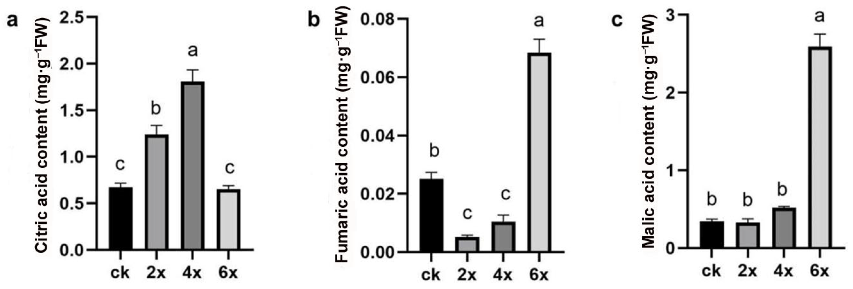
3.4. Soluble Sugar and Organic Acid Detection in Different Ploidy Embryoids Derived from Ovules

To further investigate the influence of ploidy variation on metabolic pathways, soluble sugar and organic acid contents in embryoids of different ploidy levels derived from unfertilized ovules were determined using a HPLC system. The soluble sugar components included glucose, fructose, and sucrose, while the organic acids included oxalic acid, quinic acid, malic acid, citric acid, succinic acid, and fumaric acid. Chromatograms of detected soluble sugar and organic acid standards are shown in Figure 5.
For soluble sugar contents, tetraploid embryoids had the highest level of sucrose, whereas no significant difference was found between CK and diploid embryoids (Figure 6a). Significant differences were observed in fructose (Figure 6b), glucose (Figure 6c), and total sugar (Figure 6d) contents among the embryoids with different ploidy levels, except for the glucose content between diploid and tetraploid embryoids. Comparative analysis revealed that hexaploid embryoids exhibited the highest contents of fructose, glucose, and total sugar, while the diploid embryoids showed the lowest levels. Moreover, the contents of fructose, glucose, and total sugar in the embryoid were significantly lower than in the CK group.
For organic acid detection, significant differences were detected in citric acid, fumaric acid, and malic acid contents among embryoids of different ploidy levels. Hexaploid embryoids contained the highest levels of malic acid and fumaric acid, while diploid embryoids had the lowest contents (Figure 7b,c). In contrast, tetraploid embryoids exhibited the highest citric acid content, whereas hexaploid embryoids showed the lowest level (Figure 7a).
The results indicated that, with increasing ploidy level, the contents of the majority of detected sugars and organic acids (fructose, sucrose, total sugar, fumaric acid, and malic acid) generally showed an upward trend.

4. Discussion

Unfertilized ovule culture serves as an effective method for haploid and double haploid (DH) induction, playing a significant role in haploid breeding [27,47]. However, relative reports on obtaining polyploids through this approach are limited. In Viola odorata L., a polyploid callus was successfully induced through unfertilized ovule induction [48]. In our study, unfertilized ovule culture of two loquat varieties, ‘Xingning 1’ and ‘Huabai 1’, efficiently regenerated embryoid lines. Ploidy identification revealed that 63.16% of the induced embryoid lines were tetraploids, and 6.58% were hexaploids, both confirmed as heterozygous. This study established a robust polyploid induction protocol.
Natural chromosome doubling is a crucial phenomenon in the sexual polyploidization process of plants [49]. In loquat, natural chromosome doubling and variation are relatively common, providing abundant high-quality germplasm resources [29,32,38]. Therefore, based on the results, the homologous tetraploids and hexaploids may originate from the natural doubling of the ovule integument. This exploration of polyploid induction offers a new approach for polyploid induction in loquat or other perennial fruit trees.
Initial flow cytometry analysis identified one embryoid line as haploid (derived from ‘Xingning 1’), with no other ploidy peaks detected (Figure 2a). However, re-sequencing results revealed that this embryoid line was heterozygous (Figure 5c,d), suggesting possible contamination by a small number of somatic cells.
Tetraploids in fruit crops exhibit the characteristics of larger organs, stronger stress resistance, and richer nutritional content [17,50]. These polyploid variants have shown broad application potential in rootstock breeding due to their stronger stress tolerance traits, such as salt tolerance, drought resistance, cold tolerance, and dwarfing [24,51,52,53,54,55,56]. Additionally, tetraploid plants can serve as the parental lines for triploid breeding via sexual hybridization [51]. Metabolic profiles of tetraploids vary across crops: for example, tetraploid tartary buckwheat showed an increase in the total seed flavonoid content and deepened seed color compared to the diploid variety [57]; tetraploid Dendrobium catenatum Lindl. contained higher polysaccharide content than diploids [58]; and tetraploid Ponkan mandarin fruit had higher levels of total acids, ascorbic acid, and total phenolic compounds than its diploid donor [59].
In our study, GO functional enrichment analysis showed that SNPs and InDels identified via genome resequencing were enriched in cellular and metabolic processes. Accordingly, we quantified soluble sugar and organic acid contents in regenerated embryoids of different ploidy levels. The results demonstrated that tetraploid embryoids contained significantly higher concentrations of sucrose, fructose, total sugar, and citric acid compared to diploids. Furthermore, our analyses showed a positive correlation between ploidy level and the content of both soluble sugars and organic acids, with a consistent increasing trend observed from diploid (2x) to tetraploid (4x) to hexaploid (6x) levels.

5. Conclusions

In summary, this study successfully regenerated tetraploid (4x) and hexaploid (6x) embryoids through unfertilized ovule culture in loquat, with genomic sequencing confirming their somatic chromosome doubling origin. To our knowledge, this is the first report on creating polyploid resources using an unfertilized ovule culture approach in loquat. Our study offers valuable insights into the potential of using unfertilized ovule culture for polyploid induction in this economically important fruit crop.

Supplementary Materials

The following supporting information can be downloaded at: https://www.mdpi.com/article/10.3390/horticulturae11070727/s1, Figure S1: Ploidy determination by flow cytometry analysis; Figure S2: Functional GO enrichment analysis of SNP/InDel variants of different ploidy embryoid lines; Table S1: Primer sequencing for SSR analysis.

Author Contributions

S.W. and T.R. performed tissue culture, ploidy analysis, and SSR marker analysis, analyzed the data, and wrote the manuscript. Y.Z., P.W., and J.D. performed genome analysis. Y.X., D.J., and G.L. analyzed the data and revised the manuscript. Q.G. and Q.H. conceived and supervised the research and revised the manuscript. All authors have read and agreed to the published version of the manuscript.

Funding

This research was financially supported by the National Key R&D Program of China (No. 2023YFD1600800), National NSF of China (No. 32171820), and Chongqing Science and Technology Commission (cstc2021jscx-gksbX0010, cstc2024ycih-bgzxm0202).

Institutional Review Board Statement

Not applicable.

Informed Consent Statement

Not applicable.

Data Availability Statement

The original contributions presented in the study are included in the article/Supplementary Material; further inquiries can be directed at the corresponding author.

Conflicts of Interest

The authors declare no conflicts of interest.

References

  1. Jiao, Y.; Wickett, N.J.; Ayyampalayam, S.; Chanderbali, A.S.; Landherr, L.; Ralph, P.E.; Tomsho, L.P.; Hu, Y.; Liang, H.; Soltis, P.S.; et al. Ancestral Polyploidy in Seed Plants and Angiosperms. Nature 2011, 473, 97–100. [Google Scholar] [CrossRef] [PubMed]
  2. Wood, T.E.; Takebayashi, N.; Barker, M.S.; Mayrose, I.; Greenspoon, P.B.; Rieseberg, L.H. The Frequency of Polyploid Speciation in Vascular Plants. Proc. Natl. Acad. Sci. USA 2009, 106, 13875–13879. [Google Scholar] [CrossRef] [PubMed]
  3. Yin, L.; Zhu, Z.; Huang, L.; Luo, X.; Li, Y.; Xiao, C.; Yang, J.; Wang, J.; Zou, Q.; Tao, L.; et al. DNA Repair- and Nucleotide Metabolism-Related Genes Exhibit Differential CHG Methylation Patterns in Natural and Synthetic Polyploids (Brassica napus L.). Hortic. Res. 2021, 8, 142. [Google Scholar] [CrossRef] [PubMed]
  4. Zhou, X.; Zhao, Y.; Ni, P.; Ni, Z.; Sun, Q.; Zong, Y. CRISPR-Mediated Acceleration of Wheat Improvement: Advances and Perspectives. J. Gene. Genom. 2023, 50, 815–834. [Google Scholar] [CrossRef]
  5. Bao, Y.; Zhang, Q.; Huang, J.; Zhang, S.; Yao, W.; Yu, Z.; Deng, Z.; Yu, J.; Kong, W.; Yu, X.; et al. A Chromosomal-Scale Genome Assembly of Modern Cultivated Hybrid Sugarcane Provides Insights into Origination and Evolution. Nat. Commun. 2024, 15, 3041. [Google Scholar] [CrossRef]
  6. Serra Mari, R.; Schrinner, S.; Finkers, R.; Ziegler, F.M.R.; Arens, P.; Schmidt, M.H.-W.; Usadel, B.; Klau, G.W.; Marschall, T. Haplotype-Resolved Assembly of a Tetraploid Potato Genome Using Long Reads and Low-Depth Offspring Data. Genome Biol. 2024, 25, 26. [Google Scholar] [CrossRef]
  7. Manivannan, A.; Cheeran Amal, T. Deciphering the Complex Cotton Genome for Improving Fiber Traits and Abiotic Stress Resilience in Sustainable Agriculture. Mol. Biol. Rep. 2023, 50, 6937–6953. [Google Scholar] [CrossRef]
  8. Zhuang, W.; Chen, H.; Yang, M.; Wang, J.; Pandey, M.K.; Zhang, C.; Chang, W.-C.; Zhang, L.; Zhang, X.; Tang, R.; et al. The Genome of Cultivated Peanut Provides Insight into Legume Karyotypes, Polyploid Evolution and Crop Domestication. Nat. Genet. 2019, 51, 865–876. [Google Scholar] [CrossRef]
  9. Rijzaani, H.; Bayer, P.E.; Rouard, M.; Doležel, J.; Batley, J.; Edwards, D. The Pangenome of Banana Highlights Differences between Genera and Genomes. Plant Genome 2022, 15, e20100. [Google Scholar] [CrossRef]
  10. Zou, L.; Liu, W.; Zhang, Z.; Edwards, E.J.; Gathunga, E.K.; Fan, P.; Duan, W.; Li, S.; Liang, Z. Gene Body Demethylation Increases Expression and Is Associated with Self-Pruning during Grape Genome Duplication. Hortic. Res. 2020, 7, 84. [Google Scholar] [CrossRef]
  11. Popowski, E.; Thomson, S.J.; Knäbel, M.; Tahir, J.; Crowhurst, R.N.; Davy, M.; Foster, T.M.; Schaffer, R.J.; Tustin, D.S.; Allan, A.C.; et al. Construction of a High-Density Genetic Map for Hexaploid Kiwifruit (Actinidia chinensis Var. Deliciosa) Using Genotyping by Sequencing. G3-Genes Genom. Genet. 2021, 11, jkab142. [Google Scholar] [CrossRef] [PubMed]
  12. Guo, M.; Bi, G.; Wang, H.; Hui, R.; Chen, J.; Lian, Q.; Wang, X.; Fang, W.; Zhang, J.; Dong, Z.; et al. Genomes of Autotetraploid Wild and Cultivated Ziziphus Mauritiana Reveal Polyploid Evolution and Crop Domestication. Plant Physiol. 2024, 196, 2701–2720. [Google Scholar] [CrossRef] [PubMed]
  13. Jin, X.; Du, H.; Zhu, C.; Wan, H.; Liu, F.; Ruan, J.; Mower, J.P.; Zhu, A. Haplotype-Resolved Genomes of Wild Octoploid Progenitors Illuminate Genomic Diversifications from Wild Relatives to Cultivated Strawberry. Nat. Plants 2023, 9, 1252–1266. [Google Scholar] [CrossRef]
  14. Wang, X.; Wang, H.; Shi, C.; Zhang, X.; Duan, K.; Luo, J. Morphological, Cytological and Fertility Consequences of a Spontaneous Tetraploid of the Diploid Pear (Pyrus pyrifolia Nakai) Cultivar ‘Cuiguan.’. Sci. Hortic. 2015, 189, 59–65. [Google Scholar] [CrossRef]
  15. Liu, X.; Song, L.; Xue, B.; Chi, Z.; Wang, Y.; Wen, S.; Lv, W.; Hu, Q.; Guo, Q.; Wang, S.; et al. Organic Acid and Sugar Components Accumulation and Flavor Associated Metabolites Dynamic Changes in Yellow- and White-Fleshed Seedless Loquats (Eriobotrya japonica). Food Chem. X 2024, 21, 101046. [Google Scholar] [CrossRef]
  16. Van De Peer, Y.; Ashman, T.-L.; Soltis, P.S.; Soltis, D.E. Polyploidy: An Evolutionary and Ecological Force in Stressful Times. Plant Cell 2021, 33, 11–26. [Google Scholar] [CrossRef]
  17. Song, X.; Zhang, M.; Wang, T.; Duan, Y.; Ren, J.; Gao, H.; Fan, Y.; Xia, Q.; Cao, H.; Xie, K.; et al. Polyploidization Leads to Salt Stress Resilience via Ethylene Signaling in Citrus Plants. New Phytolo. 2025, 246, 176–191. [Google Scholar] [CrossRef]
  18. Meeus, S.; Semberova, K.; Storme, N.D.; Geelen, D.; Vallejo-Marı’n, M. Effect of Whole-Genome Duplication on the Evolutionary Rescue of Sterile Hybrid Monkeyflowers. Plant Commun. 2020, 1, 100093. [Google Scholar] [CrossRef]
  19. Soomro, S.R.; Soomro, S.N.; Altaf, M.T.; Liaqat, W.; Nadeem, M.A.; Baloch, F.S.; Aasim, M.; Mohamed, H.I. Development of Tetraploids in Tissue Culture: Modern Techniques and Biotechnological Innovations. Plant Cell Tiss. Organ. Cult. 2025, 160, 51. [Google Scholar] [CrossRef]
  20. Xie, K.D.; Yuan, D.Y.; Wang, W.; Xia, Q.M.; Wu, X.M.; Chen, C.W.; Chen, C.L.; Grosser, J.W.; Guo, W.W. Citrus Triploid Recovery Based on 2x × 4x Crosses via an Optimized Embryo Rescue Approach. Sci. Hortic. 2019, 252, 104–109. [Google Scholar] [CrossRef]
  21. Liu, M.; Liu, P. Artificial Autopolyploidization in Jujube. Sci. Hortic. 2023, 314, 111916. [Google Scholar] [CrossRef]
  22. Narukulla, V.; Lahane, Y.; Uge, P.; Pandey, S.; Fiske, K.; Kawale, K.; Jagannadham, P.T.K.; Ziogas, V. Production of Triploid Seedless Sweet Orange [Citrus sinensis (L.) Osbeck] Cv. Mosambi: A Success Story. Agronomy 2024, 14, 829. [Google Scholar] [CrossRef]
  23. Narukulla, V.; Lahane, Y.; Fiske, K.; Pandey, S.; Ziogas, V. Induction of Polyploidy in Citrus Rootstocks through In Vitro Colchicine Treatment of Seed-Derived Explants. Agronomy 2023, 13, 1442. [Google Scholar] [CrossRef]
  24. Guo, W.W.; Deng, X. Intertribal Hexaploid Somatic Hybrid Plants Regeneration from Electrofusion between Diploids of Citrus sinensis and Its Sexually Incompatible Relative, Clausena lansium. Theor. Appl. Genet. 1999, 98, 581–585. [Google Scholar] [CrossRef]
  25. Grosser, J.W.; Gmitter, F.G. Protoplast Fusion for Production of Tetraploids and Triploids: Applications for Scion and Rootstock Breeding in Citrus. Plant Cell Tiss. Organ. Cult. 2011, 104, 343–357. [Google Scholar] [CrossRef]
  26. Abdullah, M.; Sliwinska, E.; Góralski, G.; Latocha, P.; Tuleja, M.; Widyna, P.; Popielarska-Konieczna, M. Effect of Medium Composition, Genotype and Age of Explant on the Regeneration of Hexaploid Plants from Endosperm Culture of Tetraploid Kiwiberry (Actinidia arguta). Plant Cell Tiss. Organ. Cult. 2021, 147, 569–582. [Google Scholar] [CrossRef]
  27. Marin-Montes, I.M.; Rodríguez-Pérez, J.E.; Robledo-Paz, A.; De La Cruz-Torres, E.; Peña-Lomelí, A.; Sahagún-Castellanos, J. Haploid Induction in Tomato (Solanum lycopersicum L.) via Gynogenesis. Plants 2022, 11, 1595. [Google Scholar] [CrossRef]
  28. Zou, T.; Min, Z.; Song, H.; Gong, S.; Tong, L.; Sun, L.; Yang, H.; Zhang, J.; Sun, X. Whole-Transcriptome Sequence Analysis of Pumpkin Ovules in Response to Heat Shock during Unfertilized Ovary Culture. Sci. Hortic. 2024, 329, 113007. [Google Scholar] [CrossRef]
  29. Wang, P.; Yang, S.; Chen, M.; Liu, Y.; He, Q.; Sun, H.; Wu, D.; Xiang, S.; Jing, D.; Wang, S.; et al. Karyotype Variation Patterns and Phenotypic Responses of Hybrid Progenies of Triploid Loquat (Eriobotrya japonica) Provide New Insight into Aneuploid Germplasm Innovation. Hortic. Res. 2025, 12, uhaf023. [Google Scholar] [CrossRef]
  30. Chi, Z.; Liu, X.; Wen, S.; Wang, Y.; Lv, W.; Guo, Q.; Xia, Y.; Jing, D.; Liang, G. Integrated Metabolomic Profiling and Transcriptome Analysis of Fruit Quality and Ripeness in Early-Maturing Seedless Triploid Loquat. Sci. Hortic. 2023, 316, 112012. [Google Scholar] [CrossRef]
  31. Xu, X.; Yang, H.; Suo, X.; Liu, M.; Jing, D.; Zhang, Y.; Dang, J.; Wu, D.; He, Q.; Xia, Y.; et al. EjFAD8 Enhances the Low-Temperature Tolerance of Loquat by Desaturation of Sulfoquinovosyl Diacylglycerol (SQDG). Int. J. Mol. Sci. 2023, 24, 6946. [Google Scholar] [CrossRef] [PubMed]
  32. Liu, C.; Wang, M.; Wang, L.; Guo, Q.; Liang, G. Extensive Genetic and DNA Methylation Variation Contribute to Heterosis in Triploid Loquat Hybrids. Genome 2018, 61, 437–447. [Google Scholar] [CrossRef] [PubMed]
  33. Wang, S.; Wang, L.; Liu, Z.; Xia, Y.; Jing, D.; Guo, Q.; Liang, G.; He, Q. An Efficient System for Mesophyll Protoplast Isolation, Purification, and Transformation in Loquat: Studies on Fluorescent Marker Analysis and Subcellular Localization. Horticulturae 2025, 11, 391. [Google Scholar] [CrossRef]
  34. Wang, H.; Dang, J.; Wu, D.; Xie, Z.; Yan, S.; Luo, J.; Guo, Q.; Liang, G. Genotyping of Polyploid Plants Using Quantitative PCR: Application in the Breeding of White-Fleshed Triploid Loquats (Eriobotrya japonica). Plant Methods 2021, 17, 93. [Google Scholar] [CrossRef]
  35. Gamborg, O.; Miller, R.; Ojima, K. Nutrient Requirement Suspension Cultures of Soybean Root Cells. Exp. Cell Res. 1968, 50, 151–158. [Google Scholar] [CrossRef]
  36. Wang, P.; Yang, Y.; Lei, C.; Xia, Q.; Wu, D.; He, Q.; Jing, D.; Guo, Q.; Liang, G.; Dang, J. A Female Fertile Triploid Loquat Line Produces Fruits with Less Seed and Aneuploid Germplasm. Sci. Hortic. 2023, 319, 112141. [Google Scholar] [CrossRef]
  37. Dang, J.; Wu, T.; Liang, G.; Wu, D.; He, Q.; Guo, Q. Identification and Characterization of a Loquat Aneuploid with Novel Leaf Phenotypes. Hortscience 2019, 54, 804–808. [Google Scholar] [CrossRef]
  38. Wen, G.; Dang, J.; Xie, Z.; Wang, J.; Jiang, P.; Guo, Q.; Liang, G. Molecular Karyotypes of Loquat (Eriobotrya japonica) Aneuploids Can Be Detected by Using SSR Markers Combined with Quantitative PCR Irrespective of Heterozygosity. Plant Methods 2020, 16, 22. [Google Scholar] [CrossRef]
  39. Ruiz, C.; Breto, M.P.; Asıns, M.J. A Quick Methodology to Identify Sexual Seedlings in Citrus Breeding Programs Using SSR Markers. Euphytica 2000, 112, 89–94. [Google Scholar] [CrossRef]
  40. Jing, D.; Liu, X.; He, Q.; Dang, J.; Hu, R.; Xia, Y.; Wu, D.; Wang, S.; Zhang, Y.; Xia, Q.; et al. Genome Assembly of Wild Loquat (Eriobotrya japonica) and Resequencing Provide New Insights into the Genomic Evolution and Fruit Domestication in Loquat. Hortic. Res. 2023, 10, uhac265. [Google Scholar] [CrossRef]
  41. Li, H.; Durbin, R. Fast and Accurate Short Read Alignment with Burrows–Wheeler Transform. Bioinformatics 2009, 25, 1754–1760. [Google Scholar] [CrossRef] [PubMed]
  42. Zhu, P.; He, L.; Li, Y.; Huang, W.; Xi, F.; Lin, L.; Zhi, Q.; Zhang, W.; Yang, Y.T.; Geng, C.; et al. OTG-Snpcaller: An Optimized Pipeline Based on TMAP and GATK for SNP Calling from Ion Torrent Data. PLoS ONE 2014, 9, 97507. [Google Scholar] [CrossRef] [PubMed]
  43. McKenna, A.; Hanna, M.; Banks, E.; Sivachenko, A.; Cibulskis, K.; Kernytsky, A.; Garimella, K.; Altshuler, D.; Gabriel, S.; Daly, M.; et al. The Genome Analysis Toolkit: A MapReduce Framework for Analyzing next-Generation DNA Sequencing Data. Genome Res. 2010, 20, 1297–1303. [Google Scholar] [CrossRef] [PubMed]
  44. Wang, K.; Li, M.; Hakonarson, H. ANNOVAR: Functional Annotation of Genetic Variants from High-Throughput Sequencing Data. Nucleic Acids Res. 2010, 38, e164. [Google Scholar] [CrossRef]
  45. Negoro, T.; Orihara, K.; Irahara, T.; Nishiyama, H.; Hagiwara, K.; Nishida, R.; Takagi, H.; Satoh, K.; Yamamoto, Y.; Shimizu, S.; et al. Influence of SNPs in Cytokine-related Genes on the Severity of Food Allergy and Atopic Eczema in Children. Pediatric Allergy Immunol. 2006, 17, 583–590. [Google Scholar] [CrossRef]
  46. Yu, G.; Wang, L.G.; Han, Y.; He, Q.Y. clusterProfiler: An R package for comparing biological themes among gene clusters. OMICS 2012, 16, 284–287. [Google Scholar] [CrossRef]
  47. Metin, D.; Atakul, Z.; Kurtar, E.S.; Seymen, M.; Alan, A.R.; Çelebi Toprak, F. Callogenesis, Embryogenesis, and Plantlet Initiation in Citron Watermelon (Citrullus lanatus Var. Citroides) via Anther and Unfertilized Ovary Culture. Sci. Hortic. 2024, 337, 113493. [Google Scholar] [CrossRef]
  48. Wijowska, M.; Kuta, E.; Przywara, L. In Vitro Culture of Unfertilized Ovules of Viola odorata L. Acta Biol. Cracov. Bot. 1999, 41, 95–101. [Google Scholar]
  49. Liu, X.; Li, D.; Zhang, Y.; Zhou, X.; Wang, S.; Zhao, J.; Guo, J.; Jiang, Q.; Ren, F. The Cytological Mechanism of the Peach Haploid Producing Triploid Offspring. Hortic. Res. 2025, 12, uhae316. [Google Scholar] [CrossRef]
  50. Zhan, N.; Li, L.; Zhang, L.; He, W.; Yang, Q.; Bi, F.; Deng, G.; Kiggundu, A.; Yi, G.; Sheng, O. Transcriptome and Metabolome Profiling Provide Insights into Hormone-Mediated Enhanced Growth in Autotetraploid Seedlings of Banana (Musa Spp.). Front. Sustain. Food Syst. 2023, 6, 1070108. [Google Scholar] [CrossRef]
  51. Ren, J.; Lu, X.; Duan, Y.-Y.; Xiao, G.-A.; Xie, K.-D.; Wu, X.-M.; Guo, W.-W. In Vivo Tetraploid Induction of Mono-Embryonic Citrus Genotypes by Colchicine Treatment. Sci. Hortic. 2024, 338, 113701. [Google Scholar] [CrossRef]
  52. Khalid, M.F.; Vincent, C.; Morillon, R.; Anjum, M.A.; Ahmad, S.; Hussain, S. Different Strategies Lead to a Common Outcome: Different Water-Deficit Scenarios Highlight Physiological and Biochemical Strategies of Water-Deficit Tolerance in Diploid versus Tetraploid Volkamer Lemon. Tree Physiol. 2021, 41, 2359–2374. [Google Scholar] [CrossRef] [PubMed]
  53. Yang, S.; Zhang, Y.; Zhang, Y.; Peng, J.; Liu, C.; Li, D.; Guo, W. Divergence in Cold Tolerance Promotes Niche Differentiation between Diploid and Polyploid Kiwifruits along an Altitudinal Gradient in Southwest China. Oikos 2024, 5, e10181. [Google Scholar] [CrossRef]
  54. Hui, T.; Bao, L.; Shi, X.; Zhang, H.; Xu, K.; Wei, X.; Liang, J.; Zhang, R.; Qian, W.; Zhang, M.; et al. Grafting Seedling Rootstock Strengthens Tolerance to Drought Stress in Polyploid Mulberry (Morus alba L.). Plant Physiol. Bioch. 2024, 208, 108441. [Google Scholar] [CrossRef]
  55. Rugini, E.; Silvestri, C.; Ceccarelli, M.; Muleo, R.; Cristofori, V. Mutagenesis and Biotechnology Techniques as Tools for Selecting New Stable Diploid and Tetraploid Olive Genotypes and Their Dwarfing Agronomical Characterization. Hortscience 2016, 51, 799–804. [Google Scholar] [CrossRef]
  56. Liu, R.; Gao, C.; Jin, J.; Wang, Y.; Jia, X.; Ma, H.; Zhang, Y.; Zhang, H.; Qi, B.; Xu, J. Induction and Identification of Tetraploids of Pear Plants (Pyrus bretschneideri and Pyrus betulaefolia). Sci. Hortic. 2022, 304, 111322. [Google Scholar] [CrossRef]
  57. Shi, X.; Wang, C.; Li, W.; Xiao, H.; Li, Y.; Niu, B. Analysis of Seed Phenotypic and Metabolic Characteristics of Diploid and Tetraploid Tartary Buckwheat. Phyton-Int. J. Exp. Bot. 2022, 91, 1973–1986. [Google Scholar] [CrossRef]
  58. Liu, Y.; Li, X.R.; Zhang, X.J.; Duan, S.D.; Su, X.; Fan, Z.H.; Hao, L.H.; Xiang, D.Y.; Chen, D.F.; Niu, S.C. Integrated Metabolomic and Transcriptomic Analysis Reveals the Mechanism of High Polysaccharide Content in Tetraploid Dendrobium Catenatum Lindl. Ind. Crops Prod. 2024, 212, 118391. [Google Scholar] [CrossRef]
  59. Tan, F.Q.; Zhang, M.; Xie, K.D.; Fan, Y.J.; Song, X.; Wang, R.; Wu, X.M.; Zhang, H.Y.; Guo, W.W. Polyploidy Remodels Fruit Metabolism by Modifying Carbon Source Utilization and Metabolic Flux in Ponkan Mandarin (Citrus reticulata Blanco). Plant Sci. 2019, 289, 110276. [Google Scholar] [CrossRef]
Figure 1. Calluses and embryoids derived from unfertilized ovules of loquat. (a) Gynogenesis ovules exhibited initiation of embryogenic callus formation; (b) embryogenic calluses developed into spherical structures; (c) primary embryoid formations; (d) embryoid formation; (e) Type I callus with capable for embryoid induction; (f) Type II callus with slow proliferation and no capacity for embryoid induction.
Horticulturae 11 00727 g001
Figure 2. Ploidy determination by flow cytometry analysis of embryoid lines derived from cv. ‘Xingning 1’: (a) haploid (x); (b) diploid (2x); (c); tetraploid (4x); and (d) hexaploid (6x).
Horticulturae 11 00727 g002
Figure 3. Chromosome counting analysis of embryoid lines derived from ‘Xingning 1’ unfertilized ovules: (a) diploid (2x) with 34 chromosomes; (b) tetraploid (4x) with 68 chromosomes; and (c) hexaploid (6x) with 102 chromosomes.
Horticulturae 11 00727 g003
Figure 4. SSR analysis and genomic sequencing of ovule-derived embryoids. (a,b) SSR analysis between the tetraploid embryoids and their donor using primer LG1006 and LG0408, respectively; number 1 in the gels represents the donor plant cv. ‘Xingning 1’, numbers 2–5 in the gels represent four tetraploid embryoid lines derived from ‘Xingning 1. (c) SNP variant-type proportion: homozygous (1/1) and heterozygous (0/1). (d) Coverage density of different types of variants in chromosomes: A-C represent SNPs, InDels, and CNVs of haploid embryoid line, respectively; D-F represent SNPs, InDels, and CNVs of diploid embryoid line, respectively; G-I represent SNPs, InDels, and CNVs of tetraploid embryoid line, respectively; and J-L represent SNPs, InDels, and CNVs of hexaploid embryoid line, respectively.
Horticulturae 11 00727 g004
Figure 5. HPLC chromatograms of soluble sugars and organic acids. (a) HPLC chromatogram of sugar reference standards; (b) HPLC chromatogram of organic acids reference standards; (c) HPLC chromatogram of sugar components in embryoids; (d) HPLC chromatogram of organic acids components in embryoids. In (a) and (c), peaks 1, 2, and 3 represent fructose, glucose, and sucrose, respectively; in (b) and (d), peaks 1, 2, 3, 4, 5, and 6 represent oxalic acid, quinic acid, malic acid, citric acid, succinic acid, and fumaric acid, respectively.
Horticulturae 11 00727 g005
Figure 6. Soluble sugar content of different ploidy embryoids: (a) sucrose content; (b) fructose content; (c) glucose content; and (d) total sugar content. CK represents diploid callus of ‘Xingning 1’. Different lowercase letters indicate statistical significance (LSD test, p ≤ 0.05).
Horticulturae 11 00727 g006
Figure 7. Organic acid content of different ploidy embryoids: (a) citric acid content; (b) fumaric acid content; and (c) malic acid content. CK represents diploid callus of ‘Xingning 1’. Different lowercase letters indicate statistical significance (LSD test, p ≤ 0.05).
Horticulturae 11 00727 g007
Table 1. In vitro culture of unfertilized ovules of loquat.
VarietiesNo. of Inoculated Unfertilized OvulesNo. of Gynogenesis LineGynogenesis Rate
(GR) (%)
No. of Derived Embryoid LineEmbryoid Induction Rate
(EIR) (%)
‘Senwei Zaosheng’5603511.05 ± 4.30 b00
‘Huabai 1’6809813.62 ± 2.61 b1212.81 ± 3.13 b
‘Xingning 1’88015821.63 ± 3.35 a7654.37 ± 6.84 a
‘Zaozhong 6’1603720.63 ± 2.26 a00
‘Daduhe’1800000
Note: Different lowercase letters indicate statistical significance (LSD test, p ≤ 0.05).
Disclaimer/Publisher’s Note: The statements, opinions and data contained in all publications are solely those of the individual author(s) and contributor(s) and not of MDPI and/or the editor(s). MDPI and/or the editor(s) disclaim responsibility for any injury to people or property resulting from any ideas, methods, instructions or products referred to in the content.

