Article

Differential Bio-Elicitor Effects on Bioactive Compound Production in Cichorium intybus Root Callus Cultures

by
Ahmed A. Elateeq
1,*,
Mostafa M. Zarad
1,
Ahmed M. M. Gabr
2,
Hanan S. Ebrahim
2,
Shakir Ullah
3,4,
Sam M. Elhamamsy
5,
Ramy S. Nada
1,
Zakaria H. Saad
5,
Mahmoud N. A. Soliman
1,
Hend A. El-khawaga
6,
Woroud S. Alshammari
7,
Wesal S. Tanko
7 and
Hebat-Allah A. Hussein
7,*
1
Horticulture Department, Faculty of Agriculture, Al-Azhar University, Cairo 11884, Egypt
2
Department of Plant Biotechnology, Biotechnology Research Institute, National Research Centre, Cairo 12622, Egypt
3
College of Chemistry, Chemical Engineering and Resource Utilization, Northeast Forestry University, Harbin 150040, China
4
Key Laboratory of Forest Plant Ecology, Ministry of Education, Northeast Forestry University, Harbin 150040, China
5
Department of Biochemistry, Faculty of Agriculture, Al-Azhar University, Cairo 11884, Egypt
6
Botany and Microbiology Department, Faculty of Science, Al-Azhar University (Girls Branch), Cairo 11651, Egypt
7
Department of Science and Technology, University College at Nairiyah, University of Hafr Al Batin (UHB), Nairiyah 31981, Saudi Arabia
*
Authors to whom correspondence should be addressed.
Horticulturae 2025, 11(6), 678; https://doi.org/10.3390/horticulturae11060678
Submission received: 15 May 2025 / Revised: 6 June 2025 / Accepted: 11 June 2025 / Published: 13 June 2025
(This article belongs to the Section Propagation and Seeds)

Abstract

:
Chicory (Cichorium intybus L.) roots are valued in medicine for their potential health benefits. Producing callus from chicory roots through tissue culture technology can streamline bioactive metabolites production and ensure a sustainable supply chain. The current study explored the impact of plant growth regulators (PGRs) and light conditions on the characteristics of callus induced from C. intybus root explants. The effect of fungal elicitors [yeast extract (YE), Fusarium oxysporum, and Aspergillus niger] on bioactive metabolite production from root-derived callus was investigated. Callus color varied notably between a 16/8 h light/dark cycle and complete dark, with differences in texture based on PGR concentrations and light conditions. High weights of callus formed were generally recorded under the 16/8 h light/dark cycle. Low concentrations of YE (1 g/L) and F. oxysporum (0.25 g/L) enhanced callus biomass fresh weight, while high concentrations of A. niger (1 g/L) improved callus dry matter significantly. The content and productivity of total phenolic were maximized at 1 g/L of YE and 1 g/L of F. oxysporum. Callus cultures elicited with a higher level of A. niger recorded the higher values of total flavonoid production. High-performance liquid chromatography (HPLC) analysis revealed significant variations in chlorogenic acid, catechin, and caffeic acid levels among the different elicited cultures. A. niger at 1 g/L notably increased chlorogenic acid content, while catechin levels were enhanced by specific concentrations of YE. Catalase (CAT) activity was significantly affected by different elicitors, while only the higher level of F. oxysporum and A. niger showed a significant increase in peroxidase (POD) activity. DPPH scavenging activity was elevated by all fungal elicitors. Principal Component Analysis delineated distinct variations in callus traits in response to different elicitors, with specific treatments showcasing enhanced biomass production, bioactive compound accumulation, and antioxidant activities. Through meticulous experimentation, this study paves the way for enhancing chicory root-derived products, ensuring sustainable production and potent bioactivity.

1. Introduction

Across diverse cultures worldwide, a significant proportion of the global population continues to rely on traditional plant-derived medicines—a testament to their enduring cultural heritage and therapeutic efficacy, which continues to inspire modern pharmaceutical research in natural product drug discovery and development [1]. Chicory (Cichorium intybus L.) is a member of the Asteraceae family and is a low-height perennial or biennial herb. About 4000 BC, chicory was first grown in Egypt, and since then, its health benefits and nutritional value have increased awareness of the plant. It is especially well-known in the Mediterranean region for adding its root powder to coffee and making caffeine-free substitutes, as well as for use as a vegetable and probiotic component [2]. Chicory phytocompounds have emerged as a cutting-edge subject owing to their extensive application. Esculin, inulin, sesquiterpenes lactones, phenolic acids, flavonoids, coumarins, and vitamins are among the several components of chicory that have therapeutic value [3,4,5]. Chicory has been recognized for its anticarcinogenic, antiviral, antifungal, antibacterial, antimutagenic, antidiabetic, anthelmintic, immune-stimulating, and antihepatotoxic properties [4,5].
Given the complicated makeup of the compounds and the high cost of chemicals, the chemical synthesis of bioactive phytochemicals presents significant difficulties [6]. Nonetheless, field agriculture is labor-intensive and time-consuming, and it is impacted by various factors such as ecotype, season, harvest date, soil fertility, and water quality [7,8]. Biotechnology approaches, particularly the plant tissue culture technique, have been reported to have a great potential to complement conventional agriculture for the industrial and pharmaceutical production of plant bioactive metabolites in the search for an alternative solution to these problems faced by the plant pharmaceutical industries [9,10]. Callus cultures have the potential to be utilized in the food, pharmaceutical, and cosmetics sectors for the large-scale and sustainable production of by-products [11]. In addition, medicinal crops’ calli cultures create bioactive phytoconstituents with strong antioxidant activity that can be applied to a range of medical conditions [12]. Chicory root callus exhibits anti-hepatotoxic properties [13] and contains bioactive compounds such as terpenoids and glycosides. Notably, the callus culture produces unique metabolites not typically found in intact chicory plants, including 3β,6β-dihydroxynortropane, saxitoxin, E-64, isomasticadienonalic acid, and onchidal. Additionally, the callus extract harbors several phytoconstituents with undetermined pharmacological activities [14]. A recent study by Abas et al. [15] demonstrated that specific combinations and concentrations of plant growth regulators (PGRs) in the culture medium not only induced root and shoot formation alongside callus proliferation but also altered phenolic compound profiles. Notably, certain PGRs increased phenolic content in both callus and shoots derived from leaf explants. Comparative analysis revealed that in vivo-grown plants accumulated higher levels of specific phenolics, such as caftaric acid, chlorogenic acid, and inulin, whereas in vitro-regenerated samples exhibited elevated concentrations of esculin and chicoric acid.
The impediments to the extensive commercialization of plant cell technology can be addressed by the application of certain productivity-boosting strategies, such as elicitation [16]. Plant cells respond physiologically and biochemically to physical, chemical, and microbiological stimuli known as “elicitors”. Elicitation is the process of introducing elicitors—either biotic or abiotic—exogenously into the growth medium. As a result, the growth medium becomes more stressful, which increases the synthesis of secondary metabolites [16]. According to Narayani and Srivastava [17], elicitor-induced stress results in the expression of certain enzymes, and information about these enzymes is used to ascertain the biosynthetic pathways of different phytomolecules as well as the transient phosphorylation/dephosphorylation of proteins and the inactivation or activation of numerous genes linked to defense- or non-defense-related genes. In order to induce and enhance the production of various secondary metabolites in plant tissue culture systems, biotic and abiotic elicitors have been used [9,12,18,19,20]. This reduces the time needed to grow culture biomass and reaches high productivity. To maximize the accumulation of various bioactive phytochemicals, researchers have employed the fungal biotic elicitors such as yeast extract, Aspergillus niger, and Fusarium oxysporum [12,20,21,22]. Furthermore, in cultures subjected to fungal elicitation, antioxidant enzymes are activated to regulate oxidative stress [21,23,24]. By carefully adjusting the quantity and concentration of components sourced from fungi like A. niger, F. oxysporum, Penicillium notatum, and Trichoderma viride, they can be used to induce secondary metabolism in cell, callus, and root cultures of various plant species [21,25,26].
The majority of studies have utilized leaf explants for initial chicory callus induction and proliferation [15,27,28,29,30,31]. Conversely, few investigations have explored the use of roots as a source of callus [13,14,32]. Moreover, the findings of Zafar and Ali [13] showed that C. intybus root callus extract provided better protection against carbon tetrachloride-induced hepatocellular damage compared to the natural root extract, suggesting that metabolites in root-derived callus possess greater anti-hepatotoxic properties than those in the natural root extract.
Previous studies consistently demonstrate that plant growth regulators (PGRs) and light conditions are critical factors influencing C. intybus callus induction, with optimal auxin–cytokinin combinations and photoperiods often being explant-specific [28,30,32].
The current study explored the impact of PGRs and light conditions on the characteristics of callus induced from C. intybus root explants. Importantly, the research focuses on employing bio-elicitors (yeast extract, A. niger, and F. oxysporum) as a strategic approach to enhance biomass production, antioxidant activity, and phenolics and flavonoids accumulation in C. intybus root-derived callus. The study hypothesizes that different fungal elicitors will variably enhance the production of bioactive compounds (e.g., total phenolics, flavonoids, chlorogenic acid, catechin, and caffeic acid) and antioxidant enzymes activity in chicory root callus, depending on the elicitor type and concentration.

2. Materials and Methods

2.1. Seed Sterilization and Explant Preparation

The C. intybus seeds were kindly provided by the Horticultural Research Institute, Agricultural Research Center, Giza, Egypt. The experiments were carried out in the Laboratory of Biotechnology, Horticulture Department, Faculty of Agriculture, Al-Azhar University, Cairo. Seeds of C. intybus were washed with detergent and tap water and surface sterilized by ethanol (70%) for 30 s, then immersed in 1% sodium hypochlorite solution containing few drops of Tween 20 for 20 min. After washing with sterile distilled water, the seeds were sown in culture jars containing Murashige and Skoog [33] basal medium (MS) and 30 g/L sucrose, solidified with 7 g/L agar and incubated under 16/8 h light/dark cycle with illumination from cool white fluorescence lights 40 µmol/m2/s at 25 ± 2 °C. The roots of in vitro-grown seedlings (4 weeks old) were excised to induce callus.

2.2. Callus Induction

Root segments (1–1.5 cm length) were transferred to MS basal medium and 30 g/L sucrose, solidified with 7 g/L agar and supplemented with different combinations of α-naphthalene acetic acid (NAA; 0, 1, and 2 mg/L) and benzyl adenine (BA; 0, 1, and 2 mg/L). Cultures were placed in a growth chamber for 4 weeks under 16/8 h light/dark cycle with illumination from cool white fluorescence lights 40 µmol/m2/s or complete dark at 25 ± 2 °C.

2.3. Fungal Elicitors

The fungal strains used in this study were obtained from the Microbiology Laboratory, Botany Department, Faculty of Agriculture, Al-Azhar University, Cairo, Egypt. A. niger and F. oxysporum were grown in malt extract medium (20 g/L) in 500 mL shake flasks containing 100 mL medium at 120 rpm and 25 ± 2 °C. After 7 days, the cultures were autoclaved at 121 °C for 20 min. The fungal biomass (mycelium) was separated by filtration through Whatman No. 1 filter paper, and washed three times with sterile distilled water to remove residual medium components. The mycelium was dried at 45 °C for 24 h and then ground into a fine powder. The final elicitor concentrations (0.25–1 g/L) were prepared based on the dry weight of this fungal powder.
Root-derived calli induced on MS medium fortified with 2 mg/L NAA + 1 mg/L BA under a 16/8 h light/dark cycle were used. The callus was trimmed from adventitious roots that appeared on it, then equal parts of it were transferred to sterile jars containing MS medium and 30 g/L sucrose, solidified with 7 g/L agar and supplemented with 2 mg/L NAA + 1 mg/L BA. Yeast extract (YE) (Sigma Chemical Co., St. Louis, MO, USA) at 1, 2, and 4 g/L, A. niger at 0.25, 0.5, and 1 g/L, and F. oxysporum at 0.25, 0.5, and 1 g/L were each individually fortified into the culture medium as fungal bio-elicitors. The dried mycelium and YE powders were first solubilized in distilled water with constant stirring until completely dissolved. This solution was then added to the culture media before being autoclaved at 121 °C for 20 min.
The pH value of all tested media was adjusted to 5.8 with NaOH or HCl (1 N). The cultures were placed in a growth chamber for 4 weeks under 25 ± 2 °C and a 16/8 h light/dark cycle with illumination from cool white fluorescence lights 40 µmol/m2/s.

2.4. Measurements and Determinations

2.4.1. Characterization of Callus Induction

The different responses of root explants cultured on MS media supplemented with different combinations of NAA and BA were characterized after the incubation period under light and darkness. Characterization of the induced callus was recorded, such as the color and texture of the callus and the degree of adventitious organ formation. The degree of adventitious root or shoot formation was evaluated using a semi-quantitative scoring system based on visual assessment: (-) no formation, (+) low, (++) moderate, (+++) high, and (++++) very high.
Growth measurements of the callus induced were recorded as fresh weight (FW), dry weight (DW), and dry matter (DM). Fresh callus was dried using the oven-drying method at 45 °C until a constant weight was achieved. The dry matter of callus was calculated according to the following equation:
Dry matter (%) = (final DW/final FW) × 100

2.4.2. Determination of Callus Biomass Production

Fungal-elicited calli were collected after the incubation period (4 weeks) and callus FW, DW, and DM were determined. Callus biomass production (FW and DW) were expressed as g callus/L medium. A standard volume of 30 mL of medium was consistently used for each culture jar, and three explants were cultured in each jar.
The dried callus was homogenized into a fine powder using a laboratory blender for subsequent chemical analysis.

2.4.3. Determination of Total Phenolic Content

A quantity of 100 mg DW of callus was immersed in 5 mL of 95% ethanol and stirred for 4 h. The tubes containing this mixture were then homogenized and centrifuged for 10 min. The total phenolic content (TPC) in the supernatants was assessed using the Folin–Ciocalteu method, following the protocol outlined by Singleton and Rossi [34]. An amount of 1 ml of the supernatant was combined with 1 mL of 95% ethanol, 5 mL of distilled water, and 0.5 mL of 50% Folin–Ciocalteu reagent. After a 5 min interval, 1 mL of Na2CO3 (5%) was added and thoroughly mixed. The resultant solution was left for an hour at 25 ± 2 °C. Subsequently, the absorbance was detected at 725 nm using a Jenway 6800 UV/Vis spectrophotometer (Bibby Scientific Ltd., Staffordshire, UK) against a blank. Gallic acid dilutions were employed to establish the standard curve. The TPC was then computed in mg of gallic acid equivalents (GAE)/g DW of callus.
Total phenolic productivity (TPP) (mg GAE/L culture medium) = TPC (mg GAE/g DW) × callus biomass DW (g/L culture medium)

2.4.4. Determination of Total Flavonoid Content

The callus powder (100 mg DW) was mixed with 5 mL of 95% ethanol and agitated for 4 h, followed by a 24 h incubation at room temperature. After centrifugation for 10 min, the total flavonoid content (TFC) was determined utilizing the aluminum chloride (AlCl3) colorimetric method, following the procedures detailed by Chang et al. [35]. The ethanol extract (0.5 mL) was mixed with 1.5 mL of 95% ethanol, 0.1 mL of AlCl3 10%, 0.1 mL of potassium acetate 1 M, and 2.8 mL of distilled water. Following a 30 min incubation at 25 ± 2 °C, the absorbance was measured at 415 nm using a spectrophotometer against a blank. The calibration curve was established using quercetin dilutions, and the TFC was expressed as mg of quercetin equivalents (QE)/g DW of callus.
Total flavonoid productivity (TFP) (mg QE/L culture medium) = TFC (mg QE/g DW) × callus biomass DW (g/L culture medium)

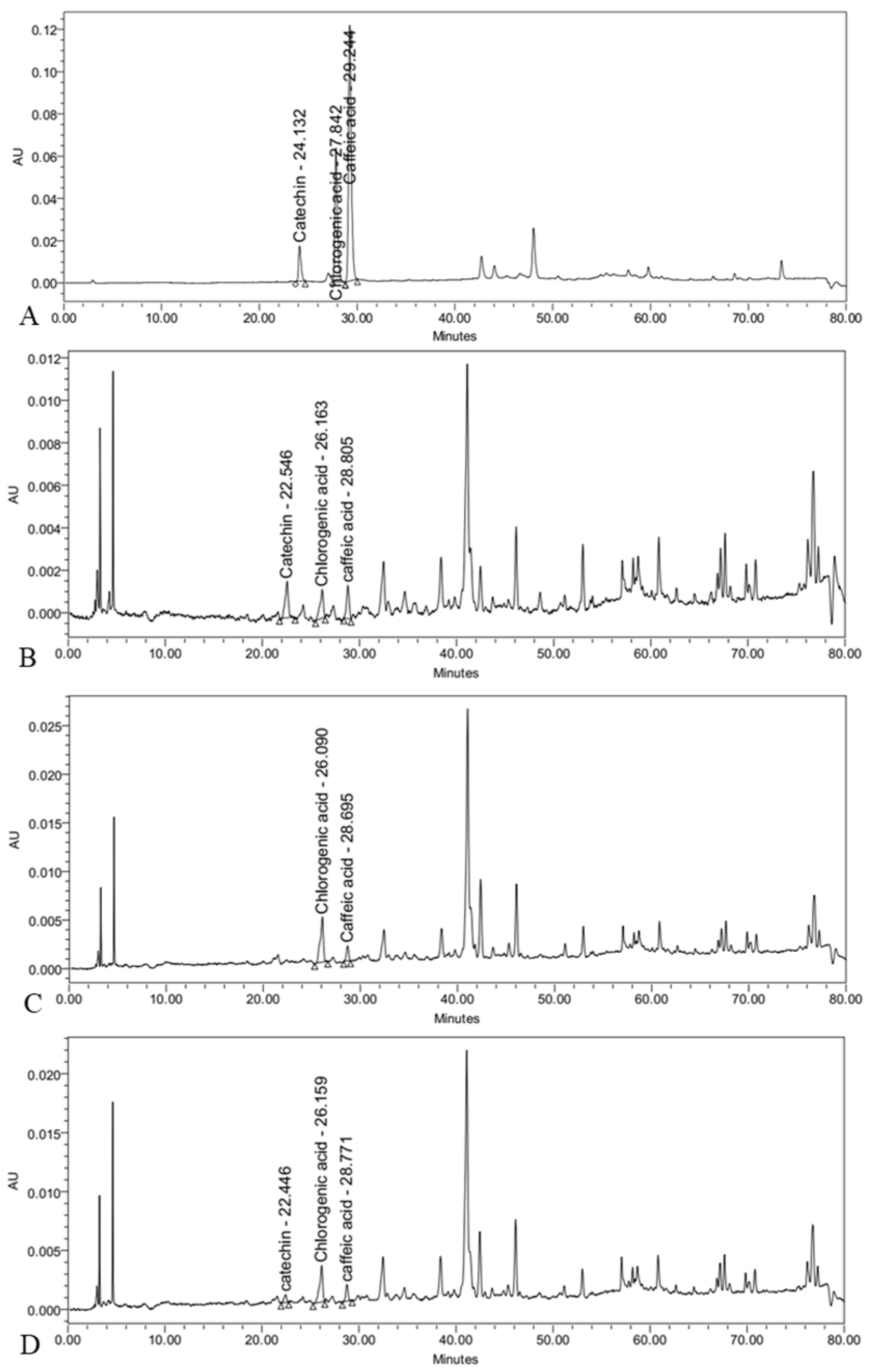
2.4.5. Determination of Chlorogenic Acid, Caffeic Acid, and Catechin

The callus powder was mixed with 95% ethanol and agitated for 4 h. After a 24 h incubation at room temperature, the mixture was filtered, and the filtrate was stored at 4 °C for subsequent analysis of chlorogenic acid, caffeic acid, and catechin content. The Agilent 1100 series HPLC system (Agilent Technologies, Palo Alto, CA, USA) with a UV detector, quaternary pump G1311A, degasser G1322A, and Empower™ 3 software (version 3, Waters Corporation, Milford, MA, USA) was used for the analysis of chlorogenic acid, caffeic acid, and catechin. A 250 mm × 4.6 mm, 5 μm LiChrospher RP-18 HPLC column (Merck, Darmstadt, Germany) was used for the separation. Acetonitrile (Solvent B) and 0.1% TFA in water (Solution A) made up the mobile phase. Elution was performed using the following protocol: 1.0 mL/min flow rate, 5% B for 0 min, 100% B for 12 min. Three duplicates of the analysis were performed at room temperature, with a detection wavelength of 280, 320, and 250 nm. Every methanolic extract had a concentration of 10 mg/mL. To produce calibration curves with a concentration range of 7.0–500 μg/mL, one milligram of each standard chemical (chlorogenic acid, caffeic acid, and catechin) was precisely weighed, dissolved in methanol, and utilized.

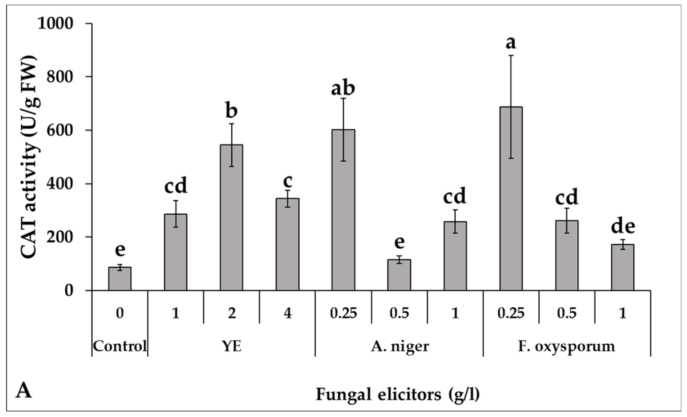
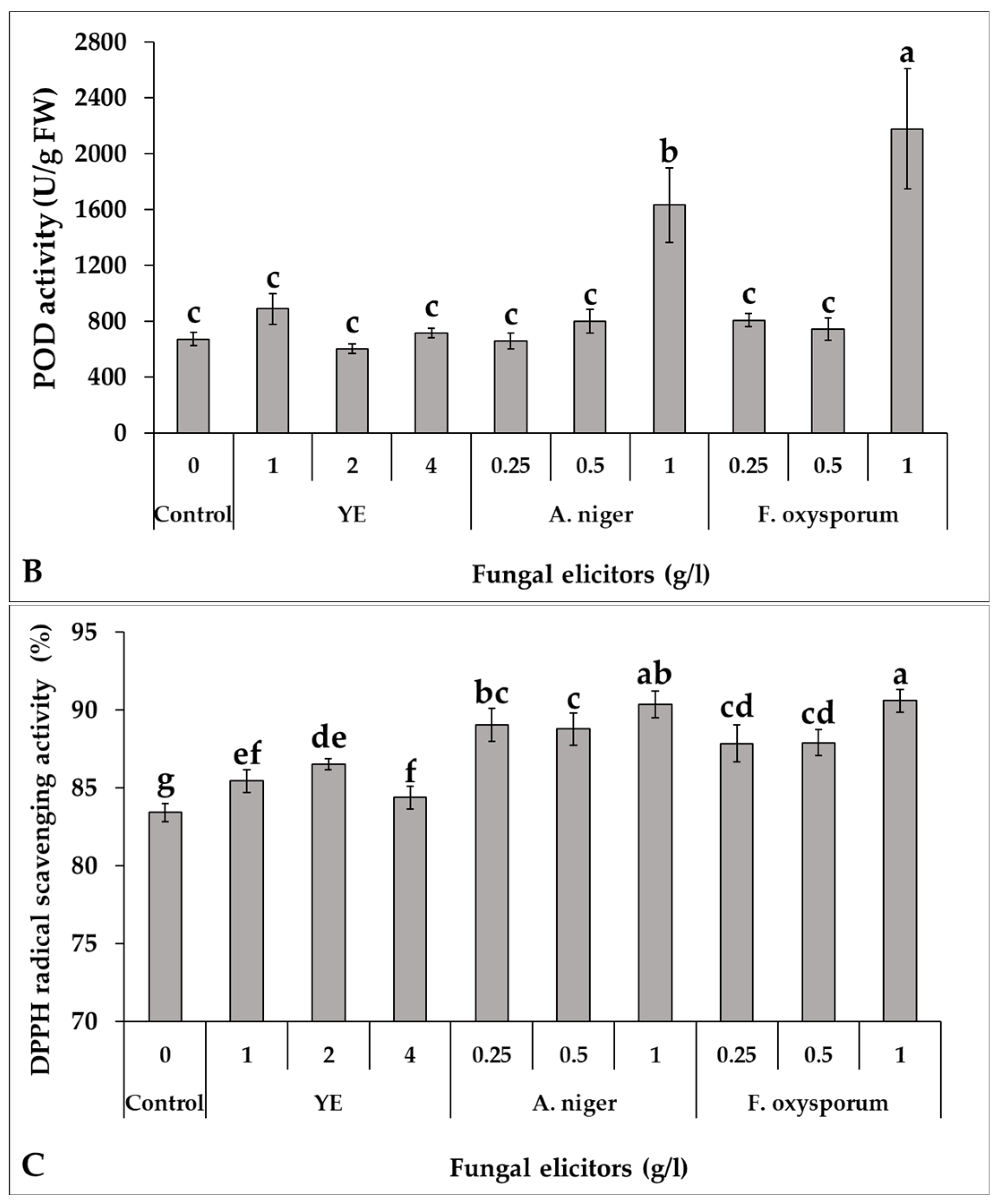
2.4.6. Determination of Antioxidant Enzymes Activity

A fresh chicory callus weighing 0.5 g was homogenized in a mortar with 5 mL of 0.1 M cold phosphate buffer (pH 7.1), followed by centrifugation at 15,000× g for 20 min at 4 °C. The resulting supernatant was utilized for the enzyme activity assay. Peroxidase activity (POD) was determined following the method outlined by Amako et al. [36], while catalase (CAT) activity was assessed in accordance with the procedure detailed by Aebi [37].

2.4.7. Determination of Free Radical Scavenging Activity

The antioxidant potential of fungal-elicited callus extracts was evaluated through the DPPH (2,2-diphenyl-1-picrylhydrazyl) assay [38]. An ethanol extract (0.7 mL) from the callus samples was combined with 3 mL of a DPPH ethanol solution (200 µM). This mixture was agitated and kept in the dark for 30 min at 25 ± 2 °C. The absorbance was then measured at 517 nm using a spectrophotometer. The percentage of DPPH radical scavenging activity was computed using the formula:
D P P H   a c t i v i t y % = A   c o n t r o l A   s a m p l e A   c o n t r o l × 100
Here, A sample represents the absorbance of the callus extract combined with the DPPH solution, and A control denotes the absorbance of the DPPH solution with 0.7 mL of 95% ethanol in place of the sample.

2.5. The Statistical Analysis

The study was arranged in a completely randomized design comprising three replications. Each replication consisted of five jars, with each jar containing three explants. Statistical analysis was executed through Analysis of Variance (ANOVA) and subsequently by Duncan’s Multiple Range Test (DMRT) at a significance level of p < 0.05. The statistical computations were conducted using CoStat software, version 6.4 (CoHort software, Monterey, CA, USA). Furthermore, Pearson’s Correlation Analysis and Principal Component Analysis (PCA) were carried out utilizing the OriginPro 2024 software (OriginLab Corporation, Northampton, MA, USA).

3. Results

3.1. Characterization of Callus Induced by Root Explants Under Different Combinations of PGRs and Light Conditions

Root explants cultured on MS medium without PGRs showed slow elongation without callus formation, while those supplemented with BA alone exhibited slight tip swelling but no callusing. Different combinations of PGRs (NAA; 1 and 2 mg/L and BA; 0, 1, and 2 mg/L) and light conditions (16/8 h light/dark cycle or complete dark) affected the characteristics (callus color, callus texture, formation of adventitious roots and shoots) of callus induced by root explants of C. intybus (Table 1 and Figure 1). All tested combinations of PGRs and light conditions resulted in 100% callus induction from root explants. A noticeable difference in callus color was observed between light and dark incubations. The majority of calli generated from cultures incubated in the 16/8 h light/dark cycle were yellowish green or yellowish white tinged with light violet, whereas callus induced under dark incubation was light yellow or light brown with light violet. When cultures were incubated under the 16/8 h light/dark cycle, callus with a friable or soft texture was induced, except for the high concentration of BA (2 mg/L), which formed callus with a compact texture. Under complete dark incubation, callus with a friable texture was induced only in cultures without BA. As can be seen from Figure 1A,B,F, adventitious roots were formed in considerable quantities in both light and dark conditions when NAA was used alone in the callus induction media. Some adventitious roots were also formed with low BA (1 mg/L) but not with high BA levels (Figure 1C,D,G). High BA concentrations (2 mg/L) caused the formation of adventitious shoots on the callus when incubated only in light (Figure 1I).
As shown in Table 2, different combinations of PGRs and light conditions significantly (p < 0.05) affected the FW, DW, and DM of callus induced by root explants of C. intybus. In general, callus induced by root explants under the 16/8 h light/dark cycle condition recorded higher weights (FW and DW) of 1.21 ± 0.27 and 0.083 ± 0.013 g/explant, respectively, compared to callus induced under complete dark (0.72 ± 0.25 and 0.061 ± 0.016 g/explant, respectively). On the other side, the DM of callus formed was increased under complete dark (8.58 ± 0.92%) compared to the 16/8 h light/dark cycle (7.02 ± 1.00%).
When the concentration of PGRs in the callus induction medium was increased, there was a discernible increase in the weight of the callus formed (Table 2). Regardless of light conditions, statistical analysis showed that the maximum significant FW and DW of callus formed (1.25 ± 0.12 and 0.087 ± 0.006 g/explant, respectively) was observed on medium containing 2 mg/L NAA + 2 mg/L BA, followed by 2 mg/L NAA + 1 mg/L BA (1.12 ± 0.39 and 0.080 ± 0.019 g/explant, respectively). On the contrary, there was a noticeable increase in the DM of the formed callus at low levels of PGRs. The combination of NAA and BA each at 1 mg/L produced the highest significant DM (9.04 ± 0.85%).
Statistical analysis of the interaction between incubation lighting conditions (16/8 h light/dark cycle or complete dark) and PGRs (NAA and BA) added to the induction media showed significant differences (p < 0.05) between treatments with respect to FW and DW of callus formed but not DM (Table 2). The highest significant values of callus formed FW was recorded for the induction media supplemented with 2 mg/L of NAA under the 16/8 h light/dark cycle. In this regard, the FW of callus formed was 1.38 ± 0.05, 1.45 ± 0.11, and 1.34 ± 0.06 g/explant with the combinations 2 mg/L NAA + 0 mg/L BA, 2 mg/L NAA + 1 mg/L BA, and 2 mg/L NAA + 2 mg/L BA, respectively. The DW of callus formed generally followed the same pattern as the FW. In addition, the induction media containing 1 mg/L NAA + 0 mg/L BA, 1 mg/L NAA + 2 mg/L BA under light incubation, and 2 mg/L NAA + 2 mg/L BA under complete incubation induced callus with a DW value significantly equal to those recorded with 2 mg/L NAA in combination with different levels of BA. In this context, the DW values ranged from 0.084 ± 0.004 to 0.096 ± 0.007 g/explant. The interaction between incubation lighting conditions and PGR combinations showed no significant variation in DM percentage. The DM value ranged from 6.15 ± 0.15% to 9.30 ± 0.49%. Callus induced in 2 mg/L NAA + 1 mg/L BA medium under 16/8 h light/dark cycle was chosen for the bio-elicitors experiment due to its superior growth characteristics, lack of shoot formation, and ease of removal of minor adventitious roots.

3.2. Effect of Fungal Elicitors on Callus Biomass Production

There were notable variations in callus biomass FW across bio-elicitors. Data presented in Table 3 show that fungal elicitors concentrations varied; low concentrations of YE and F. oxysporum enhanced the biomass production of callus FW, whereas high concentrations of all elicitors caused the FW of callus to drop compared to unelicited control callus. YE at 1 g/L and F. oxysporum at 0.25 g/L achieved the highest significant production of callus FW (251.91 ± 16.73 and 264.04 ± 21.39 g/L, respectively) compared with control (220.73 ± 29.21 g/L). Moderate level of YE (2 g/L) and F. oxysporum (0.5 g/L), as well as low (0.25 g/L) and moderate (0.5 g/L) levels of A. niger did not significantly affect the FW of callus cultures when compared with control.
In general, there were no significant differences (p < 0.05) between all fungal elicitors for callus biomass DW production. However, the findings in Table 3 for callus biomass DW indicate that YE was more prominent than A. niger and F. oxysporum. The production of callus biomass DW ranged from 11.94 ± 2.02 g/L with the treatment of 0.5 g/L of A. niger to 15.15 ± 0.55 g/L with the addition of 1 g/L of YE. Fungal elicitors applied at high concentrations significantly improved the DM of C. intybus callus (Table 3). Eliciting callus cultures with high concentrations of A. niger (1 g/L) resulted in the highest significant value of DM (8.44 ± 0.13%) followed by YE at 4 g/L (6.93 ± 0.23%) and F. oxysporum at 1 g/L (6.89 ± 0.14%) compared to the control (5.62 ± 0.31%). No significant effect of low and moderate concentrations of fungal elicitors on DM was observed when compared with the control.

3.3. Effect of Fungal Elicitors on Total Phenolic and Total Flavonoid Production

Total phenolic content (TPC) was positively increased upon fungal biotic elicitation of C. intybus callus. By increasing the eliciting dose of A. niger and F. oxysporum, TPC increased. In contrast, TPC decreased with increasing YE level (Table 4). Except for 0.25 g/L of F. oxysporum, the bio-elicitor treatments enhanced TPC by 1.26–1.77-fold than unelicited callus. The highest significant value of TPC (13.78 ± 0.56 and 14.24 ± 0.78 mg GAE/g DW; 1.71 and 1.77-fold increase than control, respectively) was obtained with low level of YE (1 g/L) and higher level of F. oxysporum (1 g/L).
Cultures elicited with 1 g/L YE produced the highest significant total phenolic productivity (TPP) (p < 0.05), measuring 208.59 ± 2.44 mg GAE/L medium. This represented a 2.10-fold increase compared to the control (99.28 ± 3.97 mg GAE/L medium). This was followed by F. oxysporum at 1 g/L, which resulted in 173.45 ± 5.48 mg GAE/L medium, a 1.75-fold increase over the control. The remaining concentrations of YE (2 and 4 g/L), A. niger (0.25, 0.5, and 1 g/L), and F. oxysporum (0.25 and 0.5 g/L) also achieved significant increments in TPP being higher than control by 1.64, 1.46, 1.32, 1.51, 1.65, 1.25, and 1.22-fold, respectively.
YE-elicited cultures showed a considerable accumulation of total flavonoid content (TFC) compared with the control culture (1.14–1.22-fold increase than control). In contrast, only high levels (1 g/L) of A. niger and F. oxysporum recorded a significant increase in TFC compared to the control (Tabel 4). The highest significant value of TFC (6.62 ± 0.21 mg QE/g DW), which was 1.38 times higher than the control (4.79 ± 0.23 mg QE/g DW), was recorded for 1 g/L of A. niger.
Likewise, fungal elicitation appeared to have a substantial impact on the total flavonoid productivity (TFP) in C. intybus callus expect for lower levels of A. niger (0.25 g/L) and moderate levels of F. oxysporum (0.5 g/L). In comparison to the control (59.19 ± 2.61 mg QE/L medium), A. niger at 1 g/L produced the greatest significant TFP (91.66 ± 1.51 mg QE/L medium), followed by 1 g/L YE (86.50 ± 0.98 mg QE/L medium), 1.55 and 1.46-fold higher than the control, respectively.

3.4. Effect of Fungal Elicitors on Phenolic Compounds Content

The content of phenolic compounds—chlorogenic acid, caffeic acid, and catechin—in the elicited callus cultures of C. intybus was determined by HPLC (Figure 2). Different types and concentrations of fungal elicitors added to the callus medium showed significant differences (p < 0.05) from each other in the content of these phenolic compounds (Table 5). Cultures elicited with higher concentrations of A. niger (1 g/L) produced the highest significant content of chlorogenic acid (295.64 ± 15.60 µg/g DW), followed by F. oxysporum at 1 g/L (221.26 ± 6.43 µg/g DW) and A. niger at 0.5 g/L (206.48 ± 5.47 µg/g DW); a 1.70, 1.27, and 1.19-fold increase compared to the control, respectively. The application of YE at all concentrations decreased the content of chlorogenic acid when compared to the control. On the other hand, regarding catechin, specific concentrations of YE (1 and 2 g/L) recorded a significant increase in its content in the elicited callus (403.95 ± 11.70 and 429.89 ± 5.98 µg/g DW: 1.31 and 1.40 times the control, respectively) compared to the rest of the treatments that caused a significant decrease in the catechin content. In addition, the absence of catechin was observed in the cultures elicited by the high concentration of A. niger (1 g/L). The content of caffeic acid was not significantly affected in the fungal-elicited callus except at moderate concentrations of A. niger and F. oxysporum (0.5 g/L) which caused a significant increase in caffeic acid accumulation compared to unelicited callus (123.73 ± 9.30, 113.75 ± 3.84, and 101.30 ± 6.85 µg/g DW, respectively).

3.5. Effect of Fungal Elicitors on the Activity of Some Antioxidant Enzymes

Data from Figure 3A revealed that all levels of YE significantly stimulated CAT activity while neither moderate concentration of A. niger nor high concentration of F. oxysporum significantly affected CAT activity compared to unelicited callus. In comparison to the control (85.96 U/g FW), the low level of F. oxysporum and A. niger (0.25 g/L) and moderate elicitation of YE (2 g/L) recorded the highest significant rate of CAT activity (687.64, 601.81, and 544.42 U/g FW, respectively). Conversely, the maximal significant activity of POD enzyme was recorded for callus cultures elicited with 1 g/L of F. oxysporum and 1 g/L of A. niger (2177.76 and 1633.29 U/g FW, respectively) compared to the control callus (673.55 U/g FW) (Figure 3B). In contrast to CAT activity, there was no significant change in POD activity in callus cultures treated with YE.

3.6. Effect of Fungal Elicitors on Antioxidant Activity

According to the data in Figure 3C, DPPH free radical scavenging activity was considerably increased by bio-elicitors at all levels compared to control callus (83.43%). Higher concentrations of A. niger and F. oxysporum (1 g/L) produced the highest significant antioxidant activity (90.35% and 90.61%, respectively). YE concentrations achieved lower antioxidant activity than those recorded for A. niger and F. oxysporum but still outperformed the control callus.

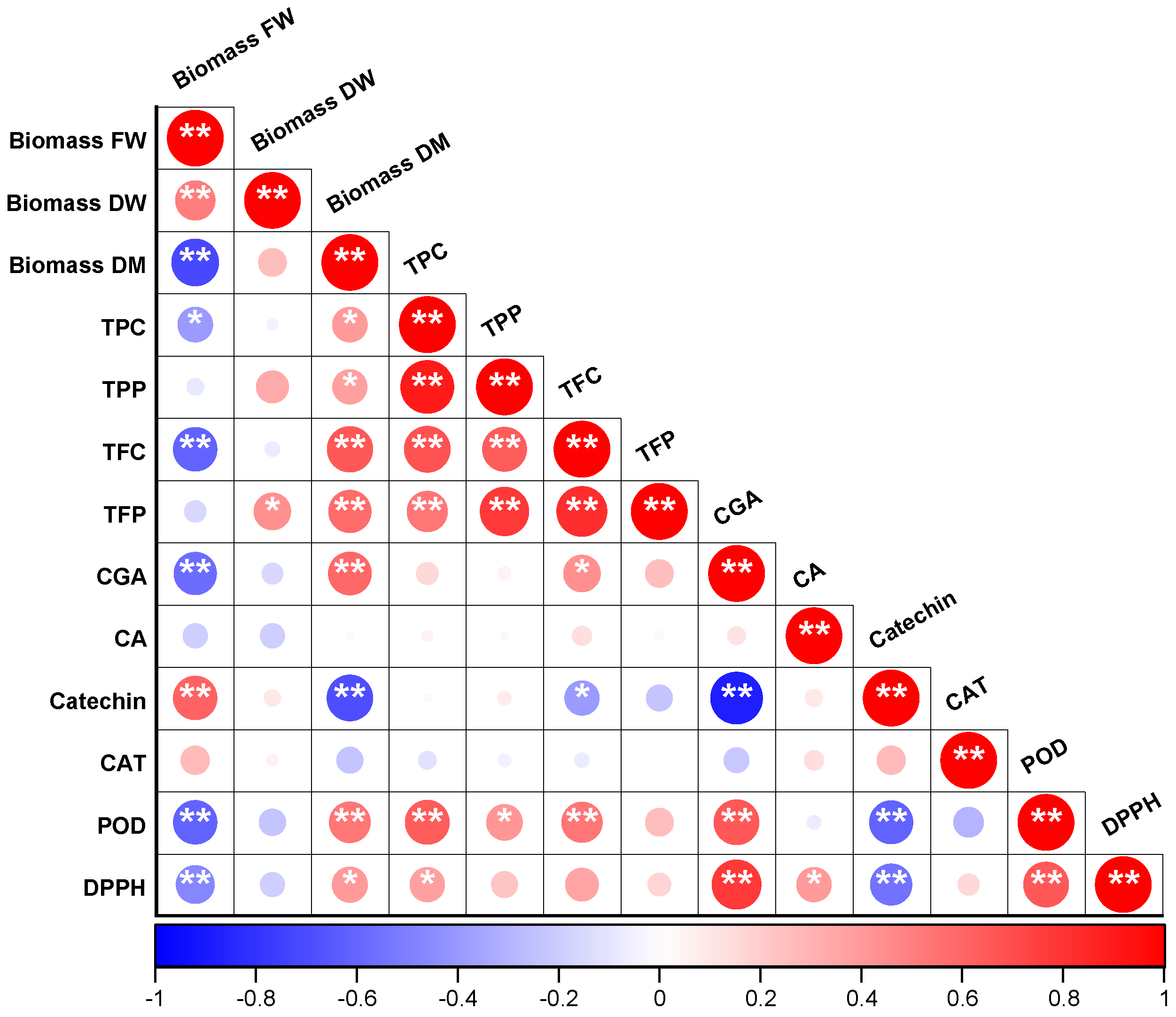
3.7. Pearson’s Correlation Analysis

Pearson’s Correlation Analysis revealed significant correlations, whether positive or negative, among various characteristics of C. intybus root-derived callus in response to different fungal elicitors (Figure 4). Positive correlations were represented by red circles, while negative correlations were indicated by blue circles in the data. A positive correlation (p < 0.05 and p < 0.01) was identified between the callus biomass DM and several parameters including callus biomass DW, TPC, TPP, TFC, TFP, chlorogenic acid content, POD activity, and DPPH scavenging activity. Furthermore, the DPPH scavenging ability of the callus extract exhibited positive correlations with various attributes such as callus biomass DM, biochemical parameters like TPC, TPP, TFC, TFP, chlorogenic acid content, caffeic acid content, as well as the activities of antioxidant enzymes (CAT and POD). Although CAT and POD activities showed a negative correlation with each other, it was noted that POD activity was positively associated with callus biomass DM (p < 0.01), the production of bioactive compounds (excluding caffeic acid and catechin), and DPPH scavenging activity (p < 0.01). Conversely, a negative correlation (p < 0.05 and p < 0.01) was observed between callus biomass FW and all parameters except callus biomass DW, catechin content, and caffeic acid content.

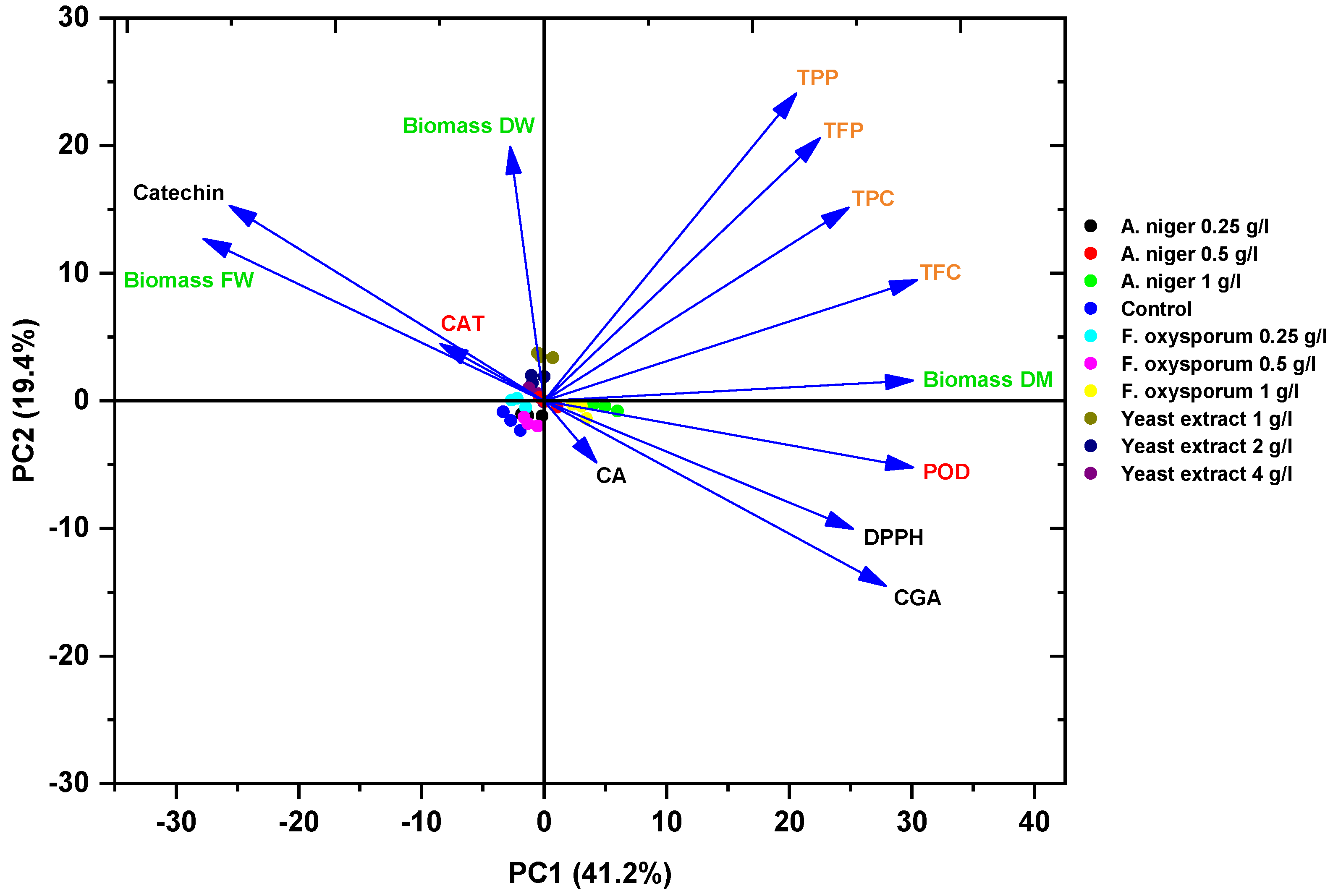
3.8. Principal Component Analysis

Principal Component Analysis (PCA) was conducted to explore the relationship between elicitor treatments and various characteristics of C. intybus callus. The PCA revealed significant differences in callus parameters, including biomass production, bioactive compounds accumulation, antioxidant enzymes activity, and DPPH scavenging capacity when exposed to different fungal elicitors (Figure 5). The data for these traits were visualized in a two-dimensional diagram generated by PCA. The PCA diagram illustrated two principal components (PC1 and PC2) that accounted for 41.2% and 19.4% of the variance, respectively, totaling 60.6% of the overall data variability. Within these components, callus biomass FW, callus biomass DW, catechin content, and CAT activity clustered together in PC2, showing positive correlations among them, while the remaining parameters aligned with PC1. Specifically, the treatment of 1 g/L of YE was mostly related to higher biomass DW and TPP, whereas catechin levels peaked at 2 g/L YE. Cultures elicited with a higher concentration (1 g/L) of A. niger or F. oxysporum were most suitable for the production of total flavonoids and chlorogenic acid and achieved high biomass DM with high POD activity and high DPPH scavenging capacity.

4. Discussion

Plant tissue culture techniques not only enable multiplication and obtain high-quality planting materials but also facilitate the development of in vitro cell cultures of medicinal plants with enhanced accumulation of bioactive compounds [8,39]. Chicory roots are recognized for their high accumulation of bioactive compounds, which have beneficial impacts on human health [14]. Callus cultures were established from in vitro root explants of C. intybus in this study. PGRs and light conditions played crucial roles in the induction and development of callus from root explants of C. intybus. Research consistently demonstrates that C. intybus callus induction is profoundly influenced by PGR combinations and photoperiods, with optimal conditions being explant-dependent. While root explants achieve maximum response with 7.5 μM IBA + 2 μM BA under the 16/8 h light/dark cycle [32], leaf tissues require different formulations—either 7.5 mg/L NAA + 2 mg/L BA for dense callus [30] or 1.5 mg/L NAA + 0.25 mg/L BA for friable structures [28].
In the present work, calli induced under a 16/8 h light/dark cycle exhibited distinct colors compared to those induced in complete darkness. Under the 16/8 h light/dark cycle, calli exhibited yellowish-green or yellowish-white hues with light violet tinges, while calli grown in complete darkness appeared light yellow or light brown with light violet. These color variations are attributed to differences in pigment accumulation, such as anthocyanins and flavonoids, which are influenced by light conditions and PGRs [18]. The anthocyanin coloration that appeared on the callus in the current experiment was also reported in leaf-derived callus of C. intybus in previous work [30]. The distinct colors under light versus dark conditions highlight the role of photomorphogenesis in regulating secondary metabolism. Light exposure typically activates pathways for chlorophyll and flavonoid biosynthesis, whereas darkness may favor other metabolites [18,40]. Additionally, the light brown calli in darkness showed higher dry matter content, which may reflect a shift toward storage metabolites like polysaccharides or lignins [11]. The texture of the callus was also influenced by the PGR concentrations and light conditions. Elateeq et al. [18] found that the combination of 1 mg/L NAA and 2 mg/L BA in the induction medium of Ginkgo biloba resulted in the highest callogenesis percentage, showcasing a compacted callus texture with varying colors under both a 16/8 h light/dark cycle and complete darkness.
NAA alone stimulated adventitious root formation across both light conditions, while low BA concentrations also promoted rooting; however, high BA under light induced shoot formation revealed light- and PGR-dependent organogenesis in C. intybus callus cultures. Kirakosyan et al. [30] also observed the development of multiple meristematic foci on leaf-derived callus of C. intybus when cultivated on media containing NAA and BA. Consistent with our results, Hadizadeh et al. [41] obtained callus with shoot regeneration of C. intybus leaves and petioles from media containing NAA and BA under a 16/8 h light/dark cycle. Additionally, Abdelhamid et al. [31] observed organogenesis of the leaf-induced callus of C. intybus and noted variations in callus texture based on light source.
The concentration of PGRs in the callus induction medium of C. intybus correlated with the weight of callus formed. The effects of auxins in promoting cell enlargement, elongation, and cell wall modification by altering cell membranes have been well-documented [42]. Plant cell division is regulated by the interplay of cytokinin and auxin, with auxin influencing DNA replication and cytokinin controlling events leading to mitosis [42,43]. In our experiment, the combination of 2 mg/L NAA + 2 mg/L BA resulted in the highest FW and DW values, likely due to synergistic effects of auxin (cell elongation) and cytokinin (cell division) [42,43], while lower PGR concentrations favored higher DM percentages, possibly by slowing growth and diverting resources to metabolite storage [11]. Similarly, Koohsari et al. [29] observed varying effects of NAA (0, 0.5, and 1 mg/L) and BA (0, 0.5, 1, and 2 mg/L) combinations on the FW, DW, and DM content of callus derived from different C. intybus explants. The interaction between PGR concentrations and light conditions showed significant differences in FW and DW of the callus formation of chicory. Callus induced under a 16/8 h light/dark cycles had higher FW and DW but lower DM (Table 2). This suggests light promotes cell division and water uptake, while darkness may enhance metabolite accumulation or reduce water content [11]. The selection of the most suitable PGR combination for callus growth, based on characteristics such as weight and dry matter content, is crucial for subsequent experiments like bio-elicitor treatments.
The impact of fungal elicitors on callus biomass and secondary metabolite production in C. intybus cultures is a critical area of study that can have implications for biotechnological applications and the production of bioactive compounds. Low concentrations of YE and F. oxysporum enhanced callus FW (Table 3), likely due to nutrient supplementation (e.g., amino acids in YE) or mild stress-induced growth stimulation [22], while high concentrations of all elicitors led to a decrease in callus FW compared to the unelicited callus. This was also observed by Khan et al. [20] on Fagonia indica callus cultures as the biomass FW and DW of callus was maximized at moderate levels of F. oxysporum and decreased at higher levels. This enhancement could be attributed to the composition of fungal mycelium, containing natural polymers like chitin, cellulose, and proteins [44]. Elevated levels of fungal elicitors have been found to inhibit cell growth due to increased oxidative stress resulting from high toxicity [22,45]. However, high concentrations of fungal elicitors significantly improved the DM of C. intybus callus, suggesting severe stress triggered water loss and metabolite compaction [45]. Research has shown that the augmentation of biomass is reliant on both the concentration of the fungal extract, as evidenced in previous studies [21,46], and the interaction of the fungus with plants, which can lead to beneficial, neutral, or detrimental effects on the host [20]. YE elicitation significantly increased biomass in Annurca apple callus [47], Glycyrrhiza glabra [48], and Ocimum basilicum [49] cell suspensions, attributable to YE’s growth-promoting components (glucans, chitin, vitamins, ergosterol, and glycopeptides) [50]. Moreover, YE contains elevated levels of amino acids, vitamins, and minerals that have the potential to enhance biomass weight depending on the plant species and concentration [22]. Our results align with the study of Elshahawy et al. [21] on Echinacea purpurea callus, where the same fungal elicitors (YE, A. niger, and F. oxysporum) were tested at identical concentration ranges. Their study demonstrated that low and moderate concentrations of F. oxysporum and A. niger (0.25–0.5 g/L) significantly enhanced callus FW and growth index. No significant change in DW across elicitors implies that biomass production was stable, but its composition (e.g., metabolite content) shifted, as evidenced by higher phenolics/flavonoids (Table 4).
The bio-elicitor treatments significantly enhanced TPC compared to unelicited callus, with the highest values obtained with low levels of YE and higher levels of F. oxysporum. Jasim and Habeeb [51] also noticed a gradual increase in TFC and total alkaloid content in Salvadora persica callus after elicitation with different concentrations of F. oxysporum. Zhong et al. [52] suggested that fungal polysaccharides derived from the endophyte F. oxysporum Fat9 could act as potent biotic elicitors, potentially being detected by surface receptors in in vitro cultures or converted into a stress signal that activates the biosynthesis of functional flavonoids in buckwheat sprouts. In the current study, TPC decreased with increasing YE levels. A similar TFC trend was observed in YE-elicited Boerhavia diffusa cell cultures [45]. Yeast cell wall components, including mannoproteins, β-1,3- and β-1,6-glucans, and chitin, along with lipids, sterols, and proteins in its membrane, can activate plant defense mechanisms through various biosynthetic pathways [53]. Treatment of B. diffusa cell cultures with A. niger led to a 2.6-fold increase in boeravinone B content, along with a significant enhancement in TPC and TFC compared to control cultures [45]. The introduction of fungal elicitors, such as A. niger, into tissue culture media can influence gene expression by promoting the production of endogenous signaling molecules, including nitric oxide, ethylene, abscisic acid, jasmonic acid, and salicylic acid. These molecules activate biosynthetic pathways for secondary compounds [54].
In the current study, we utilized a higher YE concentration range (1–4 g/L) than many previous reports. For instance, Laezza et al. [47] employed 300–500 mg/L in apple callus, Zaman et al. [49] used 1–400 mg/L in basil callus, and Bai et al. [55] applied 25–200 mg/L in Lobelia chinensis plantlet. This elevated range was a deliberate choice, primarily because plant species exhibit significant variations in their sensitivity and response to elicitors. What is effective for one species may not be for another; thus, chicory likely requires higher YE concentrations to induce a robust secondary metabolite accumulation. Furthermore, our selected concentrations are well-supported by other successful elicitation strategies in plant tissue culture. Notably, Elshahawy et al. [21] effectively utilized the identical 1–4 g/L YE concentrations to enhance metabolite production in E. purpurea callus cultures. This precedent reinforces the relevance and potential efficacy of our chosen dose range, demonstrating a targeted approach to optimize bioactive compounds production in chicory through an established, effective elicitor dose.
Although there was no significant effect of the fungal elicitors on callus biomass DW, there was a significant effect on TPP and TFP due to their impact on TPC and TFC. These findings align with previous research in plant biotechnology and elicitation studies. For instance, the work of Khan et al. [20] demonstrated the positive impact of F. oxysporum on TPC, TPP, TFC, and TFP in Fagonia indica callus cultures, highlighting the importance of optimizing elicitor concentrations for enhanced bioactive compound yields. The significant increase in TPC and TFC with YE supplementation could be attributed to the heightened activation of endogenous methyl jasmonate and/or jasmonic acid [56]. The activation of the plant’s defense mechanism, accompanied by the synthesis of signaling chemicals, is initiated when specific receptors on the plant membrane recognize and bind to molecules from the fungal extract. Consequently, this process triggers the activation of relevant genes and promotes the accumulation of secondary metabolites [20]. Moreover, studies have shown that fungal cell wall extracts act as polysaccharide elicitors, triggering an increase in calcium levels in plant cells and activating various defense mechanisms that lead to the accumulation of secondary compounds [57].
The analysis of phenolic compounds in the elicited callus cultures of C. intybus revealed significant differences in the content of chlorogenic acid, caffeic acid, and catechin among the treatments. Cultures elicited with higher concentrations of A. niger and F. oxysporum exhibited notable increases in chlorogenic acid content. Conversely, YE treatments led to a decrease in chlorogenic acid content. For catechin content, specific concentrations of YE showed significant increases, while the high concentration of A. niger resulted in the absence of catechin. Research by Shilpa and Lakshmi [58] highlighted the extraction of chlorogenic acid from C. intybus leaf calli on MS medium with various combinations of NAA and kinetin. Our findings demonstrated that catechin levels increased with YE treatment, aligning with previous findings on pulp-derived callus cultures of Annurca apple [47], Malus domestica leaf cell cultures [59], and in vitro plantlet of L. chinensis [55]. However, contrary to our results, chlorogenic acid content increased in these studies compared to the control, possibly due to variations in plant species and YE concentrations. The absence of catechin can be explained by the fact that the impact of A. niger on metabolic pathways leading to the production of defensive compounds can be altered at high concentrations due to changes in gene expression induced by the fungal elicitor. Caffeic acid content was not significantly affected by most fungal elicitors, except for moderate concentrations of A. niger and F. oxysporum. Similarly, no significant change in dihydrochalcone accumulation, such as phloridzin, was observed in YE-elicited callus of Annurca apple compared to unelicited callus [47]. However, an elevated increase in caffeic acid, catechin, and kaempferol was observed by various concentrations of F. oxysporum in callus cultures of Fagonia indica [20]. Savitha et al. [25] reported that the culture filtrate and dry cell powder of A. niger and Penicillium notatum can boost betalain accumulation in Beta vulgaris cultures while also decreasing the cell mass of the culture. YE, being a natural and cost-effective bio-elicitor, exhibits variable efficacy dependent on the plant variety and tissue type used [22]. The elicitor’s interaction with plant tissue culture type can lead to the production of diverse bioactive molecules with varying effects [47]. Treating the callus cultures of Salvadora persica with F. oxysporum enhanced the accumulation of different flavonoids, including rutin, kaempferol, quercetin, catechin, luteolin, and apigenin compared to the control callus [51]. Eliciting the cell suspension culture of Celosia cristata with F. oxysporum has been reported to enhance the production of betalain [46]. Furthermore, the elicitation with cell wall homogenate from F. oxysporum has also been shown to boost the production of glycosylated trans-resveratrol [26].
Catalase (CAT) and peroxidases (PODs) are crucial antioxidant enzymes in aerobic organisms, both playing a critical role in plant responses to various stresses by converting harmful hydrogen peroxide (H2O2) into water [60]. The results indicated that all levels of YE significantly stimulated CAT activity. This stimulation may be linked to YE’s capacity to induce cellular stress, thereby triggering the expression of CAT enzyme genes [23]. Conversely, there was no significant alteration in POD activity in chicory callus cultures treated with YE. A substantial increase in POD activity was only noticed for callus culture elicited with higher concentrations of F. oxysporum and A. niger. Fungal extracts induce oxidative stress, leading to the accumulation of endogenous abscisic acid and jasmonic acid in plant cells. These phytohormones enhance the transcriptional regulation of genes responsible for antioxidant enzyme biosynthesis, thereby boosting their activity [24,54].
Given the diverse range of specialized compounds in chicory root-derived callus, this study also assessed antioxidant activity. DPPH free radical scavenging activity, a measure of antioxidant potential, was determined for extracts of C. intybus callus cultures elicited with fungal elicitors. The data revealed that fungal elicitors significantly increased antioxidant activity compared to the control callus. Previous studies on callus cultures have reported a notable increase in antioxidant activity following fungal elicitors treatment [20,21,47,52]. However, the antioxidant capabilities of plant extracts are influenced by their composition. Reactive oxygen species (ROS) generated during plant stress exposure can potentially harm plant cells, membranes, and DNA. Plant secondary metabolites like terpenoids, phenolics, flavonoids, alkaloids, and others act as antioxidants, protecting plants from the detrimental effects of oxidation [49]. Phenolic and flavonoid complexes in plants are extensively studied due to their potent antioxidant properties, enabling them to effectively scavenge free radicals by donating hydrogen atoms. Moreover, they play a crucial role in osmotic regulation, stimulating antioxidative actions and defense mechanisms against biotic and abiotic stress factors [61]. These findings underscore the significant antioxidant activity of callus derived from C. intybus roots, suggesting their potential utilization in various fields, including the development of natural antioxidants for applications in the food and pharmaceutical industries.
Pearson’s Correlation Analysis revealed significant correlations, both positive and negative, among different parameters, shedding light on the interconnectedness of biomass production, bioactive compound accumulation, antioxidant enzyme activities, and DPPH scavenging capacity in the callus cultures. The correlation analysis suggests that an increase in callus biomass DM is associated with enhanced accumulation of bioactive compounds and improved antioxidant potential in the callus cultures. Higher FW often coincided with lower DM, while treatments with elevated DM (e.g., high A. niger) had reduced FW but higher chlorogenic acid and phenolics. This trade-off reflects resource allocation between growth and defense metabolism [20]. Additionally, the DPPH scavenging ability of the callus extract showed positive correlations with various parameters, highlighting the importance of biomass production and the presence of specific bioactive compounds in enhancing antioxidant activity. Antioxidant activity in plant extracts is linked to secondary metabolite levels such as phenolics and flavonoids [19,52]. Nadeem et al. [62] observed a positive correlation between the TPC, TFC, and antioxidant activity following YE treatment in Linum usitatissimum cell cultures. On the other hand, root-derived calli of chicory showed the presence of glycosides, terpenoids, and other bioactive metabolites not found in field-grown plants [14] which could also be involved in the antioxidant activity. The negative correlation between CAT and POD activities, despite being expected due to their opposing roles in the antioxidant defense system, underscores the complex interplay between different antioxidant enzymes in the callus cultures. POD activity exhibited positive associations with the production of bioactive compounds (excluding caffeic acid and catechin), and DPPH scavenging activity. These results suggest that POD activity plays a crucial role in enhancing the antioxidant capacity of the callus cultures, potentially through the modulation of phenolic compound levels and DPPH scavenging ability. The negative association between callus biomass FW and various attributes indicates that an increase in biomass FW may not necessarily lead to higher levels of bioactive compounds or antioxidant enzyme activities in the callus cultures.

5. Conclusions

This study confirms the profound influence of PGRs and light conditions on the characteristics of callus formation of C. intybus and explores the differential effects of bio-elicitors on bioactive compound production in root-derived callus. Key findings demonstrate that low concentrations of YE (1 g/l) and F. oxysporum (0.25 g/L) enhance callus biomass, while A. niger (1 g/L) boosts dry matter and chlorogenic acid content. Catechin levels were only enhanced by 1 and 2 g/L YE. Fungal elicitors also elevated phenolic and flavonoid levels, with A. niger and F. oxysporum showing the highest antioxidant activity. These findings underscore the potential of fungal elicitors in modulating secondary metabolite production and antioxidant properties in plant cell cultures. Future research should focus on gaining mechanistic insights into how fungal elicitors modulate secondary metabolite biosynthesis, exploring synergistic strategies by combining elicitors or stress conditions to optimize bioactive compound yields, and developing commercial applications for scaling up callus cultures to sustainably produce high-value metabolites for pharmaceuticals and nutraceuticals.

Author Contributions

Conceptualization, A.A.E. and H.-A.A.H.; methodology, A.A.E., M.M.Z., A.M.M.G., H.S.E., S.M.E. and Z.H.S.; software, S.U., W.S.A. and W.S.T.; validation, R.S.N. and M.N.A.S.; formal analysis, H.A.E.-k. and R.S.N.; investigation, A.M.M.G. and H.S.E.; resources, Z.H.S. and W.S.T.; data curation, H.A.E.-k. and W.S.A.; writing—original draft preparation, M.M.Z. and S.U.; writing—review and editing, A.A.E. and H.-A.A.H.; visualization, A.A.E., S.U. and M.N.A.S. All authors have read and agreed to the published version of the manuscript.

Funding

This research received no external funding.

Data Availability Statement

The raw data supporting the conclusions of this article will be made available by the authors on request.

Acknowledgments

The authors would like to thank Magdy A. Omar, Department of Agricultural Botany, Faculty of Agriculture, Al-Azhar University, Cairo, for providing A. niger and F. oxysporum cultures.

Conflicts of Interest

The authors declare no conflicts of interest.

References

  1. Atanasov, A.G.; Zotchev, S.B.; Dirsch, V.M.; Supuran, C.T. Natural products in drug discovery: Advances and opportunities. Nat. Rev. Drug Discov. 2021, 20, 200–216. [Google Scholar] [CrossRef] [PubMed]
  2. Liu, C.; Wang, W.; Lin, W.; Ling, W.; Wang, D. Established atherosclerosis might be a prerequisite for chicory and its constituent protocatechuic acid to promote endothelium-dependent vasodilation in mice. Mol. Nutr. Food Res. 2016, 60, 2141–2150. [Google Scholar] [CrossRef] [PubMed]
  3. Puhlmann, M.-L.; de Vos, W.M. Back to the roots: Revisiting the use of the fiber-rich Cichorium intybus L. Taproots. Adv. Nutr. 2020, 11, 878–889. [Google Scholar] [CrossRef] [PubMed]
  4. Janda, K.; Gutowska, I.; Geszke-Moritz, M.; Jakubczyk, K. The Common Cichory (Cichorium intybus L.) as a Source of Extracts with Health-Promoting Properties—A Review. Molecules 2021, 26, 1814. [Google Scholar] [CrossRef]
  5. Khan, A.M.A.; Chandra, K. Medicinal and Nutritional Importance of Cichorium intybus in Human Health. In Medicinal Plants and Their Bioactive Compounds in Human Health: Volume 1; Ansari, M.A., Shoaib, S., Islam, N., Eds.; Springer: Singapore, 2024; pp. 251–271. [Google Scholar] [CrossRef]
  6. Wawrosch, C.; Zotchev, S.B. Production of bioactive plant secondary metabolites through in vitro technologies—Status and outlook. Appl. Microbiol. Biotechnol. 2021, 105, 6649–6668. [Google Scholar] [CrossRef]
  7. Ahmadi, F.; Samadi, A.; Sepehr, E.; Rahimi, A.; Shabala, S. Morphological, phytochemical, and essential oil changes induced by different nitrogen supply forms and salinity stress in Echinacea purpurea L. Biocatal. Agric. Biotechnol. 2022, 43, 102396. [Google Scholar] [CrossRef]
  8. Elateeq, A.A.; Sun, Y. Production of bioactive metabolites in in vitro cultures of saffron (Crocus sativus L.). In Biotechnological Production of Bioactive Phytochemicals of Medicinal Value: A Comprehensive Treatise; Kishor, P.B.K., Pullaiah, T., Suprasanna, P., Rao, A.R., Romano, A., Eds.; Elsevier: Amsterdam, The Netherlands, 2024; pp. 17–65. [Google Scholar] [CrossRef]
  9. Elateeq, A.A.; Sun, Y.; Nxumalo, W.; Gabr, A.M.M. Biotechnological production of silymarin in Silybum marianum L.: A review. Biocatal. Agric. Biotechnol. 2020, 29, 101775. [Google Scholar] [CrossRef]
  10. Gabr, A.M.M.; Mabrok, H.B.; Sytar, O.; Smetanska, I. Recent Advances Toward Development of Plant Cell Culture Process for Sustainable Production of Lignans and Their Health Benefits. In Exploring Plant Cells for the Production of Compounds of Interest; Malik, S., Ed.; Springer: Cham, Switzerland, 2021; pp. 249–289. [Google Scholar] [CrossRef]
  11. Efferth, T. Biotechnology applications of plant callus cultures. Engineering 2019, 5, 50–59. [Google Scholar] [CrossRef]
  12. Arafa, N.M.; Moawad, M.; Aly, U.I. Gas chromatography-mass spectrometry analysis of phytocomponents present in Pimpinella anisum L. callus cultures as affected by yeast and phenylalanine application. Egypt. J. Chem. 2022, 65, 667–675. [Google Scholar] [CrossRef]
  13. Zafar, R.; Ali, S.M. Anti-hepatotoxic effects of root and root callus extracts of Cichorium intybus L. J. Ethnopharmacol. 1998, 63, 227–231. [Google Scholar] [CrossRef]
  14. Wadekar, P.P.; Salunkhe, V.R. Unveiling the therapeutic potential of Cichorium intybus callus: A phytochemical study. Int. J. Chem. Biochem. Sci. 2023, 24, 299–306. [Google Scholar]
  15. Abas, Y.A.; Eroğlu, A.; Dalar, A.; Türker, M.; Ozdemir, F.A.; Sołowski, G. Phenolic Compound Production Increased In Vitro Regenerated Cichorium intybus L. Appl. Biosci. 2023, 2, 84–93. [Google Scholar] [CrossRef]
  16. Selwal, N.; Supriadi, K.; Rahayu, F.; Sukmadjaja, D.; Khamidah, A.; Budiaarto, K.; Antarlina, S.S.; Tripatmasari, M.; Wani, A.K. Elicitation strategies for enhanced secondary metabolite synthesis in plant cell cultures and its role in plant defense mechanism. Plant Gene 2025, 41, 100485. [Google Scholar] [CrossRef]
  17. Narayani, M.; Srivastava, S. Elicitation: A stimulation of stress in in vitro plant cell/tissue cultures for enhancement of secondary metabolite production. Phytochem. Rev. 2017, 16, 1227–1252. [Google Scholar] [CrossRef]
  18. Elateeq, A.A.; Saad, Z.H.; Eissa, M.A.; Shakir, U. Effect of chitosan and light conditions on the production of callus biomass, total flavonoids and total phenolics in Ginkgo biloba L. Al-Azhar J. Agric. Res. 2021, 46, 28–42. [Google Scholar] [CrossRef]
  19. Zarad, M.M.; Toaima, N.M.; Refaey, K.A.; Atta, R.F.; Elateeq, A.A. Copper sulfate and Cobalt chloride effect on total phenolics accumulation and antioxidant activity of Artemisia annua L. callus cultures. Al-Azhar J. Agric. Res. 2021, 46, 26–40. [Google Scholar] [CrossRef]
  20. Khan, T.; Javed, M.U.; Mahmood, T.; Khan, B.; Khan, T.; Asad Ullah, M.; Khurshid, R.; Zaman, G.; Hano, C.; Giglioli-Guivarc’h, N.; et al. Enhancement in the production of phenolic compounds from Fagonia indica callus cultures via Fusarium oxysporum triggered elicitation. Vitr. Cell. Dev. Biol. Plant 2024, 60, 16–27. [Google Scholar] [CrossRef]
  21. Elshahawy, O.A.M.; Zeawail, M.E.-F.; Hamza, M.A.; Elateeq, A.A.; Omar, M.A. Improving the Production of Total Phenolics and Flavonoids and the Antioxidant Capacity of Echinacea purpurea Callus through Biotic Elicitation. Egypt. J. Chem. 2022, 65, 137–149. [Google Scholar] [CrossRef]
  22. Lescano, L.; Cziáky, Z.; Custódio, L.; Rodrigues, M.J. Yeast extract elicitation enhances growth and metabolite production in Limonium algarvense callus cultures. Plant Cell Tissue Organ Cult. 2025, 160, 45. [Google Scholar] [CrossRef]
  23. Maqsood, M.; Abdul, M. Yeast extract elicitation increases vinblastine and vincristine yield in protoplast derived tissues and plantlets in Catharanthus roseus. Rev. Bras. Farmacogn. 2017, 27, 549–556. [Google Scholar] [CrossRef]
  24. Aziz, E.; Batool, R.; Akhtar, W.; Rehman, S.; Gregersen, P.L.; Mahmood, T. Expression analysis of the polyphenol oxidase gene in response to signaling molecules, herbivory and wounding in antisense transgenic tobacco plants. 3 Biotech 2019, 9, 55. [Google Scholar] [CrossRef]
  25. Savitha, B.C.; Thimmaraju, R.; Bhagyalakshmi, N.; Ravishankar, G.A. Different biotic and abiotic elicitors influence betalain production in hairy root cultures of Beta vulgaris in shake-flask and bioreactor. Process Biochem. 2006, 41, 50–60. [Google Scholar] [CrossRef]
  26. Sák, M.; Dokupilová, I.; Kaňuková, Š.; Mrkvová, M.; Mihálik, D.; Hauptvogel, P.; Kraic, J. Biotic and abiotic elicitors of stilbenes production in Vitis vinifera L. cell culture. Plants 2021, 10, 490. [Google Scholar] [CrossRef]
  27. Rehman, R.; Israr, M.; Srivastava, P.; Bansal, K.; Abdin, M. In vitro regeneration of witloof chicory (Cichorium intybus L.) from leaf explants and accumulation of esculin. Vitr. Cell. Dev. Biol.-Plant 2003, 39, 142–146. [Google Scholar] [CrossRef]
  28. Dakshayini, K.; Rao, C.V.; Karun, A.; Bhavyashree, U.; Ujwal, P. High-frequency plant regeneration and histological analysis of callus in Cichorium intybus: An important medicinal plant. J. Phytol. 2016, 8, 7–12. [Google Scholar] [CrossRef]
  29. Koohsari, A.; Chalavi, V.; Akbarpour, V. Effect of Explant Types and Growth Regulators on Callus induction and Secondary Metabolites of Chicory (Cichorium intybus L.). J. Plant Prod. Res. 2020, 27, 59–72. [Google Scholar] [CrossRef]
  30. Kirakosyan, R.N.; Sumin, A.V.; Polupanova, A.A.; Pankova, M.G.; Degtyareva, I.S.; Sleptsov, N.N.; Khuat, Q.V. Influence of Plant Growth Regulators and Artificial Light on the Growth and Accumulation of Inulin of Dedifferentiated Chicory (Cichorium intybus L.) Callus Cells. Life 2022, 12, 1524. [Google Scholar] [CrossRef]
  31. Abdelhamid, S.A.; Marzouk, A.I.; Asker, M.S.; El Shabrawi, H.M. RED light promotes flavonoid and phenolic accumulation in Cichorium spp. callus culture as anti-candida agent. Sci. Rep. 2025, 15, 2194. [Google Scholar] [CrossRef]
  32. Velayutham, P.; Ranjithakumari, B.; Baskaran, P. An efficient in vitro plant regeneration system for Cichorium intybus L.—An important medicinal plant. J. Agric. Technol. 2006, 2, 287–298. [Google Scholar]
  33. Murashige, T.; Skoog, F. A revised medium for rapid growth and bioassays with tobacco tissue cultures. Physiol. Plant. 1962, 15, 473–497. [Google Scholar] [CrossRef]
  34. Singleton, V.L.; Rossi, J.A. Colorimetry of total phenolics with phosphomolybdic-phosphotungstic acid reagents. Am. J. Enol. Vitic. 1965, 16, 144–158. [Google Scholar] [CrossRef]
  35. Chang, C.-C.; Yang, M.-H.; Wen, H.-M.; Chern, J.-C. Estimation of total flavonoid content in propolis by two complementary colorimetric methods. J. Food Drug Anal. 2002, 10, 178–182. [Google Scholar] [CrossRef]
  36. Amako, K.; Chen, G.-X.; Asada, K. Separate assays specific for ascorbate peroxidase and guaiacol peroxidase and for the chloroplastic and cytosolic isozymes of ascorbate peroxidase in plants. Plant Cell Physiol. 1994, 35, 497–504. [Google Scholar] [CrossRef]
  37. Aebi, H. Catalase in vitro. In Methods in Enzymology; Academic Press: Cambridge, MA, USA, 1984; Volume 105, pp. 121–126. [Google Scholar] [CrossRef]
  38. Elateeq, A.A.; Gabr, A.M.M.; Abdelkawy, A.M.; Toaima, N.M.; Bosila, H.A.; Zarad, M.M.; Ebrahim, H.S.; Jiao, J.; Pan, H.; Ullah, S.; et al. Establishment of Gypsophila paniculata root culture for biomass, saponin, and flavonoid production. Not. Bot. Horti Agrobot. Cluj-Napoca 2022, 50, 12886. [Google Scholar] [CrossRef]
  39. Abdelkawy, A.M.; Alshammari, S.O.; Hussein, H.-A.A.; Kenawy, S.K.M.; Abou El-Enain, I.M.M.; Abdelkhalek, E.S.; Meganid, A.S.; Elateeq, A.A. Hairy root cultures as a potential tool for the biosynthesis of active compounds in Catharanthus roseus. Pak. J. Bot. 2024, 56, 1567–1574. [Google Scholar] [CrossRef]
  40. Han, J.; Li, Y.; Zhao, Y.; Sun, Y.; Li, Y.; Peng, Z. How does light regulate plant regeneration? Front. Plant Sci. 2025, 15, 1474431. [Google Scholar] [CrossRef]
  41. Hadizadeh, H.; Mohebodini, M.; Esmaeilpoor, B.; Chamani, E. Studies on Callus Induction and Regeneration of Medicinal Plant Chicory (Cichorium intybus L.) from Leaf and Petiole Explants. J. Hortic. Sci. 2016, 29, 621–630. [Google Scholar] [CrossRef]
  42. Du, M.; Spalding, E.P.; Gray, W.M. Rapid auxin-mediated cell expansion. Annu. Rev. Plant Biol. 2020, 71, 379–402. [Google Scholar] [CrossRef]
  43. Yang, W.; Cortijo, S.; Korsbo, N.; Roszak, P.; Schiessl, K.; Gurzadyan, A.; Wightman, R.; Jönsson, H.; Meyerowitz, E. Molecular mechanism of cytokinin-activated cell division in Arabidopsis. Science 2021, 371, 1350–1355. [Google Scholar] [CrossRef]
  44. Haneef, M.; Ceseracciu, L.; Canale, C.; Bayer, I.S.; Heredia-Guerrero, J.A.; Athanassiou, A. Advanced materials from fungal mycelium: Fabrication and tuning of physical properties. Sci. Rep. 2017, 7, 41292. [Google Scholar] [CrossRef]
  45. Sudheer, W.N.; Nagella, P. Biotic elicitors influence boeravinone B production from cell suspension cultures of Boerhavia diffusa Linn. S. Afr. J. Bot. 2024, 174, 96–106. [Google Scholar] [CrossRef]
  46. Warhade, M.I.; Badere, R.S. Fusarium oxysporum cell elicitor enhances betalain content in the cell suspension culture of Celosia cristata. Physiol. Mol. Biol. Plants 2018, 24, 285–293. [Google Scholar] [CrossRef] [PubMed]
  47. Laezza, C.; Imbimbo, P.; D’Amelia, V.; Marzocchi, A.; Monti, D.M.; Di Loria, A.; Monti, S.M.; Novellino, E.; Tenore, G.C.; Rigano, M.M. Use of yeast extract to elicit a pulp-derived callus cultures from Annurca apple and potentiate its biological activity. J. Funct. Foods 2024, 112, 105988. [Google Scholar] [CrossRef]
  48. Vijayalakshmi, U.; Shourie, A. Yeast extract-mediated elicitation of anti-cancerous compounds licoisoflavone B, licochalcone A, and liquirtigenin in callus cultures of Glycyrrhiza glabra. Biotechnologia 2019, 100, 441–451. [Google Scholar] [CrossRef]
  49. Zaman, G.; Farooq, U.; Bajwa, M.N.; Jan, H.; Shah, M.; Ahmad, R.; Andleeb, A.; Drouet, S.; Hano, C.; Abbasi, B.H. Effects of yeast extract on the production of phenylpropanoid metabolites in callus culture of purple basil (Ocimum Basilicum L. var purpurascens) and their in-vitro evaluation for antioxidant potential. Plant Cell Tissue Organ Cult. 2022, 150, 543–553. [Google Scholar] [CrossRef]
  50. Cai, Z.; Kastell, A.; Mewis, I.; Knorr, D.; Smetanska, I. Polysaccharide elicitors enhance anthocyanin and phenolic acid accumulation in cell suspension cultures of Vitis vinifera. Plant Cell Tissue Organ Cult. 2012, 108, 401–409. [Google Scholar] [CrossRef]
  51. Jasim, H.Y.J.; Habeeb, H.M. Effect of biotic and abiotic elicitors on Salvadora persica callus in vitro. Baghdad Sci. J. 2024, 21, 2829. [Google Scholar] [CrossRef]
  52. Zhong, L.; Niu, B.; Tang, L.; Chen, F.; Zhao, G.; Zhao, J. Effects of Polysaccharide Elicitors from Endophytic Fusarium oxysporum Fat9 on the Growth, Flavonoid Accumulation and Antioxidant Property of Fagopyrum tataricum Sprout Cultures. Molecules 2016, 21, 1590. [Google Scholar] [CrossRef] [PubMed]
  53. Ferrari, S. Biological elicitors of plant secondary metabolites: Mode of action and use in the production of nutraceutics. In Bio-Farms for Nutraceuticals. Advances in Experimental Medicine and Biology; Giardi, M.T., Rea, G., Berra, B., Eds.; Springer: Boston, MA, USA, 2010; Volume 698, pp. 152–166. [Google Scholar] [CrossRef]
  54. Aharoni, A.; Galili, G. Metabolic engineering of the plant primary-secondary metabolism interface. Curr. Opin. Biotechnol. 2011, 22, 239–244. [Google Scholar] [CrossRef]
  55. Bai, X.; Lee, H.-S.; Han, J.-E.; Murthy, H.N.; Park, S.-Y. Enhancement of Phenolic and Polyacetylene Accumulation in Lobelia chinensis (Chinese lobelia) Plantlet Cultures Through Yeast Extract and Salicylic Acid Elicitation. Horticulturae 2025, 11, 612. [Google Scholar] [CrossRef]
  56. Nabi, N.; Singh, S.; Saffeullah, P. Responses of in vitro cell cultures to elicitation: Regulatory role of jasmonic acid and methyl jasmonate: A review. Vitr. Cell. Dev. Biol.-Plant 2021, 57, 341–355. [Google Scholar] [CrossRef]
  57. Chodisetti, B.; Rao, K.; Gandi, S.; Giri, A. Improved gymnemic acid production in the suspension cultures of Gymnema sylvestre through biotic elicitation. Plant Biotechnol. Rep. 2013, 7, 519–525. [Google Scholar] [CrossRef]
  58. Shilpa, K.; Lakshmi, B.S. Influence of exogenous cinnamic acid on the production of chlorogenic acid in Cichorium intybus L cell culture. S. Afr. J. Bot. 2019, 125, 527–532. [Google Scholar] [CrossRef]
  59. Sarkate, A.; Banerjee, S.; Mir, J.I.; Roy, P.; Sircar, D. Antioxidant and cytotoxic activity of bioactive phenolic metabolites isolated from the yeast-extract treated cell culture of apple. Plant Cell Tissue Organ Cult. 2017, 130, 641–649. [Google Scholar] [CrossRef]
  60. Dhiman, V.; Singh, D.; Dhiman, V.K.; Verma, S.K.; Pandey, H. Reactive Oxygen Species (ROS) and Biotic Stress: Evolving Roles Under Climate Change. In Climate Change and Biotic Factors; Singh, A., Pandey, S., Kumar, A., Eds.; Apple Academic Press: Palm Bay, FL, USA, 2025; pp. 201–218. [Google Scholar] [CrossRef]
  61. Salam, U.; Ullah, S.; Tang, Z.-H.; Elateeq, A.A.; Khan, Y.; Khan, J.; Khan, A.; Ali, S. Plant Metabolomics: An Overview of the Role of Primary and Secondary Metabolites against Different Environmental Stress Factors. Life 2023, 13, 706. [Google Scholar] [CrossRef]
  62. Nadeem, M.; Abbasi, B.H.; Garros, L.; Drouet, S.; Zahir, A.; Ahmad, W.; Giglioli-Guivarc’h, N.; Hano, C. Yeast-extract improved biosynthesis of lignans and neolignans in cell suspension cultures of Linum usitatissimum L. Plant Cell Tissue Organ Cult. 2018, 135, 347–355. [Google Scholar] [CrossRef]
Figure 1. Different responses of root explants of Cichorium intybus cultured on MS media supplemented with different combinations of NAA and BA (mg/l); 1 NAA + 0 BA (A,B), 1 NAA + 1 BA (C,D), 2 NAA + 0 BA (E,F), 2 NAA + 1 BA (G,H), and 2 NAA + 2 BA (I,J) under 16/8 h light/dark cycle and complete dark conditions, respectively. The cultures were incubated for 4 weeks at 25 ± 2 °C.
Horticulturae 11 00678 g001
Figure 2. HPLC chromatogram fraction of Cichorium intybus callus extract. (A) Reference standards with identified peaks (catechin, chlorogenic acid, and caffeic acid), (BD) Callus extracts showing corresponding peaks; (B) 2 g/L yeast extract, (C) 1 g/L Aspergillus niger, and (D) 1 g/L Fusarium oxysporum. The x-axis represents retention time (minutes), and the y-axis shows signal intensity in Absorbance Units (AU).
Horticulturae 11 00678 g002
Figure 3. Effect of fungal elicitors on the activity of catalase (CAT; (A)), peroxidase (POD; (B)), and the DPPH radical scavenging capacity (C) of Cichorium intybus callus. YE: yeast extract, A. niger: Aspergillus niger, F. oxysporum: Fusarium oxysporum. Bar represents ± SD, n = 3. Columns annotated by different letters are significantly different (p < 0.05) according to DMRT.
Horticulturae 11 00678 g003aHorticulturae 11 00678 g003b
Figure 4. Pearson’s Correlation Analysis of callus biomass production, bioactive compounds accumulation, antioxidant enzymes activity, and DPPH scavenging activity of Cichorium intybus callus. Biomass FW: callus biomass fresh weight, Biomass DW: callus biomass dry weight, Biomass DM: callus biomass dry matter, TPC: total phenolic content, TPP: total phenolic productivity, TFC: total flavonoid content, TFP: total flavonoid productivity, CGA: chlorogenic acid, CA: caffeic acid, CAT: catalase, POD: peroxidase, DPPH: 2,2-diphenyl-1-picrylhydrazyl. The red color represents a positive correlation, and the blue color represents a negative correlation. Significant correlations were shown by * at p < 0.05 and ** at p < 0.01.
Horticulturae 11 00678 g004
Figure 5. Principal Component Analysis (PCA) of callus biomass production, bioactive compounds accumulation, antioxidant enzymes activity, and DPPH scavenging activity of Cichorium intybus callus elicited with fungal elicitors. Colored dots represent treatments: the control are the unelicited callus cultures; yeast extract used at 1, 2, and 4 g/L, Aspergillus niger added at 0.25, 0.5, and 1 g/L, Fusarium oxysporum added at 0.25, 0.5, and 1 g/L. Abbreviations in PCA are the same as mentioned in the legend in correlation analysis.
Horticulturae 11 00678 g005
Table 1. Different responses of root explants of Cichorium intybus cultured on MS media supplemented with different combinations of NAA and BA. The cultures were incubated for 4 weeks at 25 ± 2 °C under 16/8 h light/dark cycle or complete dark conditions.
PGRs (mg/L)Callus ColorCallus TextureAdventitious Root FormationAdventitious Shoot Formation
NAABA16/8 h Light/Dark CycleComplete Dark16/8 h Light/Dark CycleComplete Dark16/8 h Light/Dark CycleComplete Dark16/8 h Light/Dark CycleComplete Dark
10Light yellow tinged with light violetLight brown with light violetFriable Friable +++++++--
11Yellowish greenLight yellowFriableCompact++--
12Yellowish white tinged with light violetLight yellowCompactCompact--++++-
20Light yellow with light violetLight brown with light violetSoftFriable++++++--
21Yellowish greenLight yellowFriableSemi-compacted++---
22Yellowish white tinged with light violetLight yellowCompactCompact--++++-
All treatments showed 100% callus induction. Adventitious organ formation is indicated by symbols (-) no formation, (+) low, (++) moderate, (+++) high, (++++) very high.
Table 2. Effect of different combinations of plant growth regulators (NAA and BA) and light conditions (16/8 h light/dark cycle and complete dark) on fresh weight, dry weight, and dry matter of callus formed by root explant of Cichorium intybus.
Treatments Fresh Weight (g/Explant)Dr Weight (g/Explant)Dry Matter (%)
Light conditions (LC)16/8 h light/dark cycle1.21 ± 0.27 a0.083 ± 0.013 a7.02 ± 1.00 b
Complete dark0.72 ± 0.25 b0.061 ± 0.016 b8.58 ± 0.92 a
PGRs (mg/L)NAABA
100.88 ± 0.39 c0.067 ± 0.021 cd8.19 ± 1.45 b
110.65 ± 0.09 d0.059 ± 0.011 d9.04 ± 0.85 a
120.98 ± 0.25 c0.076 ± 0.011 bc7.91 ± 1.09 bc
200.93 ± 0.50 c0.062 ± 0.025 d7.20 ± 1.23 cd
211.12 ± 0.39 b0.080 ± 0.019 ab7.46 ± 1.14 bcd
221.25 ± 0.12 a0.087 ± 0.006 a6.97 ± 0.60 d
LC × PGRs16/8 h light/dark cycle101.23 ± 0.07 bcd0.086 ± 0.007 a6.96 ± 0.61 de
110.67 ± 0.12 ef0.060 ± 0.017 bc8.78 ± 1.16 ab
121.20 ± 0.02 cd0.084 ± 0.004 a6.96 ± 0.40 de
201.38 ± 0.05 ab0.085 ± 0.001 a6.15 ± 0.15 e
211.45 ± 0.11 a0.096 ± 0.007 a6.62 ± 0.34 de
221.34 ± 0.06 abc0.089 ± 0.003 a6.64 ± 0.40 de
Complete dark100.52 ± 0.06 fg0.049 ± 0.003 cd9.43 ± 0.56 a
110.63 ± 0.07 efg0.059 ± 0.004 bc9.30 ± 0.49 ab
120.76 ± 0.07 e0.067 ± 0.008 b8.87 ± 0.30 ab
200.48 ± 0.08 g0.039 ± 0.004 d8.26 ± 0.66 bc
210.80 ± 0.23 e0.065 ± 0.011 b8.29 ± 1.01 bc
221.17 ± 0.08 d0.085 ± 0.008 a7.30 ± 0.64 cd
SignificanceLC*********
PGRs*********
LC × PGRs******ns
***: significant and ns: non-significant. Data represent means ± SD, n = 3. Mean values followed by different letters in the column are significantly different (p < 0.05) according to DMRT.
Table 3. Effect of fungal elicitors (yeast extract, Aspergillus niger, and Fusarium oxysporum) on callus biomass production of Cichorium intybus.
Fungal Elicitors (g/L)Callus Biomass FW (g/L Medium)Callus Biomass DW (g/L Medium)Callus Biomass DM (%)
Control0220.73 ± 29.21 b12.38 ± 1.49 c5.62 ± 0.31 d
Yeast extract1251.91 ± 16.73 a15.15 ± 0.55 a6.03 ± 0.30 cd
2218.90 ± 10.41 b13.58 ± 1.46 abc6.22 ± 0.76 bcd
4194.18 ± 18.76 bc13.44 ± 0.93 abc6.93 ± 0.23 b
Aspergillus niger0.25214.29 ± 17.85 b12.94 ± 0.57 bc6.08 ± 0.70 cd
0.5217.89 ± 15.07 b14.64 ± 1.60 ab6.71 ± 0.39 bc
1164.28 ± 13.29 d13.86 ± 0.94 abc8.44 ± 0.13 a
Fusarium oxysporum0.25264.04 ± 21.39 a14.58 ± 1.51 ab5.52 ± 0.14 d
0.5217.35 ± 10.83 b11.94 ± 2.02 c5.48 ± 0.74 d
1177.13 ± 10.31 cd12.21 ± 0.90 c6.89 ± 0.14 b
Significance***ns***
***: significant and ns: non-significant. Data represent means ± SD, n = 3. Mean values followed by different letters in the column are significantly different (p < 0.05) according to DMRT.
Table 4. Effect of fungal elicitors (yeast extract, Aspergillus niger, and Fusarium oxysporum) on the content and productivity of total phenolic and total flavonoid in Cichorium intybus callus cultures.
Fungal Elicitors (g/L)Total Phenolic Content
(mg GAE/g DW)
Total Phenolic Productivity
(mg GAE/L Medium)
Total Flavonoid Content
(mg QE/g DW)
Total Flavonoid Productivity
(mg QE/L Medium)
Control08.04 ± 0.46 d99.28 ± 3.97 g4.79 ± 0.23 e59.19 ± 2.61 g
Yeast extract113.78 ± 0.56 a208.59 ± 2.44 a5.71 ± 0.23 b86.50 ± 0.98 b
211.98 ± 0.57 b162.43 ± 2.12 c5.82 ± 0.28 b78.97 ± 0.91 c
410.77 ± 0.24 c144.50 ± 6.88 d5.47 ± 0.34 bcd73.24 ± 0.88 de
Aspergillus niger0.2510.16 ± 0.63 c131.31 ± 3.76 e4.76 ± 0.22 e61.52 ± 1.28 g
0.510.24 ± 0.50 c149.85 ± 3.17 d5.19 ± 0.40 cde75.85 ± 3.69 cd
111.83 ± 0.83 b163.79 ± 8.91 c6.62 ± 0.21 a91.66 ± 1.51 a
Fusarium oxysporum0.258.53 ± 0.49 d124.31 ± 3.95 ef4.83 ± 0.28 e70.32 ± 2.28 ef
0.510.20 ± 0.53 c121.53 ± 2.83 f5.02 ± 0.57 de59.72 ± 3.58 g
114.24 ± 0.78 a173.45 ± 5.48 b5.61 ± 0.10 bc68.38 ± 3.90 f
Significance************
***: significant. Data represent means ± SD, n = 3. Mean values followed by different letters in the column are significantly different (p < 0.05) according to DMRT.
Table 5. Effect of fungal elicitors (yeast extract, Aspergillus niger, and Fusarium oxysporum) on phenolic compounds content in Cichorium intybus callus cultures.
Fungal Elicitors (g/L)Chlorogenic Acid
(µg/g DW)
Caffeic Acid
(µg/g DW)
Catechin
(µg/g DW)
Control0173.69 ± 10.60 d101.30 ± 6.85 c307.24 ± 9.00 c
Yeast extract1156.22 ± 4.46 e105.26 ± 5.41 bc403.95 ± 11.70 b
2145.17 ± 7.71 ef110.72 ± 4.28 bc429.89 ± 5.98 a
4133.62 ± 9.66 f110.56 ± 5.71 bc279.39 ± 12.04 de
Aspergillus niger0.25199.49 ± 11.49 c110.07 ± 7.09 bc287.15 ± 6.70 d
0.5206.48 ± 5.47 bc123.73 ± 9.30 a281.86 ± 4.04 de
1295.64 ± 15.60 a106.65 ± 7.30 bc-
Fusarium oxysporum0.25180.86 ± 4.01 d109.74 ± 2.06 bc289.47 ± 11.79 cd
0.5198.22 ± 5.45 c113.75 ± 3.84 ab267.77 ± 8.80 e
1221.26 ± 6.43 b105.74 ± 7.69 bc207.50 ± 19.51 f
Significance*******
- absent, * and ***: significant. Data represent means ± SD, n = 3. Mean values followed by different letters in the column are significantly different (p < 0.05) according to DMRT.
Disclaimer/Publisher’s Note: The statements, opinions and data contained in all publications are solely those of the individual author(s) and contributor(s) and not of MDPI and/or the editor(s). MDPI and/or the editor(s) disclaim responsibility for any injury to people or property resulting from any ideas, methods, instructions or products referred to in the content.

