Article

Phosphoproteomic Profiling Deciphers Heat-Stress-Responsive Mechanisms in Passion Fruit

1
Fruit Research Institute, Fujian Academy of Agricultural Sciences, Fuzhou 350013, China
2
Longyan Agricultural Science Research Institute, Longyan 361000, China
*
Authors to whom correspondence should be addressed.
Horticulturae 2025, 11(5), 553; https://doi.org/10.3390/horticulturae11050553
Submission received: 2 April 2025 / Revised: 17 May 2025 / Accepted: 19 May 2025 / Published: 21 May 2025

Abstract

:
Heat stress severely inhibited the flower bud growth and development of passion fruit (Passiflora edulis Sims) in summer, resulting in severe production damage. Protein phosphorylation plays a key role in plant protein regulatory networks in response to abiotic stress, while the mechanism of phosphorylation regulation response to heat stress in passion fruit is still unknown. In this study, 97.62% of passion fruit floral buds withered and fell off after 2 h of heat stress, compared to 3.33% after 0.5 h. A total of 10,614 phosphorylation sites across 2906 proteins were identified by phosphoproteomic analysis. Among them, 1343 differentially regulated phosphoproteins (DRPPs) were mainly located in the nucleus, cytoplasm, and chloroplast. The DRPPs whose phosphorylation sites were induced by heat stress were mainly involved in the ‘ABC transporters’, ‘Plant hormone signal transduction’, and ‘MAPK signaling’ pathways. In addition, the accumulations of ABA and H2O2 were induced under heat stress for 0.5 h. Through protein interaction prediction and qRT-PCR analyses, we identified a key protein PePP2C1, in which the levels of gene expression, protein expression, and phosphorylation were induced by heat stress. The transient assays showed that the overexpression of PePP2C1 inhibited the accumulation of H2O2. Our results suggested the potential role of phosphoproteins under heat stress in the floral buds of passion fruit. The findings in this study contribute to a better understanding of the molecular mechanism of phosphoproteins in response to heat stress.

1. Introduction

Passion fruit (Passiflora edulis Sims) is a perennial woody vine plant belonging to the Passiflora Linnaeus, which originates from Brazil and neighboring countries [1]. Owing to its short growth cycle, strong flowering ability, and substantial economic benefits, the passion fruit industry has emerged as a key fruit sector in numerous regions, including South American, Australia, Africa, and Asia countries [2]. However, passion fruit is highly susceptible to hot ambient temperatures, which adversely affects flowering and fruit sets. In Israel, flowers of the purple ‘Passion dream’ cultivar frequently experience abortion during the summer as a result of high-temperature conditions [3]. Flower primordia of many passion fruit varieties were observed to be aborted in summer in various regions, including Hawaii, Southern Australia, and Japan, due to heat stress [4]. In the Guangxi Province of China, passion fruit experiences a marked incidence of abnormal floral bud development during summer, with fruit-set rates demonstrating significant reduction compared to autumn cultivation periods [5]. The previous study has shown that after being treated at 39 °C for 4 h, the fruit-setting rate of golden passion fruit is only 28.6%. Furthermore, the numbers of floral buds and young fruits fall off when the maximum field temperature exceeds 37 °C [6]. Additionally, Li et al. reported that exceeding 36 °C of field temperatures would markedly reduce the fruit-set rate of several passion fruit varieties [7]. Therefore, elucidating the mechanism of passion fruit response to heat stress is an urgent and important topic, which are still unclear.
Plants possess a certain degree of adaptability to external temperature fluctuations. Under moderate heat stress, plants can activate intracellular signaling pathways and metabolic mechanisms to acclimate to temperature variations. However, once the adaptive threshold is exceeded, severe and long-term damage may occur, such as the disruption of seasonal growth patterns, altered flowering times, and reduced crop yield [8]. Throughout the plant growth cycle, the flowering stage is particularly sensitive to heat stress, which may cause damage to floral buds, leading to malformed flowers or flowering failure, decreased activity of pollen mother cells and microspores [9], reduced pollen grain diameter [10], and inhibited pollen tube growth [11], thereby affecting the pollination and fertilization processes. Previous laboratory studies have demonstrated that exposure to 40 °C significantly reduces the germination rate of passion fruit pollen, impairs stigma surface adhesion, and severely hinders pollen tube elongation in the style [11]. In response to abiotic stresses such as heat stress, plants activate osmoregulatory systems to maintain osmotic balance by increasing intracellular solute concentrations. This includes synthesizing soluble sugars, proteins, and proline to lower cellular osmotic potential or absorbing extracellular calcium, sodium, and potassium ions to regulate vacuolar osmotic potential [12]. For instance, heat-tolerant plants tend to accumulate higher levels of proline, soluble proteins, or soluble sugars [13]. Heat stress may also disrupt reactive oxygen species (ROS) homeostasis in plants. Under such stress, ROS and free radicals accumulate extensively, damaging photosynthetic pigments, proteins, and other macromolecules while interfering with normal metabolic activities [14]. Heat-tolerant plants produce greater amounts of antioxidant enzymes, such as superoxide dismutase (SOD), peroxidase (POD), and catalase (CAT), under high-temperature conditions to mitigate oxidative damage caused by excessive ROS [15]. Laboratory observations have confirmed the significant induction of superoxide anions and hydrogen peroxide in passion fruit leaves under heat stress [11]. Furthermore, studies have shown that heat-shock proteins (HSPs) [16] and heat-stress transcription factors (HSFs) [17] are highly expressed in plants under heat stress. These molecules maintain protein homeostasis, repair damaged proteins, and reduce heat-stress-induced damage, thereby enhancing plant thermotolerance [18,19]. However, the specific roles of structural proteins or proteases in the protein-level mechanisms of heat stress and how proteins function in heat-stress signal transduction remain to be further elucidated.
The role of protein phosphorylation in regulating plant growth, development, and environmental adaptation has been increasingly elucidated. By precisely phosphorylating key components within signaling cascades, plants activate or deactivate specific pathways required for growth and defense, thereby participating in developmental processes and stress responses [20]. In Arabidopsis thaliana, approximately 41% of proteins are phosphorylated under varying conditions, with their phosphorylation states regulated by protein kinases and phosphatases. Phosphorylation predominantly occurs at serine (S), threonine (T), and tyrosine (Y) residues [21,22]. To adapt to fluctuating external environments, plants employ rapid protein phosphorylation reactions to respond to external signals, facilitating both biotic and abiotic stress responses. Most abiotic stresses induce the rapid accumulation of abscisic acid (ABA), which triggers subsequent stress-responsive pathways. The ABA receptor protein PYR/PYL serves as a critical component in ABA perception. This receptor inhibits protein phosphatase ABI1/2, thereby releasing the activity of SNF1-related protein kinase SnRK2s, which acts as the central hub of ABA signal transduction [23]. SnRK2s can phosphorylate downstream transcription factors, activating their transcriptional activity and promoting the expression of genes involved in ABA or stress responses [24]. Key components of ABA signal transduction are also regulated post-translationally via phosphorylation to maintain plant growth, development, and defense mechanisms. For instance, the ABA receptor protein PYL can be phosphorylated by the target of rapamycin kinase (TOR) to suppress ABA-mediated stress responses [25]. Raf-type Mitogen-activated protein kinase kinase kinase (MAPKKK) can activate SnRK2s, initiating ABA signal transduction in response to osmotic stress [26]. Thus, ABA signal transduction relies on a series of protein phosphorylation modification events. Mitogen-activated protein kinases (MAPKs) serve as a conserved signaling center in plants, participating in various stress responses through signal cascades. For example, MPK6 reduces its DNA-binding affinity by phosphorylating the transcription factor MYB15, thereby promoting the expression of cold-resistant gene CBF [27]. In A. thaliana, a class of MAPKKK proteins, such as ANP1, is activated during heat stress, promoting hydrogen peroxide production and the activation of MAPK3/6. Activated MAPK3/6 further phosphorylates heat-shock transcription factors HSFA2 and HSFA4A, regulating the expression of heat-responsive genes [28]. Additionally, calcium ions, as crucial second messengers in plant cells, actively mediate responses to diverse stresses. Calcium-dependent protein kinase (CDPK), a key effector protein, phosphorylates and activates various ion channels during stress responses [29]. CDPK3/6/21/23 phosphorylates S-type anion channel proteins to mediate stomatal movement [30]. Furthermore, CDPK negatively regulates ABA signal transduction pathways by stabilizing ABI2 protein through phosphorylation [31]. Protein phosphorylation also plays a role in regulating sodium/potassium ion homeostasis [32], hormone metabolism and signal transduction [20], and pathogen resistance responses [33]. The multifaceted functions of protein phosphorylation in plant stress regulation warrant further exploration and discovery.
Due to the important role of protein phosphorylation in plant growth, development, and environmental responses, further investigating protein phosphorylation in plants can enhance our understanding of plant stress-response mechanisms. However, the protein phosphorylation dynamics of passion fruit under heat stress remain unclear. Therefore, this study aimed to investigate protein phosphorylation in passion fruit flower bud samples subjected to heat stress. Through biological analysis, qPCR testing, and transient transformation assays, phosphorylated proteins responsive to heat stress were identified in passion fruit. These findings are expected to provide novel insights into the regulatory mechanisms underlying flower bud responses to heat stress.

2. Materials and Methods

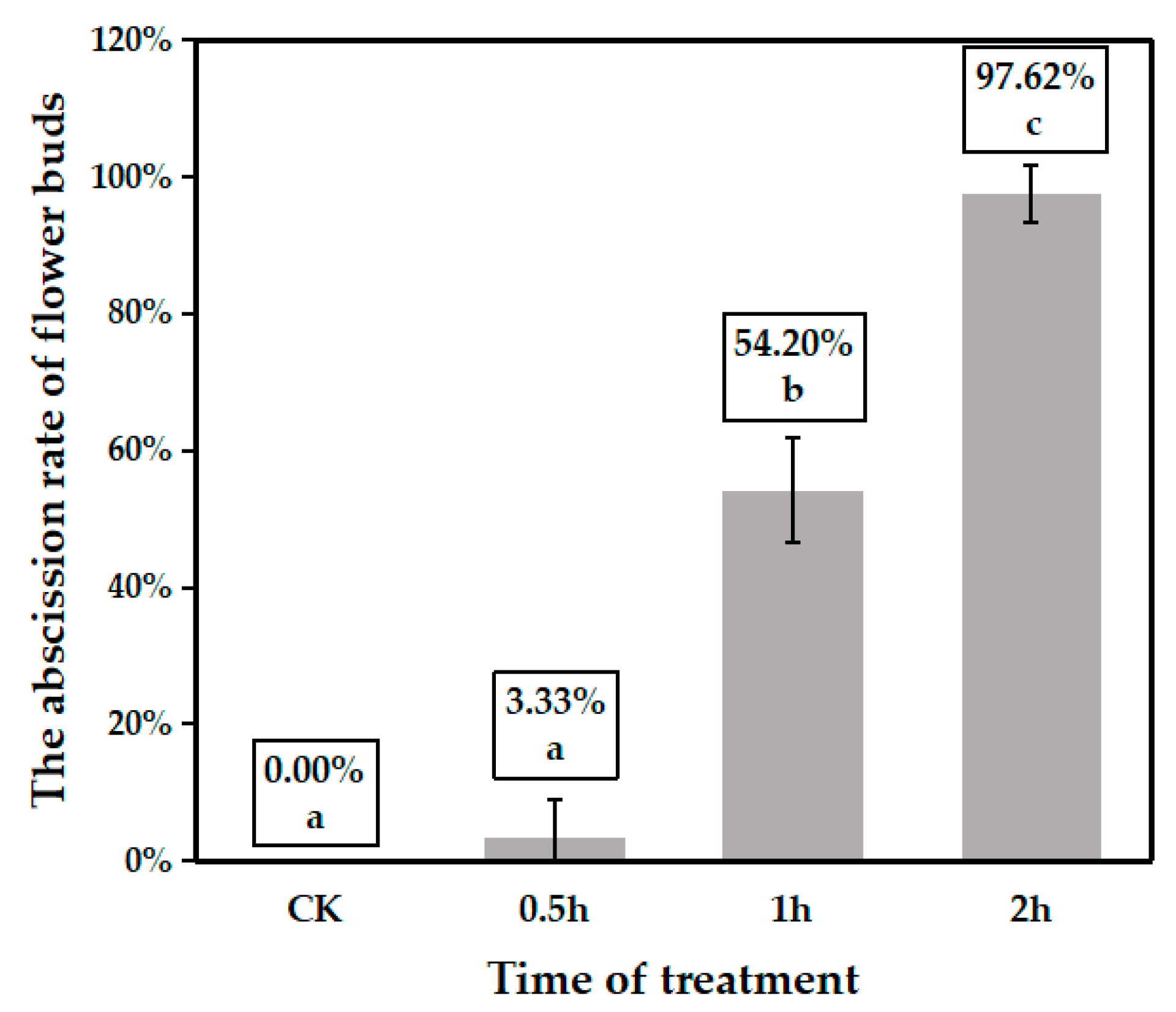
2.1. Plant Material and Treatment

In this study, the passion flower (Passiflora edulis Sims) ‘Qinmi 9’ was used as the experimental material. About 1 m high grafted seedling plants with robust growth and floral buds were selected. These plants were transferred to a climate-controlled growth chamber (BXA-800, Boxun, Shanghai, China) for heat-stress treatment (45 °C) for 0.5 h, 1 h, or 2 h, with untreated plants serving as controls. Each treatment group included three biological replicates, with twelve seedling plants per replicate. For each replicate, half of them (six seeding plants) were moved to a standard growth room (25 °C, 16 h light/8 h dark) for 2 days to quantify flower bud abscission. For the other half of the seedings from each replicate, floral buds were collected immediately after heat-stress treatment. At least 24 floral buds (20 days after flower bud formation) were collected from each replicate for subsequent ABA detection and phosphorylation experiments.
To further study the regulation of ABA in heat stress, the grafted seeding plants were treated with 150 mg/L ABA (Solarbio, Beijing, China). Then, the ABA-treated plants were treated with heat stress for 0.5 h and 2 h. The ABA-treated plants without heat stress were used as controls. Each treatment group included three biological replicates, with six seedling plants per replicate. The heat-stress treatment and sampling methods were the same as above.

2.2. Protein Extraction and Trypsin Digestion

Based on the results of heat-stress treatment, the samples from the heat stress for 0.5 h (short-term heat stress, named SH) and 2 h (long-term heat stress, named LH) were selected for proteomic and phosphoproteomic sequencing, and the samples with untreated were used as the control (CK).
The sample was fully homogenized in a mortar pre-cooled with liquid nitrogen, and then ultrasonic lysis was performed by adding 4 times the powder volume of phenol extraction buffer (10 mM dithiothreitol, 1% protease inhibitor, 1% phosphatase inhibitor, 50 μM PR-619, 3 μM TSA, 50 mM NAM). The same volume of Tris equilibrium phenol was added to the lysate, and centrifuged at 5500× g at 4 °C for 10 min. Take the supernatant and add 0.1 M ammonium acetate/methanol, 5 times the volume of the supernatant, and precipitate overnight. The precipitates were then washed with methanol and acetone solutions, respectively. Finally, 8 M urea redissolved precipitate was used to determine the protein concentration by BCA kit (P0011, Beyotime Biotechnology, Shanghai, China).
For trypsin digestion, the protein solution was gradually added to achieve a final concentration of 20% (m/v) TCA for protein precipitation. The mixture was vortexed thoroughly and incubated at 4 °C for 2 h. Subsequently, the precipitated protein was collected via centrifugation at 4500× g for 5 min at 4 °C. The protein pellet was washed three times with pre-cooled acetone and dried for 1 min. Then, the protein sample was redissolved in 200 mM TEAB and ultrasonically dispersed. The trypsin was added at a ratio of 1:50 (protease: protein, m/m) and enzymolized overnight. Dithiothreitol (DTT) was added to the final concentration of 5 mM and reduced at 56 °C for 30 min. Then, the iodoacetamide (IAA) was added to make its final concentration 11 mM, and incubated at room temperature for 15 min away from light.

2.3. Enrichment for Phosphorylated Peptides and LC-MS/MS Analysis

The peptide mixture solution was incubated with IMAC microspheres in a loading buffer (50% acetonitrile/0.5% acetic acid) with vibration. Non-specifically adsorbed peptides were removed by sequential washing with 50% acetonitrile/0.5% acetic acid and 30% acetonitrile/0.1% trifluoroacetic acid. Phosphopeptides were eluted in elution buffer (with 10% NH4OH) and collected after lyophilization for LC-MS/MS analysis.
The tryptic peptides were dissolved in solvent A (0.1% formic acid, 2% acetonitrile in water) and separated on a homemade reversed-phase column (25 cm × 100 μm i.d.) using a gradient of solvent B (0.1% formic acid in acetonitrile): 0–16 min, 2–22%; 16–22 min, 22–35%; 22–26 min, 35–90%; 26–30 min, 90%, at 450 nl/min on a NanoElute UHPLC system (Bruker Daltonics, Billerica, MA, USA). Peptides were analyzed by timsTOF Pro mass spectrometry with an electrospray voltage of 1.7 kV. Precursors and fragments were detected by the TOF detector in dia-PASEF mode. The MS scan range was 100–1700 m/z and 22 PASEF-MS/MS scans per cycle were acquired. The MS/MS scan range was 395–1395 m/z with an isolation window of 20 m/z.

2.4. Database Search

The MS/MS data generated were processed following the method described by Pan et al. [34]. The Mascot search engine (v. 2.3.0) was used to match tandem mass spectra against the Uniprot_foxtail_45551 database concatenated with a reverse bait database. Trypsin/P was set as the proteolytic enzyme, allowing for up to four missed cleavages. Precursor-ion mass tolerance was 20 ppm for the initial search and 5 ppm for the main search, with fragment-ion mass tolerance at 0.02 Da. The false discovery rate (FDR) was controlled below 1%, and modified peptides required a minimum score of >40. Phosphorylation site analysis used the lowest intensity values from MaxQuant output, and localization was determined with a threshold >0.75 [35]. The quantification of phosphorylation sites (serine, threonine, and tyrosine) was based on MaxQuant output files. Quantitative values for each sample in triplicate were derived from three experiments. Differential modification between samples was assessed in two steps: calculating mean values across triplicates, and then computing the ratio of means as the final result. A significant p-value for differential expression was calculated using log2 transformed data and a two-sample, two-tailed t-test. Up-regulation was defined as p < 0.05 and protein ratio > 1.5, while down-regulation was p < 0.05 and protein ratio < 1/1.5. Raw abundance ratios of phosphorylation sites were normalized to corresponding protein proportions.

2.5. Bioinformatics Annotation Analysis

The functional descriptions of the identified protein domains were annotated through the protein sequence alignment method by using InterProScan (http://www.ebi.ac.uk/interpro, accessed on 26 July 2024). Then, the Gene Ontology (GO) and Kyoto Encyclopedia of Genes and Genomes (KEGG) pathway analyses were performed via the online platform Sangerbox [36] (http://www.sangerbox.com/home.html, accessed on 27 March 2025). The subcellular localization of proteins was identified with the eukaryotic database Wolfpsort (http://www.genscript.com/psort/wolf_psort.html, accessed on 26 July 2024). The Motif-x algorithm Soft MoMo (http://meme-suite.org/tools/momo, accessed on 28 March 2025) was utilized to analyze the sequence model constituted by amino acids at specific positions within 6 amino acids upstream and downstream in all protein sequences. The K-means approach was adopted to conduct expression cluster analysis on the identified differentially expressed phosphorylated proteins. The expression heatmap of protein phosphorylation levels was visualized utilizing TBtool-II [37]. The online website STRING was applied to analyze the protein interaction network, and the Cytoscape software (version 3.9.1) was utilized to visualize the prediction of the protein–protein interaction network.

2.6. Detection of ABA and H2O2 Contents

The ABA content in samples was measured according to Li et al. [38]. A total of 0.5 g of frozen samples were homogenized in liquid nitrogen and mixed with a precooled water/ethyl alcohol solution (1:4, v/v, 4 °C). Subsequently, the mixture was extracted overnight at 4 °C. After centrifugation, the supernatant was evaporated to dryness under reduced pressure and reconstituted in pure methanol. Impurities were removed by passing the solution through a C18 gel column followed by filtration using a 0.22 μm organic filter membrane. The resulting methanol solution (containing the samples) was injected into an HPLC system equipped with an ODS column (Luna 5 μm C18(2) 100 A, 260 × 4.6 mm, 5 μm, Phenomenex, Torrance, CA, USA). The separation was performed at a flow rate of 1 mL/min using a gradient elution of methanol/acetonitrile/phosphate buffer (pH = 3.5).
The H2O2 content was determined using a detection kit (H2O2-1-Y, Comin Biotechnology Co., Suzhou, China) with appropriate modifications. Approximately 0.05 g of fresh plant tissue was rapidly homogenized in acetone and centrifuged at 8000× g for 10 min at 4 °C. Subsequently, 100 µL of 5% titanium sulfate solution and 100 µL of concentrated ammonia water were added to the supernatant (with acetone as the blank control), followed by centrifugation at 4000× g for 10 min at 25 °C. The precipitate was then extracted and dissolved in 1 mL of 2 mol/L sulfuric acid. The resulting solution was incubated at 25 °C for 5 min, and absorbance was measured at 415 nm by a microplate reader (Feyond-A300, AllSheng Co., Hangzhou, China).

2.7. Quantitative Real-Time PCR

The total RNA from each sample was extracted and cDNA was synthesized with FastKing gDNA Dispelling RT SuperMix kit (Tiangen Biotech, Beijing, China). The expression of relative genes was analyzed by qRT-PCR on a Roche LightCycler 96 system (Roch, Basel, Switzerland) according to the Top Green qPCR SuperMix kit (TransGen, Beijing, China). The results were analyzed by the 2−ΔΔCt method, and the expression levels of EF1a [39] were used as an internal control gene for normalization. All primers were listed in Supplementary Table S1.

2.8. Transient Transformation Assays

The CDS of PePP2C1 (GWHPAZTM015785) was obtained by 2 x Phanta Max Master Mix (Vazyme, Nanjing, China) and inserted into pBWA(V)HS vector to generate pBWA(V)HS-GWHPAZTM015785 overexpression vector. Then, the overexpression vector was transformed into Agrobacterium tumefaciens strain GV3101.
The young leaves of Nicotiana. benthamiana was selected for transient assays. First, the GV3101-carrying constructs were resuspended in the induction medium (containing 10 mM MgCl2 and 0.1 mM acetosyringone) to an OD600 of 0.5. Then, the strains containing pBWA(V)HS-GWHPAZTM015785 overexpression vector were injected into the abaxial leaf of N. benthamiana. The N. benthamiana leaves infected with empty vector were used as control. Three leaves of N. benthamiana were used as biological repeats. Three days after infection, the infiltrated area of N. benthamiana leaves was sampled for H2O2 content detection.

2.9. Statistical Analyses

The data of the contents of ABA and H2O2, as well as the expression levels of candidate proteins and genes, were analyzed using one-factor ANOVA to assess statistically significant differences among groups. Post hoc pairwise comparisons were conducted by Tukey’s honestly significant difference (HSD) test. The results were shown as the mean ± standard deviation of replicates, with significance determined at p < 0.05. All statistical analyses were performed with IBM SPSS Statistics 26 software.

3. Results

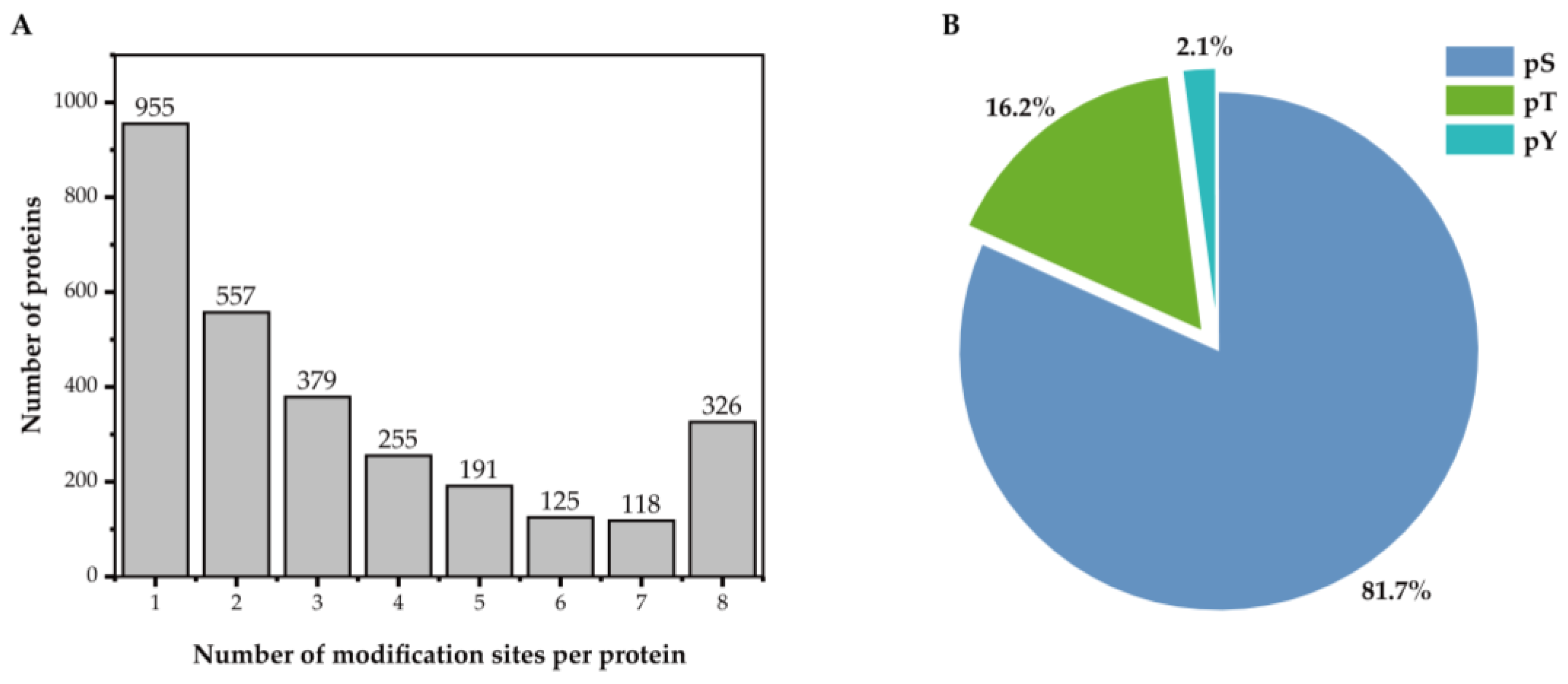
3.1. Identification of Proteins and Phosphorylated Sites in Floral Buds Under Heat Stress

In this study, we investigated the effects of short-term heat stress on passion fruit by exposing potted plants to graded durations of thermal stress in a climate-controlled growth chamber. Following heat treatment, plants were transferred to optimal temperature conditions for subsequent observation. The result showed that flower bud mortality and abscission rates exhibited progressive increases with extended heat exposure duration (Figure 1). Plants exposed to 0.5 h of thermal stress showed minimal damage with only 3.33% bud loss, which was not significantly different from the CK. This percentage rose to 54.20% following 1 h of heat treatment, reaching 97.62% mortality when the exposure period extended to 2 h.
The study aimed to investigate the adaptation mechanisms underlying heat-stress response in floral buds of passion fruit. Samples subjected to 0.5 h heat exposure (short heat stress, named SH), which largely maintained developmental competence, were compared against 2 h stressed (long heat stress, named LH) and unstressed controls (CK) via phosphoproteomic profiling. Following stringent data filtering, a total of 10,614 phosphorylation sites across 2906 proteins were identified (Table 1). An analysis of phosphorylation site distribution (Figure 2A) showed that 32.9% of proteins contained a single modification site, 19.2% exhibited two sites, and 11.2% harbored over seven sites. The characterization of phosphorylated amino acid residues (Figure 2B) revealed strong residue specificity. Most of the amino acids (81.7%, 8676) were phosphorylated at serine residues, followed by threonine residues (16.2%, 1715), while tyrosine residues were rare (2.1%, 223).
The conserved motifs flanking phosphorylation sites critically influence enzyme substrate specificity. In this study, we identified 16,042 distinct conserved motifs by extracting 13-amino-acid sequences centered on phosphorylation sites. These included 13,643 phosphoserine, 2156 phosphothreonine, and 243 phosphotyrosine sequences (Supplementary Table S2). The enrichment analysis of conserved motifs revealed that 65 types of conserved motifs were significantly enriched around phosphoserine sites, with only 13 and 4 for phosphothreonine and phosphotyrosine sites.

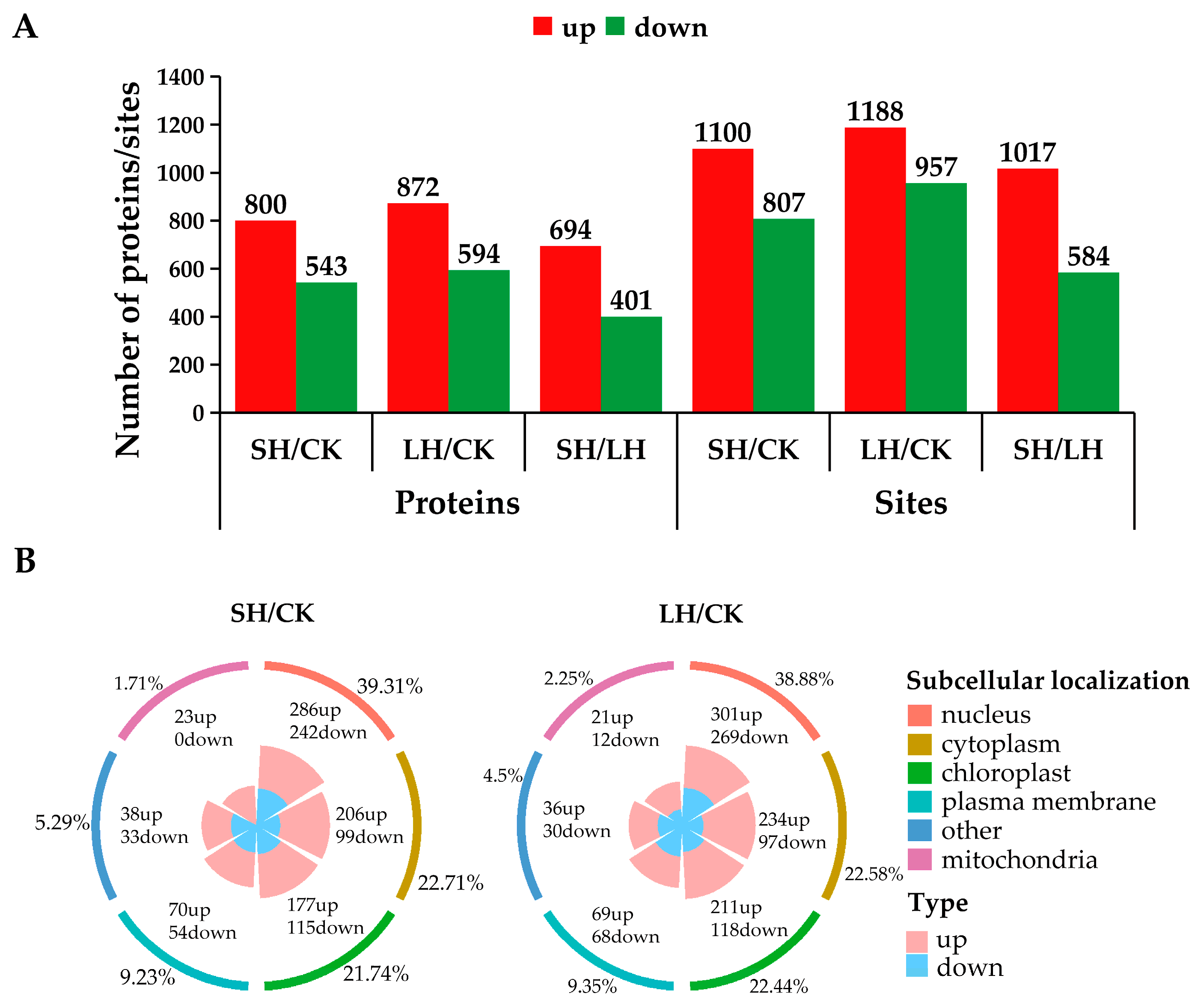
3.2. Analysis of Phosphoproteins in Response to Heat Stress

The alterations of phosphorylation status reflect the potential functions of phosphoproteins in heat stress. We analyzed the differentially regulated phosphoproteins (DRPPs) and phosphorylation sites (DRPSs) by the thresholds of |fold change| > 1.5 and p < 0.05 (Figure 3A). A total of 1343 DRPPs (800 up-regulated and 543 down-regulated) and 1907 DRPSs (1100 up-regulated and 807 down-regulated) were identified in response to SH stress. There were 1466 DRPPs (872 up-regulated and 594 down-regulated) and 2145 DRPSs (1188 up-regulated and 957 down-regulated) identified after LH treatment. The subcellular structure of DRPPs was mainly divided into three categories, including ‘nucleus’ (39.31% in SH and 38.88% in LH), ‘cytoplasm’ (22.71% in SH and 22.58% in LH), and ‘chloroplast’ (21.74% in SH and 22.44% in LH; Figure 3B).
The GO analysis of DRPPs indicated that the biological process (BP) category of GO entries for up-regulated DRPPs upon SH stress was significantly enriched in ‘regulation of protein-containing complex disassembly’, ‘response to carbohydrate’, and ‘positive regulation of gene expression’, while down-regulated DRPPs upon SH stress were enriched in ‘regulation of nuclear division’, ‘tricarboxylic acid cycle’, and ‘cell cycle G1/S phase transition’ (Supplementary Table S3). The up-regulated DRPPs under LH stress were associated with the ‘regulation of RNA biosynthetic process’ and ‘regulation of DNA-templated transcription’. The down-regulated DRPPs under LH stress were associated with ‘regulation of nuclear division’, and ‘shoot system morphogenesis’. For the KEGG enrichment analysis (Figure 4A, Supplementary Table S4), the four most abundant pathways, ‘Endocytosis’, ‘Spliceosome’, ‘Peroxisome’, and ‘Fatty acid biosynthesis’, were identified in up-regulated DRPPs under both SH and LH stress. While the majority of down-regulated DRPPs were assigned in the ‘ABC transporters’, ‘TCA cycle’, ‘Carbon metabolism’, and ‘Nucleocytoplasmic transport’. We also performed the enrichment analysis of the domain of DRPPs (Figure 4B, Supplementary Table S5). The results showed that the Tudor domain, SBP domain, PH domain, and N-terminal domain of S-adenosylmethionine synthetase were identified in up-regulated DRPPs. The RPN1 N-terminal domain, Sec23-binding domain, VHS domain, and WW domain were associated with down-regulated DRPPs. These results revealed that the flower of passion fruit regulated multiple biological pathways in response to heat stress by enhancing or weakening the phosphorylation state of proteins.

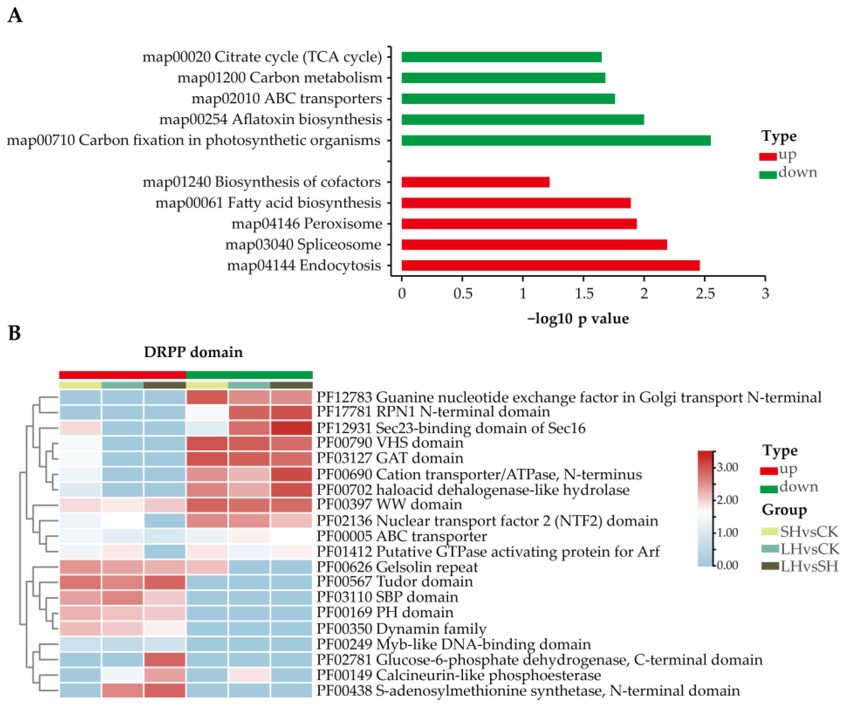
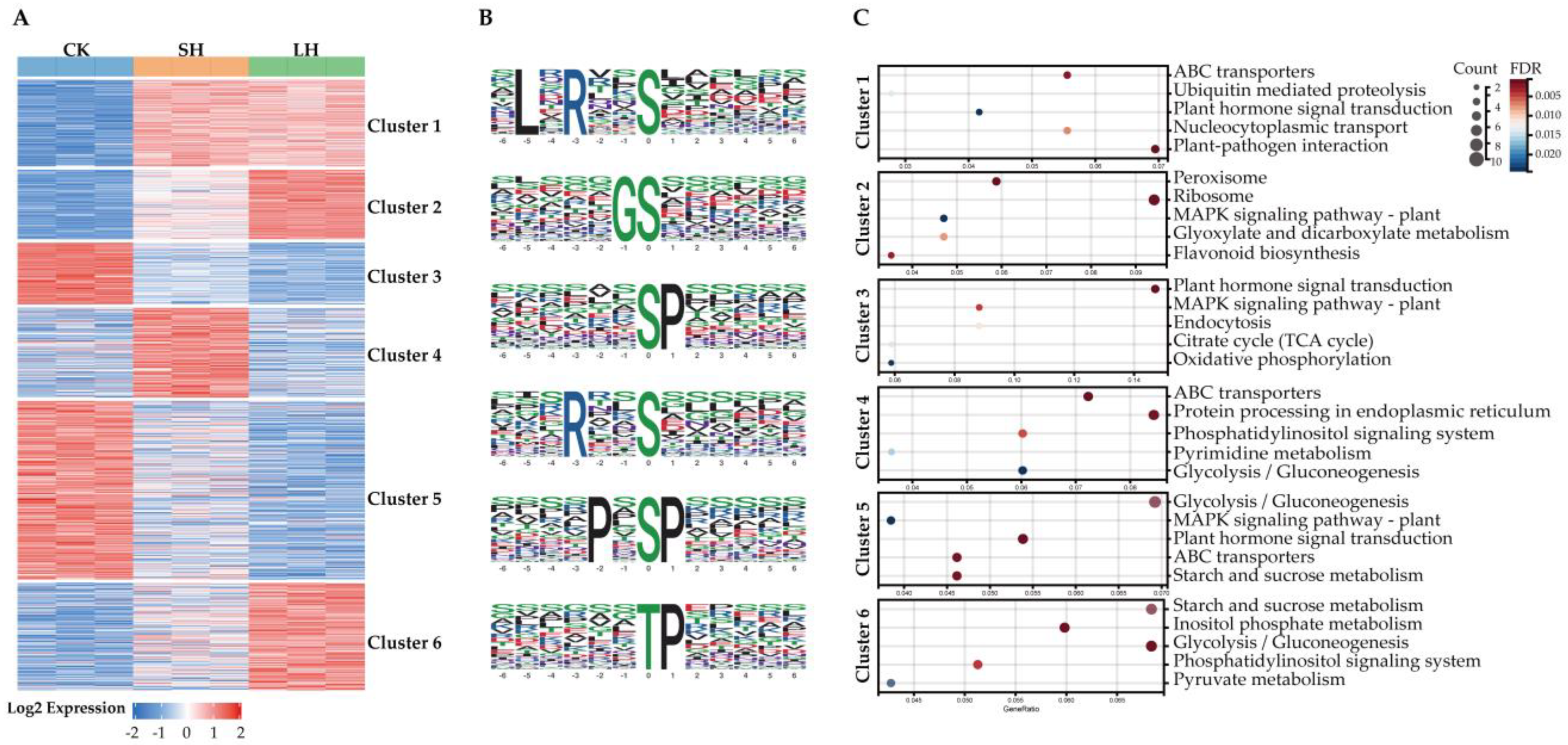
3.3. Cluster Analysis of Phosphoproteins

To further study the dynamic changes in DRPPs in response to heat stress, we performed the k-means clustering analysis. The DRPPs were divided into six expression clusters (Figure 5, Supplementary Table S6). There were 294 DRPSs of 254 DRPPs in cluster 1 that exhibited stably increased phosphorylation levels under heat stress. An analysis of the conserved motifs surrounding the DRPSs indicated that the ‘TP’ and ‘LxRxxS’ motifs were enriched in this cluster (Supplementary Table S7). ‘TP’ motif is a common phosphothreonine motif and serves as a potential substrate for MAPKs (Mitogen-activated protein kinases) [40]. The ‘LxRxxS’ motif is the potential substrate for MAPKK (Mitogen-activated protein kinase kinase), and CaMK (Calmodulin-dependent protein kinase) [41]. Further KEGG analysis of DRPPs in cluster 1 showed that ‘ABC transporters’, ‘Plant hormone signal transduction’, and ‘Plant-pathogen interaction’ were the most abundant pathways (Supplementary Table S8). The phosphorylation levels of 238 DRPSs in cluster 2 were constantly induced with the increase in heat-stress duration. The ‘TP’ and ‘GS’ motifs were identified as abundant conserved motifs in this cluster. Those DRPPs were mainly involved in the ‘Peroxisome’, ‘MAPK signaling pathway–plant’, and ‘Glyoxylate and dicarboxylate metabolism’ pathways.
The DRPPs in cluster 4 were phosphorylated in SH stress but dephosphorylated under LH stress. Those DRPPs may contribute to improving the tolerance of floral buds to short-term heat stress. The ‘RxxS’ and ‘SP’ motifs were enriched in this cluster. The ‘RxxS’ motif was the substrate of MAPKK and CaMK, while the ‘SP’ motif was the substrate of MAPKs and SnRK2 (sucrose non-fermenting1-related protein kinase 2) [42]. The KEGG result showed that those DRPPs were assigned to the ‘Protein processing in endoplasmic reticulum’ and ‘Phosphatidylinositol signaling system’ pathways. The DRPPs in cluster 6 were phosphorylated in LH stress and enriched in the ‘Starch and sucrose metabolism’ and ‘Inositol phosphate metabolism’ pathways.
The phosphorylation levels of 210 DRPSs in cluster 3 and 611 DRPSs in cluster 5 were repressed after heat stress. The ‘SP’, ‘SxE’, and ‘TP’ were over-represented in the two clusters. The most abundant pathways of those down-regulated DRPPs were ‘MAPK signaling pathway–plant’, ‘Plant hormone signal transduction’, and ‘Glycolysis/Gluconeogenesis’. It seems that the regulation of energy metabolism and signal transduction pathways is weakened in plants under prolonged heat stress.

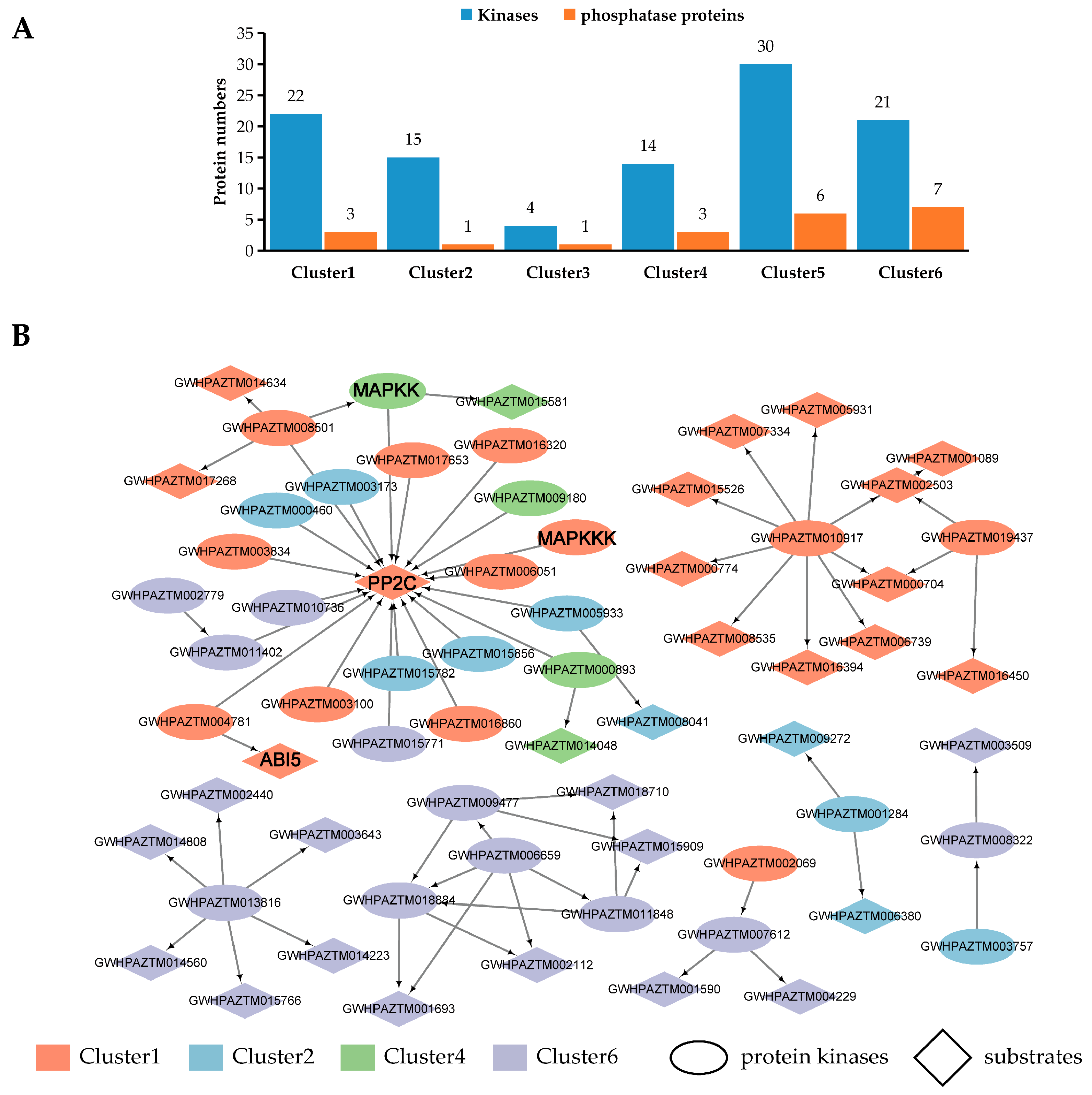
3.4. Identification of Protein Kinases and Phosphatases

The numbers of conserved motifs we identified above were recognized by protein kinases, suggesting that kinases may have important roles in mediating heat stress. So, the phosphorylation levels of protein kinases and protein phosphatases were analyzed. A total of 74 and 15 DRPPs were identified as protein kinases and protein phosphatases under heat stress, respectively (Figure 6A, Supplementary Table S9). There were 24 phosphorylation sites of twenty-two protein kinases in cluster 1, including seven Serine threonine-protein kinases (GWHPAZTM001581, GWHPAZTM003100, GWHPAZTM003834, GWHPAZTM006356, GWHPAZTM016860, GWHPAZTM017653, and GWHPAZTM019437), two CDPKs (Calcium-dependent protein kinases; GWHPAZTM001977 and GWHPAZTM016698), and two MAPKs (GWHPAZTM005069 and GWHPAZTM008501). In cluster 4, 19 phosphorylation sites of fourteen protein kinases were induced by SH stress. We noticed that MAPKKK (Mitogen-activated protein kinase kinase kinase) GWHPAZTM009546 (Ser366) was inducted by SH, while GWHPAZTM009546 (Ser559) was repressed by heat stress. In addition, MAPKKK GWHPAZTM005069Ser77 was inducted and GWHPAZTM005069Ser180,Ser299,Ser304,Ser359 were reduced by heat stress. The results indicated that protein kinases had the characteristics of multiple phosphorylation sites and multiple phosphorylation modes.
To better understand the regulation roles of phosphorylation protein kinases under heat stress, a protein–protein interaction network for the protein kinases identified in clusters (cluster 1, 2, 4, and 6) was predicted. The results showed that 16 protein kinases were predicted to interact with 21 substrate proteins in cluster 1 and 4 (Figure 6B). Notably, the protein phosphatase 2C PePP2C1 (GWHPAZTM015785) was predicted to interact with 21 protein kinases from different clusters. This suggests that PePP2C1 protein plays an important role in heat-stress signal transduction. The proteins interacting with protein kinases from clusters 2 and 6 were primarily associated with energy metabolism, suggesting that floral buds may require more energy to respond to LH stress.

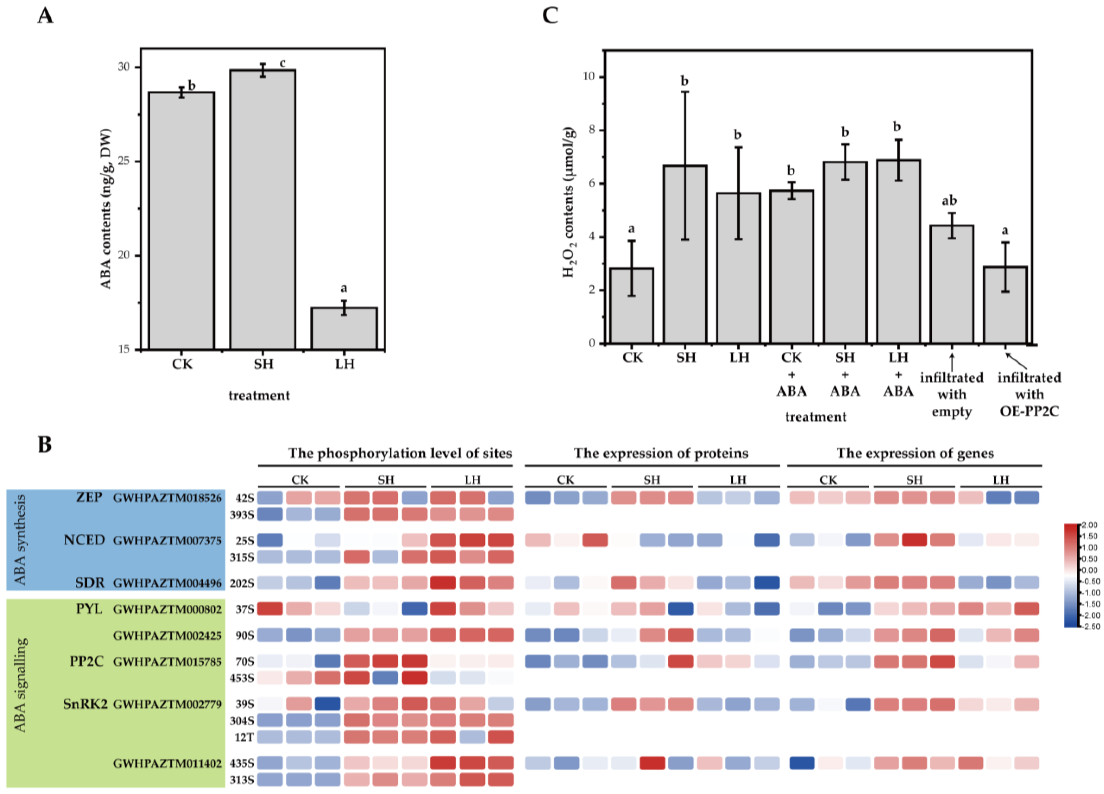
3.5. The Levels of Expression and Phosphorylation of Phosphoproteins Related to ABA Signaling

PP2Cs are important negative regulators of ABA signaling, ROS accumulation, and MAPK activation [43,44]. The PePP2C1 protein was predicted to interact with protein kinases from different clusters, suggesting that the PePP2C1 protein may actively respond to heat stress by regulating ABA signaling and ROS accumulation. The changes in ABA content under heat stress were analyzed. The results showed that ABA accumulation was induced by SH stress, which was significantly inhibited by LH stress (Figure 7A). Then, we analyzed the levels of phosphorylation, protein expression, and gene expression of key enzymes involved in ABA synthesis and signaling by phosphoproteomic profile, proteomic profile, and qRT-PCR, respectively (Figure 7B). The ABA synthetic gene PeZEP (zeaxanthin epoxidase, GWHPAZTM018526) showed a high level of gene and protein expression levels in SH stress, which was consistent with the phosphorylation level at its phosphate sites. The phosphorylation levels and gene expression of ABA receptor PePYL (pyrabactin resistance-like protein, GWHPAZTM000802) were induced by both SH and LH stress, while its proteins were abundant in SH stress. The phosphorylation levels of PePP2C1 were inhibited at LH stress, which was opposite to the phosphorylation levels of SnRK2 (SNF1-related protein kinase 2, GWHPAZTM011402).
The analyses of H2O2 (the main member of ROS) contents showed that the accumulation of H2O2 was induced by heat stress (Figure 7C). There was a slight decrease in H2O2 content under LH stress compared with SH stress. Exogenous ABA application could significantly increase H2O2 content in floral buds under non-stress conditions but had little effect on the H2O2 content of floral buds treated with heat stress. PP2C is a negative feedback regulator of ROS accumulation [45]. The up-regulation of the phosphorylation of PePP2C1 under SH stress may be a negative feedback regulatory mechanism in response to H2O2 accumulation. To further explore the role of PePP2C1 protein in H2O2 accumulation, we constructed a 35S::PePP2C1 overexpression (OE-PePP2C1) vector. The inhibition of H2O2 accumulation was observed in N. tabacum leaves infected with the OE-PePP2C1 vector, compared with leaves infected with the empty vector. These results confirmed the negative regulatory role of PePP2C1 protein in H2O2 accumulation.

4. Discussion

Under the high-temperature conditions in summer, passion fruit or other fruit trees are prone to phenomena such as flower bud drop and seedling failure, and susceptible to such problems as floral bud abscission, floral organ malformation, and pollen abortion, which significantly impact fruit set rates and productions [5,46]. Reversible protein phosphorylation plays an important role in signal transduction in response to stress [47]. Under low-nitrogen stress, 258 phosphoproteins involved in chloroplast development, carbon metabolism, and phytohormones were identified in Japonica rice [48]. The phosphoproteins related to protein processing, photosynthesis, and RNA binding were modified under high drought stress [49]. In addition, previous studies have demonstrated that phosphorylated proteins are crucial for regulating cellular pathways across different stages of flower bud development [50]. A total of 10,614 phosphorylation sites across 2906 proteins were identified in the floral buds of passion fruit under heat stress in this study. The number of phosphoproteins we identified was comparable to the number of 3468 phosphoproteins identified under NaHCO3 stress in soybean [51] and 3373 phosphoproteins identified under P starvation/resupply in barley [41]. The phosphoproteins identified in this study were primarily localized in the nucleus and cytoplasm. The up-regulated DRPPs were mainly involved in the ‘Fatty acid biosynthesis’, ‘Glyoxylate and dicarboxylate metabolism’, and ‘Plant hormone signal transduction’ pathways, which was similar to the transcriptome analysis of passion fruit leaves under heat stress [52]. This result indicated that these pathways were widely involved in the mechanism of passion fruit plants’ response to heat stress.
It has been widely confirmed that high temperature affects the production and signaling of endogenous hormones in many plants [53]. Studies have demonstrated that ethylene plays a pivotal role in mediating adaptive responses to stress [41]. For instance, roots under phosphorus (P) deficiency stress produce more ethylene compared to those under P abundance [54]. Additionally, more ethylene signal transduction-related proteins are enriched in cotton flower organs under heat stress [55]. The ethylene-insensitive gene EIN2 serves as an important positive regulator in the ethylene signal transduction pathway. In the absence of ethylene, EIN2 protein is activated by CTR1-mediated phosphorylation, which prevents the cleavage of the CEND domain of EIN2 (EIN2-CEND). Subsequently, the EIN2 protein is bound by the F-box protein ETP1/ETP2 and degraded via ubiquitination, thereby inhibiting ethylene signal transmission. In the presence of ethylene, ethylene suppresses the CTR1-mediated phosphorylation of EIN2, reduces the abundance of ETP1/ETP2 proteins, and enables the successful cleavage of EIN2-CEND from the endoplasmic reticulum, transferring it to the nucleus. There, EIN2-CEND collaborates with EIN3 to promote histone acetylation at H3K14Ac and H3K23Ac levels, facilitating ethylene signal transduction [56,57,58]. In this study, we found that the phosphorylation levels at S795 and S994 of the EIN2 protein (GWHPAZTM001783) were significantly downregulated under heat stress. These two sites are located in the C-terminal region of the EIN2 protein, suggesting that increased ethylene content under heat stress promotes EIN2 dephosphorylation and facilitates ethylene signal transduction. This study also revealed that the C-terminal T1108 site of EIN2 protein exhibited increased phosphorylation levels under heat stress. The precise mechanism by which passion fruit modulates the phosphorylation levels at various sites of the EIN2 protein under heat stress requires further investigation.
ABA is another key hormone in plants, which plays a core role during heat-stress response [59]. ABA levels usually increase under moderately elevated temperatures [53]. The elevated ABA levels were observed in A. thaliana seeds at 32 °C [53], and in red-skinned grape at 35 °C [60]. In this study, ABA levels increased at SH stress and decreased at LH stress. The reduction in ABA levels at LH stress may be attributed to extreme temperature and prolonged stress exposure. Under 45 °C conditions, a decrease in ABA levels was observed in the roots of A. thaliana [61]. This indicates that extremely high temperatures would inhibit the ABA levels in certain parts of the plants rather than promote it. This may be related to the production site and transport mechanism of ABA in plants. In rice, ABA from leaves was transported to caryopsis to ensure normal seed development in response to heat stress [62]. Therefore, it would be interesting and meaningful to investigate the ABA content, ABA biosynthetic genes, and ABA transporters in different tissues of passion fruit under extreme heat stress in the future.
In contrast to the transcriptome result of passion fruit leaves under heat stress in a previous study [52], the results of conserved motifs and KEGG analyses in our study showed that up-regulated DRPPs were enriched in the MAPK signaling pathway. This suggested that MAPK-related proteins were actively involved in the stress response of passion fruit through phosphorylation modification after being exposed to extreme heat stress (45 °C) for a short time (0.5 h and 2 h). Under salt-stress conditions, multiple MAPK proteins in wheat roots undergo phosphorylation and participate in the MAPK cascade reaction [63]. MAPK and related kinases involved in ethylene and ABA signal transduction pathways have been reported. After perceiving stress, plant cell membranes can trigger the activation of the MAPK cascade, thereby modulating the expression of genes involved in the downstream ethylene synthesis pathway and enhancing ethylene production as a response to stress [64]. In A. thaliana, the Mitogen-activated protein kinases MPK3 and MPK6 phosphorylate three serine residues located at the N-terminus of the ERF1 protein, thereby activating its transcriptional activity. This process synergistically induces the biosynthesis of the plant defense hormone via the ERF1-mediated ethylene signaling pathway, enabling resistance to pathogen infection [65]. In addition, we noticed that the MAPKKK (GWHPAZTM005069), MAPKK (GWHPAZTM015863), and MAPK (GWHPAZTM008501) were predicted to interact with PePP2C1 protein, which was a key negative regulator of ABA signaling. A total of 21 protein kinases were predicted to interact with PePP2C1 protein, indicating its potentially important role under heat stress. In A. thaliana, MAP4 kinase MAP4K phosphorylates PP2C38 protein, leading to the dissociation of PP2C38 from BIK1 (BOTRYTIS-INDUCED KINASE 1) protein [66,67]. The induction of PePP2C1 phosphorylation may result from its interaction with MAPKs. However, further studies are required to elucidate the specific role that the phosphorylation of PePP2C1 protein plays in response to heat stress. The H2O2 contents were regulated by heat stress in this study. Studies have shown that an adequate concentration of hydrogen peroxide promotes local cell-wall lignification and reduces cell-wall expansion, thereby enhancing the mechanical stability of nuclear organoids and enabling plant cells to more effectively withstand stress-induced osmotic pressure fluctuations [68]. PP2C acts as a negative regulator of ROS accumulation [45]. Our results confirmed that the overexpression of PePP2C1 gene inhibited H2O2 content in N. tabacum leaves. The interaction between MAPKs and PePP2C1 protein may have weakened the inhibition of H2O2 by PePP2C1 protein, resulting in increased H2O2 content under heat stress. In addition, MAP4 kinase SIK1 positively regulates immunity by activating the Rbohd (respiratory burst oxidase homolog) gene, thereby promoting the extracellular ROS burst [66]. The up-regulation of the phosphorylation level of Rbohd protein (GWHPAZTM001337) was identified in this study. It remains to be verified whether MAPK or other protein kinases promote ROS accumulation by enhancing the phosphorylation level of the Rbohd protein.
HSFs are the key regulators of heat-stress response and were suggested to play an important role in thermotolerance [17]. Overexpressed experiments in A. thaliana, tobacco, and Lily have confirmed the ability of the LlHSFC2 gene to increase the thermotolerance [69]. After treatment at 45 °C for 12 h, PeHSF-C1a was identified and significantly induced in the leaves of passion fruit seedlings [70]. In this study, seven phosphorylation sites from four HSF proteins were identified, and no difference in phosphorylation levels. The protein and phosphorylation levels of PeHSF-C1a were not identified here. This difference may be due to the duration of heat stress. The phosphorylation levels of proteins were only detected after 0.5 h and 2 h of heat stress in this study. Organ differences may be another reason for the differences in the expression of PeHSF-C1a. PeHSF-C1a was reported to be mainly expressed in the roots and leaves of passion fruit, but not in the floral buds [70]. The current research mainly demonstrates the heat tolerance of the leaves and roots in HSF-overexpressed plants [69,70]. However, whether the HSF gene can enhance the heat tolerance of flowers or fruits remains unclear and merits further investigation.

5. Conclusions

Our study demonstrated that, compared with short-term heat-stress treatment (45 °C for 0.5 h, named SH), long-term heat-stress treatment (45 °C for 2 h, named LH) treatment results in a significant increase in the abscission of floral buds in passion fruit plants. A total of 10,614 phosphorylation sites from 2906 proteins in floral buds were identified. The results of clustering and functional enrichment analyses suggested that floral buds responded to heat stress through biological pathways such as ‘Plant hormone signal transduction’ and ‘MAPK signaling pathway–plant’. As a negative regulator of ABA and MAPK signal transduction pathways, the inhibitory effect of the protein phosphatase PePP2C1 on the accumulation of H2O2 has been confirmed in this study, indicating its role in the heat-stress response through the suppression of ROS. The result contributed to the further understanding the role of phosphoproteins in response to heat stress in passion fruit plants.

Supplementary Materials

The following supporting information can be downloaded at https://www.mdpi.com/article/10.3390/horticulturae11050553/s1, Figure S1. Phenotype of passion fruit plants under heat stress; Table S1: Primers used for qPCR and vector construction; Table S2: Conserved motif statistics of phosphorylated peptides; Table S3: List of significant enriched category of GO annotation; Table S4: List of significant enriched category of KEGG annotation; Table S5: List of protein domains; Table S6: The clustering analysis of DRPPs; Table S7: List of conserved motifs; Table S8: List of KEGG pathways for clusters; Table S9: List of kinase and phosphatase proteins for clusters.

Author Contributions

Conceptualization, L.L.; Data curation, Y.T. and P.Z.; Formal analysis, D.Y.; Funding acquisition, L.L. and J.X.; Investigation, L.L. and Y.T.; Methodology, L.L. and Z.L.; Project administration, X.W. and J.X.; Resources, X.W.; Software, L.L. and P.Z.; Supervision, X.W.; Validation, Y.T. and X.W.; Writing—original draft, L.L.; Writing—review and editing, X.W. and J.X. All authors have read and agreed to the published version of the manuscript.

Funding

This research was funded by Free exploration science and technology innovation project of Fujian Academy of Agricultural Sciences (grant number: ZYTS2023008), the High-quality Development beyond the “5511” Collaborative Innovation Project in Fujian Province (XTCXGC2021006), the “The 14th Five-Year Plan” for Fujian Province’s Seed Industry Innovation and Industrialization Project (grant number: zycxny2021010), and Hainan Provincial Natural Science Foundation of China (grant number: 322RC765).

Data Availability Statement

The mass spectrometry proteomics data have been deposited to the ProteomeXchange Consortium (https://proteomecentral.proteomexchange.org) via the iProX partner repository [71] with the dataset identifier PXD061629 and PXD061630.

Acknowledgments

We thank Weijie Huang and Shaoyong Wu for their care of the passion fruit plants.

Conflicts of Interest

The authors declare no conflicts of interest.

References

  1. Santos, E.A.; Souza, M.M.; Abreu, P.P.; da Conceição, L.D.H.C.S.; Araújo, I.S.; Viana, A.P.; de Almeida, A.-A.F.; Freitas, J.C.d.O. Confirmation and characterization of interspecific hybrids of Passiflora L. (Passifloraceae) for ornamental use. Euphytica 2012, 184, 389–399. [Google Scholar] [CrossRef]
  2. Dong, L.; Wang, X.; Cai, Z.; Su, W.; Chen, Q.; Qiu, W.; Reng, H.; Wang, J.; Fang, W.; Huang, Z. Net chamber cultivation technique of healthy virus-free seedlings of passion fruit. S. China Fruits 2021, 50, 141–143. [Google Scholar] [CrossRef]
  3. Sobol, S.; Chayut, N.; Nave, N.; Kafle, D.; Hegele, M.; Kaminetsky, R.; Wünsche, J.N.; Samach, A. Genetic variation in yield under hot ambient temperatures spotlights a role for cytokinin in protection of developing floral primordia. Plant Cell Environ. 2014, 37, 643–657. [Google Scholar] [CrossRef]
  4. Chayut, N.; Sobol, S.; Nave, N.; Samach, A. Shielding Flowers Developing under Stress: Translating Theory to Field Application. Plants 2014, 3, 304–323. [Google Scholar] [CrossRef] [PubMed]
  5. Tian, Q.; Wu, Y.; Huang, W.; Liu, J.; Wei, S.; Mou, H.; Wei, D.; Huang, Y.; Xiong, X.; Zhang, Y. Flower formation and fruit setting in ‘Tainong No.1’ passion fruit and its relationship with meteorological factors. J. Fruit Sci. 2020, 37, 1358–1370. [Google Scholar] [CrossRef]
  6. Lan, Y.; Shi, Z.; Li, L. Preliminary Report on Test of High Temperature Affecting Fruit Set Rate of Passiflora caerulea. S. China Fruits 2021, 50, 66–68. [Google Scholar] [CrossRef]
  7. Huang, X.; Chen, G.; Huang, Y.; Gui, J.; Huang, C.; Ju, Y.; Sheng, J.; Jiang, P.; Yang, L. Effents of continuous high-temperature on flowering and fruit-setting habits and photosynthesis of four passion fruit varieties. J. Fruit Sci. 2021, 41, 1401–1409. [Google Scholar] [CrossRef]
  8. Lobell, D.B.; Schlenker, W.; Costa-Roberts, J. Climate trends and global crop production since 1980. Science 2011, 333, 616–620. [Google Scholar] [CrossRef]
  9. Abiko, M.; Akibayashi, K.; Sakata, T.; Kimura, M.; Kihara, M.; Itoh, K.; Asamizu, E.; Sato, S.; Takahashi, H.; Higashitani, A. High-temperature induction of male sterility during barley (Hordeum vulgare L.) anther development is mediated by transcriptional inhibition. Sex. Plant Reprod. 2005, 18, 91–100. [Google Scholar] [CrossRef]
  10. Zhou, J.; Zhang, Y.; Zhu, D.; Xiang, J.; Chen, X. Effects of air humidity and soil moisture on rice spikelet sterility induced by high temperature. Acta Agric. Jiangxi 2017, 29, 24–27. [Google Scholar] [CrossRef]
  11. Xiang, Y.; Huang, W.; Lai, J.; Tang, L.; Lin, T.; Li, L.; Wei, X.; Xu, J. Determination of viability of passion fruit pollens. Fujian J. Agric. Sci. 2024, 39, 302–309. [Google Scholar] [CrossRef]
  12. Ren, S.; Ma, K.; Lu, Z.; Chen, G.; Cui, J.; Tong, P.; Wang, L.; Teng, N.; Jin, B. Transcriptomic and Metabolomic Analysis of the Heat-Stress Response of Populus tomentosa Carr. Forests 2019, 10, 383. [Google Scholar] [CrossRef]
  13. Wang, H.; Gu, J.; Fang, W.; Chen, F.; Zhang, D. Response of cell membrane stability and osmotic adjustment substances in leaves of different cultivars of Anthurium andraeanum under high temperature stress. Acta Agric. Shanghai 2020, 36, 24–27. [Google Scholar] [CrossRef]
  14. Nosaka, Y.; Nosaka, A.Y. Generation and Detection of Reactive Oxygen Species in Photocatalysis. Chem. Rev. 2017, 117, 11302–11336. [Google Scholar] [CrossRef] [PubMed]
  15. Pan, X.; Wang, J.; Hu, Y.; Chi, Y.; Jin, S. Physiological and biochemical responses of leaves of four rhododendron cultivars under heat stress. Mol. Plant Breed. 2022, 20, 6877–6884. [Google Scholar] [CrossRef]
  16. Jacob, P.; Hirt, H.; Bendahmane, A. The heat-shock protein/chaperone network and multiple stress resistance. Plant Biotechnol. J. 2017, 15, 405–414. [Google Scholar] [CrossRef]
  17. Ohama, N.; Sato, H.; Shinozaki, K.; Yamaguchi-Shinozaki, K. Transcriptional Regulatory Network of Plant Heat Stress Response. Trends Plant Sci. 2017, 22, 53–65. [Google Scholar] [CrossRef] [PubMed]
  18. Ma, J.; Wang, J.; Wang, Q.; Shang, L.; Zhao, Y.; Zhang, G.; Ma, Q.; Hong, S.; Gu, C. Physiological and transcriptional responses to heat stress and functional analyses of PsHSPs in tree peony (Paeonia suffruticosa). Front. Plant Sci. 2022, 13, 926900. [Google Scholar] [CrossRef]
  19. Liu, D.; Zhang, X.; Cheng, Y.; Takano, T.; Liu, S. rHsp90 gene expression in response to several environmental stresses in rice (Oryza sativa L.). Plant Physiol. Biochem. 2006, 44, 380–386. [Google Scholar] [CrossRef]
  20. Zhang, W.J.; Zhou, Y.; Zhang, Y.; Su, Y.H.; Xu, T. Protein phosphorylation: A molecular switch in plant signaling. Cell Rep. 2023, 42, 112729. [Google Scholar] [CrossRef]
  21. Mergner, J.; Frejno, M.; List, M.; Papacek, M.; Chen, X.; Chaudhary, A.; Samaras, P.; Richter, S.; Shikata, H.; Messerer, M.; et al. Mass-spectrometry-based draft of the Arabidopsis proteome. Nature 2020, 579, 409–414. [Google Scholar] [CrossRef] [PubMed]
  22. Li, P.; Liu, J. Protein Phosphorylation in Plant Cell Signaling. Methods Mol. Biol. 2021, 2358, 45–71. [Google Scholar] [CrossRef] [PubMed]
  23. Fujii, H.; Chinnusamy, V.; Rodrigues, A.; Rubio, S.; Antoni, R.; Park, S.Y.; Cutler, S.R.; Sheen, J.; Rodriguez, P.L.; Zhu, J.K. In vitro reconstitution of an abscisic acid signalling pathway. Nature 2009, 462, 660–664. [Google Scholar] [CrossRef] [PubMed]
  24. Gong, Z.; Xiong, L.; Shi, H.; Yang, S.; Herrera-Estrella, L.R.; Xu, G.; Chao, D.Y.; Li, J.; Wang, P.Y.; Qin, F.; et al. Plant abiotic stress response and nutrient use efficiency. Sci. China Life Sci. 2020, 63, 635–674. [Google Scholar] [CrossRef]
  25. Wang, P.; Zhao, Y.; Li, Z.; Hsu, C.C.; Liu, X.; Fu, L.; Hou, Y.J.; Du, Y.; Xie, S.; Zhang, C.; et al. Reciprocal Regulation of the TOR Kinase and ABA Receptor Balances Plant Growth and Stress Response. Mol. Cell 2018, 69, 100–112.e106. [Google Scholar] [CrossRef]
  26. Takahashi, Y.; Zhang, J.; Hsu, P.K.; Ceciliato, P.H.O.; Zhang, L.; Dubeaux, G.; Munemasa, S.; Ge, C.; Zhao, Y.; Hauser, F.; et al. MAP3Kinase-dependent SnRK2-kinase activation is required for abscisic acid signal transduction and rapid osmotic stress response. Nat. Commun. 2020, 11, 12. [Google Scholar] [CrossRef]
  27. Kim, S.H.; Kim, H.S.; Bahk, S.; An, J.; Yoo, Y.; Kim, J.Y.; Chung, W.S. Phosphorylation of the transcriptional repressor MYB15 by mitogen-activated protein kinase 6 is required for freezing tolerance in Arabidopsis. Nucleic Acids Res. 2017, 45, 6613–6627. [Google Scholar] [CrossRef]
  28. Andrási, N.; Rigó, G.; Zsigmond, L.; Pérez-Salamó, I.; Papdi, C.; Klement, E.; Pettkó-Szandtner, A.; Baba, A.I.; Ayaydin, F.; Dasari, R.; et al. The mitogen-activated protein kinase 4-phosphorylated heat shock factor A4A regulates responses to combined salt and heat stresses. J. Exp. Bot. 2019, 70, 4903–4918. [Google Scholar] [CrossRef]
  29. Yip Delormel, T.; Boudsocq, M. Properties and functions of calcium-dependent protein kinases and their relatives in Arabidopsis thaliana. New Phytol. 2019, 224, 585–604. [Google Scholar] [CrossRef]
  30. Brandt, B.; Brodsky, D.E.; Xue, S.; Negi, J.; Iba, K.; Kangasjärvi, J.; Ghassemian, M.; Stephan, A.B.; Hu, H.; Schroeder, J.I. Reconstitution of abscisic acid activation of SLAC1 anion channel by CPK6 and OST1 kinases and branched ABI1 PP2C phosphatase action. Proc. Natl. Acad. Sci. USA 2012, 109, 10593–10598. [Google Scholar] [CrossRef]
  31. Li, Z.; Takahashi, Y.; Scavo, A.; Brandt, B.; Nguyen, D.; Rieu, P.; Schroeder, J.I. Abscisic acid-induced degradation of Arabidopsis guanine nucleotide exchange factor requires calcium-dependent protein kinases. Proc. Natl. Acad. Sci. USA 2018, 115, E4522–E4531. [Google Scholar] [CrossRef]
  32. Yang, Y.; Guo, Y. Elucidating the molecular mechanisms mediating plant salt-stress responses. New Phytol. 2018, 217, 523–539. [Google Scholar] [CrossRef] [PubMed]
  33. Zhou, J.M.; Zhang, Y. Plant Immunity: Danger Perception and Signaling. Cell 2020, 181, 978–989. [Google Scholar] [CrossRef] [PubMed]
  34. Pan, J.; Li, Z.; Wang, Q.; Garrell, A.K.; Liu, M.; Guan, Y.; Zhou, W.; Liu, W. Comparative proteomic investigation of drought responses in foxtail millet. BMC Plant Biol. 2018, 18, 315. [Google Scholar] [CrossRef]
  35. Hogrebe, A.; von Stechow, L.; Bekker-Jensen, D.B.; Weinert, B.T.; Kelstrup, C.D.; Olsen, J.V. Benchmarking common quantification strategies for large-scale phosphoproteomics. Nat. Commun. 2018, 9, 1045. [Google Scholar] [CrossRef] [PubMed]
  36. Shen, W.; Song, Z.; Zhong, X.; Huang, M.; Shen, D.; Gao, P.; Qian, X.; Wang, M.; He, X.; Wang, T.; et al. Sangerbox: A comprehensive, interaction-friendly clinical bioinformatics analysis platform. iMeta 2022, 1, e36. [Google Scholar] [CrossRef]
  37. Chen, C.; Wu, Y.; Li, J.; Wang, X.; Zeng, Z.; Xu, J.; Liu, Y.; Feng, J.; Chen, H.; He, Y.; et al. TBtools-II: A “one for all, all for one” bioinformatics platform for biological big-data mining. Mol. Plant 2023, 16, 1733–1742. [Google Scholar] [CrossRef]
  38. Li, L.; Liu, J.; Liang, Q.; Zhang, Y.; Kang, K.; Wang, W.; Feng, Y.; Wu, S.; Yang, C.; Li, Y. Genome-wide analysis of long noncoding RNAs affecting floral bud dormancy in pears in response to cold stress. Tree Physiol. 2021, 41, 771–790. [Google Scholar] [CrossRef]
  39. Zhang, D.; Du, L.; Lin, J.; Wang, L.; Zheng, P.; Deng, B.; Zhang, W.; Su, W.; Liu, Y.; Lu, Y.; et al. Genome-wide identification and expression analysis of calmodulin and calmodulin-like genes in passion fruit (Passiflora edulis) and their involvement in flower and fruit development. BMC Plant Biol. 2024, 24, 626. [Google Scholar] [CrossRef]
  40. Yang, J.; Xie, M.-Y.; Yang, X.-L.; Liu, B.-H.; Lin, H.-H. Phosphoproteomic Profiling Reveals the Importance of CK2, MAPKs and CDPKs in Response to Phosphate Starvation in Rice. Plant Cell Physiol. 2019, 60, 2785–2796. [Google Scholar] [CrossRef]
  41. Ma, Z.; Wang, J.; Li, C.; Ren, P.; Yao, L.; Li, B.; Meng, Y.; Ma, X.; Si, E.; Yang, K.; et al. Global Profiling of Phosphorylation Reveals the Barley Roots Response to Phosphorus Starvation and Resupply. Front. Plant Sci. 2021, 12, 676432. [Google Scholar] [CrossRef]
  42. van Wijk, K.J.; Friso, G.; Walther, D.; Schulze, W.X. Meta-Analysis of Arabidopsis thaliana Phospho-Proteomics Data Reveals Compartmentalization of Phosphorylation Motifs. Plant Cell 2014, 26, 2367–2389. [Google Scholar] [CrossRef] [PubMed]
  43. Fang, K.; Yao, X.; Tian, Y.; He, Y.; Lin, Y.; Lei, W.; Peng, S.; Pan, G.; Shi, H.; Zhang, D.; et al. Ubiquitin-specific protease UBP14 stabilizes HY5 by deubiquitination to promote photomorphogenesis in Arabidopsis thaliana. Proc. Natl. Acad. Sci. USA 2024, 121, e2404883121. [Google Scholar] [CrossRef]
  44. Wang, D.; Wei, L.; Liu, T.; Ma, J.; Huang, K.; Guo, H.; Huang, Y.; Zhang, L.; Zhao, J.; Tsuda, K.; et al. Suppression of ETI by PTI priming to balance plant growth and defense through an MPK3/MPK6-WRKYs-PP2Cs module. Mol. Plant 2023, 16, 903–918. [Google Scholar] [CrossRef]
  45. Mittler, R.; Blumwald, E. The Roles of ROS and ABA in Systemic Acquired Acclimation. Plant Cell 2015, 27, 64–70. [Google Scholar] [CrossRef] [PubMed]
  46. Xu, T.; Chen, Q.; Wu, C. Effects of high temperature stress flowering on flower organ development, reproduction and physiological mechanism of fruit trees. North. Fruits 2022, 31, 1–4. [Google Scholar] [CrossRef]
  47. Hu, X.; Wu, L.; Zhao, F.; Zhang, D.; Li, N.; Zhu, G.; Li, C.; Wang, W. Phosphoproteomic analysis of the response of maize leaves to drought, heat and their combination stress. Front. Plant Sci. 2015, 6, 298. [Google Scholar] [CrossRef] [PubMed]
  48. Xie, S.; Liu, H.; Ma, T.; Shen, S.; Zheng, H.; Yang, L.; Liu, L.; Wei, Z.; Xin, W.; Zou, D.; et al. Global Phosphoproteomic Analysis Reveals the Defense and Response Mechanisms of Japonica Rice under Low Nitrogen Stress. Int. J. Mol. Sci. 2023, 24, 7699. [Google Scholar] [CrossRef]
  49. Zhang, J.; Li, J.; Su, Y.; Song, Z.; Wang, J. The Posttranscriptional Mechanism in Salvia miltiorrhiza Bunge Leaves in Response to Drought Stress Using Phosphoproteomics. Agronomy 2022, 12, 781. [Google Scholar] [CrossRef]
  50. Arefian, M.; Bhagya, N.; Prasad, T.S.K. Phosphorylation-mediated signalling in flowering: Prospects and retrospects of phosphoproteomics in crops. Biol. Rev. 2021, 96, 2164–2191. [Google Scholar] [CrossRef]
  51. Li, Q.; Li, M.; Ma, H.; Xue, M.; Chen, T.; Ding, X.; Zhang, S.; Xiao, J. Quantitative Phosphoproteomic Analysis Provides Insights into the Sodium Bicarbonate Responsiveness of Glycine max. Biomolecules 2023, 13, 1520. [Google Scholar] [CrossRef] [PubMed]
  52. Wang, H.; Zhao, J.; Lai, M.; Zhang, Y.; Qiu, W.; Li, Y.; Tu, H.; Ling, Q.; Fu, X. Differential gene expression analysis and physiological response characteristics of passion fruit (Passiflora edulis) buds under high-temperature stress. PeerJ 2023, 11, e14839. [Google Scholar] [CrossRef] [PubMed]
  53. Castroverde, C.D.M.; Dina, D. Temperature regulation of plant hormone signaling during stress and development. J. Exp. Bot. 2021, 72, 7436–7458. [Google Scholar] [CrossRef]
  54. Gilbert, G.A.; Knight, J.D.; Vance, C.P.; Allan, D.L. Proteoid Root Development of Phosphorus Deficient Lupin is Mimicked by Auxin and Phosphonate. Ann. Bot. 2000, 85, 921–928. [Google Scholar] [CrossRef]
  55. Liang, Y.; Gong, Z.; Wang, J.; Zheng, J.; Ma, Y.; Min, L.; Chen, Q.; Li, Z.; Qu, Y.; Chen, Q.; et al. Nanopore-Based Comparative Transcriptome Analysis Reveals the Potential Mechanism of High-Temperature Tolerance in Cotton (Gossypium hirsutum L.). Plants 2021, 10, 2517. [Google Scholar] [CrossRef]
  56. Qiao, H.; Shen, Z.; Huang, S.S.; Schmitz, R.J.; Urich, M.A.; Briggs, S.P.; Ecker, J.R. Processing and subcellular trafficking of ER-tethered EIN2 control response to ethylene gas. Science 2012, 338, 390–393. [Google Scholar] [CrossRef] [PubMed]
  57. Merchante, C.; Brumos, J.; Yun, J.; Hu, Q.; Spencer, K.R.; Enríquez, P.; Binder, B.M.; Heber, S.; Stepanova, A.N.; Alonso, J.M. Gene-specific translation regulation mediated by the hormone-signaling molecule EIN2. Cell 2015, 163, 684–697. [Google Scholar] [CrossRef]
  58. Wang, L.; Zhang, Z.; Zhang, F.; Shao, Z.; Zhao, B.; Huang, A.; Tran, J.; Hernandez, F.V.; Qiao, H. EIN2-directed histone acetylation requires EIN3-mediated positive feedback regulation in response to ethylene. Plant Cell 2020, 33, 322–337. [Google Scholar] [CrossRef]
  59. Cutler, S.R.; Rodriguez, P.L.; Finkelstein, R.R.; Abrams, S.R. Abscisic acid: Emergence of a core signaling network. Annu. Rev. Plant Biol. 2010, 61, 651–679. [Google Scholar] [CrossRef]
  60. Gao-Takai, M.; Katayama-Ikegami, A.; Matsuda, K.; Shindo, H.; Uemae, S.; Oyaizu, M. A low temperature promotes anthocyanin biosynthesis but does not accelerate endogenous abscisic acid accumulation in red-skinned grapes. Plant Sci. 2019, 283, 165–176. [Google Scholar] [CrossRef]
  61. Prerostova, S.; Dobrev, P.I.; Kramna, B.; Gaudinova, A.; Knirsch, V.; Spichal, L.; Zatloukal, M.; Vankova, R. Heat Acclimation and Inhibition of Cytokinin Degradation Positively Affect Heat Stress Tolerance of Arabidopsis. Front. Plant Sci. 2020, 11, 87. [Google Scholar] [CrossRef]
  62. Qin, P.; Zhang, G.; Hu, B.; Wu, J.; Chen, W.; Ren, Z.; Liu, Y.; Xie, J.; Yuan, H.; Tu, B.; et al. Leaf-derived ABA regulates rice seed development via a transporter-mediated and temperature-sensitive mechanism. Sci. Adv. 2021, 7, eabc8873. [Google Scholar] [CrossRef]
  63. Pan, J.; Li, Z.; Wang, Q.; Guan, Y.; Li, X.; Huangfu, Y.; Meng, F.; Li, J.; Dai, S.; Liu, W. Phosphoproteomic Profiling Reveals Early Salt-Responsive Mechanisms in Two Foxtail Millet Cultivars. Front. Plant Sci. 2021, 12, 712257. [Google Scholar] [CrossRef] [PubMed]
  64. Chen, Y.; Yan, W.; Guo, D.; Li, Y.; Li, J.; Liu, H.; Wei, L.; Yu, N.; Wang, B.; Zheng, Y.; et al. An Activity-Based Sensing Fluorogenic Probe for Monitoring Ethylene in Living Cells and Plants. Angew. Chem. Int. Ed. Engl. 2021, 60, 21934–21942. [Google Scholar] [CrossRef] [PubMed]
  65. Zhou, J.; Mu, Q.; Wang, X.; Zhang, J.; Yu, H.; Huang, T.; He, Y.; Dai, S.; Meng, X. Multilayered synergistic regulation of phytoalexin biosynthesis by ethylene, jasmonate, and MAPK signaling pathways in Arabidopsis. Plant Cell 2022, 34, 3066–3087. [Google Scholar] [CrossRef] [PubMed]
  66. Zhang, M.; Chiang, Y.H.; Toruño, T.Y.; Lee, D.; Ma, M.; Liang, X.; Lal, N.K.; Lemos, M.; Lu, Y.J.; Ma, S.; et al. The MAP4 Kinase SIK1 Ensures Robust Extracellular ROS Burst and Antibacterial Immunity in Plants. Cell Host Microbe 2018, 24, 379–391.e375. [Google Scholar] [CrossRef]
  67. Pan, L.; De Smet, I. Expanding the Mitogen-Activated Protein Kinase (MAPK) Universe: An Update on MAP4Ks. Front. Plant Sci. 2020, 11, 1220. [Google Scholar] [CrossRef]
  68. Tenhaken, R. Cell wall remodeling under abiotic stress. Front. Plant Sci. 2015, 5, 771. [Google Scholar] [CrossRef]
  69. Wu, Z.; Li, T.; Ding, L.; Wang, C.; Teng, R.; Xu, S.; Cao, X.; Teng, N. Lily LlHSFC2 coordinates with HSFAs to balance heat stress response and improve thermotolerance. New Phytol. 2024, 241, 2124–2142. [Google Scholar] [CrossRef]
  70. Wang, L.; Liu, Y.; Chai, G.; Zhang, D.; Fang, Y.; Deng, K.; Aslam, M.; Niu, X.; Zhang, W.; Qin, Y.; et al. Identification of passion fruit HSF gene family and the functional analysis of PeHSF-C1a in response to heat and osmotic stress. Plant Physiol. Biochem. 2023, 200, 107800. [Google Scholar] [CrossRef]
  71. Chen, T.; Ma, J.; Liu, Y.; Chen, Z.; Xiao, N.; Lu, Y.; Fu, Y.; Yang, C.; Li, M.; Wu, S.; et al. iProX in 2021: Connecting proteomics data sharing with big data. Nucleic Acids Res. 2022, 50, D1522–D1527. [Google Scholar] [CrossRef] [PubMed]
Figure 1. The abscission rate of floral buds under heat stress. The different letters indicated significant differences by the Tukey test (p < 0.05).
Horticulturae 11 00553 g001
Figure 2. The proteome-wide identification of phosphorylation sites in the floral buds of passion fruit. (A) Distribution of proteins containing different numbers of phosphorylated sites. (B) Distribution of phosphorylated amino acids.
Horticulturae 11 00553 g002
Figure 3. The statistics of the differentially regulated phosphoproteins (DRPPs) and phosphorylation sites (DRPSs) under heat stress in floral buds. (A) The numbers of DRPPs and DRPSs under SH and LH stress, respectively. The red bars indicated the up-regulated DRPPs or DRPSs; the green bars indicated the down-regulated DRPPs or DRPSs. SH: 0.5 h heat stress. LH: 2 h heat stress. (B) The subcellular structure analysis of DRPPs. The different colors represent different subcellular localization and type, as shown in the legend on the right. The number outside the circle indicates the proportion of proteins located in that subcellular structure.
Horticulturae 11 00553 g003
Figure 4. Enrichment analysis of DRPPs under heat stress in floral buds. (A) Top 5 KEGG pathways of up- and down-regulated DRPPs under SH stress. (B) The heatmap of the DRPP domain under SH and LH stress, respectively.
Horticulturae 11 00553 g004
Figure 5. The hierarchical clustering of DRPPs under heat stress in floral buds. (A) K-means clustering analysis of DRPPs under heat stress. (B) The logo of top six conserved motifs of the phosphorylation sites. The conserved motifs presented in the figure are as follows: ‘LxRxxS’, ‘GS’, ‘SP’, ‘RxxS’, ‘PxSP’, and ‘TP’. (C) KEGG enrichment analysis of DRPPs in six clusters, respectively.
Horticulturae 11 00553 g005
Figure 6. The identification of protein kinases and phosphatases. (A) The number of protein kinases and phosphatases in six clusters. (B) The prediction of protein–protein interaction network of up-regulated protein kinases in response to heat stress. Orange represents the proteins in cluster 1; blue represents the proteins in cluster 2; green represents the proteins in cluster 4; and purple represents the proteins in cluster 6. The ellipse shape represents protein kinases, and the diamond shape represents substrate proteins. The protein pointed by the arrow is the target protein.
Horticulturae 11 00553 g006
Figure 7. The identification of phosphoproteins related to ABA signaling. (A) The changes in ABA content under heat stress. (B) The expression heatmap of phosphorylation sites, proteins, and genes of ABA syntheses and signaling genes. ZEP: zeaxanthin epoxidase; NCED: 9-cis-epoxycarotenoid dioxygenase; SDR: short-chain dehydrogenase/reductase; PYL: pyrabactin resistance-like proteins; SnRK2: sucrose nonfermenting 1 (SNF1)-related protein kinase 2. (C) The changes in H2O2 content in different treatments. ‘CK + ABA’ indicated ABA treatment under unstressed conditions; ‘SH + ABA’ indicated ABA treatment under SH stress; ‘LH + ABA’ indicated ABA treatment under LH stress; ‘infiltrated with empty’ indicated N. tabacum leaves were infiltrated with empty vector; ‘infiltrated with OE-PP2C’ indicated N. tabacum leaves were infiltrated with 35S::PePP2C1 overexpression vector. The error bar indicates the standard errors for three replicates; the different letters indicate significant differences between samples by Tukey test (p < 0.05).
Horticulturae 11 00553 g007
Table 1. Spectrum database search analysis summary.
TitlesNumber
Peptides15,367
Modified peptides11,827
Identified proteins4909
Identified sites17,765
Comparable proteins2906
Comparable sites10,614
Disclaimer/Publisher’s Note: The statements, opinions and data contained in all publications are solely those of the individual author(s) and contributor(s) and not of MDPI and/or the editor(s). MDPI and/or the editor(s) disclaim responsibility for any injury to people or property resulting from any ideas, methods, instructions or products referred to in the content.

