Article

The Exploration of Cordyceps militaris Extract as a Postharvest Preservative for Flammulina filiformis

1
College of Food Sciences and Technology, Shanghai Ocean University, 999 Huchenghuan Rd., Shanghai 201306, China
2
National Engineering Research Center of Edible Fungi, Institute of Edible Fungi, Shanghai Academy of Agricultural Sciences, 1000 Jinqi Rd., Shanghai 201403, China
3
Pureway Biotechnology Ltd., No. 1065, West Zhongshan Road, Changning District, Shanghai 200051, China
4
College of Horticulture Science, Zhejiang A&F University, Hangzhou 311300, China
*
Authors to whom correspondence should be addressed.
These authors contributed equally to this work.
Horticulturae 2025, 11(5), 472; https://doi.org/10.3390/horticulturae11050472
Submission received: 25 March 2025 / Revised: 18 April 2025 / Accepted: 25 April 2025 / Published: 27 April 2025
(This article belongs to the Section Postharvest Biology, Quality, Safety, and Technology)

Abstract

:
Postharvest Flammulina filiformis is prone to quality degradation, adversely impacting its commercial value. Cordyceps militaris, rich in antioxidant and antibacterial components, shows promise as a natural biological preservative. This study aimed to explore the potential of C. militaris extract (CME) as a preservative for F. filiformis. Through analyzing indicators such as browning, stipe elongation, and cap diameter, this study confirmed the effectiveness of CME in delaying oxidation and inhibiting microbial growth during storage. Additionally, transcriptome analysis revealed that CME modulated gene expression in F. filiformis, enhancing its antioxidant defense mechanisms. The results demonstrated that CME could effectively extend the shelf life of F. filiformis, providing valuable insights into preservation strategies for this and other edible fungi.

1. Introduction

Flammulina filiformis, an edible fungus commonly known as enoki mushroom, is widely favored for its unique taste and rich nutritional value. Rich in bioactive compounds like amino acids, vitamins, minerals, and dietary fibers, F. filiformis possesses various pharmacological effects including antioxidant and immune-regulating properties [1]. With these characteristics, F. filiformis is in great demand in the domestic market and is also exported overseas in large quantities, making it a valuable agricultural trade commodity. In recent years, driven by government policy support and technological progress, the production of F. filiformis in China has increased, with overall export volumes showing steady growth. In the first half of 2024, the export volume of F. filiformis reached 35,000 tons, with 19,500 tons of fresh or refrigerated products destined for Vietnam [2].
However, F. filiformis retains certain metabolic activities after harvest, making it prone to quality deterioration, such as stripe elongation, browning, and nutrient loss. Moreover, due to its properties, it may serve as an ideal habitat for various pathogenic bacteria including Listeria monocytogenes and Escherichia coli [3,4]. The study of Chen et al. [5] revealed that the contamination rate of L. monocytogenes reached the highest of 55.5% in F. filiformis among tested fungi cultivars. In early 2023, the United States experienced a L. monocytogenes infection outbreak associated with F. filiformis, leading to the recall of some F. filiformis exported from China [6]. These issues significantly affect the commercial value and economic benefits of F. filiformis, posing serious challenges to export trade [7]. Therefore, it is of great importance to develop safe and effective preservation techniques to maintain the postharvest quality of F. filiformis, which may help extend the preservation period, reduce the risk of quality deterioration, and mitigate the potential threat of pathogens, thus providing strong support for the high-quality development of F. filiformis industry and enhancing its international market competitiveness.
Cordyceps militaris is an edible and medicinal fungus rich in bioactive compounds such as cordycepin and polysaccharides, which exhibits antioxidant, antimicrobial, anti-inflammatory, and immunomodulatory properties [8]. In recent years, the role of C. militaris in food preservation has gained great attention. Studies have shown that C. militaris extract (CME) could effectively delay oxidation and inhibit microbial growth, making it a promising preservative for a variety of foods [9]. As a natural bio-preservative, CME is non-toxic, environmentally friendly, and has great potential in extending the shelf life of F. filiformis, aligning with the growing demand for sustainable food preservation solutions.
This study aimed to verify a novel bio-preservative based on CME. On the premise of determining its inhibitory effect on the growth of microorganisms such as L. monocytogenes and E. coli, its ability to delay browning, suppress stipe elongation, and maintain the freshness and commercial value of F. filiformis was then observed. By conducting comparative experiments, this study explored the practical application of CME in F. filiformis preservation, providing new insights and methods for the preservation of F. filiformis and other edible fungi in the future.

2. Materials and Methods

2.1. Materials and Reagents

The fruiting bodies of F. filiformis used in this experiment were purchased from Shanghai Xuerong Bio-technology Co (Shanghai, China). Dried C. militaris fruiting bodies were provided by the Dengta Chunjiang Cordyceps-culturing Specialized Cooperative Organization (Dengta, Liaoning, China). The standard strains E. coli ATCC 25922 and L. monocytogenes ATCC 19114 were purchased from the China General Microbiological Culture Collection Center (CGMCC) (Beijing, China).
The LB medium (1 L) was prepared with 10 g of peptone, 5 g of yeast extract, and 10 g of sodium chloride; for solid, 15 g of agar powder was additionally added. The PCA plate count medium (1 L) consisted of 5 g of tryptone, 2.5 g of yeast extract, 1 g of glucose, and 15 g of agar powder. All reagents used in these media, along with sodium bicarbonate for subsequent experiments, were purchased from Sinopharm Chemical Reagent Co., Ltd. (Shanghai, China). The cefotaxime sodium used in the study was obtained from Beijing Vilab Biotechnology Co., Ltd. (Beijing, China). Cordycepin and Cordyceps polysaccharide were purchased from Shanghai Yuanye Bio-Technology Co., Ltd. (Shanghai, China).

2.2. Preparation of Cordyceps militaris Extract

The fruiting bodies of C. militaris were placed in a drying oven and dried at 60 °C to constant weight. The powder used for the extraction was ground with a pulverizer and passed through an 80-mesh sieve. Using a solid-to-liquid ratio of 1 g: 40 mL, 40 g of the powder was added to 1600 mL of distilled water and extracted in a water bath at 60 °C for 2 h. The extract was filtered through a four-layer filter cloth to obtain filtrate I and the residue, which was then mixed with an appropriate amount of distilled water and extracted again in the same condition. The second extract was filtered to obtain filtrate II. Filtrate I and II were combined, and the mixture was concentrated by rotary evaporation at 60 °C to 40 mL, with a dissolution ratio of 30%. A water extract with a concentration of 300 mg/mL named CME was obtained.

2.3. Antibacterial Activity Test

2.3.1. Preparation of Bacterial Suspension

After activating the standard bacterial strains according to the instructions, they were inoculated onto LB agar plates and incubated at 37 °C for 12 h. A single colony was then picked and placed in LB broth, which was cultured at 37 °C with shaking until the OD600 value reached 0.6. The suspension was freshly prepared for immediate use.

2.3.2. Determination of the Inhibition Zone

The method was referred to Bhattacharjee [10] with modifications. LB solid medium was sterilized and cooled to a certain temperature, and bacterial suspension with an OD600 value of 0.6 was added at a 1:100 dilution. After mixing, the medium was poured into sterile Petri dishes quantitatively to ensure a uniform thickness. Once the medium solidified, three wells were made in the agar using a sterile puncher, with agar plugs removed. Then, 300 mg/mL of CME were added to the wells (filling but not overflowing). Sterile water was used as a negative control, and 100 μg/mL of cefotaxime sodium was used as a positive control.

2.3.3. Determination of Minimum Inhibitory Concentration (MIC)

The MIC experiment was conducted on a 96-well plate using the dilution method [11]. In the first column, 200 μL of 300 mg/mL CME was added. The extracts were serially diluted two-fold to the right using LB broth. After diluting the bacterial suspension with an OD value of 0.6 to 1:100, 50 μL was added to each well of the 96-well plate, resulting in the extract concentrations of 240, 120, 60, 30, 15, 7.5, 3.75, and 1.875 mg/mL. LB broth and sterile water were used as blank controls, and 100 μg/mL cefotaxime sodium was used as a positive control. An initial concentration of 5 mg/mL of cordycepin and 100 mg/mL of Cordyceps polysaccharides was set as reference for antibacterial components. The 96-well plate was incubated at 37 °C for 12 h, and the concentration at which no bacterial growth was observed was defined as the minimum inhibitory concentration (MIC).

2.3.4. Evaluation of Environmental Microorganism Inhibition

A 200 μL extract and sterile water were evenly spread on PCA plates, which were then left open outdoors for 3 h. Afterward, the plates were sealed and incubated at 25 °C for 48–72 h. The forming colonies were observed and selected for 16S and ITS amplification before being sent for sequencing.

2.3.5. Determination of Cordycepin Content

Accurately, 10 mg of cordycepin was weighed and dissolved in sterile water, then diluted to a final volume of 10.0 mL to obtain a standard solution with a concentration of 1.0 mg/mL. A series of standard solutions with concentrations of 0, 2.5, 5, 10, 20, 40, 60, 80, and 100 μg/mL were prepared by further dilution with sterile water.
All solutions were filtered through the 0.22 μm microporous membrane for sterilization before being measured using high-performance liquid chromatography (HPLC). The measurement was carried out using a Sunfire C18 column with a methanol–water (2:8) mobile phase at a flow rate of 1.0 mL/min. The detection wavelength was set to 260 nm, the column temperature was maintained at 25 ± 5 °C, and the injection volume was 10 μL. A standard curve was constructed based on the results. Meanwhile, the extracted samples underwent the same pretreatment process and were analyzed alongside the standard solutions.

2.4. Preparation of Samples for Soaking

Fruiting bodies of F. filiformis with intact morphology, no mechanical damage, bright white color, and stipe length of about 15 to 17 cm were selected and soaked for 3 min in sterile water (control check, CK) and CME (diluted based on antibacterial results), respectively. Taken out and dried naturally, they were packaged in polyethylene preservation bags and stored at 4 °C. The volume of residual extract after soaking was measured by graduated cylinder to determine the utilization efficiency of CME. The evaluation of relevant indicators was conducted every two days. Some indicators were measured up to the 18th day of the storage.

2.5. Weight Loss Rate, Cap Diameter, and Stipe Elongation Length

The weight loss rate (%) was calculated as [(WiWn)/Wi] × 100, with Wi being the initial weight and Wn the weight after n days of storage. Vernier caliper was used to measure the diameter of the cap of F. filiformis every 6 days. As for the measurement of stipe elongation length, a site 6 cm from the cap was marked and recorded as the initial length (Li). During storage, the distance from the marker to the cap was measured and noted as the measured length (Lm). Stipe elongation was then determined by calculating the difference as LmLi.

2.6. Evaluation of Quality Changes

2.6.1. Browning Index

The measurement of browning index was conducted as follows: 5 g of samples (including both stipe and cap) were placed in a mortar, ground in an ice bath with 0.2 mol/L phosphate buffer solution (pH 6.5), and volumed to 25 mL. Standing for 10 min, the suspension was then centrifuged at 4 °C for 15 min at 6500× g. The absorbance of the supernatant was then measured at 450 nm, with the browning index calculated as A   450   n m × 5 [12]. To account for pigment residues in the samples that may interfere with the initial browning index, changes relative to the initial browning index were used as the reference.

2.6.2. The Content of Soluble Solids and Soluble Protein

Samples of 5 g were pressed to extract the juice, and the soluble solids content was measured with a handheld refractometer. The results were expressed as a mass percentage (%). Sample preparation for soluble protein content followed similar steps as that for the browning index determination. The assay was accomplished by using the Thomas Brilliant Blue G-250 method, as described by Grintzalis et al. [13].

2.6.3. Antioxidant Enzyme Activities and Malondialdehyde (MDA) Content

Assay kits (Keming, Suzhou, China) were used to determine the enzyme activities of catalase (CAT), polyphenol oxidase (PPO), peroxidase (POD), superoxide dismutase (SOD), and the content of malondialdehyde (MDA). The experiments were conducted according to the manufacturer’s protocols with no modifications or deviations from the original procedures.

2.7. Transcriptome Sequencing and Analysis

2.7.1. Total RNA Extraction, Library Construction, and Illumina Sequencing

Total RNA was extracted from the stipe and cap of samples that had been soaked in sterile water and CME at different time points (day 0, 6, and 12) using a commercial kit (Personalbio, Shanghai, China). These samples were immediately snap-frozen in liquid nitrogen and ground into a fine powder prior to RNA extraction, following the manufacturer’s protocol. After the assessment of RNA concentration and purity, cDNA libraries were constructed using another kit of Personalbio. The libraries were then subjected to paired-end sequencing using second-generation sequencing technology on the Illumina high-throughput sequencing platform [14].

2.7.2. Transcriptome Sequencing Data Processing

FastQC software 0.12.1 (Babraham Bioinformatics, Cambridge, UK) was used to perform quality control on the raw sequencing data, evaluating sequence quality scores, GC content, and duplicated sequences. Low-quality reads, adapter sequences, and contaminating sequences were removed using the Cutadapt tool 4.6 ( Martin Marcel, Heidelberg, Germany) to yield high-quality clean data as clean reads. The genome of F. filiformis strain Dan3 (GenBank accession number: JBDPIE000000000) was used as the reference genome for this transcriptome. The HISAT2 software 2.2.1 (John Hopkins University, Baltimore, MD, USA) was then employed to align the quality-controlled clean reads to the reference genome.

2.7.3. Differential Gene Expression Analysis

DESeq was used to normalize the transcriptome data to correct for sequencing depth and sample variations. Differentially expressed genes were identified by applying a Fold Change threshold (|log2FC| > 1) and a significance level (p < 0.05).

2.7.4. GO Functional and KEGG Pathway Enrichment Analysis

The Gene Ontology (GO) functional enrichment analysis of differentially expressed genes was performed using topGO 2.54.0 (Technical University of Dortmund, Dortmund, Germany) [15]. The enrichment of genes was analyzed across three major categories: biological process (BP), cellular component (CC), and molecular function (MF). ClusterProfiler 4.10.0 (Yu Lab, Guangzhou, China) was used to perform the KEGG pathway enrichment analysis for the differentially expressed genes, identifying significantly enriched biological metabolic pathways from the KEGG database.

2.7.5. Quantitative Real-Time PCR Validation

Six differentially expressed genes (DEGs) were randomly selected to verify the expression of stipe and pileus genes (Table S1). These genes were chosen due to their involvement in key biological processes, such as transaminase activity, amino acid metabolism, biosynthesis, meiosis, and DNA replication, all of which are crucial for understanding the underlying molecular mechanisms that drive the observed phenotypic changes. Specific primers were designed for these genes (shown in Table S1), and the extracted RNA was reverse-transcribed into cDNA using a reverse transcription kit (Vazyme, Nanjing, China). Quantitative real-time PCR (qPCR) was performed with SYBR Green probes following the procedure of Tables S2 and S3, with actin serving as an internal reference gene for expression normalization. The relative expression levels of the target genes were calculated using the 2−ΔΔCt method. The results of qRT-PCR were then compared with the transcriptome sequencing data to perform a correlation analysis, validating the accuracy of the sequencing results.

2.8. Data Statistics and Analysis

Data statistics were conducted using Excel 2021 (Microsoft, Redmond, WA, USA), graphs were generated using GraphPad Prism 8 (GraphPad, San Diego, CA, USA), Adobe Illustrator CS6 (Adobe, San Jose, CA, USA), and Figdraw (www.figdraw.com (accessed on 17 August 2024)). One-way ANOVA was performed to analyze the significance of differences between groups using SPSS 25.0 (IBM, Armonk, NY, USA), with p < 0.05 considered statistically significant. The stipe length and cap diameter were measured with 20 replicates, while other data were analyzed with 3 replicates.

3. Results and Discussion

3.1. Antimicrobial Efficacy of CME

The antibacterial efficacy of CME was conducted by assessing the inhibition zones formed against E. coli and L. monocytogenes, with cefotaxime and sterile water as positive and negative controls, respectively (Figure 1A,B). In terms of E. coli, the measured diameters of the inhibition zone were as follows: CME (9.564 ± 0.939 mm), positive control (28.347 ± 0.79 mm), and negative control (7.023 ± 0.038 mm). For L. monocytogenes, the corresponding inhibition zones were 8.213 ± 0.699 mm, 20.526 ± 0.253 mm, and 6.967 ± 0.083 mm, respectively.
Compared to the negative control, both CME and cefotaxime exhibited statistically significant inhibition against E. coli and L. monocytogenes (p < 0.05). CME demonstrated inhibition rates of 36.1% for E. coli and 40.0% for L. monocytogenes in comparison to the positive control. Notably, CME exerted a more pronounced inhibitory effect on E. coli than on L. monocytogenes, suggesting its potential as an antibacterial agent with varying effectiveness depending on the bacterial strain involved.
The antibacterial potency of CME was assessed through the microdilution method, with results presented in Table 1. The MIC required for CME to inhibit the growth of both E. coli and L. monocytogenes was determined to be 30 mg/mL. This indicated that CME exhibited a reasonable antibacterial effect against these bacteria, suggesting its potential as a natural antimicrobial agent. Further optimization could enhance its efficacy against specific strains.
In this experiment, the MIC of cordycepin against E. coli and L. monocytogenes were determined to be 1.0 mg/mL and 2.0 mg/mL, respectively (Table 2). As for Cordyceps polysaccharide, the concentration of MIC against both bacteria was 40 mg/mL (Table 3). The equation of the standard curve for cordycepin content was y = 31826x − 4505.1 (R2 = 0.9999). Based on this, the cordycepin content in the 30 mg/mL concentration of CME was measured to be 0.1551 ± 0.001 mg/mL, significantly lower than the minimum inhibitory concentration required for antibacterial activity. As the primary water-soluble component of CME, polysaccharides constituted a large proportion of the mass after freeze-drying [16]. However, even if the entire lyophilized product was considered polysaccharides, its concentration in CME still did not reach the MIC. These experimental results suggested that the antibacterial activity of the extract primarily stemmed from the synergistic effects of various components in CME rather than solely from cordycepin or polysaccharides, among which antimicrobial peptides may also play a significant role [17].
The application of CME reduced the total colony count on plates by approximately 21.3% compared to the blank control (50 ± 6.782 vs. 63.5 ± 10.672, respectively) (Figure 1C,D). 16S sequencing results showed a higher bacterial species diversity in the control group (CK) than in the CME-treated group, while ITS sequencing revealed minimal differences in fungal species between the groups (Table 4 and Table S4). These results indicated that CME selectively inhibited bacterial growth in the laboratory environment, reducing bacterial colony formation by over 20% without significantly affecting fungal diversity. This highlighted CME’s potential as an antibacterial agent for environmental microbial control.
The contamination of pathogenic bacteria is indeed a critical factor contributing to the spoilage of F. filiformis, as emphasized in the previous study. Yoon et al. [18] demonstrated that the combined treatment of ultrasound, 3% lactic acid, and 0.1% nisin effectively inhibited the growth of L. monocytogenes and E. coli O157:H7 in F. filiformis stored at 5 °C. Wei et al. [19] investigated the effects of ε-polylysine and nisin in reducing the growth rate of Lactococcus lactis in fresh F. filiformis fruiting bodies. In comparison, CME in this study showed significant potential as an alternative antimicrobial agent, which not only inhibited the growth of L. monocytogenes and E. coli, but also effectively targeted a broad range of environmental microbes. This broad-spectrum antimicrobial activity made CME a promising alternative to current methods for controlling bacterial contamination in F. filiformis.

3.2. CME Alleviating Morphological Deterioration of F. filiformis During Storage

The weight loss rate is a critical quality indicator for assessing storage-related changes in edible fungi, as it directly impacts their economic and nutritional value [20]. Due to the lack of epidermal tissue, high water content, and intense respiratory metabolism, F. filiformis is prone to water evaporation and nutrient loss, leading to weight reduction over time. During storage, the weight loss rate in both treatment groups exhibited an upward trend (Figure 2A). The CME-treated group demonstrated a lower weight loss compared to the CK group, showing a significant reduction (p < 0.05) at day 6. At day 18, the weight loss rate in CK group was 0.829% higher than that in the CME group. The results indicated that CME could slightly inhibit the increase in weight loss rate of F. filiformis during storage, thereby delaying quality deterioration.
Cap diameter expansion is a key indicator of morphological deterioration in F. filiformis, as it reflects the extent of cap opening. Throughout storage, cap diameter in both groups increased (Figure 2B). While no significant differences were observed between treatment groups in the early storage period, by day 12, the cap diameter in the CK group was significantly larger than that in the CME-treated group, with a trend continuing until day 18. These results suggested that CME treatment effectively reduced morphological deterioration in F. filiformis by limiting cap expansion.
The stipe elongation of F. filiformis is also one manifestation of its morphological deterioration. The experimental results indicated that the stipes of F. filiformis continued to elongate during storage (Figure 2C,D). During this process, the elongation of stipes treated with CME was significantly less than that of the CK group (p < 0.05). At day 18, the average elongation of the stipes was 4.84 ± 1.264 cm in the CK group, while in the CME group, it was 2.37 ± 0.670 cm, representing a 51% reduction compared to the CK group. This suggested that soaking treatment with CME could significantly inhibit stipe elongation and thereby mitigate the morphological deterioration associated with prolonged storage. The reduced elongation in the CME group could be attributed to the suppression of cellular expansion and division, helping to maintain structural stability in F. filiformis and thus enhancing its storage quality.

3.3. Benefit of CME Treatment to Quality Maintenance of F. filiformis During Storage

3.3.1. Effect on the Inhibition of Browning

Browning is a primary factor that affects the postharvest quality and commercial value of F. filiformis, as it not only damages its appearance but also reduces its flavor and nutritional value [21,22]. The browning degree of F. filiformis increased continuously with prolonged storage time (Figure 3A). Starting from day 9, the increase in the browning degree of F. filiformis treated with CME was significantly lower than that of the CK group (p < 0.05). By day 12, the browning index increased by 0.47 ± 0.019 in the CK group, while the CME group exhibited a browning increase of only 0.333 ± 0.017, representing a 29.1% reduction compared to CK.
These results suggested that CME treatment was effective in controlling browning in F. filiformis, likely due to its potential to inhibit enzymatic activities related to browning. CME thus appeared to be a promising postharvest treatment for maintaining the visual and nutritional quality of F. filiformis during storage, thereby enhancing its commercial value.

3.3.2. Impact on the Retention of Soluble Solids, Protein Content, and Membrane Integrity

F. filiformis continuously consumes its own nutrients (such as sugars and proteins) to maintain life activities after harvest, leading to a gradual decline in its nutritional quality. Storage-induced quality deterioration in F. filiformis is reflected in the decline in soluble solids, protein content, and the increase in malondialdehyde (MDA) levels, an indicator of oxidative damage. CME treatment could effectively mitigate these changes during the process of the experiment.
The soluble solids content of F. filiformis, a marker of freshness, is influenced by its own respiratory and metabolic activities as well as the degradation of macromolecules caused by microbial infestation [23]. During storage, the content of soluble solids continuously decreased, experiencing a sharp decline in the early storage period, followed by a slower decrease from day 6 to day 12 (Figure 3B). At day 12, the soluble solids content in the CME group was 6.047%, representing a 16.4% improvement over the CK group (5.193%). The results indicated that CME treatment contributed significantly to the retention of soluble solids.
Throughout storage, due to the active decomposition of proteases within the fruiting bodies of F. filiformis, proteins are broken down into smaller molecules, leading to a continuous decline in soluble protein content [7]. The results showed an overall downward trend in the soluble protein content of F. filiformis over time (Figure 3C). The soluble protein content in CME group was significantly higher than that in the CK group during the entire storage period (with increases of 0.499, 0.363, 0.239, and 0.195 mg/g at the respective measurement points). The experimental results demonstrated that CME treatment effectively delayed the reduction in soluble protein content during storage.
During the senescence of edible fungi, the degree of cellular membrane lipid peroxidation increases, leading to the accumulation of malondialdehyde (MDA), a product of membrane lipid peroxidation [24]. This accumulation disrupts the normal distribution of polyphenol oxidase and phenolic compounds, causing them to make contact with each other and resulting in browning. Therefore, the content of MDA can reflect the damage to membrane structure and the degree of tissue senescence, serving as a key indicator for the evaluation of F. filiformis quality [25,26]. The MDA content generally increased during storage, with fluctuations observed at day 9 (Figure 3D). The CME group had significantly lower levels, reaching 13.038 ± 0.903 nmol/g by day 12, compared to 15.956 ± 1.036 nmol/g in the CK group with a reduction of approximately 18.3%. Overall, these results demonstrated that CME treatment effectively preserved F. filiformis quality during storage by reducing soluble solids and protein loss while limiting oxidative damage, as indicated by lower MDA levels.

3.4. Effect of Exacts on Antioxidant Enzyme Activities in F. filiformis

Catalase (CAT) is an important antioxidant enzyme that reduces oxidative damage in edible fungi during storage by decomposing hydrogen peroxide (H2O2), thereby protecting the structural integrity of cell membranes as a crucial component of the antioxidant system [27]. The activity of CAT exhibited a trend of initially decreasing and then increasing during storage (Figure 4A). This fluctuation may be attributed to an initial oxidative stress spike, temporarily reducing CAT activity as the enzyme was actively consumed. However, as the antioxidant system became increasingly active, CAT activity surged in later stages. Throughout this process, the CAT activity in the CME group was significantly higher than that in the CK group (p < 0.05), reaching a peak at day 12, with values of 772.206 ± 23.522 U/g, which was 1.89 times higher than that of the CK group (409.38 ± 13.585 U/g). The experiment demonstrated a substantial improvement in CAT activity due to CME treatment.
Polyphenol oxidase (PPO) is an enzyme primarily responsible for catalyzing the oxidation of phenolic compounds in various organisms. When edible fungi undergo aging or experience environmental stresses such as mechanical damage, the cell membrane structure may be disrupted, allowing PPO to interact with its substrate (phenolic substances) [28]. In the presence of oxygen, an oxidation reaction occurs, resulting in enzymatic browning that affects the color and sensory appeal of the edible fungi [29]. During storage, PPO activity in both experimental groups initially increased and subsequently declined (Figure 4B). No statistically significant differences were observed in the early storage period. However, at day 9, PPO activity in the CK group was markedly higher than that in the CME group (p < 0.05). Specifically, PPO activities were measured at 81.96 ± 1.953 U/g for CK and 59.153 ± 1.579 U/g for CME, representing a reduction of approximately 27.8% in CME compared to CK. At day 12, the CME group still maintained relatively low activity at 55.99 ± 2.684 U/g, a decrease of about 19.1% compared to the CK group (69.263 ± 4.041 U/g). These findings indicated that CME had a good inhibitory effect on the PPO activity in F. filiformis, which was consistent with the previous results showing a delay in browning.
Peroxidase (POD) is a key enzyme involved in protecting cells from oxidative damage by catalyzing the decomposition of hydrogen peroxide and organic peroxides, thereby maintaining cellular stability in edible fungi [30]. The fluctuation trends of POD activity in both experimental groups during storage were consistent (Figure 4C). During this period, the CME group exhibited notably higher POD activity than the CK group (p < 0.05). At day 3, POD activity reached the highest level, with 245.36 ± 2.487 U/g in the CK group and 602.119 ± 36.896 U/g in the CME group. Compared to CK, CME treatment resulted in a 145.5% increase in POD activity, demonstrating its role in enhancing the antioxidant defense system of F. filiformis.
Superoxide dismutase (SOD) is an important antioxidant enzyme that effectively scavenges superoxide anions (O2) within edible fungi cells, reducing oxidative stress and preventing free radical damage [31,32]. The results exhibited that during storage, SOD activity in the CME group was consistently higher than that in the CK group (p < 0.05) (Figure 4D). At day 12, the activity of SOD in the CME group (841.067 ± 37.698 U/g) was 14.3% higher than that in the CK group (736.028 ± 19.246 U/g). The sustained higher SOD activity in the CME-treated group suggested that the applied treatment might stimulate the endogenous antioxidant system or contain compounds that directly contribute to antioxidant defense.
Oxygen is a key factor in postharvest browning of F. filiformis, as coping with oxidative stress is a critical aspect of the preservation [33]. In the research of Shao et al. [34], a cinnamaldehyde emulsion composed of soybean phospholipids and pullulan was developed, which effectively improved antioxidant activity by inhibiting ROS accumulation and enhanced the activities of oxygen radical scavenging enzymes (SOD, CAT, and POD), prolonging the shelf life of F. filiformis. CME treatment effectively boosted the antioxidant defense system of F. filiformis, offering a promising strategy for combating oxidative stress during storage. The treatment enhanced the activities of key antioxidant enzymes, including SOD, CAT, and POD, which are crucial for neutralizing ROS and minimizing oxidative damage. CME’s ability to reinforce the antioxidant defense system not only helped protect F. filiformis against oxidative damage but also contributed to its overall preservation, making it a valuable addition to postharvest treatment strategies aimed at extending shelf life and maintaining product quality.
Experimental calculations showed that the usage of CME for F. filiformis was approximately 0.111 ± 0.007 mL/g. Based on market prices, preserving 1 RMB worth of F. filiformis would require at most 0.02498 RMB worth of C. militaris (Table S5). This low-cost ratio, accounting for utmost 2.50% of the total value of F. filiformis, highlighting the economic feasibility of using C. militaris as a preservation agent.

3.5. Transcriptome Analysis Revealed Cell Activity Inhibition of F. filiformis by CME Treatment

Based on the data obtained from preliminary experiments, 30 samples were selected for transcriptome sequencing, including stipes and caps (with 3 replicates) in the CK group and the CME group at day 0, 6, and 12 named B0, G0, CKB6, CLB6, CKG6, CLG6, CKB12, CLB12, CKG12, and CLG12, where CK stands for sterile water-treated, CL for CME-treated, B for cap, and G for stipe and the numbers for storage time. Here, B0 and G0 were serving as technical references for RNA-seq normalization. A total of 233.32 Gb of clean data was obtained. The percentage of bases with Q30 quality was no less than 96.71% across all samples, and the proportion of clean reads mapped to the reference genome ranged from 91.67% to 92.62% (Table S6). These results indicated a high level of transcriptome sequencing quality, ensuring that the data were suitable for subsequent analyses.
Here, a Principal Component Analysis (PCA) was performed to assess the overall relationships between the samples [35]. In all samples, the scores of PC1 exhibited significant variation between positive and negative intervals, suggesting that the primary source of variation captured by PC1 was due to the key factors driving the transcriptomic differences. Figure S1A highlighted a clear transcriptomic distinction between the stipe and the cap of F. filiformis. PC2 would reflect changes in biological features or treatment conditions specific to the groups. The distribution of scores for PC2 across the samples exhibited a broad range, with particularly large variations observed between the treatment and control groups. Significant differences were also noted between the storage times of 6 days and 12 days (Figure S1A). Therefore, the results could be grouped into four categories for analysis as Group I (CKB6 vs. CLB6), Group II (CKB12 vs. CLB12), Group III (CKG6 vs. CLG6), and Group IV (CKG12 vs. CLG12). The replicates within each group showed good consistency, making the data suitable for further analysis.
Figure S1B visually depicted the number of differentially expressed genes (DEGs), with their up- and down-regulation between the treatment and control groups, in both the stipe and cap, as well as at the time points of day 6. The results showed that 12. 752 genes were up-regulated and 504 were down-regulated in Group I, 1071 and 601 in Group II, 506 and 677 in Group III, and 447 and 926 in Group IV. Notably, the number of DEGs at day 12 was higher than that at day 6. In the stipe group, more genes were up-regulated than those that were down-regulated, while the situation was the opposite in the cap group.
The results of the GO functional enrichment analysis (Figure 5) showed that the up-regulated genes in Group I were notably associated with amino acid metabolism, transaminase activity, and small molecule metabolism, while the down-regulated genes were linked to DNA replication, chromosome organization, and nucleosome assembly. In Group II, the up-regulated genes were enriched in ribosome function, organic acid metabolism and synthesis, and ligase activity, with no significant enrichment observed for the down-regulated genes. In Group III, the up-regulated genes were predominantly involved in amino acid metabolism, transaminase activity, and organic biosynthetic processes, whereas the down-regulated genes were related to catalytic activity. In Group IV, the up-regulated genes were connected to cellular amino acid metabolism, while the down-regulated ones were enriched in chromosome organization, DNA replication, and nucleosome assembly.
The results of the KEGG pathway enrichment analysis (Figure 6) showed that the up-regulated genes in Groups I, II, and IV were primarily enriched in amino acid and carbohydrate metabolism, while the down-regulated genes were mainly enriched in DNA replication and cell cycle pathways. In Group III, the up-regulated genes were enriched in amino acid metabolism and biosynthesis, but no significant enrichment was observed for the down-regulated ones.
Ribosomes are important protein synthesis sites in cells, which provide the necessary material basis for cell growth and reproduction [36]. The inhibition of stipe elongation in the F. filiformis after CME treatment may be related to the down-regulation of gene expression in the ribosome and the inability to synthesize proteins required for cell growth. Cell cycle is an important pathway to regulate cell division, which includes G1 (pre-DAN synthesis), S (DNA synthesis Genesis), G2 (late DNA synthesis), and M (division) phases [37]. The down-regulation of gene scaffold3.g363 encoding S-phase cell division control protein (Cdc6) could slow down DNA replication and prolong cell cycle time [38]. Together with the down-regulated genes found in the DNA replication pathway, it could be deduced that CME might reduce energy expenditure and inhibit unnecessary cell proliferation by inhibiting cell division and chromosome recombination during storage to effectively maintain the steady state of mycelia, thereby prolonging the fresh-keeping time. Up-regulated genes in all four groups were significantly associated with amino acid metabolism, which could down-regulate the expression of genes related to protein degradation and reduce protein breakdown related to oxidative stress [39,40], helping to maintain the quality of F. filiformis.
Additionally, changes in the expression of several enzymes played critical roles during storage. Polyamine oxidase (PAO), which breaks down polyamines such as putrescine, spermidine, and spermine, generates reactive oxygen species (ROS) like hydrogen peroxide (H2O2) during degradation. At high concentrations, ROS induce oxidative stress, damaging cell membranes, proteins, and DNA, thereby accelerating mushroom aging, browning, and textural deterioration [41]. In Group III, the expression of PAO (scaffold5.g162) was down-regulated, potentially reducing ROS production during polyamine degradation, thereby alleviating oxidative stress and delaying mushroom senescence. Furthermore, polyamines play essential roles in cell growth and division, and the down-regulation of PAO helped maintain normal polyamine levels, supporting metabolic stability and reducing nutritional losses during storage. Glyoxal oxidase (GOX), which metabolizes the toxic compound methylglyoxal (MGO), also plays a significant role. MGO, a by-product of cellular metabolism, accumulates and induces oxidative stress, damaging cellular structures [42]. In Group I, GOX (scaffold4.g99) was up-regulated, effectively breaking down MGO and mitigating its toxic effects, thus protecting the cell membrane, DNA, and protein structures of F. filiformis from damage and functional degradation during storage. Moreover, GOX up-regulation could slow the browning process, as MGO may promote the oxidation of phenolic compounds under certain conditions, which can be inhibited by reducing MGO accumulation. Therefore, the up-regulation of GOX enhanced the antioxidant capacity of F. filiformis and extended its shelf life. Peroxidase, an essential antioxidant enzyme, primarily reduces oxidative stress by decomposing H2O2, effectively eliminating ROS produced during storage, protecting cells from oxidative damage and delaying aging and apoptosis [43]. Consistent with the enzyme activity tests, the up-regulation of peroxidase (scaffold4.g180) was observed in both groups I and III. The coordinated regulation of various enzymes, by reducing oxidative stress, maintaining cellular stability and energy metabolism, eliminating harmful metabolic by-products, and inhibiting cellular aging and browning, jointly extended the shelf life of F. filiformis. These enzymatic changes significantly enhanced the storage tolerance of F. filiformis, ensuring its commercial value and nutritional quality.
The reliability of the transcriptome data in this study was further confirmed by qRT-PCR validation. The results for the selected differentially expressed genes showed consistency with the transcriptome sequencing data in both the stipe and cap (Figure S1C,D), ensuring the robustness of the findings.
Through transcriptome sequencing and analysis, this study identified a series of differentially expressed genes in the stipes and caps of F. filiformis after treatment with CME. Gene Ontology (GO) and KEGG pathway enrichment analyses revealed that these genes were involved in multiple biological processes and metabolic pathways related to antioxidation, metabolic regulation, and cell protection, providing an important molecular basis for fungi preservation. Specifically, the up-regulation of amino acid metabolism-related genes indicated the enhanced maintenance of the nutritional components of F. filiformis, while the down-regulation of genes associated with DNA replication and the cell cycle explained the inhibition of stipe elongation and cap expansion caused by CME. Related regulation of antioxidant genes further confirmed the effectiveness of CME in the preservation of F. filiformis.

3.6. The Advantages and Challenges of CME Preservatives

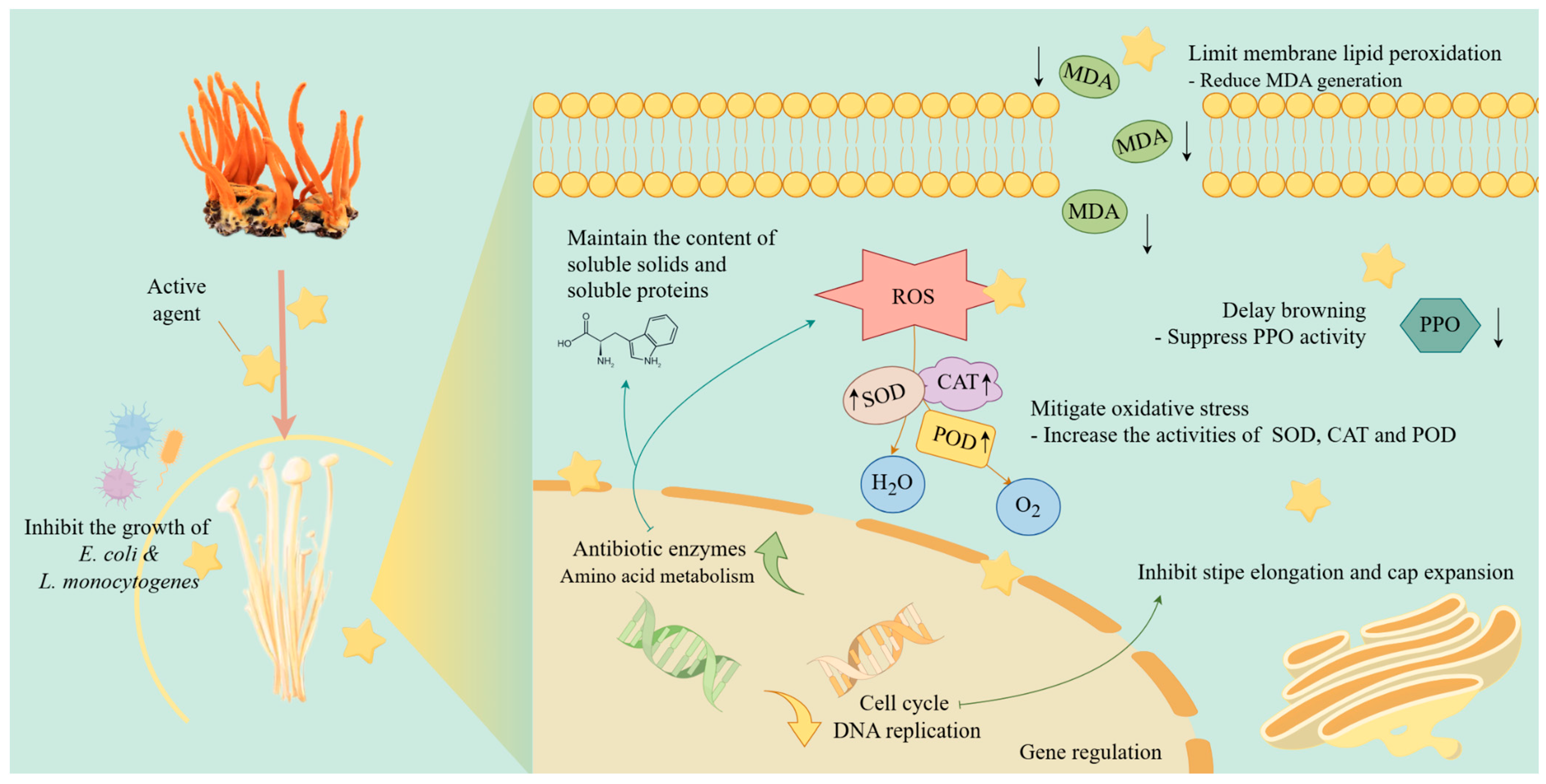
CME conducted in this study showed significant effects in inhibiting bacterial growth, delaying oxidation process, as well as maintaining the shape and nutritional quality of F. filiformis during storage at 4 °C (Figure 7). Compared with other natural preservatives mentioned above, the preparation process and treatment method of CME were simpler and less time-consuming. This research provided molecular-level explanations and support for the preservative effects of CME, making it superior to others with unknown mechanisms in terms of scientific rigor and predictability.
On the other hand, CME treatment could cause some color changes on F. filiformis, though the appearance of the treated samples remained acceptable. In subsequent verification experiments, it was found that the individual actions of cordycepin and polysaccharides did not significantly affect the maintenance of F. filiformis morphology or inhibit surface bacteria, both of which may require specific conditions or carriers to enhance their bioavailability in order to exert their effects. Based on this, it was speculated that antimicrobial peptides may play an important role during the process [44]. Further verification was needed for the specific substances and the long-term storage stability of CME. Furthermore, the utilization of the residue of C. militaris could also be explored.

4. Conclusions

This study demonstrated the preservative effect of CME on F. filiformis. Compared with the CK group, the extract notably inhibited stipe elongation and cap opening, and delayed browning as well. Based on the analysis of experimental results, this enhanced preservation ability could be attributed to the antioxidant and antimicrobial activities of the extract. Taken together, CME could inhibit morphological degradation and preserve the nutritional quality in F. filiformis during storage, showing its potential as a natural bio-preservative. Although challenges such as extraction costs remained for large-scale industrial applications, this study provided strong support for the development of future natural preservation technologies and offered new research directions for the preservation of other edible fungi.

Supplementary Materials

The following supporting information can be downloaded at: https://www.mdpi.com/article/10.3390/horticulturae11050472/s1, Figure S1: Transcriptome-related data; Table S1: Primers for qRT-PCR; Table S2: Reagents for qPCR reaction; Table S3: qPCR cycling conditions; Table S4: Comparison of antifungal effects; Table S5: Estimated cost of CME preservation for Flammulina filiformi; Table S6: Transcriptome sequencing data statistics.

Author Contributions

Conceptualization, G.Z. and Y.W.; methodology, W.C.; software, W.C.; validation, L.W., W.C. and G.Z.; formal analysis, Y.W.; investigation, Y.S.; resources, Y.L.; data curation, W.C. and M.G.; writing—original draft preparation, W.C., Y.L. and Y.S.; writing—review and editing, G.Z. and Y.W.; visualization, H.C. and N.L.; supervision, G.Z.; project administration, G.Z.; funding acquisition, G.Z. All authors have read and agreed to the published version of the manuscript.

Funding

This work was supported by the National Key R&D Program of China (2023YFF1000800), and the Excellent Team Plan of Shanghai Academy of Agricultural Sciences [(2022)014].

Institutional Review Board Statement

Not applicable.

Informed Consent Statement

Not applicable.

Data Availability Statement

The original contributions presented in this study are included in the article. Further inquiries can be directed to the corresponding authors.

Conflicts of Interest

Author Li Wang was employed by the company Pureway Biotechnology Ltd. The remaining authors declare that the research was conducted in the absence of any commercial or financial relationships that could be construed as a potential conflict of interest.

References

  1. Fukushima-Sakuno, E. Bioactive small secondary metabolites from the mushrooms Lentinula edodes and Flammulina velutipes. J. Antibiot. 2020, 73, 687–696. [Google Scholar] [CrossRef] [PubMed]
  2. Research, Y. Flammulina filiformis Market Report: Industry Production Rebounds with Sustained Growth in Exports. Available online: https://www.yhresearch.cn/news/6895/flammulina-velutipes (accessed on 14 August 2024).
  3. Chen, M.; Wu, Q.; Zhang, J.; Guo, W.; Wu, S.; Yang, X. Prevalence and contamination patterns of Listeria monocytogenes in Flammulina velutipes plants. Foodborne Pathog. Dis. 2014, 11, 620–627. [Google Scholar] [CrossRef] [PubMed]
  4. Venturini, M.E.; Reyes, J.E.; Rivera, C.S.; Oria, R.; Blanco, D. Microbiological quality and safety of fresh cultivated and wild mushrooms commercialized in Spain. Food Microbiol. 2011, 28, 1492–1498. [Google Scholar] [CrossRef] [PubMed]
  5. Chen, M.; Cheng, J.; Wu, Q.; Zhang, J.; Chen, Y.; Zeng, H.; Ye, Q.; Wu, S.; Cai, S.; Wang, J. Prevalence, potential virulence, and genetic diversity of Listeria monocytogenes isolates from edible mushrooms in Chinese markets. Front. Microbiol. 2018, 9, 1711. [Google Scholar] [CrossRef]
  6. FDA. Outbreak Investigation of Listeria monocytogenes: Enoki Mushrooms. Available online: https://www.fda.gov/food/outbreaks-foodborne-illness/outbreak-investigation-listeria-monocytogenes-enoki-mushrooms-november-2022 (accessed on 7 April 2023).
  7. Guo, Y.; Chen, X.; Gong, P.; Deng, Z.; Qi, Z.; Wang, R.; Long, H.; Wang, J.; Yao, W.; Yang, W.; et al. Recent advances in quality preservation of postharvest golden needle mushroom (Flammulina velutiper). J. Sci. Food Agric. 2023, 103, 5647–5658. [Google Scholar] [CrossRef]
  8. Barido, F.H.; Kang, S.M.; Lee, S.K. The quality and functional improvement of retorted Korean ginseng chicken soup (Samgyetang) by enzymolysis pre-treatment with Cordyceps militaris mushroom extract. Foods 2022, 11, 422. [Google Scholar] [CrossRef]
  9. Xiong, S.; Jiang, J.; Wan, F.; Tan, D.; Zheng, H.; Xue, H.; Hang, Y.; Lu, Y.; Su, Y. Cordyceps militaris extract and cordycepin alleviate oxidative stress, modulate gut microbiota and ameliorate intestinal damage in LPS-induced piglets. Antioxidants 2024, 13, 441. [Google Scholar] [CrossRef]
  10. Bhattacharjee, M.K. Better visualization and photodocumentation of zone of inhibition by staining cells and background agar differently. J. Antibiot. 2015, 68, 657–659. [Google Scholar] [CrossRef]
  11. Rodriguez-Tudela, J.; Barchiesi, F.; Bille, J.; Chryssanthou, E.; Cuenca-Estrella, M.; Denning, D.; Donnelly, J.; Dupont, B.; Fegeler, W.; Moore, C. Method for the determination of minimum inhibitory concentration (MIC) by broth dilution of fermentative yeasts. Clin. Microbiol. Infect. 2003, 9, 1–8. [Google Scholar] [CrossRef]
  12. Shan, N.; Yang, Q.; Yang, W.; Liu, Y.; Zhao, L.; Xin, Z.; Fang, Y.; Hu, Q.; An, K. Effect of nano-packing on storage quality of Flammulina velutipes. Food Sci. 2012, 33, 262–266. [Google Scholar]
  13. Grintzalis, K.; Georgiou, C.D.; Schneider, Y.-J. An accurate and sensitive Coomassie Brilliant Blue G-250-based assay for protein determination. Anal. Biochem. 2015, 480, 28–30. [Google Scholar] [CrossRef] [PubMed]
  14. Chan, A.W.; Naphtali, J.; Schellhorn, H.E. High-throughput DNA sequencing technologies for water and wastewater analysis. Sci. Prog. 2019, 102, 351–376. [Google Scholar] [CrossRef] [PubMed]
  15. Alexa, A.; Rahnenführer, J. Gene set enrichment analysis with topGO. Bioconductor Improv. 2009, 27, 776. [Google Scholar]
  16. Miao, M.; Yu, W.-Q.; Li, Y.; Sun, Y.-L.; Guo, S.-D. Structural elucidation and activities of Cordyceps militaris-derived polysaccharides: A review. Front. Nutr. 2022, 9, 898674. [Google Scholar] [CrossRef]
  17. Afzal, M.; Abusalah, M.A.H.A.; Shehzadi, N.; Absar, M.; Ahmed, N.; Khan, S.; Naseem, Y.; Mehmood, N.; Singh, K.K.B. Investigation of biometabolites and novel antimicrobial peptides derived from promising source Cordyceps militaris and effect of non-small cell lung cancer genes computationally. PLoS ONE 2025, 20, e0310103. [Google Scholar] [CrossRef]
  18. Yoon, J.; Jeong, D.; Lee, S.; Kim, S. Control of Listeria monocytogenes and Escherichia coli O157: H7 in enoki mushrooms (Flammulina velutipes) by combined treatments with organic acids, nisin, and ultrasound. Food Control 2021, 129, 108204. [Google Scholar] [CrossRef]
  19. Wei, Q.; Pan, X.; Jia, Z.; Li, C.; Chen, B.; Fang, T.; Jiang, Y. Comparative study of ε-polylysine or nisin inhibition kinetics of Lactococcus lactis and spoilage microorganisms in fresh Flammulina velutipes fruiting bodies. J. Food Qual. 2022, 1, 9135887. [Google Scholar] [CrossRef]
  20. Yang, W.; Wu, Y.; Hu, Q.; Pei, F.; Mariga, A.M. Preharvest treatment of Agaricus bisporus with methyl jasmonate inhibits postharvest deterioration. LWT 2019, 106, 158–163. [Google Scholar] [CrossRef]
  21. Guo, Y.; Chen, X.; Gong, P.; Wang, R.; Han, A.; Deng, Z.; Qi, Z.; Long, H.; Wang, J.; Yao, W. Advances in the role and mechanisms of essential oils and plant extracts as natural preservatives to extend the postharvest shelf life of edible mushrooms. Foods 2023, 12, 801. [Google Scholar] [CrossRef]
  22. Wang, B.; Yun, J.; Ye, C.; Xu, S.; Guo, W.; Zhao, F.; Qu, Y.; Bi, Y. A novel polyethylene nanopackaging combined with ozone fumigation delayed the browning and softening of Agaricus bisporus during postharvest storage. Postharvest Biol. Technol. 2024, 210, 112771. [Google Scholar] [CrossRef]
  23. Xue, L.; Shi, K.; Zhang, Y.; Song, H.; Liao, Y.; Shi, H.; Shi, W. Evaluation of the umami in edible fungi and study on umami extraction of Agaricus bisporus. J. Food Composit. Anal. 2024, 128, 106069. [Google Scholar] [CrossRef]
  24. Yu, H.; Zhao, X.; Huang, W.; Zhan, J.; He, Y. Drought stress influences the growth and physiological characteristics of Solanum rostratum dunal seedlings from different geographical populations in China. Front. Plant Sci. 2021, 12, 733268. [Google Scholar] [CrossRef]
  25. Srinontong, P.; Wandee, J.; Aengwanich, W. The effect of gallic acid on malondialdehyde, hydrogen peroxide and nitric oxide that influence viability of broiler blood cells at high ambient temperatures. Br. Poult. Sci. 2023, 64, 512–517. [Google Scholar] [CrossRef]
  26. Fang, D.; Yang, W.; Benard Muinde, K.; An, X.; Hu, Q.; Zhao, L. Effect of nanocomposite packaging on postharvest quality and reactive oxygen species metabolism of mushrooms (Flammulina velutipes). Postharvest Biol. Technol. 2016, 119, 49–57. [Google Scholar] [CrossRef]
  27. Zhu, P.; Wang, P.; Teng, Q.; Chen, T.; Tian, G.; Yao, C.; Yalimaimaiti, N.; Liu, Q. Postharvest preservation of Flammulina velutipes with isoamyl isothiocyanate. Agronomy 2023, 13, 1771. [Google Scholar] [CrossRef]
  28. Qian, X.; Hou, Q.; Liu, J.; Huang, Q.; Jin, Z.; Zhou, Q.; Jiang, T.; Zheng, X. Inhibition of browning and shelf life extension of button mushroom (Agaricus bisporus) by ergothioneine treatment. Sci. Hortic. 2021, 288, 110385. [Google Scholar] [CrossRef]
  29. Prabsangob, N.; Sittiketgorn, S. Profile of taste-related compounds and bioactivity of split gill mushroom (Schizophyllum commune) as affected by blanching and drying. Int. J. Food Prop. 2023, 26, 2078–2090. [Google Scholar] [CrossRef]
  30. Hu, Y.; Xue, F.; Chen, Y.; Qi, Y.; Zhu, W.; Wang, F.; Wen, Q.; Shen, J. Effects and mechanism of the mycelial culture temperature on the growth and development of Pleurotus ostreatus (Jacq.) P. Kumm. Horticulturae 2023, 9, 95. [Google Scholar] [CrossRef]
  31. Stephenie, S.; Chang, Y.P.; Gnanasekaran, A.; Esa, N.M.; Gnanaraj, C. An insight on superoxide dismutase (SOD) from plants for mammalian health enhancement. J. Funct. Foods 2020, 68, 103917. [Google Scholar] [CrossRef]
  32. Saxena, P.; Selvaraj, K.; Khare, S.K.; Chaudhary, N. Superoxide dismutase as multipotent therapeutic antioxidant enzyme: Role in human diseases. Biotechnol. Lett. 2022, 44, 1–22. [Google Scholar] [CrossRef]
  33. Fu, Y.; Tan, H.; Wang, B.; Peng, W.; Sun, Q.; Yu, Y. Integrated multi-omic analyses on yellow Flammulina filiformis cultivar reveal postharvest oxidative damage responses. Postharvest Biol. Technol. 2023, 195, 112111. [Google Scholar] [CrossRef]
  34. Shao, X.; Niu, B.; Fang, X.; Wu, W.; Liu, R.; Mu, H.; Gao, H.; Chen, H. Pullulan-stabilized soybean phospholipids/cinnamaldehyde emulsion for Flammulina velutipes preservation. Int. J. Biol. Macromol. 2023, 246, 125425. [Google Scholar] [CrossRef] [PubMed]
  35. Greenacre, M.; Groenen, P.J.; Hastie, T.; d’Enza, A.I.; Markos, A.; Tuzhilina, E. Principal component analysis. NRMP Natl. Resid. Matching Program 2022, 2, 100. [Google Scholar] [CrossRef]
  36. Mikhaylina, A.; Nikonova, E.; Kostareva, O.; Tishchenko, S. Regulation of ribosomal protein synthesis in prokaryotes. Mol. Biol. 2021, 55, 16–36. [Google Scholar] [CrossRef]
  37. Schwabe, D.; Formichetti, S.; Junker, J.P.; Falcke, M.; Rajewsky, N. The transcriptome dynamics of single cells during the cell cycle. Mol. Syst. Biol. 2020, 16, e9946. [Google Scholar] [CrossRef] [PubMed]
  38. Berti, M.; Cortez, D.; Lopes, M. The plasticity of DNA replication forks in response to clinically relevant genotoxic stress. Nat. Rev. Mol. Cell Biol. 2020, 21, 633–651. [Google Scholar] [CrossRef]
  39. Xiao, M.; Wang, T.; Tang, C.; He, M.; Li, Y.; Li, X. Effects of different dryingmethods on amino acid metabolite content and quality of Ophiocordyceps sinensis by LC-MS/MS combined with multivariate statistical methods. Metabolites 2024, 14, 459. [Google Scholar] [CrossRef] [PubMed]
  40. Wassie, T.; Duan, X.; Xie, C.; Wang, R.; Wu, X. Dietary Enteromorpha polysaccharide-Zn supplementation regulates amino acid and fatty acid metabolism by improving the antioxidant activity in chicken. J. Anim. Sci. Biotechnol. 2022, 13, 18. [Google Scholar] [CrossRef]
  41. Sies, H.; Belousov, V.V.; Chandel, N.S.; Davies, M.J.; Jones, D.P.; Mann, G.E.; Murphy, M.P.; Yamamoto, M.; Winterbourn, C. Defining roles of specific reactive oxygen species (ROS) in cell biology and physiology. Nat. Rev. Mol. Cell Biol. 2022, 23, 499–515. [Google Scholar] [CrossRef]
  42. Wohlschlager, L.; Kracher, D.; Scheiblbrandner, S.; Csarman, F.; Ludwig, R. Spectroelectrochemical investigation of the glyoxal oxidase activation mechanism. Bioelectrochemistry 2021, 141, 107845. [Google Scholar] [CrossRef]
  43. Sellami, K.; Couvert, A.; Nasrallah, N.; Maachi, R.; Abouseoud, M.; Amrane, A. Peroxidase enzymes as green catalysts for bioremediation and biotechnological applications: A review. Sci. Total Environ. 2022, 806, 150500. [Google Scholar] [CrossRef] [PubMed]
  44. Amigo, L.; Hernández-Ledesma, B. Current evidence on the bioavailability of food bioactive peptides. Molecules 2020, 25, 4479. [Google Scholar] [CrossRef] [PubMed]
Figure 1. Antibacterial activity analysis. (A) The plates of inhibition zone. (B) The diameters of the inhibition zone. (C) The number of environmental microbial colonies. (D) The growth situations of environmental microorganisms at 48 h and 72 h. CK, sterile water; CME, water extract of C. militaris dried product; Cef, cefotaxime sodium. Based on Duncan’s multiple range test, different letters indicate significant differences (p < 0.05) among the treatments. Asterisks denote statistically significant differences compared to the CK group, as * for p < 0.05. Each error bar represents the standard deviation of three replicates.
Horticulturae 11 00472 g001
Figure 2. Effects of sterile water (CK) and water extract of C. militaris dried product (CME) on weight loss (A), cap diameter (B), stipe elongation (C), and overall appearance of F. filiformis (D) during storage at 4 °C. Asterisks denote statistically significant differences compared to the CK group, with * for p < 0.05, and *** for p < 0.001. Each error bar represents the standard deviation of three replicates.
Horticulturae 11 00472 g002
Figure 3. Effects of sterile water (CK) and water extract of C. militaris dried product (CME) on browning variations (A), content of soluble solid (B), soluble protein (C), and MDA (D) during storage of F. filiformis. Asterisks denote statistically significant differences compared to the CK group, with * for p < 0.05, ** for p < 0.01, and *** for p < 0.001. Each error bar represents the standard deviation of three replicates.
Horticulturae 11 00472 g003
Figure 4. CAT activity (A), PPO activity (B), POD activity (C), and SOD activity (D) in F. filiformis stored at 4 °C with treatment of sterile water (CK) and water extract of C. militaris dried product (CME). Asterisks denote statistically significant differences compared to the CK group, with * for p < 0.05, ** for p < 0.01, and *** for p < 0.001. Each error bar represents the standard deviation of three replicates.
Horticulturae 11 00472 g004
Figure 5. GO functional enrichment analysis of DEGs in F. filiformis within Group I (CKB6 vs. CLB6) (A), Group II (CKB12 vs. CLB12) (B), Group III (CKG6 vs. CLG6) (C), and Group IV (CKG12 vs. CLG12) (D). CK is treated with sterile water, while CL is treated with the water extract of C. militaris dried product. B and G indicate the stalk and cap of F. filiformis, respectively, with 6 and 12 denoting the storage time. BP, biological process; CC, cellular component; MF, molecular function.
Horticulturae 11 00472 g005
Figure 6. KEGG pathway enrichment analysis of DEGs in F. filiformis within Group I (CKB6 vs. CLB6) (A), Group II (CKB12 vs. CLB12) (B), Group III (CKG6 vs. CLG6) (C), and Group IV (CKG12 vs. CLG12) (D). CK is treated with sterile water, while CL is treated with the water extract of C. militaris dried product. B and G indicate the stalk and cap of F. filiformis, respectively, with 6 and 12 denoting the storage time. GIP, genetic information processing; M, metabolism; CP, cellular processes.
Horticulturae 11 00472 g006
Figure 7. Diagram of the mechanism of CME. CME in this study inhibited the growth of harmful bacteria such as E. coli and L. monocytogenes, demonstrating its antimicrobial properties. It also reduced membrane lipid peroxidation and MDA generation, thereby protecting cell membranes. Additionally, CME helped maintain soluble solids and protein levels while enhancing the activities of antioxidant enzymes such as superoxide dismutase (SOD), catalase (CAT), and peroxidase (POD), thereby mitigating oxidative stress. The agent delayed browning by suppressing polyphenol oxidase (PPO) activity, and regulated the cell cycle and DNA replication, thus promoting proper gene expression and cellular stability. Altogether, the preservation of F. filiformis was achieved by CME. Images in the figure were from Figdraw platform.
Horticulturae 11 00472 g007
Table 1. MIC of CME.
Test StrainConcentration of CME (mg/mL)MIC (mg/mL)
2401206030157.53.751.875
Escherchiacoli----+++++++++++30
Listeria monocytogenes----+++++++++++++30
- indicated no obvious colony growth; + indicated colony growth. The numbers implied the degree of turbidity of the bacterial solution.
Table 2. MIC of cordycepin.
Test StrainConcentration of Cordycepin (mg/mL)MIC (mg/mL)
4.02.01.00.50.250.1250.06250.03125
E. coli---++++++++++++++1.0
L. monocytogenes--++++++++++++++++++++2.0
- indicated no obvious colony growth; + indicated colony growth. The numbers implied the degree of turbidity of the bacterial solution.
Table 3. MIC of Cordyceps polysaccharide.
Test StrainConcentration of Cordyceps Polysaccharide (mg/mL)MIC (mg/mL)
804020105.02.51.250.625
E.coli--++++++++++++++++40
L. monocytogenes--++++++++++++++++++++40
- indicated no obvious colony growth; + indicated colony growth. The numbers implied the degree of turbidity of the bacterial solution.
Table 4. Comparison of bacterial inhibition effects.
TreatmentWater Extract of Dried C. militarisSterile Water
Detected BacteriaMicrococcus luteus
uncultured Staphylococcus sp.
uncultured bacterium
Alkalicoccobacillus gibsonii
Staphylococcus hominis
Staphylococcus epidermidis
Staphylococcus sp. ZWS13
Roseomonas mucosa
Bacillus sp. (in: firmicutes)
uncultured soil bacterium
uncultured bacterium
uncultured Propionibacterium sp.
Disclaimer/Publisher’s Note: The statements, opinions and data contained in all publications are solely those of the individual author(s) and contributor(s) and not of MDPI and/or the editor(s). MDPI and/or the editor(s) disclaim responsibility for any injury to people or property resulting from any ideas, methods, instructions or products referred to in the content.