Article

A Potassium Phosphite Solution as a Dual-Action Strategy Against Bean Anthracnose: Antifungal Activity and Defense Gene Priming

by
Catalina Saldarriaga-Gómez
,
Paula Natalia Paez-Monroy
and
Adriana González-Almario
*
Department of Agronomy, Faculty of Agricultural Science, Universidad Nacional de Colombia, Bogotá 16486, Colombia
*
Author to whom correspondence should be addressed.
Horticulturae 2025, 11(5), 462; https://doi.org/10.3390/horticulturae11050462
Submission received: 8 February 2025 / Revised: 14 March 2025 / Accepted: 20 March 2025 / Published: 25 April 2025
(This article belongs to the Special Issue Plant–Microbial Interactions: Mechanisms and Impacts)

Abstract

:
Anthracnose in beans is an important disease caused by Colletotrichum lindemuthianum, which affects crop productivity and infects the plant in all growth stages, affecting the quality of the pod and grains. The most viable strategy to control this disease is using bean cultivars; however, fungal variability is a limitation. Among the strategies proposed is using phosphite-based compounds, which can act as fungicides or priming stimulators. This study aimed to evaluate the antifungal activity of a phosphite-based solution (potassium phosphite (H3PO3), potassium hydroxide, and potassium citrate, in a formulation of phosphorus (P2O5) 28% and potassium (K2O) 26%) on C. lindemuthianum under in vitro conditions. In addition, its effects as a defense inducer in Sutagao bean plants was determined by changes in disease severity and the expression of PR1, PR3, PR4, and POD (defense-related genes) in plants treated with the phosphite solution before infection with the fungus. The results showed that the potassium phosphite solution had a statistically significant antifungal effect on C. lindemuthianum, reducing mycelial growth by 42% and germination by 48%, at a dose of 5 mL L−1. Foliar application of the phosphite-based solution showed a 17% reduction in anthracnose severity associated with high expression of the PR1, PR3, PR4, and POD defense genes, which increased in plants that were subsequently infected with the pathogen, demonstrating a priming effect. In conclusion, a potassium phosphite solution can be included in a management program to control bean anthracnose.

1. Introduction

Phaseolus vulgaris is the most widely cultivated legume for direct consumption worldwide, as it is considered a source of dietary protein, particularly in developing countries [1]. Colletotrichum lindemuthianum (Sacc. & Magnus) LambScrib, the anamorph of Glomerella cingulata, is the causal agent of anthracnose in the common bean (P. vulgaris L.). Anthracnose is a devastating disease that leads to total crop loss under conditions of high relative humidity (>80%), leading to severe economic losses [2,3]. C. lindemuthianum affects all plant organs, and the disease symptoms include necrotic lesions on petioles, stems, branches, and the primary and secondary veins of leaves. Round, sunken cankers appear on the pods, which can lead to pod malformation, low seed number, and tissue death [4].
The global demand for nutritious legumes, the adaptability of pathogens, and climate change make it necessary to control this disease worldwide [3]. The most viable and cost-effective strategy for anthracnose control in beans is the use of fungus-resistant cultivars. However, due to the high pathogenic variability of C. lindemuthianum, this is difficult to implement [5]. Chemical fungicides were the first agents found to control plant diseases; however, Colletotrichum species show a loss of sensitivity to fungicides, possibly due to continuous use [6]. In addition, these chemical agents are toxic to the environment and to human health [7]. Among the strategies proposed for disease control, priming is a novel technique in which the induction of a physiological state allows a plant to deploy a faster and more efficient defense response against stress compared to an unprepared plant [8].
Recently, phosphite-derived products have been introduced as an alternative to disease management [9]. These products are derived from the neutralization of phosphorous acid (H3PO3) with a base, such as sodium hydroxide, potassium hydroxide, or ammonium hydroxide. One of the advantages of phosphites is their dual activity. At high concentrations, they act as antifungal agents by inhibiting fungal mycelial growth and germination of fungal spores [7,10]. Phosphite exposure in fungal cells is related to changes in phosphorus metabolism and in compounds containing this element, including the inhibition of crucial phosphorylation reactions in fungi [9,11]. Additionally, these compounds have been linked to alterations in fungal gene expression associated with the production of cell wall and cytoskeleton proteins [12].
In contrast, phosphites at low concentrations act as fertilizers and/or biostimulants, which increase the absorption and assimilation of nutrients, improve the quality of products, and induce a defense response to biotic and abiotic stresses [7,13]. The application of phosphites induces various plant defense mechanisms, even in the absence of pathogens [9,10,11], and increases systemic acquired resistance (SAR) signaling activities [7].
Therefore, studying the antifungal activity of a product derived from potassium phosphite (H3PO3) on C. lindemuthianum and its effect as an inducer of disease resistance in bean seedlings could contribute to an understanding of the use of this product in the management of anthracnose.

2. Materials and Methods

2.1. C. lindemuthianum Isolate

The C. lindemuthianum isolate was provided by the Alliance Bioversity International-CIAT and reactivated in Petri dishes containing PDA (potato dextrose agar) medium. A monosporic culture was obtained to ensure genetic uniformity of the fungus [14].

2.2. Effect of Potassium Phosphite Solution on C. lindemuthianum Under In Vitro Conditions

The direct effect of a phosphite-based solution (potassium phosphite (H3PO3), potassium hydroxide, and potassium citrate, in a formulation of phosphorus (P2O5) 28% and potassium (K2O) 26%) on the growth, morphology, and germination of C. lindemuthianum was evaluated by in vitro tests. For this, PDA medium was supplemented with the potassium phosphite solution at a dose of 5 mL L−1. As a control, the fungus was grown on PDA culture medium without potassium phosphite [13]. The mycelial growth of C. lindemuthianum was evaluated by transferring a 7 mm mycelial plug from a 7-day-old culture to a Petri dish containing PDA. The dishes were incubated at 25 °C in a growth chamber. The final ratio of the colony was measured once the control mycelia had fully colonized the medium. The mycelial growth rate was calculated by the equation
MGR   ( cm / day ) = M G 2 M G 1 t 2 t 1 ,
where MG = mycelial growth, and t = time in days.
The percentage of mycelial growth inhibition was calculated by the equation
MGI   ( % ) = M G c M G t M G c × 100 ,
where MGc = average diameter of the control C. lindemuthianum, and MGt = average diameter of the treated C. lindemuthianum. A randomized design was employed with three replicates, and the experiment was conducted three times.
The effect of the potassium phosphite solution on C. lindemuthianum was determined after 18 days of growth by changes in colony morphology (color and appearance) and at the microscopic level by evaluating changes in mycelia and acervuli formation at 40× magnification. A suspension of 5 × 10⁴ conidia mL−1 was prepared for the conidia germination test. Twenty microliters of this suspension was sown and homogenized on the surface of agar–water culture medium using a sterile rake and incubated for 24 h at 25 °C [15]. Finally, 100 conidia were counted in an area of 2 cm2, considering germinated conidia as those whose germ tube was twice the width of the conidia.
The data were analyzed using RStudio program (RSTUDIO-2023.09.1-494.EXE) and Microsoft Excel. The assumptions were validated through the Shapiro–Wilk normality test and Levene’s test for homogeneity of variances. Subsequently, an analysis of variance (ANOVA) was performed, followed by a multiple comparison test using Tukey’s method, with a significance level of p < 0.05 to establish significant differences.

2.3. Inoculum Increase and Pathogenicity Test

To increase the inoculum of C. lindemuthianum for the pathogenicity tests, fragments of the monosporic fungi grown on PDA medium for over 2 weeks were placed into glass tumblers containing pre-sterilized beans (P. vulgaris) [16]. The tumblers were then incubated in darkness at 25 °C for 21 days, allowing formation of acervuli and conidia of the pathogen. A conidial suspension was then prepared by maceration and filtration of the inoculated beans, counted in a Neubauer chamber, and adjusted to a concentration of 1 × 10⁷ conidia mL−1 [17]. This suspension was applied by brushing it onto both the upper and lower sides of the leaves of 14-day-old bean plants [18]. The plants were maintained at a relative humidity above 80% to promote infection.

2.4. Effect of Potassium Phosphite Solution on Anthracnosis Disease

To evaluate the potassium phosphite effect, 10-day-old plants of the Sutagao bean cultivar were sprayed with 1 mL of the phosphite-based solution at a concentration of 5 mL L−1, as per the manufacturer’s instructions. The solution was applied using an airbrush, targeting both the upper and lower sides of the leaves from a distance of 20 cm, in the afternoon. Five days after applying the potassium phosphite solution, the plants were inoculated with C. lindemuthianum, as previously described. The plants were kept in a propagation greenhouse with an average temperature of 18–25 °C, a relative humidity of 80%, and a natural photoperiod of 12 h. The treatments included: (−PK/+Cl), plants without application of potassium phosphite and infected with C. lindemuthianum; (+PK/+Cl), plants treated with phosphite potassium and infected with C. lindemuthianum; (+PK/−Cl), plants treated with potassium phosphite and without C. lindemuthianum infection; and (−PK/−Cl), plants without potassium phosphite application and not infected with C. lindemuthianum. The experiment was performed twice, with 15 biological replicates for each treatment.
The effect of the potassium phosphite solution on anthracnose in bean plants was determined by disease severity quantification. A descriptive ordinal scale was used for this purpose [19]. The evaluation was conducted every 4 days up to a maximum of 14 days post-inoculation (dpi). The area under the disease progress curve (AUDPC) was calculated from the severity data [20]. The RStudio program (RSTUDIO-2023.09.1-494.EXE) was used to analyze the AUDPC data. A repeated means analysis of variance (ANOVA) followed by Tukey’s test was performed to determine significant statistical differences among the treatments.

2.5. Effect of the Potassium Phosphite Solution on Defense Gene Expression

To determine changes in defense gene expression induced by the application of potassium phosphite in all treatment groups, the RNA was extracted from three leaves of bean seedlings at 0, 24, 48, 72, and 96 h post-inoculation (hpi) (three biological replicates) using a CTAB-based protocol with LiCl [21]. Three leaves from each treatment group were collected at each time point for RNA extraction. Subsequently, the RNA was treated with DNase I (ThermoFisher®, Carlsbad, CA, USA), and its quality and concentration were measured using a NanoDrop™ Spectrophotometer (Thermo Scientific™, Wilmington, DE, USA). The elongation factor gene (EF1α) of P. vulgaris was amplified from each RNA sample to confirm the absence of DNA. The PCR reaction was conducted in a 20 μL reaction volume containing 1× buffer, 2 mM MgCl2, 0.2 mM dNTPs, 0.2 µM of each primer (Forward 5′ CGGGTATGCTGGTGACTTTT 3′ and Reverse 5′ CACGCTTGAGATCCTTGACA 3′), 1 U Taq polymerase, and 2.0 μL of DNase-treated RNA. The reaction conditions were as follows: 95 °C for 10 min, followed by 35 amplification cycles at 95 °C for 1 min, 60 °C for 30 s, and 72 °C for 2 min. Finally, cDNA was synthesized from each RNA using M-MLV Reverse Transcriptase (ThermoFisher®, Carlsbad, CA, USA) according to the manufacturer’s protocol.
Before the gene expression assays were performed, the amplification efficiency of each primer set (EF1-α, POD, PR-1, PR-3, and PR-4) [18] was determined using a ten-fold dilution series of P. vulgaris DNA (7.5 ng mL−1) and SYBR Green qPCR. Three replicates were prepared for each dilution, and a standard curve was generated with the threshold cycle (Ct) values for each dilution. The efficiency was calculated by the equation
E = 10 1 / s p l o p e
The qPCR reactions were carried out in a final volume of 10 μL containing 1× BlasTaq™ 2 qPCR Master Mix (Applied Biological Materials Inc. (abm), Richmond, BC, Canada), 0.2 μM of each primer, and 3 μL of cDNA. Amplification conditions were an initial incubation at 95 °C for 10 min, followed by 40 cycles of 95 °C for 1 min, 60 °C for 30 s, and 72 °C for 2 min. Dissociation curve (melting curve) analysis was performed with the thermal profile increasing from 60 °C to 95 °C in 15 s intervals [21]. The reactions were performed on a qTOWER3 thermal cycler (Jena Analytik, Lohhof, Germany).
For differential expression analysis of the defense genes, qPCR was performed using the relative quantification method, with three cDNA replicates for each treatment at 24, 48, 72, and 96 hpi (three technical replicates). In this analysis, the crossing point (CP) values of EF1-α (housekeeping gene) were compared to those of the defense-related genes. The qPCR reactions were conducted in a final volume of 10 μL, consisting of 5 μL BlasTaq™ 2× qPCR Master Mix, 0.2 μL of each primer at 10 μM, and 3 μL cDNA. The qPCR amplification program was the same as the one used previously to describe the efficiency test, and a dissociation curve (melting curve) was generated at the end with the thermal profile increasing from 60 °C to 95 °C in 15 s intervals. The relative expression of each gene was calculated using three repetitions of Ct values obtained for each treatment by the Pfaffl comparative method [22]. An ANOVA analysis of repeated means, followed by Tukey’s test, was performed to determine significant statistical differences among the treatments. The data were analyzed using the RStudio program (Rstudio-2023.09.1-494.EXE).

3. Results

3.1. Effect of the Potassium Phosphite Solution on C. lindemuthianum

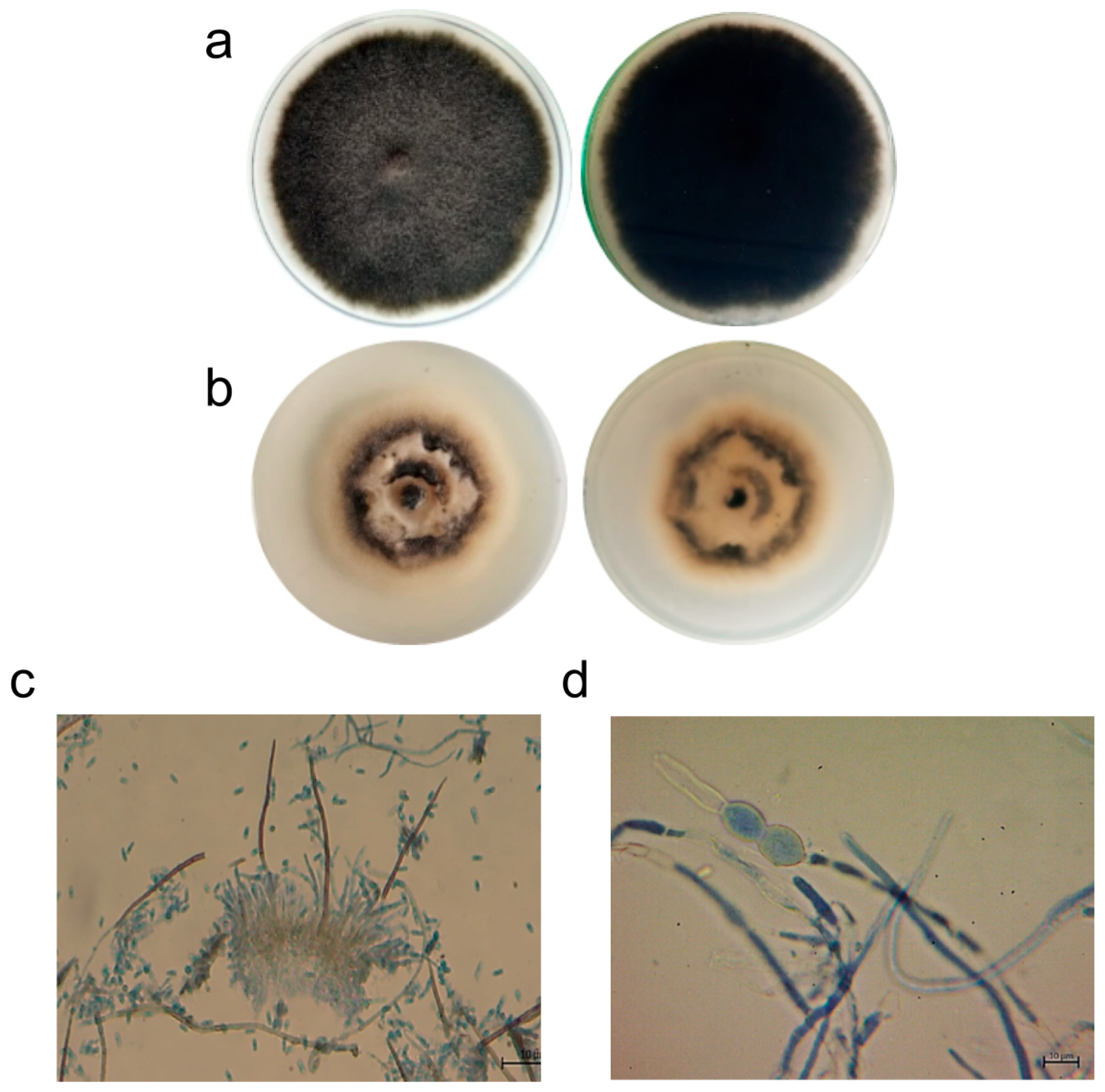
3.1.1. Effect on C. lindemuthianum Morphology

The macroscopic and microscopic characteristics of C. lindemuthianum differed between the cultures grown with and without potassium phosphite solution (Figure 1). The colony grown on PDA without supplementation exhibited a cottony growth pattern, and was black with a slightly gray center, 18 days after sowing (Figure 1a). Microscopically, this colony displayed hyaline septate mycelia with asexual fruiting bodies (acervuli), which are clusters of conidiophores from which conidia emerge (Figure 1c). The conidia observed were typical of the genus and were classified as hyaline with a cylindrical shape. In contrast, colonies grown on PDA supplemented with potassium phosphite (5 mL L−1) had a black cotton-like appearance with a white center and a white edge (Figure 1b). Microscopically, the fungus showed deformations in the mycelium, characterized by overgrowth and widening of the hyphae (Figure 1d).

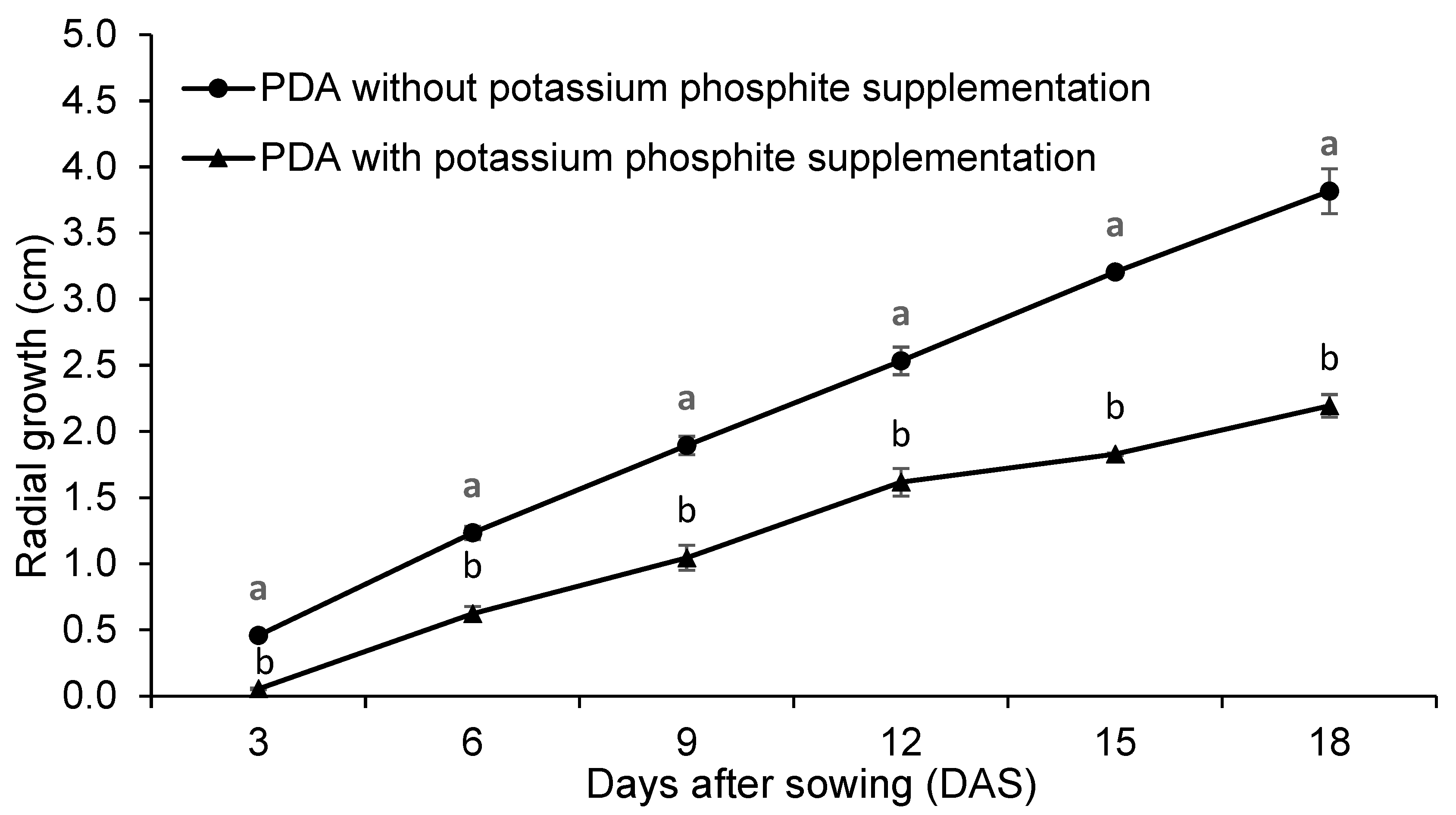
3.1.2. Effect on C. lindemuthianum Growth Rate and Conidia Germination

Colonies of C. lindemuthianum grown on PDA without supplementation with potassium phosphite showed greater mycelial growth (Figure 2), as well as a higher mycelial growth rate and germination percentage, compared to colonies grown on PDA supplemented with potassium phosphite (5 mL L−1) (Table 1). The growth inhibition percentage in the PDA-supplemented culture media was 42%, and the germination percentage decreased by 48%, compared to the non-supplemented culture media (Table 1).

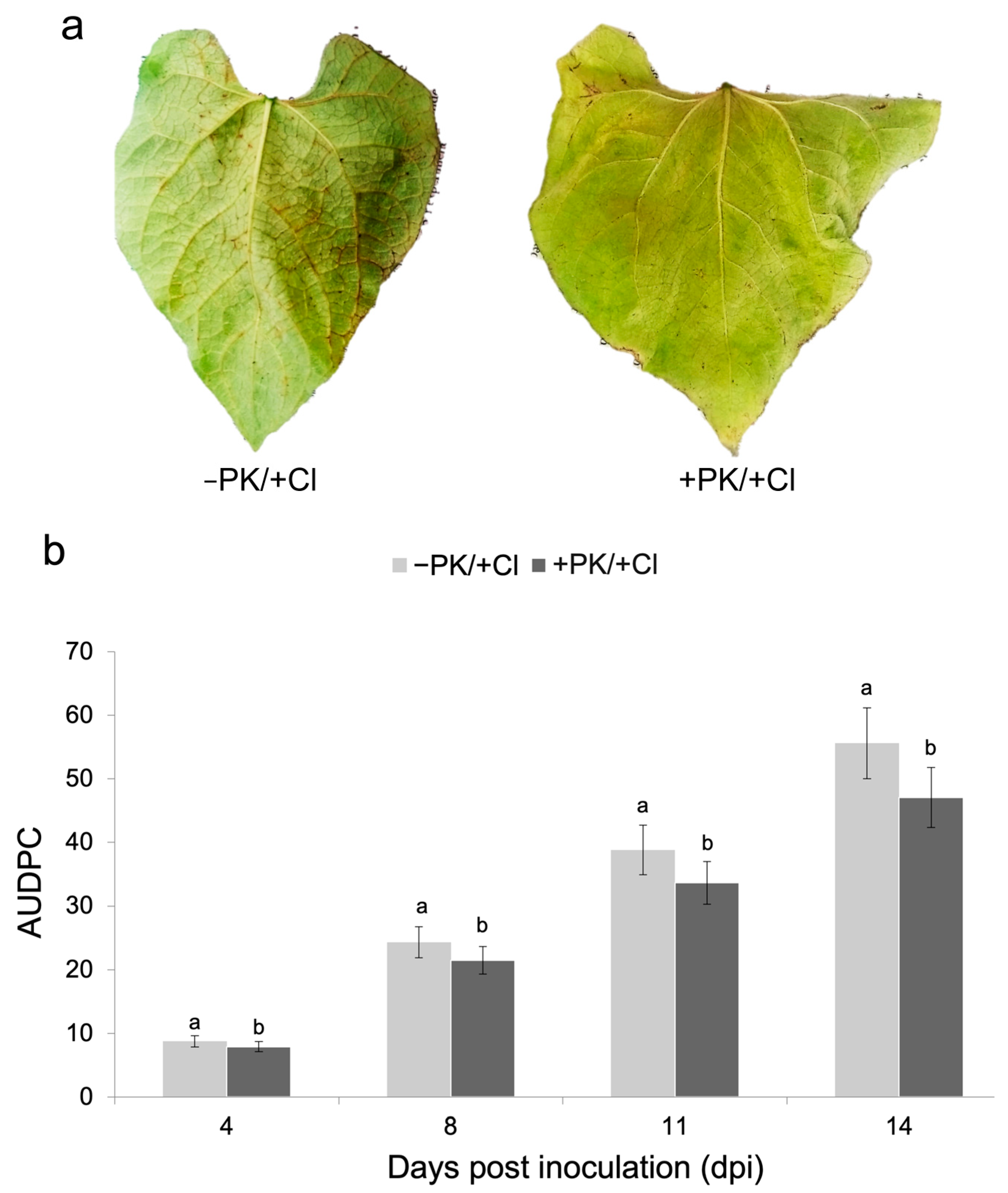
3.2. Effect of Potassium Phosphite Solution on Bean Anthracnose

The effect of potassium phosphite on anthracnose was estimated in bean seedlings of the Sutagao cultivar infected with C. lindemuthianum. Leaves of plants previously treated with potassium phosphite before fungal infection (+PK/+Cl) showed smaller necrotic lesions in the secondary veins compared to those of infected plants that were not treated with potassium phosphite (−PK/+Cl) (Figure 3a). In addition, the AUDPC data indicated a 17% reduction in disease severity in plants treated with the phosphite potassium before infection with C. lindemuthianum (+PK/+Cl), compared to untreated plants (−PK/+Cl). Significant differences between the two treatments were observed as early as 4 days post-inoculation (dpi) and persisted throughout the disease evaluation period (Figure 3b).

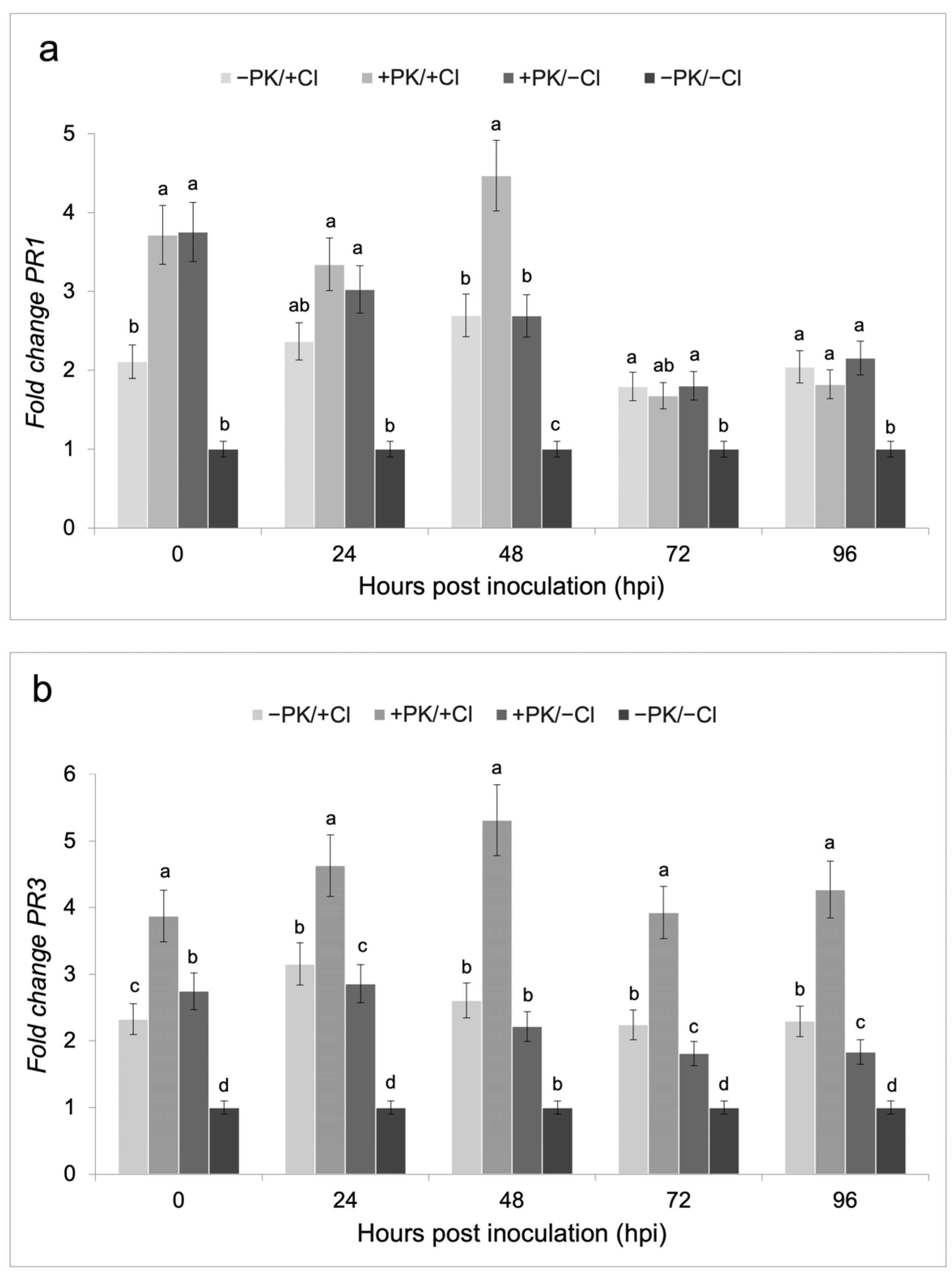
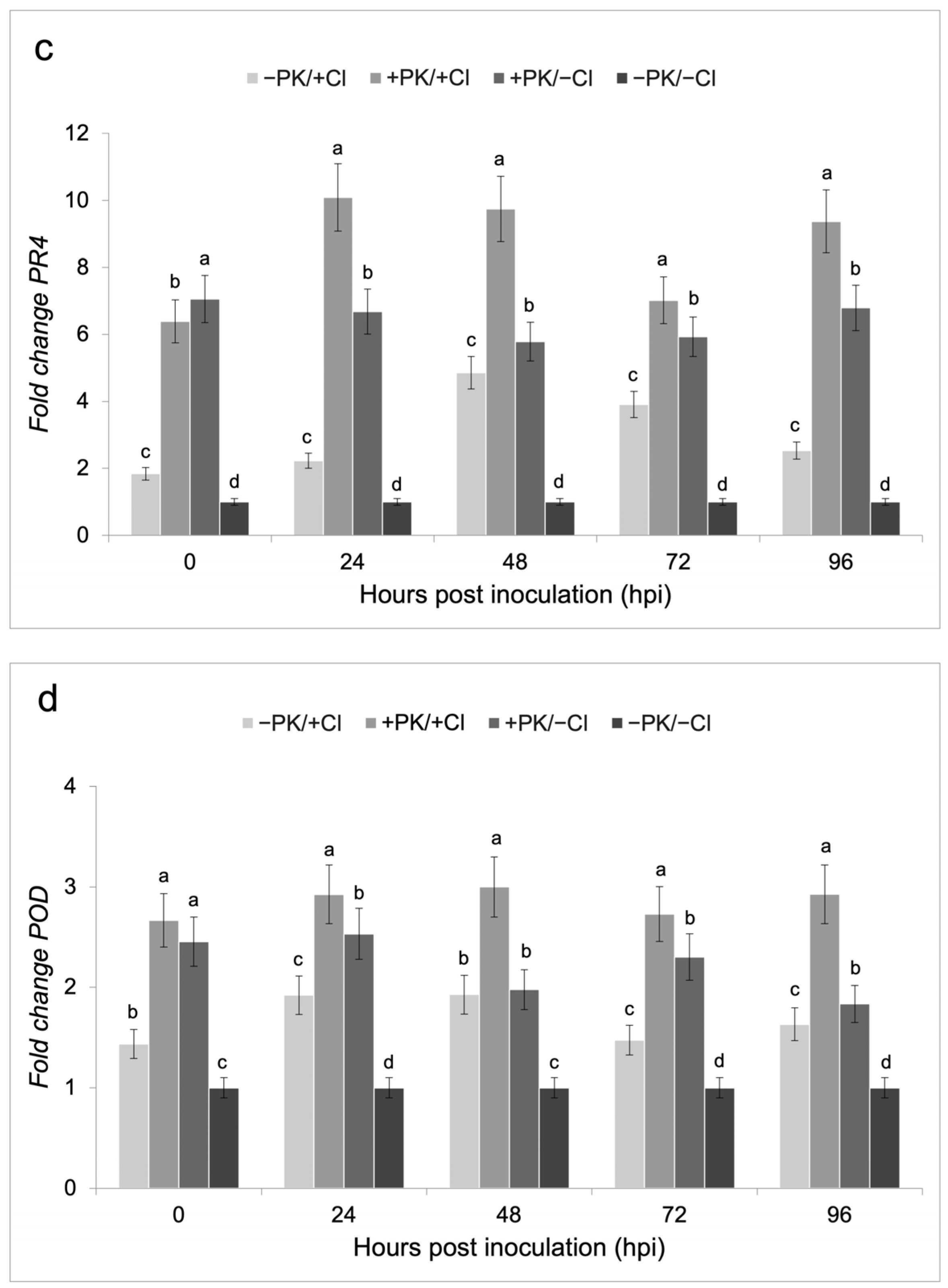
3.3. Defense Gene Expression on Bean Plants Treated with the Potassium Phosphite Solution and Infected with C. lindemuthianum

The potassium phosphite solution significantly increased expression of the plant defense genes PR1, PR3, PR4, and POD in plants treated with this product without prior C. lindemuthianum infection (+PK/−Cl), demonstrating a defense-inducing effect (Figure 4). This gene induction was further enhanced when the plants were infected with the fungus (+PK/+Cl) (Figure 4). PR1 and PR3 exhibited a significant increase in expression at 48 hpi, although the expression of PR1 declined at 72 hpi, whereas that of PR3 remained elevated (Figure 4a,b). PR4 and POD displayed early expression at 24 hpi, with PR4 showing the highest expression level among all the genes evaluated (Figure 4). These findings suggest strong activation of the defense response, possibly linked to a priming effect (Figure 4).

4. Discussion

4.1. Antifungal Effect of the Potassium Phosphite Solution on C. lindemuthianum

Various studies have demonstrated the efficacy of phosphite-based solutions similar to the product evaluated in this study (28% P2O5, and 26% K2O) in controlling anthracnose caused by Colletotrichum species, as well as their antifungal activity in in vitro evaluations [9]. In this work, C. lindemuthianum showed a significant reduction in growth (42%) and conidial germination percentage (48%) on PDA supplemented with 5 mL L1 of the potassium phosphite solution, similar to the decrease in mycelial growth (62.1%) and germ tube formation in C. lindemuthianum reported by other authors [5]. However, another study showed that a dose of 5 mL L1 of phosphite A solution (26% P2O5 and 19% K2O) or phosphite B solution (33.6% P2O5 and 29% K2O) had a significant effect, inhibiting mycelial growth by 81.1% and 100%, respectively [13]. Evaluation of the dose of phosphite ion in a 0-40-20 formulation of N-P-K (pH 3) on C. gloeosporioides, the causal agent of anthracnose in apples, reduced the colony diameter by 94% [23]. In the same way, doses of 5.0 mL L1 and 10.0 mL L1 of potassium phosphite (35.10% P2O5, 25.65% K2O) inhibited conidia germination by around 51.1% and 63.1%, respectively, in C. gloeosporioides isolated from coffee anthracnose [24]. The effects of phosphite exposure on fungal cells have been associated with alterations in phosphorus metabolism and in compounds containing this element, including inhibition of key phosphorylation reactions in fungi [9,11]. Likewise, these compounds have been associated with changes in gene expression related to synthesis of the cell wall and cytoskeletal proteins [12]. Other authors have reported that potassium phosphite produces increases in inorganic polyphosphates, alterations in nucleotide reserves, alterations in pentose phosphate metabolism, disruption of ionic balance, and changes in protein kinase activation, which regulate the formation of infective structures of the pathogen [7,9,11,15].

4.2. Reduction in Bean Anthracnose Induced by the Potassium Phosphite Solution

The reduction in bean anthracnose severity induced by application of the potassium phosphite solution that we observed was smaller (17%) than those reported by other authors, which included 5669% [13], 74–81% [25], and 71% [26]. These differences could be related to the bean material used and the concentration of the pathogen inoculum. The genetic and variety of bean plants used in this study (Sutagao cultivar, derived from the cross between Cabrera and G2333) differs from Perola [13], BRS Majestoso [25], and IPR Tangará [26], characteristics that determine resistance or susceptibility to the pathogen depending on its race. In addition, the inoculum concentration of C. lindemuthianum used to infect the plants in this study (1 × 10⁷ conidia mL1) was higher than the 7 × 10⁵ conidia mL1 [13,25] and 1 × 10⁶ conidia mL−1 [26] doses used by other authors.
Furthermore, the quantity of phosphite and potassium in the product, as well as the dose and time of application, may have also influenced the results. Other studies [13,25,26] evaluated various phosphite formulations with different concentrations of P2O5 and K2O than those used in this study. However, one of the phosphites evaluated by another group [25] corresponded to the same one used in this work, but applied at three different times to plants at more advanced stages of development: V4 (three trifoliate leaves), R5 (pre-flowering), and R7 (pod formation).
Similarly, phosphites have been evaluated in seeds with reapplication 18 days after sowing at the moment of expansion of the second trifoliate leaf [26], which differs from the present study, in which only one application was performed. This suggest that, to obtain better results in reducing anthracnose in beans caused by C. lindemuthianum, it is necessary to carry out several applications of this compound in seeds or at more advanced phenological stages, to induce a priming effect. This strategy improves the defense response of plants by triggering changes in the plant at physiological, transcriptional, metabolic, and epigenetic levels, preparing it with a better response capacity when exposed to a subsequent stimulus, such as attack by a pathogen, activating immune mechanisms more quickly and strongly [7,27].
The effect of phosphite treatment as a strategy to reduce the severity of diseases in plants has been demonstrated mainly against oomycete pathogens, such as species of the genera Phytophthora, Pythium, and Peronosclerospora sorghi. However, it also acts against fungi such as Alternaria alternata, Fusicladium effusum, Fusarium spp., F. oxysporum, Rhizoctonia solani, and Verticillium spp.; some protists such as Plasmodiophora brassicae; and bacteria such as Pectobacterium carotovorum and Pseudomonas syringae [7,11,28]. Nevertheless, changes in the expression of defense genes in plants treated with these potassium phosphite products have not been thoroughly evaluated in plant–pathogen interactions that do not consider oomycetes.

4.3. Induction of Defense Gene Expression by the Potassium Phosphite Solution

This study demonstrated that the application of the potassium phosphite solution to bean plants induced expression of the PR1, PR3, PR4, and POD defense genes. However, their expression increased when the plants were infected with C. lindemuthianum. Similar results have been described in potato plants that were previously foliar-sprayed with a potassium phosphite (KPhi)-based fungicide and infected with Phytophthora infestans [7].
In a transcriptomic analysis carried out at 6 hpi in bean seedlings susceptible and resistant to C. lindemuthianum and subjected to previous stimulus with AS (salicylic acid) and MeJA (methyl jasmonate), 463 genes were differentially expressed [29]. Of these, 19 genes were up-regulated in the resistant genotype in response to stimulation with AS, and 17 with MeJA. Ten of these were R genes, eleven genes encoded protein kinases, and ten genes belonging to the transcription factor family. This provides evidence for very early activation of signaling by a defense inducer, which promotes transcription and expression of defense responses in plants [29]. However, no other time points after infection were assessed to determine the duration and intensity of expression of these genes.
In the present study, the expression of PR3 was higher at 48 hpi, which was also reported to be the highest enzymatic activity for this protein (PR3) in potatoes treated with phosphite and infected with P. infestans [7]. Additionally, for PR4, another chitinase evaluated in this work, the expression level increased earlier than that for PR3 (24 hpi) and was maintained until the last evaluation time (96 hpi), as well as for PR3. Similarly, high expression levels of genes associated with glucanases and chitinases were observed in cucumber (Cucumis sativus L.) plants treated with potassium phosphite and in plants treated and inoculated with Pseudoperonospora cubensis [30], indicating sensitization of the plants that triggers a rapid defense response against pathogens. Therefore, chitinases are important in the early defense response, and their enhanced effect in plants previously treated with phosphite solution results in increased sensitivity and activity against phytopathogens [31].
Concerning PR1, the potassium phosphite solution promoted its expression before infection with the pathogen. The highest expression of the gene was evident at 48 hpi, although it subsequently declined at 72 hpi, indicating that it did not maintain its expression. The PR1 gene in bean plants infected with C. lindemuthianum has been identified as a key marker that is expressed during the early stages of infection in both susceptible and resistant hosts. However, its expression is stronger and occurs earlier in resistant plants, reaching peak expression up to 72 h post-inoculation (hpi) [32]. In this study, transient PR1 expression was detected in susceptible Sutagao bean plants treated with phosphite. This transient response correlated with severity scores, as potassium phosphite–treated plants exhibited symptoms, albeit less severe than untreated plants, with a 17% reduction in severity. This suggests that the resistance induced was neither robust nor long lasting. A higher dose or multiple applications (e.g., at seed, transplant, or other vegetative stage) may be required to enhance the priming effect to achieve a more sustained and effective defense response.
Increased expression of PR1 in plants treated with phosphite before infection with a pathogen has been reported in plants principally infected with an oomycete. For example, pepper (Capsicum annuum) treated with potassium phosphite and inoculated with Phytophthora capsici and Phytophthora cinnamomi showed strong upregulation of soluble proteins and pathogenesis-related 1 (CaPR1) over time, which is associated with callose deposition and ROS production [33,34]. Although detection of callose and H2O2 was not carried out in this study, previous work has compared the defense responses against C. lindemuthianum between the susceptible bean cultivar Sutagao, the same used in this study, and the resistant bean genotype G2333 [18]. The authors showed an increase in expression of the PR1, PR3, and POD genes at 1 dpi, coinciding with the time of H2O2 production in the resistant bean cultivar, in contrast to delayed expression of these genes in the susceptible bean cultivar. This indicates that PR1, PR3, and POD are markers associated with defense against the fungus, and that phosphite increased their expression in the Sutagao cultivar before and after infection.
It is important to highlight PR1 as a marker of systemic resistance, the priming response, and the transgenerational priming response against pathogen challenge [35]. It has been reported that an increase in some enzymatic activities, such as peroxidase and polyphenol oxidase, after treatment with phosphite and infection by a pathogen is related to induction of a systemic defense response [28]. In this study, high POD expression was detected both in plants treated with phosphite and after inoculation with C. lindemuthianum from 24 hpi until the final evaluation time (96 hpi). This expression is related to induction of ROS detoxification activity by the enzyme peroxidase (POD), which is generated as an antioxidant defense response to pathogen infection and further stimulated by phosphite [7,36]. In beans, an increase in POD enzyme activity has been reported in plants stimulated with phosphite, which is associated with detoxification [7,37]. At the transcriptomic level, genes encoding oxidoreductase activity were confirmed to be expressed in bean seedlings resistant to C. lindemuthianum [29]. In other plants stimulated with phosphite and previously infected with the pathogen, an increase in POD activity has been reported, for example in cucumber plants with Pythium ultimum var [38]; apple (Malus pumila) with Venturia inaequalis [39]; and potato plants (S. tuberosum) with P. infestans [7].

5. Conclusions

The potassium phosphite solution (P2O5 28% and K2O 26%) had direct antifungal activity against C. lindemuthianum. It reduced mycelial growth by 42% and germination by 48% at a dose of 5 mL L−1. The effect of this compound on Sutagao bean plants stimulated expression of the PR1, PR3, PR4 and POD defense genes, but this increased when the plants were infected with C. lindemuthianum. The highest expression levels were observed for the chitinase genes (PR3 and PR4), whose expression levels were maintained until 96 hpi, followed by the POD gene. These results demonstrate the dual activity of potassium phosphite as an antifungal agent and priming inducer against bean anthracnose and its potential for inclusion in management programs.

Author Contributions

Conceptualization, A.G.-A. and C.S.-G.; methodology, C.S.-G. and P.N.P.-M.; data analysis, C.S.-G., P.N.P.-M. and A.G.-A.; writing—original draft preparation, C.S.-G., P.N.P.-M. and A.G.-A.; writing—review and editing, C.S.-G. and A.G.-A.; supervision, A.G.-A.; project administration, A.G.-A.; funding acquisition, A.G.-A. All authors have read and agreed to the published version of the manuscript.

Funding

This research was funded by Universidad Nacional de Colombia, sede Bogotá, Project ID 57226 Convocatoria para la financiación de proyectos de tesis de doctorado y maestría de la Facultad de Ciencias Agrarias, sede Bogotá. Fondo de Investigación UGI-2022.

Data Availability Statement

The original contributions presented in this study are included in the article. Further inquiries can be directed to the corresponding author.

Acknowledgments

Thanks to Gustavo Ligarreto for supplying the seeds of bean cultivars and the Antonio Angarita Zerda-Plant Biotechnology Laboratory at the Faculty of Agricultural Sciences of the Universidad Nacional de Colombia for the use of their facilities and equipment.

Conflicts of Interest

The authors declare no conflicts of interest.

References

  1. Rahmanzadeh, A.; Khahani, B.; Taghavi, S.M.; Khojasteh, M.; Osdaghi, E. Genome-wide meta-QTL analyses provide novel insight into disease resistance repertoires in common bean. BMC Genom. 2022, 23, 680. [Google Scholar] [CrossRef]
  2. Gupta, C.; Salgotra, R.K.; Damm, U.; Rajeshkumar, K.C. Phylogeny and pathogenicity of Colletotrichum lindemuthianum causing anthracnose of Phaseolus vulgaris cv. Bhaderwah-Rajmash from northern Himalayas, India. 3 Biotech 2022, 12, 169. [Google Scholar] [CrossRef]
  3. Nabi, N.; Nabi, A.; Fayaz, T.; Lateef, I.; Nisa, Q.; Bashir, A.; Rashid, Z.; Shah, M.D.; Itoo, H.; Shah, R.A.; et al. Pathogenically altered Colletotrichum lindemuthianum transformants help in understanding the biochemical defense and colonization dynamics in Phaseolus vulgaris. Physiol. Mol. Plant Pathol. 2024, 129, 102208. [Google Scholar] [CrossRef]
  4. Nabi, A.; Lateef, I.; Nisa, Q.; Banoo, A.; Rasool, R.S.; Shah, M.D.; Ahmad, M.; Padder, B.A. Phaseolus vulgaris-Colletotrichum lindemuthianum pathosystem in the post-genomic era: An update. Curr. Microbiol. 2022, 79, 36. [Google Scholar] [CrossRef]
  5. Silva, L.L.; Moreno, H.L.A.; Correia, H.L.N.; Santana, M.F.; Queiroz, M.V. Colletotrichum: Species complexes, lifestyle, and peculiarities of some sources of genetic variability. Appl. Microbiol. Biotechnol. 2020, 104, 1891–1904. [Google Scholar] [CrossRef]
  6. Lopez-Zapata, S.P.; Castaño-Zapata, J. In vitro effect of four fungicides on Colletotrichum gloeosporioides causing anthracnosis on the Red Globe grape variety. Rev. Acad. Colomb. Cienc. Exactas. Fis. Nat. 2020, 44, 747–758. [Google Scholar] [CrossRef]
  7. Mohammadi, M.A.; Cheng, Y.; Aslam, M.; Jakada, B.H.; Wai, M.H.; Ye, K.; He, X.; Luo, T.; Ye, L.; Dong, C.; et al. ROS and oxidative response systems in plants under biotic and abiotic stresses: Revisiting the crucial role of phosphite triggered plants defense response. Front. Microbiol. 2021, 12, 631318. [Google Scholar] [CrossRef]
  8. Balmer, A.; Pastor, V.; Gamir, J.; Flors, V.; Mauch-Mani, B. The ’prime-ome’: Towards a holistic approach to priming. Trends Plant Sci. 2015, 20, 443–452. [Google Scholar] [CrossRef]
  9. Achary, V.M.M.; Ram, B.; Manna, M.; Datta, D.; Bhatt, A.; Reddy, M.K.; Agrawal, P.K. Phosphite: A novel P fertilizer for weed management and pathogen control. Plant Biotechnol. J. 2017, 15, 1493–1508. [Google Scholar] [CrossRef]
  10. Eshraghi, L.; Aanderson, J.; Aryamaseh, N.; Shearer, B.; McComb, J.; Hardy, G.E.S.; O’Brien, P.A. Phosphite primed defence responses and enhanced expression of defence genes in Arabidopsis thaliana infected with Phytophthora cinnamomi. Plant Pathol. 2011, 60, 1086–1095. [Google Scholar] [CrossRef]
  11. Havlin, J.L.; Schlegel, A.J. Review of phosphite as a plant nutrient and fungicide. Soil Syst. 2021, 5, 52. [Google Scholar] [CrossRef]
  12. King, M.; Reeve, W.; Van Der Hoek, M.; Williams, N.; McComb, J.; O’Brien, P.A.; Hardy, G.E.S.J. Defining the phosphite-regulated transcriptome of the plant pathogen Phytophthora cinnamomi. Mol. Genet. Genom. 2010, 284, 425–435. [Google Scholar] [CrossRef]
  13. Costa, B.H.G.; De Resende, M.L.V.; Monteiro, A.C.A.; Junior, P.M.R.; Botelho, D.M.S.; Silva, B.M. Potassium phosphites in the protection of common bean plants against anthracnose and biochemical defence responses. J. Phytopathol. 2017, 66, 95–102. [Google Scholar] [CrossRef]
  14. Ortiz, H.E.; Rivero, R.; Hoyos, C.L. Manual de Prácticas de Fitopatología General; Facultad de Ciencias Agrarias Universidad Nacional de Colombia: Bogotá, Colombia, 2011. [Google Scholar]
  15. Fu, T.; Shin, J.H.; Lee, N.H.; Lee, K.H.; Kim, K.S. Mitogen-activated protein kinase CsPMK1 is essential for pepper fruit anthracnose by Colletotrichum scovillei. Front. Microbiol. 2022, 13, 770119. [Google Scholar] [CrossRef]
  16. Castellanos, G.; Jara, C.E.; Mosquera, G. Colletotrichum lindemuthianum. Enfermedad antracnosis. In Guías Prácticas de Laboratorio Para el Manejo de Patógenos del Frijol; Castellanos, G., Jara, C.E., Mosquera, G., Eds.; Alliance Bioversity International—CIAT: Palmira, Colombia, 2011; pp. 1–39. [Google Scholar]
  17. Romero, G.; González, S.; Royero, W.; González, A. Morphological and transcriptional analysis of Colletotrichum lindemuthianum race 7 during early stages of infection in common bean. Genet. Mol. Biol. 2024, 47, e20220263. [Google Scholar] [CrossRef]
  18. Pedroza-Padilla, M.C.; Rodriguez-Arevalo, K.A.; Rincon-Rivera, L.J.; Gonzalez-Almario, A. Co-52 resistance allele contributes to induce basal defense against Colletotrichum lindemuthianum race 7. Pesqui. Agropecu. Trop. 2022, 52, e71746. [Google Scholar] [CrossRef]
  19. Van Schoonhoven, A.; Pastor-Corrales, M.A. Standard System for the Evaluation of Bean Germplasm; Alliance Bioversity International—CIAT: Palmira, Colombia, 1987; pp. 29–31. [Google Scholar]
  20. Shaner, G.; Finney, R.E. The effect of nitrogen fertilization on the expression of slow-mildewing resistance in knox whetl. Phytopathology 1977, 67, 1051–1056. [Google Scholar] [CrossRef]
  21. Saldarriaga-Gomez, C.; Gonzalez-Almario, A. Evaluation of defense genes expression and the virulence factor Cac1 in Phaseolus vulgaris and Colletotrichum lindemuthianum interaction. Agron. Colomb. 2025. [Google Scholar] [CrossRef]
  22. Pfaffl, M.W. A new mathematical model for relative quantification in real-time RT-PCR. Nucleic Acids Res. 2001, 29, e45. [Google Scholar] [CrossRef]
  23. Araujo, L.; Valdebenito-Sanhueza, R.M.; Stadnik, M.J. Evaluation of potassium phosphite formulations against Colletotrichum gloeosporioides in vitro and for post-infection control of Glomerella leaf spot in apple. Trop. Plant Pathol. 2010, 35, 54–59. [Google Scholar] [CrossRef]
  24. Ogoshi, C.; Abreu, M.S.; Silva, B.M.; Neto, H.S.; Ribeiro-Junior, P.M.; Resende, M.L.V. Potassium phosphite: A promising product in the management of diseases caused by Colletotrichum gloeosporioides in coffee plants. Biosci. J. 2013, 29, 1558–1565. [Google Scholar]
  25. Gadaga, S.J.C.; Abreu, M.S.; De Resende, M.L.V.; Junior, P.M.R. Phosphites for the control of anthracnose in common bean. Pesqui. Agropecu. Bras. 2017, 52, 36–44. [Google Scholar] [CrossRef]
  26. Figueira, E.P.P.; Kuhn, O.J.; Martinazzo-Portz, T.; Stangarlin, J.R.; Pereira, M.D.P.; Lampugnani, C. Histochemical changes induced by Trichoderma spp. and potassium phosphite in common bean (Phaseolus vulgaris) in response to the attack by Colletotrichum lindemuthianum. Semin. Ciênc. Agrár. 2020, 41, 811–828. [Google Scholar] [CrossRef]
  27. Hilker, M.; Schmulling, T. Stress priming, memory, and signalling in plants. J. Integr. Plant Biol. 2019, 42, 753–761. [Google Scholar] [CrossRef]
  28. Yañez-Juarez, M.G.; Lopez-Orona, C.A.; Ayala-Tafoya, F.; Partida-Ruvalcaba, L.; Velasquez-Alcatraz, T.J.; Medina-Lopez, R. Los fosfitos como alternativa para el manejo de problemas fitopatológicos. Rev. Mex. Fitopatol. 2018, 36, 79–94. [Google Scholar] [CrossRef]
  29. Chang, Y.; Liu, Y.; Wang, L.; Wang, S.; Wu, J. Global transcriptome analysis reveals resistance genes in the early response of common bean (Phaseolus vulgaris L.) to Colletotrichum lindemuthianum. BMC Genom. 2024, 25, 579. [Google Scholar] [CrossRef]
  30. Ramezani, M.; Ramezani, F.; Rahmani, F.; Dehestani, A. Exogenous potassium phosphite application improved PR-protein expression and associated physiobiochemical events in cucumber challenged by Pseudoperonospora cubensis. Sci. Hortic. 2018, 234, 335–343. [Google Scholar] [CrossRef]
  31. Vaghela, B.; Vashi, R.; Rajput, K.; Joshi, R. Plant chitinases and their role in plant defense: A comprehensive review. Enzym. Microb. Technol. 2022, 159, 110055. [Google Scholar] [CrossRef]
  32. Alvarez-Diaz, J.C.; Laugé, R.; Delannoy, E.; Huguet, S.; Paysant-Le Roux, C.; Gratias, A.; Geffroy, V. Genome-wide transcriptomic analysis of the effects of infection with the hemibiotrophic fungus Colletotrichum lindemuthianum on common bean. Plants 2022, 11, 1995. [Google Scholar] [CrossRef]
  33. Liu, P.; Li, B.; Lin, M.; Chen, G.; Ding, X.; Weng, Q.; Chen, Q. Phosphite-induced reactive oxygen species production and ethylene and ABA biosynthesis, mediate the control of Phytophthora capsici in pepper (Capsicum annuum). Funct. Plant Biol. 2016, 43, 563–574. [Google Scholar] [CrossRef]
  34. Eshraghi, L.; Anderson, J.P.; Aryamanesh, N.; McComb, J.A.; Shearer, B.; Hardy, G. Defence signalling pathways involved in plant resistance and phosphite-mediated control of Phytophthora cinnamomi. Plant Mol. Biol. Rep. 2014, 32, 342–356. [Google Scholar] [CrossRef]
  35. Martinez-Aguilar, K.; Hernandez-Chavez, J.L.; Alvarez-Venegas, R. Priming of seeds with INA and its transgenerational effect in common bean (Phaseolus vulgaris L.) plants. Plant Sci. 2021, 305, 110834. [Google Scholar] [CrossRef] [PubMed]
  36. Novaes, M.; Debona, D.; Fagundes-Nacarath, I.; Brás, V.; Rodrigues, F. Physiological and biochemical responses of soybean to white mold affected by manganese phosphite and fluazinam. Acta Physiol. Plant. 2018, 41, 186. [Google Scholar] [CrossRef]
  37. Avila, F.W.; Faquin, V.; Da Silva-Lobato, A.K.; Avila, P.A.; Marques, D.J.; Silva-Guedes, E.M.; Yuen-Tan, D.K. Effect of phosphite supply in nutrient solution on yield, phosphorus nutrition and enzymatic behavior in common bean (’Phaseolus vulgaris’. L.) plants. Aust. J. Crop Sci. 2013, 7, 713–722. [Google Scholar]
  38. Mofidnakhaei, M.; Abdossi, V.; Dehestani, A.; Pirdashti, H.; Babaeizad, V. Potassium phosphite affects growth, antioxidant enzymes activity and alleviates disease damage in cucumber plants inoculated with Pythium ultimum. Arch. Phytopathol. Plant Prot. 2016, 49, 207–221. [Google Scholar] [CrossRef]
  39. Felipini, R.B.; Boneti, J.I.; Katsurayama, Y.; Neto, A.C.R.; Veleirinho, B.; Maraschin, M.; Piero, R. Apple scab control and activation of plant defence responses using potassium phosphite and chitosan. Eur. J. Plant Pathol. 2016, 145, 929–939. [Google Scholar] [CrossRef]
Figure 1. Macroscopic and microscopic characteristics of C. lindemuthianum grown on PDA with and without the potassium phosphite solution (5 mL L−1), 18 days after sowing. (a) C. lindemuthianum colony on PDA culture media without supplementation; the images on the left show the front of the colony, while those on the right show the back; (b) C. lindemuthianum colony on PDA culture media supplemented with the potassium phosphite solution; the images on the left display the front of the colony, and those on the right display the back; (c) acervuli and conidia of C. lindemuthianum observed at 40× magnification, grown on PDA without supplementation; (d) deformations in C. lindemuthianum mycelia observed at 40× magnification, grown on PDA supplemented with the potassium phosphite solution.
Horticulturae 11 00462 g001
Figure 2. Radial growth curve of C. lindemuthianum on PDA culture media with and without the potassium phosphite solution. Data are shown as the mean ± SD of three replicates. Different letters represent values with significant differences between treatments (p < 0.05).
Horticulturae 11 00462 g002
Figure 3. Effects of the potassium phosphite solution on anthracnose disease in Sutagao bean plants. (a) Necrotic lesions caused by C. lindemuthianum in leaves at 14 dpi. The image on the left shows a leaf infected with C. lindemuthianum but not pre-treated with the potassium phosphite solution (−PK/+Cl); the image on the right shows a leaf infected and pre-treated with potassium phosphite solution (−PK/+Cl); (b) AUDPC values obtained after inoculation with C. lindemuthianum on 10-day-old plants of the Sutagao bean cultivar, previously treated with the potassium phosphite solution at different time points. Different letters represent values with significant differences between treatments (p < 0.05).
Horticulturae 11 00462 g003
Figure 4. Differential expression of defense genes in the Sutagao bean cultivar inoculated with C. lindemuthianum and treated with the potassium phosphite solution at different time points. (a) Changes in PR1 expression; (b) changes in PR3 expression; (c) changes in PR4 expression; (d) changes in POD expression. Different letters indicate significant differences between treatments (p < 0.05).
Horticulturae 11 00462 g004aHorticulturae 11 00462 g004b
Table 1. Mycelial growth rate, growth inhibition percentage, and conidial germination percentage of C. lindemuthianum on PDA culture media with and without potassium phosphite supplementation.
TreatmentMycelial Growth Rate (cm/Day)Growth
Inhibition
Percentage (%)
Conidial
Germination
Percentage (%)
PDA without supplementation0.22 a-49.44 a
PDA supplemented with
potassium phosphite
0.14 b42.3925.78 b
Different letters represent values with significant differences between treatments (p < 0.05).
Disclaimer/Publisher’s Note: The statements, opinions and data contained in all publications are solely those of the individual author(s) and contributor(s) and not of MDPI and/or the editor(s). MDPI and/or the editor(s) disclaim responsibility for any injury to people or property resulting from any ideas, methods, instructions or products referred to in the content.

