Article

Brassica oleracea var. sabellica: A New Host of Agroathelia delphinii in Soilless Cultivation Systems in Central Thailand

by
Santiti Bincader
1,2,
Ratiya Pongpisutta
3,
Thipwara Tiansawang
1,
Sirorat Khienman
1,
Panida Boonyaritthongchai
4,
Vipaporn Phuntumart
5 and
Chainarong Rattanakreetakul
6,*
1
Program in Plant Science, Faculty of Agricultural Technology and Agro-Industry, Rajamangala University of Technology Suvarnabhumi, Phra Nakhon Si Ayutthaya 13000, Thailand
2
Agricultural Research Division, Center of Excellence in Agriculture and Food Safety, Rajamangala University of Technology Suvarnabhumi, Phra Nakhon Si Ayutthaya 13000, Thailand
3
Department of Plant Pathology, Faculty of Agriculture, Kasetsart University, Bangkok 10900, Thailand
4
Postharvest Technology Program, School of Bioresources and Technology, King Mongkut’s University of Technology Thonburi, Bangkok 10140, Thailand
5
Department of Biological Sciences, 129 Life Sciences Building, Bowling Green State University, Bowling Green, OH 43403, USA
6
Department of Plant Pathology, Faculty of Agriculture at Kamphaeng Saen, Kasetsart University, Kamphaeng Sean Campus, Nakhon Pathom 73140, Thailand
*
Author to whom correspondence should be addressed.
Horticulturae 2025, 11(4), 411; https://doi.org/10.3390/horticulturae11040411
Submission received: 18 March 2025 / Revised: 7 April 2025 / Accepted: 10 April 2025 / Published: 11 April 2025
(This article belongs to the Special Issue Fungal Diseases in Horticultural Crops)

Abstract

:
Kale (Brassica oleracea var. sabellica), known for its high nutritional value and health benefits, has gained significant popularity. Recently, kale grown in soilless systems has also become increasingly popular, as these systems offer better environmental control and improve overall quality, making them an ideal method for cultivating kale. However, in 2023–2024, several kale plants exhibited severe symptoms of seedling and stem rot leading to losses of over 70% in both quality and yield. In this study, the infectious isolates were obtained from stem rot kale grown in soilless cultivation greenhouses across three provinces in central Thailand. The pathogens were identified through a combination of morphological characteristics and molecular techniques, utilizing nucleotide sequences from the internal transcribed spacer (ITS1-5.8S-ITS2) and large subunit ribosomal RNA (LSU rDNA). Pathogenicity tests and Koch’s postulates on 2-month-old kale plants confirmed that the fungus was responsible for causing brown stem lesions and rot. Morphological features and multilocus sequence analysis (MLSA) identified the pathogen as Agroathelia delphinii. This research represents the first report of A. delphinii infecting kale in Thailand, offering crucial insights for accurate disease diagnosis and the development of effective management strategies in soilless cultivation systems, which is essential for improving productivity in increasingly variable environments.

1. Introduction

Kale plants (Brassica oleracea var. sabellica) are rapidly gaining popularity as both a highly nutritious vegetable and a celebrated superfood. A member of the Brassicaceae family, which includes other notable vegetables such as bok choy (Brassica rap var. chinensis), broccoli (Brassica oleracea var. italica), cabbage (Brassica oleracea var. capitata) or cauliflower (Brassica oleracea var. botrytis), kale has garnered attention for its rich nutrient profile and related health benefits. From 2015 to 2024, numerous studies have highlighted the multifaceted value of kale, emphasizing its nutritional benefits, economic significance, and role in promoting human health [1,2]. The economic value of kale has surged due to rising consumer demand for health-promoting and organic foods. The global market for kale has expanded, providing lucrative opportunities for farmers and agricultural enterprises, particularly those employing sustainable and soilless cultivation methods. Kale’s adaptability to various growing conditions and its relatively short growth cycle makes it an attractive crop for producers aiming to meet the increasing demand for nutrient-dense foods [3,4,5]. Additionally, kale has garnered significant attention for its nutritional benefits and versatile culinary applications. The increasing demand for health-promoting foods has led to innovative cultivation practices, including soilless systems, which offer numerous advantages such as enhanced control over growing conditions and reduced soil-borne diseases.
Soilless cultivation systems, including hydroponics and aeroponics, have gained popularity for their efficiency and ability to optimize plant growth. These systems allow precise regulation of nutrient supply, pH, and other environmental factors, leading to improved crop yields and quality. Research has demonstrated that soilless cultivation can significantly enhance the nutritional content of kale, making it an attractive method for modern agricultural practices [6,7,8,9]. However, the adoption of soilless systems is not without challenges, particularly concerning plant diseases [10,11,12,13,14]. Despite the benefits of soilless cultivation, kale remains susceptible to various diseases, which can severely impact yield and quality. Seedling rot and stem rot are common issues that can arise in controlled environments, often caused by fungal or bacterial soil-borne pathogens. These diseases pose significant threats to kale production, leading to substantial economic losses and highlighting the need for effective disease management strategies [11,15,16,17,18].
Sclerotium species, (anamorph: Agroathelia) first introduced by Tode (1790) [19], are notable for their ability to cause destructive diseases in a wide range of agricultural and horticultural plants. Diseases caused by Sclerotium, such as Southern blight, stem rot, and seedling damping-off, can lead to significant yield losses and economic damage. The epidemiology of these fungi involves complex interactions between the pathogen, the host, and the environment. Sclerotia, the hardened fungal structures that serve as survival and dissemination units, can persist in soil and plant debris for extended periods. These structures germinate under favorable conditions, leading to infection of susceptible plants. Factors such as temperature, humidity, and soil moisture play pivotal roles in the germination and pathogenicity of Sclerotium fungi. For instance, high temperatures and moist conditions typically promote the outbreak of diseases like Southern blight, highlighting the importance of environmental monitoring in disease management [20,21,22,23,24,25].
Agroathelia delphinii (Synonym: Sclerotium delphinii) is a species with a distribution primarily restricted to temperate regions such as parts of China [26] and the northern and midwestern region of the United States [27]. There have been reports of Sclerotium fungi in Thailand, with Sclerotium rolfsii being the most identified species. This fungus is responsible for affecting a wide range of economically important crops, including legumes and plants grown in diverse conditions, both in conventional agricultural systems and soilless systems. It is the cause of several plant diseases, including root and collar rot. Nevertheless, no conclusive reports or unambiguous identification of Agroathelia delphinii in Thailand have been reported [28]. It is still unknown why this geographic distribution exists. Variations in temperature may have an impact on the survival of A. delphinii, thereby influencing its dispersion. Nevertheless, there is no proof to support or refute this theory [29,30,31].
In this research, kale disease in soilless cultivation greenhouse systems was identified by nucleotide sequencing (two regions of the gene), morphological characterization, and pathogenicity tests. Accurate species identification is a crucial first step for effective disease management and understanding the ecology of phytopathogenic fungal disease. Morphological characteristics and host infection continue to play a significant role in fungal identifications. Moreover, internal transcribed spacer (ITS1-5.8S-ITS2) and large subunit ribosomal DNA (LSU rDNA) have been widely used as markers for fungal species identification in recent years, due to both gene/regions being extremely conserved in a variety of biological types [32,33,34]. This study aims to report and confirm, for the first time, that Agroathelia fungi are responsible for causing stem rot in kale and to document a new host for these fungi in central Thailand.

2. Materials and Methods

2.1. Fungal Isolation

The fungal pathogen was isolated from kale stem rot symptoms using a tissue transplanting technique [35]. A small piece (5 × 5 mm in size) of infected tissue was cut from the edge of the lesion. The surface was disinfected with 1.2% sodium hypochlorite for 3 min, then rinsed three times with sterile distilled water. The tissue was wiped with sterile paper and allowed to air-dry, and placed on potato dextrose agar (PDA) containing 200 pp of streptomycin sulfate (Sigma-Aldrich, St. Louis, MO, USA). It was incubated at 27 °C under a 12 h light/12 h dark photoperiod for 5 days to induce mycelial development. The fungus was purified using a hyphal tip isolation technique on water agar (WA) containing 200 ppm of streptomycin sulfate under a stereo microscope, and the mycelia were transferred onto potato carrot agar (PCA) and kept at 14 °C for further study.

2.2. Morphology Characteristics

Petri dishes containing 15 mL of PDA were inoculated with a 5 mm-diameter core taken from the edge of an actively growing 5-day-old culture. The culture was incubated at 25 °C under a 12 h light/12 h dark photoperiod. Colony diameters from five replicates of each isolate were recorded at day 5. The mycelium clamp connection structure was observed from 5-day-old cultures, which were transferred onto 25.4 × 76.2 mm sterile microscope slides and covered with 22 × 22 mm cover slips. Photographs were taken under binocular compound microscope system CX23 at 40× magnification (Olympus Corporation, Tokyo, Japan) with Olympus CellSens standard software version 1.16. Additionally, 50 sclerotia were randomly selected from each replicate, and their length and width were measured on day 10 using an Olympus SZ61 binocular stereo microscope at 20× magnification, as well as Olympus CellSens standard software version 1.16. The fungal morphological features and sclerotia were identified according to Watanabe [36].

2.3. DNA Isolations, Sequencing, and Phylogenetic Analyses

The fungal genomic DNA was prepared and extracted following Rattanakreetakul et al. [35] and using the kit “DNA Secure Plant Kit” (Tiangen, Co., Ltd., Beijing, China) following the manufacturer’s instructions. The resulting genomic DNA was investigated by 1.2% agarose gel electrophoresis with 1× TBE buffer (Tris base, 0.5 mM EDTA, Boric acid). The gel was loaded with DNA sample, loading dye (10 mM Tris-HCl pH7.6, 0.03% bromophenol blue, 0.03% xylene cyanol FF, 60% glycerol, 60 mM EDTA), and GelStar in 5:2:3 ratio. Electrophoresis was carried out at 80 volts in 1× TBE buffer for 45 min. Total genomic DNA was evaluated using GeneFlash Gel Documentation (Syngene, Cambridge, UK) and compared with standard DNA (100 bp DNA Ladder, Thermo Fisher Scientific, Waltham, MA, USA) to investigate DNA quantity and quality.
The internal transcribed spacer (ITS) region and the rDNA large subunit (LSU) were amplified using the primer ITS1 (5′-TCCGTAGGTGAACCTGCGG-3′)/ITS4 (5′-TCCTC CGCTTATTGATATGC-3′) [37] and LROR (5′-ACCCGCTGAACTTAAGC-3′)/LR5 (5′-TCCTGAGGGAAACTTCG-3′) [38]. The PCR master mix had a total volume of 25 µL containing 20 ng of total genomic DNA, 0.96X Taq buffer (Thermo Fisher Scientific, MA, USA), 2.4 mM MgCl2, 0.48 µM of each primer, 0.48 µM dNTPs, 1 U of Taq polymerase (Thermo Fisher Scientific, MA, USA), with distilled sterile water added to adjust the volume.
PCR was carried out using the PCR thermal cycler (Sensoquest GmbH, Göttingen, Germany) under the following conditions: pre-denaturation at 94 °C for 5 min; 30 cycles of denaturation at 94 °C for 1 min, annealing at 56 °C for ITS and 49 °C for LSU for 1 min, and extension at 72 °C for 1 min; and a final extension step of 72 °C for 5 min. PCR products were stained with GelStar and separated by electrophoresis and visualized as described above. PCR product sequencing was performed at ATGC Co., Ltd., in Pathum Thani, Thailand.

2.4. Phylogenetic Analyses

The nucleotide sequences of the ITS1-5.8S-ITS2 and LSU regions were manually inspected using EditSeq (DNAStar Inc., Madison, WI, USA). Basic BLASTn https://blast.ncbi.nlm.nih.gov/Blast.cgi (accessed on 20 August 2024) was performed to compare nucleotide sequences to the GenBank National Center for Biotechnology Information (NCBI) database. Ex-type or epitype isolate sequences from related species were obtained from GenBank (Table 1) and aligned using Clustal W alignment [39], embedded in MEGA version X [40]. Bayesian inference analyses (BI) were used to reconstruct the phylogenetic trees using MrBayes version 3.2.7 [41], applied in the CIPRES Science Gateway version 3.3. (https://www.phylo.org/portal2/home.action, accessed on 5 September 2024). The nucleotide substitution model was determined by jModelTest v. 2.1.6 [42]. Following Drummond and Rambaut [43], 1,000,000 generations (2 independent runs) were set up, and the analyses were sampled every 1000 generations, with the first 25% of the samples discarded. Maximum likelihood analyses were conducted by the MEGA version X [40] using a General Time Reversible model (GTR) combined with Gamma Distributed with Invariant Sites (G+I) based on 1000 bootstrap replicates, with a cut-off of 50%. The accession numbers of sequences obtained in this study are available in the GenBank database (Table 1).

2.5. Pathogenicity Test

Confirmation of all phytopathogenic fungi was carried out through pathogenicity tests on 2-month-old kale plants. The soilless growing medium, consisting of coconut coir, was autoclaved twice at 121 °C and 15 pound force per square inch pressure for 40 min. The inoculation process was modified according to the methodology by Abdelghany et al. [44] and Anderson [45]. A 6 mm mycelial plug, cut from the margin of 5-day-old PDA culture, was placed onto the stem of the kale, which was then incubated at 28 ± 2 °C under a 12 h light/12 h dark photoperiod in a moisture box. After 24 h of incubation, the plants were transferred to a controlled greenhouse under the conditions of 28–30 °C with 75–90% humidity. PDA plugs without fungi were used as the negative control.
Five days after incubation period, the infected plants were evaluated by measuring the lesion length. This experiment was conducted in a completely randomized design (CRD) with ten replicates. One-way ANOVA was performed using R software version 3.5.2 [46] with the Agricolae package (Statistical procedures for agricultural research) [47]. The means of the lesion diameters (LDs) were compared using the mean squared error (MSE) test (Supplementary Data S1). After 10 days of inoculation, Koch’s postulates method was conducted. Sclerotia from the isolates were transferred and cultivated in PDA and incubated at 25 °C under the previously mentioned conditions for 5 days to induce mycelial growth. The morphological characteristics of each re-isolated fungus were subsequently studied in the laboratory (see Section 2.2 above).

3. Results

3.1. Fungal Isolation

The symptom of the disease included brown to light brown lesions on stem tissue near the growing substrate, along with white fungal mycelium (Figure 1a,b). Newly infected tissue at the edge of the symptoms was collected for fungal isolation. Under stress conditions such as nutrient limitation, the fungus forms sclerotia that appeared rounded and ranged in color from yellowish-brown to reddish-brown in the soil (Figure 1c) as well as on culture media (Figure 1d). A total of 10 fungal isolates were obtained in three provinces in central Thailand, with 4 isolates from Phra Nakhon Si Ayutthaya, 4 isolates from Nakhon Pathom, and 2 isolates from Suphan Buri (Table 1).

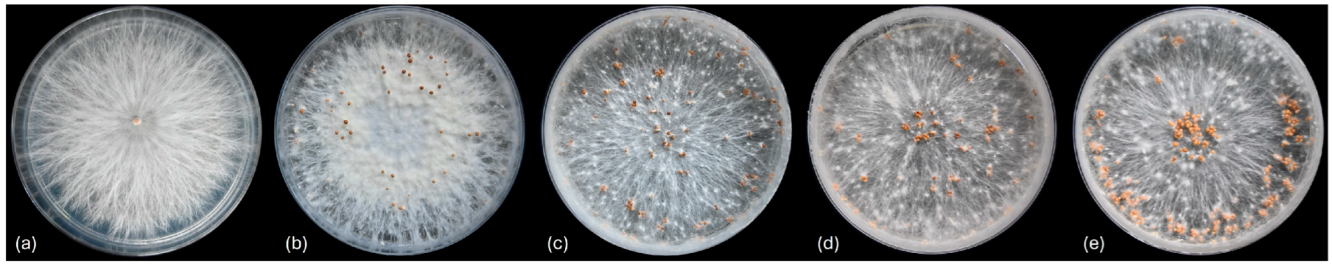
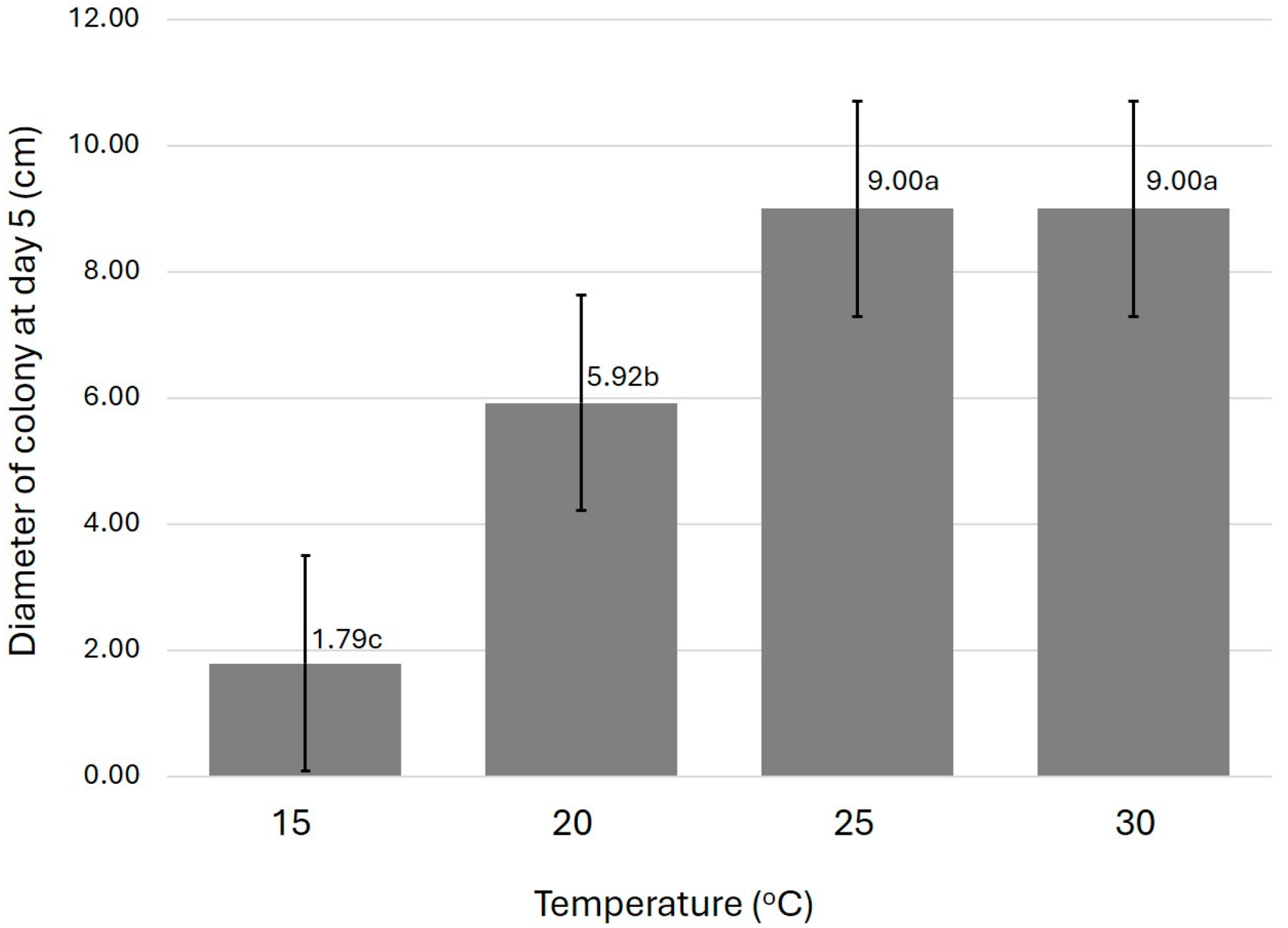
3.2. Morphology Characteristics

The fungi isolated from stem rot symptoms in the three provinces were grown and maintained on PDA medium. After 3 days of incubation, colony sizes ranging from 6.20 to 6.98 cm were observed (Figure 2). The fastest-growing isolate was BOS-NPT003 from Nakhon Pathom, with a colony diameter of 6.98 cm; the slowest-growing isolate, BOS-AYA004 from Phra Nakhon Si Ayutthaya, had the smallest colony, measuring 6.20 cm. All isolates exhibited similar colony characteristics, with white, velvety mycelium spreading flat over the surface and uneven colony edges (Figure 1e). No spores were produced, but resting structures (sclerotia) were formed, initially white (Figure 1f), and later turning brown to yellowish-brown (Figure 1g). These structures were smooth, round to oval with rounded ends, and measured approximately 0.90–3.16 × 1.02–3.27 µm (Figure 1g). The isolate BOS-NPT003 from Nakhon Pathom produced the most resting structures on the culture medium (Level 5) (Figure 3). Under a compound microscope, smooth-walled, hyaline mycelium, hyphae with septa, and clamp connections were observed (Figure 1h). Based on morphological comparisons following the descriptions of Barnett and Hunter [48], the 10 fungi were identified as the genus Sclerotium (Table S1).
Moreover, the effect of temperature on fungal growth was investigated at four different temperatures, 15, 20, 25, and 30 °C. The result indicated that all isolates grew well at 25 °C and above, exhibiting white mycelium, velvety colonies spreading flat over the surface, with uneven colony edges (average colony diameter = 9.00 cm; n = 10) on day 5 after incubation. At 20 °C, the fungal mycelium reached approximately 4.90–6.80 cm, with isolate BOS-AYA002 from Phra Nakhon Si Ayutthaya showing the smallest diameter. Nevertheless, at 15 °C, hyphal growth was limited to about 1.28–2.23 cm, with isolate BOS-AYA004 from Phra Nakhon Si Ayutthaya also exhibiting the smallest diameter (Table S2; Figure 4).

3.3. Molecular Identification and Phylogenetic Analyses

Nucleotide sequences of ITS1-5.8S-ITS2 and LSU gene were analyzed using the Nucleotide: Basic Local Alignment Search Tool https://blast.ncbi.nlm.nih.gov/Blast.cgi (accessed on 20 August 2024) to compare related species in a public database, available at the GenBank National Center for Biotechnology Information (NCBI). The results indicated that all isolates were identified as Agroathelia delphinii with an identity range of 98.85 and 100%. Sequences of ex-type or epitype strains of related genera (Table 1) were selected for phylogenetic analysis. The resulting phylogenetic tree of related genera, based on maximum likelihood (ML) using the GTR+G+I model tree for the concatenated data set of ITS1-5.8S-ITS2, and LSU gene sequences, is shown in Figure 5.
According to the analysis, the ten isolates were classified into one group: Agroathelia delphinii (Figure 5). They were closely related to the A. delphinii strains (CBS221.46 and CBS272.30), showing 2.200 posterior probability (PP) and 92% bootstrap (BS). In contrast, they were separated from the fungi in the related Athelia rolfsii, which included strains CBS 115.22, CBS132553, and CBS 191.62, with a posterior probability (PP) of 13.389 and a bootstrap (BS) value of 96%. Additionally, Agroathelia delphinii strains CBS 305.32 could not be distinctly separated from the two genera, resulting in a posterior probability (PP) of 1.750 and a bootstrap (BS) value of 91%. However, all isolates were separated from the fungus Agroathelia coffeicola CBS115.19 at 100% bootstrap (BS) and 18.815 posterior probability (PP).
While the morphological characteristics of the 10 fungal isolates provided initial identification at the genus level, nucleotide sequence analysis of the ITS1-5.8S-ITS2 and LSU regions were applied to differentiate them from related fungal genera with similar morphology. Notably, Athelia neuhoffii strain CBS124594, Athelia arachnoidea strain CBS 418.72, and CBS105.18 and Athelia decipiens strain CBS103869 exhibited 6.625 posterior probability (PP) and 100% bootstrap (BS). Moreover, Sclerotium perniciosum strain CBS 275.93, S. glucanicum strain CBS 520.71, S. wakkeri strain CBS 386.63 revealed 72% bootstrap (BS) and 4.095 posterior probability (PP), respectively. The sequence data obtained in this study are now available in the NCBI database (Table 1).

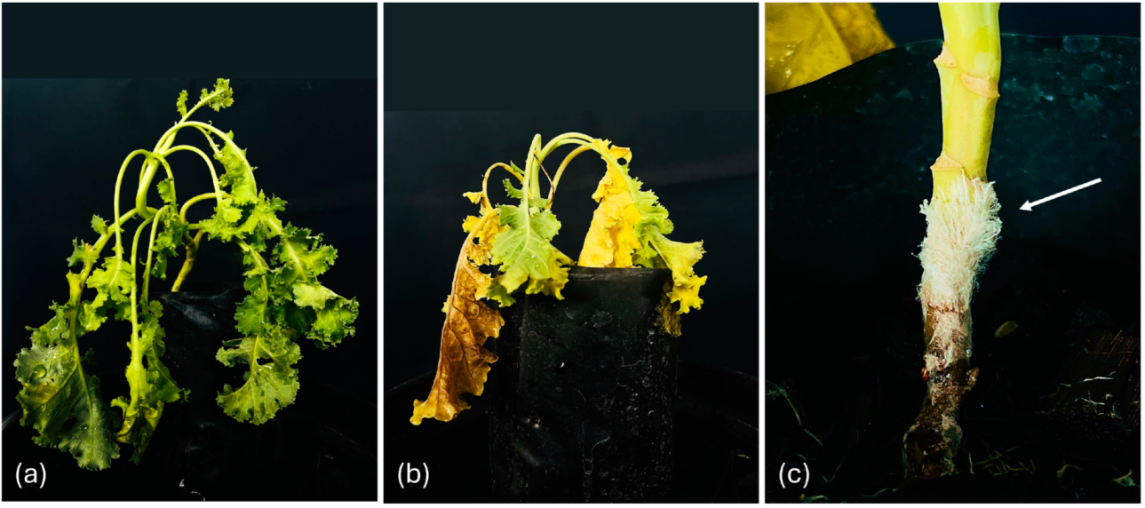
3.4. Pathogenicity Test

A mycelial plug of a 5-day-old culture was inoculated onto unwounded stems. Rot lesions exhibiting brown to brown-yellowish coloration were observed around the inoculation sites 5 days after inoculation (Table 2 and Figure 6). No disease symptoms were detected on the control kale (Figure 5). Among the inoculated plants, Agroathelia delphinii isolate BOS-NPT002 from Nakhon Pathom province produced the largest lesions, with an average lesion diameter of 8.02 cm and 100% disease incidence. This was followed by isolate BOS-NPT001 from the same area, which had an average lesion diameter of 7.38 cm2 and 100% disease incidence, while isolate BOS-SPB001 and BOS-SPB002 from Suphan Buri province exhibited an average lesion diameter of 5.62 and 5.00 cm2 and 100% disease incidence, respectively. The fungal isolates from Phra Nakhon Si Ayutthaya showed average lesion diameters of 4.47, 5.79, and 5.55 cm2, respectively, with 100% disease incidence.
After the pathogenicity test, all inoculated plants were re-isolated from the infected tissues on PDA medium, confirming that they exhibited identical morphological characteristics to the original isolates according to Koch’s postulates.

4. Discussion

In modern plant production, achieving high quality and standards that align with consumer preferences often involves the successful implementation of greenhouse systems, particularly for popular vegetables like kale [49,50]. To meet consumer demand, new technologies have been invented, including efficiency-enhancing substances that stimulate nutrient uptake, advancement in agricultural practices, improved fertilization techniques, and especially the adoption of soilless systems such as hydroponics and soil substitutes. These approaches help mitigate the risks of heavy metal residues and other contaminants, both biological and chemical [50]. Furthermore, maintaining environmental balances is critical when cultivating plants in greenhouse systems, especially in soilless cultivation. The primary objective is to create conditions that are conducive and adequate for plant growth. However, this method may also inadvertently create an environment that favors pathogen development.
Currently, it is acknowledged that diseases affecting kale are understudied, and the identification of phytopathogenic agents remains inconclusive. This poses a significant challenge in managing plant diseases and pathogens, particularly in relation to the abnormalities observed in kale cultivated in soilless systems within the greenhouse in the central region of Thailand. This region has seen an increase in the cultivation of vegetables, including kale, in greenhouses and soilless systems [51,52]. Furthermore, Thailand has high agricultural production, resulting in significant waste from the processes, such as rice straw, rice husks, coconut husks, and others, which can serve as alternatives for soilless growing materials. However, the acquisition of agricultural waste materials may not involve sufficient sterilizing processes which could lead to pest residues [53,54,55,56,57,58].
Our studies revealed that kale from soilless cultivation systems had a 60% incidence of stem rot with brown lesions, particularly in Phra Nakhon Si Ayutthaya, Nakhon Pathom, and Suphan Buri provinces, where rice straw and coconut husks from various sources were used as soil replacement materials. Initially, we investigated the pathogens that formed mycelium and sclerotium from the soil substitutes. To confirm the pathogenicity according to Koch’s postulates, we isolated the pathogens from kale exhibiting stem rot symptoms and examined their morphological and molecular characteristics by sequencing the ITS1-5.8S-ITS2 and LSU gene regions, as described by several previous researchers [24,25,59,60,61,62,63,64,65]. In this study, the fungus was isolated from infected kale grown in a soilless system, identified as Agroathelia delphinii, and its pathogenicity was confirmed according to Koch’s postulates, indicating that these pathogens are indeed pathogenic to kale.
Agroathelia delphinii is one of the genera which underwent re-classification in the year 2016 by Song et al. [66]. According to the Index Fungorum [66] and Mycobank database [67], the previously recorded name was Sclerotium delphinii Welch [66]. The first report of Sclerotium delphinii was published in 1924, entitled “A sclerotial disease of cultivated Delphinium” [66]. In 2023, research by Redhead and Mullineux [68] further elaborated on the differentiation between Agroathelia and related genera, building on the work of Song et al. [66]. Species of Agroathelia produced brown to brownish sclerotia that vary in size due to hyphal strand-type ontogeny. Under a compound microscope, the sclerotia were observed to have four distinct zones and were slightly stalked during vegetative growth. The outer surface of the sclerotia is polygonal, exhibiting various shapes and colors, ranging from brownish to yellowish, on which clamp connections also observed [22,66,68].
Agroathelia delphinii has been reported as a causal agent of disease in vegetable and ornamental plants, including agronomic crops [69,70,71,72]. The epidemiology of this fungus is primarily limited to subtropical regions, such as northern China and midwestern United States and various other countries [69]. Disease symptoms are mainly associated with southern blight and stem rot, characterized by mycelium developing at the base of the plant stem, followed by the appearance of sclerotia 7 to 10 days after inoculation [73]. In some strains, needle-like propagules are produced on the leaves in addition to sclerotia [22,59,69].
This research is the first to report that Agroathelia delphinii can infect kale in Thailand. Our study utilized soilless cultivation systems to rule out other soil-borne root rot pathogens. The data show that Agroathelia delphinii can persist in these systems and may be resistant to environmental conditions in a temperate region like Thailand. In support of this, our investigation into culturing the fungus at four different temperatures indicated that its optimum temperature, from this study, is at 25 °C and above (Figure 4). Potential sources of the pathogen could include contaminated seeds produced using rice straw, which may be contaminated with Agroathelia delphinii, to cover the plant beds, as well as contaminated water and infected tools and equipment. To minimize the risk of fungal pathogens in soilless systems, practices such as sterilizing equipment and media, filtering water, maintaining proper system hygiene, and using disease-resistant cultivars can be effective. Regular monitoring and early detection of fungal symptoms also help in managing outbreaks. The findings provide valuable insights that will contribute not only to the development of research but also to the advancement of kale production processes, ensuring they are effective and meet the consumer demand. This includes exploring effective disease management strategies in greenhouse and soilless cultivation systems, which are becoming increasingly important due to significant changes in weather and environmental conditions.

5. Conclusions

In this study, Agroathelia delphinii was isolated from stem rot of kale from three provinces. Koch’s postulates were performed using soilless cultivation systems to rule out other potential root rot pathogens, as well as to support growers using soilless systems, which have become popular in Thailand. The identification of the pathogen was conducted using a combination of multilocus sequence analysis (MLSA), morphological characteristics, and Koch’s postulates, confirming that the causal pathogen is Agroathelia delphinii. This study represents the first report showing that Agroathelia delphinii causes stem rot in kale in Thailand. These findings contribute not only to the research community but also to risk assessments of soilless cultivation systems, highlighting the significance of contaminated fungal pathogens on materials used to produce seeds, such as rice straw, which is known to be an alternative host for Agroathelia.

Supplementary Materials

The following supporting information can be downloaded at: https://www.mdpi.com/article/10.3390/horticulturae11040411/s1, Table S1: Morphological characteristics of Agroathelia delphinii 10 isolates on PDA incubated at 27 °C under a 12 h light/12 h dark photoperiod for 3 days and pathogenicity test on 2-month-old Kale plant after inoculation at day 7. Table S2: Diameter of Agroathelia delphinii 10 isolates on PDA incubated at 4 different temperatures. Supplementary Data S1: The formulas for calculating disease incidence and disease severity.

Author Contributions

Conceptualization, S.B., S.K. and C.R.; methodology, S.B., S.K. and T.T.; software, S.B.; validation, S.B., R.P., P.B. and C.R.; formal analysis, S.B.; investigation, S.K. and T.T.; resources, S.B., R.P., S.K., T.T. and C.R.; writing—original draft preparation, S.B., R.P., P.B., V.P. and C.R.; writing—review and editing, S.B., R.P., T.T., S.K., P.B., V.P. and C.R.; project administration, S.B. and C.R.; funding acquisition, S.B. All authors have read and agreed to the published version of the manuscript.

Funding

This research was funded by the Research and Development Institute of Rajamangala University of Technology Suvarnabhumi, Phra Nakhon Si Ayutthaya, Thailand (Frontier Research) [Grant No. R66202009] (to S.B.).

Data Availability Statement

The original contributions presented in this study are included in the article/Supplementary Material. Further inquiries can be directed to the corresponding author.

Acknowledgments

We thank Liesa Pötschke of Institute of Applied Microbiology, RWTH Aachen University, Germany for critical reading of the manuscripts.

Conflicts of Interest

The authors declare no conflicts of interest.

References

  1. Avato, P.; Argentieri, M.P. Brassicaceae: A rich source of health improving phytochemicals. Phytochem. Rev. 2015, 14, 1019–1033. [Google Scholar] [CrossRef]
  2. Zhou, R.; Qin, X.; Hou, J.; Liu, Y. Research progress on Brassicaceae plants: A bibliometrics analysis. Front. Plant Sci. 2024, 15, 1285050. [Google Scholar] [CrossRef]
  3. Mehmood, M.A.; Zhao, H.; Cheng, J.; Xie, J.; Jiang, D.; Fu, Y. Sclerotia of a phytopathogenic fungus restrict microbial diversity and improve soil health by suppressing other pathogens and enriching beneficial microorganisms. J. Environ. Manag. 2020, 259, 109857. [Google Scholar] [CrossRef]
  4. Bais, H.P.; Weir, T.L.; Perry, L.G.; Gilroy, S.; Vivanco, J.M. The role of root exudates in rhizosphere interactions with plants and other organisms. Annu. Rev. Plant Biol. 2006, 57, 233–266. [Google Scholar] [CrossRef]
  5. Cook, G.E. Survival of Whetzelinia sclerotiorum and initial infection of dry edible beans in Western Nebraska. Phytopathology 1975, 65, 250. [Google Scholar] [CrossRef]
  6. Hall, N.M.; Griffiths, H.; Corlett, J.A.; Jones, H.G.; Lynn, J.; King, G.J. Relationships between water-use traits and photosynthesis in Brassica oleracea resolved by quantitative genetic analysis. Plant Breed. 2005, 124, 557–564. [Google Scholar] [CrossRef]
  7. Dodd, I.C.; Puértolas, J.; Huber, K.; Pérez-Pérez, J.G.; Wright, H.R.; Blackwell, M.S.A. The importance of soil drying and re-wetting in crop phytohormonal and nutritional responses to deficit irrigation. J. Exp. Bot. 2015, 66, 2239–2252. [Google Scholar] [CrossRef]
  8. Barickman, T.C.; Ku, K.M.; Sams, C.E. Differing precision irrigation thresholds for Kale (Brassica oleracea L. var. Acephala) induces changes in physiological performance, metabolites, and yield. Environ. Exp. Bot. 2020, 180, 104253. [Google Scholar] [CrossRef]
  9. Dukes, M.D.; Zotarelli, L.; Morgan, K.T. Use of irrigation technologies for vegetable crops in Florida. Horttechnology 2010, 20, 133–142. [Google Scholar] [CrossRef]
  10. Frezza, D.; León, A.; Logegaray, V.; Chiesa, A.; Desimone, M.; Diaz, L. Soilless culture technology for high quality lettuce. Acta Hortic. 2005, 697, 43–48. [Google Scholar] [CrossRef]
  11. Fussy, A.; Papenbrock, J. An Overview of soil and soilless cultivation techniques—Chances, challenges and the neglected question of Sustainability. Plants 2022, 11, 1153. [Google Scholar] [CrossRef] [PubMed]
  12. Gonnella, M.; Renna, M.; Fernández, J.A.; San Bautista, A. The evolution of soilless systems towards ecological sustainability in the perspective of a circular economy. Is it really the opposite of organic agriculture? Agronomy 2021, 11, 950. [Google Scholar] [CrossRef]
  13. Savvas, D.; Gruda, N. Application of soilless culture technologies in the modern greenhouse industry. Eur. J. Hortic. Sci. 2018, 83, 280–293. [Google Scholar] [CrossRef]
  14. Van Os, E.A. Disease management in soilless culture systems. Acta Hortic. 2010, 883, 385–394. [Google Scholar] [CrossRef]
  15. Ferreira Queiroz, M.; Araujo Gonçalves Lima, M.; Edinalva Pereira, J.; Alves Barroso, K.; Domingos Paz, C.D.; Maria Luchese, A.; Rosa Peixoto, A. Essential oils in the management of soft rot of kale in the Brazilian Semiarid Region. Biosci. J. 2020, 36, 143–155. [Google Scholar] [CrossRef]
  16. Kale|Diseases and Pests, Description, Uses, Propagation. Available online: https://plantvillage.psu.edu/topics/kale/infos (accessed on 2 September 2024).
  17. Kalayanamitra, P.; Kalayanamitra, K.; Nontajak, S.; Taylor, P.W.J.; Jonglaekha, N.; Bussaban, B. Identification, characterization, and control of black spot on chinese kale caused by Sphaerobolus Cuprophilus sp. nov. Plants 2023, 12, 480. [Google Scholar] [CrossRef]
  18. Nuñez, A.M.P.; Rodríguez, G.A.A.; Monteiro, F.P.; Faria, A.F.; Silva, J.C.P.; Monteiro, A.C.A.; Carvalho, C.V.; Gomes, L.A.A.; Souza, R.M.; De Souza, J.T.; et al. Bio-based products control black rot (Xanthomonas campestris pv. campestris) and increase the nutraceutical and antioxidant components in kale. Sci. Rep. 2018, 8, 10199. [Google Scholar] [CrossRef]
  19. Tode, H.J. Fungi Mecklenbvrgenses Selecti; Lemke: Lüneburg, Germany, 1790. [Google Scholar]
  20. Mahadevakumar, S.; Sarma, P.V.S.R.N.; Danteswari, C.; Joy, J.; Mahesh, M.; Mamathabhanu, L.S.; Santhosh, C.R.; Chandranayaka, S. First report of Athelia rolfsii (=Sclerotium rolfsii) associated with foot rot disease of Chrysanthemum morifolium in India. Plant Dis. 2023, 107, 2250. [Google Scholar] [CrossRef]
  21. Flores-Moctezuma, H.E.; Montes-Belmont, R.; Jiménez-Pérez, A.; Nava-Juárez, R. Pathogenic Diversity of Sclerotium rolfsii isolates from Mexico, and potential control of Southern blight through solarization and organic amendments. Crop Prot. 2006, 25, 195–201. [Google Scholar] [CrossRef]
  22. Fery, R.L.; Dukes, P.D. Southern blight (Sclerotium rolfsii Sacc.) of cowpea: Yield-loss estimates and sources of resistance. Crop Prot. 2002, 21, 403–408. [Google Scholar] [CrossRef]
  23. Blum, L.E.B.; Rodríguez-Kábana, R. Effect of organic amendments on sclerotial germination, mycelial growth, and Sclerotium rolfsii-induced diseases. Fitopatol. Bras. 2004, 29, 66–74. [Google Scholar] [CrossRef]
  24. Akem, C.N. First report of Southern blight caused by Sclerotium rolfsii on soybeans in Nigeria. Plant Dis. 1991, 75, 537D. [Google Scholar] [CrossRef]
  25. Paparu, P.; Acur, A.; Kato, F.; Acam, C.; Nakibuule, J.; Nkuboye, A.; Musoke, S.; Mukankusi, C. Morphological and pathogenic characterization of Sclerotium rolfsii, the causal agent of Southern blight disease on common bean in Uganda. Plant Dis. 2020, 104, 2130–2137. [Google Scholar] [CrossRef]
  26. Rehner, S.A.; Buckley, E.; White, T.J. First report of Agroathelia rolfsii causing Southern blight on cowpea in China. Plant Dis. 2005, 97, 225. [Google Scholar] [CrossRef]
  27. Urbina, H.; Jones, C.; Moore, M.R.; Hansen, J. Southern blight of water lily: The first host record of Agroathelia rolfsii on Nelumbo nucifera discovered in Florida, U.S.A. Plant Dis. 2024, 108, 1404. [Google Scholar] [CrossRef] [PubMed]
  28. Changtor, P.; Rodriguez-Mateos, P.; Buddhachat, K.; Wattanachaiyingcharoen, W.; Iles, A.; Kerdphon, S.; Yimtragool, N.; Pamme, N. Integration of IFAST-based nucleic acid extraction and LAMP for on-chip rapid detection of Agroathelia rolfsii in Soil. Biosens. Bioelectron. 2024, 250, 116051. [Google Scholar] [CrossRef]
  29. Bengtsson-Palme, J.; Ryberg, M.; Hartmann, M.; Branco, S.; Wang, Z.; Godhe, A.; De Wit, P.; Sánchez-García, M.; Ebersberger, I.; de Sousa, F.; et al. Improved software detection and extraction of ITS1 and ITS2 from ribosomal ITS sequences of fungi and other eukaryotes for analysis of environmental sequencing data. Methods Ecol. Evol. 2013, 4, 914–919. [Google Scholar] [CrossRef]
  30. Sulistyo, B.P.; Larsson, K.H.; Haelewaters, D.; Ryberg, M. Multigene phylogeny and taxonomic revision of Atheliales s.l.: Reinstatement of three Families and one new Family, Lobuliciaceae fam. nov. Fungal Biol. 2021, 125, 239–255. [Google Scholar] [CrossRef]
  31. Paul, S.K.; Gupta, D.R.; Mahapatra, C.K.; Rani, K.; Islam, T. Morpho-molecular, cultural, and pathological characterization of Athelia rolfsii causing Southern blight disease on common bean. Heliyon 2023, 9, e16136. [Google Scholar] [CrossRef]
  32. Wang, A.R.; Lin, W.W.; Chen, X.T.; Lu, G.D.; Zhou, J.; Wang, Z.H. Isolation, and identification of Sclerotinia stem rot causal pathogen in Arabidopsis thaliana. J. Zhejiang Univ. Sci. B 2008, 9, 818. [Google Scholar] [CrossRef]
  33. Lydeard, C.; Holznagel, W.E.; Schnare, M.N.; Gutell, R.R. Phylogenetic analysis of Molluscan Mitochondrial LSU rDNA sequences and secondary structures. Mol. Phylogenet. Evol. 2000, 15, 83–102. [Google Scholar] [CrossRef] [PubMed]
  34. Woese, C.R.; Kandler, O.; Wheelis, M.L. Towards a natural system of organisms: Proposal for the domains archaea, bacteria, and eucarya. Proc. Natl. Acad. Sci. USA 1990, 87, 4576–4579. [Google Scholar] [CrossRef]
  35. Rattanakreetakul, C.; Keawmanee, P.; Bincader, S.; Mongkolporn, O.; Phuntumart, V.; Chiba, S.; Pongpisutta, R. Two newly identified Colletotrichum species associated with mango anthracnose in Central Thailand. Plants 2023, 12, 1130. [Google Scholar] [CrossRef]
  36. Watanabe, T. Pictorial atlas of soil and seed fungi. In Morphologies of Cultured Fungi and Key to Species, 2nd ed.; CRC Press: Washington, DC, USA, 2002; 500p. [Google Scholar]
  37. White, T.J.; Bruns, T.; Lee, S.J.W.T.; Taylor, J. Amplification and direct sequencing of fungal ribosomal RNA genes for phylogenetics. In PCR Protocols: A Guide to Methods and Applications; Innis, M.A., Gelfand, D.H., Sninsky, J.J., White, T.J., Eds.; Academic Press: Cambridge, MA, USA, 1990; pp. 315–322. [Google Scholar]
  38. Vilgalys, R.; Hester, M. Rapid genetic identification, and mapping of enzymatically amplified ribosomal DNA from several Cryptococcus species. J. Bacteriol. 1990, 172, 4238–4246. [Google Scholar] [CrossRef] [PubMed]
  39. Thompson, J.D.; Higgins, D.G.; Gibson, T.J. Clustal W: Improving the sensitivity of progressive multiple sequence alignment through sequence weighting, position-specific gap penalties and weight matrix choice. Nucleic Acids Res. 1994, 22, 4673–4680. [Google Scholar] [CrossRef]
  40. Kumar, S.; Stecher, G.; Li, M.; Knyaz, C.; Tamura, K. MEGA X: Molecular evolutionary genetics analysis across computing platforms. Mol. Biol. Evol. 2018, 35, 1547–1549. [Google Scholar] [CrossRef] [PubMed]
  41. Ronquist, F.; Teslenko, M.; Van Der Mark, P.; Ayres, D.L.; Darling, A.; Höhna, S.; Larget, B.; Liu, L.; Suchard, M.A.; Huelsenbeck, J.P. MrBayes 3.2: Efficient bayesian phylogenetic inference and model choice across a large model space. Syst. Biol. 2012, 61, 539–542. [Google Scholar] [CrossRef]
  42. Darriba, D.; Taboada, G.L.; Doallo, R.; Posada, D. JModelTest 2: More models, new heuristics, and parallel computing. Nat. Methods 2012, 9, 772. [Google Scholar] [CrossRef]
  43. Drummond, A.J.; Ho, S.Y.W.; Phillips, M.J.; Rambaut, A. Relaxed phylogenetics and dating with confidence. PLoS Biol. 2006, 4, e88. [Google Scholar] [CrossRef]
  44. Abdelghany, M.M.A.; Kurikawa, M.; Watanabe, M.; Matsui, H.; Yamamoto, M.; Ichinose, Y.; Toyoda, K.; Kouzai, Y.; Noutoshi, Y. Surveillance of pathogenicity of Rhizoctonia solani Japanese isolates with varied anastomosis groups and subgroups on Arabidopsis thaliana. Life 2022, 12, 76. [Google Scholar] [CrossRef]
  45. Anderson, N.A. The genetics and pathology of Rhizoctonia solani. Annu. Rev. Phytopathol. 1982, 20, 329–347. [Google Scholar] [CrossRef]
  46. R Core Team. R: The R Project for Statistical Computing. Available online: https://www.r-project.org/ (accessed on 11 September 2024).
  47. Agricolae: Statistical Procedures for Agricultural Research. Agricolae: Statistical Procedures for Agricultural Research. Available online: https://myaseen208.com/agricolae//index.html (accessed on 11 September 2024).
  48. Barnett, H.L.; Hunter, B.B. Illustrated Genera of Imperfect Fungi, 4th ed.; Macmillan Publishing Co.: New York, NY, USA, 1972; 241p. [Google Scholar]
  49. Asgharipour, M.R.; Amiri, Z.; Campbell, D.E. Evaluation of the sustainability of four greenhouse vegetable production ecosystems based on an analysis of emergy and social characteristics. Ecol. Modell. 2020, 424, 109021. [Google Scholar] [CrossRef]
  50. Fuentes-Peñailillo, F.; Gutter, K.; Vega, R.; Silva, G.C. New generation sustainable technologies for soilless vegetable production. Horticulturae 2024, 10, 49. [Google Scholar] [CrossRef]
  51. Tzortzakis, N.; Nicola, S.; Savvas, D.; Voogt, W. Editorial: Soilless cultivation through an intensive crop production scheme. Management strategies, challenges, and future directions. Front. Plant Sci. 2020, 11, 363. [Google Scholar] [CrossRef]
  52. Transporting Vegetables from Thailand: Maintaining Quality and Freshness. Available online: https://sse.co.th/transporting-vegetables/?srsltid=AfmBOooRC_f2fmQ0Q7Mbw_CaioigpPiMKMVJeR1R03TRMT1B0riIAo3F (accessed on 19 September 2024).
  53. Thailand—Agriculture. Available online: https://www.trade.gov/country-commercial-guides/thailand-agriculture (accessed on 19 September 2024).
  54. Nizamani, M.M.; Hughes, A.C.; Zhang, H.L.; Wang, Y. Revolutionizing agriculture with nanotechnology: Innovative approaches in fungal disease management and plant health monitoring. Sci. Total Environ. 2024, 928, 172473. [Google Scholar] [CrossRef] [PubMed]
  55. Wang, H. Epidemiology, and control of fungal diseases in crop plants. Agronomy 2023, 13, 2327. [Google Scholar] [CrossRef]
  56. Li, Z. Assessing potential soil pollution from plant waste disposal: A modeling analysis of pesticide contamination. Sci. Total Environ. 2024, 907, 167859. [Google Scholar] [CrossRef]
  57. Riseh, R.S.; Vazvani, M.G.; Hassanisaadi, M.; Thakur, V.K. Agricultural wastes: A practical and potential source for the isolation and preparation of cellulose and application in agriculture and different industries. Ind. Crops Prod. 2024, 208, 117904. [Google Scholar] [CrossRef]
  58. Garvey, M.; Meade, E.; Rowan, N.J. Effectiveness of front line and emerging fungal disease prevention and control interventions and opportunities to address appropriate eco-sustainable solutions. Sci. Total Environ. 2022, 851, 158284. [Google Scholar] [CrossRef]
  59. Mwangi, R.W.; Mustafa, M.; Charles, K.; Wagara, I.W.; Kappel, N. Selected emerging and reemerging plant pathogens affecting the food basket: A threat to food security. J. Agric. Food Res. 2023, 14, 100827. [Google Scholar] [CrossRef]
  60. Mahadevakumar, S.; Yadav, V.; Tejaswini, G.S.; Janardhana, G.R. Morphological and molecular characterization of Sclerotium rolfsii associated with fruit rot of Cucurbita maxima. Eur. J. Plant Pathol. 2016, 145, 215–219. [Google Scholar] [CrossRef]
  61. Hernández-Morales, J.; Ochoa-Martínez, D.L.; Ayala-Escobar, V.; Ortega-Acosta, S.Á. First report of Southern blight caused by Sclerotium rolfsii on sesame in Mexico. Plant Pathol. J. 2018, 100, 323. [Google Scholar] [CrossRef]
  62. Okabe, I.; Matsumoto, N. Phylogenetic relationship of Sclerotium rolfsii (Teleomorph Athelia rolfsii) and S. delphinii based on ITS sequences. Mycol. Res. 2003, 107, 164–168. [Google Scholar] [CrossRef] [PubMed]
  63. Okabe, I. Variation in Sclerotium rolfsii isolates in Japan. Mycoscience 1998, 39, 399–407. [Google Scholar] [CrossRef]
  64. Garibaldi, A.; Gilardi, G.; Gullino, M.L. First report of Southern blight incited by Sclerotium rolfsii on potato (Solanum tuberosum) in Northern Italy. Plant Dis. 2006, 90, 1114. [Google Scholar] [CrossRef]
  65. Sun, S.; Sun, F.; Deng, D.; Zhu, X.; Duan, C.; Zhu, Z. First report of Southern blight of mung bean caused by Sclerotium rolfsii in China. Crop Prot. 2020, 130, 105055. [Google Scholar] [CrossRef]
  66. Song, J.; Liu, X.Y.; Wang, M.; Cui, B.K. Phylogeny, and taxonomy of the Genus Anomoloma (Amylocorticiales, Basidiomycota). Mycol. Prog. 2016, 15, 11. [Google Scholar] [CrossRef]
  67. Index Fungorum—Names Record. Available online: https://www.indexfungorum.org/Names/namesrecord.asp?RecordID=251780 (accessed on 19 September 2024).
  68. Sclerotium delphinii. Available online: https://www.mycobank.org/page/Name%20details%20page/field/Mycobank%20%23/251780 (accessed on 19 September 2024).
  69. Scott, A. Redhead & Sahra-Taylor Mullineux 2023: Nomenclatural novelties. Index Fungorum 550: 1.—Biota of NZ. Available online: https://biotanz.landcareresearch.co.nz/references/6d322bd2-05d3-4ed5-b0d5-f0b73c8d3e32 (accessed on 19 September 2024).
  70. Chen, J.; Cong, L.; Zhou, R.; Li, Z.; Piao, J.; Hao, N. Identification, and characterization of Sclerotium delphinii causing Southern blight on Aconitum kusnezoffii in Northeast China. Plant Dis. 2022, 106, 2031–2038. [Google Scholar] [CrossRef]
  71. Severo, R.; Shibutani, L.J.S.; Sousa, E.S.; Matos, K.S.; Beserra, J.E.A.; de Melo, M.P. Sclerotium delphinii causing concentric leaf spots in Piper nigrum in Brazil. Austral. Plant Pathol. 2021, 50, 661–670. [Google Scholar] [CrossRef]
  72. Javed, Z.U.R.; Coley-Smith, J.R. Studies on germination of Sclerotia of Sclerotium delphinii. Trans. Br. Mycol. Soc. 1973, 60, 441-IN6. [Google Scholar] [CrossRef]
  73. Obee, D.J. Studies on the host range of Sclerotium delphinii Welch. Trans. Kans. Acad. Sci. 1937, 40, 89. [Google Scholar] [CrossRef]
Figure 1. Symptom of stem rot on Brassica oleracea var. sabellica (a); mycelial structure on stem (b); sclerotia (c,d); colony characteristic on PDA after 7 days of incubation (e); sclerotia development on agar (f,g); and clamp connection under compound microscope at 40× (h).
Horticulturae 11 00411 g001
Figure 2. Bar chart showing the colony diameter of the ten Agroathelia delphinii isolates used in this study. The colonies were incubated at 27 °C after purification under a 12 h light/12 h dark photoperiod for 3 days (during which fungal hyphae showed the best growth differentiation). Vertical lines represent standard errors. Means followed by the different common letters are significantly different according to the Duncan Multiple Range Test using R software version 4.2.3.
Horticulturae 11 00411 g002
Figure 3. Level of sclerotia production on PDA medium. Level 1 = no sclerotia (a); Level 2 = 0.1–15% of sclerotia production (b); Level 3 = 16–30% of sclerotia production (c); Level 4 = 31–45% of sclerotia production (d); and Level 5 > 45% of sclerotia production (e).
Horticulturae 11 00411 g003
Figure 4. Box plots showing the variation in average colony diameter at four temperatures associated with Agroathelia development 5 days after inoculation on PDA medium. Vertical lines represent standard errors. Means followed by the different common letters are significantly different according to the multicomp test using R software version 4.2.3.
Horticulturae 11 00411 g004
Figure 5. The maximum likelihood (GTR+G+I model) tree for the concatenated data set of ITS1-5.8S-ITS2, and LSU gene sequences from a total of fungal isolates with the ex-type/epitype were retrieved from GenBank database. Numbers on the node are bootstrap values (left) and posterior probability (right). Agroathelia isolates from this study are in bold. The scale bar shows the number of substitutions per site. Phytophthora citrophthora isolate CBS 581.69 was used as an outgroup. Evolutionary analyses were conducted using MEGA version X.
Horticulturae 11 00411 g005
Figure 6. Pathogenicity assay of Agroathelia delphinii on 2-month-old kale plants: control (a), visual symptoms on kale at 5 days after inoculation (b), and fungal mycelial development on stem (c). The test was performed on all isolates; only a representative of each species is shown here.
Horticulturae 11 00411 g006
Table 1. Collection details and GenBank accession numbers of all isolates used in this study for phylogenetic analysis.
Species NameCulture
Collection No. 1
Host/Substrate 2OriginAccession Number
ITS-RegionLSU
Agroathelia coffeicolaCBS 115.19N/ASurinameMH854677MH866193
Agroathelia delphiniiBOS-AYA001B. oleracea var. sabellicaThailandLC835020LC835030
BOS-AYA002B. oleracea var. sabellicaThailandLC835021LC835031
BOS-AYA003B. oleracea var. sabellicaThailandLC835022LC835032
BOS-AYA004B. oleracea var. sabellicaThailandLC835019LC835029
BOS-NPT001B. oleracea var. sabellicaThailandLC835015LC835025
BOS-NPT002B. oleracea var. sabellicaThailandLC835016LC835026
BOS-NPT003B. oleracea var. sabellicaThailandLC835017LC835027
BOS-NPT004B. oleracea var. sabellicaThailandLC835018LC835028
BOS-SPB001B. oleracea var. sabellicaThailandLC835013LC835023
BOS-SPB002B. oleracea var. sabellicaThailandLC835014LC835024
CBS 221.46Ranunculus sp.The NetherlandsMH856168-
CBS 272.30Iris germanicaCanadaMH855140MH866588
CBS 305.32Delphinium sp.USANR_189755MH866785
Athelia rolfsiiCBS 115.22Oryza sativa L.USAMH854711-
CBS 132553Vigna unguiculataLaosJX566993-
CBS 191.62Ficus repensItalyMH858139MH869724
CBS 305.32Delphinium sp.USANR_189755MH866785
Athelia arachnoideaCBS 418.72Populus sp.The NetherlandsMH860510-
CBS 105.18N/AGermanyMH854664MH866181
Athelia decipiensCBS 103869Picea sp.FinlandKY025593-
Athelia neuhoffiiCBS 463.72Angiosperm woodThe NetherlandsMH860532-
Rhizoctonia solaniCBS 124594Coprosma repensItalyMH863394-
Rhizoctonia fragariaeCBS 335.62Fragaria x ananassaCanadaMH858171MH869763
Rhizoctonia carotaeCBS 464.48Daucus carotaUSAMH856434MH867980
Rhizoctonia globurisCBS 262.60N/ACanadaMH857978MH869533
Rhizoctonia callaeCBS 310.35Calla aethiopicaItalyMH855686MH867201
Sclerotium cacticolaCBS 304.32Opuntia sp.The NetherlandsMH855330-
Sclerotium costantiniiCBS 288.38N/AFranceMH855965MH867461
Sclerotium glucanicumCBS 520.71N/AUSAMH860245-
Sclerotium hydrophilumCBS 385.63Submerged leaf in garden pondItalyFJ212350-
Sclerotium perniciosumCBS 275.93Tulipa sp.The NetherlandsMH862400FJ212355
Sclerotium tuliparumCBS 206.25N/AUSAMH854847MH866346
Sclerotium wakkeriCBS 386.63Tulipa cv. CarraraThe NetherlandsMH858312-
Stromatinia cepivoraCBS 276.93Allium sp.The NetherlandsMH862401MH874059
CBS 402.85N/AThe NetherlandsMH861892-
CBS 342.47N/AThe NetherlandsMH856279FJ212344
Phytophthora citrophthoraCBS 581.69Hevea brasiliensisMalaysiaMH401211-
1 CBS is Westerdijk Fungal Biodiversity Institute-KNAW, Utrecht, The Netherlands. 2 N/A = not available. Isolates obtained in this study are indicated in bold.
Table 2. Lesion sizes on kale (Brassica oleracea var. sabellica) after inoculation with Agroathelia delphinii at day 7.
IsolateDisease Incident (%)Disease Severity (cm2) 1
BOS-AYA0010.000.00 ± 0.00 e
BOS-AYA002100.004.47 ± 0.03 d
BOS-AYA003100.005.79 ± 0.78 b
BOS-AYA004100.005.55 ± 0.50 bc
BOS-NPT001100.007.38 ± 0.41 a
BOS-NPT002100.008.02 ± 0.41 a
BOS-NPT003100.004.67 ± 0.55 c
BOS-NPT004100.004.62 ± 0.53 d
BOS-SPB001100.005.62 ± 0.57 b
BOS-SPB002100.005.00 ± 0.66 bc
C.V. (%) 4.9705
F-test ***
MSE 3.9975
1 Values with the same column followed by different common letters mean that they are significantly different based on variance with the least significant difference test at p = 0.05. Pathogenicity was evaluated with 10 replicates per isolate (n = 10). *** represents the F-test value.
Disclaimer/Publisher’s Note: The statements, opinions and data contained in all publications are solely those of the individual author(s) and contributor(s) and not of MDPI and/or the editor(s). MDPI and/or the editor(s) disclaim responsibility for any injury to people or property resulting from any ideas, methods, instructions or products referred to in the content.

