Article

In Vitro Multiplication, Antioxidant Activity, and Phytochemical Profiling of Wild and In Vitro-Cultured Plants of Kaempferia larsenii Sirirugsa—A Rare Plant Species in Thailand

by
Surapon Saensouk
1,2,
Phiphat Sonthongphithak
2,
Theeraphan Chumroenphat
3,
Nooduan Muangsan
4,
Phetlasy Souladeth
5 and
Piyaporn Saensouk
2,6,*
1
Walai Rukhavej Botanical Research Institute, Mahasarakham University, Maha Sarakham 44150, Thailand
2
Diversity of Family Zingiberaceae and Vascular Plant for Its Applications Research Unit, Mahasarakham University, Maha Sarakham 44150, Thailand
3
Aesthetic Sciences and Health Program, Faculty of Thai Traditional and Alternative Medicine, Ubon Ratchathani Rajabhat University, Ubon Ratchathani 34000, Thailand
4
School of Biology, Institute of Science, Suranaree University of Technology, Nakhon Ratchasima 30000, Thailand
5
Faculty of Forest Science, National University of Laos, Vientiane 7322, Laos
6
Department of Biology, Faculty of Science, Mahasarakham University, Maha Sarakham 44150, Thailand
*
Author to whom correspondence should be addressed.
Horticulturae 2025, 11(3), 281; https://doi.org/10.3390/horticulturae11030281
Submission received: 6 February 2025 / Revised: 24 February 2025 / Accepted: 25 February 2025 / Published: 5 March 2025
(This article belongs to the Section Propagation and Seeds)

Abstract

:
Kaempferia larsenii Sirirugsa, a rare species in Thailand belonging to the Zingiberaceae family, is known for its effective pharmaceutical properties. However, its slow natural growth and threats from overharvesting and habitat encroachment pose significant challenges. This study investigated the effects of plant growth regulators (PGRs) on the plant regeneration, transplantation success, phytochemical profiling, and antioxidant properties of wild and in vitro-cultured plants. Plantlets (~1 cm long) were cultivated for 8 weeks in different types of MS media (solid, liquid, and liquid-over-solid) combined with various PGRs (BA, kinetin, TDZ, NAA, and IAA). Solid MS medium enriched with 2 mg/L BA, 3 mg/L TDZ, and 0.2 mg/L NAA produced the highest number of shoots (13.10 shoots/explant). By comparison, liquid MS medium containing 1 mg/L BA and 0.5 mg/L IAA also promoted high shoot production (4.70 shoots/explant). The strongest root induction (16.90 roots/explant) was achieved using a liquid MS medium supplemented with 2 mg/L BA, 2 mg/L kinetin, and 1 mg/L NAA. Sandy soil as a planting material yielded the highest survival rate (100%) during transplantation. The total phenolic content (TPC) and total flavonoid content (TFC) were higher in mother plants than in in vitro-cultured plants. The addition of PGRs significantly enhanced the production of secondary metabolites. The leaves of K. larsenii exhibited superior antioxidant properties compared to other organs under both growing conditions. Cinnamic acid was identified as abundant in in vitro-cultured plants via HPLC analysis. FTIR analysis revealed functional groups associated with phenolic acids and flavonoids, which are useful for phytochemical screening and antioxidant evaluation. This research highlighted the potential of biotechnology as a crucial strategy for conserving K. larsenii and demonstrated its sustainable application in the medical and cosmetics industries.

1. Introduction

Zingiberaceae, widely referred to as the ginger family, is the largest family in the order Zingiberales, with extensive dispersion across the tropics and subtropics, especially in Southeast Asia [1]. Worldwide, the family comprises approximately 1900 species across 57 genera [2,3,4]. In Thailand, more than 400 species covering 28 genera have been reported [3]. The Kaempferia genus, one of the most recognized in the ginger family, has been utilized extensively in foods, functional drinks, traditional medicine, and horticulture [5,6,7]. Currently, the Kaempferia genus has 63 accepted species listed by Plants of the World Online (POWO) [4]. Thailand is regarded as one of the centers of distribution for this genus, with 46 native species, 34 of which are endemic [2,8,9,10].
Kaempferia larsenii Sirirugsa was discovered by Sirirugsa P. [9]. This species is distributed in northeastern Thailand (Ubon Ratchathani and Amnat Charoen provinces) and Southern Laos (Champasak Province). It is a rare and vulnerable plant species. This plant is used as food and in traditional medicine, with its rhizomes applied topically to relieve inflammation caused by insect bites [6,11]. Presently, K. larsenii is declining in its natural habitat due to overharvesting for uses such as food, as well as habitat encroachment from intensive agriculture, farming, and road construction [11]. The direct vegetative propagation of this plant species relies on rhizome division, which is slow-growing and produces only one growth cycle per year, posing challenges for large-scale cultivation [12,13]. Conservation efforts are critical to ensure the survival of this species in its natural habitat. Plant tissue culture techniques offer a potential solution to these challenges by enabling rapid propagation, year-round growth, and the production of disease-free plants, making them an appealing option for scaling up production. Previous studies on the in vitro multiplication of the Kaempferia genus have reported the effects of plant growth regulators (PGRs), including auxins and cytokinins, in enhancing shoot propagation and root induction. Research on K. angustifolia Roscoe [14], K. galanga L. [15,16,17], K. grandifolia Saensouk & Jenjitikul [18], K. koratensis Picheans [19], K. larsenii [20], K. marginata Carey ex Roscoe [21], K. parviflora Wall. ex Baker [22,23], K. rotunda L. [17,24], K. siamensis Sirirugsa [25], and K. sisaketensis Picheans. & Koonterm [26] indicated that altering the level of these PGRs had a significant impact on the quality of shoot and root regenerations. The contained phytochemicals, antioxidant activity, and antibacterial activity of K. larsenii have been previously reported [27,28,29]. This plant shows potential for application in various herb products.
This research investigated the effects of PGRs under different conditions and combinations on the multiplication of K. larsenii, including shoot regeneration, root production, and transplantation. The impact of adding PGRs to the growing medium with varying concentrations of bioactive compounds was also examined. The phytochemical profiles and antioxidant properties of both wild and in vitro-cultured plants were compared for the first time. This research conservation study also explored the potential use of K. larsenii in medicine, cosmetics, and other industries.

2. Materials and Methods

2.1. Tissue Culture Regeneration

Plant Material Collection: The starting plant materials were collected from Ubon Ratchathani Province, Thailand, for use as explants in shoot and root induction investigations.
Plant Preparation and Surface Decontamination: Rhizomes of K. larsenii were collected from wild habitats and cleaned in running tap water for 1 h. The rhizomes were then disinfected by spraying with ethanol (70% v/v) for 30 s, followed by sterilization using sodium hypochlorite (NaOCl). The rhizomes were sterilized in NaOCl solution at concentrations of 20% and 15% for 20 and 15 min, respectively. Following sterilization with NaOCl solution, the rhizomes were thoroughly washed with sterilized distilled water three times for 5 min each time to remove any remaining disinfectant. The rhizome buds were then divided into 1 × 1 cm pieces for transfer to regeneration on the culture medium. To promote rapid regeneration of the plant materials, segments of the rhizome buds were cultured onto Murashige and Skoog (MS) medium [30]. The MS medium contained 6–benzylamino–purine (BA, Sigma–Aldrich, Budapest, Hungary) at a concentration of 2.0 mg/L, in combination with Thidiazuron (TDZ, Sigma–Aldrich, Munich, Germany) at a concentration of 4.0 mg/L. After being cultured on MS medium supplemented with BA and TDZ for four weeks per cycle over six cycles for shoot multiplication, the explants were transferred to MS medium without PGRs for eight weeks to minimize any residual effects before proceeding with further experiments. The new sprouts were cut to a length of 1 cm as explants for future experimental use.
Medium Culture and Tissue Culture Conditions: For in vitro multiplication, the explants were inoculated onto standard MS medium, first described by Murashige and Skoog in 1962. The standard MS medium was combined with 30 g/L of sucrose to contribute the carbon source for plant induction. For solid medium conditions, 7 g/L of bacto agar to solidify. Glass culture containers (120 mL) were used for cultivation in solid medium conditions. One sprout (1 cm long) was placed on the medium in the middle of each container, with 10 replications for each treatment. In liquid medium conditions, the standard liquid MS medium was not added with a solidifying agent. Similarly, 250 mL Erlenmeyer flasks were used for the liquid medium conditions. Three sprouts were inoculated into each flask, with five replications for each treatment. The acidity and alkalinity of the culture medium were prepared at 5.7–5.8 to maintain a suitable environment for plant tissue development. The process of culture decontamination was stabilized using an autoclave at 121 °C with 15 pounds per square inch (psi) for 15 min to confirm that all materials were decontaminated before in vitro cultivation. All tissue culture experiments were performed under monitored environmental conditions to provide optimal cultivation. The culture room was maintained at a constant temperature of 25 ± 2 °C and illuminated with fluorescent light (Philips TLD 36W/54-765 Cool Daylight, Bangkok, Thailand) intensity at 27 µmol s−1m−2 for 16 h per day to provide the energy required for photosynthesis, physiological, and other light-dependent processes.
Optimization of PGRs on In Vitro Propagation: To evaluate the optimal multiplication of K. larsenii under various culture conditions, sprouts were excised from the MS-medium-treated conditions. The sprouts were cultivated on solid MS medium supplemented with various types of PGRs, including cytokinins (BA, kinetin (Sigma–Aldrich, Buchs, Switzerland), and TDZ), auxins (1–naphthaleneacetic acid (NAA, Sd Fine–Chem Limited, Mumbai, India)), and indole–3–acetic acid (IAA, Sigma–Aldrich, Buchs, Switzerland) at varying concentrations and combinations. The culture medium formulation for cultivated explants consisted of MS medium supplemented with BA or kinetin at concentrations of 1 to 6 mg/L in combination with NAA (0.1 and 0.5 mg/L). MS medium containing varying concentrations of TDZ (0.1, 0.2, 1, 2, 3, 4, or 5 mg/L) was also combined with IAA (0.5 mg/L). The culture medium formulations included BA (1, 2, or 3 mg/L) and NAA (0.2 mg/L), with kinetin (2 or 4 mg/L) or TDZ (1 or 3 mg/L) (Table 1).
To study shoot and root regeneration of K. larsenii under liquid medium conditions, the liquid medium was supplemented with BA at concentrations of 1, 2, and 3 mg/L, combined with kinetin (2 and 4 mg/L), IAA (0.5 mg/L), and NAA (1 mg/L). The liquid culture conditions were maintained at a rotating speed of 120 rpm on an orbital shaker (Table 1).

2.2. Effect of Explant Division by Cutting and Culturing on Different Medium Types for Plant Regeneration

To study plant regeneration, the explants (approximately 1 cm in size) were divided by cutting them into longitudinal sections before culturing on different types of culture media, including liquid medium, solid medium, and liquid-over-solid medium. Each medium type was supplemented with TDZ (1 mg/L) and IAA (0.5 mg/L) (Table 1).

2.3. Transplantation and Acclimatization

To study the transplantation of in vitro-cultured plants onto different plant materials, plantlets with well-developed structures after 8 weeks of cultivation on MS medium were subjected to an acclimatization phase at room temperature for 2 weeks. This process assisted their adaptation to the ex vitro environment. The plantlets (length 5–6 cm) were taken out of the MS medium without PGRs and the roots were carefully cleaned with running tap water to remove any agar or residual particles. After cleaning, the plantlets were transferred into pots containing various growth substrates, including loamy soil, sandy soil, compost, burned rice husk, and a 1:1 (w/w) mixture of each combination of loamy soil + sandy soil, loamy soil + compost, sandy soil + compost, and loamy soil + sandy soil + compost. The plants were maintained in a greenhouse located at the Department of Biology, Faculty of Science, Mahasarakham University, Thailand, and the plantlets underwent an 8-week acclimatization period. During this period, environmental conditions were closely monitored, with regular watering with tap water to ensure proper hydration. Throughout the acclimatization process, the growth parameters were evaluated to assess the plantlets’ performance and adaptation to the new conditions for regeneration. The growth parameters included the survival rate of the plantlets, the mean number of shoots produced per plant, the mean plant shoot height per plant (measured in cm), and the average number of leaves per plant. Detailed monitoring provides insights into plantlet development and adaptation.

2.4. Phytochemical Profiling and Antioxidant Activity of K. larsenii

Plant Material: Wild plants were collected from Ubon Ratchathani Province, Thailand, in July 2020. The plant specimens were identified and authenticated by Associate Professor Dr. Surapon Saensouk at the Walai Rukhavej Botanical Research Institute, Mahasarakham University, Thailand. Voucher specimens were deposited in the herbarium at the Department of Biology, Faculty of Science, Mahasarakham University, Thailand. Specimens of wild plants collected for this study included various parts, such as leaves, pseudostems, rhizomes, and storage roots. These were collected alongside in vitro-cultured plants from two different sources, including those grown on MS medium alone and MS medium supplemented with BA (2 mg/mL) and NAA (0.1 mg/L).
Plant Preparation and Extraction: All samples from wild plants and in vitro-cultured plants were washed with running tap water followed by distilled water. The cleaned samples were divided into different parts (leaves, pseudostems, rhizomes, and roots) and dried by a freeze-drying method for 24–48 h. The dried samples were ground into a fine powder using an electric blender and stored at −20 °C. The sample extraction process was modified from the methods of Chumroenphat et al. [31] and Siriamornpun and Kaewseejan [32]. A 1 g aliquot of powdered samples was mixed with 10 mL of 80% ethanol, and the mixture was incubated at 37 °C for 15 h with shaking at 150 rpm to extract the phytochemicals. After maceration, the ethanolic extract was filtered using filter paper (Whatman® No. 1). The filtered ethanolic extract was collected and stored at −20 °C for use in the phytochemical and antioxidant activity analyses.
Determination of Total Phenolic Content (TPC): The total phenolic content of the samples was evaluated using the Folin–Ciocalteu assay, as described by Al–Duais et al. [33] and Kubola and Siriamornpun [34] with modifications. Briefly, the ethanolic extract (20 µL) was added to a 96-well plate containing 100 µL of 20% (v/v) Folin–Ciocalteu’s reagent (Sigma–Aldrich, Munich, Germany). The mixture was shaken and incubated for 5 min, 75 µL of 10% (w/v) sodium carbonate (Na2CO3) solution was added, and the mixture was kept in dark condition at room temperature for 2 h. Following the reaction period, the absorbance of the mixture was determined using a UV–Vis microplate reader (Varioskan™ LUX, Thermo Fisher Scientific Inc., Marsiling, Singapore) at 750 nm. The total phenolic content was calculated from a gallic acid (Merck KGaA, Beijing, China) calibration curve and expressed as milligrams of gallic acid equivalent per gram of dry weight (mg GAE/g DW). The gallic acid standard solutions were prepared at concentrations of 1.95 to 1000 mg/L.
Determination of Total Flavonoid Content (TFC): The total flavonoid content of the ethanolic extract was determined by the aluminum chloride colorimetric method, as described by Jia et al. [35], Abu Bakar et al. [36], and Kubola and Siriamornpun [34] with some modifications. In brief, 25 µL of ethanolic extract was mixed with 100 µL of deionized water, and 10 µL of 5% (w/v) sodium nitrite (NaNO2) solution was added onto a 96-well plate. The mixture was incubated for 5 min, and 15 µL of 10% (w/v) aluminum chloride hexahydrate (AlCl3·6H2O) solution was added. After a 6-minute incubation, the mixture was added to 50 µL of 1 M sodium hydroxide (NaOH), followed by 50 µL of deionized water. The absorbance was measured at 510 nm by a UV–Vis microplate reader, with results expressed as milligrams of rutin equivalent per gram of dried weight (mg RE/g DW). The rutin standard (Sigma–Aldrich, Beijing, China) solutions were prepared at concentrations of 1.95 to 1000 mg/L.
Evaluation of Antioxidant Activity: The antioxidant activity was analyzed using the 2,2–diphenyl–1–picrylhydrazyl (DPPH, Alfa Aesar, Thermo Fisher Scientific, Waltham, MA, USA) assay to determine the free radical scavenging activity of the samples, according to Rivero–Péreze et al. [37] and Kubola and Siriamornpun [34] with slight modifications. Firstly, 180 µL of 0.15 M DPPH solution was pipetted into a 96-well plate, followed by the addition of ethanolic extract (20 µL), and mixed homogeneously. The mixture was then incubated in dark condition at room temperature for 30 min. The absorption spectra of the samples were measured at 517 nm using a UV–Vis microplate reader (Varioskan™ LUX, Thermo Fisher Scientific Inc., Marsiling, Singapore) with scavenging calculated from the calibration curve of the Trolox standard (Sigma–Aldrich, Munich, Germany) at concentrations of 1.95–1000 mg/L. The results of the free radical scavenging were expressed as milligrams of Trolox equivalent per gram dry weight of samples (mg TE/g DW). To calculate the percentage inhibition of the DPPH radical, the absorbance of the initial 0.15 M DPPH solution was compared with the absorbance of the DPPH solution after reacting with each sample using the following equation:
% inhibition = [(Ainitial)(Afinal)/(Ainitial)] × 100
where Ainitial is the absorbance of the control and Afinal is the absorbance of the DPPH solution and the ethanolic extract.
The ferric-reducing antioxidant power (FRAP) assay was also used to determine the antioxidant ability of the sample through a redox-linked colorimetric reaction in which Fe3+ is reduced to Fe2+ [38]. The protocol followed Pellegrini [39] and Kubola and Siriamornpun [34] with some modifications. For this assay, 180 µL of FRAP reagent (Fe3+–TPTZ complex) was pipetted into a 96-well plate containing 5 µL of the ethanolic extract and mixed for 1 min, followed by incubation at 37 °C for 15 min. Next, an aliquot was measured for absorbance using a UV–Vis microplate reader (Varioskan™ LUX, Thermo Fisher Scientific Inc., Marsiling, Singapore) at 593 nm. Ferrous sulfate (FeSO4) served as the standard, and the antioxidant properties of the samples were reported as milligrams of ferrous sulfate equivalent per gram of plant dry weight (mg FeSO4/g DW).

2.5. Quantitative HPLC Analysis of Phenolic Compounds and Flavonoid Compounds of Kaempferia larsenii

High-performance liquid chromatography (HPLC) was performed to analyze the phytochemicals in the samples, including phenolic acid and flavonoids. The phenolic acid standards included gallic acid, protocatechuic acid, p-hydroxybenzoic acid, chlorogenic acid, caffeic acid, p-coumaric acid, ferulic acid, sinapic acid, and cinnamic acid. The flavonoid standards included rutin, quercetin, myricetin, and apigenin. All reference standards used were of analytical reagent (AR) grade and purchased from Sigma–Aldrich.
Plant Extraction: The extracts from different plant parts of Kaempferia larsenii including the leaves, pseudostems, rhizomes, and storage roots (or roots) were prepared from both wild plants and in vitro-cultured plants grown on MS medium alone and MS medium with PGRs. To prepare the plant extracts, 0.2 g of fine powder from each sample was mixed with 10 mL of 1% (v/v) hydrochloric acid in methanol solution. The mixture was then incubated at 37 °C with shaking at 150 rpm for 15 h, filtered using filter paper, and passed through a 0.22 µm nylon membrane filter. This process followed Chumroenphat et al. [40].
HPLC Operating Conditions: The HPLC analysis was performed using a Shimadzu LC–20A series system (Shimadzu Corp., Kyoto, Japan). Chromatographic separation was achieved on a C18 column (4.6 mm × 250 mm, 5 µm; GL Sciences Inc., Tokyo, Japan) protected with a guard column. The mobile phase consisted of 0.1% (v/v) acetic acid (solvent A) and acetonitrile (solvent B) at a flow rate of 0.8 mL/min. Gradient elution was carried out as described by Siriamornpun and Kaewseejan [32]. The column temperature was maintained at 38 °C, with an injection volume of 20 µL. UV–diode array detection was performed at 280 nm for hydroxybenzoic acids, 320 nm for hydroxycinnamic acids, and 370 nm for flavonoids at a flow rate of 0.8 mL/min. Spectra were recorded over a wavelength range of 200–600 nm. Phenolic acid and flavonoids in the samples were identified by comparing their relative retention times (RTs) and UV spectra to the authentic standards and quantified using the external standard method.

2.6. FTIR Analysis of the Functional Groups from Different Parts of Kaempferia larsenii

The functional groups of organic compounds from Kaempferia larsenii were analyzed using a Fourier transform infrared spectrometer (FTIR) equipped with a UATR accessory (PerkinElmer, Waltham, MA, USA) following the method described by Chumroenphat et al. [40]. Dried powder samples from various plant parts were placed on a Diamond/KRS–crystal composite for spectrum screening. Spectral data were collected from 32 scans at a resolution of 4 cm−1, covering the spectral range of 4000–400 cm−1, with background subtraction performed automatically by the software.

2.7. Statistical Analysis

Plant Tissue Culture: The experimental studies were designed with a completely randomized design (CRD) to achieve reliable and impartial outcomes, and all experimental results were reported as the mean ± standard deviation (SD). To evaluate plant multiplications, parameters including the number of shoots, number of roots, shoot length, and root length were measured in centimeters for 10 replications. SPSS software (IBM Crop., New York, NY, USA, version 28) was used for the statistical analysis using one-way analysis of variance (ANOVA) and Duncan’s multiple range test (DMRT) with a significance level of p < 0.05
Phytochemical Profiling and Antioxidant Activity Analysis: All results were displayed as the mean ± standard deviation (SD) derived from three independent replicates. Data were analyzed using one-way ANOVA with DMRT for post hoc comparisons, and the significance level was set at p < 0.05 using SPSS software (version 28). Pearson’s correlation coefficient (r) was also determined to indicate the strength and direction of the correlations between variables, including TPC, TFC, DPPH, and FRAP values.

3. Results

3.1. In Vitro Propagation of Kaempferia larsenii

3.1.1. The Effect of PGR Combinations at Different Concentrations on Shoot and Root Induction of Kaempferia larsenii Cultured on Solid MS Medium

Sprouts of Kaempferia larsenii (1 cm long) were cultured on solid MS medium supplemented with BA and NAA at varying concentrations. After 8 weeks of cultivation, there was a significant increase in the number of shoots in the medium supplemented with PGRs. The number of shoots cultured on MS medium containing PGRs increased at higher concentrations of BA. The optimal combination of BA at 6 mg/L and NAA at 0.5 mg/L resulted in the highest number of shoots, with an average of 4.63 shoots/explant (Table 2 and Figure 1). The average shoot length was 2.52 cm, significantly less than the 3.72 cm attained on MS medium (control) (Table 2 and Figure 1). The plantlets were cultured on MS medium combined with kinetin and NAA at various concentrations for 8 weeks of cultivation. The results showed that the number of shoots and roots increased significantly on MS medium supplemented with PGRs compared to MS medium without PGRs. The highest average number of shoots was 4.10 per explant, with an average shoot length of 4.46 cm (Table 3 and Figure 2) on medium mixed with kinetin at 6 mg/L and NAA at 0.1 mg/L. On MS medium without PGRs, the average number of shoots was 2.57, with an average shoot length of 3.85 cm (Table 3 and Figure 2). The effect of auxins (BA and kinetin) on shoot multiplication indicated that, as the concentration of hormones increased, the number of shoots also increased. Higher concentrations of PGRs disrupted shoot length, as shown in Table 2 and Table 3. The influence of the NAA combination with BA and kinetin on plant regeneration showed that the root production was lower on a medium containing BA and NAA compared to a medium enriched with kinetin and NAA, as presented in Table 2 and Table 3. After 8 weeks of cultivation, the plantlets had a healthy morphology, similar to the mother plant. The leaves were elliptic-linear to elliptic or lanceolate, and the lower surface was glabrous to pilose, plain green to purple, or with purple veins as the characteristics of this plant [9].
Microshoots of K. larsenii were cultured on solid MS medium containing TDZ at 0–4 mg/L with IAA at 0.5 mg/L to evaluate plant regeneration. Growth parameters, including the number of shoots and number of roots, were measured after 8 weeks of culture (Table 4). The results demonstrated a significantly different number of shoots on MS medium without PGRs compared to MS medium containing PGRs. The highest shoot production was recorded in the medium supplemented with TDZ at 0.5 mg/L and IAA at 0.5 mg/L, with the average number of shoots 4.00 shoots/explant and 5.46 cm average length (Table 4 and Figure 3). This medium formulation produced healthy plant structures, such as fresh green leaves, fresh leaf sheaths, and strong roots. Those explants cultured on higher concentrations of TDZ at 1 to 4 mg/L showed decreased shoot production.
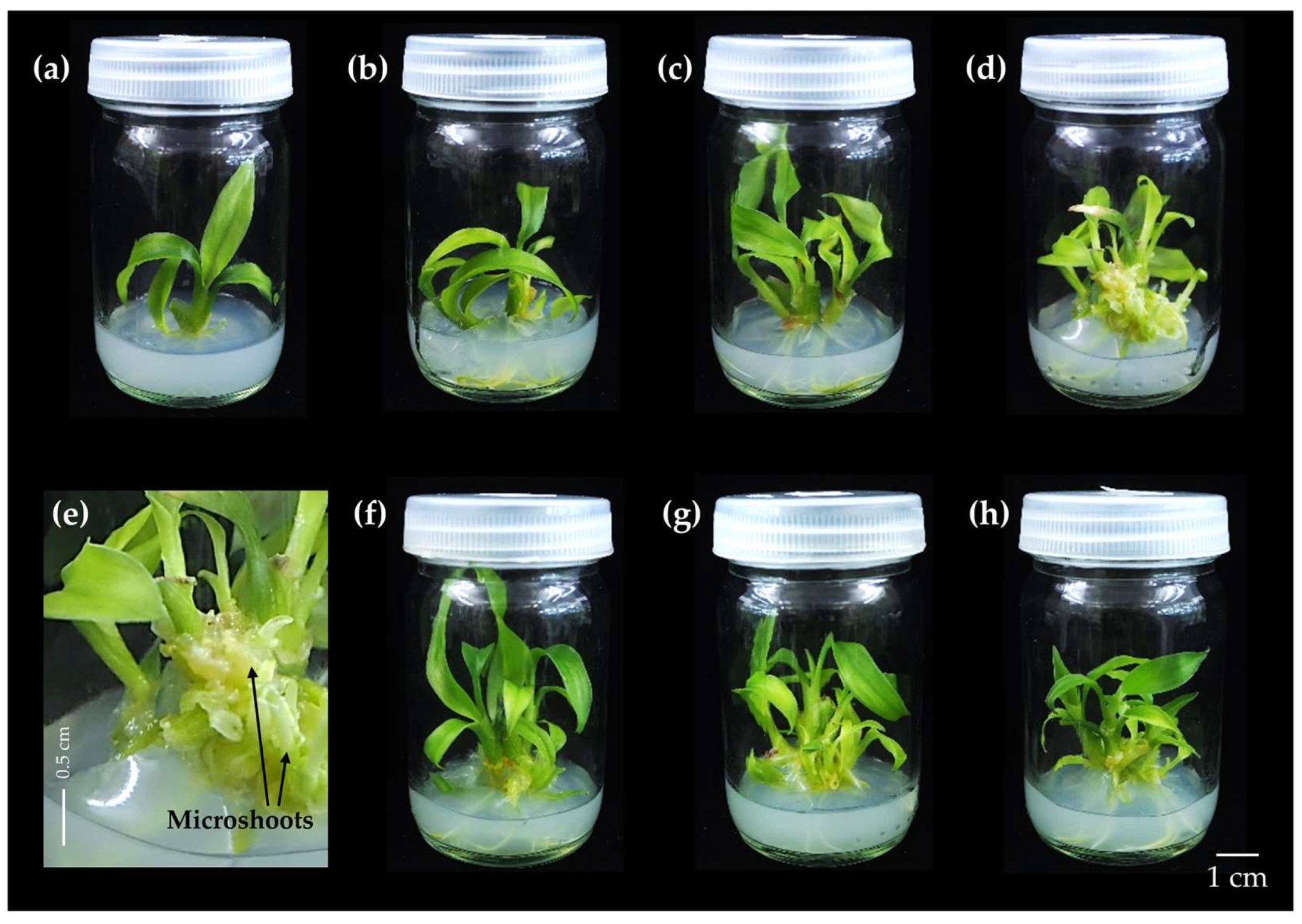
Adding combinations of PGRs, including BA, kinetin, and NAA, at different concentrations in the solid MS medium significantly increased the number of shoots and roots compared to the medium without PGRs. The highest average number of shoots was observed in explants cultured on MS medium supplemented with 3 mg/L BA, 2 mg/L kinetin, and 0.2 mg/L NAA, resulting in 11.00 shoots/explant and a shoot length of 3.76 cm (Table 5 and Figure 4). In the control treatment, the average number of shoots was 1.90 per explant, with an average of 8.30 roots/explant (Table 5 and Figure 4). The characteristics of plant regeneration were healthy with fresh green leaves and free of contamination. At concentrations of 3 mg/L BA, 2 mg/L kinetin, and 0.2 mg/L NAA on MS medium, numerous microshoots were produced (Figure 4e). Combinations of BA and kinetin cytokinins enhanced the shoot production at higher concentrations, with a decrease in the number of shoots and shoot length (Table 5 and Figure 4).
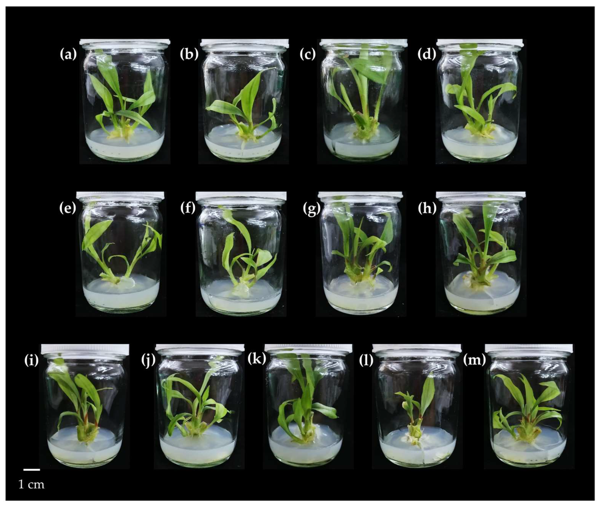
To determine the effect of combinations of BA, TDZ, and NAA at different concentrations on plant multiplication, the explants were transferred into a solid MS medium with PGRs and cultured for 8 weeks under controlled conditions. The results showed that the highest average number of shoots was observed in explants cultured with 2 mg/L BA, 3 mg/L TDZ, and 0.2 mg/L NAA, producing 13.10 shoots/explant with an average length of 1.97 cm (Table 6 and Figure 5). For explants cultured on the control medium, the average number of shoots was 1.80, the average shoot length was 3.34 cm, and there were 5.30 roots/explant. When comparing MS medium with and without PGRs, a significant increase in plant production was observed on the medium supplemented with plant hormones (Table 6 and Figure 5). The combination of BA and TDZ at higher concentrations resulted in decreases in both the number of shoots and shoot length, with small shoot tips observed around the newly formed pseudostems. The leaf characteristics of Kaempferia larsenii cultured under this treatment exhibited smaller and shorter leaves compared to the combination of BA and kinetin.

3.1.2. The Effect of PGR Combinations at Different Concentrations on Shoot and Root Induction of Kaempferia larsenii Cultured on Liquid MS Medium

The liquid medium was prepared to compare the type of medium substrate to the growth and development of Kaempferia larsenii. The plantlets were cultivated on a liquid medium in combinations of various PGRs, including BA, kinetin, IAA, and NAA, to evaluate optimal plant regeneration. Plantlets incubated in BA at 1 mg/L with 0.5 mg/L of IAA yielded 4.70 shoots/explant, the highest average shoot number for this treatment, with 11.33 cm average length (Table 7 and Figure 6). PGR-free liquid MS medium and liquid MS medium supplemented with PGRs showed no significant differences in shoot regeneration. The number of shoots/explant on standard liquid medium had an average shoot count of 4.00 and 8.93 cm shoot length. The combination of BA at 3 mg/L and kinetin at 4 mg/L with liquid medium yielded an 11.82 cm average shoot height, higher than the other formulations.

3.2. Effect of Explant Division by Cutting and Culturing on Different Medium Types for Plant Regeneration

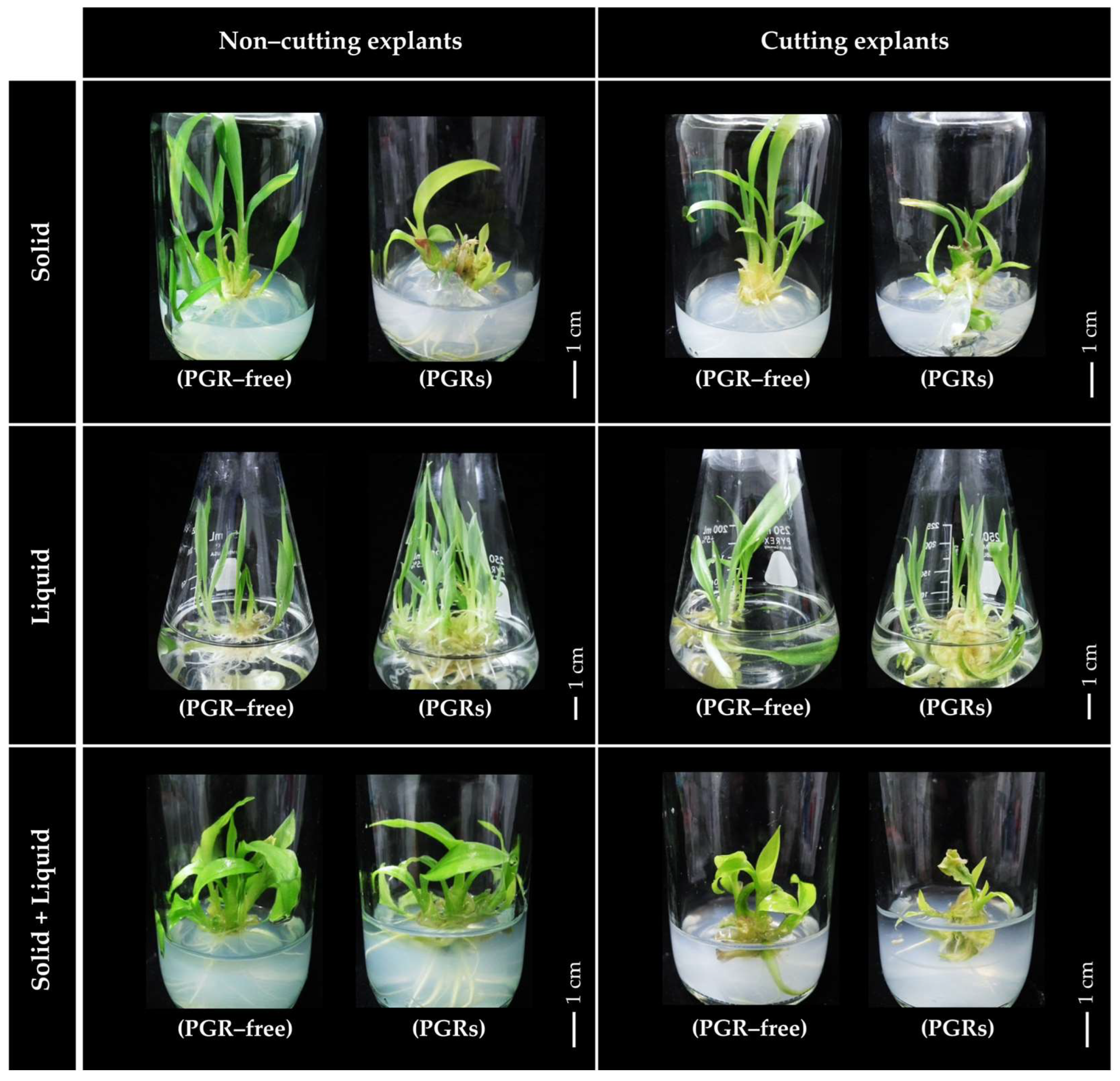
To study the effects of cutting into long sections on the production and development of Kaempferia larsenii, microshoots were incised into long sections and transferred to a culture medium containing 1 mg/L TDZ and 0.5 mg/L IAA. The samples were cultivated for 8 weeks. The addition of PGRs in various types of media, including solid, liquid, and liquid-over-solid, to simulate the natural habitat significantly increased the number of shoots. The highest average shoot production was observed in liquid medium, with an average of 9.60 shoots/explant (Table 8 and Figure 7). When comparing plantlets cut into long sections with non-cut plantlets cultured in solid media, the solid MS medium (PGR-free) with cut plantlets showed higher shoot production, with an average of 5.00 shoots per explant. By contrast, the non-cut plantlets exhibited an average of 3.10 shoots/explant. Comparing the cut and non-cut plantlets before culture in the simulated natural habitat medium and liquid medium showed that cutting into long sections decreased the shoot production, as shown in Table 8 and Figure 7.

3.3. Acclimatization and Transplantation of In Vitro-Cultured Kaempferia larsenii

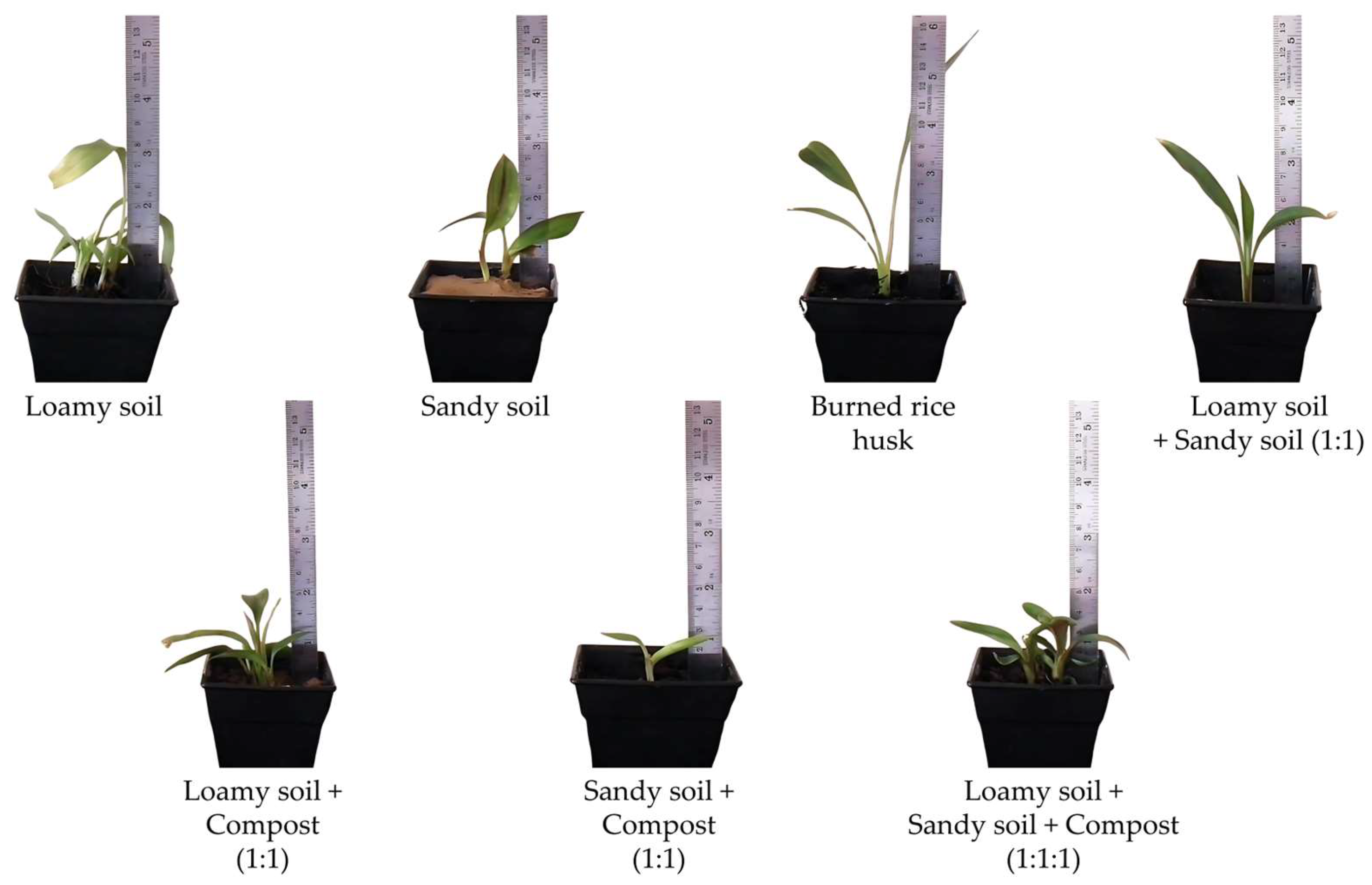
The in vitro-cultured PGR-free plantlets were acclimatized at room temperature for 2 weeks before the experiment. The selected plantlets, heights 5–6 cm, with similar morphology regarding number of shoots, number of roots, and freshness, were transferred to various plant materials, and plant development was observed after 8 weeks of transplantation. After the cultivation period, Kaempferia larsenii presented different shoot production and survival rates depending on the plant material types used. Sandy soil showed the highest survival rate of 100% (Table 9). The plants cultivated in sandy soil produced an average of 1.50 shoots/explant, with a shoot height of 6.70 cm and an average of 2.17 leaves/explant (Table 9 and Figure 8). The morphology of the in vitro-cultured plants and the mother plants of K. larsenii showed no significant differences, indicating that the in vitro-cultured plants retained similar morphological traits to those propagated through traditional methods (Table 9 and Figure 8).

3.4. Phytochemical Profiling and Antioxidant Activity of Kaempferia larsenii

3.4.1. Total Phenolic Content (TPC) and Total Flavonoid Content (TFC)

Phenolic and flavonoid compounds that belong to polyphenols are considered to contribute highly to the biological activity of plants. The TPC and TFC of ethanolic extracts from several parts of Kaempferia larsenii (cultured on MS medium, MS medium supplemented with PGRs, and the mother plant) were evaluated. The results showed that the leaves extracted from the mother plant contained higher phenolic content and flavonoid levels than those derived from in vitro cultures. The TFC values of the leaves from the mother plant, the medium supplemented with PGRs, and the control medium yielded 716.03, 192.84, and 157.88 mg GAE/g DW, respectively (Table 10). The TFC values of the leaves from the mother plant, the medium supplemented with PGRs, and the control medium yielded 47.89, 11.47, and 6.28 mg QE/g DW, respectively (Table 10). The leaves cultured on PGR-enriched medium exhibited higher TPC and TFC values than those cultivated on MS medium alone. The TFC in the root extract derived from PGR-containing medium was higher than in the rhizomes and storage roots from the mother plant, while the pseudostems of MS medium alone and MS medium enhanced with PGRs showed no significant differences in TPC and TFC values, as detailed in Table 10.

3.4.2. Antioxidant Activity

The potential antioxidant capacity of Kaempferia larsenii was measured using the free radical scavenging activity (DPPH assay) and the ferric-reducing antioxidant power (FRAP) assay. The antioxidant activity of various plant parts is detailed in Table 10. The results showed that the leaf extract from natural plants yielded the highest antioxidant activity by the DPPH assay (121.34 mg TE/g DW) or 59.04% inhibition with the FRAP assay (134.35 FeSO4/g DW). For the in vitro-cultured plants, the leaf extract grown on MS medium supplemented with PGRs showed the highest DPPH scavenging activity at 19.76 mg TE/g DW or 40.47% inhibition. The highest FRAP value was observed in roots grown on MS medium containing PGRs (17.15 mg FeSO4/g DW). Conversely, the lowest free radical scavenging and FRAP values were recorded in pseudostems of the PGR-free-medium plants, as detailed in Table 10. Comparing the DPPH and FRAP values showed that the extract antioxidant activity of several parts from the PGR-containing medium was significantly higher than the MS medium alone, indicating that adding PGRs increased antioxidant activity.
Pearson’s correlation coefficient (r) is a statistical measure of the linear association of two variables. This correlation coefficient was used to analyze the phytochemical content and antioxidant activity of Kaempferia larsenii. Strong positive correlations (r > 0.5) were observed between TPC and DPPH (0.996), TPC and FRAP (0.993), and DPPH and FRAP (0.998) with a 0.01 level of significance (Table 11).

3.5. Quantitative Analysis of Phenolic Acid and Flavonoid Compounds Determined by HPLC Analysis

The extracts from various parts of Kaempferia larsenii grown under different conditions were quantitatively evaluated for their phenolic acid and flavonoid contents by comparing the retention times of several standard phenolic acids and flavonoids from the HPLC chromatogram. HPLC is used to analyze many phytochemical compounds with a C18 column, as presented in Table 12 and Table 13. The results demonstrated that the natural plant extract parts contained gallic acid, protocatechuic acid, caffeic acid, p-coumaric acid, ferulic acid, and sinapic acid for phenolic acid compounds and rutin, quercetin, myricetin, and apigenin for flavonoid compounds. Sinapic acid was the most dominant phenolic acid in the parts of the natural plants, with the highest concentration in the leaf extract (47.21 µg/g DW) (Table 12). Rutin was the most abundant flavonoid in the leaves, while myricetin was dominant in the pseudostems and rhizomes of the natural plants. For the in vitro-cultured plants, the results revealed the presence of gallic acid, p-hydroxybenzoic acid, p-coumaric acid, ferulic acid, and cinnamic acid in the leaves and pseudostems. Rutin, quercetin, myricetin, and apigenin were detected in varying concentrations in different plant parts. The main phenolic acid compound in the in vitro-cultured plants was cinnamic acid, which was most abundant in the root extract from the MS medium (882.52 µg/g DW) (Table 12), while myricetin was highest in the leaves derived from MS medium (1798.64 µg/g DW) (Table 13). Chlorogenic acid was not detected in either the wild or in vitro-cultured plants. Protocatechuic acid and caffeic acid were present in the mother plants but absent in the in vitro-cultured plants. p-Hydroxybenzoic acid and cinnamic acid were observed in the in vitro-cultured plants but were absent in the mother plants, while rutin and quercetin were not detected in the in vitro-cultured plants. Our results highlighted the high contents of various phytochemical compounds, such as cinnamic acid, myricetin, and apigenin, in several parts of the in vitro-cultured plants, demonstrating their potential use in antioxidant, antimicrobial, and other applications. The types and concentrations of phytochemical compounds produced depend on the growth environment and the specific plant parts.

3.6. Functional Group Screening of Kaempferia larsenii Determined by FTIR Analysis

FTIR analysis was used to screen and identify the types of chemical bonds in the phytochemicals (organic compounds), such as phenolic compounds and flavonoids. These functional groups indicate the presence of bioactive compounds, which contribute to biological activities or antioxidant properties. Plants of K. larsenii grown under different conditions were divided into several parts and measured using UATR-mode FTIR from 4000–400 cm−1. The FTIR spectra of K. larsenii grown in their natural habitat, as illustrated in Figure 9a, showed that several plant parts presented peaks around 3600–3200 cm−1, indicating the vibration of O–H stretching (alcohols and phenols) [41,42]. The peak at 3000–2850 cm−1 was attributed to the C–H stretching vibration [41]. The absorption band around the 2000–1650 cm−1 range referred to the C=O stretching vibration of aldehyde [43]. The peak at 1310–1250 cm−1 revealed aromatic ester (C–O stretching vibration) [42]. Vibration peaks at 1318 cm−1 was observed in rhizome parts, indicating the aromatic amine groups (C–N stretching) [42]. The strong vibrations at 1250–1020 cm−1 were related to the alkyl aryl ether (C–O stretching bonds) [43,44] (data shown in Table 14). The FTIR spectra of the in vitro-cultured plants (Figure 9b) showed that several parts of the plant presented alcohol and phenol groups (O–H bonds), C–H stretching, aldehyde groups (C=O stretching), vibration of O–H bending (alcohol groups), and alkyl aryl ether groups (C–O stretching). The peak positions, types of vibration bonds, and types of functional groups of the in vitro-cultured plants are presented in Table 14. A comparison of the FTIR spectra of both the natural plants and in vitro-cultured plants with and without PGRs displayed similar spectra and peak positions, such as O–H stretching of phenol groups, C=O stretching of aldehyde groups, and C–O stretching of alkyl aryl ether, indicating similar phytochemical compositions of K. larsenii under different growth conditions. Certain peaks were observed exclusively in natural plants, such as those corresponding to aromatic amine and aromatic ether groups, while O–H bending of alcohol groups was found only in tissue-cultured plants.

4. Discussion

This research explored the effects of plant growth regulators on in vitro plant multiplication, contributing to the conservation of Kaempferia larsenii, a rare and vulnerable species of Thailand. The phytochemical composition and antioxidant activity were also assessed to evaluate the potential of this plant for future sustainable use. Plantlets of K. larsenii showed significant shoot production responses to different types and concentrations of PGRs under various culture media. Our findings demonstrated that those explants cultivated on a PGR-free medium showed the lowest shoot regeneration, with similar observations reported in other Kaempferia species, such as K. koratensis, an endemic plant of Thailand, where the explants cultured on MS medium showed minimal shoot production without any PGR supplementation [19]. Similarly, the explants of K. angustifolia, a medicinal plant, exhibited low shoot multiplication when grown on a solid MS medium without PGR supplementation [14]. The types and concentrations of PGRs, including auxins (IAA, NAA, and others) and cytokinins (BA, kinetin, TDZ, and others) supplemented in the culture medium, promoted shoot and root production, as previously reported in K. galanga [16,17], K. marginata [21], K. rotunda [17,24], and K. larsenii [20]. Auxin hormones play a vital role in plant development by regulating cell division, elongation, and differentiation. They are crucial for root development, organogenesis, vascular tissue formation, and apical dominance by supporting overall growth and adaptation [45,46,47]. Cytokinins, a group of phytohormones, are potent growth factors that are essential for cell growth and differentiation. They regulate various vital activities in plants, including cell division, meristem formation, photosynthesis, senescence, nutrient uptake (both macro- and micronutrients), and responses to biotic and abiotic stressors [48,49].
For the in vitro multiplication of Kaempferia larsenii cultivated under solid medium conditions, the medium enriched with BA (6 mg/L) and NAA (0.5 mg/L) resulted in the highest average shoot production, yielding 4.63 shoots/explant. These results differed from Pudpong et al. [20], who reported that MS medium supplemented with BA at concentrations of 0.5 to 5 mg/L increased shoot production in K. larsenii, with the highest shoot production observed on MS medium containing 4 mg/L BA, yielding 4.90 shoots/explant. In another study, the explants of K. koratensis cultured on MS medium supplemented with BA at 4 mg/L and NAA at 0.2 mg/L showed the highest shoot production, yielding 4.00 shoots/explant [19]. Rahman et al. [50] reported that K. angustifolia explants cultured on medium enriched with 3 mg/L BA and 0.5 mg/L NAA demonstrated the maximum shoot quantity (11.40 shoots), while explants of K. parviflora inoculated on MS medium in combination with BA at 8 mg/L and IAA at 0.8 mg/L showed the highest average number of shoots/explant (27.3 shoots), as reported by Koh et al. [51]. The combination of kinetin and NAA at different concentrations was compared with other cytokinin hormones. The results showed that the medium containing 6 mg/L kinetin with 0.1 mg/L NAA had the highest shoot production (4.10 shoots). This result differed from Pudpong et al. [20], who reported that K. larsenii cultured on MS medium treated with kinetin at a concentration of 3 mg/L demonstrated the maximum production of shoots (5.00 shoots/explant). Rajasshree and Reena [52] documented the highest average shoot multiplication (5.8 shoots) of K. parviflora cultured on MS medium supplemented with kinetin at 3 mg/L and NAA at 0.5 mg/L. When combining TDZ with IAA at various concentrations, the optimal results were observed in the medium with 0.5 mg/L TDZ and IAA at 0.5 mg/L, yielding 4.00 shoots/explant. This result differed from Pudpong et al. [20], who noted that K. larsenii inoculated in solid medium mixed with 1 mg/L TDZ achieved high shoot production. Park et al. [23] studied the in vitro propagation of K. parviflora cultivated on MS medium containing 2 µM (0.44 mg/L) of TDZ. They reported the highest shoot multiplication at 4.7 shoots/explant, similar to our study. When comparing BA, kinetin, and TDZ hormones for inducing shoot development, the results indicated that concentrations of BA and kinetin at more than 2 mg/L significantly increased the number of shoots, similar to previous studies in Kaempferia plants [15,19,20,50,51,52], while the TDZ hormone yielded optimal shoot production at low concentrations of 0.1 to 1 mg/L [14,20].
The combination of two cytokinin substances and auxins at different concentrations regarding plant regeneration showed that the combination of BA at 3 mg/L, kinetin 2 at mg/L, and NAA at 0.2 mg/L exhibited the highest number of shoots (11.00 shoots/explant), while the combination of BA at 2 mg/L, TDZ at 3 mg/L, and NAA at 0.2 mg/L presented the highest shoot production (13.10 shoots). The results indicated that the combination of two cytokinin substances enhanced the shoot production more efficiently than using a single hormone alone, aligning with Rajashree and Reena [52], who reported that sprouted buds of Kaempferia parviflora inoculated into solid MS medium in combination with BA and TDZ or kinetin and TDZ showed higher shoot production than using BA or kinetin alone. By contrast to previous results, studies on the micropropagation of K. larsenii cultured on MS medium containing a single cytokinin (BA, kinetin, or TDZ) demonstrated higher shoot regeneration compared to the results of this study. The addition of NAA or IAA (an auxin hormone) to various solid medium formulations for the regeneration of K. larsenii indicated that solid MS medium enriched with BA (2 mg/L), kinetin (4 mg/L), and NAA (0.2 mg/L) promoted extensive root production, yielding 14.70 roots per explant with an average root elongation of 3.21 cm. This observation concurred with previous studies that reported the use of auxin hormones in combination with various cytokinins. Auxin hormones at low concentrations (0.1 to 1 mg/L) enhanced shoot and root production, effectively supporting root development. Higher concentrations of auxins reduced shoot and root production, similar to the effects observed with high cytokinin concentrations in solid MS medium, which have been reported in various plants, including K. angustifolia, K. galanga, K. larsenii, and K. parviflora [14,20,23,53,54,55].
For in vitro multiplication of Kaempferia larsenii under liquid medium conditions, the explants were cultured in liquid medium supplemented with PGRs at different concentrations. The results showed that the highest shoot induction was achieved at an average of 4.70 shoots/explant and shoot length of 11.33 cm in liquid medium complemented with 1 mg/L BA and 0.5 mg/L IAA. When comparing liquid medium formulations for K. larsenii shoot and root induction, including cytokinins alone, combinations of cytokinins or cytokinins with auxins revealed no significant differences in shoot regeneration. The MS liquid medium containing PRGs at various combinations and concentrations showed a higher average shoot length compared to the solid MS plus PGR medium. This finding aligned with earlier studies on K. angustifolia [14], indicating that the liquid medium enhanced nutrient absorption through consistent exposure to the growth medium, supporting superior growth compared to solid media, which restricted nutrient access and light availability, potentially limiting the overall development [56]. Using cultivation containers, which increase the growth area compared to solid media, may also play a role. The in vitro multiplication rate in liquid medium was greater for plant production compared to solid medium, as previously reported in Kaempferia angustifolia [14], Zingiber montanum Koenig [57], Zingiber officinale [58], and Curcuma longa [59].
To evaluate the effects of long-section division on plant multiplication, plantlets of Kaempferia larsenii were divided into long sections and cultivated on various types of culture media. The results showed that cutting plantlets into long sections did not significantly increase the plant induction across the different media, whether supplemented with PGRs or PGR-free, while K. larsenii plantlets cultured in PGR-free solid medium showed increased shoot induction compared to uncut explants.
Contrary to these findings, Jala [60] observed that trimming Curcuma longa explants into two longitudinal sections resulted in the highest average number of new shoots on MS medium supplemented with 3 mg/L BA compared to untrimmed explants. Similarly, long-divided explants of C. sparganiifolia Gagnep. grown in liquid MS medium with kinetin and IAA demonstrated significantly higher production compared to undivided explants [61]. Daungban et al. [62] also reported that longitudinal cutting of explants from the Musa AAA group (Kluai Hom Thong banana) in a temporary immersion bioreactor (TIB) system on liquid medium containing TDZ significantly increased shoot induction compared to explants that were not cut.
The advantages of using divided explants for in vitro multiplication include more efficient utilization of explants for rapid plant production and enhanced direct nutrient access, promoting enhanced shoot and root induction [60,61]. Our results showed that cutting Kaempferia larsenii into long sections before cultivation on MS medium generally resulted in decreased plant induction, likely due to tissue damage caused by the cutting process, creating suboptimal conditions for K. larsenii multiplication under these experimental settings. Thus, the longitudinal division of explants can enhance in vitro propagation in certain species, but the effectiveness of this technique is highly dependent on the specific plant species and cultivation conditions.
To ensure the complete conservation of this rare plant, this study examined the impact of planting materials on growth efficiency. Two-week-old healthy cultured plantlets (5–6 cm in height) were transplanted into various materials, resulting in survival rates of 0% to 100%. Compost inhibited growth entirely, with a survival rate of 0%. By contrast, sandy soil demonstrated the highest survival rate of 100%, likely due to its superior porosity and aeration properties, which supported root health and improved survival rates. These results concurred with previous studies on K. angustifolia [14], K. koratensis [19], K. sisaketensis [26], C. sparganiifolia [61], and C. larsenii Maknoi & Jenjitikul [63]. Saensouk et al. [21] reported that a mixture of burned rice husk and sand in a 1:1 ratio resulted in a 100% survival rate, whereas sand alone showed a lower survival rate (20%) for K. marginata.
The phytochemical content of K. larsenii was evaluated and compared between the wild plants and in vitro-cultured plants for the first time to explore potential applications in various fields. The methanolic extract of K. larsenii from different plant parts was analyzed for TPC, TFC, and antioxidant activities, including DPPH free radical scavenging and FRAP assays. The leaves extracted from the mother plants exhibited higher total phenolic content compared to the other plant parts. These findings aligned with Chan et al. [64], who reported that the TPC values and antioxidant activities of wild ginger plants, including Alpinia nobilis Ridl., Amomum slahmong (C.K.Lim) Škorničk. & Hlavatá, Etlingera maingayi (Baker) R.M.Sm., Scaphochlamys kunstleri (Baker) Holttum, and Zingiber spectabile Griff., were higher in the leaf extracts than in the rhizome extracts. Similarly, Mabibi and Barbosa [65] reported that the leaf extract of Etlingera philippinensis (Ridl.) R.M.Sm. had a higher TPC value compared to the rhizome extract. Nonthalee et al. [66] found that the TPC and antioxidant activity of the leaf extracts from K. grandifolia and K. siamensis were greater than found in the rhizome parts, most likely caused by the existence of secondary metabolites in plants from natural habitats, such as phenolics, flavonoids, tannins, glycosides, and alkaloids, which act as antioxidant agents to neutralize various free radical intermediates. High-intensity sunlight can also stimulate the production of free radicals. Consequently, the protective mechanism against free radicals in leaf tissues may require a greater concentration of these compounds compared to other plant parts [64,65,67]. Contrary to previous findings, Yaowachai et al. [68] reported that the rhizome natural extract from Globba globulifera Gagnep. exhibited higher TPC and TFC values compared to other plant parts. In conclusion, the TPC and TFC of natural plants have different phytochemical compounds depending on the plant species, growth habits, spatial and climatic conditions, and growing season [69,70]. The TPC and TFC from various parts of the in vitro-cultured plants were investigated and compared under PGR-free conditions and PGR-supplemented conditions. The results showed that the addition of PGRs in the culture medium significantly increased the TPC and TFC values, concurring with Saensouk et al. [14], who observed that leaves of K. angustifolia derived from a PGR-supplemented medium exhibited higher TPC and TFC values compared to leaves grown in a PGR-free medium. Similarly, Usman et al. [71] reported that the addition of plant hormones increased phytochemical synthesis in callus derived from Solanum virginianum L., while Karalija and Parić [72] indicated that Thymus vulgaris cultured on MS medium supplemented with BA and IBA hormones exhibited increases in both TPC and TFC values. A comparison of their TPC and TFC values revealed that wild plants contained higher levels in their plant parts compared to in vitro-cultured plants, likely attributable to variations in growth habitats, plant age, and the controlled and optimized conditions of in vitro cultivation, which may reduce stress-induced metabolite production. These factors collectively influence the accumulation of secondary metabolites [73,74]. Our findings aligned with previous studies on K. grandifolia and K. siamensis [66].
The antioxidant activity of K. larsenii from both the mother plants and the in vitro-cultured plants showed a direct correlation with the TPC and TFC values, with higher antioxidant activity corresponding to greater secondary metabolite content. Phenolic compounds and flavonoid compounds are characterized by an aromatic ring with at least one hydroxyl group, which enables them to act as good electron donors, thereby contributing to antioxidant action [75]. The TPC and TFC values were strongly correlated with the DPPH and FRAP values, suggesting that phenolic compounds are the main antioxidant components responsible for antioxidant activity. Previous research also reported a positive correlation between TPC and DPPH and FRAP scavenging capacities [76,77]. Previous studies also reported that the antioxidant activity of the rhizome extract from K. larsenii showed significantly higher DPPH scavenging activity compared to the standard control, with a half-maximal effective concentration (EC50) of 18.56 µg/mL [29]. Theanphong et al. [28] studied the antioxidant activity using the DPPH assay in essential oils from the roots and rhizomes of K. larsenii, with both parts showing effective free radical scavenging. The root essential oil had an EC50 of 21.26 µg/mL, while the rhizome essential oil had an EC50 of 19.93 µg/mL. These studies indicated that this rare plant exhibits high concentrations of phytochemical compounds and has excellent antioxidant properties.
The HPLC analysis evaluated the quantities of phenolic acids and flavonoids in various parts of the K. larsenii plant. The results showed that several parts of the wild-growing plant contained common phenolic acids found in ginger plants, such as gallic acid, protocatechuic acid, caffeic acid, p-coumaric acid, ferulic acid, and sinapic acid. These results concurred with Nonthalee et al. [66], who reported that the leaves and rhizomes of K. grandifolia contained p-coumaric acid and ferulic acid, while K. siamensis contained p-coumaric acid, ferulic acid, and syringic acid. Previous studies also identified the phenolic profiles of medicinal plants such as K. parviflora, which included gallic acid, protocatechuic acid, p-hydroxybenzoic acid, vanillic acid, chlorogenic acid, caffeic acid, syringic acid, and p-coumaric acid [78]. The flavonoid compounds found in wild plants included rutin, quercetin, myricetin, and apigenin in various plant parts. These compounds are widely recognized as common flavonoids in higher plants. In alignment with these findings, Theanphong and Somwong [28] and Mustafa et al. [79] reported that K. galanga and Boesenbergia rotunda (L.) Mansf. contained catechin, epicatechin, quercetin, myricetin, kaempferol, apigenin, luteolin, and naringenin. These results suggested that K. larsenii, a rare plant, possesses significant potential as a source of phenolic and flavonoid compounds, making it a valuable source of natural antioxidants with anti-inflammatory, anti-tumor, anti-aging, and other properties and offering wide applications in human health.
The phenolic and flavonoid compounds in different parts of the in vitro-cultured plants included gallic acid, p-hydroxybenzoic acid, p-coumaric acid, ferulic acid, sinapic acid, and cinnamic acid phenolic compounds, as well as rutin, quercetin, myricetin, and apigenin flavonoid compounds. These results indicated that the phytochemical profiles from the in vitro-cultured plants were similar to those of the wild plants, with some compounds absent in the wild plants, such as p-hydroxybenzoic acid and cinnamic acid. The differences in the phytochemical compounds under both growth conditions were likely influenced by environmental parameters. Previous reports indicated that ecological limiting factors such as temperature, carbon dioxide levels, light intensity, ozone concentration, soil water availability, soil salinity, and soil fertility significantly impacted the physiological and biochemical responses of medicinal plants, as well as their secondary metabolic processes [80].
FTIR spectroscopy was used to determine the functional groups of various plant parts under different growing conditions. The results indicated that O–H stretching, C–H stretching, C=O stretching, C–O stretching, and C–N stretching were vibrations associated with phenolic and flavonoid compounds. This correlated with the phytochemicals identified by HPLC, such as gallic acid, protocatechuic acid, p-hydroxybenzoic acid, caffeic acid, p-coumaric acid, sinapic acid, cinnamic acid, quercetin, and myricetin. C–O stretching (alkyl aryl ether) was also observed in both wild plants and in vitro-cultured plants, indicating the presence of lignin in the cell wall [44]. Lignins are polymers formed by the cross-linking of phenolic precursors. The results demonstrated that the infrared spectrum in FTIR spectroscopy successfully screened the functional groups of phytochemical compounds in various plant parts, which is useful for evaluating their medicinal potential.

5. Conclusions

The study successfully propagated K. larsenii using tissue culture techniques with various PGR combinations across different media. It involved greenhouse transplantation, phytochemical profiling, functional group screening, and antioxidant activity evaluation. The optimal regeneration was achieved with solid MS medium supplemented with 2 mg/L BA, 3 mg/L TDZ, and 0.2 mg/L NAA, resulting in the highest shoot production of 13.10 shoots/explant. Dividing the explants into long sections increased the shoot production in PGR-free solid MS medium. In the greenhouse trial, sandy soil was the best substrate, achieving a 100% survival rate. The HPLC analysis identified key compounds, including phenolic acids and flavonoids, along with unique compounds like p-hydroxybenzoic acid and cinnamic acid detected in the in vitro-cultured plants, indicating their positive presence. Additionally, protocatechuic acid and caffeic acid were found exclusively in wild plants. The antioxidant activity, assessed using the DPPH and FRAP assays, was highest in leaves from naturally occurring plants. Meanwhile, leaves from in vitro-cultured plants grown in PGR-supplemented medium showing significantly higher activity than those grown without PGRs. FTIR spectroscopy confirmed the presence of functional groups such as O–H and C=O bonds. These findings highlight the ecological and medicinal importance of K. larsenii and support future research into its conservation and potential applications in medicine and cosmetics.

Author Contributions

Conceptualization, P.S. (Piyaporn Saensouk), S.S. and P.S. (Phiphat Sonthongphithak); methodology, P.S. (Piyaporn Saensouk), S.S., P.S. (Phiphat Sonthongphithak), T.C., N.M. and P.S. (Phetlasy Souladeth); software, P.S. (Piyaporn Saensouk), P.S. (Phiphat Sonthongphithak) and T.C.; validation, P.S. (Piyaporn Saensouk), S.S., P.S. (Phiphat Sonthongphithak), T.C., N.M. and P.S. (Phetlasy Souladeth); formal analysis, P.S. (Piyaporn Saensouk), S.S. and P.S. (Phiphat Sonthongphithak); investigation, P.S. (Piyaporn Saensouk), S.S., P.S. (Phiphat Sonthongphithak), T.C., N.M. and P.S. (Phetlasy Souladeth); resources, S.S. and P.S. (Piyaporn Saensouk); data curation, P.S. (Piyaporn Saensouk), S.S. and P.S. (Phiphat Sonthongphithak); writing—original draft preparation, P.S. (Phiphat Sonthongphithak) and P.S. (Piyaporn Saensouk); writing—review and editing, P.S. (Piyaporn Saensouk), S.S., P.S. (Phiphat Sonthongphithak) and N.M.; visualization, P.S. (Piyaporn Saensouk) and P.S. (Phiphat Sonthongphithak); supervision, P.S. (Piyaporn Saensouk) and S.S.; project administration, P.S. (Piyaporn Saensouk) and S.S.; funding acquisition, P.S. (Piyaporn Saensouk) and S.S. All authors have read and agreed to the published version of the manuscript.

Funding

This research project was financially supported by Mahasarakham University.

Data Availability Statement

Data are contained within the article.

Acknowledgments

The authors would like to thank the Department of Biology, Faculty of Science, and the Laboratory Equipment Center at Mahasarakham University, Maha Sarakham, Thailand, for providing the necessary facilities for this research. We also acknowledge Jiraporn Pudpong, Thathika Pu-ongtong, and Rattiya Thungjan for their assistance in preparing laboratory equipment in the tissue culture laboratory and greenhouse.

Conflicts of Interest

The authors declare no conflicts of interest.

References

  1. Zhou, Y.; Liu, H.; He, M.; Wang, R.; Zeng, Q.; Wang, Y.; Ye, W.; Zhang, Q. A review of the botany, phytochemical, and pharmacological properties of galangal. In Handbook of Food Bioengineering, Natural and Artificial Flavoring Agents and Food Dyes; Grumezescu, A.M., Holban, A.M., Eds.; Academic Press: Cambridge, MA, USA, 2018; Volume 7, pp. 351–396. [Google Scholar] [CrossRef]
  2. Newman, M.F.; Barfod, A.S.; Esser, H.J.; Simpson, D.; Parnell, J.A.N. Flora of Thailand Volume 16 Part 2; The Forest Herbarium, Department of National Parks, Wildlife and Plant Conservation: Bangkok, Thailand, 2023; pp. 333–747. [Google Scholar]
  3. Saensouk, P.; Saensouk, S.; Boonma, T.; Techa, C.; Rakarcha, S.; Ragsasilp, A.; Nguyen, D.D. Cornukaempferia puangpeniae sp. nov. and C. aurantiiflora var. vespera var. nov. (Zingiberaceae) from Northern Thailand. Heliyon 2025, 11, e41603. [Google Scholar] [CrossRef]
  4. POWO. Plants of the World Online. Facilitated by the Royal Botanic Gardens, Kew. 2024. Available online: https://powo.science.kew.org/ (accessed on 16 December 2024).
  5. Picheansoonthon, C.; Koonterm, S. Note on the genus Kaempferia L. (Zingiberaceae) in Thailand. J. Thai Trad. Alt. Med. 2008, 6, 73–83. [Google Scholar]
  6. Wattanathorn, J.; Tong–Un, T.; Thukham–Mee, W.; Weerapreeyakul, N. A functional drink containing Kaempferia parviflora extract increases cardiorespiratory fitness and physical flexibility in adult volunteers. Foods 2023, 12, 3411. [Google Scholar] [CrossRef] [PubMed]
  7. Singh, A.; Singh, N.; Singh, S.; Srivastava, R.P.; Singh, L.; Verma, P.C.; Devkota, H.P.; Rahman, L.; Rajak, K.B.; Singh, A.; et al. The industrially important genus Kaempferia: An ethnopharmacological review. Front. Pharmacol. 2023, 14, 1099523. [Google Scholar] [CrossRef]
  8. Nopporncharoenkul, N.; Sukseansri, W.; Nopun, P.; Meewasana, J.; Jenjittikul, T.; Chuenboonngarm, N.; Viboonjun, U.; Umpunjun, P. Cytotaxonomy of Kaempferia subg. Protanthium (Zingiberaceae) supports a new limestone species endemic to Thailand. Willdenowia 2024, 54, 121–149. [Google Scholar] [CrossRef]
  9. Sirirugsa, P. The genus Kaempferia (Zingiberaceae) in Thailand. Nord. J. Bot. 1989, 9, 257–260. [Google Scholar] [CrossRef]
  10. Saensouk, P.; Saensouk, S.; Boonma, T.; Rakarcha, S.; Srisuk, P.; Imieje, V.O. Kaempferia sakolchaii sp. nov. and K. phuphanensis var. viridans var. nov. (Zingiberaceae), two new taxa from Northeastern Thailand. Horticulturae 2024, 10, 430. [Google Scholar] [CrossRef]
  11. Saensouk, S. Endermic and rare plant of ginger family in Thailand. KKU Res. J. 2011, 16, 308–330. [Google Scholar]
  12. Preetha, T.S.; Hemanthakumar, A.S.; Krishnan, P.N. A comprehensive review of Kaempferia galanga L. (Zingiberaceae): A high sought medicinal plant in Tropical Asia. J. Med. Plants Stud. 2016, 4, 270–276. [Google Scholar]
  13. Subramanian, M.; Koh, K.S.; Gantait, S.; Sinniah, U.R. Advances on in vitro regeneration and microrhizome production in Zingiberaceae family. In Vitro Cell Dev. Biol. Plant 2024, 60, 601–619. [Google Scholar] [CrossRef]
  14. Saensouk, S.; Yaowachai, W.; Chumroenphat, T.; Nonthalee, S.; Saensouk, P. In vitro regeneration, transplantation and phytochemical profiles of Kaempferia angustifolia Roscoe. Not. Bot. Horti. Agrobo. 2023, 51, 13190. [Google Scholar] [CrossRef]
  15. Vincent, K.A.; Mathew, K.M.; Hariharan, M. Micropropagation of Kaempferia galanga L.—A medicinal plant. Plant Cell Tiss. Organ Cult. 1992, 28, 229–230. [Google Scholar] [CrossRef]
  16. Swapna, T.S.; Binitha, M.; Manju, T.S. In vitro multiplication in Kaempferia galanga Linn. Appl. Biochem. Biotechnol. 2004, 118, 233–241. [Google Scholar] [CrossRef] [PubMed]
  17. Chirangini, P.; Sinha, S.; Sharma, G. In vitro propagation and microrhizome induction in Kaempferia galanga Linn. and K. rotunda Linn. Indian J. Biotechnol. 2005, 4, 404–408. [Google Scholar]
  18. Nonthalee, S.; Saensouk, S.; Maneechai, S.; Souladeth, P.; Saensouk, P. A tissue culture technique for rapid clonal propagation, microrhizome induction, and RAPD analysis of Kaempferia grandifolia Saensouk & Jenjittikul—A rare plant species in Thailand. Horticulturae 2025, 11, 6. [Google Scholar] [CrossRef]
  19. Saliwan, S.; Saensouk, S.; Saensouk, P. In vitro propagation of Kaempferia koratensis Picheans., an endemic plant of Thailand. Koch Cha Sarn J. Sci. 2022, 44, 1–13. [Google Scholar]
  20. Pudpong, J.; Saensouk, S.; Saensouk, P. In vitro propagation of Kaempferia larsenii Sirirugsa for conservation of rare plant species in Thailand. KKU Sci. J. 2015, 43, 673–678. [Google Scholar]
  21. Saensouk, P.; Muangsan, N.; Saensouk, S.; Sirinajun, P. In vitro propagation of Kaempferia marginata Carey ex Roscoe, a native plant species to Thailand. J. Anim. Plant Sci. 2016, 26, 1405–1410. [Google Scholar]
  22. Labrooy, C.; Abdullah, T.L.; Stanslas, J. Influence of N6–benzyladenine and sucrose on in vitro direct regeneration and microrhizome induction of Kaempferia parviflora Wall. ex Baker, an important ethnomedicinal herb of Asia. Trop. Life Sci. Res. 2020, 31, 123–139. [Google Scholar] [CrossRef]
  23. Park, H.Y.; Kim, K.S.; Ak, G.; Zengin, G.; Cziáky, Z.; Jekő, J.; Adaikalam, K.; Song, K.; Kim, D.H.; Sivanesan, I. Establishment of a rapid micropropagation system for Kaempferia parviflora Wall. ex Baker: Phytochemical analysis of leaf extracts and evaluation of biological activities. Plants 2021, 10, 698. [Google Scholar] [CrossRef]
  24. Sahoo, S.; Lenka, J.; Kar, B.; Nayak, S. Clonal fidelity and phytochemical analysis of in vitro propagated Kaempferia rotunda Linn.—An endangered medicinal plant. In Vitro Cell. Dev. Biol.-Plant 2023, 59, 329–339. [Google Scholar] [CrossRef]
  25. Nonthalee, S.; Saensouk, S.; Maneechai, S.; Saensouk, P. In vitro propagation, microrhizome induction, and evaluation of genetic variation by RAPD markers of Kaempferia siamensis Sirirugsa. Propag. Ornam. Plants 2022, 22, 11–22. [Google Scholar]
  26. Thungjan, R.; Saensouk, S.; Saensouk, P. Effect of different concentration of plant growth regulators on in vitro culture of Kaempferia sisaketensis Picheans. & Koonterm. Burapha Sci. J. 2024, 29, 74–98. [Google Scholar]
  27. Theanphong, O.; Palanuvej, C.; Ruangrungsi, N.; Rungsihirunrat, K.; Thanakijcharoenpath, W. Essential oil compositions of Kaempferia larsenii Sirirugsa and Kaempferia marginata Carey rhizomes from Thailand. In Proceedings of the Pure and Applied Chemistry International Conference (PACCON2014), Khon Kaen, Thailand, 8–9 January 2014; Available online: https://www.researchgate.net/publication/301894384_Essential_oil_Compositions_of_Kaempferia_larsenii_Sirirugsa_and_Kaempferia_marginata_Carey_Rhizomes_from_Thailand (accessed on 20 December 2024).
  28. Theanphong, O.; Mingvanish, W.; Jenjittikul, T. Antimicrobial and radical scavenging activities of essential oils from Kaempferia larsenii Sirirugsa. Trends Sci. 2023, 20, 5212. [Google Scholar] [CrossRef]
  29. Theanphong, O.; Somwong, P. Radical scavenging activities of Kaempferia larsenii Sirirugsa extract and prominent flavonoids in its rhizomes. Plant Sci. Today 2023, 10, 179–184. [Google Scholar] [CrossRef]
  30. Murashige, T.; Skoog, F. A revised medium for rapid growth and bioassay tobacco tissue culture. Physiol. Plant 1962, 15, 473–497. [Google Scholar] [CrossRef]
  31. Chumroenphat, T.; Sombonwatthanakul, I.; Saensouk, S.; Siriamornpun, S. The diversity of biologically active compounds in the rhizomes of recently discovered Zingiberaceae plants native to Northeastern Thailand. Pharmacogn. J. 2019, 11, 1014–1022. [Google Scholar] [CrossRef]
  32. Siriamornpun, S.; Kaewseejan, N. Quality, bioactive compounds and antioxidant capacity of selected climacteric fruits with relation to their maturity. Sci. Hortic. 2017, 221, 33–42. [Google Scholar] [CrossRef]
  33. Al-Duais, M.; Müller, L.; Böhm, V.; Jetschke, G. Antioxidant capacity and total phenolics of Cyphostemma digitatum before and after processing: Use of different assays. Eur. Food Res. Technol. 2009, 228, 813–821. [Google Scholar] [CrossRef]
  34. Kubola, J.; Siriamornpun, S. Phytochemicals and antioxidant activity of different fruit fractions (peel, pulp, aril and seed) of Thai gac (Momordica cochinchinensis Spreng). Food Chem. 2011, 127, 1138–1145. [Google Scholar] [CrossRef]
  35. Jia, Z.; Tang, M.; Wu, J. The determination of flavonoid contents in mulberry and their scavenging effects on superoxide radicals. Food Chem. 1999, 64, 555–559. [Google Scholar] [CrossRef]
  36. Bakar, M.F.A.; Mohamed, M.; Rahmat, A.; Fry, J. Phytochemicals and antioxidant activity of different parts of bambangan (Mangifera pajang) and tarap (Artocarpus odoratissimus). Food Chem. 2009, 113, 479–483. [Google Scholar] [CrossRef]
  37. Rivero-Pérez, M.D.; Muñiz, P.; Gonzalez-Sanjosé, M.L. Antioxidant profile of red wines evaluated by total antioxidant capacity, scavenger activity, and biomarkers of oxidative stress methodologies. J. Agric. Food Chem. 2007, 55, 5476–5483. [Google Scholar] [CrossRef]
  38. Benzie, I.F.F.; Strain, J.J. Ferric reducing/antioxidant power assay: Direct measure of total antioxidant activity of biological fluids and modified version for simultaneous measurement of total antioxidant power and ascorbic acid concentration. Methods Enzy. Mol. 1999, 299, 15–27. [Google Scholar] [CrossRef]
  39. Pellegrini, N.; Serafini, M.; Colombi, B.; Del Rio, D.; Salvatore, S.; Bianchi, M.; Brighenti, F. Total antioxidant capacity of plant foods, beverages and oils consumed in Italy assessed by three different in vitro assays. J. Nutr. 2003, 133, 2812–2819. [Google Scholar] [CrossRef]
  40. Chumroenphat, T.; Somboonwatthanakul, I.; Saensouk, S.; Siriamornpun, S. Changes in curcuminoids and chemical components of turmeric (Curcuma longa L.) under freeze–drying and low–temperature drying methods. Food Chem. 2021, 339, 128121. [Google Scholar] [CrossRef]
  41. Mabasa, X.E.; Mathomu, L.M.; Madala, N.E.; Musie, E.M.; Sigidi, M.T. Molecular spectroscopic (FTIR and UV-Vis) and hyphenated chromatographic (UHPLC-qTOF-MS) analysis and in vitro bioactivities of the Momordica balsamina leaf extract. Biochem. Res. Int. 2021, 2021, 2854217. [Google Scholar] [CrossRef]
  42. Kainat, S.; Gilani, S.R.; Asad, F.; Khalid, M.Z.; Khalid, W.; Ranjha, M.M.A.N.; Bangar, S.P.; Lorenzo, J.M. Determination and comparison of phytochemicals, phenolics, and flavonoids in Solanum lycopersicum using FTIR spectroscopy. Food Anal. Methods 2022, 15, 2931–2939. [Google Scholar] [CrossRef]
  43. Singh, P.K.; Singh, J.; Medhi, T.; Kumar, A. Phytochemical screening, quantification, FT-IR analysis, and in silico characterization of potential bio–active compounds identified in HR-LC/MS analysis of the polyherbal formulation from Northeast India. ACS Omega 2022, 7, 33067–33078. [Google Scholar] [CrossRef]
  44. Collier, W.E.; Schultz, T.P.; Kalasinsky, V.F. Infrared study of lignin: Reexamination of aryl-alkyl ether C–O stretching peak assignments. Holzforschung 1992, 46, 523–528. [Google Scholar] [CrossRef]
  45. Woodward, A.W.; Bartel, B. Auxin: Regulation, action, and interaction. Ann. Bot. 2005, 95, 707–735. [Google Scholar] [CrossRef] [PubMed]
  46. Teale, W.D.; Paponov, I.A.; Palme, K. Auxin in action: Signaling, transport, and the control of plant growth and development. Nat. Rev. Mol. Cell Biol. 2006, 7, 847–859. [Google Scholar] [CrossRef]
  47. Mockaitis, K.; Estelle, M. Auxin receptors and plant development: A new signaling paradigm. Annu. Rev. Cell Dev. Biol. 2008, 24, 55–80. [Google Scholar] [CrossRef]
  48. Aoyama, T.; Oka, A. Cytokinin signal transduction in plant cells. J. Plant Res. 2003, 116, 221–231. [Google Scholar] [CrossRef]
  49. Kieber, J.; Schaller, G.E. Cytokinin signaling in plant development. Development 2018, 145, dev149344. [Google Scholar] [CrossRef]
  50. Rahman, Z.A.; Othman, A.N.; Ghazalli, M.N.; Adlan, N.A.S.; Rahman, Z.A.; Othman, A.N.; Ghazalli, M.N.; Adlan, N.A.S. Micropropagation of Kaempferia angustifolia Roscoe via direct regeneration. Am. J. Plant Sci. 2022, 13, 734–743. [Google Scholar] [CrossRef]
  51. Koh, K.S.; Ismail, M.F.; Naharudin, N.S.; Gantait, S.; Sinniah, U.R. Harnessing the potential of transverse thin cell layer culture for high−frequency micropropagation of Thai ginseng (Kaempferia parviflora Wall. ex Baker). Ind. Crops Prod. 2024, 213, 118375. [Google Scholar] [CrossRef]
  52. Rajashree, P.; Reena, P. Influence of Thidiazuron on in vitro regeneration potential of Kaempferia parviflora Wall. ex Baker from Eastern India. Res. J. Biotechnol. 2023, 18, 18–22. [Google Scholar] [CrossRef]
  53. Haque, S.M.; Ghosh, B. Micropropagation of Kaempferia angustifolia Roscoe—An aromatic, essential oil yielding, underutilized medicinal plant of Zingiberaceae family. J. Crop Sci. Biotechnol. 2018, 21, 147–153. [Google Scholar] [CrossRef]
  54. Anbazhagan, M.; Balachandran, B.; Sudharson, S.; Arumugam, K. In vitro propagation of Kaempferia galanga (L.)—An endangered medicinal plant. Int. J. Curr. Sci. 2015, 5, 63–69. [Google Scholar]
  55. Senarath, R.M.U.S.; Karunarathna, B.M.A.C.; Senarath, W.T.P.S.K.; Jimmy, G.C. In vitro propagation of Kaempferia galanga (Zingiberaceae) and comparison of larvicidal activity and phytochemical identities of rhizomes of tissue cultured and naturally grown plants. J. Appl. Biotechnol. Bioeng. 2017, 2, 157–162. [Google Scholar] [CrossRef]
  56. Rezali, N.I.; Jaafar Sidik, N.; Saleh, A.; Osman, N.I.; Mohd Adam, N.A. The effects of different strength of MS media in solid and liquid media on in vitro growth of Typhonium flagelliforme. Asian Pac. J. Trop. Biomed. 2017, 7, 151–156. [Google Scholar] [CrossRef]
  57. Hamirah, M.N.; Sani, H.B.; Boyce, P.C.; Sim, S.L. Micropropagation of red ginger (Zingiber montanum Koenig), a medicinal plant. AsPac. J. Mol. Biol. Biotechnol. 2010, 18, 125–128. [Google Scholar]
  58. Sathyagowri, S.; Seran, T.H. In vitro plant regeneration of ginger (Zingiber officinale Rosc.) with emphasis on initial culture establishment. Int. J. Med. Arom. Plants 2011, 1, 195–202. [Google Scholar]
  59. Salvi, N.D.; George, L.; Eapen, S. Micropropagation and field evaluation of micropropagated plants of turmeric. Plant Cell Tiss. Organ Cult. 2002, 68, 143–151. [Google Scholar] [CrossRef]
  60. Jala, A. Effects of NAA BA and sucrose on shoot induction and rapid micropropagation by trimming shoot of Curcuma longa L. Sci. Technol. Asia 2012, 17, 54–60. [Google Scholar]
  61. Saensouk, S.; Phookabhin, B.; Muangsan, N.; Chumroenphat, T.; Saensouk, P. In vitro propagation of Curcuma sparganiifolia Gagnep., a rare plant species from Thailand. J. Anim. Plant Sci. 2023, 33, 367–377. [Google Scholar] [CrossRef]
  62. Daungban, S.; Paweena, P.; Topoonyanont, N.; Poonnoy, P. Effects of explants division by cutting, concentrations of TDZ and number of sub–culture cycles on propagation of ‘Kluai Hom Thong’ banana in a temporary immersion bioreactor system. Thai J. Sci. Technol. 2017, 6, 89–99. [Google Scholar] [CrossRef]
  63. Saensouk, S.; Benjamin, S.; Chumroenphat, T.; Saensouk, P. In vitro propagation, evaluation of antioxidant activities, and phytochemical profiling of wild and in vitro–cultured plants of Curcuma larsenii Maknoi & Jenjitikul—A rare plant species in Thailand. Horticulturae 2024, 10, 1181. [Google Scholar] [CrossRef]
  64. Chan, E.W.C.; Lim, Y.Y.; Lim, T.Y. Total phenolic content and antioxidant activity of leaves and rhizomes of some ginger species in Peninsular Malaysia. Gard. Bull. Singap. 2007, 59, 47–56. [Google Scholar]
  65. Mabini, J.M.A.; Barbosa, G.B. Antioxidant activity and phenolic content of the leaves and rhizomes of Etlingera philippinensis (Zingiberaceae). Bull. Env. Pharmacol. Life Sci. 2018, 7, 39–44. [Google Scholar]
  66. Nonthalee, S.; Maneechai, S.; Saensouk, S.; Saensouk, P. Comparative phytochemical profiling (GC–MS and HPLC) and evaluation of antioxidant activities of wild, in vitro cultured and greenhouse plants of Kaempferia grandifolia Saensouk and Jenjitt. and Kaempferia siamensis Sirirugsa; Rare plant species in Thailand. Pharmacogn. Mag. 2023, 19, 156–167. [Google Scholar] [CrossRef]
  67. Chan, E.W.C.; Lim, Y.Y.; Wong, S.K. Antioxidant properties of ginger leaves: An overview. Free Rad. Antiox. 2011, 1, 6–11. [Google Scholar] [CrossRef]
  68. Yaowachai, W.; Saensouk, S.; Saensouk, P. In vitro propagation and determination of total phenolic compounds, flavonoid contents, and antioxidative activity of Globba globulifera Gagnep. Pharmacogn. J. 2020, 12, 1740–1747. [Google Scholar] [CrossRef]
  69. Kumar, S.; Yadav, M.; Yadav, A.; Yadav, J.P. Impact of spatial and climatic conditions on phytochemical diversity and in vitro antioxidant activity of Indian Aloe vera (L.) Burm.f. S. Afr. J. Bot. 2017, 111, 50–59. [Google Scholar] [CrossRef]
  70. Gatabazi, A.; Marais, D.; Steyn, M.; Araya, H.; Du Plooy, C.; Ncube, B.; Mokgehle, S. Effect of water regimes and harvest times on yield and phytochemical accumulation of two ginger species. Sci. Hortic. 2022, 304, 111353. [Google Scholar] [CrossRef]
  71. Usman, H.; Jan, H.; Zaman, G.; Khanum, M.; Drouet, S.; Garros, L.; Tungmunnithum, D.; Hano, C.; Abbasi, B.H. Comparative analysis of various plant–growth–regulator treatments on biomass accumulation, bioactive phytochemical production, and biological activity of Solanum virginianum L. callus culture extracts. Cosmetics 2022, 9, 71. [Google Scholar] [CrossRef]
  72. Karalija, E.; Parić, A. The effect of BA and IBA on the secondary metabolite production by shoot culture of Thymus vulgaris L. Biol. Nyssana 2011, 2, 29–35. [Google Scholar]
  73. Akula, R.; Ravishankar, G.A. Influence of abiotic stress signals on secondary metabolites in plants. Plant Signal. Behav. 2011, 6, 1720–1731. [Google Scholar] [CrossRef]
  74. Pavarini, D.P.; Pavarini, S.P.; Niehues, M.; Lopes, N.P. Exogenous influences on plant secondary metabolite levels. Anim. Feed Sci. Technol. 2012, 176, 5–16. [Google Scholar] [CrossRef]
  75. Aryal, S.; Baniya, M.K.; Danekhu, K.; Kunwar, P.; Gurung, R.; Koirala, N. Total phenolic content, flavonoid content and antioxidant potential of wild vegetables from Western Nepal. Plants 2019, 8, 96. [Google Scholar] [CrossRef]
  76. Chavan, J.J.; Gaikwad, N.B.; Kshirsagar, P.R.; Dixit, G.B. Total phenolics, flavonoids and antioxidant properties of three Ceropegia species from Western Ghats of India. S. Afr. J. Bot. 2013, 88, 273–277. [Google Scholar] [CrossRef]
  77. Tamuly, C.; Saikia, B.; Hazarika, M.; Bora, J.; Bordoloi, M.J.; Sahu, O.P. Correlation between phenolic, flavonoid, and mineral contents with antioxidant activity of underutilized vegetables. Int. J. Veg. Sci. 2013, 19, 34–44. [Google Scholar] [CrossRef]
  78. Wanyo, P.; Chomnawang, C.; Huaisan, K.; Chamsai, T. Comprehensive analysis of antioxidant and phenolic profiles of Thai medicinal plants for functional food and pharmaceutical development. Plant Foods Hum. Nutr. 2024, 79, 394–400. [Google Scholar] [CrossRef] [PubMed]
  79. Mustafa, R.A.; Hamid, A.A.; Mohamed, S.; Bakar, F.A. Total phenolic compounds, flavonoids, and radical scavenging activity of 21 selected tropical plants. J. Food Sci. 2010, 75, 28–35. [Google Scholar] [CrossRef]
  80. Pant, P.; Pandey, S.; Dall’Acqua, S. The influence of environmental conditions on secondary metabolites in medicinal plants: A literature review. Chem. Biodivers. 2021, 18, e2100345. [Google Scholar] [CrossRef]
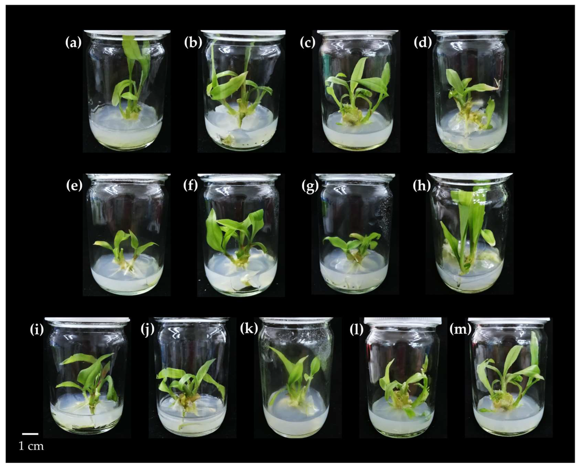
Figure 1. Characteristics of Kaempferia larsenii cultured on solid MS medium supplemented with different concentrations of BA in combination with NAA (0.1 and 0.5 mg/L) after 8 weeks of culture: (a) PGR-free MS medium; (b) 1 BA + 0.1 NAA; (c) 2 BA + 0.1 NAA; (d) 3 BA + 0.1 NAA; (e) 4 BA + 0.1 NAA; (f) 5 BA + 0.1 NAA; (g) 6 BA + 0.1 NAA; (h) 1 BA + 0.5 NAA; (i) 2 BA + 0.5 NAA; (j) 3 BA + 0.5 NAA; (k) 4 BA + 0.5 NAA; (l) 5 BA + 0.5 NAA; (m) 6 BA + 0.5 NAA mg/L. Scale bar = 1 cm.
Horticulturae 11 00281 g001
Figure 2. Characteristics of Kaempferia larsenii cultured on solid MS medium supplemented with different concentrations of kinetin in combination with NAA (0.1 and 0.5 mg/L) after 8 weeks of culture: (a) PGR-free MS medium; (b) 1 kinetin + 0.1 NAA; (c) 2 kinetin + 0.1 NAA; (d) 3 kinetin + 0.1 NAA; (e) 4 kinetin + 0.1 NAA; (f) 5 kinetin + 0.1 NAA; (g) 6 kinetin + 0.1 NAA; (h) 1 kinetin + 0.5 NAA; (i) 2 kinetin + 0.5 NAA; (j) 3 kinetin + 0.5 NAA; (k) 4 kinetin + 0.5 NAA; (l) 5 kinetin + 0.5 NAA; (m) 6 kinetin + 0.5 NAA mg/L. Scale bar = 1 cm.
Horticulturae 11 00281 g002
Figure 3. Characteristics of Kaempferia larsenii cultured on solid MS medium containing TDZ in combination with IAA at different concentrations after 8 weeks of culture: (a) PGR-free MS medium; (b) 0.1 TDZ + 0.5 IAA; (c) 0.5 TDZ + 0.5 IAA; (d) 1 TDZ + 0.5 IAA; (e) 2 TDZ + 0.5 IAA; (f) 3 TDZ + 0.5 IAA; (g) 4 TDZ + 0.5 IAA mg/L. Scale bar = 1 cm.
Horticulturae 11 00281 g003
Figure 4. Characteristics of K. larsenii cultured on MS medium containing BA, kinetin, and NAA after 8 weeks of culture. (a) PGR-free MS medium; (b) 1 BA + 2 KIN + 0.2 NAA; (c) 2 BA + 2 kinetin + 0.2 NAA; (d) 3 BA + 2 kinetin + 0.2 NAA; (e) microshoots on 3 BA + 2 kinetin + 0.2 NAA medium (scale bar = 0.5 cm); (f) 1 BA + 4 kinetin + 0.2 NAA; (g) 2 BA + 4 kinetin + 0.2 NAA; (h) 3 BA + 4 kinetin + 0.2 NAA mg/L. Scale bar = 1 cm.
Horticulturae 11 00281 g004
Figure 5. Characteristics of K. larsenii cultured on MS medium containing BA, TDZ, and NAA after 8 weeks of culture: (a) PGR-free MS medium; (b) 1 BA + 1 TDZ + 0.2 NAA; (c) 2 BA + 1 TDZ + 0.2 NAA; (d) 3 BA + 3 TDZ + 0.2 NAA; (e) 1 BA + 3 TDZ + 0.2 NAA; (f) 2 BA + 3 TDZ + 0.2 NAA; (g) 3 BA + 3 TDZ + 0.2 NAA mg/L; (h) microshoots on 3 BA + 3 TDZ + 0.2 NAA mg/L medium. Scale bar = 1 cm.
Horticulturae 11 00281 g005
Figure 6. Characteristics of Kaempferia larsenii cultured on liquid medium supplemented with BA, kinetin, IAA, and NAA for 8 weeks: (a) PGR-free MS liquid medium; (b) 1 BA + 0.5 IAA; (c) 2 kinetin + 1 NAA; (d) 2 BA + 2 kinetin + 1 NAA; (e) 3 BA; (f) 3 BA + 4 kinetin mg/L. Scale bar = 1 cm.
Horticulturae 11 00281 g006
Figure 7. Effects of cutting microshoots in half on shoot and root induction of Kaempferia larsenii cultured on different types of MS medium. The condition labeled “PGR-free” refers to explants cultured on MS medium without PGRs; “PGRs” refer to explants cultured on MS medium supplemented with 1 mg/L TDZ and 0.5 mg/L IAA after 8 weeks of culture. Scale bar = 1 cm.
Horticulturae 11 00281 g007
Figure 8. Characteristics of Kaempferia larsenii after transplanting various plant materials in the greenhouse for 8 weeks.
Horticulturae 11 00281 g008
Figure 9. FTIR analysis spectra of several plant parts of Kaempferia larsenii derived from different growing conditions: (a) mother plants; (b) in vitro-cultured plants.
Horticulturae 11 00281 g009
Table 1. Treatments of PGR components integrated into MS medium for in vitro propagation of K. larsenii.
Medium Formulation (Solid)PGRs (mg/L)
  • Combination of BA and NAA (13 treatments)
BA0, 1, 2, 3, 4, 5, 6
NAA0, 0.1, 0.5
2.
Combination of kinetin and NAA (13 treatments)
kinetin0, 1, 2, 3, 4, 5, 6
NAA0, 0.1, 0.5
3.
Combination of TDZ and IAA (7 treatments)
TDZ0, 0.1, 0.5, 1, 2, 3, 4, 5
IAA0, 0.5
4.
Combination of BA, kinetin, and NAA (7 treatments)
BA0, 1, 2, 3
kinetin0, 2, 4
NAA0, 0.2
5.
Combination of BA, TDZ, and NAA (7 treatments)
BA0, 1, 2, 3
TDZ0, 1, 3
NAA0, 0.2
Medium Formulation (Liquid)PGRs (mg/L)
Combination of BA, kinetin, IAA, and NAA (6 treatments)BA0, 1, 2, 3
kinetin0, 2, 4
IAA0, 0.5
NAA0, 1
All control treatments utilized PGR-free MS medium.
Table 2. The effect of BA at different concentrations in combination with NAA (0.1 and 0.5 mg/L) on shoot and root induction of Kaempferia larsenii after 8 weeks of cultivation.
BA
(mg/L)
NAA
(mg/L)
No. of
Shoots/Explant
Shoot Height
(cm)
No. of
Roots/Explant
Root Length
(cm)
002.33 ± 0.76 c3.72 ± 1.28 a5.89 ± 1.33 b3.45 ± 1.20 a
10.13.10 ± 0.73 bc3.69 ± 0.85 a7.90 ± 3.26 a2.78 ± 0.76 b
20.13.70 ± 0.95 b3.28 ± 0.33 ab8.40 ± 2.06 a2.74 ± 0.66 b
30.14.10 ± 0.98 a2.86 ± 1.04 b8.30 ± 2.15 a2.80 ± 0.70 b
40.12.60 ± 1.08 c2.72 ± 0.35 b3.60 ± 2.85 d1.76 ± 0.85 c
50.14.00 ± 1.17 a3.07 ± 1.01 ab8.00 ± 2.06 a3.15 ± 1.01 a
60.13.71 ± 1.64 b2.29 ± 0.29 c7.14 ± 3.54 a2.67 ± 1.77 b
10.53.13 ± 1.23 bc3.58 ± 1.33 a8.25 ± 3.10 a2.72 ± 1.27 b
20.53.75 ± 1.96 b3.11 ± 0.73 ab7.25 ± 3.48 a2.51 ± 0.85 b
30.54.13 ± 1.27 a2.90 ± 1.11 b5.00 ± 3.04 b1.78 ± 0.60 c
40.54.50 ± 1.58 a3.27 ± 1.01 ab4.75 ± 2.85 c1.38 ± 0.82 c
50.53.88 ± 1.52 b3.52 ± 0.98 a7.00 ± 1.68 a2.40 + 0.73 b
60.54.63 ± 1.90 a2.52 ± 0.44 bc4.38 ± 1.58 c1.91 ± 0.73 c
Mean values ± SD followed by different letters in the same column indicate significant differences based on DMRT analysis (p ≤ 0.05). Abbreviation: DMRT = Duncan’s multiple range test.
Table 3. The effect of kinetin at different concentrations in combination with NAA (0.1 and 0.5 mg/L) on shoot and root induction of Kaempferia larsenii after 8 weeks of cultivation.
Kinetin
(mg/L)
NAA
(mg/L)
No. of
Shoots/Explant
Shoot Height
(cm)
No. of
Roots/Explant
Root Length
(cm)
002.57 ± 1.36 b3.85 ± 1.08 b7.43 ± 5.57 c3.87 ± 1.77 b
10.12.71 ± 0.91 b2.78 ± 1.40 c9.86 ± 3.73 b3.68 ± 1.33 b
20.13.38 ± 1.33 ab5.00 ± 2.09 a9.88 ± 4.71 b4.30 ± 2.47 ab
30.13.40 ± 1.08 ab4.52 ± 1.27 ab9.20 ± 4.30 b3.14 ± 0.82 b
40.12.89 ± 0.98 b3.89 ± 1.42 b5.56 ± 3.22 d5.36 ± 1.40 a
50.13.40 ± 0.85 ab3.55 ± 0.80 b8.60 ± 2.56 c3.05 ± 0.95 b
60.14.10 ± 1.51 a4.46 ± 1.52 ab10.90 ± 3.04 a3.31 ± 0.63 b
10.53.14 ± 1.08 ab4.67 ± 1.96 ab11.14 ± 4.11 a4.09 ± 1.33 ab
20.53.14 ± 1.08 ab4.75 ± 1.40 ab12.00 ± 7.23 a5.15 ± 1.61 a
30.53.29 ± 0.92 ab3.67 ± 1.52 b10.86 ± 1.61 a3.72 ± 1.08 b
40.53.38 ± 1.20 ab4.62 ± 0.57 ab11.75 ± 1.68 a3.91 ± 0.67 ab
50.52.71 ± 0.92 b2.81 ± 0.85 c7.14 ± 3.48 c3.26 ± 1.49 b
60.53.57 ± 1.17 ab3.25 ± 1.46 b9.43 ± 5.38 b3.40 ± 1.80 b
Mean values ± SD followed by different letters in the same column indicate significant differences based on DMRT analysis (p ≤ 0.05).
Table 4. The effect of plant growth regulators (TDZ and IAA) at different concentrations on shoot and root induction of Kaempferia larsenii after 8 weeks of cultivation.
Plant Growth
Regulators (mg/L)
No. of
Shoots/Explant
Shoot Height
(cm)
No. of
Roots/Explant
Root Length
(cm)
TDZIAA
002.41 ± 1.17 b5.14 ± 1.64 ab7.25 ± 1.65 a2.23 ± 0.51 ab
0.10.53.57 ± 1.64 ab4.49 ± 1.42 b5.20 ± 8.13 b2.65 ± 1.20 a
0.50.54.00 ± 1.55 a5.49 ± 0.85 ab7.09 ± 2.85 a2.68 ± 0.47 a
10.52.41 ± 0.98 b6.21 ± 0.54 a5.58 ± 2.69 b2.78 ± 0.82 a
20.53.28 ± 1.47 ab4.83 ± 1.90 b5.83 ± 4.17 b2.89 ± 0.60 a
30.53.30 ± 2.25 ab5.11 ± 1.27 ab7.80 ± 2.44 a2.57 ± 0.66 a
40.52.91 ± 0.80 ab4.25 ± 0.70 b5.87 ± 3.73 b1.67 ± 0.47 b
Mean values ± SD followed by different letters in the same column indicate significant differences based on DMRT analysis (p ≤ 0.05).
Table 5. The effect of plant growth regulators (BA, kinetin, and NAA) at different concentrations on shoot and root induction of Kaempferia larsenii after 8 weeks of cultivation.
Plant Growth
Regulators (mg/L)
No. of
Shoots/Explant
Shoot Height
(cm)
No. of
Roots/Explant
Root Length
(cm)
BAKinetinNAA
0001.90 ± 0.89 d3.64 ± 0.32 bc8.30 ± 2.91 c3.75 ± 0.32 a
120.23.10 ± 0.32 c3.80 ± 0.22 ab11.30 ± 1.33 b3.40 ± 0.25 bc
220.23.60 ± 0.70 c3.70 ± 0.32 bc10.60 ± 1.71 b3.19 ± 0.25 c
320.211.00 ± 2.40 a3.76 ± 0.41ab9.90 ± 2.12 bc2.87 ± 0.35 d
140.25.40 ± 2.03 b3.99 ± 0.29 a14.50 ± 2.03 a3.51 ± 0.22 b
240.29.00 ± 2.31 a3.58 ± 0.20 bc14.70 ± 0.64 a3.21 ± 0.25 c
340.26.40 ± 1.71 b3.44 ± 0.16 c13.50 ± 2.28 a3.31 ± 0.13 bc
Mean values ± SD followed by different letters in the same column indicate significant differences based on DMRT analysis (p ≤ 0.05).
Table 6. The effect of plant growth regulators (BA, TDZ, and NAA) at different concentrations on shoot and root induction of Kaempferia larsenii after 8 weeks of cultivation.
Plant Growth
Regulators (mg/L)
No. of
Shoots/Explant
Shoot Height
(cm)
No. of
Roots/Explant
Root Length
(cm)
BATDZNAA
0001.80 ± 0.79 d3.34 ± 0.17 a5.30 ± 1.49 b3.47 ± 0.15 a
110.29.80 ± 2.10 b3.02 ± 0.41 b10.10 ± 2.28 a3.50 ± 0.23 a
210.26.60 ± 2.17 c2.88 ± 0.25 bc11.00 ± 1.76 a3.28 ± 0.19 ab
310.211.70 ± 2.11 a2.65 ± 0.25 c6.50 ± 1.27 b2.77 ± 0.38 d
130.29.40 ± 1.51 b2.22 ± 0.41 d3.20 ± 1.14 c2.42 ± 0.33 e
230.213.10 ± 1.20 a1.97 ± 0.22 d2.30 ± 0.48 c3.15 ± 0.34 bc
330.212.00 ± 2.67 a2.02 ± 0.24 d2.60 ± 1.43 c2.89 ± 0.54 cd
Mean values ± SD followed by different letters in the same column indicate significant differences based on DMRT analysis (p ≤ 0.05).
Table 7. The effect of plant growth regulators (BA, kinetin, IAA, and NAA) on shoot and root development of Kaempferia larsenii cultured on liquid medium for 8 weeks.
Plant Growth Regulators (mg/L)No. of
Shoots/Explant
Shoot Height
(cm)
No. of
Roots/Explant
Root Length
(cm)
BAKinetinIAANAA
00004.00 ± 0.67 a8.93 ± 0.43 c10.90 ± 1.29 b1.90 ± 0.13 d
100.504.70 ± 0.82 a11.33 ± 1.16 ab16.40 ± 1.78 a3.45 ± 0.33 a
02014.20 ± 0.63 a10.92 ± 1.09 b15.60 ± 1.84 a2.63 ± 0.33 c
22014.50 ± 0.53 a10.98 ± 0.46 b16.90 ± 0.99 a2.74 ± 0.34 c
30004.20 ± 0.42 a11.16 ± 0.75 ab16.20 ± 2.04 a3.02 ± 0.17 b
34004.60 ± 0.52 a11.82 ± 0.63 a10.60 ± 1.07 b3.16 ± 0.33 b
Mean values ± SD followed by different letters in the same column indicate significant differences based on DMRT analysis (p ≤ 0.05).
Table 8. The effect of cutting microshoots in half on shoot and root induction of Kaempferia larsenii grown on different types of MS medium for 8 weeks.
MicroshootsTypes of MS MediumPlant Growth
Regulators (mg/L)
No. of
Shoots/Explant
Shoot Height
(cm)
No. of
Roots/Explant
Root Length
(cm)
TDZIAA
Non-cuttingSolid003.10 ± 0.85 cd4.08 ± 1.01 b–d6.30 ± 1.68 a–d2.95 ± 0.66 a
10.55.00 ± 3.16 b–d2.70 ± 1.36 e–g9.20 ± 6.39 ab2.47 ± 0.92 ab
Liquid005.75 ± 6.10 b–d5.64 ± 2.75 a6.00 ± 6.83 a–d2.43 ± 0.95 a–c
10.59.60 ± 6.77 a5.12 ± 1.20 ab10.50 ± 1.58 a3.05 ± 2.69 a
Solid + Liquid005.12 ± 1.23 b–d5.17 ± 1.20 ab6.25 ± 4.36 a–d2.74 ± 0.66 ab
10.57.40 ± 4.84 ab4.78 ± 1.20 bc8.30 ± 1.14 a–c2.52 ± 0.95 ab
CuttingSolid005.00 ± 2.06 b–d4.00 ± 0.98 b–d4.40 ± 3.38 b–d2.04 ± 0.66 a–c
10.55.00 ± 3.23 b–d2.81 ± 0.63 e–g7.17 ± 6.80 a–d3.13 ± 1.11 a
Liquid003.25 ± 1.55 cd5.08 ± 0.82 ab7.17 ± 5.79 a–d1.15 ± 0.73 c
10.55.16 ± 2.50 b–d5.63 ± 3.23 a7.42 ± 1.93 a–c2.10 ± 1.49 a–c
Solid + Liquid001.30 ± 0.47 d3.22 ± 1.74 cd2.00 ± 1.80 d1.50 ± 2.40 bc
10.52.00 ± 1.17 cd2.19 ± 0.25 g3.33 ± 2.34 cd2.42 ± 1.30 ab
Mean values ± SD followed by different letters in the same column indicate significant differences based on DMRT analysis (p ≤ 0.05).
Table 9. The effect of plant materials on shoot–root development and survival rate of Kaempferia larsenii in the greenhouse for 8 weeks.
Planting MaterialSurvival Rate
(%)
No. of Shoots/PlantShoot Height
(cm)
No. of Leaves/Plant
Loamy soil41.672.00 ± 2.37 ab7.26 ± 1.90 b3.33 ± 1.61 a
Sandy soil100.001.50 ± 3.48 ab6.70 ± 1.52 b2.17 ± 0.44 ab
Compost0.000.00 c0.00 d0.00 c
Burned rice husk8.301.00 ± 0.00 b13.0 ± 0.00 a3.00 ± 0.00 ab
Loamy soil + sandy soil58.331.42 ± 2.94 ab7.60 ± 2.00 b2.00 ± 1.23 b
Loamy soil + compost16.671.00 ± 0.00 b3.35 ± 2.69 c2.50 ± 1.58 ab
Sandy soil + compost50.002.17 ± 4.33 ab5.01 ± 0.98 bc2.80 ± 0.38 ab
Loamy soil + sandy soil + compost50.002.50 ± 4.40 a5.41 ± 3.80 bc2.45 ± 0.70 ab
Mean values ± SD followed by different letters in the same column indicate significant differences based on DMRT analysis (p ≤ 0.05).
Table 10. Total phenolic content, total flavonoid content, and antioxidant activity of methanolic extracts from various explant parts of Kaempferia larsenii cultured in vitro compared to the mother plant.
ConditionPlant Parts TPC
(mg GAE/g DW)
TFC
(mg QE/g DW)
DPPH
(mg TE/g DW)
DPPH
(% Inhibition)
FRAP
(mg FeSO4/g DW)
Mother plant Leaves716.03 ± 5.08 a47.89 ± 0.80 a121.34 ± 1.42 a59.04 ± 0.62 a134.35 ± 2.67 a
Pseudostem203.33 ± 3.31 b30.97 ± 0.54 b24.73 ± 0.31 b49.20 ± 0.55 b17.44 ± 1.47 b
Rhizomes and
Storage roots
201.88 ± 2.32 b7.01 ± 0.35 d22.58 ± 1.46 c45.43 ± 2.55 c14.97 ± 0.55 c
In VitroMSLeaves157.88 ± 2.88 e6.28 ± 0.49 d14.24 ± 1.50 f30.77 ± 2.62 f11.27 ± 1.11 de
Pseudostems114.76 ± 2.18 f4.43 ± 0.38 e11.47 ± 1.26 g25.91 ± 2.22 g7.77 ± 0.31 f
Roots168.13 ± 1.18 d6.40 ± 0.21 d16.64 ± 1.04 e34.99 ± 1.82 e12.12 ± 0.66 d
PGRsLeaves192.84 ± 3.15 c11.47 ± 0.43 c19.76 ± 1.65 d40.47 ± 2.90 d16.48 ± 1.04 bc
Pseudostems117.59 ± 1.16 f4.46 ± 0.43 e14.33 ± 0.52 f30.94 ± 0.90 f9.78 ± 0.40 e
Roots155.32 ± 3.76 e10.67 ± 0.33 c18.04 ± 0.31 de37.45 ± 0.54 de17.15 ± 0.71 b
Mean values ± SD followed by different letters in the same column indicate significant differences based on DMRT analysis (p ≤ 0.05). Abbreviations: DPPH = 2,2–diphenyl–1–picrylhydrazyl assay; FRAP = ferric-reducing antioxidant power assay; TFC = total flavonoid content; TPC = total phenolic content.
Table 11. Correlations between total phenolic content, total flavonoid content, DPPH scavenging, and % inhibition of DPPH and FRAP activities in Kaempferia larsenii.
TPCTFCDPPH% Inhibition (DPPH)FRAP
TPC10.881 **0.996 **0.800 **0.993 **
TFC 10.878 **0.867 **0.864 **
DPPH 10.782 **0.998 **
% inhibition (DPPH) 10.749 **
FRAP 1
** Correlation is significant at the 0.01 level (2–tailed).
Table 12. Phenolic acid compounds of mother plants and in vitro-derived plants of Kaempferia larsenii analyzed using the HPLC technique.
ConditionExplantPhenolic Acid Compound Contents (µg/g DW)
GAPCAHBACGACACMAFASACNATotal
Mother plantsLeaves3.05 ±
0.09 f
5.01 ±
0.12 e
NDND7.88 ±
0.35 d
28.98 ±
0.36 c
35.22 ±
0.17 b
47.21 ±
0.26 a
ND127.36 ± 0.05
Pseudo
stems
1.16 ±
0.07 f
1.67 ±
0.09 e
NDND3.28 ±
0.10 c
2.17 ±
0.14 d
4.52 ±
0.35 b
5.86 ±
0.12 a
ND18.66 ± 0.01
Rhizomes +
Storage roots
4.06 ±
0.05 c
2.77 ±
0.05 d
NDND1.67 ±
0.07 e
4.07 ±
0.26 c
4.96 ±
0.42 b
6.27 ±
0.29 a
ND23.81 ± 0.04
In VitroMSLeaves25.30 ±
1.28 e
ND61.95 ±
0.50 d
NDND14.00 ±
0.26 f
202.97 ±
4.38 b
114.40 ±
0.40 c
489.35 ±
15.68 a
904.81 ± 1.34
Pseudo
stems
44.79 ±
1.21 d
ND65.00 ±
0.23 c
NDND6.18 ±
0.16 e
114.77 ±
3.00 b
ND236.09 ±
4.39 a
466.83 ± 0.70
Roots13.29 ±
0.68 c
NDNDNDNDND48.40 ±
0.43 b
ND882.52 ±
11.83 a
944.21 ± 1.77
2 BA +
0.1 NAA
(mg/L)
Leaves6.80 ±
0.24 e
ND60.45 ±
0.68 d
NDND8.90 ±
0.16 e
253.15 ±
2.44 b
114.40 ±
0.40 c
515.87 ±
7.86 a
959.57 ± 0.68
Pseudo
stems
44.10 ±
1.58 d
ND62.10 ±
0.35 c
NDND6.53 ±
0.50 e
149.75 ±
2.94 b
ND237.99 ±
15.36 a
500.48 ± 1.57
Roots63.48 ±
1.39 b
NDNDNDNDND50.03 ±
0.35 c
ND437.46 ±
4.78 a
550.98 ± 0.40
Mean values ± SD followed by different letters in the same row indicate significant differences based on DMRT analysis (p ≤ 0.05). Abbreviations: CA = caffeic acid; CNA = cinnamic acid; CGA = chlorogenic acid; CMA = p-coumaric acid; FA = ferulic acid; GA = gallic acid; HBA = p-hydroxybenzoic acid; ND = not detected; PCA = protocatechuic acid; SA = sinapic acid.
Table 13. Flavonoid compounds of the mother plants and the in vitro-derived plants of Kaempferia larsenii analyzed using the HPLC technique.
ConditionExplantFlavonoid Compound Contents (µg/g DW)
RutinQuercetinMyricetinApigeninTotal
Mother plantsLeaves1987.73 ±
2.74 a
259.90±
1.85 d
1937.71 ±
3.12 b
334.45 ±
2.69 c
4519.79 ± 0.27
Pseudostems105.04 ±
1.94 b
21.96 ±
0.80 d
483.20 ±
2.46 a
86.15 ±
2.44 c
696.34 ± 0.39
Rhizomes +
Storage roots
4.61 ±
0.07 d
23.10 ±
0.76 c
665.77 ±
2.32 a
112.08 ±
3.40 b
805.55 ± 0.75
In VitroMSLeavesNDND1798.64 ±
3.57
1181.68 ±
6.34
2980.31 ± 0.98
PseudostemsND152.74 ±
2.34 c
833.82 ±
8.02 b
866.74 ±
3.33 a
1853.30 ± 1.52
Roots1.46 ±
0.12 c
ND818.25 ±
2.01 b
1531.14 ±
9.77 a
2350.85 ± 2.57
2 BA +
0.1 NAA
(mg/L)
LeavesNDND1283.81 ±
5.23
957.85 ±
6.11
2241.66 ± 0.31
Pseudostems1.00 ±
0.07 d
146.00 ±
3.95 c
588.53 ±
8.92 b
911.79 ±
5.61 a
1647.31 ± 1.84
Roots1.59 ±
0.02 d
180.89 ±
1.68 c
649.08 ±
2.36 b
1791.27 ±
8.05 a
2622.83 ± 1.75
Mean values ± SD followed by different letters in the same row indicate significant differences based on DMRT analysis (p ≤ 0.05); ND = not detected.
Table 14. Peak wavenumbers, types of vibrational bonds, and types of functional groups from different plant parts of K. larsenii determined by FTIR analysis.
Wavenumber (cm−1)Vibrational BondFunctional Group
Mother PlantMSPGRs (2 BA + 0.1 NAA)
LsPsRsLsPsRLsPsR
328232823285327432753284327632743282O–H
(Stretching)
Alcohols
Phenols
291729202921291829182919291829182920C–H
(Stretching)
Alkane
28492850285028472850C–H
(Stretching)
Alkane
173117291719173617351735173517351730C=O
(Stretching)
Aldehyde
16331627163416221623162816291629C=C
(Stretching)
Alkene
13101308C–O
(Stretching)
Aromatic
ester
1318C–N
(Stretching)
Aromatic
amine
135713621363135913591357O–H
bending
Alcohols
124112421247123212321233123212331233C–O
(Stretching)
Alkyl aryl ether
103210311024102810301031103010281030C–O
(Stretching)
Alkyl aryl ether
Abbreviations: Ls = leaves; Ps = pseudostems; R = roots; Rs = rhizomes and storage roots of Kaempferia larsenii.
Disclaimer/Publisher’s Note: The statements, opinions and data contained in all publications are solely those of the individual author(s) and contributor(s) and not of MDPI and/or the editor(s). MDPI and/or the editor(s) disclaim responsibility for any injury to people or property resulting from any ideas, methods, instructions or products referred to in the content.