Article

Hydrogen Sulfide Mitigates Manganese-Induced Toxicity in Malus hupehensis Plants by Regulating Osmoregulation, Antioxidant Defense, Mineral Homeostasis, and Glutathione Ascorbate Cycle

College of Horticulture, Northwest A&F University, Xianyang 712100, China
*
Author to whom correspondence should be addressed.
Horticulturae 2025, 11(2), 133; https://doi.org/10.3390/horticulturae11020133
Submission received: 1 December 2024 / Revised: 22 January 2025 / Accepted: 24 January 2025 / Published: 26 January 2025
(This article belongs to the Special Issue Tolerance and Response of Ornamental Plants to Abiotic Stress)

Abstract

:
Manganese (Mn) is a toxic metal element that adversely affects plant growth. Hydrogen sulfide (H2S) is considered an important signaling molecule with significant potential in alleviating various abiotic stresses. However, there is limited information available on the role of H2S in alleviating manganese stress in plants. In this study, the effects of exogenous H2S and its scavenger, homocysteine thiolactone (HT), on the physiological and biochemical parameters of Malus hupehensis var. pingyiensis seedlings were evaluated. Our results show that H2S treatment significantly alleviates growth inhibition and oxidative damage induced by manganese stress in Malus hupehensis seedlings, primarily by enhancing antioxidant enzyme activity and up-regulating the ascorbate-glutathione (ASA-GSH) cycle. H2S treatment increased photosynthetic pigment content and helped maintain osmotic balance in leaves, thereby enhancing key gas exchange parameters and mitigating manganese-induced suppression of photosynthesis. H2S treatment enhanced the absorption of Ca, Mg, Fe and Zn under manganese stress, significantly reduced manganese accumulation in Malus hupehensis seedlings, and modulated the transcriptional expression of MTPs, facilitating the transfer of manganese to the leaves. Thus, H2S reduces oxidative damage and promotes growth under Mn stress, highlighting its important role in plant stress tolerance.

1. Introduction

Manganese (Mn), the second most abundant metal widely available worldwide, is an essential trace element for all living organisms [1]. In plants, Mn plays an important role in growth, development, and metabolism mainly in the form of Mn2+ [2]. Within chloroplasts, Mn supports chlorophyll formation and structural stability and acts as a cofactor in the redox reactions of the electron transport chain during photosynthesis [3,4]. However, excessive Mn levels in the environment can be detrimental to plants, a phenomenon that often occurs in acidic and flooded soils. Under low pH and reducing conditions, the concentration of soil Mn2+ increases, making it more accessible to plants and leading to toxicity [5]. With the development of industry and long-term overuse of fertilizers and pesticides, more than half of the potentially arable soils around the world are acidic, and manganese stress has become the second most important plant growth limiting factor after aluminum toxicity [6,7]. Therefore, understanding Mn toxicity and plant tolerance mechanisms is important for agricultural development.
Many studies have shown that Mn toxicity disrupts a variety of physiological processes in plant cells, such as triggering oxidative stress, inhibiting enzyme activities, hindering chlorophyll biosynthesis and photosynthesis [8], and hindering the uptake and transport of other essential minerals [9]. This results in a reduction in the number of lateral roots of the plant, reduced root vigor, leaf greening and necrosis, and growth inhibition [10]. Prolonged exposure to metal stress leads to the production of reactive oxygen species (ROS), such as hydrogen peroxide (H2O2), superoxide (O2), and singlet oxygen (1O2). Excessive accumulation of ROS leads to lipid peroxidation, which damages proteins involved in cellular activities and impairs their normal functions [11].
It has been found in past studies that plants can mitigate Mn toxicity by complexing ligands and chelates, regulating manganese transport proteins, regulating antioxidant systems, and altering biochemical pathways [12]. Organic acids such as citrate, malate, and oxalate form stable Mn complexes that reduce its bioavailability and toxicity [13,14]. Heavy metal transport proteins segregate Mn into subcellular compartments such as the cell wall, Golgi apparatus, and vesicles, which in turn reduces the concentration of Mn2+ in the cytoplasm and other organelles, decreasing the effective concentration and toxicity [1]. The manganese tolerance protein (MTP) transporter plays a critical role in Mn transport, distribution, and the maintenance of Mn balance in plants. A total of 20 MTP family members have been identified in the apple genome. Based on the substrate specificity of its members, the apple MTP family is primarily classified into three subfamilies: Mn-MTP, Zn-MTP, and Zn/Fe-MTP. Among these, MTP8 to MTP11 belong to the Mn-MTP subfamily [15]. MTP8, MTP9, and MTP11 are localized to the vacuolar membrane, cell membrane, and Golgi apparatus, respectively, and play roles in the compartmentalization and efflux of manganese ions [15,16,17]. Plants also induce multiple antioxidant enzyme activities to interact with non-enzymatic antioxidants to scavenge excess ROS and reduce oxidative stress [18].
Hydrogen sulfide (H2S) has emerged as an important signaling molecule involved in regulating various plant physiological processes, such as seed germination, root development, stomatal movement, photosynthesis, and stress responses [19,20]. Studies demonstrate that H2S acts as a signal transducer during abiotic stresses such as salinity, osmotic pressure, and extreme temperatures [21,22]. Under metal stress, endogenous H2S levels rise, while the exogenous application of H2S in appropriate concentrations can enhance plant tolerance and reduce stress-induced damage [23]. For instance, exogenous H2S reduces damage caused by Cd stress in Brassica oleracea, Oryza sativa, and Triticum aestivum L., alleviates Al toxicity in Brassica napus and Glycine max, and promotes wheat seed germination under Cr stress [23,24,25]. These protective effects are attributed to H2S-mediated reductions in metal uptake and accumulation or by increasing the activity of antioxidant systems [26].
While the benefits of exogenous H2S under various metal stresses are well-documented, research on its role in mitigating plant manganese stress remains limited. Malus hupehensis var. pingyiensis is a unique germplasm resource in northern China, known for its well-developed root system, strong waterlogging resistance, and high disease tolerance. It is frequently used as rootstock and in research on resistance mechanisms [27]. Therefore, this study will evaluate how exogenous H2S affects the physiological and biochemical responses of Malus hupehensis under Mn stress. The results will provide insight into the potential use of H2S as a tool for mitigating Mn toxicity in agricultural settings.

2. Materials and Methods

2.1. Materials and Treatments

We selected approximately 300 healthy and uniform Malus hupehensis var. pingyiensis seeds (previously stored in the laboratory) for surface sterilization for 10 min using 5% sodium hypochlorite (NaClO) solution, and washed them thoroughly with distilled water, then randomly placed them in 15cm diameter Petri dishes containing gauze moistened with distilled water for one week. Sprouted seedlings were transplanted into cavity trays filled with media comprising nutrient soil (Xingyuxing, Wuhan, China), peat moss (Xinghe, Jinan, China), and vermiculite (Luqing, Nanning, China) at a 3:1:1 ratio (v:v:v), and placed in a light incubator (Yiheng, Shanghai, China) under a 16 h light/8 h dark photoperiod at 25 ± 2 °C. Following two weeks of normal growth, uniform seedlings were transferred to full Hoagland’s nutrient solution (HB8870, Haibo, Qingdao, China) for hydroponic culture, and after one week, Mn stress was induced by adding an extra 1 mM MnSO4 to the Hoagland’s solution, while control plants continued to grow in Hoagland’s solution. To assess the role of H2S under Mn stress, the H2S donor sodium hydrogen sulfide (NaHS) was introduced into the Hoagland’s solution for both the control and Mn-stressed plants. Additionally, the H2S scavenger taurine (HT; 200 µM) was supplied under conditions with exogenous H2S to verify the positive effects of H2S under Mn stress. We added the reagent combinations specified in Table 1 to the complete Hoagland solution for plant treatment. The pH of the nutrient solution for all treatment groups was adjusted to 5.9 prior to use.
The nutrient solution was replaced every three days throughout the experimental period, and the environmental conditions were set to remain the same as before. After two weeks of treatment, samples were collected to measure the specified indicators. Each treatment was repeated three times under identical experimental conditions, with ten plants per replicate, using a completely randomized design to ensure reliability.

2.2. Estimation of Growth and Photosynthetic Pigment Parameters

Plants were harvested after 14 days of treatment and fresh weight (FW) and dry weight (DW) of the plants were determined using a standard weighing balance.
FW was measured using whole plants, while DW was measured by drying the plants at 70 °C for 48 h. Fresh leaf (100 mg) samples were impregnated in 80% acetone following a previously described method of Copper [28]. The absorbance of chlorophyll a and b was measured using a spectrophotometer (UV3600, Shimadzu, Kyoto, Japan) at 663 nm and 645 nm, respectively. The chlorophyll estimates were calculated using the following equation:
Chlorophyll a (mg g−1 FW) = 100 × [(A663 × 0.0127 − A645 × 0.00269)]/0.5
The net photosynthetic rate (Pn), transpiration rate (E), stomatal conductance (gsw), and intercellular CO2 concentration (Ci) of M. hupehensis seedlings were measured using the LI-6800 Photosynthesis Measurement System (LI-COR, Tucson, AZ, USA) during the periods of 8:30–11:30 a.m. or 2:30–4:30 p.m. All measurements were performed on the third fully expanded mature leaf.

2.3. Estimation of Leaf RWC

The leaf relative water content (RWC) was measured as previously described by Lazcano-Ferrat [29].
The FW of the leaves was recorded before immersing the leaves in distilled water for 24 h at room temperature to measure the turgid weight (TW). The leaves were then dried at 70 °C for 48 h to measure the DW. RWC was calculated using the following equation:
RWC% = (FW − DW)/(TW − DW) × 100%

2.4. Estimation of Soluble Sugar and Proline Contents

The soluble sugar content was determined using the plant soluble sugar assay kit (BC0030, Solarbio, Beijing, China), based on the anthrone colorimetric method. Leaves (0.1 g) from treated seedlings were ground in 1 mL of distilled water and incubated in a boiling water bath for 10 min. The supernatant was then collected and the final volume was adjusted to 10 mL with distilled water. The reaction system was prepared according to the kit instructions and incubated at 95 °C for 10 min. After cooling to room temperature, absorbance was measured at 620 nm, and soluble sugar content was calculated using a standard curve.
Proline content was determined using the proline assay kit (BC0290, Solarbio, Beijing, China), based on the ninhydrin method. Leaves (0.1 g) from treated seedlings were homogenized in 1 mL of extraction buffer on ice, followed by a 10-min boiling water bath and centrifugation at 10,000× g for 10 min. The reaction system was prepared according to the kit instructions and incubated at 100 °C for 30 min. After cooling to room temperature, absorbance was measured at 520 nm, and proline content was calculated using a standard curve.

2.5. Estimation of Metal Element Content

The plant tissue was processed using the ashing method. The treated plants were rinsed three times with 10mM ethylene diamine tetraacetic acid (EDTA) and then dried at 80 °C for 48 h and ground into powder. Then, we accurately weighed 0.1 g in a crucible and heated in a muffle furnace at 300 °C for 48 h or more. We dissolved the ash residue in 0.5 mol L−1 nitric acid, transferred the solution to a centrifuge tube, and diluted it with the same concentration of nitric acid to a final volume of 10 mL. The metal ion concentrations were analyzed using inductively coupled plasma atomic emission spectrometry (ICP-AES; Prodigy, Leemanlabs Inc., Hudson City, NY, USA).

2.6. Evaluation of MDA and H2O2 Contents

Malondialdehyde (MDA) content was measured using the MDA assay kit (BC0020, Solarbio, Beijing, China), based on the thiobarbituric acid (TBA) method. MDA reacts with TBA under acidic and high-temperature conditions, forming a reddish-brown compound with maximum absorption at 532 nm. According to the kit instructions, leaves (0.1 g) from treated seedlings were homogenized in 1 mL of extraction buffer on ice, followed by centrifugation at 8000× g for 10 min at 4 °C. The supernatant was collected, and 200 µL of the supernatant was mixed with 800 µL of the reaction solution and incubated at 100 °C for 60 min. After cooling in ice and centrifugation, absorbance was measured at 520 nm to estimate MDA content.
Hydrogen peroxide (H2O2) content was measured using the H2O2 assay kit (BC3590, Solarbio, Beijing, China). H2O2 reacts with titanium sulfate to form a yellow titanium peroxide complex, which absorbs light at 415 nm. According to the kit instructions, leaves (0.1 g) from treated seedlings were ground in 1 mL of cold acetone and homogenized, followed by centrifugation at 8000× g for 10 min at 4 °C. The supernatant was collected, and the reaction solution was added. Absorbance was measured at 415 nm to estimate H2O2 content in the sample.

2.7. Determination of Electrolyte Leakage

Electrolyte leakage (EL) was assessed using the method of Dionisio-Sese and Tobita [30]. From the removed plants, 10 small discs were taken from 4 to 6 leaves with the same functional leaf position using a 0.8 cm hole punch and immersed in a medium of 10 mL of deionized distilled water. They were immersed at room temperature for 4 h, during which they were shaken several times and the initial conductance value (EC1) of the solution was measured after 4 h. The samples were then boiled for 30 min, cooled to room temperature, and the final conductivity (EC2) was measured. EL was calculated as follows:
EL (%) = (EC1/EC2) × 100

2.8. RNA Extraction and RT-qPCR Analysis

Total RNA was extracted using the FastPure Universal Plant Total RNA Isolation Kit (Vazyme Biotech Co., Nanjing, China) following the manufacturer’s instructions. The cDNA was obtained by reverse transcription using the HiScript II 1st Strand cDNA Synthesis Kit (Vazyme Biotech Co., Nanjing, China). Quantitative real-time PCR (RT-qPCR) was performed using the ArtiCanATM SYBR qPCR Mix kit (Tsingke Biotech Co., Ltd., Beijing, China) on the QuantStudio 3 system (Life Technologies, Carlsbad, CA, USA), with actin serving as the internal reference. The qRT-PCR procedure included 95 °C for 1 min, 95 °C for 10 s, and 58 °C for 20 s for 40 cycles. Three replicates were run for each sample. RT-qPCR primers are shown in Table 2.

2.9. Estimation of Enzymatic and Non-Enzymatic Antioxidant Activity

The activities of superoxide dismutase (SOD), peroxidase (POD), catalase (CAT) and ascorbate peroxidase (APX) were measured using assay kits (BC5160, BC0090, BC0020 and BC0220, Solarbio, Beijing, China). Leaves (0.5 g) from treated seedlings were ground to a powder in liquid nitrogen. Then, 5 mL of 50 mM/L phosphate-buffered saline containing 1% β-mercaptoethanol and 1% polyvinylpyrrolidone (Ph = 7.8) was added to the sample and incubated for 60 min at 4 °C. After centrifugation at 8000× g for 20 min at 4 °C, the supernatant (enzyme extract) was used for further analysis. The reaction systems were prepared according to the kit instructions, and absorbance changes were measured at 450 nm, 470 nm, 240 nm, and 290 nm over a two-minute period using a spectrophotometer. Enzyme activities were calculated based on the rate of absorbance change.
MDHAR and DHAR activities were measured using activity kits (BC0650, BC0660, Solarbio, Beijing, China). According to the kit instructions, leaves (0.1 g) from treated seedlings were homogenized with 1 mL of 50 mM Tris-HCl buffer (pH = 7.8) on ice. After centrifugation at 10,000 rpm for 10 min at 4 °C, the supernatant (enzyme extract) was used for analysis. The reaction solution was added according to the manufacturer’s instructions, and the absorbance changes at 340 nm and 412 nm were measured for two minutes using a spectrophotometer. The enzyme activities were calculated based on the rate of absorbance.
The ascorbate acid (ASA) content was determined using the assay kit (BC1230, Solarbio, Beijing, China), based on the oxidation of AsA by ascorbic acid oxidase (AAO), which converts AsA to dehydroascorbic Acid (DHA). The content of AsA was calculated by measuring the oxidation rate. According to the kit instructions, leaves (0.1 g) from treated seedlings were homogenized with 1 mL of extraction buffer on ice, following the kit instructions. After centrifugation at 8000× g for 10 min at 4 °C, the supernatant was collected for analysis. The reaction solution was prepared according to the manufacturer’s instructions, and absorbance changes at 265 nm were measured over two minutes using a spectrophotometer. The ASA content was calculated from the rate of absorbance.
Glutathione (GSH) and oxidized glutathione (GSSG) contents were determined using the assay kits (BC1170, BC1180, Solarbio, Beijing, China). GSH reacts with 5,5′-dithiobis-2-nitrobenzoic acid (DTNB), producing 2-nitro-5-thiobenzoic acid (DNCB) and GSSG. The DNCB is a yellow product with maximum absorption at 412 nm. According to the kit instructions, leaves (0.1 g) from treated seedlings were homogenized with 1 mL of extraction buffer on ice, following the kit instructions. After centrifugation at 8000× g for 10 min at 4 °C, the supernatant was collected for analysis. The reaction solution was prepared according to the manufacturer’s instructions, and absorbance changes at 412 nm were measured over two minutes using a spectrophotometer. The GSH and GSSG contents were calculated from the rate of absorbance.

2.10. Statistical Analysis

Statistical analysis of all experiments was performed using SPSS. Data were analyzed using a one-way ANOVA. Significant differences among the treatments were determined using the least significant difference (LSD) test (p < 0.05). All presented values are repeated means ± standard error of three independent experiments.

3. Results

3.1. Effect of H2S on Plant Biomass and Leaf RWC Under Manganese Stress

Mn severely affected plant biomass and leaf RWC (Table 3). The FW and DW of plants were reduced after exposure to Mn compared to control seedlings (CK). The reduction of leaf RWC verified the water deficit condition of M. hupehensis seedlings under Mn stress. Leaf RWC was reduced by 25% in Mn-stressed seedlings compared to the control. However, all four levels of H2S significantly improved water balance by increasing leaf RWC compared to the stress treatment without H2S. In addition, the program HT treatments with H2S can be used in a similar representation to the Mn treatments.

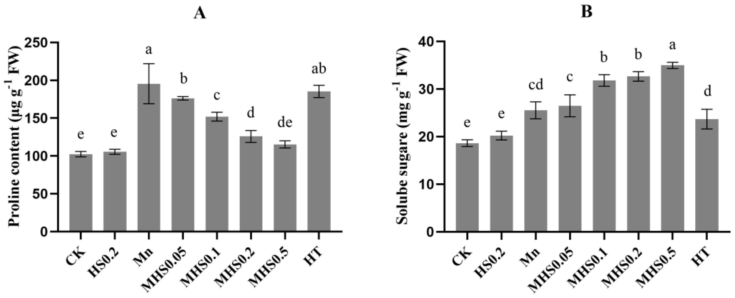
3.2. Effect of H2S on Proline and Soluble Sugar Contents Under Manganese Stress

Mn stress significantly increased proline levels of seedlings (Figure 1A). However, the application of 0.2 mM H2S (HS0.2) reduced proline content significantly, with the 0.5 mM H2S treatment (MHS0.5) leading to a 41% decrease compared to Mn-only stressed plants. Under non-stressed conditions, NaHS application had little effect on soluble sugars and proline content in plant leaves (Figure 1A,B). Mn stress also elevated soluble sugar levels by 24% compared to control plants (CK), and co-application of NaHS further increased this content compared to Mn alone. However, the inclusion of taurine (HT, an H2S scavenger) reversed the beneficial effects of H2S.

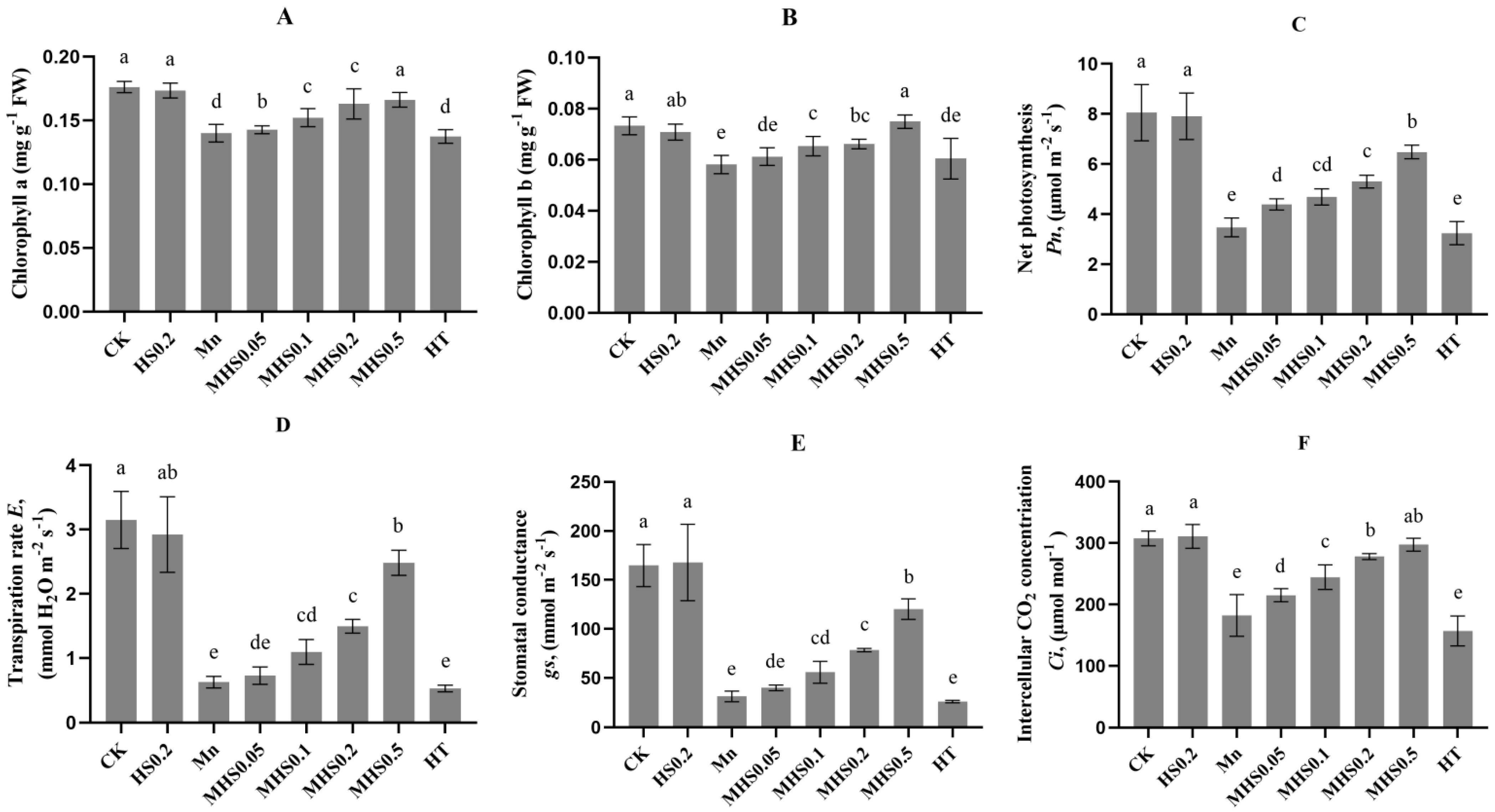
3.3. Effect of H2S on Chlorophyll Content and Gas Exchange Parameters Under Manganese Stress

Chlorophyll a and chlorophyll b content decreased by 21% in Mn-stressed plants compared to control plants (CK), reflecting the impact of Mn toxicity (Figure 2A,B). Supplementation with H2S significantly restored chlorophyll levels, with the 0.5 mM H2S treatment (MHS0.5) showing a more potent effect in restoring both chlorophyll a and chlorophyll b content. Mn stress negatively impacted gas exchange parameters, reducing Pn, E, gsw, and Ci by 52%, 80%, 79%, and 28%, respectively, compared to the control plants (Figure 2C–F). However, the addition of H2S improved these parameters under Mn stress, with the MHS0.5 treatment increasing Pn, E, gsw, and Ci by 41%, 75%, 74% and 39%, respectively, compared to Mn-only treated plants.

3.4. Effect of H2S on Mineral Homeostasis and Manganese Accumulation Under Mn Toxicity

Mn stress reduced the contents of Fe, Zn, Mg, and Ca by 41%, 44%, 53%, and 25%, respectively, compared to control plants (Table 4). Under non-stress conditions, 0.2 mM H2S application (HS0.2) had little effect on plant metal content. H2S supplementation alleviated this reduction, with MHS0.5 treatment reducing these losses to 8%, 12%, 49%, and 10%, respectively, compared to the control. There was almost no difference in the metal contents of the plants in the HT treatment group and the Mn alone treatment group.
Additionally, Mn content in the leaves, stems, and roots decreased by 41%, 76%, and 57%, respectively, under MHS0.5 treatment compared to Mn-alone treatment (Table 5). Mn accumulation in the roots showed the greatest reduction, followed by the stems and leaves. Interestingly, the proportion of Mn in leaves relative to the total plant Mn content increased with rising H2S concentrations, ranging from 24% to 39%.

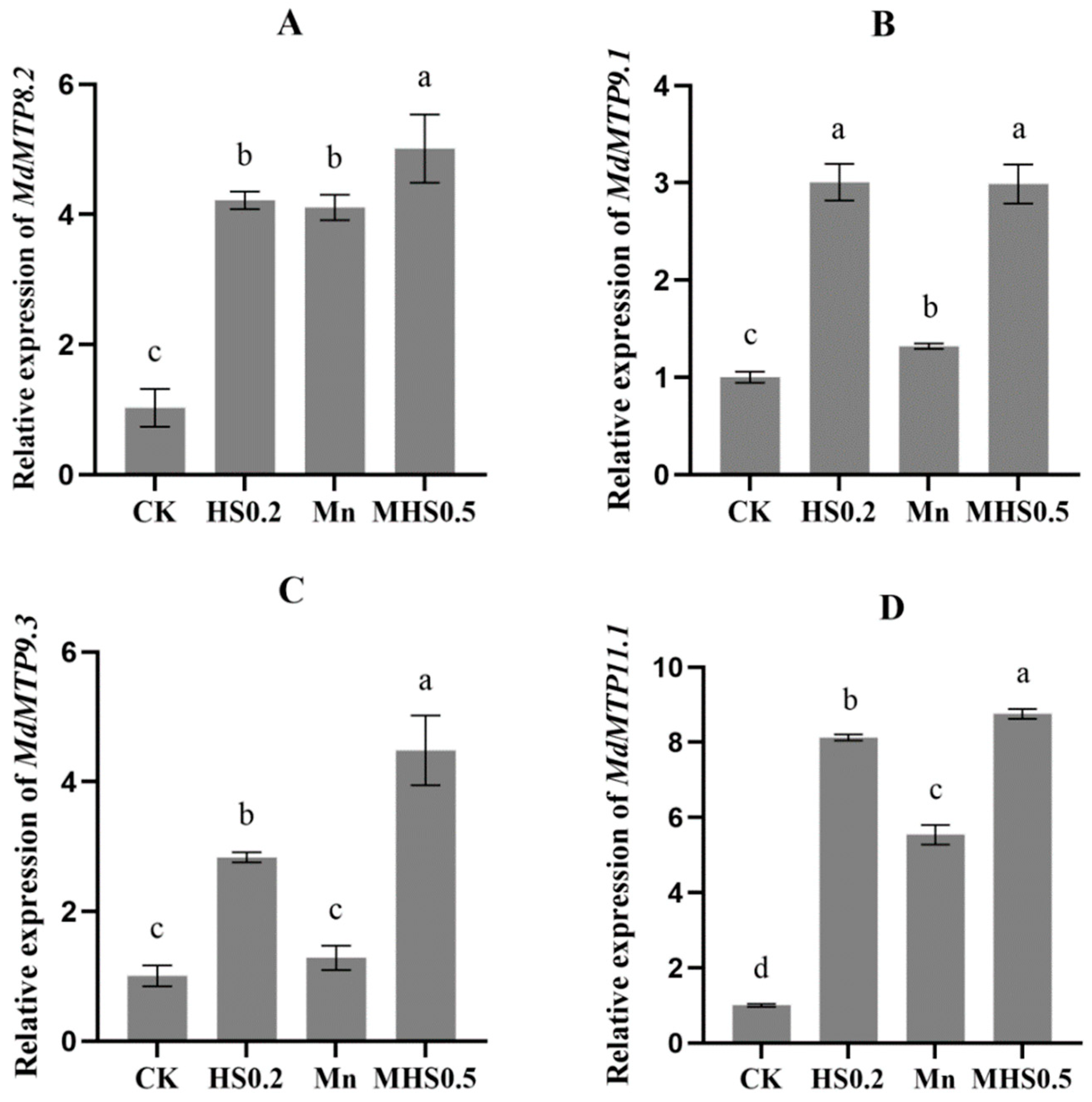
3.5. Effect of H2S on the Expression of Manganese Tolerance Protein Genes Under Mn Stress

Mn stress significantly up-regulated the expression of MdMTP8.2 and MdMTP11.1 by 4.1-fold and 5.5-fold, respectively, compared to the control plants (Figure 3A,D). The co-application of Mn and H2S further enhanced their expression. Mn treatment increased MdMTP9.1 expression by 1.3-fold compared to the control, and MHS0.5 treatment elevated this by 2.3-fold relative to Mn alone (Figure 3B). While Mn stress did not alter MdMTP9.3 expression, H2S application significantly up-regulated its expression under both stressed and non-stressed conditions (Figure 3C).

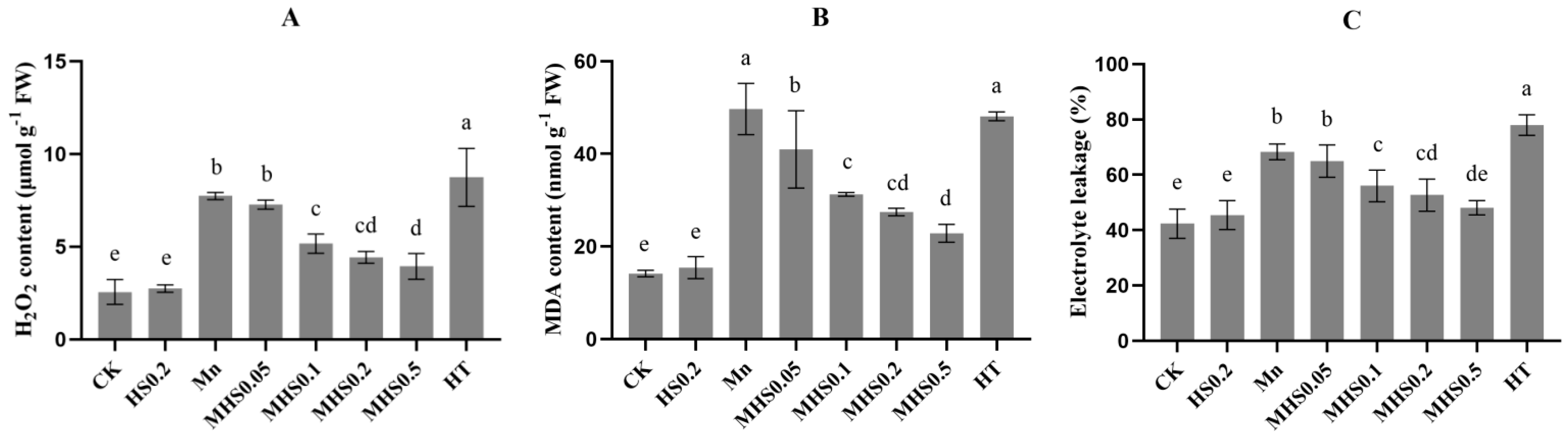
3.6. Effect of H2S on ROS Accumulation and Electrolyte Leakage Under Mn Stress

Mn stress elevated H2O2 and MDA levels by 65% and 71%, respectively, compared to the control plants (Figure 4A,B). However, H2S supplementation reduced these levels significantly, with the MHS0.5 treatment showing the greatest reduction. In addition, EL increased from 42% to 68% under Mn stress; however, 0.5 Mm H2S application (MHS0.5) decreased EL to 48% (Figure 4C). Similar results were observed in the HT-treated group of plants as Mn-alone stressed plants in terms of H2O2, MDA, and EL levels.

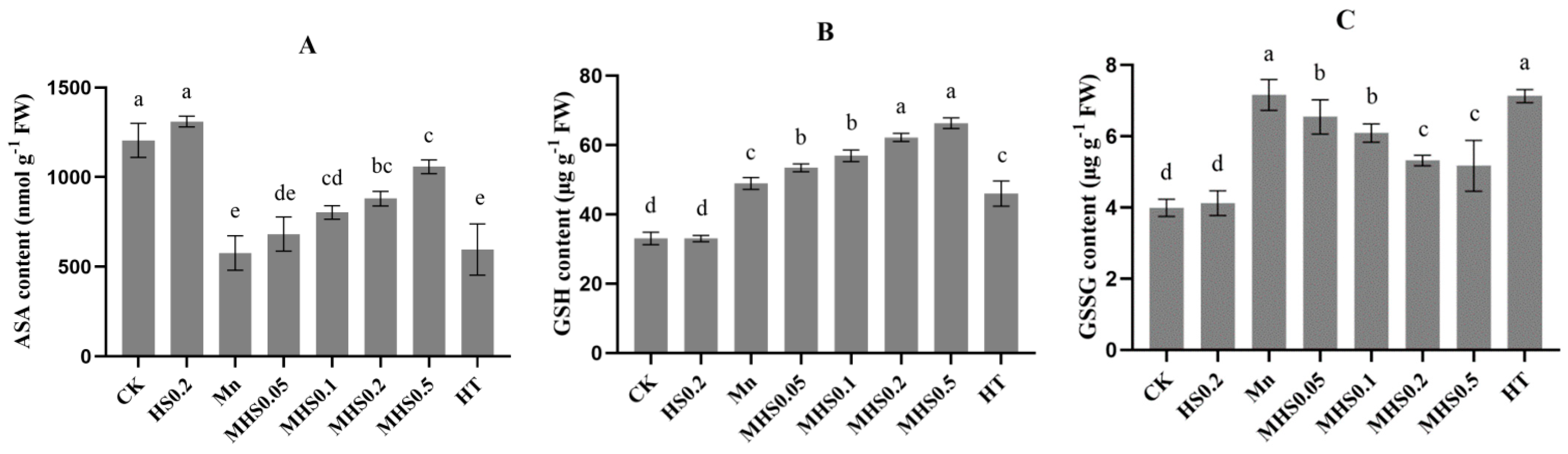
3.7. Effect of H2S on Antioxidant Enzyme Activities and Antioxidant Content Under Mn Stress

Mn stress increased SOD, POD, and CAT by 48%, 48% and 37%, respectively, compared to the controls (Figure 5A–D). Supplementation with H2S further enhanced these enzyme activities under Mn stress, with increases of 36%, 27%, 74%, and 35% in SOD, CAT and POD activities, respectively, under MHS0.5 treatment. Mn stress reduced MDHAR and DHAR activities, but H2S supplementation increased these by 56% and 69%, respectively, under MHS0.5 treatment (Figure 5E,F). There was no significant difference in antioxidant enzyme activity between plants treated with HT and Mn alone.
GSH and GSSG contents increased by 33% and 44%, respectively, while ASA content decreased by 53% under Mn stress (Figure 6). Increasing the concentration of H2S applied to Mn-stressed plants further significantly increased the effect on antioxidant content compared to Mn-treated plants only, with the supply of 0.5 mM H2S increasing the levels of ASA and GSH by 52% and 26%, respectively, and decreasing the level of GSSG by 28%. The HT treatment reversed these improvements.

4. Discussion

The literature reports that H2S is involved in a variety of growth and developmental processes and adversity responses in plants [31]. In this study, it was observed that there was significant potential for H2S to alleviate manganese (Mn) toxicity in M. hupehensis. Our results suggest that Mn toxicity reduces the biomass of M. hupehensis (Table 3), findings that are consistent with earlier reports on other species such as Triticum polonicum [32]. Manganese-induced plant growth arrest or damage may be due to causing oxidative stress, mineral element imbalance, inhibition of photosynthesis, and the destruction of the cellular structure [33,34]. In this study, we found that H2S treatment mitigated the adverse effects of Mn toxicity on the morphological and physiological properties of seedlings. Supplementation with H2S significantly improved the growth of Mn-stressed M. hupehensis, consistent with previous studies demonstrating that exogenous H2S enhances plant growth under toxic metal stress [23,24,35]. Furthermore, substantial evidence indicates that the application of H2S scavengers (HT) effectively reverses the positive effects of H2S on plant growth under heavy metal stress [25,36,37].
In the present study, H2S treatment significantly alleviated the negative effects of Mn toxicity, which may be due to the activation of complex mechanisms by H2S to resist metal toxicity. Manganese toxicity disturbs the water balance of plants. In response to this stress, plants usually accumulate osmoprotectants such as proline and soluble sugars [13]. In this study, H2S maintained soluble sugar and proline synthesis in leaves of seedlings under Mn stress to help maintain cellular osmotic pressure and protect organelles from damage (Figure 1). Notably, increased leaf RWC (Table 3) may have improved the stomatal conductance (Figure 2C) of M. hupehensis seedlings under Mn treatment with H2S application, thereby increasing photosynthetic activity and, thus, improving plant growth [38]. This is in agreement with findings in other plant systems, where H2S has been shown to mitigate drought and salinity stress by modulating osmotic balance and water relations [39,40].
Mn stress has been shown to adversely affect chloroplasts and inhibit chlorophyll biosynthesis [41]. Our study showed that Mn stress significantly reduced the chlorophyll content of M. hupehensis seedlings (Figure 2A,B), which is in line with previous findings on Arabidopsis thaliana [42] and Marchantia polymorpha [43]. Under Mn stress, Mn in chloroplasts generates ROS through photo-oxidation, and free photosynthetic electron transport chain electrons will react with the large amount of O2 in the chloroplasts and reduce the photosynthetic rate [41]. Intercystic swelling and basal deformation in chloroplasts have also been associated with Mn toxicity [44]. In addition, Mn may replace Mg in the chlorophyll molecule or bind to ferredoxin in the stroma of the cysts, ultimately destroying the ultrastructure of the chloroplasts [45]. H2S treatment significantly reduced the adverse effects of Cd on photosynthesis in Brassica rapa [37] and As in Pisum sativum L. [46], and in agreement with these studies, we observed that under manganese toxicity, the chlorophyll content and the gas exchange parameters(Figure 2) were at their lowest levels. In contrast, elevated H2S concentration significantly increased chlorophyll content (Figure 2A,B).
It was found that a decrease in E and gsw was detected only in the more damaged young leaves under Mn stress, while no decrease in gsw was detected in the mature leaves with higher Mn accumulation, suggesting that the reduction in gsw by Mn may be a direct result of leaf damage [47]. Thus, H2S treatment alleviated the decrease in photosynthesis and gas exchange parameters due to Mn stress, which is likely due to the ability of H2S to reduce ROS levels, maintain chloroplast integrity, and enhance the activity of photosynthetic enzymes (Figure 4). Similar improvements in photosynthesis and chlorophyll content have been observed in plants exposed to other heavy metals, such as Cd-stressed Triticum aestivum L. [25] and Ni-stressed Oryza sativa L. [48]. Furthermore, H2S has been shown to promote chloroplast biogenesis and CO2 fixation efficiency in Spinacia oleracea [49].
Our results showed that the exogenous application of H2S increased the plant uptake of Ca, Fe, Mg, and Zn (Table 4). Ca2+ signaling was triggered in Ziziphus jujuba in response to environmental stimuli that triggered corresponding cellular responses in plants [50]. In addition, it has been shown that Ca can significantly reduce the cell membrane permeability of date palm under salt stress [51]. The increase in Ca content in H2S-treated plants may have reduced the EL of M. hupehensis seedlings (Figure 4C). H2S increased the uptake of Mg and Fe in plants, which contributes to the synthesis of photosynthetic pigments under Mn stress. The increase in Zn content in H2S-treated plants contributed to the increase in biomass [52]. In this study, H2S treatment improved the uptake of essential nutrients and promoted a more favorable distribution of Mn within the plant. Specifically, H2S reduced Mn accumulation in the roots and stems while increasing its translocation to leaves, where it can be stored in non-toxic forms.
The observed changes in Mn distribution are likely mediated by the up-regulation of manganese tolerance protein (MTP) genes, such as MdMTP8.2, MdMTP9.1, MdMTP9.3, and MdMTP11.1. These proteins are involved in Mn sequestration into vacuoles, the Golgi apparatus, and extracellular spaces, thereby reducing its cytoplasmic concentration and associated toxicity [15]. Previous studies have highlighted the role of MTP8.2 in conferring Mn tolerance in Pyrus spp. [16]. It has been shown that in Arabidopsis thaliana, the calcium-dependent signaling protein complex CBL2/3-CIPK3/9/26 and CPK5 regulate the vesicular transporter protein AtMTP8 through sequential phosphorylation, enhancing their activity [53]. H2S treatment significantly up-regulated the expression of the MdMTP8.2 gene in M. hupehensis under Mn stress, suggesting that H2S may be involved in the maintenance of Mn homeostasis by regulating the Ca2+ signaling pathway. H2S can affect the signaling pathway by modulating the sulfhydrylation modification of proteins. Exploring whether H2S regulates key signaling pathways in response to Mn stress through sulfhydrylation may be a worthwhile direction of research.
Heavy metal stress can lead to excessive accumulation of ROS, which will cause serious damage to plant physiological metabolism [54]. H2O2 and MDA are potential biomarkers of oxidative stress. Therefore, we examined the ROS content and antioxidant enzyme activities in plants after Mn2+ stress, and the results showed that in addition to reducing ROS accumulation, H2S treatment significantly enhanced membrane stability, as evidenced by the decreased levels of H2O2 and MDA (Figure 4A,B), thereby protecting plant cells from oxidative damage induced by Mn stress.
ROS scavenging in plants protects against oxidative stress and this scavenging system is regulated by enzymes and non-enzymatic antioxidants [11]. Among these enzymes, SOD is considered to be the first line of defense for the diversification of O2- to H2O2 [55]. H2O2 is highly destructive because of its ability to be transferred across the cell membrane to other parts of the cell in which it is produced. H2O2 and O2- are then converted by other antioxidant enzymes into less harmful molecules, thereby protecting cellular components. In our study, the activities of SOD, POD, and CAT were significantly increased under Mn stress, which is similar to previously observed responses in other species, such as Pisum sativum L. [46] and Brassica rapa [37]. The plants treated with Mn + NaHS (0.5 mM) showed a further increase in the three enzyme activities (Figure 5A–C) and a corresponding further decrease in ROS content. The observed increase in antioxidant enzyme activities aligns with previous studies on H2S-mediated stress tolerance in other species, such as Brassica napus and Spinacia oleracea [37,49]. While the exact molecular mechanisms are not fully elucidated, it is likely that H2S modulates the activity of key enzymes involved in antioxidant defense (e.g., SOD, CAT, POD) through post-translational modifications such as sulfhydrylation, which may influence the efficiency of ROS scavenging
Additionally, the AsA-GSH cycle is a critical pathway for ROS detoxification and redox homeostasis [56]. MDHAR, DHAR and APX, as well as two nonenzymatic antioxidants, AsA and GSH, are important components of the AsA-GSH cycle. In this study, Mn stress significantly suppressed MDHAR and DHAR activities while increasing APX activity (Figure 5D–F). This imbalance led to reduced levels of AsA and GSH, compromising the plant’s ability to scavenge ROS. However, H2S supply attenuated the inhibition of MDHAR and DHAR activities and further increased APX activity in Mn-treated plants, and these findings suggest that H2S may regulate the redox state of AsA and GSH by inducing enzyme activities in the AsA-GSH cycle (Figure 5D–F). AsA is a non-enzymatic water-soluble antioxidant that directly interacts with ROS, thereby reducing the ROS content in cells [56]. GSH is another non-enzymatic antioxidant that can act as a cofactor in the glyoxalase system, reducing ROS levels and alleviating oxidative stress [57]. Our results clearly showed that Mn stress disrupted the redox balance in M. hupehensis by depleting AsA and GSH levels (Figure 6A,B). The addition of H2S restored redox homeostasis, as evidenced by increased AsA and GSH levels, which enhanced the redox buffering capacity of the plants. The restoration of the AsA-GSH cycle by H2S is indicative of a broader redox-regulatory role. The increased activities of MDHAR, DHAR, and APX under H2S treatment suggest that H2S not only enhances the enzymatic scavenging of ROS but also helps restore the levels of AsA and GSH, which is critical for cellular detoxification mechanisms. Consistent with our findings, H2S application has been shown to attenuate arsenate toxicity in Pisum sativum L. by up-regulating the AsA-GSH cycle [58].
However, under Mn stress, the positive regulatory effects of H2S on both ROS levels and the antioxidant system in plants were reversed by HT, suggesting that H2S plays an important role in mitigating Mn damage to plants. However, how H2S specifically affects plant antioxidant enzyme activities still needs further investigation.

5. Conclusions

H2S treatment mitigates the toxic effects of manganese stress on M. hupehensis seedlings. The mitigating effect of H2S on manganese stress likely involves several mechanisms: (1) H2S restores the absorption of essential minerals, such as Ca, Mg, Fe, and Zn, under manganese stress, thereby helping to maintain nutrient balance in plants. H2S also modulates the expression of MTP family genes, which regulate manganese transport, reduce toxic accumulation, and promote safe sequestration in leaves. (2) H2S enhances antioxidant enzyme activity and strengthens the ASA-GSH cycle, mitigating oxidative damage induced by manganese stress. (3) H2S restores key gas exchange parameters and chlorophyll content, improving photosynthesis and alleviating the adverse effects of manganese stress on plant growth. These findings are crucial for enhancing crop resilience under heavy metal stress, particularly in soils susceptible to manganese toxicity. Future research should focus on identifying specific signaling pathways and molecular targets involved in H2S-mediated stress responses, which may provide new strategies for enhancing plant tolerance to other abiotic stresses.

Author Contributions

Conceptualization, software, validation, formal analysis, resources, data curation, writing—original draft preparation and writing—review and editing were performed by B.L. and B.W.; Methodology and investigation were performed by T.C.; Supervision, project administration and funding acquisition were performed by M.Z. All authors have read and agreed to the published version of the manuscript.

Funding

This research was funded by the Apple Research System (CARS-27): K3010723020.

Data Availability Statement

Data are contained within the article.

Conflicts of Interest

The authors declare no conflicts of interest.

References

  1. Li, J.; Jia, Y.; Dong, R.; Huang, R.; Liu, P.; Li, X.; Wang, Z.; Liu, G.; Chen, Z. Advances in the Mechanisms of Plant Tolerance to Manganese Toxicity. Int. J. Mol. Sci. 2019, 20, 5096. [Google Scholar] [CrossRef] [PubMed]
  2. You, X.; Yang, L.-T.; Lu, Y.-B.; Li, H.; Zhang, S.-Q.; Chen, L.-S. Proteomic changes of Citrus roots in response to long-term manganese toxicity. Trees 2014, 28, 1383–1399. [Google Scholar] [CrossRef]
  3. Millaleo, R.; Alvear, M.; Aguilera, P.; González-Villagra, J.; de la Luz Mora, M.; Alberdi, M.; Reyes-Díaz, M. Mn Toxicity Differentially Affects Physiological and Biochemical Features in Highbush Blueberry (Vaccinium corymbosum L.) Cultivars. J. Soil. Sci. Plant Nutr. 2019, 20, 795–805. [Google Scholar] [CrossRef]
  4. Chu, H.H.; Car, S.; Socha, A.L.; Hindt, M.N.; Punshon, T.; Guerinot, M.L. The Arabidopsis MTP8 transporter determines the localization of manganese and iron in seeds. Sci. Rep. 2017, 7, 11024. [Google Scholar] [CrossRef] [PubMed]
  5. Sparrow, L.A.; Uren, N.C. Manganese oxidation and reduction in soils: Effects of temperature, water potential, pH and their interactions. Soil. Res. 2014, 52, 483–494. [Google Scholar] [CrossRef]
  6. Wang, F.; Ge, S.; Lyu, M.; Liu, J.; Li, M.; Jiang, Y.; Xu, X.; Xing, Y.; Cao, H.; Zhu, Z.; et al. DMPP reduces nitrogen fertilizer application rate, improves fruit quality, and reduces environmental cost of intensive apple production in China. Sci. Total Environ. 2022, 802, 149813. [Google Scholar] [CrossRef] [PubMed]
  7. Wei, Y.; Han, R.; Xie, Y.; Jiang, C.; Yu, Y. Recent Advances in Understanding Mechanisms of Plant Tolerance and Response to Aluminum Toxicity. Sustainability 2021, 13, 1782. [Google Scholar] [CrossRef]
  8. Wan, H.; Yang, F.; Zhuang, X.; Cao, Y.; He, J.; Li, H.; Qin, S.; Lyu, D. Malus rootstocks affect copper accumulation and tolerance in trees by regulating copper mobility, physiological responses, and gene expression patterns. Environ. Pollut. 2021, 287, 117610. [Google Scholar] [CrossRef]
  9. Min, S.F.; Chang, Z.X.; Chen, L.; Feng, L.Z.; Tang, B.; Ping, Z.Z. Effects of manganese pollution on soil mincrobial community fuxtion and plant growth. J. Environ. Prot. Ecol. 2019, 20, 63–74. [Google Scholar]
  10. Noor, I.; Sohail, H.; Zhang, D.; Zhu, K.; Shen, W.; Pan, J.; Hasanuzzaman, M.; Li, G.; Liu, J. Silencing of PpNRAMP5 improves manganese toxicity tolerance in peach (Prunus persica) seedlings. J. Hazard. Mater. 2023, 454, 131442. [Google Scholar] [CrossRef] [PubMed]
  11. Ali, M.A.; Fahad, S.; Haider, I.; Ahmed, N.; Ahmad, S.; Hussain, S.; Arshad, M. Oxidative Stress and Antioxidant Defense in Plants Exposed to Metal/Metalloid Toxicity. In Reactive Oxygen, Nitrogen and Sulfur Species in Plants; John Wiley & Sons Ltd.: Hoboken, NJ, USA, 2019; pp. 353–370. [Google Scholar]
  12. Liu, Y.; Chen, J.; Li, X.; Yang, S.; Wu, Z.; Xue, Y.; Chen, J. Physiological Mechanisms in Which Manganese Toxicity Inhibits Root Growth in Soybean. J. Soil Sci. Plant Nutr. 2023, 23, 4141–4156. [Google Scholar] [CrossRef]
  13. Rosas, A.; Rengel, Z.; de la Luz Mora, M. Manganese supply and pH influence growth, carboxylate exudation and peroxidase activity of ryegrass and white clover. J. Plant Nutr. 2007, 30, 253–270. [Google Scholar] [CrossRef]
  14. Chen, Z.; Sun, L.; Liu, P.; Liu, G.; Tian, J.; Liao, H. Malate Synthesis and Secretion Mediated by a Manganese-Enhanced Malate Dehydrogenase Confers Superior Manganese Tolerance in Stylosanthes guianensis. Plant Physiol. 2015, 167, 176–188. [Google Scholar] [CrossRef] [PubMed]
  15. Song, R.; Li, Z.; Su, X.; Liang, M.; Li, W.; Tang, X.; Li, J.; Qiao, X. The Malus domestica metal tolerance protein MdMTP11.1 was involved in the detoxification of excess manganese in Arabidopsis thaliana. J. Plant Physiol. 2023, 288, 154056. [Google Scholar] [CrossRef] [PubMed]
  16. Li, J.; Zheng, L.; Fan, Y.; Wang, Y.; Ma, Y.; Gu, D.; Lu, Y.; Zhang, S.; Chen, X.; Zhang, W. Pear metal transport protein PbMTP8.1 confers manganese tolerance when expressed in yeast and Arabidopsis thaliana. Ecotoxicol. Environ. Saf. 2021, 208, 111687. [Google Scholar] [CrossRef]
  17. Ueno, D.; Sasaki, A.; Yamaji, N.; Miyaji, T.; Fujii, Y.; Takemoto, Y.; Moriyama, S.; Che, J.; Moriyama, Y.; Iwasaki, K.; et al. A polarly localized transporter for efficient manganese uptake in rice. Nat. Plants 2015, 1, 15170. [Google Scholar] [CrossRef] [PubMed]
  18. Seneviratne, M.; Rajakaruna, N.; Rizwan, M.; Madawala, H.M.S.P.; Ok, Y.S.; Vithanage, M. Correction to: Heavy metal-induced oxidative stress on seed germination and seedling development: A critical review. Environ. Geochem. Health 2019, 41, 1813–1831. [Google Scholar] [CrossRef]
  19. Arif, Y.; Hayat, S.; Yusuf, M.; Bajguz, A. Hydrogen sulfide: A versatile gaseous molecule in plants. Plant Physiol. Biochem. 2021, 158, 372–384. [Google Scholar] [CrossRef] [PubMed]
  20. da-Silva, C.J.; Modolo, L.V. Hydrogen sulfide: A new endogenous player in an old mechanism of plant tolerance to high salinity. Acta Bot. Bras. 2017, 32, 150–160. [Google Scholar] [CrossRef]
  21. Li, Z.G.; Fang, J.R.; Bai, S.J. Hydrogen sulfide signaling in plant response to temperature stress. Front. Plant Sci. 2024, 15, 1337250. [Google Scholar] [CrossRef]
  22. Li, H.; Shi, J.; Wang, Z.; Zhang, W.; Yang, H. H2S pretreatment mitigates the alkaline salt stress on Malus hupehensis roots by regulating Na+/K+ homeostasis and oxidative stress. Plant Physiol. Biochem. 2020, 156, 233–241. [Google Scholar] [CrossRef] [PubMed]
  23. He, H.; Li, Y.; He, L.F. The central role of hydrogen sulfide in plant responses to toxic metal stress. Ecotoxicol. Environ. Saf. 2018, 157, 403–408. [Google Scholar] [CrossRef] [PubMed]
  24. Yu, Y.; Dong, J.; Li, R.; Zhao, X.; Zhu, Z.; Zhang, F.; Zhou, K.; Lin, X. Sodium hydrosulfide alleviates aluminum toxicity in Brassica napus through maintaining H2S, ROS homeostasis and enhancing aluminum exclusion. Sci. Total Environ. 2023, 858, 160073. [Google Scholar] [CrossRef]
  25. Kaya, C.; Ashraf, M.; Alyemeni, M.N.; Ahmad, P. Responses of nitric oxide and hydrogen sulfide in regulating oxidative defence system in wheat plants grown under cadmium stress. Physiol. Plant 2020, 168, 345–360. [Google Scholar] [CrossRef] [PubMed]
  26. Kharbech, O.; Sakouhi, L.; Mahjoubi, Y.; Ben Massoud, M.; Debez, A.; Zribi, O.T.; Djebali, W.; Chaoui, A.; Mur, L.A.J. Nitric oxide donor, sodium nitroprusside modulates hydrogen sulfide metabolism and cysteine homeostasis to aid the alleviation of chromium toxicity in maize seedlings (Zea mays L.). J. Hazard. Mater. 2022, 424, 127302. [Google Scholar] [CrossRef]
  27. Hongqiang, Y.; Kaixuan, D.; Wei, Z. Biology and Physiology of Malus Hupehensis for the Apogamic Plant Resource. Acta Hortic. 2008, 769, 441–447. [Google Scholar] [CrossRef]
  28. DI, A. Copper enzymes in isolated chloroplasts Polyphenoloxidase in Beta vulgaris. Plant Physiol. 1949, 24, 1–15. [Google Scholar]
  29. Lazcano-Ferrat, I.; Lovatt, C.J. Relationship between relative water content, nitrogen pools, and growth of Phaseolus vulgaris L. and P. acutifolius A. Gray during water deficit. Crop Sci. 1999, 39, 467–475. [Google Scholar] [CrossRef]
  30. Maribel, L.; Dionisio-Sese, S.T. Antioxidant responses of rice seedlings to salinity stress. Antioxid. Responses Rice Seedl. Salin. Stress 1998, 135, 1–9. [Google Scholar]
  31. Fang, T.; Cao, Z.; Li, J.; Shen, W.; Huang, L. Auxin-induced hydrogen sulfide generation is involved in lateral root formation in tomato. Plant Physiol. Biochem. 2014, 76, 44–51. [Google Scholar] [CrossRef]
  32. Sheng, H.; Zeng, J.; Yan, F.; Wang, X.; Wang, Y.; Kang, H.; Fan, X.; Sha, L.; Zhang, H.; Zhou, Y. Effect of exogenous salicylic acid on manganese toxicity, mineral nutrients translocation and antioxidative system in polish wheat (Triticum polonicum L.). Acta Physiol. Plant. 2015, 37, 32. [Google Scholar] [CrossRef]
  33. Rajpoot, R.; Srivastava, R.K.; Rani, A.; Pandey, P.; Dubey, R.S. Manganese-induced oxidative stress, ultrastructural changes, and proteomics studies in rice plants. Protoplasma 2021, 258, 319–335. [Google Scholar] [CrossRef] [PubMed]
  34. Doncheva, S.; Poschenrieder, C.; Stoyanova, Z.; Georgieva, K.; Velichkova, M.; Barceló, J. Silicon amelioration of manganese toxicity in Mn-sensitive and Mn-tolerant maize varieties. Environ. Exp. Bot. 2009, 65, 189–197. [Google Scholar] [CrossRef]
  35. Alamri, S.; Ali, H.M.; Khan, M.I.R.; Singh, V.P.; Siddiqui, M.H. Exogenous nitric oxide requires endogenous hydrogen sulfide to induce the resilience through sulfur assimilation in tomato seedlings under hexavalent chromium toxicity. Plant Physiol. Biochem. 2020, 155, 20–34. [Google Scholar] [CrossRef] [PubMed]
  36. Mostofa, M.G.; Rahman, A.; Ansary, M.M.; Watanabe, A.; Fujita, M.; Tran, L.S. Hydrogen sulfide modulates cadmium-induced physiological and biochemical responses to alleviate cadmium toxicity in rice. Sci. Rep. 2015, 5, 14078. [Google Scholar] [CrossRef] [PubMed]
  37. Li, G.; Shah, A.A.; Khan, W.U.; Yasin, N.A.; Ahmad, A.; Abbas, M.; Ali, A.; Safdar, N. Hydrogen sulfide mitigates cadmium induced toxicity in Brassica rapa by modulating physiochemical attributes, osmolyte metabolism and antioxidative machinery. Chemosphere 2021, 263, 127999. [Google Scholar] [CrossRef] [PubMed]
  38. Duan, B.; Ma, Y.; Jiang, M.; Yang, F.; Ni, L.; Lu, W. Improvement of photosynthesis in rice (Oryza sativa L.) as a result of an increase in stomatal aperture and density by exogenous hydrogen sulfide treatment. Plant Growth Regul. 2014, 75, 33–44. [Google Scholar] [CrossRef]
  39. Luo, S.; Liu, Z.; Wan, Z.; He, X.; Lv, J.; Yu, J.; Zhang, G. Foliar Spraying of NaHS Alleviates Cucumber Salt Stress by Maintaining N+/K+ Balance and Activating Salt Tolerance Signaling Pathways. Plants 2023, 12, 2450. [Google Scholar] [CrossRef] [PubMed]
  40. Ekinci, M.; Turan, M.; Ors, S.; Dursun, A.; Yildirim, E. Improving salt tolerance of bean (Phaseolus vulgaris L.) with hydrogen sulfide. Photosynthetica 2023, 61, 25–36. [Google Scholar] [CrossRef]
  41. Alejandro, S.; Holler, S.; Meier, B.; Peiter, E. Manganese in Plants: From Acquisition to Subcellular Allocation. Front. Plant Sci. 2020, 11, 300. [Google Scholar] [CrossRef]
  42. Hou, L.; Wang, Z.; Gong, G.; Zhu, Y.; Ye, Q.; Lu, S.; Liu, X. Hydrogen Sulfide Alleviates Manganese Stress in Arabidopsis. Int. J. Mol. Sci. 2022, 23, 5046. [Google Scholar] [CrossRef]
  43. Messant, M.; Hani, U.; Hennebelle, T.; Guerard, F.; Gakiere, B.; Gall, A.; Thomine, S.; Krieger-Liszkay, A. Manganese concentration affects chloroplast structure and the photosynthetic apparatus in Marchantia polymorpha. Plant Physiol. 2023, 192, 356–369. [Google Scholar] [CrossRef] [PubMed]
  44. Zambrosi, F.C.B.; Mesquita, G.L.; Marchiori, P.E.R.; Tanaka, F.A.O.; Machado, E.C.; Ribeiro, R.V. Anatomical and physiological bases of sugarcane tolerance to manganese toxicity. Environ. Exp. Bot. 2016, 132, 100–112. [Google Scholar] [CrossRef]
  45. Costa, G.B.; Simioni, C.; Ramlov, F.; Maraschin, M.; Chow, F.; Bouzon, Z.L.; Schmidt, É.C. Effects of manganese on the physiology and ultrastructure of Sargassum cymosum. Environ. Exp. Bot. 2017, 133, 24–34. [Google Scholar] [CrossRef]
  46. Alsahli, A.A.; Bhat, J.A.; Alyemeni, M.N.; Ashraf, M.; Ahmad, P. Hydrogen Sulfide (H2S) Mitigates Arsenic (As)-Induced Toxicity in Pea (Pisum sativum L.) Plants by Regulating Osmoregulation, Antioxidant Defense System, Ascorbate Glutathione Cycle and Glyoxalase System. J. Plant Growth Regul. 2020, 40, 2515–2531. [Google Scholar] [CrossRef]
  47. González, A.; Lynch, J.P. Effects of manganese toxicity on leaf CO2 assimilation of contrasting common bean genotypes. Physiol. Plant. 2006, 101, 872–880. [Google Scholar] [CrossRef]
  48. Rizwan, M.; Mostofa, M.G.; Ahmad, M.Z.; Zhou, Y.; Adeel, M.; Mehmood, S.; Ahmad, M.A.; Javed, R.; Imtiaz, M.; Aziz, O.; et al. Hydrogen sulfide enhances rice tolerance to nickel through the prevention of chloroplast damage and the improvement of nitrogen metabolism under excessive nickel. Plant Physiol. Biochem. 2019, 138, 100–111. [Google Scholar] [CrossRef] [PubMed]
  49. Chen, J.; Wu, F.H.; Wang, W.H.; Zheng, C.J.; Lin, G.H.; Dong, X.J.; He, J.X.; Pei, Z.M.; Zheng, H.L. Hydrogen sulphide enhances photosynthesis through promoting chloroplast biogenesis, photosynthetic enzyme expression, and thiol redox modification in Spinacia oleracea seedlings. J. Exp. Bot. 2011, 62, 4481–4493. [Google Scholar] [CrossRef] [PubMed]
  50. Chen, X.; Ding, Y.; Yang, Y.; Song, C.; Wang, B.; Yang, S.; Guo, Y.; Gong, Z. Protein kinases in plant responses to drought, salt, and cold stress. J. Integr. Plant Biol. 2021, 63, 53–78. [Google Scholar] [CrossRef] [PubMed]
  51. Jin, J.; Cui, H.; Lv, X.; Yang, Y.; Wang, Y.; Lu, X. Exogenous CaCl2 reduces salt stress in sour jujube by reducing Na+ and increasing K+, Ca2+, and Mg2+ in different plant organs. J. Hortic. Sci. Biotechnol. 2016, 92, 98–106. [Google Scholar] [CrossRef]
  52. Haider, M.U.; Hussain, M.; Farooq, M.; Nawaz, A. Zinc Nutrition for Improving the Productivity and Grain Biofortification of Mungbean. J. Soil Sci. Plant Nutr. 2020, 20, 1321–1335. [Google Scholar] [CrossRef]
  53. Zhang, Z.; Fu, D.; Sun, Z.; Ju, C.; Miao, C.; Wang, Z.; Xie, D.; Ma, L.; Gong, Z.; Wang, C. Tonoplast-associated calcium signaling regulates manganese homeostasis in Arabidopsis. Mol. Plant 2021, 14, 805–819. [Google Scholar] [CrossRef]
  54. Emamverdian, A.; Ding, Y.; Mokhberdoran, F.; Xie, Y. Heavy metal stress and some mechanisms of plant defense response. Sci. World J. 2015, 2015, 756120. [Google Scholar] [CrossRef] [PubMed]
  55. Shahid, M.; Natasha; Khalid, S.; Abbas, G.; Niazi, N.K.; Murtaza, B.; Rashid, M.I.; Bibi, I. Redox Mechanisms and Plant Tolerance Under Heavy Metal Stress: Genes and Regulatory Networks. In Plant Metallomics and Functional Omics: A System-Wide Perspective; Sablok, G., Ed.; Springer International Publishing: Cham, Switzerland, 2019; pp. 71–105. [Google Scholar]
  56. Mahmud, J.A.; Hasanuzzaman, M.; Nahar, K.; Bhuyan, M.; Fujita, M. Insights into citric acid-induced cadmium tolerance and phytoremediation in Brassica juncea L.: Coordinated functions of metal chelation, antioxidant defense and glyoxalase systems. Ecotoxicol. Environ. Saf. 2018, 147, 990–1001. [Google Scholar] [CrossRef]
  57. Hasanuzzaman, M.; Nahar, K.; Hossain, M.S.; Mahmud, J.A.; Rahman, A.; Inafuku, M.; Oku, H.; Fujita, M. Coordinated Actions of Glyoxalase and Antioxidant Defense Systems in Conferring Abiotic Stress Tolerance in Plants. Int. J. Mol. Sci. 2017, 18, 200. [Google Scholar] [CrossRef] [PubMed]
  58. Singh, V.P.; Singh, S.; Kumar, J.; Prasad, S.M. Hydrogen sulfide alleviates toxic effects of arsenate in pea seedlings through up-regulation of the ascorbate-glutathione cycle: Possible involvement of nitric oxide. J. Plant Physiol. 2015, 181, 20–29. [Google Scholar] [CrossRef] [PubMed]
Figure 1. The effects of different treatments on proline (A) and soluble sugar (B) contents of M. hupehensis seedlings under manganese stress. Data are expressed as average values ± standard errors. In the charts, different letters indicate the significant difference at p < 0.05.
Horticulturae 11 00133 g001
Figure 2. The effects of different treatments on chlorophyll a (A), chlorophyll b (B), Net photosynthesis (C), Transpiration rate (D), Stomatal conductance (E) and Intercellular CO2 concentration (F) of M. hupehensis seedlings under manganese stress. Data are expressed as average values ± standard errors. In the charts, different letters indicate the significant difference at p < 0.05.
Horticulturae 11 00133 g002
Figure 3. The effects of different treatments on the Expression of Manganese Tolerance Protein (MTP) family genes, MdMTP8.2 (A), MdMTP9.1 (B), MdMTP9.3 (C) and MdMTP11.1 (D), of M. hupehensis seedlings under manganese stress. Data are expressed as average values ± standard errors. In the charts, different letters indicate the significant difference at p < 0.05.
Horticulturae 11 00133 g003
Figure 4. The effects of different treatments on hydrogen peroxide (H2O2) content (A), malondialdehyde (MDA) content (B) and electrolyte leakage (C) of M. hupehensis seedlings under manganese stress. Data are expressed as average values ± standard errors. In charts, different letters indicate the significant difference at p < 0.05.
Horticulturae 11 00133 g004
Figure 5. The effects of different treatments on SOD (A), POD (B), CAT (C), APX (D), MDHAR (E), and DHAR (F) activities of M. hupehensis seedlings under manganese stress. Data are expressed as average values ± standard errors. In the charts, different letters indicate the significant difference at p < 0.05.
Horticulturae 11 00133 g005
Figure 6. The effects of different treatments on ASA (A), GSH (B), and GSSG (C) contents of M. hupehensis seedlings under manganese stress. Data are expressed as average values ± standard errors. In the charts, different letters indicate the significant difference at p < 0.05.
Horticulturae 11 00133 g006
Table 1. Experimental treatments.
Sample NumberProcessing Reagents
CKMn 0 mmol/L + NaHS 0 mmol/L
HS0.2Mn 0 mmol/L + NaHS 0.2 mmol/L
MnMn 1 mmol/L + NaHS 0 mmol/L
MHS0.05Mn 1 mmol/L + NaHS 0.05 mmol/L
MHS0.1Mn 1 mmol/L + NaHS 0.1 mmol/L
MHS0.2Mn 1 mmol/L + NaHS 0.2 mmol/L
MHS0.5Mn 1 mmol/L + NaHS 0.5 mmol/L
HTMn 1 mmol/L + NaHS 0.2 mmol/L + HT 0.2 mmol/L
Table 2. Primer sequences of RT-qPCR.
Gene NamePrimers’ Sequences (5′-3′)
MdMTP8.2FTCTTATTTGTTTCGCTGCGTGTAG
MdMTP8.2RGCATTGTGACCGTTCGATTTTC
MdMTP9.1FGACAGTGGACGCCGTCGTTT
MdMTP9.1RACTGCCATCCGCTCGCTCTT
MdMTP9.3FGGCCTCATGTTGAATTAACCTGTA
MdMTP9.3RGGCCTCATGTTGAATTAACCTGTA
MdMTP11.1FTCCCACACACTCTCTCCTTTTACCT
MdMTP11.1RCGTCGAAGTTCAACCGCCAC
Table 3. The effects of different treatments on plant biomass, and leaf relative water content (%) of the aerial parts and roots of M. hupehensis seedlings under manganese stress.
TreatmentsAerial PartsRootsLeaf RWC
(%)
FW (mg)DW (mg)FW (mg)DW (mg)
control1.74 ± 0.08 a0.66 ± 0.02 ab0.7 ± 0.13 a0.18 ± 0.03 a91.52 ± 1.88 a
HS0.21.77 ± 0.05 a0.69 ± 0.03 a0.68 ± 0.08 a0.18 ± 0.02 a90.4 ± 3.77 a
Mn0.82 ± 0.07 e0.29 ± 0.02 e0.33 ± 0.07 e0.05 ± 0.01 d64.34 ± 5.16 de
MHS0.050.96 ± 0.1 d0.32 ± 0.04 e0.39 ± 0.04 de0.06 ± 0.01 cd65.88 ± 4.06 de
MHS0.11.16 ± 0.08 c0.47 ± 0.03 d0.42 ± 0.1d e0.08 ± 0.02 c67.66 ± 2.89 d
MHS0.21.35 ± 0.07 b0.54 ± 0.04 c0.49 ± 0.04 cd0.11 ± 0.01 b75.01 ± 3 c
MHS0.51.44 ± 0.1 b0.63 ± 0.05 b0.58 ± 0.07 bc0.12 ± 0.02 b81.81 ± 2.48 b
HT0.87 ± 0.04 de0.31 ± 0.02 e0.35 ± 0.09 e0.06 ± 0.02 cd60.78 ± 3.93 e
Dates are expressed as average values ± standard errors. In the table, different letters indicate the significant difference at p < 0.05.
Table 4. The effects of different treatments on nutrient contents (Fe, Zn, Mg and Ca) of M. hupehensis seedlings under manganese stress.
TreatmentsFe
(mg g−1 DW)
Zn
(mg g−1 DW)
Mg
(mg g−1 DW)
Ca
(mg g−1 DW)
CK1.82 ± 0.46 a0.09 ± 0.01 a6.68 ± 0.23 a4.89 ± 0.49 ab
HS0.21.71 ± 0.13 ab0.09 ± 0.02 a6.58 ± 0.5 a5.44 ± 1 a
Mn1.07 ± 0.08 d0.05 ± 0 c3.16 ± 0.51 c3.65 ± 0.5 cd
MHS0.051.36 ± 0.42 bcd0.05 ± 0.01 c3.4 ± 0.6 bc3.89 ± 0.26 cd
MHS0.11.41 ± 0.03 abcd0.05 ± 0 c3.41 ± 0.61 bc4.05 ± 0.35 bcd
MHS0.21.63 ± 0.11 abc0.06 ± 0 b3.83 ± 0.42 bc3.82 ± 0.11 cd
MHS0.51.71 ± 0.14 ab0.08 ± 0.01 a4.1 ± 0.52 b4.43 ± 0.38 bc
HT1.15 ± 0.06 cd0.05 ± 0 c2.27 ± 0.43 d3.51 ± 0.22 d
Dates are expressed as average values ± standard errors. In the table, different letters indicate the significant difference at p < 0.05.
Table 5. The effects of different treatments on the accumulation of manganese in different parts of M. hupehensis seedlings under manganese stress.
TreatmentsMn (mg g−1 DW)Percentage of Mn in Leaves (%)
LeafRootStemTotalLeaf
CK0.06 ± 0.01 d0.01 ± 0 f0.07 ± 0.02 f0.14 ± 0.03 e0.44 ± 0.07 a
HS0.20.07 ± 0.01 d0.01 ± 0.01 f0.06 ± 0 f0.15 ± 0.02 e0.65 ± 0.31 a
Mn0.79 ± 0.16 ab1.7 ± 0.03 a0.77 ± 0.03 b3.26 ± 0.17 a0.24 ± 0.05 c
MHS0.050.74 ± 0.22 ab1.2 ± 0.15 c0.57 ± 0.02 d2.51 ± 0.25 b0.3 ± 0.09 bc
MHS0.10.66 ± 0.15 bc1.24 ± 0.07 c0.69 ± 0.07 c2.59 ± 0.29 b0.25 ± 0.06 c
MHS0.20.52 ± 0.06 c0.59 ± 0.01 d0.63 ± 0.02 d1.74 ± 0.09 c0.3 ± 0.03 bc
MHS0.50.47 ± 0.11 c0.42 ± 0.04 e0.33 ± 0.01 e1.22 ± 0.14 d0.39 ± 0.09 ab
HT0.91 ± 0.05 a1.38 ± 0.02 b0.9 ± 0 a3.18 ± 0.07 a0.28 ± 0.02 bc
Data are expressed as average values ± standard errors. In the table, different letters indicate the significant difference at p < 0.05.
Disclaimer/Publisher’s Note: The statements, opinions and data contained in all publications are solely those of the individual author(s) and contributor(s) and not of MDPI and/or the editor(s). MDPI and/or the editor(s) disclaim responsibility for any injury to people or property resulting from any ideas, methods, instructions or products referred to in the content.

