Article

Blue Light Suppresses Pepper Resistance Against Phytophthora capsici Through CRY2-Mediated ROS and SA Signaling Pathways

1
Key Laboratory of Crop Physiology, Ecology and Genetic Breeding, Ministry of Education, College of Agronomy, Jiangxi Agricultural University, Nanchang 330045, China
2
Jiangxi Provincial Key Laboratory for Postharvest Storage and Preservation of Fruits & Vegetables, College of Agronomy, Jiangxi Agricultural University, Nanchang 330045, China
3
Key Laboratory of Crop Physiology, Ecology and Genetic Breeding, Ministry of Education, College of Bioscience and Bioengineering, Jiangxi Agricultural University, Nanchang 330045, China
4
College of Agronomy, Jiangxi Agricultural University, Nanchang 330045, China
*
Authors to whom correspondence should be addressed.
Horticulturae 2025, 11(12), 1434; https://doi.org/10.3390/horticulturae11121434
Submission received: 10 October 2025 / Revised: 22 November 2025 / Accepted: 24 November 2025 / Published: 27 November 2025
(This article belongs to the Special Issue A Decade of Research on Vegetable Crops: From Omics to Biotechnology)

Abstract

Phytophthora capsici is frequently found in pepper (Capsicum annuum L.) cultivation, causing severe yield loss and fruit quality deterioration. Light quality is known to influence pepper growth and stress responses, but its role in pepper resistance against P. capsici remains poorly understood. This study displayed that, among pepper plants treated with red, green, and blue light (BL) and infected with P. capsici, those under BL exposure showed the highest disease index accompanied by lower H2O2 and salicylic acid (SA) contents. Correspondingly, the blue light photoreceptor CaCRY2 was induced by both BL exposure and P. capsici infection (PCI). Silencing of CaCRY2 in pepper led to a decrease in disease index and lesion area with higher ROS and SA accumulation, while overexpression of CaCRY2 in tobacco increased disease index. In addition, we also found that CaCRY2 manipulated the resistance of pepper against P. capsici through ROS and SA signaling pathways. These results provide a new perspective on the involvement of blue light exposure in pepper resistance to P. capsici.

1. Introduction

Pepper (Capsicum annuum L.) is one of the most economically important vegetables worldwide [1]. Phytophthora is a highly destructive pathogen that mainly threatens important crops such as those in the Solanaceae, Leguminosae, and Cucurbitaceae families [2,3]. Phytophthora capsici is a type of soil-borne oomycete that has a short disease cycle and fast transmission [4,5], especially widespread in open-air cultivation. This pathogen occurs throughout the entire growth period of peppers, which causes root, stem, and fruit rot and leaf blight [4,6]. Therefore, it is crucial to identify the genes regulating pepper resistance against P. capsici.
To detect and resist pathogen invasion, pattern recognition receptors (PRRs) on the plant cell membrane recognize pathogen- or microbe-associated molecular patterns (PAMPs) on the surface of pathogens, inducing broad-spectrum defense in plants, which is called pattern recognition receptor-triggered immunity (PTI). Subsequently, the nucleotide-binding domain and leucine-rich repeats (NLRs) in plants, which specifically recognize and bind to effector proteins, initiate effector-triggered immunity (ETI) [5,6]. Both PTI and ETI responses require the participation of reactive oxygen species (ROS). ROS have an antimicrobial effect and act as signaling molecules to induce and amplify plant immune responses, playing an important role in plant resistance to pathogen invasion [5]. Hydrogen peroxide (H2O2) is a relatively stable ROS [7] that functions in plant defense against various biotic stresses. The WRKY1-lncRNA33732-RBOH module regulates H2O2 accumulation to mediate tomato resistance against Phytophthora infestans [8], and a natural allele of bsr-d1 confers broad-spectrum resistance to rice blast through restraining H2O2 degradation [9]. The accumulation of H2O2 suppresses Pseudomonas syringae pv. tabaci infection in tobacco leaves [10] as well as improving powdery mildew resistance in oriental melon seedlings by upregulating RBOHD expression [11]. Our previous study also discovered that endogenous H2O2 promoted root-knot nematode Meloidogyne incognita resistance in watermelon [12], while excessive H2O2 caused lipid peroxidation and damaged cellular structure [13]. Hence, certain key antioxidant enzymes like peroxidase (POD), superoxide dismutase (SOD), and ascorbate peroxidase (APX), which are the main scavenging systems in plant cells, are also responsible for defense in plants [10,12]. Salicylic acid (SA) is a common resistant substance that frequently cooperates with ROS signals to manipulate responses against biotrophic pathogens. It was reported that the SA and H2O2 accumulation increased powdery mildew resistance in oriental melon seedlings via CmWRKY-mediated transcriptional regulation [11,14]. Recent research has shown that tomato WRKY transcription factor 3 (SlWRKY3) is proven to negatively regulate tomato defense against Botrytis cinerea through modulation of the SA and ROS signaling pathways [15].
Plants adjust their growth and development by constantly sensing changes in the surrounding light environment, thereby improving their adaptability to the external environment [16]. Red and blue lights are the most efficiently utilized wavelengths in the process of plant photosynthesis because the absorption spectra of photosynthetic pigments are mainly concentrated in the spectrum between them [17,18]; therefore, they are frequently reported to regulate plant growth and development. Red light (RL) has been widely proved to be involved in plant defense against diseases, including inducing powdery mildew resistance in cucumber [19] and oriental melon [11,12,14] and promoting rice resistance to sheath blight [20]. Moreover, our previous report demonstrated that RL increased pepper resistance against P. capsici by facilitating SA accumulation [21]. However, BL was only proved to participate in a number of developmental and physiological processes in plants such as phototropism, photomorphogenesis, leaf photosynthetic functions [22], and stomatal opening [23]. Less research has been carried out on the involvement of BL in plant disease resistance. A recent study indicated that BL mediates the photophore StPho1 to enhance the sensitivity of tobacco against potato late blight by activating NPH3/RPT2-like (NRL) family members [24]. Nevertheless, how blue light regulates pepper resistance to Phytophthora blight disease remains poorly understood.
Cryptochromes (CRYs) are blue/UV-A photoreceptors of flavoprotein, similar to photolyases, commonly found in prokaryotes and eukaryotes [25,26]. These proteins consist of two structural domains, namely, the conserved photolyase-homologous region (PHR) domain at the N-terminus and the distinguishing cryptochrome c-terminal extension (CCE) domain at the C-terminus [27,28,29]. CRYs manipulate various growth and development processes in animals and plants [25,30]. Previous studies revealed that CRYs regulate various biological processes in plants including photomorphogenesis [31], flowering [32], biological rhythm [33], stomatal opening [34], and development [35]. Additionally, CRYs have been demonstrated to be participating in the regulation of abiotic stress. The CRY1 PIF4 module inhibits hypocotyl elongation in high ambient temperature [36]. The CRY2-COP1-HY5-BBX7/8 module enhances blue light-dependent cold tolerance in Arabidopsis [37]. Moreover, CRYs were also reported to mediate biotic stress. CRY1 positively regulated inducible resistance to Pseudomonas syringae through induction of PR-1 expression and SA accumulation in Arabidopsis [38]. CRY2 negatively regulated the E3 ubiquitin ligase to modulate Arabidopsis resistance protein-mediated antiviral defense [39]. CRY1 played a positive role in regulating Arabidopsis thaliana defense against cucumber mosaic virus (CMV) infection by increasing the SA and H2O2 contents [40]. However, there is little known about how CRY2 functions against P. capsici in peppers.
Here, we detected that BL decreased pepper defense to P. capsici through H2O2 and SA signaling pathways. Subsequently, we identified a blue light receptor, CaCRY2, that enhances the sensitivity of peppers to P. capsici via virus-induced gene silencing (VIGS) in peppers and heterologous expression in tobacco. The results demonstrate that silencing of CaCRY2 enhanced the resistance of peppers to P. capsici, while heterologous expression of CaCRY2 weakened resistance of tobacco to P. capsici. We also found that CaCRY2 manipulates pepper resistance to PCI by participating in H2O2 and SA signaling pathways. These findings provide new insights into the study of pepper resistance to P. capsici.

2. Materials and Methods

2.1. Plant Material and Growth Conditions

Pepper cultivar ‘Hangjiao 12’ was used for light treatments and VIGS. Nicotiana benthamiana was used for transient genetic transformation and subcellular localization. The photoperiod in the greenhouse was set at the regime of 12 h both light and dark, with an average day and night temperature of 25/20 °C. Light intensity was set at 200 μmol m−2 s−1. Pepper seedlings at the 6-leaf stage were subjected to a P. capsici inoculation assay and immediately exposed to one of three monochromatic lights, i.e., red light (λmax = 660 nm), blue light (λmax = 460 nm), or green light (λmax = 520 nm), for light treatment. White light was used as control. The LED equipment was provided by the Huizhou Kedao technology company (Huizhou, China) [41]. Three replicates were selected for each of the above-mentioned four light treatments, and each replicate contained six pepper seedlings. After 7 days of light treatment, the pepper plants were used to calculate the disease index.

2.2. Cultivation of P. capsici and Inoculation Assay

P. capsici strain JX202105 [21] was initially inoculated on Potato Dextrose Agar (PDA) medium for dark cultivation for about 4–7 days. Then the strain was transferred to V8 medium and incubated in the dark at 28 °C for 7 days until mycelia grew all over the medium, which was washed with sterile water every 30 minutes (min) and repeated three times [42]. Sterile water was added again at 4 °C for 30 min, and then the mycelia were transferred to room temperature (25–28 °C) for 20 min to stimulate the release of spores from the sporangia. Spores were collected, and the suspension was diluted to 105 spores/mL. Inoculation experiments were carried out when the plants grew to the 6-leaf stage. The root irrigation method was used to inoculate the pepper and tobacco seedlings. Briefly, 5 mL spore suspension was added to the substrate near the roots, and an equal volume of sterile water was added to the control group. The disease index was calculated as previously described [21]. The detached leaves were placed on 0.8% agar medium to moisturize them. Then 20 μL spore suspension was dropped on the widest part next to the vein. Three days after infection, the lesion area was recorded and photographed.

2.3. Real-Time Quantitative PCR Assay

The total RNA was extracted from different tissues using Trizol reagent (Aidlab, Beijing, China). First-strand cDNA was obtained following the manufacturer protocol of HiScript® II 1st Strand cDNA Synthesis Kit (Vazyme, Nanjing, China). Real-time quantitative reverse transcription polymerase chain reaction (RT-qPCR) was carried out in 96-well blocks with the Bio-Rad iCycler iQTM Real-Time PCR Detection System to determine the transcript levels of genes. The expression level of genes was obtained via the cycle threshold (Ct) 2−ΔΔCt method [43]. The Actin gene CA12g08730/Capana12g001934 was used as a reference gene for peppers. The Actin gene PP837836.1 was used as an internal control to quantify the relative biomass of P. capsici. Primer sequences are displayed in the supporting information in Table S1.

2.4. Subcellular Localization

The coding sequence without the stop codon of CaCRY2 was gained and introduced into the Super1300-GFP vector to produce a GFP fusion construct (p1300GFP:CaCRY2). The GFP fusion construct was introduced into Arabidopsis protoplasts together with the nuclear marker Ghd7-RFP through co-transformation [44]. The Super1300-GFP vector was used as control [43]. Confocal microscopy imaging was performed on protoplasts after overnight incubation at room temperature (FV1200, Olympus, Tokyo, Japan). The primers used are displayed in the supporting materials in Table S1.

2.5. Sequence Comparison and Phylogenetic Analysis

Homologous CRY amino acid sequences from pepper, tomato, and Arabidopsis thaliana were retrieved from the NCBI (https://www.ncbi.nlm.nih.gov/, accessed on 20 February 2022). ClustalX 2.1 was applied to multiple sequence alignments [45]. The phylogenetic tree was constructed using MEGA X 10.0.2 software, with branch support assessed through bootstrap analysis (2000 replicates).

2.6. Virus-Induced Gene Silencing

A 232 bp fragment of CaCRY2 was amplified from the pepper cultivar ‘Zunla’ using gene-specific primers and cloned into the vector tobacco rattle virus 2 (TRV2) as previously described by Zhou et al. [42]. Agrobacterium tumefaciens GV3101 was transformed with the following constructs: pTRV2 (empty vector control), pTRV2-CaCRY2, pTRV2-CaPDS (photobleaching-positive control), and pTRV1. The former three were each mixed with pTRV1 in a 1:1 ratio and co-infiltrated into the leaves of 2-week-old pepper seedlings. The primers used are listed in the supplementary data in Table S1.

2.7. Tobacco Transformation

The full-length coding sequence of CaCRY2 was amplified and introduced into the pFGC1008-HA vector driven by the cauliflower mosaic virus 35S promoter [46]. The recombinant vector was transferred into Agrobacterium tumefaciens GV3101 and used to infect sterile tobacco explants. The infection concentration of Agrobacterium tumefaciens was OD600 = 0.8. The infected explants were dark-cultured at 25 °C for 2 days and then placed under a regime of 12 h both light and dark, with a temperature of 25 °C. Transformed shoots were transferred to Murashige and Skoog medium containing chloramphenicol for screening and then detected by a PCR-based assay [42]. T3 transgenic plants were used for subsequent experiments.

2.8. Measurement of Reactive Oxygen Content and Enzyme Activity

The content of H2O2 was quantified in leaves according to a previous report [12]. The activities of SOD and APX were determined following the described protocols [47]. POD activity was assayed as previously described [40].

2.9. SA Measurement

Briefly, 1.5 mL of pre-cooled 90% methanol was added to 0.1 g pepper leaves. Both of them were incubated overnight at 4 °C. Then the samples were centrifuged, and the supernatants were collected. The residue was re-extracted twice, and all supernatants were combined. Supernatants were evaporated under reduced pressure (40 °C) until the organic phase was completely removed; then 20 μL of 1 mg/mL trichloroacetic acid aqueous solution was added to thoroughly vortex for 1 min. Subsequently, 1 mL of mixed solution (cyclohexane:ethyl acetate, 1:1 v/v) was added for extraction. The upper organic phase was transferred into a centrifuge tube, blown to dryness with nitrogen, and reconstituted with 0.2 mL of mobile phase. The free SA was determined via High-Performance Liquid Chromatography (HPLC, Waters 2695, MA, USA).

2.10. Trypan Blue Staining

After 3 days of inoculation, the detached leaves of peppers showed obvious water stains. Photos were taken of the lesions, and their area was calculated. Subsequently, the diseased leaves were placed in pre-heated trypan blue solution (trypan blue mother liquor:anhydrous ethanol = 1:2), boiled for 1–2 min, and soaked overnight. Then leaves were soaked in a hydrated chloral solution until they became transparent, followed by immersion in 95% ethanol for 12 h before being photographed. The experiment was carried out as per Yang et al. [21].

2.11. Statistical Analysis

Statistical comparisons between groups were performed using Student’s t-test in GraphPad Prism 8.0. Significant differences relative to controls were determined at two thresholds: p < 0.05 and p < 0.01.

2.12. Accession Numbers

Gene and amino acid sequences were obtained from NCBI (https://www.ncbi.nlm.nih.gov/, accessed on 9 September 2021) and SGN (http://solgenomics.net/, accessed on 9 September 2021) using the following accession numbers: CaCRY2, Capana03g003827/CA03g05280; CaPR1, CA01g31060/Capana08g002192; CaPR4, CA08g10190/Capana08g001234; CaPAL1, CA10g12380/Capana00g003499; CaPAL2, CA12g15510/Capana03g003491; CaPAL3, CA05g20790/Capana05g002560; CaPAL4, CA00g95510/Capana09g002190; CaPAL5, CA09g02410/Capana09g002199; CaPAL6, CA09g02430/Capana09g002200; CaPAL7, CA09g02420/Capana11g001287; qPc, PP837836.1.

3. Results

3.1. Light Quality Regulates Pepper Resistance to P. capsici

To study the effect of different light qualities on the resistance of pepper to P. capsici infection (PCI), four light qualities (white, red, green, and blue) were used to treat pepper plants inoculated with P. capsici. The results showed that the pepper plants treated with RL exhibited the highest resistance, followed by those under white light (WL), green light (GL), and BL, respectively (Figure 1A). The disease index of pepper plants under BL treatment was significantly higher than those for other treatments, which was up to 3.42 at 4 days postinfection (dpi) (Figure 1B). Detached leaves with PCI were also treated with four light qualities. Lesion areas under the BL and GL treatments were significantly larger than those under WL control, while RL resulted in the smallest lesions (Figure 1C). At 3 dpi, the lesion area under BL was 4.97 cm2, recording a 1.94-fold increase over WL treatment (Figure 1D). In addition, the relative biomass of P. capsici on BL-treated leaves was significantly higher than that on leaves exposed to other lights, which was 8.47 (Figure 1E). These results indicated that BL treatment aggravates PCI on pepper plants compared with other light qualities.

3.2. BL Regulates Pepper Resistance to P. capsici Through ROS and SA Signaling Pathways

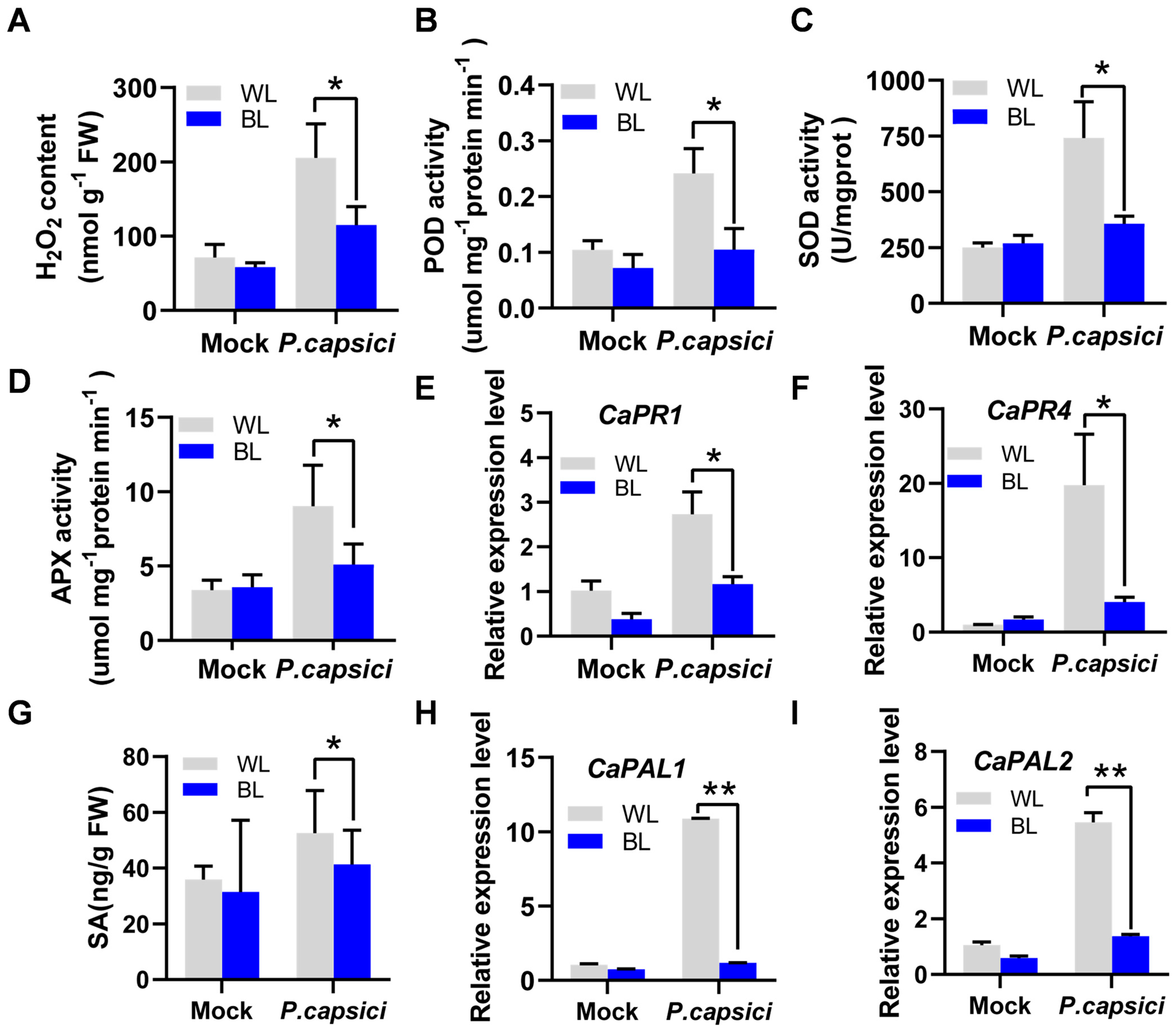
To further investigate the mechanism by which BL weakens pepper resistance to PCI, the H2O2 content was detected. In the infected group, H2O2 accumulation was significantly lower under BL than under WL, which was only 114.85 mmol g−1 FW (Figure 2A). Additionally, the activities of antioxidant enzymes such POD, SOD, and APX of pepper plants with PCI under BL were significantly lower than those under WL, with POD reduced to 41.6% and SOD and APX reduced to 48% and 55.6%, respectively (Figure 2B–D). RT-qPCR analysis revealed that BL also suppressed the expression of pathogenesis-related genes CaPR1 and CaPR4 relative to WL in the infected group (Figure 2E,F). Free SA content in BL treatment was only 41.32 ng/g fresh weight (FW), which was significantly lower than that in the WL control with PCI (Figure 2G). Since CaPALs and CaICS are SA biosynthesis genes, we examined their expression levels. The expression levels of CaPAL1 and CaPAL2 also significantly decreased under BL treatment with PCI compared with those under WL (Figure 2H,I). However, other genes (CaPAL3-7 and CaICS) did not display notably reduced expressions in the infected group under BL relative to WL (Supplementary Figure S1). We speculate that these genes may not contribute significantly to SA synthesis during BL treatment with PCI in pepper. These results showed that BL inhibited antioxidant enzymes activities, H2O2 and SA accumulation, and SA-mediated defense responses, leading to the reduced resistance of peppers to PCI.

3.3. Characterization of CaCRY2

Cryptochromes (CRYs) are one of the most important blue light receptors [25]. To investigate the evolutionary relationships between CRYs and their orthologs in pepper, tomato, and Arabidopsis, a phylogenetic tree was constructed. The results showed that these CRYs could be divided into three groups: CRY1, CRY2, and CRY3 (Figure 3A). In addition, CaCRY2 was significantly induced by BL treatment and PCI (Figure 3B,C). An NCBI conserved domain analysis revealed that CaCRY2 contains a PHR structure at the N-terminus, including MTHF and FAD chromophores, and a CCE structure at the C-terminus (Figure 3D). Amino acid sequence alignment of CaCRY2 and its homolog AtCRY2 indicated high similarity between the two proteins (Supplementary Figure S2). To explore the subcellular localization of CaCRY2, Arabidopsis protoplasts were transfected with a CaCRY2-GFP fusion construct and Ghd7-RFP (a nuclear marker). The green fluorescence signal was almost captured in the nuclei of protoplasts, while strong green fluorescence was observed in both the nucleus and cytoplasm of the positive control (Figure 3E). Meanwhile, RT-qPCR analysis revealed that CaCRY2 was expressed in all tissues, with particularly high levels in the leaves and placenta (Supplementary Figure S3).

3.4. Silencing of CaCRY2 Enhances Pepper Plant Resistance to P. capsici

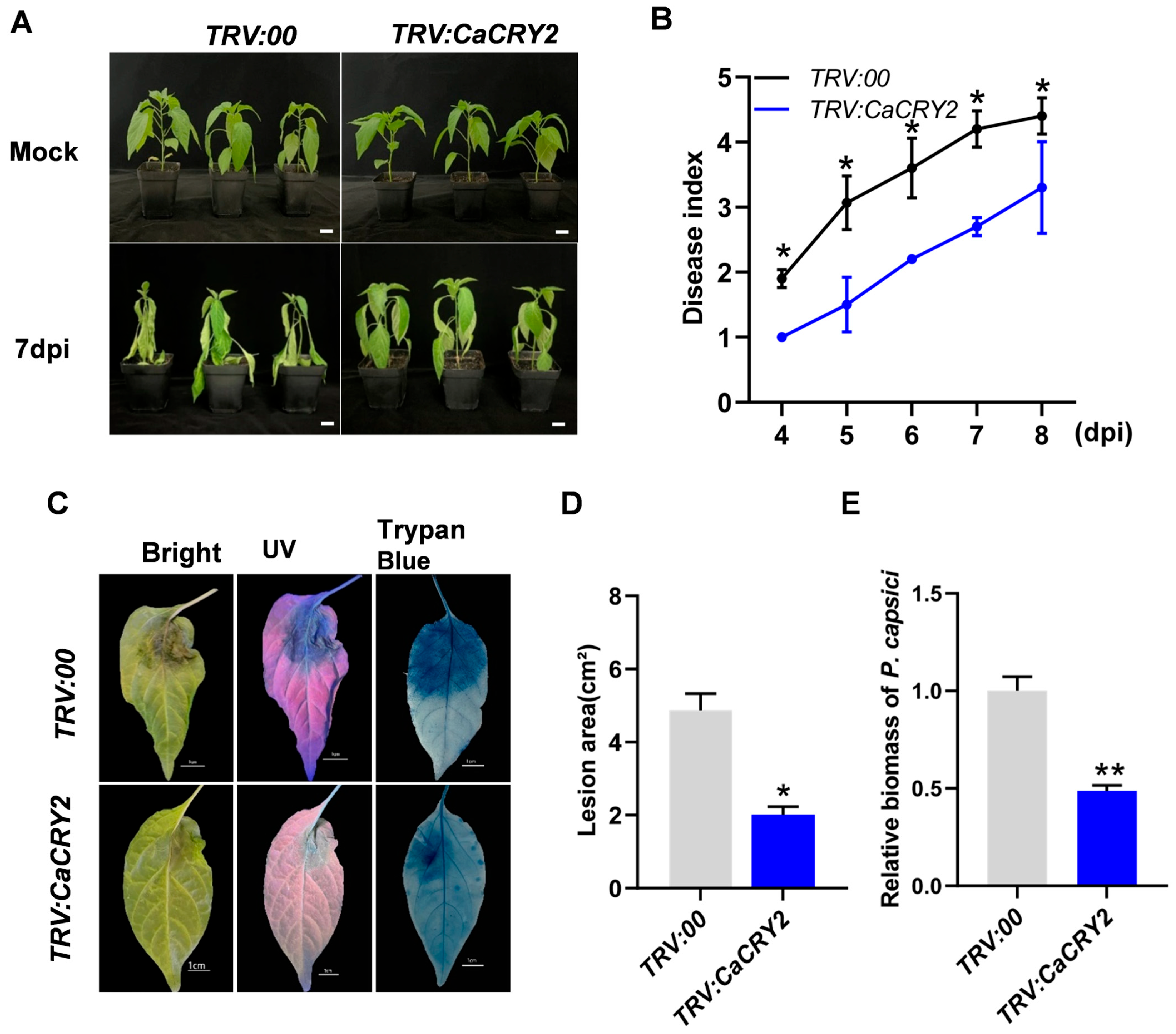
To explore the role of the blue light receptor CaCRY2 in resistance to P. capsici, VIGS technology was used to create CaCRY2-silenced lines (TRV:CaCRY2). TRV:PDS was used to identify silence efficiency (Supplementary Figure S4A). RT-qPCR analysis confirmed that the expression levels of CaCRY2 in the TRV:CaCRY2 lines were significantly lower than those in wild type (WT), averaging only 33% of WT levels (Supplementary Figure S4B). When CaCRY2-silenced and control plants were inoculated with P. capsici, the control plants exhibited more-severe disease symptoms (e.g., wilting and leaf distortion) at 7 dpi, whereas CaCRY2-silenced plants showed enhanced resistance (Figure 4A). Additionally, the disease index was significantly lower in CaCRY2-silenced plants than in controls (Figure 4B). To further verify CaCRY2’s role in resistance to PCI, lesion development on inoculated leaves under UV light after trypan blue staining was observed. The lesion areas of CaCRY2-silenced leaves were obviously decreased compared with those on control leaves (Figure 4C). The lesion area of CaCRY2-silenced leaves was 2.0 cm2 while that in the control was up to 4.86 cm2, with a statistically significant difference (Figure 4D). Moreover, the relative biomass of P. capsici on TRV:CaCRY2 leaves was also significantly decreased relative to that in the control group (Figure 4E). These results indicated that silencing CaCRY2 actually enhances pepper resistance to P. capsici, which is consistent with the previous conclusion that BL treatment reduced pepper resistance to PCI.

3.5. CaCRY2 Participates in ROS and SA Signaling Pathways

To investigate whether CaCRY2 regulates pepper resistance to PCI via the same mechanism as BL, H2O2 contents were measured in TRV:CaCRY2 plants and control plants. The results showed that the TRV:CaCRY2 group accumulated higher H2O2 levels than the control group with PCI. The H2O2 content in the TRV:CaCRY2 group with PCI was 32.75 nmol g−1 FW, while it was only 24.05 nmol g−1 FW in the TRV:00 group with PCI, implying that CaCRY2 negatively regulates H2O2 accumulation in pepper under PCI (Figure 5A). Moreover, the antioxidant enzyme activities of POD, SOD, and APX were also analyzed. Compared with the control TRV:00 plants, TRV:CaCRY2 plants showed a 0.7- to 1.66-fold increase in antioxidant enzyme activities (Figure 5B–D). Simultaneously, CaPR1 and CaPR4 were significantly induced by PCI (Figure 5E,F), and SA accumulation also increased 2.36-fold compared with that in the TRV:00 control group (Figure 5G). Additionally, PCI significantly increased the expression of CaPAL1 and CaPAL4-6 in the TRV:CaCRY2 group compared with that in the controls (Figure 5H–K). However, CaPAL2, CaPAL3, and CaPAL7 showed no significant changes in expression relative to the control, and CaICS expression remained low with or without PCI (Supplementary Figure S5). These results indicate that CaCRY2 and blue light participate in pepper resistance to PCI through the same mechanism, suggesting blue light may regulate pepper resistance to PCI through CaCRY2-mediated ROS and SA signaling pathways.

3.6. Heterologous Expression of CaCRY2 Weakens Tobacco Resistance to P. capsici

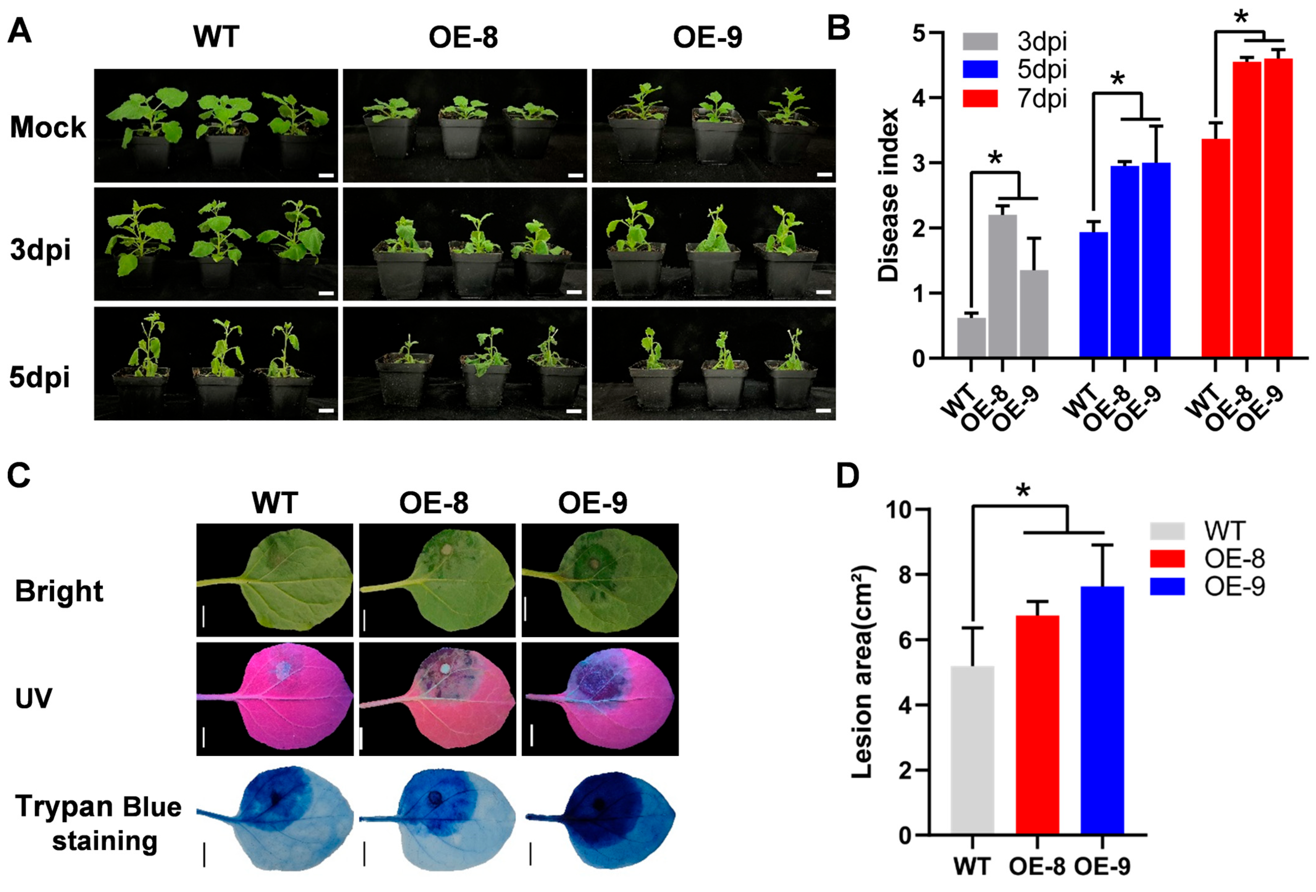
To further characterize the function of CaCRY2, it was ectopically expressed in tobacco. Polymerase chain reaction (PCR) was carried out to identify the transgenic lines containing the 35S:CaCRY2 fusion vector, and two independent lines were selected for subsequent experiments (Supplementary Figure S6A). Quantitative analysis demonstrated that CaCRY2 expression was significantly upregulated in transgenic lines compared with that in the control (Supplementary Figure S6B). Subsequently, homozygous transgenic tobacco plants were inoculated with P. capsici via root irrigation. Three days after inoculation, the CaCRY2 transgenic line exhibited more-severe wilting than the control. Five days after inoculation, the leaves of the control showed initial wilting symptoms, while the leaves of the CaCRY2 transgenic line wilted more severely and fell off, with necrosis and lodging at the growth points (Figure 6A). The disease indexes of the transgenic lines were also significantly higher than those in the control group at 3, 5, and 7 dpi (Figure 6B), consistent with the observed results in Figure 4. In addition, compared with WT, the lesion areas in CaCRY2-overexpressed tobacco were substantially larger than those in wild-type plants (Figure 6C), with average lesion areas of 6.75 cm2 (OE-8) and 7.63 cm2 (OE-9) compared with 5.20 cm2 in controls (Figure 6D).

4. Discussion

4.1. BL Suppresses Pepper Defense Against PCI

Light provides energy for plant growth and development and also participates in regulating plant defense responses [48,49]. Numerous reports indicate that light signals play a considerable role in plants’ response to abiotic stress. For instance, light signaling was vital for cold-induced gene expression in tomato, with far-red light positively regulating cold tolerance, while red light negatively mediated cold tolerance [50]. In Arabidopsis, overexpression of phytochrome-interacting factor OsPIL1 resulted in increased drought tolerance [51]. Furthermore, light signaling also participates in the response to biotic stress. Previous reports revealed that red light reduced the harm of Pseudomonas syringae pv. tomato (Pst) DC3000 to tomato leaves [52], enhanced the resistance of strawberry leaves to Botrytis cinerea [53], and strengthened the resistance of oriental melon leaves to powdery mildew [11]. It is reported that red light mainly regulates plant resistance to pathogens through participation in SA and H2O2 signaling pathways [11,21]. In this study, we discovered that blue light functions in Phytophthora blight disease through H2O2 and SA signaling pathways in pepper plants. Firstly, we detected that BL treatment enhances P. capsici susceptibility in pepper plants compared with the other three lights (Figure 1). Moreover, BL reduced H2O2 accumulation and antioxidant enzyme activities in pepper plants compared with white light treatment with PCI (Figure 2A–D). Simultaneously, BL treatment also regulated SA signaling pathways by repressing SA accumulation and SA-related gene expression levels relative to those in the control group (Figure 2E–I). This was similar to red light promoting H2O2 and SA accumulation and enhanced resistance to powdery mildew in oriental melon leaves [11,14].

4.2. CaCRY2 Regulates Pepper Resistance to P. capsici

Cryptochromes (CRYs) are evolutionarily conserved blue light receptors, which are a class of compounds widely present in animals and plants [31], first discovered in Arabidopsis [54]. Amino acid sequence alignment indicated high homology between CaCRY2 and AtCRY2 (Supplementary Figure S2), which is consistent with their high level of conservation. This study identified the localization of CaCRY2 in the nucleus (Figure 3E) in accordance with a previous study that determined that AtCRY2 in Arabidopsis was a nuclear localization protein [55]. Tissue-specific expression analysis revealed that CaCRY2 was expressed in various tissues of pepper plants but mainly expressed in the leaves and placenta (Supplementary Figure S3), implying potential roles in both photomorphogenesis and fruit development.
Many studies have shown that CRYs coordinate various growth and developmental processes and responses to environmental stimulus in plants [29,31,56,57]. Moreover, CRYs have been found to play a vital role in pathogen resistance. In Arabidopsis, CRY1 induced systemic acquired resistance (SAR) and positively regulated salicylic acid (SA)-induced pathogenesis-related gene PR-1 expression to enhance Pseudomonas syringae defense [38]. Since our former research has identified CaCRY1 as a negative regulator that modulates pepper resistance against Phytophthora capsici [58] and CaCRY2 shares about 59.03% amino acid identity with CaCRY1 (Supplementary Figure S7), we speculate that CaCRY2 and CaCRY1 may have the same function in pepper resistance to PCI. However, CaCRY3 shares only 29.36% amino acid identity with CaCRY1 and lacks a CCE domain at the C-terminus, which is noticeably different in CaCRY2 and CaCRY1 (Supplementary Figure S7). Therefore, we did not investigate the biological role of CaCRY3, and this study only focused on the function of CaCRY2. In this study, we found that CaCRY2 participated in pepper resistance to PCI by regulating CaPR1 and CaPR4 (Figure 5E,F), implying that it mediates the SA pathway, in accordance with CRY1 being involved in the SA pathway in Arabidopsis [38]. In addition, CRY2 negatively regulated an E3 ubiquitin ligase to maintain the stability of the R protein HRT, enhancing turnip crinkle virus (TCV) resistance in Arabidopsis [39]. A previous study revealed that cry1 and cry2 mutants had greater CMV (cucumber mosaic virus) accumulation and damage than that in the wild type, suggesting that CRYs have a positive regulatory effect on resistance to CMV in Arabidopsis [40]. Recently, it has been found that CRY1a promotes tomato resistance to gray mold [59]. Therefore, CRYs have been well characterized regarding their role in defense against pathogens in Arabidopsis and tomato. Moreover, CaCRY1 has been identified as a negative regulator of PCI in our previous study [58]; however, CaCRY2’s functions in pepper defense against P. capsici remain unknown. Interestingly, our study found that CaCRY2 was negatively correlated with pepper defense against P. capsici (Figure 4), contrasting with the function of certain orthologous genes. We speculate that it may be because CRY1 and CRY2 are grouped separately (Figure 3A) and because of the divergence between species.

4.3. CaCRY2 Enhances Pepper Susceptibility to P. capsici via ROS and SA Pathways

The plant–pathogen interaction causes a burst of reactive oxygen species, especially H2O2 accumulation [40,60]. Consistent with this, H2O2 levels were also notably increased in TRV:CaCRY2 plants with PCI compared with those in TRV:00 plants (Figure 5A). However, excessive H2O2 impairs cellular structure and causes oxidative stress. Antioxidative enzymes, such as SOD, POD, and APX are primarily responsible for scavenging ROS [12,61]. Recently a study has indicated that there is a relationship between pepper defense against P. capsici and the antioxidant system: SOD and peroxidase including POD and APX played a positive role in pepper defense responses to P. capsici [62]. We also detected that these antioxidant enzymes’ activities changed after P. capsici infection compared with those in the control group (Figure 2B–D and Figure 5B–D). SA is an important hormone for plant defense and plays a crucial role in many aspects of plant immunity [11,21]. We detected that some CaPR expression levels and SA accumulation changed after P. capsici infection relative to those in the control group (Figure 2E–G and Figure 5E–G). These results reveal that BL treatment and CaCRY2 manipulate H2O2 and SA signaling pathways to resist PCI in pepper plants. According to previous reports, CRY often forms complexes through protein interactions to participate in downstream gene regulation, such as CRY-COP1-SPY complexes [63], CRY-PIF complexes [64], and CRY1-COP1-HY5 complexes [65]. PIFs (phytochrome-interacting factors) can specifically bind to the G-box elements, and HY5 (elongated hypocotyl 5) can specifically bind to the G/E-box elements in promoters of downstream genes. In addition, G/E-box elements are common in some ROS/SA signaling pathway genes. Therefore, we speculate that CRY may form complexes with other proteins such as PIF or HY5 to modulate ROS/SA pathways. However, some gene expression patterns in the supplemental figures showed unexpected variations. For example, a few CaPALs showed no significant correlation with PCI compared with the control group (Supplementary Figures S1 and S5). These observations possibly reflect complex regulatory networks or experimental conditions that require further investigation. Additionally, transgenic tobacco overexpressing CaCRY2 showed a higher plant disease index and a high sensitivity to P. capsici relative to WT (Figure 6). Taking together, when CaCRY2 was silenced, the ability of pepper to receive blue light signals weakened, and the inhibitory effect of blue light on the accumulation of H2O2 and SA also weakened. Based on the above conditions, pepper resistance to PCI was increased (Figure 7A). But, when the blue light receptor CaCRY2 was not silenced and could receive blue light signals normally, BL treatment reduced the accumulation of H2O2 and SA and led to decreased pepper resistance (Figure 7B). Therefore, we determine that the CaCRY2 plays a negative regulatory role in pepper’s resistance to P. capsici.

5. Conclusions

Collectively, this study exhibited that the blue light receptor CaCRY2 negatively regulates P. capsici resistance in pepper via VIGS and transgenic technologies. Furthermore, we discovered that silencing CaCRY2 increased H2O2 and SA accumulation, elevated antioxidant enzyme activities, and induced SA biosynthesis gene expression to participate in H2O2 and SA signaling pathways (Figure 7). Our findings reveal a previously uncharacterized function of CaCRY2 and identify potential target genes for genetic modification to enhance disease resistance in pepper breeding programs.

Supplementary Materials

The following supporting information can be downloaded at https://www.mdpi.com/article/10.3390/horticulturae11121434/s1: Figure S1: Expression levels of CaPAL3, CaPAL4, CaPAL5, CaPAL6, CaPAL7, and CaICS in peppers with PCI under BL treatment. Figure S2: Amino acid sequence alignment of CaCRY2 and AtCRY2. Figure S3: Expression levels of CaCRY2 in different pepper tissues. Figure S4: Expression levels of CaCRY2 in silenced pepper plants. Figure S5: Expression levels of CaPAL3, CaPAL4, CaPAL5, and CaICS in TRV:CaCRY2 peppers with PCI. Figure S6: Heterologous expression of CaCRY2 in tobacco. A: PCR-based analysis of CaCRY2 overexpression lines. B: Expression levels of CaCRY2 in heterologous tobacco plants. Figure S7: Amino acid sequence alignment of three CaCRYs. Table S1: Primers used in this study.

Author Contributions

Y.Y., S.L., and J.C. planned and designed the research. T.Y. analyzed the data and wrote the original manuscript. Y.C. conducted most of the experiments; Y.L. (Ying Luo) and H.L. cultivated plants; Y.Z. and Y.L. (Yachun Lin) analyzed the data; X.W. revised the manuscript. All authors have read and agreed to the published version of the manuscript.

Funding

This work was supported by grants from the National Natural Science Foundation of China (32372797, 32160739, 32460797, and 32460798), the China Postdoctoral Science Foundation (2024M761229), the Natural Science Foundation of Jiangxi Province, China (20212ACB215006, 20224BAB215022, 20252BAC200399, 20223BBF61017), the earmarked fund for Jiangxi Agriculture Research System (JXARS-06), and the Science and Technology Research Project of Jiangxi Provincial Department of Education (GJJ210411).

Data Availability Statement

The original contributions presented in this study are included in the article/Supplementary Materials. Further inquiries can be directed to the corresponding authors.

Conflicts of Interest

The authors declare no competing interests.

References

  1. Chen, W.; Wang, X.; Sun, J.; Wang, X.; Zhu, Z.; Ayhan, D.; Yi, S.; Yan, M.; Zhang, L.; Meng, T.; et al. Two telomere-to-telomere gapless genomes reveal insights into Capsicum evolution and capsaicinoid biosynthesis. Nat. Commun. 2024, 15, 4295. [Google Scholar] [CrossRef] [PubMed]
  2. Hausbeck, M.; Lamour, K. Phytophthora capsici on vegetable crops: Research progress and management challenges. Plant Dis. 2004, 88, 1292–1303. [Google Scholar] [CrossRef] [PubMed]
  3. Vogel, G.; Giles, G.; Robbins, K.; Gore, M.; Smart, C. Quantitative genetic analysis of interactions in the pepper-Phytophthora capsici pathosystem. Mol. Plant Microbe Interact. 2021, 35, 1018–1033. [Google Scholar] [CrossRef] [PubMed]
  4. Barchenger, D.W.; Lamour, K.H.; Bosland, P.W. Challenges and strategies for breeding resistance in Capsicum annuum to the multifarious pathogen, Phytophthora capsici. Front. Plant Sci. 2018, 9, 628. [Google Scholar] [CrossRef]
  5. Li, Q.; Ai, G.; Shen, D.; Zou, F.; Wang, J.; Bai, T.; Chen, Y.; Li, S.; Zhang, M.; Jing, M.; et al. A Phytophthora capsici effector targets ACD11 binding partners that regulate ROS-mediated defense response in Arabidopsis. Mol. Plant 2019, 12, 565–581. [Google Scholar] [CrossRef]
  6. Lei, G.; Zhou, K.; Chen, X.; Huang, Y.; Yuan, X.; Li, G.; Xie, Y.; Fang, R. Transcriptome and metabolome analyses revealed the response mechanism of pepper roots to Phytophthora capsici infection. BMC Genom. 2023, 24, 626. [Google Scholar] [CrossRef]
  7. Rogers, H.; Munné-Bosch, S. Production and scavenging of reactive oxygen species and redox signaling during leaf and flower senescence: Similar but different. Plant Physiol. 2016, 171, 1560–1568. [Google Scholar] [CrossRef]
  8. Cui, J.; Jiang, N.; Meng, J.; Yang, G.; Liu, W.; Zhou, X.; Ma, N.; Hou, X.; Luan, Y. LncRNA33732-respiratory burst oxidase module associated with WRKY1 in tomato- Phytophthora infestans interactions. Plant J. 2019, 97, 933–946. [Google Scholar] [CrossRef]
  9. Li, W.; Zhu, Z.; Chern, M.; Yin, J.; Yang, C.; Li, R.; Cheng, M.; He, M.; Wang, K.; Wang, J.; et al. A natural allele of a transcription factor in rice confers broad-spectrum blast resistance. Cell 2017, 170, 114–126. [Google Scholar] [CrossRef]
  10. Cheng, D.; Liu, M.; Sun, X.; Zhao, M.; Chow, W.; Sun, G.; Zhang, Z.; Hu, Y. Light suppresses bacterial population through the accumulation of hydrogen peroxide in tobacco leaves infected with Pseudomonas syringae pv. tabaci. Front. Plant Sci. 2016, 7, 512. [Google Scholar] [CrossRef]
  11. Wang, L.; Wu, X.; Yu, B.; Han, Y.; Zhang, Y.; Xu, D.; Qi, H. Red light activates H2O2 signal involved in powdery mildew resistance in oriental melon seedlings. Environ. Exp. Bot. 2023, 215, 105508. [Google Scholar] [CrossRef]
  12. Yang, Y.; Wu, C.; Ahammed, G.; Wu, C.; Yang, Z.; Wan, C.; Chen, J. Red light-induced systemic resistance against root-knot nematode is mediated by a coordinated regulation of salicylic acid, jasmonic acid and redox signaling in watermelon. Front. Plant Sci. 2018, 9, 899. [Google Scholar] [CrossRef] [PubMed]
  13. Li, J.; Luan, Y.; Liu, Z. Overexpression of SpWRKY1 promotes resistance to Phytophthora nicotianae and tolerance to salt and drought stress in transgenic tobacco. Physiol. Plantarum 2015, 155, 248–266. [Google Scholar] [CrossRef] [PubMed]
  14. Wang, L.; Wu, X.; Xing, Q.; Zhao, Y.; Yu, B.; Ma, Y.; Wang, F.; Qi, H. PIF8-WRKY42-mediated salicylic acid synthesis modulates red light induced powdery mildew resistance in oriental melon. Plant Cell Environ. 2023, 46, 1726–1742. [Google Scholar] [CrossRef] [PubMed]
  15. Luo, D.; Cai, J.; Sun, W.; Yang, Q.; Hu, G.; Wang, T. Tomato SlWRKY3 negatively regulates Botrytis cinerea resistance via TPK1b. Plants 2024, 13, 1597. [Google Scholar] [CrossRef]
  16. Jing, Y.; Lin, R. Transcriptional regulatory network of the light signaling pathways. New Phytol. 2020, 227, 683–697. [Google Scholar] [CrossRef]
  17. Wang, J.; Lu, W.; Tong, Y.; Yang, Q. Leaf morphology, photosynthetic performance, chlorophyll fluorescence, stomatal development of lettuce (Lactuca sativa L.) exposed to different ratios of red light to blue light. Front. Plant Sci. 2016, 7, 250. [Google Scholar] [CrossRef]
  18. Wang, S.; Fang, H.; Xie, J.; Wu, Y.; Tang, Z.; Liu, Z.; Lv, J.; Yu, J. Physiological responses of cucumber seedlings to different supplemental light duration of red and blue LED. Front. Plant Sci. 2021, 12, 709313. [Google Scholar] [CrossRef]
  19. Wang, H.; Jiang, Y.; Yu, H.; Xia, X.; Shi, K.; Zhou, Y.; Yu, J. Light quality affects incidence of powdery mildew, expression of defence-related genes and associated metabolism in cucumber plants. Eur. J. Plant Pathol. 2010, 127, 125–135. [Google Scholar] [CrossRef]
  20. Jung, J.; Li, Z.; Chen, H.; Yang, S.; Li, D.; Priatama, R.; Kumar, V.; Xuan, Y. Mutation of phytochrome B promotes resistance to sheath blight and saline–alkaline stress via increasing ammonium uptake in rice. Plant J. 2022, 113, 277–290. [Google Scholar] [CrossRef]
  21. Yang, Y.; Li, Y.; Guang, Y.; Lin, J.; Zhou, Y.; Yu, T.; Ding, F.; Wang, Y.; Chen, J.; Zhou, Y.; et al. Red light induces salicylic acid accumulation by activating CaHY5 to enhance pepper resistance against Phytophthora capsici. Hortic. Res. 2023, 10, uhad213. [Google Scholar] [CrossRef]
  22. Hogewoning, S.; Trouwborst, G.; Maljaars, H.; Poorter, H.; van Ieperen, W.; Harbinson, J. Blue light dose-responses of leaf photosynthesis, morphology, and chemical composition of Cucumis sativus grown under different combinations of red and blue light. J. Exp. Bot. 2010, 61, 3107–3117. [Google Scholar] [CrossRef]
  23. Inoue, S.; Kinoshita, T. Blue light regulation of stomatal opening and the plasma membrane H+-ATPase. Plant Physiol. 2017, 174, 531–538. [Google Scholar] [CrossRef]
  24. Naqvi, S.; He, Q.; Trusch, F.; Qiu, H.; Pham, J.; Sun, Q.; Christie, J.; Gilroy, E.; Birch, P. Blue-light receptor phototropin 1 suppresses immunity to promote Phytophthora infestans infection. New Phytol. 2022, 233, 2282–2293. [Google Scholar] [CrossRef]
  25. Chaves, I.; Pokorny, R.; Byrdin, M.; Hoang, N.; Ritz, T.; Brettel, K.; Essen, L.; van der Horst, G.; Batschauer, A.; Ahmad, M. The cryptochromes: Blue light photoreceptors in plants and animals. Annu. Rev. Plant Biol. 2011, 62, 335–364. [Google Scholar] [CrossRef]
  26. Lin, C.; Shalitin, D. Cryptochrome structure and signal transduction. Annu. Rev. Plant Biol. 2003, 54, 469–496. [Google Scholar] [CrossRef] [PubMed]
  27. Brudler, R.; Hitomi, K.; Daiyasu, H.; Toh, H.; Kucho, K.; Ishiura, M.; Kanehisa, M.; Roberts, V.; Todo, T.; Tainer, J.; et al. Identification of a new cryptochrome class: Structure, function, and evolution. Mol. Cell 2003, 11, 59–67. [Google Scholar] [CrossRef] [PubMed]
  28. Liu, H.; Liu, B.; Zhao, C.; Pepper, M.; Lin, C. The action mechanisms of plant cryptochromes. Trends Plant Sci. 2011, 16, 684–691. [Google Scholar] [CrossRef]
  29. Wang, Q.; Lin, C. A structural view of plant CRY2 photoactivation and inactivation. Nat. Struct. Mol. Biol. 2020, 27, 401–403. [Google Scholar] [CrossRef]
  30. Ye, Y.; Li, R.; Pu, W.; Zhang, Y.; Jiang, L.; Li, H.; Liu, Y.; Ye, Y.; Yue, M.; Lin, Y.; et al. Expression analysis and interaction protein screening of CRY1 in strawberry. Horticulturae 2022, 8, 460. [Google Scholar] [CrossRef]
  31. Xu, P.; Chen, H.; Li, T.; Xu, F.; Mao, Z.; Cao, X.; Miao, L.; Du, S.; Hua, J.; Zhao, J.; et al. Blue light-dependent interactions of CRY1 with GID1 and DELLA proteins regulate gibberellin signaling and photomorphogenesis in Arabidopsis. Plant Cell 2021, 33, 2375–2394. [Google Scholar] [CrossRef]
  32. Zuo, Z.; Liu, H.; Liu, B.; Liu, X.; Lin, C. Blue light-dependent interaction of CRY2 with SPA1 regulates COP1 activity and floral initiation in Arabidopsis. Curr. Biol. 2011, 21, 841–847. [Google Scholar] [CrossRef]
  33. Somers, D.; Devlin, P.; Kay, S. Phytochromes and cryptochromes in the entrainment of the Arabidopsis circadian clock. Science 1998, 282, 1488–1490. [Google Scholar] [CrossRef]
  34. Mao, J.; Zhang, Y.; Sang, Y.; Li, Q.; Yang, H. A role for Arabidopsis cryptochromes and COP1 in the regulation of stomatal opening. Proc. Natl. Acad. Sci. USA 2005, 102, 12270–12275. [Google Scholar] [CrossRef] [PubMed]
  35. Kang, C.; Lian, H.; Wang, F.; Huang, J.; Yang, H. Cryptochromes, phytochromes, and COP1 regulate light-controlled stomatal development in Arabidopsis. Plant Cell 2009, 21, 2624–2641. [Google Scholar] [CrossRef] [PubMed]
  36. Ma, D.; Li, X.; Guo, Y.; Chu, J.; Fang, S.; Yan, C.; Noel, J.; Liu, H. Cryptochrome 1 interacts with PIF4 to regulate high temperature-mediated hypocotyl elongation in response to blue light. Proc. Natl. Acad. Sci USA 2016, 113, 224–229. [Google Scholar] [CrossRef] [PubMed]
  37. Li, Y.; Shi, Y.; Li, M.; Fu, D.; Wu, S.; Li, J.; Gong, Z.; Liu, H.; Yang, S. The CRY2-COP1-HY5-BBX7/8 module regulates blue light-dependent cold acclimation in Arabidopsis. Plant Cell 2021, 33, 3555–3573. [Google Scholar] [CrossRef]
  38. Wu, L.; Yang, H. Cryptochrome 1 is implicated in promoting R protein-mediated plant pesistance to Pseudomonas syringae in Arabidopsis. Mol. Plant 2010, 3, 539–548. [Google Scholar] [CrossRef]
  39. Jeong, R.; Chandra-Shekara, A.; Barman, S.; Navarre, D.; Klessig, D.; Kachroo, A.; Kachroo, P.; Jones, J. Cryptochrome 2 and phototropin 2 regulate resistance protein-mediated viral defense by negatively regulating an E3 ubiquitin ligase. Proc. Natl. Acad. Sci. USA 2010, 107, 13538–13543. [Google Scholar] [CrossRef]
  40. Zhou, X.; Zhu, T.; Zhu, L.; Luo, S.; Deng, X.; Lin, H.; Xi, D. The role of photoreceptors in response to cucumber mosaic virus in Arabidopsis thaliana. J. Plant Growth Regul. 2016, 36, 257–270. [Google Scholar] [CrossRef]
  41. Nie, W.; Li, Y.; Chen, Y.; Zhou, Y.; Yu, T.; Zhou, Y.; Yang, Y. Spectral light quality regulates the morphogenesis, architecture, and flowering in pepper (Capsicum annuum L.). J. Photochem. Photobiol. B Biol. 2023, 241, 112673. [Google Scholar] [CrossRef] [PubMed]
  42. Zhou, Y.; Li, Y.; Yu, T.; Li, J.; Qiu, X.; Zhu, C.; Liu, J.; Dang, F.; Yang, Y. Characterization of the B-BOX gene family in pepper and the role of CaBBX14 in defense response against Phytophthora capsici infection. Int. J. Biol. Macromol 2023, 237, 124071. [Google Scholar] [CrossRef] [PubMed]
  43. Livak, K.; Schmittgen, T. Analysis of relative gene expression data using real-time quantitative PCR and the 2−ΔΔCT method. Methods 2001, 25, 402–408. [Google Scholar] [CrossRef] [PubMed]
  44. Xue, W.; Xing, Y.; Weng, X.; Zhao, Y.; Tang, W.; Wang, L.; Zhou, H.; Yu, S.; Xu, C.; Li, X.; et al. Natural variation in Ghd7 is an important regulator of heading date and yield potential in rice. Nat. Genet. 2008, 40, 761–767. [Google Scholar] [CrossRef]
  45. Thompson, J.; Gibson, T.; Higgins, D. Multiple sequence alignment using ClustalW and ClustalX. Curr. Protoc. Bioinform. 2003, 2.3.1–2.3.22. [Google Scholar] [CrossRef]
  46. Wang, F.; Chen, X.; Dong, S.; Jiang, X.; Wang, L.; Yu, J.; Zhou, Y. Crosstalk of PIF4 and DELLA modulates CBF transcript and hormone homeostasis in cold response in tomato. Plant Biotechnol. J. 2020, 18, 879–1105. [Google Scholar] [CrossRef]
  47. Li, H.; Chang, J.; Chen, H.; Wang, Z.; Gu, X.; Wei, C.; Zhang, Y.; Ma, J.; Yang, J.; Zhang, X. Exogenous melatonin confers salt stress tolerance to watermelon by improving photosynthesis and redox homeostasis. Front. Plant Sci. 2017, 8, 295. [Google Scholar] [CrossRef]
  48. Fernández-Milmanda, G.; Crocco, C.; Reichelt, M.; Mazza, C.; Köllner, T.; Zhang, T.; Cargnel, M.; Lichy, M.; Koo, A.; Austin, A.; et al. A light-dependent molecular link between competition cues and defence responses in plants. Nat. Plants 2019, 6, 223–230. [Google Scholar] [CrossRef]
  49. Roeber, V.; Bajaj, I.; Rohde, M.; Schmülling, T.; Cortleven, A. Light acts as a stressor and influences abiotic and biotic stress responses in plants. Plant Cell Environ. 2020, 44, 645–664. [Google Scholar] [CrossRef]
  50. Wang, F.; Guo, Z.; Li, H.; Wang, M.; Onac, E.; Zhou, J.; Xia, X.; Shi, K.; Yu, J.; Zhou, Y. Phytochrome A and B function antagonistically to regulate cold tolerance via abscisic acid-aependent jasmonate signaling. Plant Physiol. 2016, 170, 459–471. [Google Scholar] [CrossRef]
  51. Kudo, M.; Kidokoro, S.; Yoshida, T.; Mizoi, J.; Todaka, D.; Fernie, A.; Shinozaki, K.; Yamaguchi-Shinozaki, K. Double overexpression of DREB and PIF transcription factors improves drought stress tolerance and cell elongation in transgenic plants. Plant Biotechnol. J. 2016, 15, 458–471. [Google Scholar] [CrossRef]
  52. Yang, Y.; Wang, M.; Yin, Y.; Onac, E.; Zhou, G.; Peng, S.; Xia, X.; Shi, K.; Yu, J.; Zhou, Y. RNA-seq analysis reveals the role of red light in resistance against Pseudomonas syringae pv. tomato DC3000 in tomato plants. BMC Genom. 2015, 16, 120. [Google Scholar] [CrossRef]
  53. Meng, L.; Mestdagh, H.; Ameye, M.; Audenaert, K.; Höfte, M.; Labeke, M. Phenotypic variation of Botrytis cinerea isolates is influenced by spectral light quality. Front. Plant Sci. 2020, 11, 1233. [Google Scholar] [CrossRef]
  54. Ahmad, M.; Cashmore, A. HY4 gene of A. thaliana encodes a protein with characteristics of a blue-light photoreceptor. Nature 1993, 366, 162. [Google Scholar] [CrossRef]
  55. Yu, X.; Klejnot, J.; Zhao, X.; Shalitin, D.; Maymon, M.; Yang, H.; Lee, J.; Liu, X.; Lopez, J.; Lin, C. Arabidopsis cryptochrome 2 completes its posttranslational life cycle in the nucleus. Plant Cell 2007, 19, 3146–3156. [Google Scholar] [CrossRef]
  56. Fantini, E.; Sulli, M.; Zhang, L.; Aprea, G.; Jiménez-Gómez, J.; Bendahmane, A.; Perrotta, G.; Giuliano, G.; Facella, P. Pivotal roles of cryptochromes 1a and 2 in tomato development and physiology. Plant Physiol. 2019, 179, 732–748. [Google Scholar] [CrossRef] [PubMed]
  57. Wang, W.; Lu, X.; Li, L.; Lian, H.; Mao, Z.; Xu, P.; Guo, T.; Xu, F.; Du, S.; Cao, X.; et al. Photoexcited Cryptochrome 1 interacts with dephosphorylated BES1 to regulate brassinosteroid signaling and photomorphogenesis in Arabidopsis. Plant Cell 2018, 30, 1989–2005. [Google Scholar] [CrossRef] [PubMed]
  58. Sun, F.; Chen, Y.; Luo, Y.; Yang, F.; Yu, T.; Han, H.; Yang, Y.; Zhou, Y. Cryptochromes (CRYs) in pepper: Genome-wide identification, evolution and functional analysis of the negative role of CaCRY1 under Phytophthora capsici infection. Plant Sci. 2025, 355, 112460. [Google Scholar] [CrossRef] [PubMed]
  59. Cai, Y.; Ying, J.; Ye, Y.; Wen, S.; Qian, R. Blue light regulates jasmonic acid synthesis via CRY1a and boosts antioxidant enzymes activity in Solanum lycopersicum to resist Botrytis cinerea. Plant Cell Rep. 2025, 44, 160. [Google Scholar] [CrossRef]
  60. Baxter, A.; Mittler, R.; Suzuki, N. ROS as key players in plant stress signalling. J. Exp. Bot. 2013, 65, 1229–1240. [Google Scholar] [CrossRef]
  61. Gao, Y.; Liu, J.; Yang, F.; Zhang, G.; Wang, D.; Zhang, L.; Ou, Y.; Yao, Y. The WRKY transcription factor WRKY8 promotes resistance to pathogen infection and mediates drought and salt stress tolerance in Solanum lycopersicum. Physiol. Plant 2019, 168, 98–117. [Google Scholar] [CrossRef]
  62. Zhang, H.; Feng, X.; Ali, M.; Jin, J.; Wei, A.; Khattak, A.; Gong, Z. Identification of pepper CaSBP08 gene in defense response against Phytophthora capsici infection. Front. Plant Sci. 2020, 11, 183. [Google Scholar] [CrossRef]
  63. Wang, H.; Ma, L.; Li, J.; Zhao, H.; Deng, X. Direct interaction of Arabidopsis Cryptochromes with COP1 in mediation of photomorphogenic development. Science 2001, 294, 154–158. [Google Scholar] [CrossRef] [PubMed]
  64. Pedmale, U.; Huang, S.; Zander, M.; Cole, B.; Hetzel, J.; Ljung, K.; Reis, P.; Sridevi, P.; Nito, K.; Nery, J.; et al. Cryptochromes interact directly with PIFs to control plant growth in limiting blue light. Cell 2016, 164, 233–245. [Google Scholar] [CrossRef]
  65. Liu, S.; Wang, Q.; Zhong, M.; Lin, G.; Ye, M.; Wang, Y.; Zhang, J.; Wang, Q. The CRY1-COP1-HY5 axis mediates blue-light regulation of Arabidopsis thermotolerance. Plant Commun. 2025, 6, 101264. [Google Scholar] [CrossRef]
Figure 1. Different light spectra regulate pepper resistance to P. capsici. (A) Phenotype of pepper plants with different light treatments at 4 days postinfection (dpi). WL, white light; RL, red light; BL, blue light; GL, green light. Scale bars = 1 cm. (B) Disease index with different light treatments at 4 dpi. Letters above the bars indicate significant differences as determined by Student’s t-test (p < 0.05). The bars present mean values ± SDs (n = 6). (CE) Phenotype of detached leaves, lesion area, and the relative biomass of P. capsici under different light treatments at 3 dpi. Scale bars = 1 cm. The bars present mean values ± SDs (n = 3).
Horticulturae 11 01434 g001
Figure 2. BL affects ROS and SA signaling pathways in pepper plants with PCI. (AI) H2O2 accumulation; the activities of POD, SOD, and APX; the expression levels of CaPR1 and CaPR4; SA accumulation; and the expression levels of CaPAL1 and CaPAL2 in pepper plants exposed to blue and white light with PCI. Statistically significant differences are established by asterisks: ** at p < 0.01 and * at p < 0.05. The bars present mean values ± SDs (n = 3).
Horticulturae 11 01434 g002
Figure 3. CaCRY2 protein family, expression profile, and subcellular localization. (A) CRY protein sequences from pepper, Arabidopsis, and tomato were subjected to phylogenetic reconstruction. Red frame is used to highlight. (B,C) Expression levels of CaCRY2 in pepper leaves induced by BL and PCI. *, p < 0.05; **, p < 0.01. The bars present mean values ± SDs (n = 3). (D) Gene structure of CaCRY2. (E) Subcellular localization of the CaCRY2 protein. Transient expression of 35S:GFP and 35S:CaCRY2-GFP in Arabidopsis protoplasts. Imaging was performed on bright-field and GFP fluorescence channels. Ghd7-RFP was used as the nucleus marker. Bright-field, green fluorescent protein, red fluorescent protein, and merged images are shown. Scale bars: top, 7.5 μm; bottom, 10 μm.
Horticulturae 11 01434 g003
Figure 4. CaCRY2 negatively regulates pepper resistance to PCI. (A) Phenotype of CaCRY2-silenced pepper plants at 7 dpi. Scale bars = 1 cm. (B) Disease index of CaCRY2-silenced pepper plants with PCI from 4 to 8 dpi (n = 6). (CE) Phenotype of CaCRY2-silenced detached leaves. Scale bars = 1 cm; lesion area and the relative biomass of P. capsici with PCI at 3 dpi. *, p < 0.05; **, p < 0.01. The bars present mean values ± SDs (n = 3).
Horticulturae 11 01434 g004
Figure 5. CaCRY2 affects ROS and SA signaling pathways in pepper plants with PCI. (AK) Contents of H2O2; activities of POD, SOD, and APX; expression levels of CaPR1 and CaPR4; contents of SA; and expression levels of CaPAL1, CaPAL4, CaPAL5, and CaPAL6 in TRV:CaCRY2 pepper plants with PCI or without PCI. Statistically significant differences are established by asterisks: ** at p < 0.01 and * at p < 0.05. The bars present mean values ± SDs (n = 3).
Horticulturae 11 01434 g005
Figure 6. CaCRY2 negatively regulates transgenic tobacco resistance to PCI. (A) Phenotype of CaCRY2-OE tobacco plants at 3 and 5 dpi. Scale bars = 1 cm. (B) Disease indexes of CaCRY2-OE tobacco plants with PCI at 3, 5, and 7dpi (n = 6). (C,D) Phenotype and lesion area of CaCRY2-overexpressed detached leaves. Scale bars = 1 cm; OE, overexpressed. Asterisks indicate statistically significant differences. *, p < 0.05. The bars present mean values ± SDs (n = 3).
Horticulturae 11 01434 g006
Figure 7. A proposed model of CaCRY2’s response to Phytophthora capsici infection (PCI) through ROS and SA signaling pathways in pepper. (A) CaCRY2 is silenced. The dashed lines represent a weakened effect. (B) CaCRY2 is normally expressed.
Horticulturae 11 01434 g007
Disclaimer/Publisher’s Note: The statements, opinions and data contained in all publications are solely those of the individual author(s) and contributor(s) and not of MDPI and/or the editor(s). MDPI and/or the editor(s) disclaim responsibility for any injury to people or property resulting from any ideas, methods, instructions or products referred to in the content.