Article

Induction and Transformation of Friable Callus in Chrysanthemum ‘Jimba’

1
College of Horticulture, Hunan Agricultural University, Changsha 410128, China
2
Yuelushan Laboratory, Changsha 410128, China
3
Hunan Mid-Subtropical Quality Plant Breeding and Utilization Engineering Technology Research Center, Changsha 410128, China
4
Engineering Center for Germplasm Innovation and New Variety Breeding of Horticultural Crops, Ministry of Education, Changsha 410128, China
5
Hunan Horticultural Research Institute, Hunan Academy of Agricultural Sciences, Changsha 410125, China
*
Author to whom correspondence should be addressed.
These authors contributed equally to this work.
Horticulturae 2025, 11(10), 1267; https://doi.org/10.3390/horticulturae11101267
Submission received: 4 September 2025 / Revised: 30 September 2025 / Accepted: 1 October 2025 / Published: 21 October 2025
(This article belongs to the Special Issue Genetic Innovation and Breeding in Ornamental Plants)

Abstract

The friability of callus is closely associated with its genetic transformation efficiency, and optimizing induction and transformation conditions is essential for establishing an efficient transformation system. In this study, we developed a high-efficiency friable callus induction and Agrobacterium-mediated transformation system for Chrysanthemum ‘Jimba’. Three plant growth regulator (PGR) combinations—6-Benzylaminopurine (6-BA) + Naphthaleneacetic Acid (NAA), 6-BA + 2,4-Dichlorophenoxyacetic Acid (2,4-D), and Thidiazuron (TDZ) + 2,4-D—were evaluated for their effects on callus morphology, proliferation, and transformation efficiency. The optimal PGR combination was identified as 1.0 mg/L 6-BA + 0.4 mg/L NAA, which produced highly friable calli with a loose structure, rapid proliferation, and the highest nuclear-to-cytoplasmic ratio. The optimal subculture time for maintaining friability and high proliferation was the 7th week, while the best Agrobacterium infection conditions were OD600 = 0.5 with a 10 min infection period, which achieved a transformation efficiency of 91%. This optimized protocol provides an efficient and rapid transformation method for future gene function studies using callus transformation.

1. Introduction

Agrobacterium-mediated genetic transformation is a cornerstone technique in genetic engineering and functional genomics, and it largely depends on an efficient transformation system. A key factor in this process is the selection of recipient plant material, which directly affects transformation efficiency. Common recipient materials include stem segments, leaves, petioles, and calli. Among these, calli are favored due to their higher capacity for exogenous DNA uptake, lower chimeric rate, and wide availability in plant tissues [1,2,3,4,5,6]. Recently, callus-based transformation has become a focal point of research, particularly as it has been used for gene function identification in species such as pear, apple, and Solanum betaceum [7,8,9]. Despite its advantages, callus-based transformation faces challenges, including the optimization of callus induction, subculture time, and infection conditions.
Calli can be categorized into two types based on cell characteristics: friable calli and compact calli [10]. Friable calli typically have a friable cell arrangement, loose structure, high cell division activity, and a high capacity for exogenous gene integration, leading to higher transformation efficiency compared to compact calli. In contrast, compact calli consist of a densely packed cell arrangement with a firmer texture and lower cell activity. Its growth primarily relies on cell enlargement rather than division, resulting in lower transformation efficiency [11,12]. Therefore, inducing friable calli is crucial for improving genetic transformation efficiency.
The induction and growth of calli are influenced by multiple factors, including the explant source, the types and concentrations of plant growth regulators (PGRs), the composition of the culture medium, etc. Among these, PGRs are key determinants of callus induction efficiency and quality [13]. A suitable combination of auxins and cytokinins can effectively promote friable callus induction, while excessive or insufficient concentrations can slow down growth or result in a compact structure. Commonly used PGRs for callus induction include 6-Benzylaminopurine (6-BA), Naphthaleneacetic Acid (NAA), 2,4-Dichlorophenoxyacetic Acid (2,4-D), and Thidiazuron (TDZ). However, the optimal PGR combinations for inducing friable callus differ across plant species. For example, in lilac (Syringa spp.), blueberry (Vaccinium spp.), and rose (Rosa hybrida), a combination of TDZ and 2,4-D is effective in inducing friable calli [14,15,16]. In grape (Vitis vinifera) and pomegranate (Punica granatum), a combination of 6-BA and NAA is more effective [17,18], while in crabapple (Malus spectabilis) and white-spine sophora (Sophora davidii), 6-BA and 2,4-D induce high rates of friable callus formation [19,20]. These findings indicate the importance of screening the optimal PGRs for callus induction in specific plant species.
The subculture time is crucial for maintaining the friable state of calli. After a period of growth, friable calli need to be transferred to fresh medium due to nutrient depletion, water loss, and accumulation of toxic metabolites. If the callus is subcultured too early, the nutrients in the medium are not fully utilized. Conversely, subculturing too late can lead to nutrient deficiency and the accumulation of toxic metabolites, negatively affecting callus proliferation. Therefore, optimal subculture time ensures efficient nutrient utilization and supports continuous callus proliferation [21,22,23].
A callus, composed of a mass of cells, is more susceptible to Agrobacterium infiltration compared to traditional recipient materials like leaf discs or stem segments, making it more sensitive to both infection concentration and duration. Insufficient bacterial concentration and duration lead to incomplete infection and reduced transformation efficiency. Conversely, excessive bacterial concentration and overly long infection duration cause overproliferation of Agrobacterium, resulting in contamination, which inhibits callus growth and reduces transformation efficiency. Thus, optimal infection concentration and duration are crucial for improving transformation efficiency [24].
Chrysanthemum (Chrysanthemum × morifolium Ramat), one of the four major cut flowers, holds significant ornamental and economic value. Among the various cultivars, ‘Jimba’ is particularly well-known and extensively studied. Several studies have investigated tissue culture conditions and genetic transformation systems for ‘Jimba’ [25,26]. For instance, Teng et al. optimized the culture medium for callus induction and adventitious bud differentiation from ‘Jimba’ leaves [25], while Zhang et al. established a genetic transformation system using leaf discs [26]. However, research focusing on the induction of friable calli in ‘Jimba’ is still lacking. This study aims to optimize the induction conditions for friable calli in ‘Jimba’, determine the optimal subculture time, and identify the best conditions for Agrobacterium-mediated transformation. Furthermore, the transformation efficiency of friable and compact calli was then compared.

2. Materials and Methods

2.1. Plant Materials

In this study, sterilized seedlings of Chrysanthemum ‘Jimba’ were cultivated in a tissue culture room at the Laboratory of Hunan Mid-subtropical Quality Plant Breeding and Utilization Engineering Technology Research Center, Hunan Agricultural University. The stem segments were cultured in a basic MS medium (seen in Section 2.3) formulation for subcultivation. The in vitro cultivation conditions were as follows: 20–25 °C room temperature, 60–70% relative humidity, and a 12 h light/dark photoperiod, with a total incubation period of approximately 30 days.

2.2. Gene Expression Vector and Bacterial Strain

The expression vector used for transformation was pGreenII0029-62-SK, and the plasmid of 35S::CmMYB6-CmbHLH2 was constructed according to a previous study [27]. The Agrobacterium tumefaciens strain used was GV3101 (Cat No.AC1002S, Weidi, Shanghai, China).

2.3. Basic Medium Formulation and Culture Conditions

The media were prepared by adding 30 g sucrose, 4.4 g Murashige and Skoog (Cat No. PM1011, Coolaber, Beijing, China) medium [28], and 7.0 g agar to ultrapure water, with the final volume adjusted to 1 L. The pH was adjusted to 5.8 using a 4% (w/v) NaOH solution, and the medium was then autoclaved at 121 °C for 20 min. The explants [25] were cultured in vitro under the following conditions: 20–25 °C room temperature, 60–70% relative humidity, a 12 h light/dark photoperiod, and 100 μmol m−2 s−1 photosynthetically active radiation.

2.4. Screening of Friable Callus Induction Medium

MS medium was used as the base, supplemented with 7.0 g/L agar and varying concentrations of growth regulators (6-BA (Coolaber, China), NAA (Solabio, Beijing, China), 2,4-D (Solabio, China), and TDZ (Solabio, China)). The pH was adjusted to 5.8. The volume of culture medium was 30 mL in each Petri dish. ‘Jimba’ internodal stem segments (approximately 0.5 cm) were used for callus induction. Five to eight explants were inoculated per dish, with six dishes per replicate and three replicates. After 30 days, the browning rate, proliferation ratio, and callus induction time were recorded, and callus growth status was observed.
The growth regulator treatments are shown in Table 1.

2.5. Observation of Callus Cells Under a Microscope

The method was modified from Xue [29]. Briefly, peripheral callus tissues were excised and stained using an acetocarmine solution (Coolaber, Beijing, China). The stained cells were placed on a glass slide, covered with a coverslip, and gently pressed to ensure uniform dispersion. The prepared slides were observed under a light microscope to record cell size, morphology, structure, and nuclear dimensions. The nuclear-to-cytoplasmic ratio was subsequently calculated.

2.6. Growth Curve Plotting for Proliferation of Friable Callus

Aseptic tissue-cultured seedlings of Chrysanthemum ‘Jimba’ with uniform growth were selected, and stem segments were excised. Explants were weighed every 7 days, with 10 randomly selected explants per measurement, and the average weight was calculated from three replicates. After 30 days, a growth curve was plotted with culture duration (days) on the X-axis and average proliferative weight on the Y-axis. Based on the growth stages, the optimal subculture time was determined.

2.7. Screening of Subculture Time for Friable Callus

Using the optimal culture medium for friable callus induction, calli with a similar texture and color from the same batch were selected. Four sampling points corresponding to different growth phases (slow, rapid, and stable proliferation) were chosen. Callus fragments (approximately 0.1 g) were subcultured, and fresh weight was measured every 3 days to record the proliferation increment. The proliferation dynamics were analyzed and visualized using GraphPad Prism 9.5.

2.8. Culture of Agrobacterium Colonies and Preparation of Agrobacterium Infection Solution

The Agrobacterium strain stored at −80 °C was streaked onto LB solid medium supplemented with 50 mg/L kanamycin and 25 mg/L gentamicin, followed by inverted incubation at 28 °C for 48 h. A single colony was picked and inoculated into 1 mL LB liquid medium containing the same antibiotics, and cultured with shaking at 200 rpm at 28 °C for 12 h. Subsequently, 30 µL of the bacterial suspension was transferred to 100 mL LB liquid medium with antibiotics and expanded under the same conditions until the OD600 reached the target value. The bacterial culture was centrifuged at 5000 rpm for 5 min, and the supernatant was discarded. The pellet was resuspended in MS liquid medium containing 50 mmol/L acetosyringone (AS (Coolaber, Beijing, China)) to prepare the infection solution.

2.9. Screening of Agrobacterium Infection Concentration and Duration

Friable calli induced from leaf segments of Chrysanthemum ‘Jimba’ were cultured on 1.0 mg/L 6-BA and 0.4 mg/L NAA media for 3–4 days and then selected as the experimental material. Infection treatments were set with durations of 5, 10, and 20 min, and bacterial suspension concentrations were adjusted to OD600 = 0.4, 0.5, and 0.6. After 30 days of culture, the Agrobacterium contamination rate, callus proliferation rate, callus browning rate, and transformation efficiency were recorded, and the total anthocyanin content in the induced calli was measured. Furthermore, the relative expression of reporter gene CmMYB6-CmbHLH2 was analyzed. Based on these, the optimal infection concentration and duration for the genetic transformation of friable calli of Chrysanthemum ‘Jimba’ were determined.

2.10. Total Anthocyanin Content Measurement

The anthocyanin content was measured according to the method described by Akira Nakatsuka [30] with minor modifications. Briefly, 0.1 g of callus was weighed and ground in liquid nitrogen, followed by extraction in 1 mL of 1% (v/v) HCl–methanol solution. The mixture was subjected to dark extraction at 4 °C for 24 h, centrifuged at 6000 rpm for 5 min, and the supernatant was collected. The absorbance at 525 nm was measured using a spectrophotometer.

2.11. RNA Extraction and RT-qPCR

Total RNA was extracted from the callus using a Plant RNA Extraction Kit (Aikerui, Changsha, Hunan, China). First-strand cDNA was synthesized from the extracted RNA using the R323 Reverse Transcription Kit (Vazyme, Nanjing, China) following the manufacturer’s protocol. RT-qPCR analysis was performed with SYBR Green dye (Vazyme, Nanjing, China) using a real-time PCR system. Gene expression levels were calculated using the (Ct)2−ΔΔCt method, with CmActin serving as the internal reference gene [31]. Primer sequences are listed in Table 2.

2.12. Data Statistics and Calculation Formulas

Browning rate (%) = Number of browned callus/Total number of callus explants × 100; Proliferation ratio = Area of callus after 30 days of culture/Initial area of callus; Nuclear-to-cytoplasmic ratio (%) = Nuclear area/Cell area × 100; Agrobacterium contamination rate (%) = Number of contaminated explants/Total number of inoculated explants × 100; Transformation efficiency (%) = Number of explants with anthocyanin accumulation/Total number of explants × 100.
The areas of calli and cells were measured with Fiji Is Just Image J software, and graphs were plotted using Graphpad Prism 9.5. The experimental data were statistically analyzed using Excel 2013 and SPSS Statistics Base software. Multiple comparisons following a one-way ANOVA were conducted using Tukey’s test.

3. Results

3.1. Optimal PGR Combination for Friable Callus Induction in Chrysanthemum ‘Jimba’ Is 1.0 mg/L 6-BA + 0.4–0.6 mg/L NAA

To screen the optimal PGR combinations and concentrations for friable callus induction in Chrysanthemum ‘Jimba’, ‘Jimba’ stem segments were cultured on 24 culture media with different PGR combinations and concentrations, including a growth regulator-free medium (CK, control). After 30 days, callus browning rate, proliferation ratio, and induction time were observed and analyzed.
As shown in Table 3, the control medium (CK) had a callus browning rate of 42% and a proliferation ratio of 1.91, which was significantly lower than that in media containing PRGs. Among the 24 hormone-containing media, the NAA + 6-BA combination (Y1-Y4) induced higher callus proliferation ratios compared to the 6-BA + 2,4-D (Y5-8) and TDZ + 2,4-D (Y9-Y24). The highest proliferation ratios (5.45 and 5.40) were observed at NAA concentrations of 0.4 mg/L and 0.6 mg/L, respectively. Further increasing the NAA concentration resulted in a decrease in the proliferation ratio and an increase in the browning rate. Additionally, the NAA + 6-BA (Y1-Y4) combination had the shortest callus induction time.
The proliferation ratio of calli can reflect the friability of calli to a certain extent [32]. Based on this, calli were classified into four types: Type I (proliferation ratio > 5), Type II (4–5), Type III (3–4), and Type IV (<3). The NAA + 6-BA combinations predominantly induced Type I calli; 2,4-D + 6-BA primarily induced Type II and Type III calli; and 2,4-D + TDZ mainly induced Type II–Type IV calli. Overall, Type I calli had the highest proliferation ratio with a loose structure, a soft texture, and green or yellow-green coloration, making it the closest to friable calli.
In conclusion, the 6-BA + NAA combination is more suitable for inducing Type I calli. The combination of 1.0 mg/L 6-BA with 0.4–0.6 mg/L NAA induced calli with the shortest induction time and the fastest proliferation ratio.

3.2. Type I Calli Exhibit the Highest Nuclear-to-Cytoplasmic Ratio and Most Friable Morphology

Friable and compact calli exhibit distinct cellular morphological characteristics beyond differences in the proliferation ratio. Generally, friable calli tend to have smaller cells, larger nuclei, denser cytoplasm, and more regular cell shapes compared to compact calli [33]. To further explore these structural differences, we observed the cellular morphology and calculated the nuclear-to-cytoplasmic ratio of four callus types (Type I–IV).
Random samples from each type were selected (Figure 1) and observed under a microscope. Type I calli exhibited densely arranged, regularly shaped cells with larger nuclei (Figure 1b-I). In contrast, Type II and III callus cells were irregular in shape, exhibited considerable size variation, and had smaller nuclei (Figure 1b-II,III). Type IV callus cells were larger with smaller nuclei. Type I calli showed the highest nuclear-to-cytoplasmic ratio, 21.07%, suggesting active metabolic and division activity, followed by Type II (8.79%), Type III (7.74%), and Type IV (5.44%; Figure 1b-II,III,IV).
The results showed significant morphological differences. Type I calli exhibited smaller cells and a higher nuclear-to-cytoplasmic ratio (Table 4), indicating that it is more friable than other types.

3.3. Comparative Analysis of Transformation Efficiency in Callus Induced by Different PGR Combinations

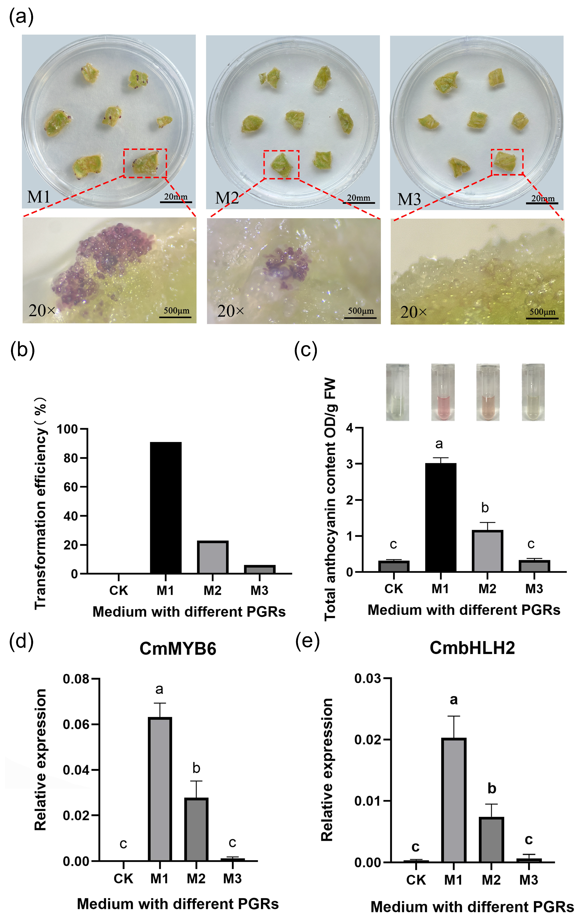
To evaluate whether different plant growth regulator (PGR) combinations affect transformation efficiency, leaf explants of Chrysanthemum ‘Jimba’ were cultured on media with three commonly used PGR combinations: 1.0 mg/L 6-BA + 0.4 mg/L NAA (M1), 1.0 mg/L 6-BA + 0.2 mg/L 2,4-D (M2), and 0.5 mg/L 2,4-D + 0.5 mg/L TDZ (M3), and then transformed with the anthocyanin reporter gene 35S::CmMYB6-CmbHLH2. Transformation efficiency was assessed by quantifying anthocyanin content and the relative expression levels of CmMYB6 and CmbHLH2.
As shown in Figure 2a, calli induced on M1 accumulated visibly more pigmentation compared to those induced on M2 and M3, suggesting higher anthocyanin production. Quantitative analysis (Figure 2b) revealed that explants on M1 medium exhibited the highest transformation efficiency (91%), significantly surpassing M2 (22.9%) and M3 (6.2%). Anthocyanin content followed a similar trend (Figure 2c), with M1-treated explants reaching 3.02 OD/g FW, much higher than those on M2 (1.17 OD/g FW) and M3 (0.33 OD/g FW). Accordingly, the expression levels of CmMYB6 and CmbHLH2 (Figure 2d,e) were significantly upregulated in M1-induced explants compared to the other two groups (p < 0.05).
These results demonstrate that transformation efficiency is significantly influenced by the PGR combination used for callus induction. The M1 (1.0 mg/L 6-BA + 0.4 mg/L NAA) produced the most favorable growth conditions for transformation, as evidenced by the highest levels of reporter gene expression and anthocyanin accumulation.

3.4. The 7th Week Is the Optimal Subculture Time for Friable Callus

The callus proliferation curve is an essential tool for determining the optimal subculture time. In this study, the proliferation curve of friable calli was plotted based on its proliferation rate.
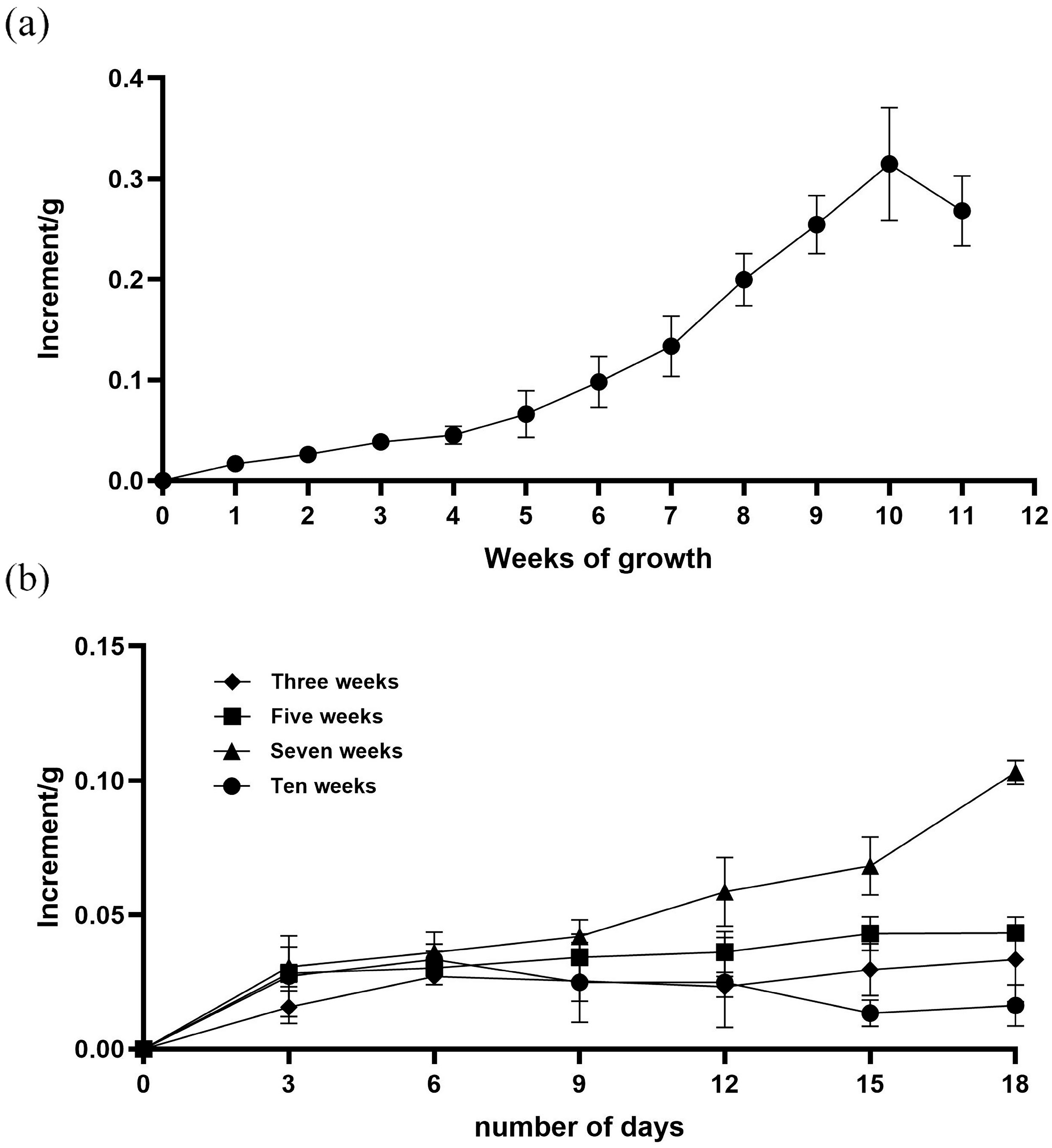
As shown in Figure 3a, the callus fresh weight initially increased, reaching a peak at the 10th week, and then began to decline. Prior to the 5th week, the proliferation remained relatively stable. From the 5th to the 10th week, calli entered a rapid proliferation phase, with the highest fresh weight proliferation at the 10th week, and then declined. These results suggest that the optimal subculture period falls between the 5th and 10th week, providing a basis for further subculture time optimization.
Subculture time significantly influences the growth of subsequent callus generation. Based on the callus proliferation curve, four subculture time points (3rd, 5th, 7th, and 10th week) were selected for analysis of their effects on calli. As shown in Figure 3b, calli subcultured at the 7th week exhibited the highest proliferation over time. On day 18, the proliferation amount of calli subcultured at the 7th week was 0.09 g, compared to 0.03 g, 0.04 g, and 0.01 g for calli subcultured at the 3rd, 5th, and 10th weeks, respectively.
In conclusion, the 7th week was identified as the optimal subculture time.

3.5. Optimal Agrobacterium Infection Conditions for Chrysanthemum Callus Were OD600 0.5 and 10-Min Infection

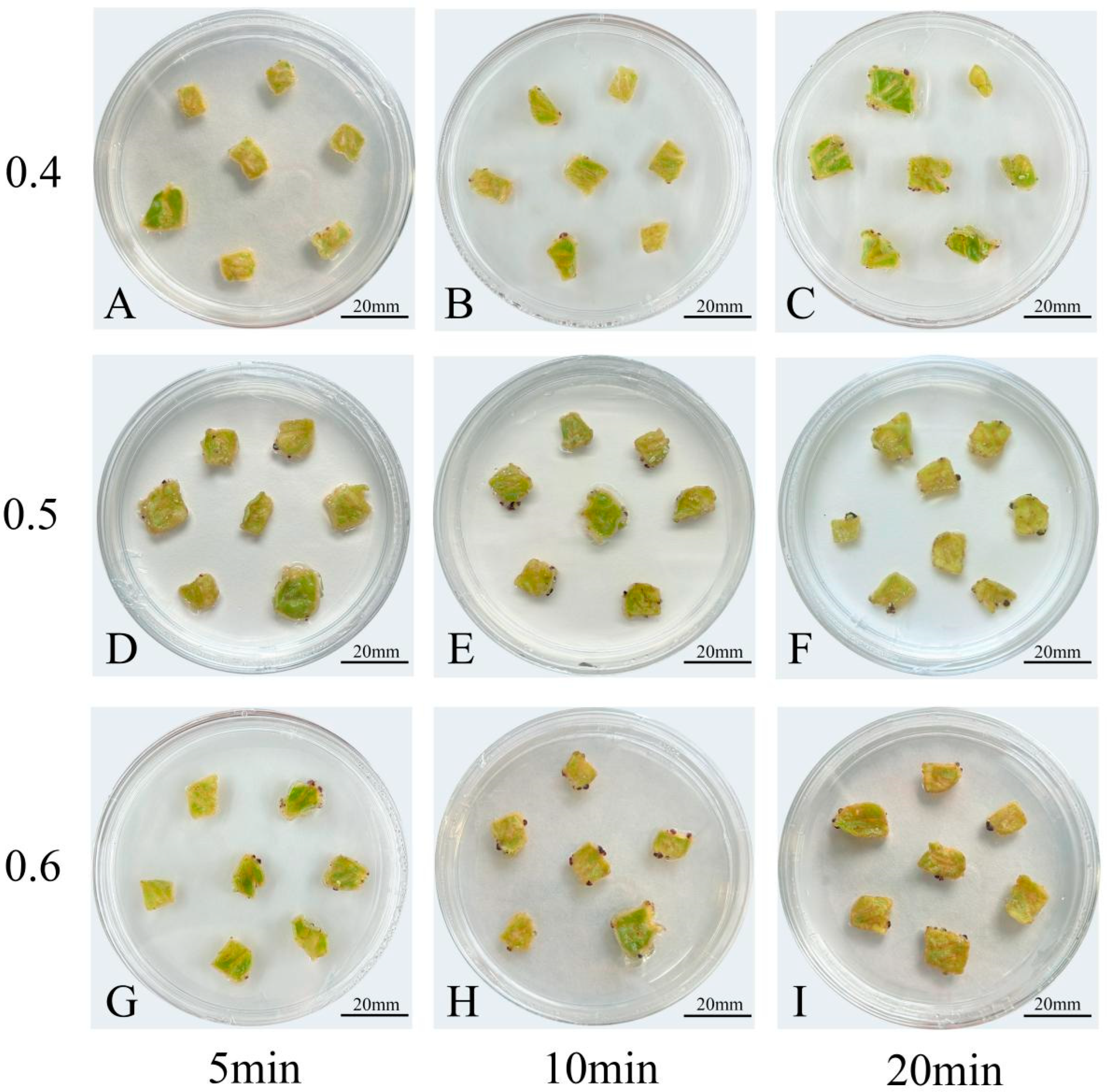
To screen the Agrobacterium infection conditions, three Agrobacterium concentrations (OD600 = 0.4, 0.5, and 0.6) and three infection durations (5, 10, and 20 min) were tested. The Agrobacterium contamination rate, anthocyanin content, and transformation efficiency were assessed under each condition. The results are as follows (Table 5, Figure 4).
At OD600 = 0.4, with infection times of 5, 10, and 20 min, the transformation efficiency was 72%, 82%, and 89%; the total anthocyanin content was 0.31 OD/gFW, 2.16 OD/gFW, and 2.46 OD/gFW; and the contamination rate was 0%, 0%, and 7%, respectively.
At OD600 = 0.5, with infection times of 5, 10, and 20 min, the transformation efficiency was 75%, 91%, and 85%. The total anthocyanin content was 2.23 OD/gFW, 3.25 OD/gFW, and 3.43 OD/gFW, while the contamination rate was 0%, 0%, and 11%. Among them, the highest transformation efficiency (91%) was achieved at an infection time of 10 min, with significantly higher total anthocyanin content than other treatments (p < 0.05). Although the anthocyanin content was slightly lower than that at 20 min, there was no significant difference (p > 0.05), and Agrobacterium contamination occurred at 20 min.
At OD600 = 0.6, with infection times of 5, 10, and 20 min, the transformation efficiency was 75%, 72% and 54%, the total anthocyanin content decreased from 2.82 OD/gFW to 1.96 OD/gFW, and the contamination rate increased from 9% to a maximum of 33%. This indicates that a bacterial solution concentration of 0.6 easily causes excessive growth of Agrobacterium, and the increased Agrobacterium contamination leads to a decrease in transformation efficiency.
Based on these results, it is suggested that OD600 = 0.5, with 10 min infection, was the optimal infection condition, which resulted in the highest transformation efficiency.

4. Discussion

4.1. The Optimal PRGs Combination for Inducing Friable Callus in Chrysanthemum ‘Jimba’ Is 6-BA + NAA

In Chrysanthemum ‘Jimba’, the combination of 6-BA and NAA is the most effective plant growth regulator (PGR) formulation for inducing friable calli. Previous studies have shown that the combination of 6-BA and NAA has a higher callus induction efficiency in chrysanthemum. Liu et al. reported that supplementing MS medium with 6-BA and NAA significantly enhanced callus induction from ‘Japanese Yellow’ chrysanthemum leaves, compared to the 6-BA and 2,4-D combination [34]. Similarly, Wang et al. found that 6-BA and NAA were more effective than 6-BA and 2,4-D for callus induction from leaf discs of ‘Jimba’ chrysanthemum [35]. In this study, three commonly used PGR combinations, 6-BA + NAA, 6-BA + 2,4-D, and 2,4-D + TDZ, were tested at different concentrations to evaluate their effects on friable callus induction [21,36,37,38,39]. The results clearly demonstrated that 6-BA + NAA had the best overall performance, not only in induction efficiency but also in generating calli with loose structure and higher cell activity. Unlike previous studies that primarily focused on callus induction rates, this research introduced additional evaluation dimensions, including callus texture, proliferation ratio, and transformation efficiency.
A novel classification system based on proliferation-associated friability was established, dividing calli into four types (Type I–IV). Type I calli exhibited rapid proliferation, a loose structure, and a high nucleus-to-cytoplasm ratio, indicating strong division activity. Most calli induced by the 6-BA + NAA combination were classified as Type I, confirming its superior ability to induce friable calli.

4.2. Effect of Subculture Time on the Proliferation Rate of Friable Callus in Chrysanthemum ‘Jimba’

The 7th week is the optimal subculture time for maintaining the proliferation rate and friability of calli in Chrysanthemum ‘Jimba’. Previous research has shown that the optimal subculture time for callus proliferation varies significantly among plant species. For example, in Taxus, subculturing on the 10th day promoted rapid proliferation and reduced browning [22]; in Gentiana rhodantha, the optimal time was 42 days [40]; and in Hemerocallis, a 21-day cycle maintained high viability [41]. These differences highlight the species-specific nature of subculture timing.
In this study, callus proliferation of Chrysanthemum ‘Jimba’ was monitored over time, revealing three distinct phases: a slow proliferation phase (weeks 1–4), a rapid proliferation phase (weeks 5–10), and a decline phase (after week 10). Based on this growth pattern, subculture was tested at weeks 3, 5, 7, and 10. The results demonstrated that the week 7 subculture produced the highest proliferation rate and best-maintained callus friability. Unlike prior studies that only focused on assessing general viability, this study provides a detailed proliferation curve for Chrysanthemum ‘Jimba’ and correlates subculture timing with callus increment.
In summary, the 7-week subculture time is optimal for promoting active, friable calli in Chrysanthemum ‘Jimba’, offering a valuable reference for chrysanthemum tissue culture systems. However, whether this interval should be maintained in subsequent subcultures remains to be determined through further research.

4.3. Effects of Infection Concentration and Duration on the Growth and Proliferation of Friable Callus in Chrysanthemum ‘Jimba’

Calli, due to their loose cellular structure, are more prone to Agrobacterium attachment than leaf discs or stem segments [36], making infection parameters critical. In previous studies, infection conditions for callus transformation varied widely across species—for example, OD600 = 0.4 in Manchurian ash [33], OD600 = 0.6 for 25 min in rose [16], and OD600 = 0.6 for 5 min in walnut [42]. In chrysanthemum, using leaves or stem segments, optimal conditions generally fall between OD600 = 0.5–0.7 and 7–30 min [43,44,45,46]. For callus-based transformation, cultivars ‘Purple Elf’ and ‘National Day Red’ achieved the best results at OD600 = 0.4–0.5 with 5–7 min of infection [46].
For friable calli of Chrysanthemum ‘Jimba’, we found the highest transformation efficiency at OD600 = 0.5 with a 10 min infection. Under these conditions, contamination was 0%, transformation efficiency reached 91%, and total anthocyanin content was 3.25 OD/g FW (Table 5, Figure 4). These findings provide precise infection conditions for friable callus transformation in Chrysanthemum ‘Jimba’, offering a reliable reference for improving genetic transformation protocols in this cultivar.

4.4. Comparison of Transformation Efficiency Among Callus Induced by Different PGRs

The type and concentration of PRGs not only affect the morphological characteristics of calli but may also directly influence transformation efficiency by regulating cell division, differentiation, and metabolic activity. However, many studies on callus induction in chrysanthemum primarily focus on growth status, but the transformation efficiency of different callus types has not been further explored [25,26,33].
In this study, we further investigated the transformation efficiency of calli induced by three PGR combinations. By examining the expression of the reporter gene in different types of calli, the results showed that calli induced by 6-BA + 2,4-D exhibited the highest transformation efficiency, with higher reporter gene expression and more pronounced anthocyanin accumulation. This result indicates that the PGRs directly affect transformation efficiency. Specifically, 6-BA + 2,4-D induced calli characterized by stronger cell activity, faster division rates, and more vigorous metabolic capacity, enhance the transformation efficiency of the reporter gene.
Compared to previous studies, this research provides a more detailed analysis of the performance of different PGR-induced calli in genetic transformation, focusing not only on the growth state and cell activity but also on their direct impact on transformation efficiency. In conclusion, this study reveals that the transformation efficiency of calli was influenced by different PGR combinations’ impact on their metabolic state, cell activity, and proliferation rate.

Author Contributions

Conceptualization, L.X. (Lili Xiang); Data curation, R.F., Q.D. and J.W.; Funding acquisition, L.X. (Lili Xiang); Investigation, R.F., Q.D. and J.W.; Methodology, R.F., Q.D., J.W., Y.L. and L.X. (Lu Xu); Project administration, L.X. (Lili Xiang); Resources, L.X. (Lili Xiang), X.Y., G.T. and W.L.; Software, R.F. and J.W.; Supervision, L.X. (Lili Xiang); Validation, R.F., Q.D. and L.X. (Lili Xiang); Visualization, R.F. and Q.D.; Writing—original draft, R.F. and Q.D.; Writing—review and editing, R.F., Q.D. and L.X. (Lili Xiang). All authors have read and agreed to the published version of the manuscript.

Funding

This research was financially supported by the National Natural Science Foundation of China (Grant No. 32202533), Hunan Provincial Natural Science Foundation Project (Grant No. 2023JJ40320), project of Hunan Provincial Department of Education (Grant No. 23B0232), Hunan Province Science and Technology Plan Project (Grant No. 2023NK4250), and 2024 National Key R&D Program of China (2024YFD1600903-3).

Data Availability Statement

The original contributions presented in this study are included in the article. Further inquiries can be directed to the corresponding author.

Conflicts of Interest

The authors declare no conflicts of interest.

References

  1. Zhang, S.; Yan, S.; An, P.; Cao, Q.; Wang, C.; Wang, J.; Zhang, H.; Zhang, L. Embryogenic callus induction from immature zygotic embryos and genetic transformation of Larix kaempferiLarix gmelinii 9. PLoS ONE 2021, 16, e258654. [Google Scholar] [CrossRef]
  2. Zhang, A.J.; Jiang, X.N.; Gai, Y. Optimization of genetic transformation method for embryogenic callus of Larix kaempferi. Mol. Plant Breed. 2024, 1–13. Available online: http://kns.cnki.net/kcms/detail/46.1068.S.20220606.1454.004.html (accessed on 30 September 2025).
  3. Armstrong, C.L.; Green, C.E. Establishment and maintenance of friable, embryogenic maize callus and the involvement of L-proline. Planta 1985, 164, 207–214. [Google Scholar] [CrossRef]
  4. Xu, S.R.; Zhao, W.Z.; Gong, X.Y.; Li, L.; Xiao, W. Resistance analysis of apple B-type cytokinin response factor MdARR11 to drought stress. J. Nucl. Agric. Sci. 2024, 38, 226–234. [Google Scholar]
  5. Zhang, K.C.; Li, X.Y.; Zhang, Y.G.; Zhang, D.Y.; Wang, Y.C. Cloning and Expression Analysis of MsCBF1 Gene from Malus sieversii (Ldb.) Roem. Plant Physiol. J. 2018, 54, 803–811. [Google Scholar]
  6. Wang, Y.; Yuan, J.; Wei, X.; Chen, Y.; Chen, Q.; Ge, X. GhLBDs Promote Callus Initiation and Act as Selectable Markers to Increase Transformation Efficiency. Front. Plant Sci. 2022, 13, 861706. [Google Scholar] [CrossRef]
  7. Xue, Y.; Shan, Y.; Yao, J.L.; Wang, R.; Xu, S.; Liu, D.; Ye, Z.; Lin, J.; Li, X.; Xue, C.; et al. The transcription factor PbrMYB24 regulates lignin and cellulose biosynthesis in stone cells of pear fruits. Plant Physiol. 2023, 192, 1997–2014. [Google Scholar] [CrossRef]
  8. Cordeiro, D.; Alves, A.; Ferraz, R.; Casimiro, B.; Canhoto, J.; Correia, S. An efficient Agrobacterium-mediated genetic transformation method for Solanum betaceum Cav. embryogenic callus. Plants 2023, 12, 1202. [Google Scholar] [CrossRef]
  9. Li, X.L.; Ding, N.; Jia, M.R.; Wei, L.Z.; Jiang, J.Z.; Li, B.B.; Jia, W.S. Establishment of gene transformation system in fruit callus and its application in gene functional analysis for apple plant. J. China Agric. Univ. 2015, 20, 108–113. [Google Scholar]
  10. Ikeuchi, M.; Sugimoto, K.; Iwase, A. Plant callus: Mechanisms of induction and repression. Plant Cell 2013, 25, 3159–3173. [Google Scholar] [CrossRef]
  11. Yi, Y.H.; Xu, D.F.; Dong, H.Y.; Liu, Y.J. Induction of loose embryogenic callus in melon (Cucumis melo L.). Seed 2023, 42, 144–150. [Google Scholar]
  12. Ramírez-Mosqueda, M.A. Overview of Somatic Embryogenesis. Somat. Embryog. Methods Protoc. 2022, 2527, 1–8. [Google Scholar]
  13. Tokuji, Y.; Kuriyama, K. Involvement of gibberellin and cytokinin in the formation of embryogenic cell clumps in carrot (Daucus carota). J. Plant Physiol. 2003, 160, 133–141. [Google Scholar] [CrossRef]
  14. Yu, Q.Y.; Guo, M.M.; Xu, K.X.; Liu, J.B.; Wang, C.; Zheng, J.; Zhang, K.Z.; Zhang, Y. Callus induction and suspension culture of inflorescence and rachis from Syringa vulgaris ‘Downfield’. J. Northeast. For. Univ. 2023, 51, 47–53. [Google Scholar]
  15. Ghosh, A.; Igamberdiev, A.U.; Debnath, S.C. Thidiazuron-induced somatic embryogenesis and changes of antioxidant properties in tissue cultures of half-high blueberry plants. Sci. Rep. 2018, 8, 16978. [Google Scholar]
  16. Yi, X. Basic Research on Embryogenic Callus Induction and Genetic Transformation of Rosa chinensis ‘Yueyuehong’. Master’s Thesis, Hunan Agricultural University, Changsha, China, 2014. [Google Scholar]
  17. Wang, L.; Li, Y.; Dai, W.N.; Yan, J.; Zhang, C.H. Establishment and optimization of grape cell suspension culture system. Biotechnol. Bull. 2018, 34, 80–86. [Google Scholar]
  18. Cheng, J.H.; Ren, Q.; Li, B.H.; Xun, H.F.; Ding, Z.E. Optimization of loose callus induction conditions for Punica granatum L. Nonwood For. Res. 2009, 27, 75–78. [Google Scholar]
  19. Wang, J.; Zhang, J.; Ji, Q.L.; Yao, Y.C. Induction and proliferation of callus in ornamental (Malus spp.). J. Beijing Univ. Agric. 2016, 31, 95–101. [Google Scholar]
  20. Wu, L.F.; Wei, X.M.; Ying, Q.Q.; Yang, C.R.; Ye, M. Analysis of callus induction quality from Sophora davidii leaves with different growth regulators. North. Hortic. 2018, 10, 63–69. [Google Scholar]
  21. Basnayake, S.W.; Moyle, R.; Birch, R.G. Embryogenic callus proliferation and regeneration conditions for genetic transformation of diverse sugarcane cultivars. Plant Cell Rep. 2011, 30, 439–448. [Google Scholar] [PubMed]
  22. Yang, S.S.; Li, S.Z.; Zhang, J.B.; Li, J.C. In vitro induction culture and browning inhibition of Taxus media Rehd., an anticancer plant. Chin. J. Biotechnol. 2024, 40, 3823–3832. [Google Scholar] [CrossRef]
  23. Mahood, H.E.; Sarropoulou, V.; Tzatzani, T.T. Effect of explant type (leaf, stem) and 2,4-D concentration on callus induction: Influence of elicitor type (biotic, abiotic), elicitor concentration and elicitation time on biomass growth rate and costunolide biosynthesis in gazania (Gazania rigens) cell suspension cultures. Bioresour. Bioprocess. 2022, 9, 100. [Google Scholar] [CrossRef] [PubMed]
  24. Li, M.; Wang, D.; Long, X.; Hao, Z.; Lu, Y.; Zhou, Y.; Peng, Y.; Cheng, T.; Shi, J.; Chen, J. Agrobacterium-mediated genetic transformation of embryogenic callus in a Liriodendron hybrid (L. chinense × L. tulipifera). Front. Plant Sci. 2022, 13, 802128. [Google Scholar] [CrossRef]
  25. Teng, R.P.; Zhang, J.Q.; Liu, X.F.; Yu, L.; Zhang, C.; Li, F. Optimization of tissue culture regeneration system for chrysanthemum ‘Jimba’. Mol. Plant Breed. 2025, 23, 1550–1557. [Google Scholar] [CrossRef]
  26. Zhang, C. Optimization of Genetic Transformation System and Preliminary Establishment of ‘Jimba’ Mutant Library Using Gene Editing Technology in Chrysanthemum. Master’s Thesis, Shandong Agricultural University, Taian, China, 2021. [Google Scholar]
  27. Xiang, L.; Liu, X.; Li, H.; Yin, X.; Grierson, D.; Li, F.; Chen, K. CmMYB#7, an R3 MYB transcription factor, acts as a negative regulator of anthocyanin biosynthesis in chrysanthemum. J. Exp. Bot. 2019, 70, 3111–3123. [Google Scholar] [CrossRef]
  28. Madke, S.S.; Cherian, J.K.; Badere, S.R. A modified Murashige and Skoog media for efficient multiple shoot induction in G. arborea Roxb. J. For. Res. 2014, 25, 557–564. [Google Scholar] [CrossRef]
  29. Xue, Y. Study on Induction of Loose Embryogenic Callus in Lycium barbarum L. from Ningxia. Master’s Thesis, Tianjin Agricultural University, Tianjin, China, 2023. [Google Scholar]
  30. Nakatsuka, A.; Mizuta, D.; Kii, Y.; Miyajima, I.; Kobayashi, N. Isolation and expression analysis of flavonoid biosynthesis genes in evergreen azalea. Sci. Hortic. 2008, 118, 314–320. [Google Scholar] [CrossRef]
  31. Xiang, L.L.; Liu, X.F.; Li, X.; Yin, X.R.; Grierson, D.; Li, F.; Chen, K.S. A Novel bHLH Transcription Factor Involved in Regulating Anthocyanin Biosynthesis in Chrysanthemums (Chrysanthemum morifolium Ramat). PLoS ONE 2015, 10, e0143892. [Google Scholar] [CrossRef] [PubMed]
  32. Hu, S.S. Key Factors and Mechanism of Loose Callus Formation in Pinellia ternata. Master’s Thesis, Guizhou University, Guiyang, China, 2018. [Google Scholar]
  33. Tao, X.K.; Gao, G.Z.; Zhang, X.T.; Wang, H.C. Studies on Induction and Proliferation of Loose Callus in Pinellia ternate. J. Shanxi Datong Univ. 2020, 36, 77–80. [Google Scholar]
  34. Liu, J. Establishment of Chrysanthemum Regeneration System and Agrobacterium-Mediated Transformation of Green Fluorescent Protein (GFP) Gene. Master’s Thesis, Shandong Agricultural University, Taian, China, 2004. [Google Scholar]
  35. Wang, S. Improvement of Regeneration Efficiency During Chrysanthemum Transformation and Study on SVP Transformation of ‘Jinba’. Master’s Thesis, Shandong Agricultural University, Taian, China, 2020. [Google Scholar]
  36. An, Y. Establishment of a Genetic Transformation System for Embryogenic Callus of Fraxinus mandshurica. Master’s Thesis, Northeast Forestry University, Harbin, China, 2023. [Google Scholar]
  37. Gill, R.; Saxena, P.K. Somatic embryogenesis in Nicotiana tabacum L.: Induction by thidiazuron of direct embryo differentiation from cultured leaf discs. Plant Cell Rep. 1993, 12, 154–159. [Google Scholar] [CrossRef]
  38. Murthy, B.N.S.; Saxena, P.K. Somatic embryogenesis and plant regeneration of neem (Azadirachta indica A. Juss.). Plant Cell Rep. 1998, 17, 469–475. [Google Scholar] [CrossRef]
  39. Cai, Y.F.; Tang, L.T.; Chen, H.X.; Li, Y.F.; Liu, R.; Chen, J.R. Somatic embryogenesis in Rosa chinensis cv. ‘Old Blush’. Plant Cell Tissue Organ Cult. 2022, 149, 645–656. [Google Scholar] [CrossRef]
  40. Jin, H.J.; Zhang, J.; Shen, X.F.; Xiao, C.H.; Zhang, Y.P. Induction of callus and analysis of physiological and biochemical characteristics in Gentiana rhodantha Franch. Mol. Plant Breed. 2021, 19, 5482–5488. [Google Scholar]
  41. Chen, Y. Response of ‘Qiuhong’ Hemerocallis to Low Temperature Stress and Isolation of Pathogenic Bacteria Causing Leaf Blight and Screening of Fungicides. Master’s Thesis, Shanghai Institute of Technology, Shanghai, China, 2023. [Google Scholar]
  42. Wei, R. Construction of Callus Genetic Transformation System for Juglans sigillata and Genetic Transformation of JsFLS5. Master’s Thesis, Guizhou University, Guiyang, China, 2023. [Google Scholar]
  43. Wei, Q.; Li, C.; Yang, Y.J.; Xu, Y.J.; Gao, J.P.; Hong, B. Isolation and homologous genetic transformation of DREB1A gene from chrysanthemum ‘Jinba’. Plant Physiol. J. 2011, 47, 153–159. [Google Scholar]
  44. He, S. Agrobacterium-Mediated Genetic Transformation of Chrysanthemum with CmWRKY15-1 Gene. Master’s Thesis, Shenyang Agricultural University, Shenyang, China, 2020. [Google Scholar]
  45. Bi, M.M. Functional Study of Chrysanthemum White Rust Resistance-RELATED Gene CmWRKY15-1. Master’s Thesis, Shenyang Agricultural University, Shenyang, China, 2020. [Google Scholar]
  46. Fan, X.X. Construction of RNAi Vector for Chrysanthemum CmMET1 Gene and Its Genetic Transformation in Chrysanthemum and Artemisia annua. Master’s Thesis, Henan University, Kaifeng, China, 2016. [Google Scholar]
Figure 1. Morphological and microscopic observation of different callus types. Note: (a) Morphology of different callus types: Type I—loose, granular structure with rapid proliferation; Type II—loose structure, soft texture, and rapid growth; Type III—compact structure, hard texture, and slow proliferation; Type IV—compact structure, soft texture, and slow proliferation. Scale bar: 5 mm. (b) Microscopic images of callus cells: Type I; Type II; Type III; Type IV. Scale bar: 10 µm.
Horticulturae 11 01267 g001
Figure 2. Transformation efficiency and anthocyanin accumulation in Chrysanthemum ‘Jimba’ explants induced by different PGR combinations. Note: (a) Morphology of calli induced by different PGR combinations: M1 (1.0 mg/L 6-BA + 0.4 mg/L NAA), M2 (1.0 mg/L 6-BA + 0.2 mg/L 2,4-D), M3 (0.5 mg/L 2,4-D + 0.5 mg/L TDZ); (b) transformation efficiency of explants cultured on different PGR media. CK: untransformed medium of M1; (c) total anthocyanin content in callus after transformation; (d) the relative expression of CmMYB6 gene; (e) the relative expression of CmbHLH2 gene. Note: Different letters represent significant differences (p < 0.05) between different PGR media.
Horticulturae 11 01267 g002
Figure 3. Callus proliferation curve and subculture time effect on callus fresh weight proliferation. Note: (a) Proliferation curve of friable callus over 11 weeks of growth; (b) the proliferation of friable callus and fresh weight after subculture at different time points (3rd, 5th, 7th, and 10th weeks) over 18 days.
Horticulturae 11 01267 g003
Figure 4. 35S::CmMYB6-CmbHLH2 visual reporter gene-assisted screening of Agrobacterium infection concentration and time. Note: The concentration of Agrobacterium (AC) was OD600 = 0.4, and the infection time was 5 min, 10 min, and 20 min. The concentration of Agrobacterium (DF) was OD600 = 0.5, and the infection time was 5 min, 10 min, and 20 min. The concentration of Agrobacterium (GI) was OD600 = 0.6, and the infection time was 5 min, 10 min, and 20 min, and the material was cultured for 30 days.
Horticulturae 11 01267 g004
Table 1. Setting of types and concentrations of PRGs in callus induction medium.
Serial NumberPlant Growth Regulators (mg/L)
6-BANAA2,4-DTDZ
Ck0000
Y110.200
Y210.400
Y310.600
Y410.800
Y5100.20
Y6100.40
Y7100.60
Y8100.80
Y9000.20.2
Y10000.20.5
Y11000.20.8
Y12000.21.0
Y13000.50.2
Y14000.50.5
Y15000.50.8
Y16000.51.0
Y17001.00.2
Y18001.00.5
Y19001.00.8
Y20001.01.0
Y21001.50.2
Y22001.50.5
Y23001.50.8
Y24001.51.0
Table 2. Primers sequences used for RT-qPCR.
PurposePrimer NameSequence (5′–3′)
RT-qPCRCmActinF: CACCCCCAGAGAGAAAATAC
R: ATCTGTTGGAAGGTGCTGAG
CmMYB6F: ATGGGGGAGTACAGAAAAATG
R: TCATAGTTGGTCCGAATTTA
CmbHLH7F: GGCTGCCAGCGGACCACCTCG
R: GTAGTATCCATCTCCCCATACC
Table 3. Friable callus induction results of different media formulations.
Serial NumberBrowning Rate (%)Proliferation RatioCallus Induction Time/dCallus Growth StatusCallus Type
CK421.9115Compact structure, soft texture, and white translucent water-stained appearanceIV
Y104.288Compact structure, soft texture, yellow-green coloration, with occasional adventitious bud formationII
Y205.457Loose structure, relatively soft texture, green colorationI
Y305.407Loose structure, soft texture, green colorationI
Y445.167Loose structure, soft texture, yellow-green coloration, with occasional adventitious bud formationI
Y5163.2410Compact structure, firm texture, dark green coloration, with adventitious bud formationIII
Y6104.009Compact structure, firm texture, dark green coloration, with adventitious bud formationIII
Y7124.329Compact structure, soft texture, green coloration, with occasional adventitious bud formationII
Y874.289Compact structure, soft texture, green coloration, with occasional adventitious bud formationII
Y903.019Compact structure, firm texture, yellow-green colorationIII
Y1004.897Loose structure, soft texture, yellowish-white colorationII
Y1103.6310Compact structure, firm texture, yellowish-white colorationIII
Y1202.8810Compact structure, soft texture, yellowish-white colorationIV
Y1303.957Loose structure, firm texture, yellow-green colorationIII
Y1404.307Loose structure, soft texture, yellow-green colorationII
Y1503.249Compact structure, soft texture, yellowish-white colorationIV
Y1602.3310Compact structure, soft texture, yellowish-white colorationIV
Y1704.009Compact structure, firm texture, dark green colorationIII
Y1803.469Compact structure, firm texture, yellow-green colorationIII
Y1902.1110Compact structure, soft texture, yellowish-white translucent water-stained appearanceIV
Y2002.1510Loose structure, soft texture, white translucent water-stained appearanceIV
Y2102.3910Loose structure, soft texture, yellowish-white colorationIV
Y2202.239Compact structure, soft texture, yellow translucent water-stained appearanceIV
Y2302.559Compact structure, soft texture, yellowish-white translucent water-stained appearanceIV
Y2402.0910Compact structure, soft texture, and white translucent water-stained appearanceIV
Note: Type I calli have a proliferation ratio > 5; Type II calli have a proliferation ratio between 4 and 5; Type III calli have a proliferation ratio between 3 and 4; Type IV calli have a proliferation ratio < 3.
Table 4. Nuclear-to-cytoplasmic ratio and microscopic cell morphology of different callus types.
Callus TypeNuclear-to-Cytoplasmic Ratio (%)Microscopic Cell Morphology
Type I21.07 ± 0.07 aSmall, regularly shaped cells with larger nuclei and dense cytoplasm
Type II8.79 ± 0.23 bIrregularly shaped cells with thinner cytoplasm and smaller nuclei
Type III7.74 ± 0.04 cIrregularly shaped cells with smaller nuclei
Type IV5.44 ± 0.15 dIrregularly shaped cells; some lacked nuclei, with thinner cytoplasm and smaller nuclei
Note: Nuclear-to-cytoplasmic ratio is presented as mean ± standard deviation; different lowercase letters in the same column indicate significant differences at p < 0.05, while the same letters indicate no significant difference. Multiple comparisons following a one-way ANOVA were performed using Tukey’s test.
Table 5. 35S::CmMYB6-CmbHLH2 visual reporter gene screening Agrobacterium infection concentration and time.
Agrobacterium Concentration
/OD600
Infection Duration
/min
Agrobacterium Contamination Rate (%)Transformation Efficiency (%)Total Anthocyanin Content (OD/g FW)
0.450720.31 ± 0.14 d
0.4100822.16 ± 0.10 c
0.4207892.46 ± 0.46 abc
0.550752.23 ± 0.22 bc
0.5100913.25 ± 0.65 ab
0.52011853.43 ± 0.27 a
0.659752.82 ± 0.19 abc
0.61020722.86 ± 0.24 abc
0.62033541.96 ± 0.18 c
Note: The total anthocyanin content is the mean ± standard deviation. Lowercase letters in the same column of data indicate p < 0.05 significance level, the difference between the same letters indicates no significant difference, and the difference between different letters indicates a significant difference. Multiple comparisons following one-way ANOVA were performed using Tukey’s test.
Disclaimer/Publisher’s Note: The statements, opinions and data contained in all publications are solely those of the individual author(s) and contributor(s) and not of MDPI and/or the editor(s). MDPI and/or the editor(s) disclaim responsibility for any injury to people or property resulting from any ideas, methods, instructions or products referred to in the content.