Article

Effect of Plant Growth-Promoting Rhizobacteria Synthetic Consortium on Growth, Yield, and Metabolic Profile of Lettuce (Lactuca sativa L.) Grown Under Suboptimal Nutrient Regime

by
Renée Abou Jaoudé
*,
Francesca Luziatelli
,
Anna Grazia Ficca
and
Maurizio Ruzzi
*
Department for Innovation in Biological, Agrofood and Forest Systems (DIBAF), University of Tuscia, via C. de Lellis, snc, I-01100 Viterbo, Italy
*
Authors to whom correspondence should be addressed.
Horticulturae 2025, 11(1), 64; https://doi.org/10.3390/horticulturae11010064
Submission received: 28 November 2024 / Revised: 23 December 2024 / Accepted: 7 January 2025 / Published: 9 January 2025

Abstract

:
Soilless cultivation allows for the exploitation of the benefits of plant growth-promoting rhizobacteria (PGPR) without the loss of efficacy observed with soil inoculation. In this study, we investigated the effects of a PGPR consortium on the plant growth, ecophysiology, and metabolic profile of lettuce (Lactuca sativa L.) grown in an aeroponic system under a low-nutrient regime. Overall, the plant biomass increased by 25% in the PGPR-inoculated plants due to enhanced leaf and root growth. The rise in the leaf biomass was primarily due to an increase in the leaf number and average leaf mass, coupled with a higher total leaf area. In addition, the inoculated plants exhibited an altered leaf anatomy characterized by an increased palisade parenchyma thickness and reduced airspace area, suggesting an improved photosynthetic efficiency and changes in the mesophyll conductance. The root morphology was also altered, with the PGPR-inoculated plants showing higher lateral root development. Furthermore, PGPR inoculation induced significant metabolic reprogramming in the leaves, affecting several pathways related to growth, development, and stress responses. These findings provide valuable insights into the intricate metabolic dialog between plants and beneficial microbes and demonstrate that the integration of soilless culture with an analysis of the ecophysiological, anatomical, and metabolomic plant responses can be a powerful approach to accelerate the design of new PGPR consortia for use as microbial biostimulants.

1. Introduction

The decline in arable land observed in recent years, resulting from the simultaneous effects of losing soil fertility, climate change, and pollution, coupled with the uneven post-pandemic economic recovery, has raised the alarm about the escalating food crisis and the decreasing food security in many areas of the world [1,2]. As a result, researchers are increasingly exploring alternative strategies to enhance crop productivity, focusing on methods that are more readily accessible and sustainable, such as precision agriculture and the use of microorganisms and microbial-based products [3].
Among the latter, plant growth-promoting microorganisms (bacteria and fungi) are particularly efficient at increasing the productivity of agricultural systems by acting as biofertilizers, biostimulants, and pesticides, while enabling the sustainable preservation of the soil’s physical, chemical, and biological structure [4,5,6]. Along with arbuscular mycorrhizal fungi (AMF), plant growth-promoting rhizobacteria (PGPR) are the most widely used microbial biostimulants because they have been shown to enhance biomass accumulation and plant survival by promoting nutrient supply or availability to plants, and by reducing the adverse effects of abiotic and biotic stresses. The improvement in plant nutrition by a PGPR application can be achieved through microbial-mediated nutrient delivery [7,8], the conversion of nutrients to more assimilable forms [9,10,11,12], through improved root uptake efficiency driven by changes in root length [13,14,15,16], or increased lateral root development [13,17,18] and C allocation to the below-ground biomass [19,20,21]. One of the most important microbial drivers inducing architectural and anatomical changes is the ability of certain PGP strains to excrete plant hormones. Among these, indole-3-acetic acid (IAA), which stimulates cell division and controls cell elongation, is responsible for apical dominance; the development of flower organs and fruits; and the emission of lateral and adventitious roots [22,23,24,25,26], cytokinins, which regulate the synthesis of the proteins involved in mitosis and thus stimulate cell division in meristematic tissues [23,27]. At the same time, gibberellins are responsible for increased plant height, shoot growth, and leaf number [28,29]. In addition, plants secrete a variety of primary (e.g., organic acids, carbohydrates, and amino acids) and secondary metabolites (e.g., alkaloids, terpenoids, and phenolics) into the rhizosphere that can affect the composition and metabolism of the soil microbiota [30], with associated feedback on plant activity. Plant responses to PGPR applications are not univocal, and inoculants can exhibit inconsistencies due to variable environments and application methods [31]. To effectively elucidate the precise mechanisms by which PGPR inoculation affects plant growth and development, an integrative approach that analyzes the variations in leaf anatomical features, physiological responses, and metabolomic profiles is necessary. The ability of PGPR to influence plant structure, particularly at the leaf level, is crucial, as altering the leaf anatomy to improve light conversion efficiency into biomass has been proposed as a potential strategy to increase crop yield [32,33,34]. Recently, an increasing number of studies have focused on an analysis of plant ecophysiological responses to the application of PGPR [15,20,35,36,37]. Although the integration of plant physiology and architecture [20,29,38,39,40], anatomy, and morphology [20,36,41] can provide a better understanding of the effect of a PGPR application on plant metabolism, these approaches are currently scarce.
A consortia of selected microbial strains or artificial microbiomes can enhance plant growth better than singularly applied microorganisms [14,42,43,44]. As stated by Liu et al. [31], single strains have limitations in terms of their functions and niches, while in a consortium, the diversity and functional complementarity of the microorganisms involved can compensate for the traits lacking in others, positively contributing to the inoculation effect. These data support the general idea that tailored multi-trait PGPR consortia can synergistically harness the beneficial effects for plant growth more than single strains [43,44,45].
Precision agriculture can exploit technologies and automation to maximize crop yield and quality, protect crops from biotic and abiotic stresses, and optimize environmental factors [46]. Hydroponics and aeroponics, among others, can effectively reduce water use, fertilizer inputs, and pesticide applications [47]. However, unlike soil-based systems, soilless culture systems are inherently devoid of beneficial microorganisms in the rhizosphere, precluding plants from accessing the potential benefits of these microbial communities [48]. Consequently, the application of PGPR in various soilless media has gained considerable attention in recent years. The PGPR treatment of seedlings grown in hydroponic systems has been reported to increase biomass production [49,50]; improve the nutritional status of plants [36,49,50], particularly under low-nutrient availability [6]; minimize abiotic stress [51]; and reduce plant contamination by pathogens [52,53].
Moreover, while redesigning the structure of soil and plant microbiota to increase their functionality is undoubtedly challenging [54], inoculating PGPR-engineered microbiomes in aeroponic or hydroponic systems can present an opportunity to exploit the potential beneficial effects of these microorganisms, avoiding the adverse effects usually observed when inoculation takes place in the soil. In addition, since plants grow faster in hydroponic/aeroponic systems, their use can accelerate the analysis of the plant response to synthetic PGPR consortia, overcoming the problem of inconsistency typically observed in field applications.
This work aimed to evaluate the impact of a PGPR consortium on the growth of lettuce (Lactuca sativa L.) plants in an aeroponic growing system under low-nutrient input. The experimental system consisted of a plant growth medium inoculated with eight bacterial strains showing different PGP and biocontrol traits and their metabolites. The parameters analyzed included the biomass production, plant structure and architecture, leaf ecophysiological traits (chlorophyll fluorescence, stomatal conductance, and electron transport rate), and leaf metabolome (Figure 1).

2. Materials and Methods

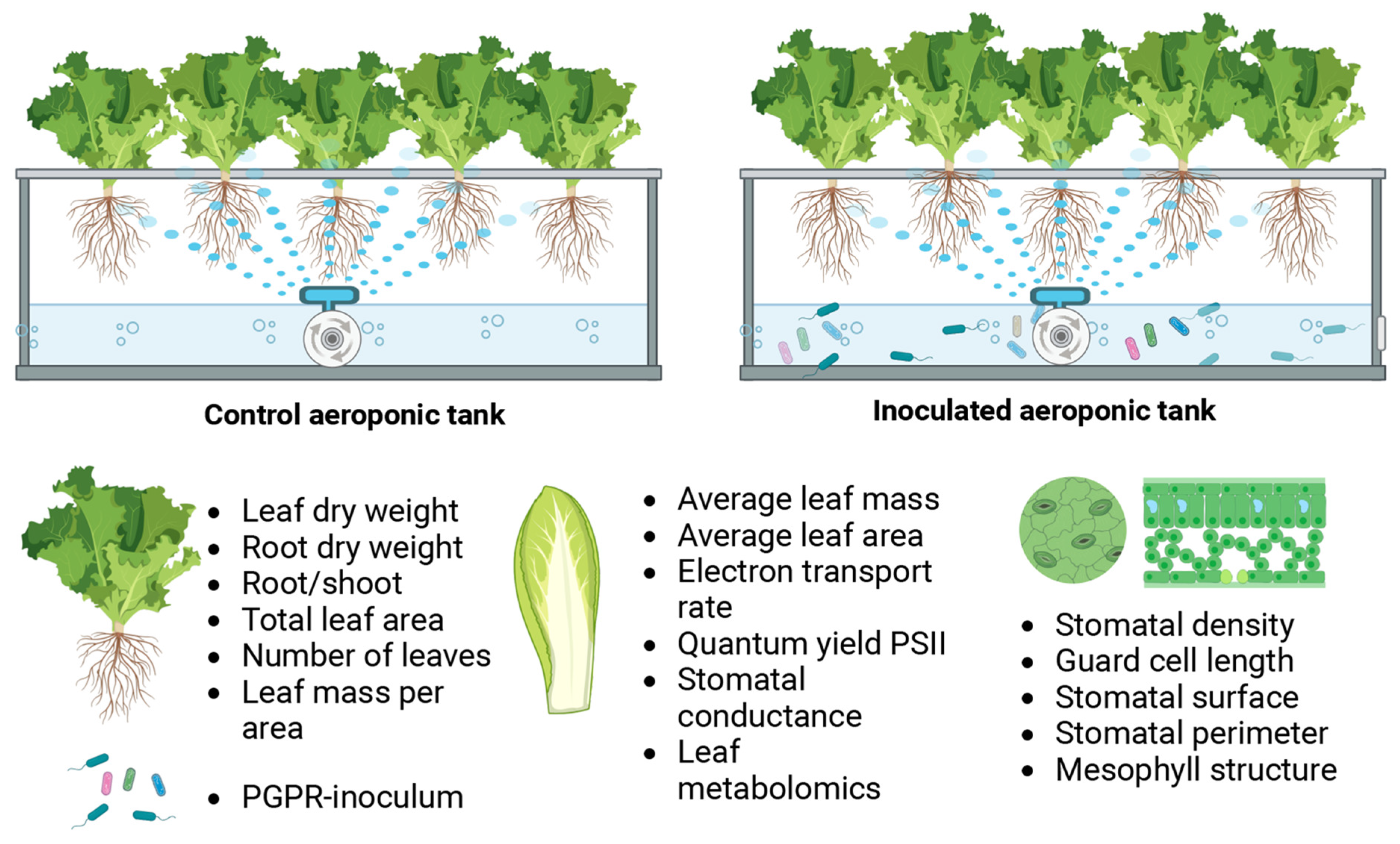
2.1. Experimental Design

Lettuce (Lactuca sativa L.) seeds, variety Spring Mortarella (series Hit; Four, Blumen Vegetable Seeds, Blumen Group S.p.A, Milan, Italy), were surface-sterilized by immersion for 10 min in sodium hypochlorite (NaClO) solution (50% v/v) amended with Twin 20 (0.025% v/v). The seeds were then rinsed ten times with sterile deionized water and left to imbibe with sterile deionized water for 4 h. The seeds were sown in 50 sterilized rock–wool cubes imbibed with sterile deionized water and placed in a grow tent (Mars Hydro EU, Ginsheim-Gustavsburg, Germany) equipped with a Mars Hydro Smart FC 3000 Samsung LED Grow Light powered by Samsung LM301B LED. The temperature was set to 26 °C, and a ventilation system (DF150A, Inline Duct Fan, Mars Hydro EU, Ginsheim-Gustavsburg, Germany) guaranteed an air exchange in the tent. The photosynthetic photon flux density (PPFD) was fixed to 150 µmol photon m−2 s−1, and the photoperiod to 14/10 h (light/dark). After six days, 40 plantlets were selected according to size uniformity, divided into two blocks (n = 20), and transplanted into two aeroponic systems (Nutriculture X-Streams propagator by G.A.S., Rotherham, UK). The PPFD was increased to 350 µmol photon m−2 s−1, while the photoperiod and temperature were kept unchanged. The plantlets were left to adapt to the new conditions for 24 h, then the PPFD was increased to 500 µmol photon m−2 s−1 and kept constant until the end of the experiment. The tanks were filled with 11 L of sterile deionized water, to which was added 0.2% (v/v) 0.2 µm filter-sterilized nutrient solution Hydro A (B’cuzz, Atami B.V., Rosmalen, The Netherlands), containing nitrogen (N) 4.9%, potassium oxide (K2O) 4.7%, calcium oxide (CaO) 3.8%, magnesium oxide (MgO) 1.3%, sulfur trioxide (SO3) 0.11%, and iron (Fe) 0.04% (w/w), and 0.2% (v/v) 0.2 µm filter-sterilized nutrient solution Hydro B (B’cuzz, Atami B.V., Rosmalen, The Netherlands), containing phosphorous pentoxide (P2O5) 4.1%, potassium oxide (K2O) 5.71%, boron (B) 0.01%, manganese (Mn) 0.03%, molybdenum (Mo) 0.001%, and zinc (Zn) 0.039% (w/w). To highlight the potential positive effect of the PGPR consortium on plant growth, a suboptimal quantity of nutrients was provided to the two aeroponic systems. The concentrations of nitrogen (N), phosphorus (P), and potassium (K) utilized in this study were 100, 30, and 100 parts per million (ppm), respectively. A Hoagland solution, widely regarded as the benchmark for the optimal growth of lettuce in hydroponic systems [55], contains 210, 31, and 235 ppm of nitrogen, phosphorus, and potassium, respectively. The pH of the growth solution was measured daily with a portable pH meter (PH400, Apera Instruments, Ohio, USA). A buffering agent (pH—Terra Aquatica, Fleurance, France) was applied to adjust the pH value and keep it in the range of 5.0–6.5, optimal for plant and microbial growth. Electrical conductivity (EC), a parameter that indirectly measures nutrient availability and salinity levels in the growth medium, was determined using an EC400 probe (Apera Instruments, Ohio, OH, USA). Water lost through evapotranspiration was measured and replaced with sterile water supplemented with a filter-sterilized nutrient solution (0.2% v/v). During the growth period, the EC level was maintained consistently at 1.2 dS m−1, which is about 20% less than the minimal optimum value (1.5–2.5 dS m−1 [56]), and about 25–30% less than the nutrient supply recommended by the manufacturer (1.6–1.7 dS m−1). Plants were grown in the system for 14 days and harvested.

2.2. Treatment with the PGPR Synthetic Consortium

The day after plantlets were transferred into the aeroponic system (the day after treatment 1, DAT1), an inoculum of the PGPR synthetic consortium was applied to one of the two aeroponic systems (PGPR inoculated, T). At the same time, no microorganisms were added to the second system (non-inoculated, C). The synthetic PGPR consortium comprised strains that had previously been tested for their growth-promoting activity in vitro and in vivo [57,58,59]. These strains were selected based on their functional complementarity to guarantee enhanced efficacy for plant growth promotion (i.e., hormone production) and biocontrol activity. All the PGPR strains derived from stock cultures stored at −80 °C in Lennox broth (LB: 10 g L−1 tryptone, 5 g L−1 yeast extract, and 5 g L−1 sodium chloride) and glycerol (20% w/w)—Pantoea agglomerans C1 (an IAA producer; [58]), Bacillus toyonensis 5A and 5B (biocontrollers), and Bacillus F13 and F14 (biocontrollers)—were isolated from the phyllosphere of lettuce plants treated with vegetal-derived protein hydrolysates [58,59]; Leucobacter sp. SS1 (IAA and gibberellin producer) and Enterobacter sp. LT1 and LT4 (IAA producers) were isolated from vegetable material (unpublished).
Pre-cultures of the single microorganisms were prepared by inoculating 20 mL of LB with 1.8 mL of the frozen stock culture. Cells were grown at 30 °C in agitation (150 rpm) until the late-exponential growth phase. To prepare the aeroponic inoculum, appropriate aliquots of each culture (1.1 × 10¹⁰ cells) were mixed, and the cells were collected by centrifugation. The pellet (about 8.8 × 10¹⁰ cells) was resuspended in sterile water, and the resulting suspension was directly poured into the aeroponic system tank. Thus, the concentration of inoculated microbial cells in the PGPR-inoculated tank was equal to 8 × 10⁶ CFU/mL. A second inoculum of the same strains was applied on DAT7 to promote greater colonization of the roots.

2.3. Plant Material Collection

One plant was collected on DAT13 for leaf anatomical analysis.
On DAT14, all plants were removed from the aeroponic systems and photographed (Figure S1b). Subsequently, the plants were separated into two subgroups: the first group (n = 12) was utilized to determine plant biomass, structural characteristics, and leaf C and N content; the second (n = 6) was frozen and used to extract leaf metabolites.

2.4. Plant Biomass Determination and Plant Structure Analysis

Each plant was measured for its height and root length and then separated into leaves and roots. The fresh weight of each portion was recorded to obtain root fresh weight (RFW), leaf fresh weight (LFW), and total fresh weight (TFW) for every plant.
The leaves of each plant were counted, carefully arranged on a flatbed scanner, and digitized. The resulting images were analyzed using ImageJ software (vers. 1.53t; Wayne Rasband and contributors, National Institutes of Health, USA) to determine the total leaf area per plant. The average leaf area per plant was assessed by dividing the total leaf area by the number of leaves.
To determine root dry weight (RDW) and leaf dry weight (LDW), root and leaf subsamples from six plants per treatment were dried using Sartorius MA 100 (Göttingen, Germany), an infrared moisture analyzer equipped with a ceramic heating element for gentle heating of temperature-sensitive samples. Significant correlations were observed between root fresh weight (RFW) and RDW (RDW = 0.0385 × RFW − 0.0042; R2 = 0.889; F value = 9.805; p < 0.01) and between leaf fresh weight (LFW) and LDW (LDW = 0.0655 × LFW − 0.0676; R2 = 0.637; F value = 5.694; p < 0.05). These regression equations were subsequently employed to estimate RDW, LDW, and total plant dry weight (TDW = RDW + LDW) of all plants belonging to the first group.
The average leaf biomass was then calculated by dividing LDW by the number of leaves. Specific leaf area (SLA) was determined as the ratio of total leaf area to leaf dry weight, while specific root length was calculated as the ratio of root maximum length to root dry weight.
The root-to-shoot ratio (R/S) was calculated for each plant using the estimated RDW to LDW ratio to assess biomass allocation.

2.5. Leaf Anatomical Analysis

On DAT13, one plant per treatment was collected. From each plant, one leaf with a similar dimension and development stage was selected and prepared for scanning electron microscopy and electron microscopy. Leaf subsamples were selected and fixed with 2% paraformaldehyde and 2.5% glutaraldehyde in 0.1 M cacodylate buffer (Electron Microscopy Science, Hatfield, PA, USA) at pH 7.4 overnight at 4 °C. The specimens were then washed four times with cacodylate buffer and then post-fixed for 1 h with 1% osmium tetroxide (Electron Microscopy Science) in 0.1 M cacodylate buffer at pH 7.2 for one hour at 4 °C. After washing in distilled water, the samples were dehydrated for 10 min in progressively increasing concentrations of ethanol solutions (10%, 30%, 50%, 70%, and 90%) and for 15 min in 100% ethanol. Then, the samples were dried using the method of liquid carbon dioxide (CO2) in a Critical Point Dryer (Balzers Union CPD 020; Balzers Union Ltd, Balzers, Principality of Liechtenstein) coated with gold in a Sputter Coater (Balzer Union MD 010), and examined using a Jeol JSM 6010 LA (Tokyo, Japan) and photographed. Four 230 × 300 µm areas of lamina abaxial epidermis were selected from one leaf per treatment, and stomata were counted to determine the stomatal density (expressed as the number of stomata per surface unit mm2). Each stomate’s guard cell length in the selected areas was measured. For each of the four images chosen per treatment, a sub-area of 170 × 225 µm was considered; the stomata present in the sub-area were manually selected for the determination of stomatal aperture surface and total stomatal pore surface, considering the internal guard cell perimeter as the boundary of the cavity.
The same leaf samples were initially prepared for light microscopy as described for SEM analysis. After dehydration in ethanol solutions, the specimens were sequentially transferred into mixtures of acrylic resin LRWhite (London Resin White, Agar Scientific Ltd, Stansted, UK) and ethanol (v:v = 1:2, v:v = 1:1, and v:v = 2:1) for two hours, respectively, and finally into pure acrylic resin overnight. The embedding was carried out using hermetic gelatin capsules. The samples were left to polymerize for 36 h at 50 °C. Semithin sections, 2 µm thick, were obtained using an ultramicrotome (Reichert-Jung Ultracut E, Reichert Inc., Depew, NY, USA), stained with toluidine blue, and observed using a light microscope. The images were analyzed using the software ImageJ (vers. 1.53t; Wayne Rasband and contributors, National Institutes of Health, USA). Three lamina thickness measurements were made for three leaf microsections per treatment to obtain the average leaf thickness in PGPR-inoculated and non-inoculated plants.
A leaf cross section having a width of 375 µm and a height equal to leaf thickness, representing more than 50% of the leaf cross section surface image, was selected for three sections per treatment. ImageJ was used to convert the chosen areas into binary form and automatically recognize the edges of the cells in the lamina section. The palisade mesophyll cells, the spongy parenchyma cells, and the air spaces were manually selected, and the total surface of each category was determined. Changes in the leaf anatomy were determined for three leaf sections, calculating the percentage of palisade mesophyll, spongy parenchyma, and air spaces relative to the total area occupied by the leaf tissues.

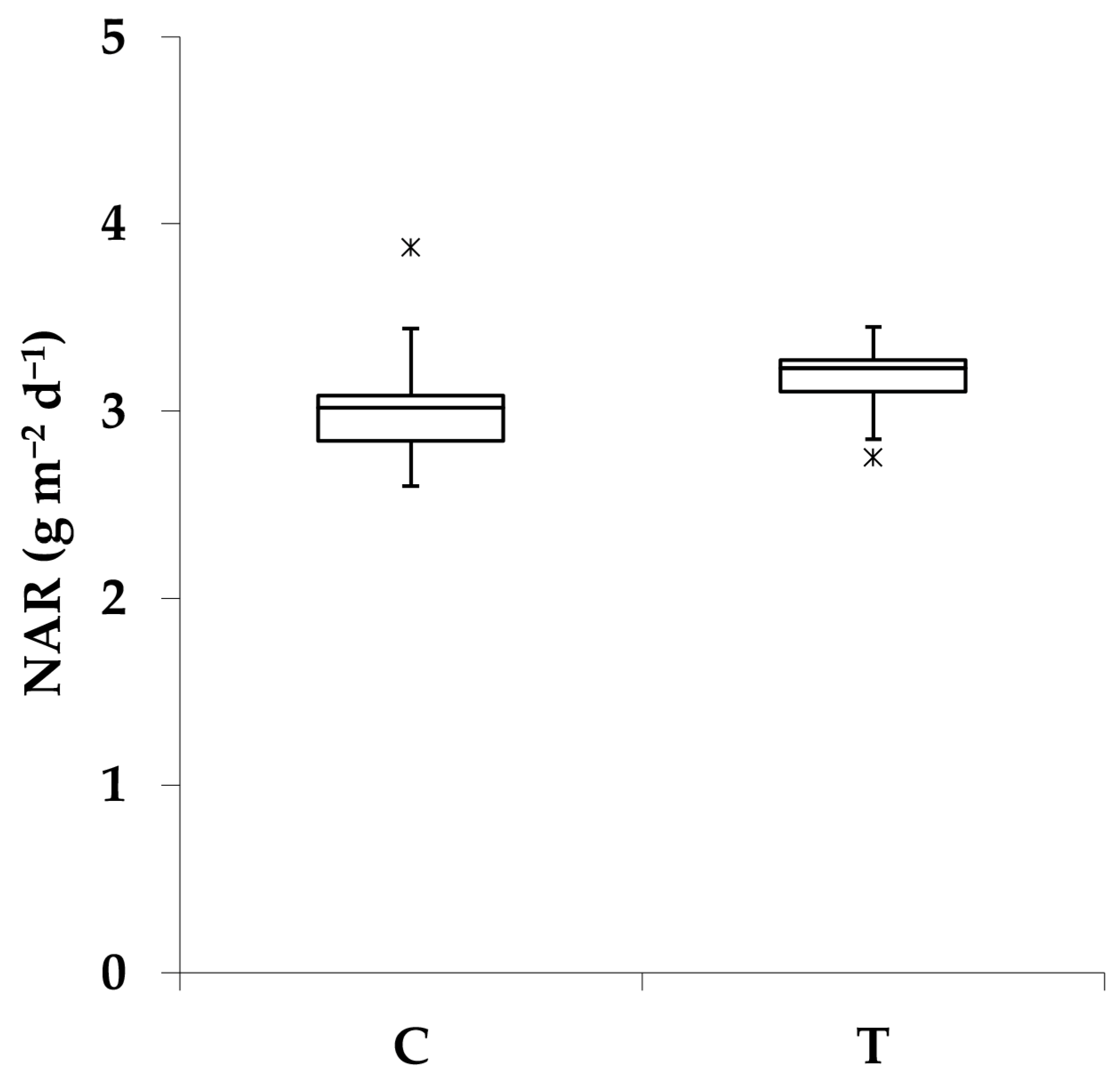
2.6. Leaf Physiological Measurements and Net Assimilation Rate

On DAT8 (the first day after the second inoculation) and DAT13 (the day before the conclusion of the experiment), stomatal conductance (gs), the quantum yield of photosystem II (ΦPSII), and electron transport rate (ETR) were measured for one leaf per plant per treatment, using an LI-600 porometer (Li-Cor, Lincoln, Oregon, OR, USA) set to a flow rate of 150 μmol s−1.
The net assimilation rate (NAR) was determined following White et al. [60]. The change in plant mass (dWp) over time (dt) can be calculated as follows:
dWp dt = SLA × LMR × NAR × Wp
SLA is the specific leaf area, LMR is the leaf-to-plant mass ratio, and NAR is the net assimilation rate.
Therefore, NAR can be calculated as
NAR = dWp dt   1 SLA × LMR × Wp
dWp is considered the biomass produced since the seed germination and is therefore equal to the plant dry weight at harvest.

2.7. Leaf C and N Content Determination

The leaves used for the dry weight determination were ground to powder. Leaf carbon and nitrogen contents were determined for three replicates per sample using the dry combustion method with Thermo Soil NC—Flash EA1112 Elemental Analyzer (Thermo Fisher Scientific, Bath, UK).

2.8. Untargeted Metabolomics

Frozen leaf material was ground into a fine powder using liquid nitrogen. Metabolites for the LC-MS analysis were extracted by sonication using a modified protocol of Bansal et al. [61]. In short, 1 g of leaf powder was transferred to a 15 mL tube, and 9 mL of ethanol/water (1:1, v/v) was added to the sample. After sonication for 30 min (in agreement with the recommendations of De Vos et al. [62]), the samples were centrifuged (12,000 rpm for 10 min). The supernatants were collected, filtered (0.22 µm), and stored for further analysis. The LC-MS analysis was performed by using an ACQUITY I-Class PLUS UPLC System (Waters, Milford, MA, USA) coupled to an ACQUITY RDa mass spectrometer (Waters, Milford, MA, USA) equipped with an ESI probe in positive and negative ion modes to perform reversed-phase chromatography. All samples (2 µL) were analyzed in positive and negative ion modes with triplicate injections. Paracetamol was used as an internal standard. An Acquity Premier HSS T3 column with VanGuard FIT (2.1 mm × 150 mm, 1.8 μm, 1/pK—Waters; Milford, MA, USA) was used for metabolite separation. LC separations were achieved at a column temperature of 40 °C and a flow rate of 0.25 mL/min. The temperature of the autosampler was 6 °C. A 0–100% linear gradient of solvent A (ddH2O, 0.1% formic acid) to B (acetonitrile, 0.1% formic acid) was employed and set as follows: 100% A and 0% B from 0 to 6 min, 95–5% A–B from 6 to 9 min, 60–40% A–B from 9 to 14 min, 5–95% A–B from 14 to 17.10 min, and 100–0% A–B from 17.10 to 19 min. The ACQUITY RDa mass spectrometer was used in a full scan with fragmentation mode to detect small-mass molecules (50–800 m/z). The scan rate was 5 Hz in both positive and negative polarity, and the cone voltage was 20 and 40 V, respectively, in positive and negative polarity. The fragmentation voltage was 60 to 80 V and 40 to 40 V, respectively, in positive and negative polarity. The absorbance read was made at a wavelength of 254 nm. The capillary voltage was set at 1.0 kV in positive and 0.8 kV in negative polarity, and a default capillary desolvation temperature of 550 °C was used.
The software UNIFI Scientific Information System (vers. 3.3.0; Waters Corp., Milford, MA, USA) analyzed the data, which were processed for peak picking, alignment, and normalization. Compound identification was performed using the UNIFI library searching algorithm. The resulting data matrices were then exported for further statistical analysis, including the component name, the expected retention time, the observed m/z, the mass error, and the detector count. Data were first corrected by removing the molecules identified in the blank. Moreover, data were filtered according to the difference between observed and expected retention times, and all molecules showing a difference higher than 5 and lower than −5 min were discarded. Moreover, the analysis did not consider a sample if the mass error was greater than 5 mDa. Normalization by median, cube root transformation, and auto-scaling was performed on the metabolomic data sets using MetaboAnalyst 6.0 (https://www.metaboanalyst.ca/home.xhtml; accessed on 30 September 2024). The resulting data were used in the MetaboAnalyst package to compute the fold change (FC) and the t-test values and to design the volcano plot. For the FC analysis, an FC threshold of 2.0 was used. For the two-samples t-test, the p-value threshold for false discovery rate (FDR) was set at <0.05. The FC and p-value threshold for the volcano plot were set at 2.0 and 0.1, respectively.

2.9. Statistical Analysis

To test the effect of PGPR inoculation on physiological, chemical, and structural parameters, a Wilcoxon rank-sum test was performed on the website www.socscistatistics.com (accessed on 28 September 2024). Differences within anatomical parameters were tested using the two-sample t-test. Statistically significant differences were reported for p ≤ 0.05.

3. Results

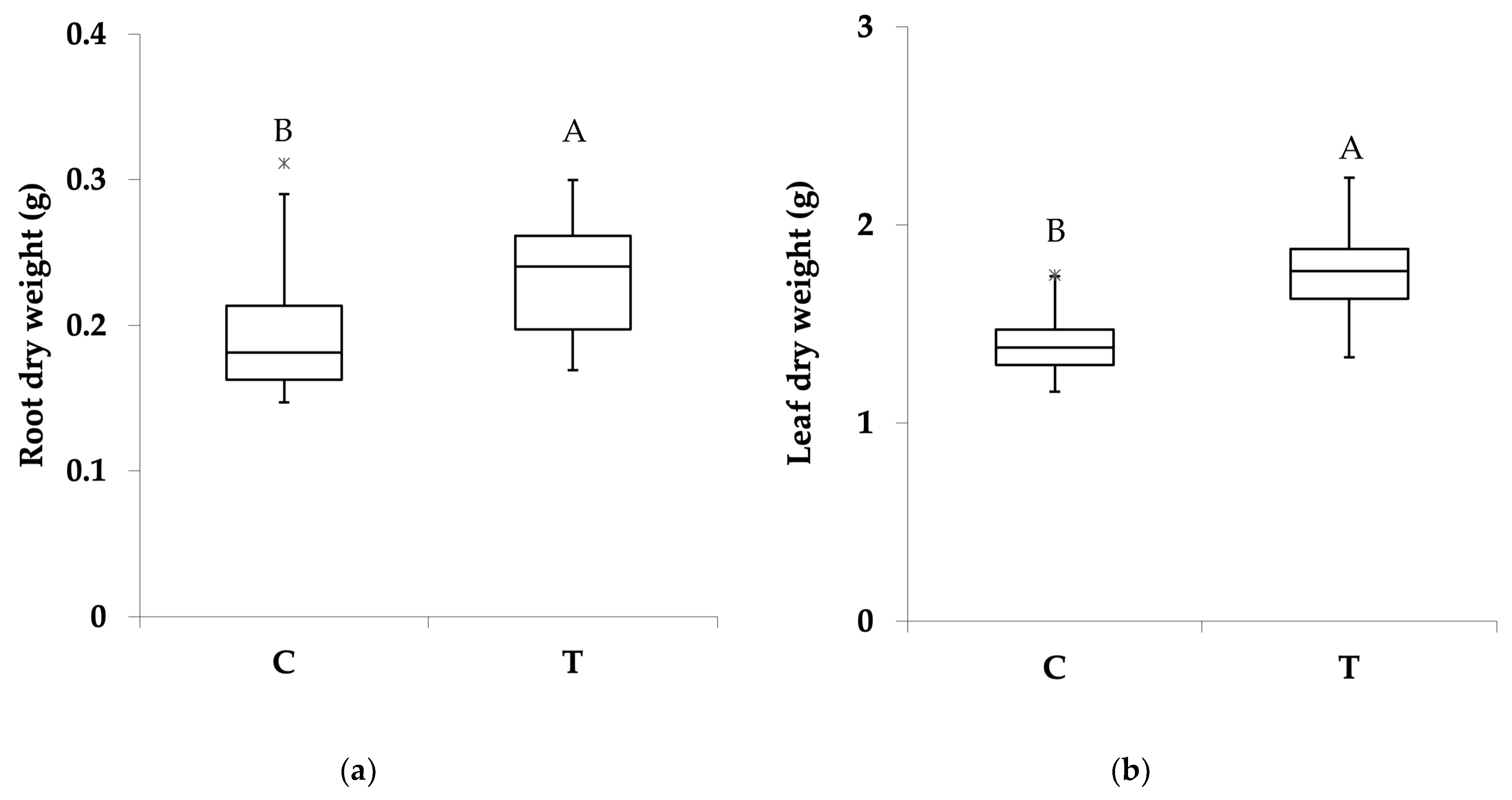
3.1. Effect of PGPR Consortium on Biomass Production and Plant Structure

Inoculation with the PGPR consortium significantly increased the lettuce biomass. Overall, 14 days after the first inoculation (day after treatment 14; DAT14), the plant biomass was 24.7%, significantly higher (p < 0.01) in the PGPR-inoculated (2.0 ± 0.1 g) compared to the non-inoculated plants (1.6 ± 0.1 g). The inoculation effect was observed in both the below- and above-ground biomass production. Significant differences were observed in the root dry weight, equal to 0.20 ± 0.015 g and 0.23 ± 0.012 g (p < 0.05), respectively, in the non-inoculated and PGPR-inoculated plants (Figure 2a). The leaf biomass was significantly higher (p < 0.05) in the PGPR-inoculated plants (1.8 ± 0.1 g) compared to the non-inoculated ones (1.4 ± 0.1 g; Figure 2b). The changes in the root and leaf dry weight were consistent across all the replicates and were not influenced by the variability in plant response.
Although the biomass allocation did not change in the presence of PGPR, as suggested by the root/shoot ratio (R/S), which was similar in the PGPR-inoculated and non-inoculated plants (on average 0.14 ± 0.01; Table 1), the inoculation gave rise to changes in the plant phenotype (Figure S1). PGPR inoculation caused a significant increase in the total leaf area from 376.0 ± 10.3 cm2 (non-inoculated) to 450.3 ± 12.3 cm2 (p < 0.01, PGPR-inoculated plants; Table 1). The number of leaves per plant was significantly higher (p < 0.05) in the inoculated lettuces (15.6 ± 0.5) compared to the non-inoculated plants (14.0 ± 0.4; Table 1). The average leaf mass was significantly higher (p < 0.05) in the plants inoculated with microorganisms (0.11 ± 0.003 g), although the leaves had, on average, a similar surface area (28.2 ± 0.9 cm2) under the two growth conditions (Table 1). Consequently, the specific leaf area (the ratio between the leaf area and leaf dry weight) was significantly lower (p < 0.05; 256.5 ± 4.7 cm2 g−1) in the inoculated compared to the non-inoculates plants (271.3 ± 7.0 cm2 g−1; Table 1).
Changes were also observed in the maximum root length, which was lower (p = 0.056) in the inoculated (32.8 ± 1.4 cm) than in the non-inoculated lettuces (36.9 ± 1.7 cm; Table 1). The specific root length (the ratio between the root length and root dry weight) was significantly lower (p < 0.05) in the plants grown in the presence of the PGPR consortium (143.3 ± 9.3 cm g−1) compared to the non-inoculated plants (199.1 ± 17.2 cm g−1; Table 1).

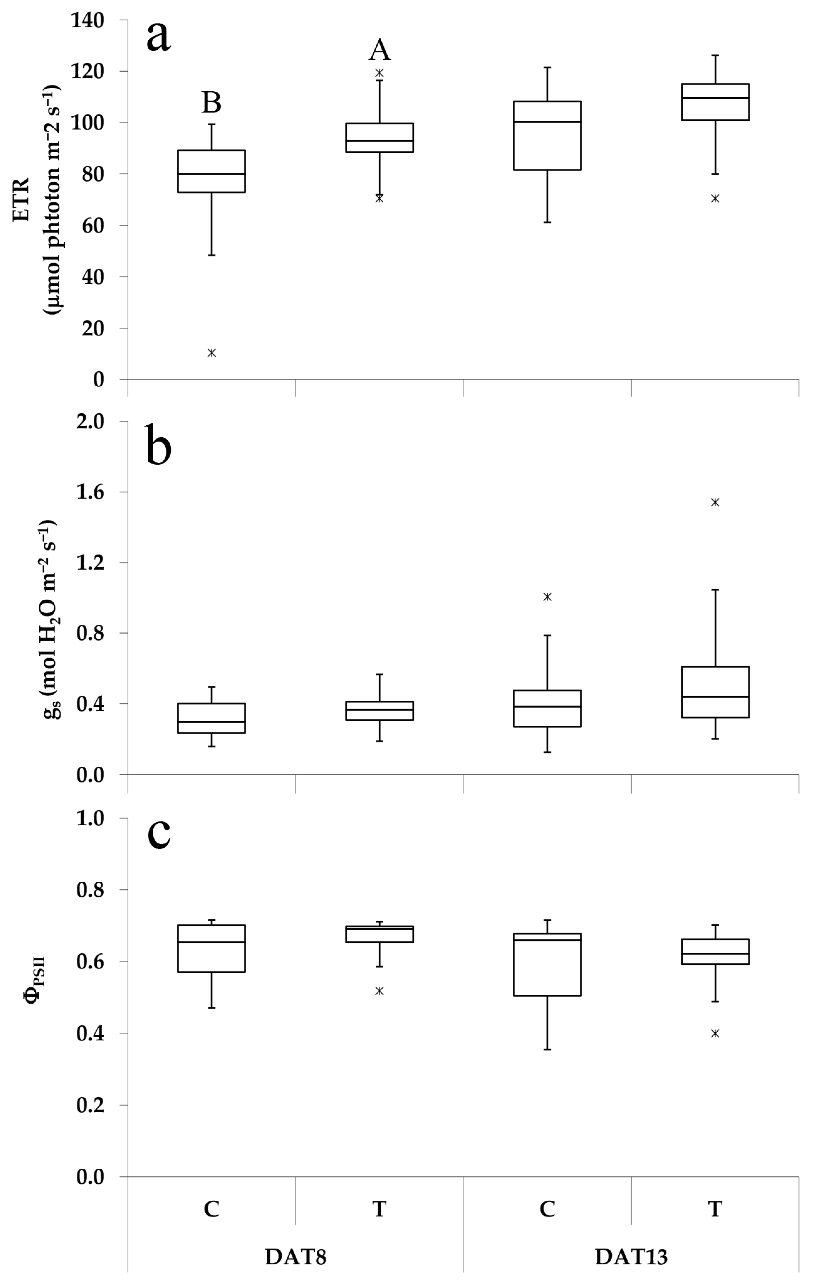
3.2. Effect of PGPR Consortium on Leaf Physiological Responses

The leaf physiological responses were influenced by inoculation with PGPR. The electron transport rate (ETR) was significantly higher (94.3 ± 2.5 µmol photons m−2 s−1, p < 0.001; Figure 3a) in the inoculated plants compared to the non-inoculated plants (72.4 ± 6.6 µmol photons m−2 s−1) on DAT8. On DAT13, the difference between the two treatments was less marked, with the ETR equal to 95 ± 4.2 µmol photons m−2 s−1 in the C and 104.8 ± 3.9 µmol photons m−2 s−1 in the T (p < 0.1; Figure 3a). However, no significant changes in the quantum yield of PSII (ΦPSII, Figure 3c) or stomatal conductance (gs, Figure 3b) were observed on DAT8 or DAT13; on average, the ΦPSII was equal to 0.62 ± 0.02. In contrast, the gs was 0.41 ± 0.05 mol H2O m−2 s−1.
Moreover, a significantly higher NAR was estimated in the inoculated (3.2 ± 0.1 g m−2 d−1, p < 0.05; Figure 4) compared to the non-inoculated plants (3.0 ± 0.1 g m−2 d−1).

3.3. Effect of PGPR on Leaf Carbon (C) and Nitrogen (N) Contents

Inoculation with PGPR significantly increased both the leaf N (2.85 ± 0.1%; p < 0.01; Figure 5a) and C (41.3 ± 0.3%, p < 0.05) contents compared to the non-inoculated plants (2.43 ± 0.1% and 40.0 ± 0.4%, respectively; Figure 5b).

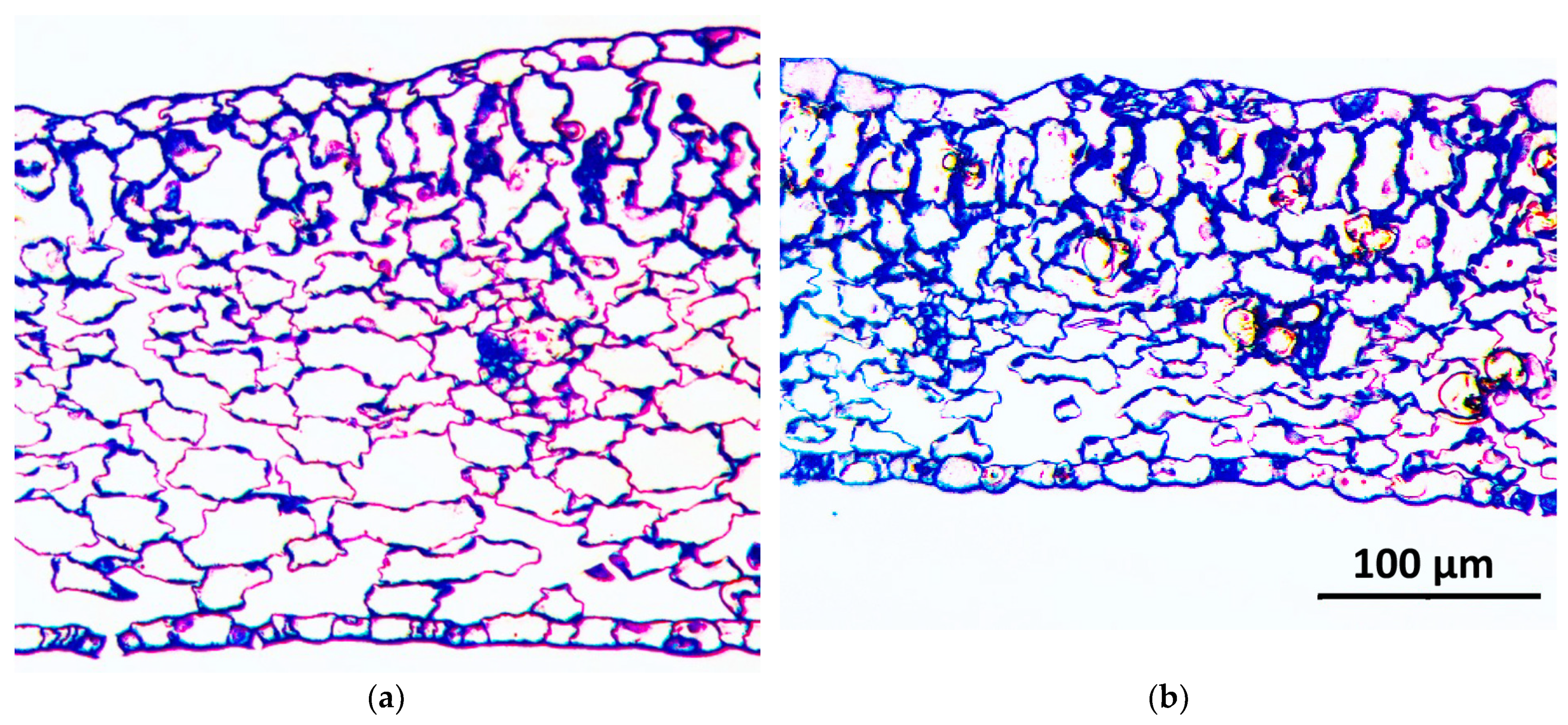
3.4. Effect of PGPR Consortium on Leaf Anatomy

The leaf epidermis showed a different stomatal density and dimension in the PGPR-inoculated plants. The consortium of PGPR induced a decrease in the stomatal density (239 ± 9.4 mm−2, p = 0.01; Table 2) and an increase in the guard cell length (19.7 ± 0.2 µm, p < 0.001) compared to the non-inoculated plants (297 ± 12.6 mm−2 and 17.0 ± 0.3 µm, respectively; Table 2); moreover, the surface of the stomatal aperture was more than double (57.4 ± 5.8 µm2, p < 0.001; Table 2) compared to that of the non-inoculated plants (25.1 ± 2.6 µm2), while the PGPR inoculation significantly increased (p < 0.001) the perimeter of the stomata by 26% (36.2 ± 1.3 µm) compared to that of the non-inoculated plants (28.6 ± 1.1 µm; Table 2).
The leaf mesophyll anatomy changed due to inoculation (Figure 6; Table 2). The leaf thickness was significantly lower in the PGPR-inoculated (195.6 ± 5.4 µm) compared to the non-inoculated plants (275.1 ± 8.9 µm, p < 0.001; Table 2). The leaf cross sections revealed that the spongy parenchyma structure resulted in a reduced thickness in the PGPR-inoculated plants (Figure 6b), principally due to a lower intercellular airspace (28.1 ± 1.9%, p < 0.05) compared to that of the non-inoculated plants (40.1 ± 4.0%, Figure 6a, Table 2). In contrast, the palisade parenchyma cell surface was thicker in the PGPR-inoculated (32.6 ± 2.4% of leaf section surface) compared to the non-inoculated lettuces (14.4 ± 0.9%, p < 0.05).

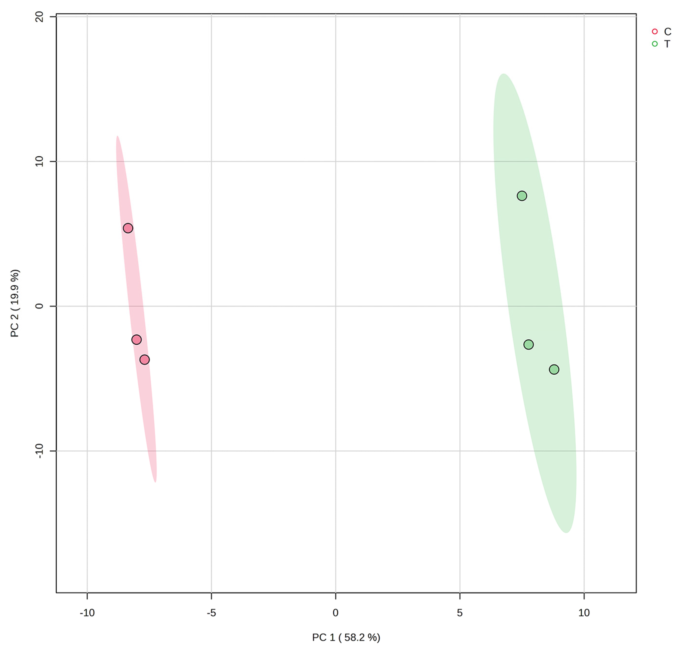
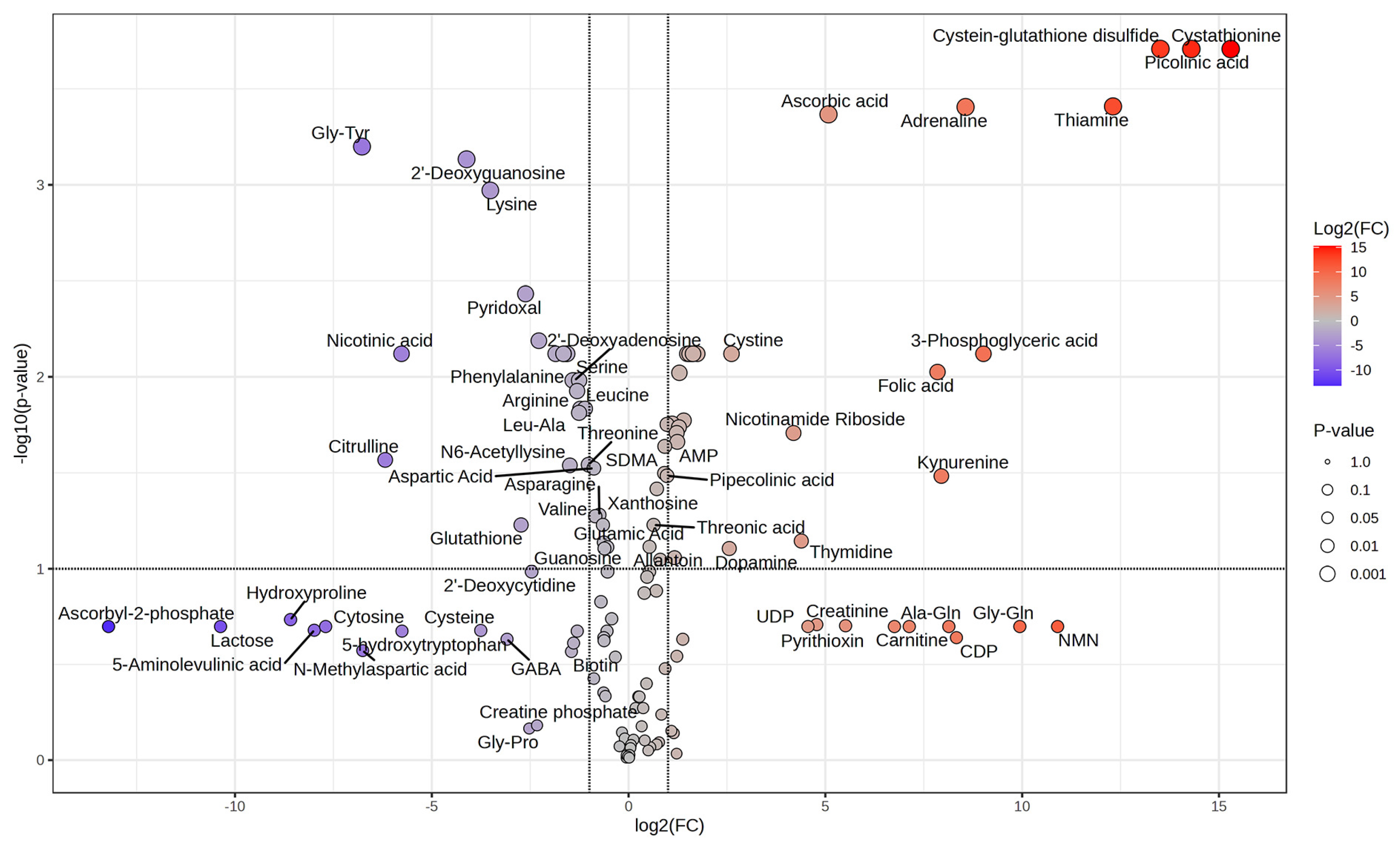
3.5. Effect of PGPR Consortium on Leaf Metabolome

The untargeted metabolomic analysis showed significant leaf metabolic changes in response to the PGPR inoculation. In total, 126 and 133 identified molecules were detected in the non-inoculated and PGPR-inoculated plants, respectively. The PCA analysis, which explained 78.1% of the variability, clearly indicated that the two datasets clustered separately (Figure 7). The fold change analysis revealed the presence of 79 molecules with an FC ≥ 2.0 or ≤0.5; 41 features had a higher abundance in the PGPR-inoculated samples and 38 were enriched in the leaves from the non-inoculated plants (Table S1). The two-sample t-test showed significant changes (p < 0.05) in the abundance of 52 compounds (Table S2). A volcano plot analysis (Figure 8) displayed 50 significantly altered metabolites (an FC of either ≥2.0 or ≤0.5 at p < 0.1), among which 27 increased and 23 decreased in response to the PGPR inoculation (Figure 8; Table S3).
The data in the volcano plot were used to detect the specific chemical classes affected by the PGPR inoculation. This analysis showed that in the amino acids and peptides class, eight proteinogenic (arginine, leucine, lysine, phenylalanine, serine, threonine, tryptophan, and tyrosine) and three non-proteinogenic amino acids (citrulline, methionine sulfoxide, and N6-acetyllysine) significantly decreased in the foliar tissues due to the PGPR inoculation (Table S3). Five additional non-proteinogenic amino acids (cystathionine, cystine, DOPA, kynurenine, and phenylacetylglycine) significantly increased in the PGPR-inoculated plants (Table S3).
Additionally, the untargeted metabolic data showed that the PGPR inoculation dramatically changed the relative abundance of several purines and pyrimidines. Specific nucleosides (2′-deoxyadenosine, 2′-deoxyguanosine, 5-methylcytosine, and 5-methyl-2′-deoxycytidine) displayed a significant reduction in their relative abundance in the leaf tissue of the PGPR-inoculated plants (Table S3).
On the other hand, the leaf tissue of the PGPR-inoculated plants exhibited a considerable increase in specific nucleotide monophosphate (AMP, CMP, and UMP). The same effect was noted for the relative abundance of thiamine (vitamin B1) and the nucleoside thymidine (Table S3).
Furthermore, the PGPR inoculation resulted in a notable alteration in the leaf tissue’s relative abundance of specific nicotinic acid alkaloid class molecules. In particular, there was a significant increase in the nicotinamide riboside and picolinic acid levels, with an 18-fold and 20,000-fold increase, respectively (Table S3). The comparative metabolomics also revealed an increase in the relative abundance of vitamins (ascorbic acid and thiamine) and catecholamines (adrenaline and dopamine) in the PGPR-inoculated plants (Table S3). In addition, the leaf tissues of the plants treated with PGPR presented a 55-fold drop in nicotinic acid content and a 6.3-fold decrease in pyridoxal relative abundance (Table S3).
The metabolites whose abundance increased most significantly (more than 5000-fold) in the leaf tissues of the PGPR-inoculated plants were cystathionine, picolinic acid, cysteine–glutathione disulfide, and thiamine (Figure 8; Table S3). Conversely, the most significant decrease in the fold change was observed for Gly-Tyr, citrulline, and nicotinic acid (Figure 8; Table S3).

4. Discussion

The utilization of microbial biostimulants derived from singular or multiple PGPR strains constitutes a sustainable strategy for enhancing horticultural crop productivity. However, the effectiveness of these biostimulants can vary, particularly when a single strain is employed [63,64,65]. Vanegas and Uribe-Vélez [66] demonstrated the positive effects of PGPR consortia inoculation on plant biomass. Their analysis of the impact of 24 single strains and 10 microbial assemblies on rice plants’ dry weight revealed an increased root biomass with 8 single strains and 6 microbial consortia treatments. The stem biomass increased with six single strains and five microbial assembly treatments, suggesting that a PGPR consortia may be more effective at promoting plant growth than single strains. This observation aligns with the findings from Azizi et al. [67], who reported enhanced above- and below-ground biomass in Myrtus communis seedlings inoculated with a mixed inoculum of Pseudomonas putida and Pseudomonas fluorescens, whereas single-strain applications only increased the below-ground biomass. Mehnaz et al. [68] noted increased plant biomass following sterilized soil inoculation with a combination of PGPR. However, neither single strains nor a consortium enhanced the yield under field conditions, suggesting that interactions between the introduced microbial assembly and the indigenous soil microbiome can negatively impact the treatment efficacy. A decline in the PGPR concentration post-inoculation is commonly observed in soil due to competition with the resident microbiome or predation [69], mirroring observations in the human gut following probiotic treatments [70,71]. The aeroponic system employed in this study avoided interference due to interactions with the soil microbiome, allowing for a more precise assessment of plant responses to the application of our PGPR consortium and its potential to reprogram plant metabolism.
The data presented here demonstrate the positive influence of PGPR inoculation on lettuce growth under suboptimal nutrient levels, with a substantial 25% increase in the overall biomass observed in the PGPR-inoculated plants (Figure 2). This aligns with Ikiz et al.’s [6] findings on lettuce grown in a hydroponic system, where a higher biomass was only seen under low mineral nutrient concentrations and PGPR inoculation compared to plants supplied with an optimal fertilization level. The improved yield was attributed to increased leaf and root growth. In their study on the effect of Pseudomonas psychrotolerans IALR632 on lettuce, Mei et al. [72] reported an enhancement in the lateral root development of lettuce cultivated in hydroponic systems as a direct consequence of inoculation with the PGPR strain. In conditions of unrestricted nutrient availability, they observed an increase in the shoot and root fresh weight of 13.5% and 13.8%, respectively, in an indoor vertical nutrient film technique (NFT) system, and an increase of 15.3% and 13.6% in a deep-water cultivation (DWC) system. It is noteworthy that the PGPR consortium used in this study determined a similar effect on the root biomass (+15% in dry weight) and a more pronounced increase in the leaf biomass (+28.6% in dry weight) under nutrient-limiting conditions. The data reported in Figure 2b clearly show that the higher leaf biomass resulted from a 12.8% increase in the average leaf mass, which was further amplified by an 11.3% augmentation in the number of leaves in the PGPR-inoculated plants (Table 1). Furthermore, a 19.4% expansion of the total leaf area was observed, even though the individual leaf size was similar to that of the non-inoculated lettuces (Table 1). An increase in the leaf number per plant and the total leaf area were also reported in a soilless tomato culture treated with biostimulants, among which PGPR, due to an increase in nutrient absorption as a result of their amino acids, organic acids, and hormones production [73]. Similarly to our results, under low mineral fertilization (−50% compared to the control), Desgan et al. [74] reported that hydroponically grown lettuce inoculated with a consortium of Bacillus subtilis, Bacillus megaterium, and P. fluorescens increased the biomass and the leaf N and ascorbic acid contents compared to non-inoculated plants, and was similar to non-inoculated lettuces grown under an optimal nutrient supply. In our study, the PGPR-inoculated plants might have produced more leaves due to the enhanced activity of the shoot apical meristem or axillary meristems, which are responsible for the developmental patterns of different angiosperms [75]. Several authors have observed positive changes in PGPR-inoculated plants’ leaf mass, number, or area under optimal growth conditions [41], abiotic stress [48], and nutrient deficiency [76]. In our research, the specific leaf area decreased because the increase in plant biomass outweighed the increase in the total leaf area (Table 1). Villar et al. [77] examined the leaf mass per area (LMA, the reciprocal of specific leaf area) of 26 woody species, reporting that the LMA can be factorized into leaf thickness and density. These, in turn, are influenced by the volume and density of the leaf structures, such as the epidermis, mesophyll, air spaces, and vascular and sclerenchymatous tissues. In our experiment, the changes in the leaf area production per unit of invested biomass were related to the modifications in the leaf anatomy triggered by the PGPR consortium. The reduction in the specific leaf area in the PGPR-inoculated plants was not correlated with a higher leaf thickness but was associated with (1) an increase in the relative area occupied by the palisade parenchyma and (2) a decrease in the airspace surface (Figure 6; Table 2). These anatomical changes increased the leaf tissue density. Research on herbaceous plants has shown an increased spongy parenchyma volume due to larger air spaces and cell expansion with age. This can lead to thicker leaves, but does not necessarily translate to a higher leaf mass per area [34]. In addition, Tholen et al. [32] reported that a lower photosynthetic capacity than that of the upper palisade tissue generally characterizes the spongy tissue. In this study, the ratio between the palisade and spongy cell surfaces was higher in the PGPR-inoculated plants (Figure 6, Table 2), resulting in thinner leaves likely characterized by increased photosynthetic activity. Inoculation with the PGPR consortia enhanced plant growth and triggered a plant response, modifying the epidermis structure. The stomatal density decreased while the guard cell length and stomatal aperture size increased (Table 2). These parameters are crucial because the size and abundance of the stomata interactively govern the maximum potential for foliar CO2 uptake [78]. Despite the larger stomatal pore surface observed in the PGPR-inoculated lettuces (Table 2), the stomatal conductance, i.e., the rate of water vapor or carbon dioxide diffusion between the atmosphere and the substomatal cavity, was comparable to the levels observed in the non-inoculated plants (Figure 4). Brodribbet et al. [79] demonstrated that larger stomata in leaves results in a lower maximum capacity to absorb CO2 and release H2O.
Moreover, the efficiency of gas movement within a leaf is directly influenced by its structural features, which determine the path that gases must take [33]. Dow et al. [80] demonstrated that stomata development modulates the differentiation of air spaces. The arrangement of air spaces, cell wall thickness, and the composition of the plasma and chloroplast membranes affect how easily gases can move into cells [81]. Furthermore, the diffusivity of CO2 is faster in the liquid phase than in the gas phase, while the shortest path length is the most effective [82]. The reduced leaf thickness and the increase in the ratio between the palisade tissue to the total leaf tissue surface observed in the PGPR-inoculated lettuces might have shortened the pathway of CO2 to the carboxylation sites, where RuBisCO (ribulose-1,5-bisphosphate carboxylase oxygenase) catalyzes the fixation of atmospheric CO2 to ribulose-1,5-bisphosphate (RuBP), potentially improving the mesophyll conductance and positively affecting the net assimilation rates (Figure 4). At the same time, the higher electron transport rate measured in the PGPR-inoculated lettuce (Figure 3a) could have supported an enhancement in energy availability for ATP synthesis and carbon assimilation. The concomitant increase in mesophyll conductance, the electron transport rate, and the NAR supports the conclusion that PGPR inoculation produced an increase in photosynthetic efficiency. The latter effect was the result of the up-regulation of specific biosynthetic pathways, which led to the accumulation of compounds, such as ascorbic acid, modulating the expression of photosynthesis-related genes [83].
The changes in the leaf tissues and epidermal structure induced by inoculation also provide evidence for modifications in the balance between CO2 uptake and water losses. Lundgren et al. [84] suggested that wheat evolution has selected for leaves with a decreased stomatal density and an increased stomatal size, associated with a decrease in stomatal conductance and mesophyll porosity, yielding a denser leaf. According to the authors, these traits are related to increased water use efficiency. Apart from the lower stomatal conductance, similar relations among the anatomical traits were also observed in our experiment on the PGPR-inoculated plants, suggesting that inoculation increased water use efficiency under non-limiting water availability. Nevertheless, as bigger stomata are less sensitive to their closure [85], a less efficient uptake of carbon dioxide and unnecessary water loss are expected during a stomatal opening or closure [86], which could limit water use efficiency during a transition in light or a plant’s water status. The increase in the above-ground biomass was mirrored by a corresponding rise in the below-ground biomass of 19.2%, maintaining a consistent ratio between the two compartments. The microbial treatment also induced changes in the root morphology. The PGPR-inoculated plants exhibited a shorter root length and lower specific root length (Table 1), suggesting increased lateral root development. The observed change in the root architecture increased the surface area for nutrient uptake and plant–microbial interactions. Zhang et al. [87] observed a positive correlation between enhanced lateral root formation, increased root surface area, and the potential for nutrient uptake. The changes in root phenotyping could be attributed to altered signaling pathways or resource allocation triggered by the PGPR. Moreover, previous studies have identified IAA production as a primary mechanism by which PGPR stimulates root growth [33]. This hormone significantly influences the extent of root growth and the overall structure of a root system [80]. In recent research on lettuce cultivated in a hydroponic system and inoculated with two PGPB strains, Pseudomonas lundensis UB 53 and Pseudomonas migulae UB 54, the analysis of the root metabolome revealed an enhanced accumulation of IAA [88]. The metabolites from indole auxin-producing Pantoea agglomerans C1 have been shown to increase adventitious rooting and leaf area in various plant cuttings [89,90]. The inclusion of four IAA producers within the PGPR consortium utilized in the present study may have resulted in the observed phenomenon of secondary rooting in the PGPR-inoculated lettuce plants.
In the PGPR-inoculated plants, the higher availability of organic N can explain the increase in leaf mass (Figure 2b) and leaf N concentration (Figure 5a), as well as the reduction in leaf proteins and nucleic acid precursors (amino acids and nucleosides; Table S3). Conversely, an increase in the leaf amino acid concentration was observed in the non-inoculated plants. An increase in the concentration of amino acids was also reported in nitrogen-deficient barley leaves, which were rich in amino acids [91]. This can result from protein breakdown, one of the most important catabolic processes for recycling nutrients, associated with leaf senescence [92]. Cipriano et al. [93] found a decrease in the relative concentration of many amino acids, including citrulline, glutamate, and aspartate, in sugar cane inoculated with plant growth-promoting endophytic bacteria. Despite the plant growth promotion observed upon inoculation, the authors did not find the leaf nitrogen concentration to increase. Ikiz et al. [6] demonstrated that the inoculation of lettuce plants with PGPR in hydroponic systems resulted in elevated leaf N concentrations and increased plant yield, but only under conditions of suboptimal mineral fertilization. The authors attributed the enhanced plant nutrition to the capacity of PGPR to facilitate the solubilization, uptake, and bioavailability of essential mineral nutrients.
The PCA analysis of the metabolomics data sets revealed that the PGPR inoculation significantly altered the profile of the detectable leaf metabolites (Figure 7). The volcano plot allowed us to identify the most significantly impacted metabolites (Figure 8). The inoculation with PGPR affected the relative abundance of oxidative stress-related molecules in the leaves, resulting in a decrease in citrulline, methionine sulfoxide, and glutathione, and an increase in cystathionine, cysteine-glutathione disulfide, and ascorbic acid. These results reflect the synergistic effect of reduced fertilization and PGPR, mirroring the adjustment of the redox state in plant cells induced by microorganisms. A significant variation in the citrulline/putrescine ratio was observed in the PGPR-inoculated plants. Thus, our data suggest that the metabolic rerouting produced by the PGPR inoculation can positively affect plant responses to several stresses, including nutrient deficiency. This hypothesis is supported by a reduction in the abundance of the oxidative stress marker methionine sulfoxide [94]. The decrease in the abundance of hydroxyproline (FC = 0.003, Table S1) at the leaf level can be associated with an increase in the synthesis of hydroxyproline-rich glycoproteins, which are known to be involved in plant defense [95]. Our metabolomic data also indicate that the leaves’ relative abundance of ascorbic acid and its precursors (gulono-1,4-lactone and glucuronic acid) increased (Figure 8, Table S3). Ascorbic acid, a non-enzymatic antioxidant that helps plants scavenge ROS-free radicals and maintain the ionic balance in their cells [96,97], has been reported to increase in Sorghum bicolor inoculated with the endophytes Bacillus sp. and Pseudacidovorax intermedius under drought stress, and after rewetting, promoting plant growth [98]. In seeds, ascorbic acids can act as a co-substrate required for the activity of 1-aminocyclopropane carboxylate oxidase, gibberellic acid hydroxylases, and 9-cis-epoxycarotenoid dioxygenases, which are involved in the synthesis of ethylene, gibberellins, and abscisic acid, respectively [99]. Thus, ascorbic acid, through its influence on plant hormones, can indirectly affect the regulation of leaf growth and development, influencing the signaling pathways that control leaf morphology and size. Moreover, ascorbic acid is also directly involved in cell proliferation and root growth [100]. The vitamin also modulates anthocyanin synthesis under high light acclimation [101]. More importantly, ascorbic acid has been proven to induce stomatal aperture. Senn et al. [102] found that deficient mutants showed a lower stomatal conductance and a higher stomatal density, possibly compensating for the lower stomatal aperture to maintain normal gas diffusion. The observed variations in the leaf gas exchanges (Figure 5) and leaf anatomy (Figure 6) in the PGPR-inoculated lettuce can be attributed to the accumulation of ascorbic acid.
In the leaves of the PGPR-inoculated plants, we also observed a significant increase in adrenaline and dopamine, which play important roles in plants, including their adaptation to various environmental stimuli [103], and a reduction in the glutathione (FC = 0.15)/cysteine–glutathione disulfide (FC = 11,690) ratio (Table S3). An increase in the concentration of cysteine–glutathione disulfide was also observed in the halophyte Mesembryanthemum crystallinum inoculated with PGP endophytes, which significantly increased biomass production under a 200 mM NaCl concentration compared to the non-inoculated plants [104]. Moreover, Wang et al. [105] stated that a higher abundance of this compound induced a better growth performance of Poa pratensis under cadmium stress. In accordance with these observations, the increase in the abundance of cysteine–glutathione disulfide observed in the PGPR-inoculated lettuce (Figure 8) had a positive effect on the plant’s adaptation to the suboptimal nutrient conditions used in this study.
Further investigation incorporating comparative proteomic and transcriptomic analyses may elucidate the molecular mechanisms underlying the metabolic reprogramming induced by PGPR inoculation and extend these findings to other nutritionally demanding vegetable crops and soil cultivation systems.

5. Conclusions

The present work highlights the potential of engineered PGPR consortia as a valuable tool for sustainable agriculture, particularly for improving horticultural crop production in nutrient-deficient environments. The integration of aeroponic culture, ecophysiological responses, and metabolomics has been proven to be a powerful approach to accelerate the design of new PGPR consortia for use as microbial biostimulants. When studying plant–PGPR interactions, metabolomics can provide useful clues to understand the complex metabolic reprogramming induced by PGPR inoculation and how these changes lead to enhanced growth under suboptimal nutrient conditions. In the future, it would be beneficial to explore ways to expand the use of PGPR consortia in field settings to assess their efficacy and cost-effectiveness in real-world agricultural scenarios. Additionally, investigating the effects of PGPR consortia on other crop species under various stress conditions, such as drought or salinity, would broaden our understanding of their potential for sustainable agriculture. In conclusion, the development of novel microbial biostimulants that can improve horticultural production and overcome the negative effects of environmental change requires innovative approaches that integrate omics with soilless culture techniques.

Supplementary Materials

The following supporting information can be downloaded at https://www.mdpi.com/article/10.3390/horticulturae11010064/s1, Figure S1: Plant growth dynamics and final structure; Table S1: Fold change statistics based on detector count differences between PGRP-inoculated vs. non-inoculated leaf metabolite datasets; Table S2: Two-sample t-test statistics based on detector count differences between PGRP-inoculated vs. non-inoculated leaf metabolite datasets; Table S3: Volcano plot statistics.

Author Contributions

Conceptualization, R.A.J. and M.R.; Methodology, R.A.J., F.L. and M.R.; Validation, R.A.J., F.L., A.G.F. and M.R.; Formal Analysis, R.A.J. and M.R.; Investigation, R.A.J., F.L., A.G.F. and M.R.; Resources, M.R.; Data Curation, R.A.J., F.L., A.G.F. and M.R.; Writing—Original Draft Preparation, R.A.J.; Writing—Review and Editing, R.A.J., F.L., A.G.F. and M.R.; Visualization, R.A.J.; Supervision, M.R.; Project Administration, M.R.; Funding Acquisition, M.R. All authors have read and agreed to the published version of the manuscript.

Funding

This study was carried out within the Agritech National Research Center and received funding from the European Union Next-GenerationEU (PIANO NAZIONALE DI RIPRESA E RESILIENZA (PNRR)—MISSIONE 4 COMPONENTE 2, INVESTIMENTO 1.4—D.D. 1032 17/06/2022, CN00000022).

Data Availability Statement

All data are contained within this article.

Acknowledgments

The authors gratefully acknowledge Waters (Waters, Milford, MA, USA) for their invaluable contribution in providing the ACQUITY I-Class PLUS UPLC System coupled to the ACQUITY RDa mass spectrometer used in this research. Figure 1 has been created in BioRender https://biorender.com/p90i469). The last access was done on 8 December 2024.

Conflicts of Interest

The authors declare that the research was conducted without any commercial or financial relationships that could be construed as a potential conflict of interest. The funders had no role in the design of the study, the collection, analysis, or interpretation of data, the writing of the manuscript, or the decision to publish the results.

References

  1. FAO. The State of the World’s Land and Water Resources for Food and Agriculture—Systems at Breaking Point; Synthesis Report 2021; FAO: Rome, Italy, 2021. [Google Scholar]
  2. OECD; FAO. Food Security and Trade 2023; OECD: Paris, France; FAO: Rome, Italy, 2023. [Google Scholar]
  3. Ortiz, A.; Sansinenea, E. Recent advancements for microorganisms and their natural compounds useful in agriculture. Appl. Microbiol. Biotechnol. 2021, 105, 891–897. [Google Scholar] [CrossRef] [PubMed]
  4. O’Callaghan, M.; Ballard, R.A.; Wright, D. Soil microbial inoculants for sustainable agriculture: Limitations and opportunities. Soil Use Manag. 2022, 38, 1340–1369. [Google Scholar] [CrossRef]
  5. Moore, J.A.M.; Abraham, P.E.; Michener, J.K.; Muchero, W.; Cregger, M.A. Ecosystem consequences of introducing plant growth promoting rhizobacteria to managed systems and potential legacy effects. New Phytol. 2022, 234, 1914–1918. [Google Scholar] [CrossRef]
  6. Ikiz, B.; Dasgan, H.Y.; Gruda, N.S. Utilizing the power of plant growth promoting rhizobacteria on reducing mineral fertilizer, improved yield, and nutritional quality of Batavia lettuce in a floating culture. Sci. Rep. 2024, 14, 1616. [Google Scholar] [CrossRef] [PubMed]
  7. Fürnkranz, M.; Wanek, W.; Richter, A.; Abell, G.; Rasche, F.; Sessitsch, A. Nitrogen fixation by phyllosphere bacteria associated with higher plants and their colonizing epiphytes of a tropical lowland rainforest of Costa Rica. ISME J. 2008, 2, 561–570. [Google Scholar] [CrossRef] [PubMed]
  8. Lorenzi, A.S.; Bonatelli, M.L.; Chia, M.A.; Peressim, L.; Quecine, M.C. Opposite sides of Pantoea agglomerans and its associated commercial outlook. Microorganisms 2022, 10, 2072. [Google Scholar] [CrossRef] [PubMed]
  9. Raymond, N.S.; Gómez-Muñoz, B.; van der Bom, F.J.T.; Nybroe, O.; Jensen, L.S.; Müller-Stöver, D.S.; Oberson, A.; Richardson, A.E. Phosphate-solubilising microorganisms for improved crop productivity: A critical assessment. New Phytol. 2021, 229, 1268–1277. [Google Scholar] [CrossRef]
  10. Wang, Y.P.; Huang, Y.; Augusto, L.; Goll, D.S.; Helfenstein, J.; Hou, E. Toward a global model for soil inorganic phosphorus dynamics: Dependence of exchange kinetics and soil bioavailability on soil physicochemical properties. Glob. Biogeochem. Cycles 2022, 36, e2021GB007061. [Google Scholar] [CrossRef]
  11. Kumawat, K.C.; Sharma, P.; Sirari, A.; Singh, I.; Gill, B.S.; Singh, U.; Saharan, K. Synergism of Pseudomonas aeruginosa (LSE-2) nodule endophyte with Bradyrhizobium sp. (LSBR-3) for improving plant growth, nutrient acquisition and soil health in soybean. World J. Microbiol. Biotechnol. 2019, 35, 47. [Google Scholar] [CrossRef] [PubMed]
  12. de Andrade, L.A.; Santos, C.H.B.; Frezarin, E.T.; Sales, L.R.; Rigobelo, E.C. Plant growth-promoting rhizobacteria for sustainable agricultural production. Microorganisms 2023, 11, 1088. [Google Scholar] [CrossRef] [PubMed]
  13. Kumari, S.; Vaishnav, A.; Jain, S.; Varma, A.; Choudhary, D.K. Bacterial-mediated induction of systemic tolerance to salinity with expression of stress alleviating enzymes in soybean (Glycine max L. Merrill). J. Plant Growth Regul. 2015, 34, 558–573. [Google Scholar] [CrossRef]
  14. Shahzad, S.M.; Khalid, A.; Arif, M.S.; Riaz, M.; Ashraf, M.; Iqbal, Z.; Yasmeen, T. Co-inoculation integrated with P-enriched compost improved nodulation and growth of chickpea (Cicer arietinum L.) under irrigated and rainfed farming systems. Biol. Fertil. Soils 2014, 50, 1–12. [Google Scholar] [CrossRef]
  15. Wang, B.; Mei, C.; Seiler, J.R. Early growth promotion and leaf level physiology changes in Burkholderia phytofirmans strain PsJN inoculated switchgrass. Plant Physiol. Biochem. 2015, 86, 16–23. [Google Scholar] [CrossRef]
  16. Bisht, S.; Singh, S.; Singh, M.; Sharma, J.G. Augmentative role of Piriformospora indica fungus and plant growth promoting bacteria in mitigating salinity stress in Trigonella foenum-graecum. J. Appl. Biol. Biotechnol. 2022, 10, 85–94. [Google Scholar]
  17. Mantelin, S.; Desbrosses, G.; Larcher, M.; Tranbarger, T.J.; Cleyet-Marel, J.C.; Touraine, B. Nitrate-dependent control of root architecture and N nutrition are altered by a plant growth-promoting Phyllobacterium sp. Planta 2006, 223, 591–603. [Google Scholar] [CrossRef] [PubMed]
  18. Contesto, C.; Milesi, S.; Mantelin, S.; Zancarini, A.; Desbrosses, G.; Varoquaux, F.; Bellini, C.; Kowalczyk, M.; Touraine, B. The auxin-signaling pathway is required for the lateral root response of Arabidopsis to the rhizobacterium Phyllobacterium brassicacearum. Planta 2010, 232, 1455–1470. [Google Scholar] [CrossRef] [PubMed]
  19. Ait Barka, E.; Nowak, J.; Clément, C. Enhancement of chilling resistance of inoculated grapevine plantlets with a plant growth-promoting rhizobacterium, Burkholderia phytofirmans strain PsJN. Appl. Environ. Microbiol. 2006, 72, 7246–7252. [Google Scholar] [CrossRef] [PubMed]
  20. Bresson, J.; Varoquaux, F.; Bontpart, T.; Touraine, B.; Vile, D. The PGPR strain Phyllobacterium brassicacearum STM196 induces a reproductive delay and physiological changes that result in improved drought tolerance in Arabidopsis. New Phytol. 2013, 200, 558–569. [Google Scholar] [CrossRef] [PubMed]
  21. Naveed, M.; Mitter, B.; Reichenauer, T.G.; Wieczorek, K.; Sessitsch, A. Increased drought stress resilience of maize through endophytic colonization by Burkholderia phytofirmans PsJN and Enterobacter sp. FD17. Environ. Exp. Bot. 2014, 97, 30–39. [Google Scholar] [CrossRef]
  22. Davies, P.J. The Plant Hormones: Their Nature, Occurrence, and Functions. In Plant Hormones; Davies, P.J., Ed.; Springer: Dordrecht, The Netherlands, 2010; pp. 1–15. [Google Scholar]
  23. Rademacher, W. Plant growth regulators: Backgrounds and uses in plant production. J. Plant Growth Regul. 2015, 34, 845–872. [Google Scholar] [CrossRef]
  24. Gallei, M.; Luschnig, C.; Friml, J. Auxin signalling in growth: Schrödinger’s cat out of the bag. Curr. Opin. Plant Biol. 2020, 53, 43–49. [Google Scholar] [CrossRef] [PubMed]
  25. Keswani, C.; Singh, S.P.; Cueto, L.; García-Estrada, C.; Mezaache-Aichour, S.; Glare, T.R.; Borriss, R.; Blázquez, M.A.; Sansinenea, E. Auxins of microbial origin and their use in agriculture. Appl. Microbiol. Biotechnol. 2020, 104, 8549–8565. [Google Scholar] [CrossRef] [PubMed]
  26. Shahid, R.; Kumar, R.; Altaf, M.M.; Kumar, A.; Khan, L.U.; Saqib, M.; Nawaz, M.A.; Saddiq, B.; Bahadur, S.; Tiwari, R.K.; et al. Phytohormones mediated modulation of abiotic stress tolerance and potential crosstalk in horticultural crops. J. Plant Growth Regul. 2023, 42, 4724–4750. [Google Scholar]
  27. Small, C.C.; Degenhardt, D. Plant growth regulators for enhancing revegetation success in reclamation: A review. Ecol. Eng. 2018, 118, 43–51. [Google Scholar] [CrossRef]
  28. Batlang, U.; Emongor, V.E.; Pule-Meulenburg, F. Effect of benzyladenine plus gibberellins and gibberellic acid on yield and yield components of cucumber (Cucumis sativus L. cv. ‘tempo’). J. Agron. 2006, 5, 418–423. [Google Scholar]
  29. Li, Y.; Li, H.; Han, X.; Han, G.; Xi, J.; Liu, Y.; Zhang, Y.; Xue, Q.; Guo, Q.; Lai, H. Actinobacteria biofertilizer improves the yields of different plants and alters the assembly processes of rhizosphere microbial communities. Appl. Soil. Ecol. 2022, 171, 104345. [Google Scholar] [CrossRef]
  30. Venturi, V.; Keel, C. Signaling in the rhizosphere. Trends Plant Sci. 2016, 21, 187–198. [Google Scholar] [CrossRef]
  31. Liu, X.; Mei, S.; Salles, J.F. Inoculated microbial consortia perform better than single strains in living soil: A meta-analysis. Appl. Soil Ecol. 2023, 190, 105011. [Google Scholar] [CrossRef]
  32. Tholen, D.; Zhu, X.-G. The mechanistic basis of internal conductance: A theoretical analysis of mesophyll cell photosynthesis and CO2 diffusion. Plant Physiol. 2011, 156, 90–105. [Google Scholar] [CrossRef] [PubMed]
  33. Ren, T.; Weraduwage, S.M.; Sharkey, T.D. Prospects for enhancing leaf photosynthetic capacity by manipulating mesophyll cell morphology. J. Exp. Bot. 2019, 70, 1153–1165. [Google Scholar] [CrossRef]
  34. Clarke, V.C.; Danila, F.R.; von Caemmerer, S. CO2 diffusion in tobacco: A link between mesophyll conductance and leaf anatomy. Interface Focus 2021, 11, 20200040. [Google Scholar] [CrossRef] [PubMed]
  35. Abou Jaoudé, R.; Luziatelli, F.; Ficca, A.G.; Ruzzi, M. A Plant’s perception of growth-promoting bacteria and their metabolites. Front. Plant Sci. 2024, 14, 1332864. [Google Scholar] [CrossRef] [PubMed]
  36. Paradiso, R.; Arena, C.; De Micco, V.; Giordano, M.; Aronne, G.; De Pascale, S. Changes in leaf anatomical traits enhanced photosynthetic activity of soybean grown in hydroponics with plant growth-promoting microorganisms. Front. Plant Sci. 2017, 8, 674. [Google Scholar] [CrossRef]
  37. Liu, J.; Zhang, J.; Shi, Q.; Liu, X.; Yang, Z.; Han, P.; Li, J.; Wei, Z.; Hu, T.; Liu, F. The interactive effects of deficit irrigation and Bacillus pumilus inoculation on growth and physiology of tomato plant. Plants 2023, 12, 670. [Google Scholar] [CrossRef]
  38. Larcher, M.; Muller, B.; Mantelin, S.; Rapior, S.; Cleyet-Marel, J.-C. Early modifications of Brassica napus root system architecture induced by a plant growth-promoting Phyllobacterium strain. New Phytol. 2003, 160, 119–125. [Google Scholar] [CrossRef] [PubMed]
  39. Knoth, J.L.; Kim, S.H.; Ettl, G.J.; Doty, S.L. Effects of cross host species inoculation of nitrogen-fixing endophytes on growth and leaf physiology of maize. GCB Bioenergy 2013, 5, 408–418. [Google Scholar] [CrossRef]
  40. Lamont, B.B.; Pérez-Fernández, M.; Rodríguez-Sánchez, J. Soil bacteria hold the key to root cluster formation. New Phytol. 2015, 206, 1156–1162. [Google Scholar] [CrossRef] [PubMed]
  41. del Rosario Cappellari, L.; Santoro, M.V.; Reinoso, H.; Travaglia, C.; Giordano, W.; Banchio, E. Anatomical, morphological, and phytochemical effects of inoculation with plant growth- promoting rhizobacteria on peppermint (Mentha piperita). J. Chem. Ecol. 2015, 41, 149–158. [Google Scholar] [CrossRef] [PubMed]
  42. Saryanah, N.A.; Sulastri; Himawati, S.; Bidara, I.S.; Roswanjaya, Y.P.; Asiani, N.; Sukmadi, R.B.; Irawati, A.F.C. Salinity stress mitigation on Zea mays L. seedling by halotolerant bacteria. In IOP Conference Series: Earth and Environmental Science; IOP Publishing: Bristol, UK, 2023; Volume 1160, p. 012004. [Google Scholar]
  43. Yankey, R.K.; Karanja, J.K.; Okal, E.J.; Omoor, I.; Lin, H.; Bodjremou, D.M.; Li, J.; Lin, D.; Cao, X.M.; Lin, Z.X. A consortium of plant growth-promoting rhizobacteria strains synergistically assists jujuncao (Pennisetum giganteum) to remediate cadmium contaminated soils. Appl. Ecol. Environ. Res. 2021, 19, 2425–2442. [Google Scholar] [CrossRef]
  44. Patel, M.; Vurukonda, S.S.K.P.; Patel, A. Multi-trait halotolerant plant growth-promoting bacteria mitigate induced salt stress and enhance growth of Amaranthus viridis. J. Soil Sci. Plant Nutr. 2023, 23, 1860–1883. [Google Scholar] [CrossRef]
  45. Martínez-Viveros, O.; Jorquera, M.A.; Crowley, D.E.; Gajardo, G.M.L.M.; Mora, M.L. Mechanisms and practical considerations involved in plant growth promotion by rhizobacteria. J. Soil Sci. Plant Nutr. 2010, 10, 293–319. [Google Scholar] [CrossRef]
  46. Topalcengiz, Z.; Chandran, S.; Gibson, K.E. A comprehensive examination of microbial hazards and risks during indoor soilless leafy green production. Int. J. Food Microbiol. 2024, 411, 110546. [Google Scholar] [CrossRef]
  47. Lee, S.; Lee, J. Beneficial bacteria and fungi in hydroponic systems: Types and characteristics of hydroponic food production methods. Sci. Hortic. 2015, 195, 206–215. [Google Scholar] [CrossRef]
  48. Ikiz, B.; Dasgan, H.Y.; Balik, S.; Kusvuran, S.; Gruda, N.S. The use of biostimulants as a key to sustainable hydroponic lettuce farming under saline water stress. BMC Plant Biol. 2024, 24, 808. [Google Scholar] [CrossRef] [PubMed]
  49. Baset, M.M.; Shamsuddin, Z.H.; Wahab, Z.; Marziah, M. Effect of plant growth promoting rhizobacterial (PGPR) inoculation on growth and nitrogen incorporation of tissue-cultured musa plantlets under nitrogen-free hydroponics condition. Aust. J. Crop Sci. 2010, 4, 85–90. [Google Scholar]
  50. Tkachenko, O.V.; Evseeva, N.V.; Terentyeva, E.V.; Burygin, G.L.; Shirokov, A.; Burov, A.; Matora, L.Y.; Shchyogolev, S.Y. Improved production of high-quality potato seeds in aeroponics with plant-growth-promoting rhizobacteria. Potato Res. 2021, 64, 55–66. [Google Scholar] [CrossRef]
  51. Moncada, A.; Miceli, A.; Vetrano, F. Use of plant growth-promoting rhizobacteria (PGPR) and organic fertilization for soilless cultivation of basil. Sci. Hortic. 2020, 275, 109733. [Google Scholar] [CrossRef]
  52. Gravel, V.; Martinez, C.; Antoun, H.; Tweddell, R.J. Control of greenhouse tomato root rot [Pythium ultimum] in hydroponic systems, using plant-growth-promoting microorganisms. Can. J. Plant Pathol. 2006, 28, 475–483. [Google Scholar] [CrossRef]
  53. Zhang, N.; Wu, K.; He, X.; Li, S.-Q.; Zhang, Z.-H.; Shen, B.; Yang, X.-M.; Zhang, R.-F.; Huang, Q.-W.; Shen, Q.-R. A new bioorganic fertilizer can effectively control banana wilt by strong colonization with Bacillus subtilis N11. Plant Soil 2011, 344, 87–97. [Google Scholar] [CrossRef]
  54. Jousset, A.; Lee, S.-W. Coming of age for the rhizosphere microbiome transplantation. Soil Ecol. Lett. 2023, 5, 4–5. [Google Scholar] [CrossRef]
  55. Sapkota, S.; Sapkota, S.; Liu, Z. Effects of nutrient composition and lettuce cultivar on crop production in hydroponic culture. Horticulturae 2019, 5, 72. [Google Scholar] [CrossRef]
  56. Samarakoon, U.; Palmer, J.; Ling, P.; Altland, J. Effects of electrical conductivity, pH, and foliar application of calcium chloride on yield and tip burn of Lactuca sativa grown using the nutrient–film technique. HortScience 2020, 55, 1265–1271. [Google Scholar] [CrossRef]
  57. Luziatelli, F.; Ficca, A.G.; Colla, G.; Baldassarre Švecová, E.; Ruzzi, M. Foliar application of vegetal-derived bioactive compounds stimulates the growth of beneficial bacteria and enhances microbiome biodiversity in lettuce. Front. Plant Sci. 2019, 10, 60. [Google Scholar] [CrossRef]
  58. Luziatelli, F.; Ficca, A.G.; Melini, F.; Ruzzi, M. Genome sequence of the plant growth-promoting rhizobacterium Pantoea agglomerans C1. Microbiol. Resour. Announc. 2019, 8, e00828-19. [Google Scholar] [CrossRef]
  59. Luziatelli, F.; Ficca, A.G.; Colla, G.; Svecova, E.; Ruzzi, M. Effects of a protein hydrolysate-based biostimulant and two micronutrient-based fertilizers on plant growth and epiphytic bacterial population of lettuce. Acta Hortic. 2016, 1148, 43–48. [Google Scholar] [CrossRef]
  60. White, A.C.; Rogers, A.; Rees, M.; Osborne, C.P. How can we make plants grow faster? A source–sink perspective on growth rate. J. Exp. Bot. 2016, 67, 31–45. [Google Scholar] [CrossRef] [PubMed]
  61. Bansal, Y.; Mujib, A.; Mamgain, J.; Syeed, R.; Mohsin, M.; Nafees, A.; Dewir, Y.H.; Mendler-Drienyovszki, N. Integrated GC-MS and UPLC-ESI-QTOF-MS based untargeted metabolomics analysis of in vitro raised tissues of Digitalis purpurea L. Front. Plant Sci. 2024, 15, 1433634. [Google Scholar] [CrossRef] [PubMed]
  62. De Vos, R.C.; Moco, S.; Lommen, A.; Keurentjes, J.J.; Bino, R.J.; Hall, R.D. Untargeted large-scale plant metabolomics using liquid chromatography coupled to mass spectrometry. Nat. Protoc. 2007, 2, 778–791. [Google Scholar] [CrossRef] [PubMed]
  63. de Freitas, J.R. Yield and N assimilation of winter wheat (Triticum aestivum L., var. Norstar) inoculated with rhizobacteria. Pedobiol. 2000, 44, 97–104. [Google Scholar] [CrossRef]
  64. Oliveira, A.L.M.; Urquiaga, S.; Döbereiner, J.; Baldani, J.I. The effect of inoculating endophytic N2-fixing bacteria on micropropagated sugarcane plants. Plant Soil. 2002, 242, 205–215. [Google Scholar] [CrossRef]
  65. Felici, C.; Vettori, L.; Giraldi, E.; Forino, L.M.C.; Toffanin, A.; Tagliasacchi, A.M.; Nuti, M. Single and co-inoculation of Bacillus subtilis and Azospirillum brasilense on Lycopersicum esculentum: Effects on plant growth and rhizosphere microbial community. Appl. Soil. Ecol. 2008, 40, 260–270. [Google Scholar] [CrossRef]
  66. Vanegas, J.; Uribe-Vélez, D. Selection of mixed inoculants exhibiting growth-promoting activity in rice plants from undefined consortia obtained by continuous enrichment. Plant Soil 2014, 375, 215–227. [Google Scholar] [CrossRef]
  67. Azizi, S.; Tabari, M.; Abad, A.R.F.N.; Ammer, C.; Guidi, L.; Bader, M.K.-F. Soil inoculation with beneficial microbes buffers negative drought effects on biomass, nutrients, and water relations of common myrtle. Front. Plant Sci. 2022, 13, 892826. [Google Scholar] [CrossRef] [PubMed]
  68. Mehnaz, S.; Baig, D.N.; Lazarovits, G. Genetic and phenotypic diversity of plant growth promoting rhizobacteria isolated from sugarcane plants growing in Pakistan. J. Microbiol. Biotechnol. 2010, 20, 1614–1623. [Google Scholar] [CrossRef] [PubMed]
  69. Jiang, G.; Zhang, Y.; Gan, G.; Li, W.; Wan, W.; Jiang, Y.; Yang, T.; Zhang, Y.; Xu, Y.; Wang, Y.; et al. Exploring rhizo-microbiome transplants as a tool for protective plant-microbiome manipulation. ISME Commun. 2022, 2, 10. [Google Scholar] [CrossRef]
  70. Lee, Y.K.; Lim, C.Y.; Teng, W.L.; Ouwehand, A.C.; Tuomola, E.M.; Salminen, S. Quantitative approach in the study of adhesion of lactic acid bacteria to intestinal cells and their competition with enterobacteria. Appl. Environ. Microbiol. 2000, 66, 3692–3697. [Google Scholar] [CrossRef] [PubMed]
  71. Zmora, N.; Zilberman-Schapira, G.; Suez, J.; Mor, U.; Dori-Bachash, M.; Bashiardes, S.; Kotler, E.; Zur, M.; Regev-Lehavi, D.; Brik, R.B.; et al. Personalized gut mucosal colonization resistance to empiric probiotics is associated with unique host and microbiome features. Cell 2018, 174, 1388–1405.e21. [Google Scholar] [CrossRef] [PubMed]
  72. Mei, C.; Zhou, D.; Chretien, R.L.; Turner, A.; Hou, G.; Evans, M.R.; Lowman, S. A Potential Application of Pseudomonas psychrotolerans IALR632 for Lettuce Growth Promotion in Hydroponics. Microorganisms 2023, 11, 376. [Google Scholar] [CrossRef] [PubMed]
  73. Dasgan, H.Y.; Aksu, K.S.; Zikaria, K.; Gruda, N.S. Biostimulants Enhance the Nutritional Quality of Soilless Greenhouse Tomatoes. Plants 2024, 13, 2587. [Google Scholar] [CrossRef]
  74. Dasgan, H.Y.; Yilmaz, D.; Zikaria, K.; Ikiz, B.; Gruda, N.S. Enhancing the Yield, Quality andAntioxidant Content of Lettucethrough Innovative and Eco-FriendlyBiofertilizer Practices in Hydroponics. Horticulturae 2023, 9, 1274. [Google Scholar] [CrossRef]
  75. Kean-Galeno, T.; Lopez-Arredondo, D.; Herrera-Estrella, L. The shoot apical meristem: An evolutionary molding of higher plants. Int. J. Mol. Sci. 2024, 25, 1519. [Google Scholar] [CrossRef]
  76. Dasgan, H.Y.; Aldiyab, A.; Elgudayem, F.; Ikiz, B.; Gruda, N.S. Effect of biofertilizers on leaf yield, nitrate amount, mineral content and antioxidants of basil (Ocimum basilicum L.) in a floating culture. Sci. Rep. 2022, 12, 20917. [Google Scholar] [CrossRef]
  77. Villar, R.; Ruiz-Robleto, J.; Ubera, J.L.; Poorter, H. Exploring variation in leaf mass per area (LMA) from leaf to cell: An anatomical analysis of 26 woody species. Am. J. Bot. 2013, 100, 1969–1980. [Google Scholar] [CrossRef] [PubMed]
  78. Jordan, G.J.; Carpenter, R.J.; Koutoulis, A.; Price, A.; Brodribb, T.J. Environmental adaptation in stomatal size independent of the effects of genome size. New Phytol. 2015, 205, 608–617. [Google Scholar] [CrossRef] [PubMed]
  79. Brodribb, T.J.; Jordan, G.J.; Carpenter, R.J. Unified changes in cell size permit coordinated leaf evolution. New Phytol. 2013, 199, 559–570. [Google Scholar] [CrossRef] [PubMed]
  80. Dow, G.J.; Berry, J.A.; Bergmann, D.C. Disruption of stomatal lineage signaling or transcriptional regulators has differential effects on mesophylldevelopment, but maintains coordination of gas exchange. New Phytol. 2017, 216, 69–75. [Google Scholar] [CrossRef] [PubMed]
  81. Terashima, I.; Hanba, Y.T.; Tholen, D.; Niinemets, Ü. Leaf functional anatomy in relation to photosynthesis. Plant Physiol. 2011, 155, 108–116. [Google Scholar] [CrossRef]
  82. Gago, J.; Daloso, D.M.; Carriquí, M.; Nadal, M.; Morales, M.; Araújo, W.L.; Nunes-Nesi, A.; Flexas, J. Mesophyll conductance: The leaf corridors for photosynthesis. Biochem. Soc. Trans. 2020, 48, 429–439. [Google Scholar] [CrossRef] [PubMed]
  83. Kiddle, G.; Pastori, G.M.; Bernard, S.; Pignocchi, C.; Antoniw, J.; Verrier, P.J.; Foyer, C.H. Effects of leaf ascorbate content on defense and photosynthesis gene expression in Arabidopsis thaliana. Antioxid. Redox Signal. 2003, 5, 23–32. [Google Scholar] [CrossRef] [PubMed]
  84. Lundgren, M.R.; Mathers, A.; Baillie, A.L.; Dunn, J.; Wilson, M.J.; Hunt, L.; Pajor, R.; Fradera-Soler, M.; Rolfe, S.; Osborne, C.P.; et al. Mesophyll porosity is modulated by the presence of functional stomata. Nat. Commun. 2019, 10, 2825. [Google Scholar] [CrossRef] [PubMed]
  85. Henry, C.; John, G.P.; Pan, R.; Bartlett, M.K.; Fletcher, L.R.; Scoffoni, C.; Sack, L. A stomatal safety-efficiency trade-off constrains responses to leaf dehydration. Nat. Commun. 2019, 10, 3398. [Google Scholar] [CrossRef] [PubMed]
  86. Lawson, T.; Vialet-Chabrand, S. Speedy stomata, photosynthesis and plant water use efficiency. New Phytol. 2019, 221, 93–98. [Google Scholar] [CrossRef]
  87. Zhang, H.; Kim, M.S.; Krishnamachari, V.; Payton, P.; Sun, Y.; Grimson, M.; Farag, M.A.; Ryu, C.-M.; Allen, R.; Melo, I.S.; et al. Rhizobacterial volatile emissions regulate auxin homeostasis and cell expansion in Arabidopsis. Planta 2007, 226, 839–851. [Google Scholar] [CrossRef]
  88. Putra, A.M.; Anastasya, N.A.; Rachmawati, S.W.; Yusnawan, E.; Syibli, M.A.; Trianti, I.; Setiawan, A.; Aini, L.Q. Growth performance and metabolic changes in lettuce inoculated with plant growth promoting bacteria in a hydroponic system. Sci. Hortic. 2024, 327, 112868. [Google Scholar] [CrossRef]
  89. Luziatelli, F.; Ficca, A.G.; Bonini, P.; Muleo, R.; Gatti, L.; Meneghini, M.; Tronati, M.; Melini, F.; Ruzzi, M. A genetic and metabolomic perspective on the production of indole-3-acetic acid by Pantoea agglomerans and use of their metabolites as biostimulants in plant nurseries. Front. Microbiol. 2020, 11, 1475. [Google Scholar] [CrossRef] [PubMed]
  90. Luziatelli, F.; Gatti, L.; Ficca, A.G.; Medori, G.; Silvestri, C.; Melini, F.; Muleo, R.; Ruzzi, M. Metabolites secreted by a plant-growth-promoting Pantoea agglomerans strain improved rooting of Pyrus communis L. cv Dar Gazi cuttings. Front. Microbiol. 2020, 11, 539359. [Google Scholar] [CrossRef]
  91. Comadira, G.; Rasool, B.; Karpinska, B.; Morris, J.; Verrall, S.R.; Hedley, P.E.; Foyer, C.H.; Hancock, R.D. Nitrogen deficiency in barley (Hordeum vulgare) seedlings induces molecular and metabolic adjustments that trigger aphid resistance. J. Exp. Bot. 2015, 66, 3639–3655. [Google Scholar] [CrossRef]
  92. Diaz-Mendoza, M.; Velasco-Arroyo, B.; Santamaria, M.E.; González-Melendi, P.; Martinez, M.; Diaz, I. Plant senescence and proteolysis: Two processes with one destiny. Genet. Mol. Biol. 2016, 39, 329–338. [Google Scholar] [CrossRef]
  93. Cipriano, M.A.P.; Freitas-Iório, R.P.; Dimitrov, M.R.; de Andrade, S.A.L.; Kuramae, E.E.; Silveira, A.P.D.D. Plant-growth endophytic bacteria improve nutrient use efficiency and modulate foliar N-metabolites in sugarcane seedling. Microorganisms 2021, 25, 479. [Google Scholar] [CrossRef]
  94. Akter, S.; Khan, M.S.; Smith, E.N.; Flashman, E. Measuring ROS and redox markers in plant cells. RSC Chem. Biol. 2021, 2, 1384–1401. [Google Scholar] [CrossRef] [PubMed]
  95. Deepak, S.; Shailasree, S.; Kini, R.K.; Muck, A.; Mithöfer, A.; Shetty, S.H. Hydroxyproline-rich glycoproteins and plant defence. J. Phytopathol. 2010, 158, 585–593. [Google Scholar] [CrossRef]
  96. Viviani, A.; Verma, B.C.; Giordani, T.; Fambrini, M. L-ascorbic acid in plants: From biosynthesis to its role in plant development and stress response. Agrochimica 2021, 65, 151–171. [Google Scholar] [CrossRef]
  97. Smirnoff, N. Botanical briefing: The function and metabolism of ascorbic acid in plants. Ann. Bot. 1996, 78, 661–669. [Google Scholar] [CrossRef]
  98. Umapathi, C.N.; Chandrasekhar, A.; Senthil, T.; Kalaiselvi, M.K.; Kalarani, R.; Sivakumar, R.; Karthikeyan, R.; Kuttimani, S.; Anandakumar, S. Bacillus sp. and Pseudacidovorax intermedius colonization effect on biochemical and metabolites expression in drought-stressed Sorghum bicolor (L.) Moench. Plant Stress. 2024, 11, 100424. [Google Scholar] [CrossRef]
  99. De Tullio, M.C.; Arrigoni, O. The ascorbic acid system in seeds: To protect and to serve. Seed Sci. Res. 2003, 13, 249–260. [Google Scholar] [CrossRef]
  100. Kka, N.; Rookes, J.; Cahill, D. The influence of ascorbic acid on root growth and the root apical meristem in Arabidopsis thaliana. Plant Physiol. Biochem. 2018, 129, 323–330. [Google Scholar] [CrossRef]
  101. Page, M.; Sultana, N.; Paszkiewicz, K.; Florance, H.; Smirnoff, N. The influence of ascorbate on anthocyanin accumulation during high light acclimation in Arabidopsis thaliana: Further evidence for redox control of anthocyanin synthesis. Plant Cell Environ. 2012, 35, 388–404. [Google Scholar] [CrossRef]
  102. Senn, M.E.; Grozeff, G.G.; Alegre, M.L.; Barrile, F.; De Tullio, M.C.; Bartoli, C.G. Effect of mitochondrial ascorbic acid synthesis on photosynthesis. Plant Physiol. Biochem. 2016, 104, 29–35. [Google Scholar] [CrossRef]
  103. Akula, R.; Mukherjee, S. New insights on neurotransmitters signaling mechanisms in plants. Plant Signal. Behav. 2020, 15, 1737450. [Google Scholar] [CrossRef] [PubMed]
  104. Kataoka, R.; Akashi, M.; Taniguchi, T.; Kinose, Y.; Yaprak, A.E.; Turgay, O.C. Metabolomics analyses reveal metabolites affected by plant growth-promoting endophytic bacteria in roots of the halophyte Mesembryanthemum crystallinum. Int. J. Mol. Sci. 2021, 22, 11813. [Google Scholar] [CrossRef] [PubMed]
  105. Wang, Y.; Cui, T.; Niu, K.; Ma, H. Integrated proteomics, transcriptomics, and metabolomics offer novel insights into Cd resistance and accumulation in Poa pratensis. J. Hazard. Mater. 2024, 474, 134727. [Google Scholar] [CrossRef] [PubMed]
Figure 1. Experimental design and structural and physiological parameters were analyzed in non-inoculated (C) and PGPR-inoculated (T) lettuces grown in aeroponic systems.
Horticulturae 11 00064 g001
Figure 2. Effect of PGPR inoculation on lettuce root and leaf dry weight. The boxplot illustrates the distribution of the root (a) and leaf (b) dry weight estimated in the non-inoculated (C) and PGPR-inoculated (T) lettuces on DAT14. The upper-case letters refer to significant differences (p < 0.05) between the treatments detected by the non-parametric Wilcoxon rank-sum test; n = 12. The asterisks in the figure represent the outliers.
Horticulturae 11 00064 g002
Figure 3. Effect of PGPR inoculation on lettuce leaf ecophysiological parameters. The boxplot illustrates the distribution of the electron transport rate (ETR, (a)), the stomatal conductance (g s , (b)), and the quantum yield of PSII (Φ PSII , (c)) measured in the noninoculated (C) and PGPR-inoculated (T) lettuces on DAT8 and DAT13. The upper-case letters refer to significant differences (p < 0.05) between the treatments (n = 19 on DAT8 and n = 18 on DAT13) detected by the non-parametric Wilcoxon rank-sum test for each measuring date. The asterisks in the figure represent the outliers.
Horticulturae 11 00064 g003
Figure 4. Effect of PGPR inoculation on the lettuces’ estimated net assimilation rates. The boxplot illustrates the distribution of the net assimilation rate (NAR) in the non-inoculated (C) and PGPR-inoculated (T) lettuces estimated according to White et al. [59]. The upper-case letters refer to significant differences (p < 0.05) between the treatments detected by the non-parametric Wilcoxon rank-sum test; n = 12. The asterisks in the figure represent the outliers.
Horticulturae 11 00064 g004
Figure 5. Effect of PGPR inoculation on lettuces’ leaf nitrogen and carbon contents. The boxplot illustrates the distribution of the leaf nitrogen ((a); N) and carbon ((b); C) content in the non-inoculated (C) and PGPR-inoculated (T) lettuces. The upper-case letters refer to significant differences (p < 0.05) between the treatments detected by the non-parametric Wilcoxon rank-sum test; n = 12. The asterisks in the figure represent the outliers.
Horticulturae 11 00064 g005
Figure 6. Effect of PGPR inoculation on lettuces’ leaf anatomical structures. Light microscope images of toluidine blue-stained leaf lamina sections of non-inoculated (a) and PGPR-inoculated (b) lettuces. Scale bar = 100 μm.
Horticulturae 11 00064 g006
Figure 7. Principal component analysis (PCA) plot based on the Euclidean distances of the leaf metabolomic datasets. The two major principal components explain 78.1% of the total variance. The different colors indicate the groups: PGPR-inoculated plants (T), green; non-inoculated plants (C), red. PERMANOVA F-value: 11.6; R-squared: 0.75; p-value: 0.1.
Horticulturae 11 00064 g007
Figure 8. Volcano plot illustrating the impact of the PGPR inoculation on the leaf metabolite concentrations (detector count > 1000). The measure of statistical significance was obtained by using Welch’s t-test on the fold change. In the plot, the y-axis displays the negative logarithm (base 10) of the p-value (−log10 (p-Value)), while the x-axis displays the logarithm (base 2) of the fold change (log2 (FC)). The red and blue colors indicate the significantly increased and decreased metabolites, respectively. The data points located towards the top of the plot represent the analytes with low p-values, indicating highly statistically significant differences between the conditions. Those towards the sides represent large fold changes. The dashed horizontal line represents the p-value threshold, which indicates statistical significance. The dashed vertical lines indicate the fold-change threshold. The default p-value threshold was set to 0.05, and the log2 (FC) range defaulted to −1 to 1.
Horticulturae 11 00064 g008
Table 1. Effect of PGPR inoculation on lettuce phenotype.
CT
Means.e.Means.e.
Root/shoot ratio (R/S)0.14a0.0080.13a0.006
Total leaf area (cm2)376.0b10.3450.2a12.3
Number of leaves per plant14.0b0.415.6a0.5
Average leaf mass (g)0.10b0.0030.11a0.003
Average leaf area (cm2)27.3a1.029.1a0.7
Specific leaf area (cm2/g)271.3a7.0256.5b4.7
Root length (cm)36.9a1.732.8b1.4
Specific root length (cm/g)199.1a17.2143.3b9.3
Results are expressed as mean ± standard error (s.e.); n = 12. Lowercase letters refer to significant differences (p < 0.05) between treatments.
Table 2. Effect of PGPR inoculation on lettuce leaf anatomical parameters.
CT
Means.e.nMeans.e.n
Stomatal density (number of stomata, mm−2)297.112.64239.19.44**
Guard cell length (µm)17.00.38219.70.266***
Average stomatal pore surface (µm2)25.12.64657.45.837***
Total stomatal pore surface (µm2 mm−2)7593486413,6228994n.s.
Leaf thickness (µm)276.18.99195.65.49***
Airspace surface to total leaf tissue (%)40.14.0328.11.93*
Spongy parenchyma surface to total leaf tissue (%)45.63.3339.23.13n.s.
Palisade parenchyma surface to total leaf tissue (%)14.40.9332.62.43*
Results for non-inoculated (C) and PGPR-inoculated (T) lettuces are expressed as mean ± standard error (s.e.), while n represents number of replicates. Asterisks refer to significant differences (* for p < 0.05, ** for p < 0.01, and *** for p < 0.001) between the treatments; n.s., not significant. Statistical analysis was performed using non-parametric Wilcoxon rank-sum test for stomatal density, total stomatal pore surface, leaf thickness, airspace surface to total leaf tissue, spongy parenchyma surface to total leaf tissue, and palisade parenchyma surface to total leaf tissue. Guard cell length and average stomatal pore surface were analyzed by using t-test.
Disclaimer/Publisher’s Note: The statements, opinions and data contained in all publications are solely those of the individual author(s) and contributor(s) and not of MDPI and/or the editor(s). MDPI and/or the editor(s) disclaim responsibility for any injury to people or property resulting from any ideas, methods, instructions or products referred to in the content.

