Article

How Sage and Rosemary Essential Oils Regulate Postharvest Senescence and Extend the Vase Life of Cut Gladiolus Spikes

by
Mohamed M. Moussa
1,
Ragia M. Mazrou
1 and
Fahmy A. S. Hassan
2,*
1
Horticulture Department, Faculty of Agriculture, Menoufia University, Shebin El Kom 32516, Egypt
2
Horticulture Department, Faculty of Agriculture, Tanta University, Tanta 31527, Egypt
*
Author to whom correspondence should be addressed.
Horticulturae 2024, 10(6), 638; https://doi.org/10.3390/horticulturae10060638
Submission received: 23 May 2024 / Revised: 8 June 2024 / Accepted: 11 June 2024 / Published: 13 June 2024

Abstract

:
The production of cut flowers has substantial economic potential, and therefore, extending their lifespan has been the main focus of several floriculture researchers. Despite the increased marketable value of gladioli, their spikes rapidly lose their visual value and postharvest quality, accompanied by a short vase life. Unfortunately, most floral preservatives used to extend the flower lifespan have hazardous impacts; thus, providing eco-friendly alternatives has spurred immense interest among scientists. Sage and rosemary essential oils (EOs) seem to be effective eco-friendly flower preservatives due to their content of antimicrobial and antioxidant compounds. This study was therefore conducted to investigate whether using sage or rosemary EOs as novel preservative solutions can enhance the quality and prolong the vase life of cut gladiolus spikes. Gladiolus spikes were subjected to several concentrations (0, 50, 100, 150 and 200 mg L−1) of sage or rosemary EOs in a vase solution. All levels of both EOs significantly prolonged the vase life of gladiolus spikes, increased their water uptake and enhanced floret opening compared to the control. The vase life was increased by 88.16 and 84.76% by applying 150 or 100 mg L−1 of sage or rosemary EOs, respectively, compared to the untreated spikes. Sage and rosemary EO treatments markedly decreased bacterial populations, preserved the chlorophyll content, decreased H2O2 production and retarded the accumulation of malondialdehyde (MDA), and therefore preserved the membrane stability relative to the control. Furthermore, the total phenols and the antioxidant enzyme activities of catalase, glutathione reductase and ascorbate peroxidase were significantly increased due to sage or rosemary EO applications. In conclusion, sage or rosemary EOs may be applied as innovative, eco-friendly alternative preservatives to the communal chemicals used as preservatives in the cut flower industry.

1. Introduction

In the last few decades, floriculture has been considered a competitive industry in several countries, with substantial economic potential due to the increase in consumer demand [1]. Delaying the senescence of cut flowers to prolong their vase life is the main focus of several floriculture investigators worldwide. Therefore, preserving the postharvest quality of flowers is an imperative topic and is considered a vital challenge faced by florists in supplying their consumers with long-lived and high-quality flowers [2,3]. Gladiolus grandiflorus L. belongs to the Iridaceae family, is a bulbous flower crop with important economic value and is considered as the queen of flowering bulbs [4]. The gladiolus is a popular cut flower crop due to its majestic spike that comprises dazzling, elegant and attractive florets. It is a specialty ornamental geophyte with increased marketable value, standing among the top five cut flowers in the global market [5]. The longevity of gladiolus spikes is affected by the number of opened florets on the spike and the life of each opened floret [6], and both factors should be considered when intending to extend the vase life of the spike. Several causes can create a loss in gladiolus flowers, such as cleaning, assembling, packing and pressing caused by the wholesaling process, loading, sorting, handling, transportation and inefficient storage. Further losses occur due to stem occlusion by microbial plugging. These considerable losses deteriorate the flower quality and minimize the flower price. Therefore, the fast postharvest deterioration and floret senescence mostly reduce the spike quality and marketability and hence limit the commercial value of gladioli [7]. Through the marketing chain of gladiolus spikes, the preservation of spike quality has been a major concern among various researchers.
The decline in postharvest quality and decrease in the vase life of cut gladioli are caused by certain physiological and biochemical processes that encourage flower senescence. In this context, the interruption in water relations caused by a limited water supply is a key factor in flower senescence [8]. Water imbalance may increase during handling, and the transport of cut flowers leads to flowers wilting [9]. The major reasons for stem xylem occlusion in cut flowers are microorganisms, air embolism and physiological wound healing [10]. Oxidative stress is another factor that causes flower senescence and quality deterioration during postharvest handling. Additionally, spike cutting from the plant leads to oxidative injury, which accelerates oxidative stress. It is established that the overproduction of reactive oxygen species (ROS) occurs under oxidative stress and attacks nucleic acids, cellular proteins and membrane lipids, leading to the deterioration of cell membranes [11]. During the postharvest life of cut gladioli, H2O2 levels and membrane permeability considerably increase, followed by high activity in antioxidant machinery [8]. In fact, the thought that ROS triggers the senescence of cut flowers has been recognized by the overproduction of ROS in senescent gladioli and cut flowers of roses [7,12]. Therefore, mitigating oxidative damage may be a vital factor in preserving the quality of cut gladiolus spikes.
Despite the most practical and economical solution in extending the vase life of gladioli being floral preservatives, most of the biocides used are toxic to flowers and have hazardous impacts on human health [13,14]. Thus, providing alternative and effective preservatives is a challenge for researchers in controlling the proliferation of microorganisms and maintaining the postharvest quality of flowers. Several applications have been used to prolong the flower lifespan and enhance the quality of gladiolus spikes by blocking microbial agents, enhancing antioxidant machinery, regulating the water balance and delaying senescence [6,7,15,16]. However, using eco-friendly preservatives is crucial for the sustainable production of cut flowers; thus, providing natural preservatives instead of common chemicals for cut flowers has spurred immense interest among scientists. In this context, moringa extract was used to extend the vase life of cut gladiolus spikes [8]. Nevertheless, some of the substances used have been applied with limited success, either because there is still a need to confirm their effectiveness or because these substances have not yet been found to be suitable for commercial application due to low customer preference. Therefore, exploring new natural compounds as effective preservatives are required for commercial applications to maintain the quality and prolong the vase life of cut gladiolus spikes.
Recently, using natural and safe substances such as essential oils (EOs) as a novel application has been suggested to control bacterial and fungal contamination and, therefore, reduce postharvest quality loss in several horticultural crops [17]. EOs have gained widespread attention due to their useful applications. In fact, EOs extracted from aromatic plants are organic substances that mainly consist of terpenoids and their oxygenated derivatives, and the phenolic structure of EOs is mainly responsible for their antibacterial properties [18,19]. Phenolic compounds such as 1,8-Cineole, Camphor and Borneol are the main components of rosemary (Rosmarinus officinalis L.) essential oil and were found to possess effective antimicrobial and antioxidant activities [20]. Thujone and Camphor are the main active ingredients in sage (Salvia officinalis L.) essential oil, and reports have shown their antifungal, antibacterial and antioxidant activities [21]. Several reports have revealed that the addition of EOs to a vase solution enhanced cut flower longevity and quality. The EOs of different aromatic plants, such as Shirazi thyme in gerbera [22], Salvia rosmarinus in carnation [23], Mentha piperita in gladiolus [16], Mentha spicata in rose [24], and lemon grass in gladiolus [25], have been used to extend the vase life and quality due to their effective antibacterial properties. The EOs of sage and rosemary seem to be effective eco-friendly flower preservatives due to their content of antimicrobial and antioxidant compounds. However, the use of EOs for this purpose is still elusive because there are not enough studies to clarify their underlying mechanisms in preserving the quality of cut flowers. To the best of the authors’ knowledge, there are no published reports revealing the use of sage and rosemary EOs as flower preservatives to maintain the quality of cut gladiolus spikes. Therefore, this study was conducted to investigate whether using sage and rosemary EOs as novel preservative solutions can enhance the postharvest quality and prolong the vase life of cut gladiolus spikes. Moreover, the possible mechanisms that sage and rosemary EOs may exert to control the flower senescence were also examined.

2. Materials and Methods

2.1. Flower Materials

Homogenous cut spikes of Gladiolus grandiflorus L. cv. “White Prosperity”, with 14–17 buds each, were selected from a commercial farm at the commercial cut flower stage (appearing as the color in the first floret) and transported to the laboratory. Upon arrival, the lower leaves were removed, and the spikes were trimmed to a uniform length of 70 cm and maintained at 22 °C, 75 ± 5% relative humidity (RH), 12 h photoperiod, and 12 µmol m−2 s−1 irradiance for further treatments.

2.2. Extraction and Analyses of Sage and Rosemary EOs

The essential oil samples of sage (Salvia officinalis L.) and rosemary (Rosmarinus officinalis L.) were separately extracted from fresh leaves (200 g FW) using the hydro distillation method in a Clevenger apparatus for 3 h. The collected oil of each species was dried with anhydrous sodium sulfate and directly stored at 4 °C in dark conditions until gas chromatography–mass spectrometry (GC–MS, Varian, Inc. 2700 Mitchell Drive, Walnut Creek, CA, USA) analysis. A Varian GC (CP-3800, Varian, Inc. 2700 Mitchell Drive, Walnut Creek, CA, USA) and MS (Saturn 2200, Varian, Inc. 2700 Mitchell Drive, Walnut Creek, CA, USA) were used to perform the GC–MS analysis of both essential oils. The components of both essential oils were identified by relating the retention times and components’ mass spectra with standards and the NIST library of the GC–MS system.

2.3. Treatments and Experimental Design

Stock solutions of sage and rosemary EOs were prepared by dissolving 0.2 g of each EO in 100 mL of ethyl alcohol with Tween 20 to ensure the complete dissolution of the EO. Several solution levels of 0, 50, 100, 150 and 200 mg L−1 from each EO were prepared from the stock solutions by distilled water dilution. The treatments were designed in a completely randomized system, and each treatment had six replications (three spikes/replicate). Gladiolus spikes were placed in glass jars with 500 mL of each EO concentration, while distilled water was used as a control. Vase solutions were prepared at the beginning of the study and did not renew through the vase life period. To prevent evaporation and contamination, the mouth of the jars was covered with a plastic film.

2.4. Vase Life Evaluation

The longevity of gladiolus cut spikes was evaluated at the vase life room of the Horticulture Department Laboratory, Faculty of Agriculture, Menoufia University. The vase life was evaluated at 22 °C, 75 ± 5% RH, 12 h photoperiod, and 12 µmol m−2 s−1 irradiance. The number of days required for losing the turgor and wilting of 50% of florets on the spike was expressed as spike longevity, as previously reported [7,8].

2.5. Number of Opened and Unopened Florets

The number of both opened and unopened florets was counted on each spike until the end of their vase life.

2.6. Water Uptake

The weight of the jars without spikes was recorded daily throughout the evaluation period using a digital balance. The volume of the vase solution was recorded daily for each spike. The average daily water uptake was determined as the difference between the solution volume on the initial day (day 0) and daily until day 6. Values were expressed as mL day−1stem−1 [25].

2.7. Physiological and Biochemical Characteristics

Samples for all physiological and biochemical analyses were collected from the control treatment and the best level of each essential oil based on the vase life results. For the bacterial count and chlorophyll investigations, samples were collected on days 0, 1, 3, 5, 7 and 9, while for the rest of the parameters, samples were taken on days 0, 1, 2, 3 and 4 from the third floret from the spike base.

2.7.1. Bacterial Counts

To count the bacterial populations, sections of 0.5 g from stem ends were removed on days 1, 3, 5, 7 and 9 over the longevity period and washed three times with distilled water. Then, the sections were ground and diluted using 0.9% normal saline. Aliquots (0.1 mL) were extracted and spread on nutrient agar culture media. After that, the plates were incubated at 37 °C for 24 h, and the bacterial colonies were investigated thereafter. Normal saline (0.9%) was utilized to dilute the samples and obtain 30–300 bacterial clones in each plate [26]. The bacterial population counts were recorded as colony-forming units per mL (CFU mL−1).

2.7.2. Chlorophyll Contents

Samples of 0.2 g were used to extract chlorophyll from gladiolus leaves using acetone solvent (80%), as reported by Metzner et al. [27]. Afterward, the extracts were centrifuged at 15,000× g for 10 min and monitored at 663 and 645 nm using a spectrophotometer (ST150SA Model 7205, Cole-Parmer Ltd. Stone, Staffordshire, UK). The equations reported by Lichtenthaler [28] were used to calculate the contents of chlorophyll a and b as follows:
Chl a = 12.25.A663 − 2.79.A647
Chl b = 21.50.A647 − 5.10.A663
Total Chl = Chl a + Chl b
where A663 and A647 are the optical densities at 663 and 647 nm wavelengths, respectively. To calculate the total chlorophyll, both values were combined and reported as mg g−1 FW.

2.7.3. Hydrogen Peroxide (H2O2)

The methodology reported by Patterson et al. [29] was used to assess the H2O2 production in the samples collected from the third floret from the spike base. The floret extract was organized by homogenizing each sample (0.5 g) in chilled acetone (6 mL of 100%) and then centrifuged for 10 min at 12,000× g at 4 °C. A volume of one mL from the obtained extract was added to NH4OH (0.2 mL) and Ti(SO4)2 (0.1 mL of 5%) and then centrifuged for 10 min at 3000× g. A volume of 4 mL from H2SO4 (2 M) was used to dissolve the pellet thereafter. Finally, the optical density was spectrophotometrically (Cole-Parmer Ltd. ST150SA, Model 7205Stone, Staffordshire, UK) monitored at 412 nm. Several known levels of H2O2 were used to perform a standard curve in order to calibrate the absorbance, and the H2O2 production was presented in mmol kg−1 FW.

2.7.4. Lipid Peroxidation

The content of malondialdehyde (MDA) in fresh floret samples was measured to assess lipid peroxidation, as reported by Hodges et al. [30]. Samples (0.2 g each) were homogenized in trichloroacetic acid (2 mL of 0.1%) and then centrifuged at 14,000× g for 15 min. Next, 2 mL of the supernatant was mixed with 3 mL of 0.5% thiobarbituric acid and 5% trichloroacetic acid in a water bath for 30 min at 95 °C. Afterward, the reaction was ended by cooling on ice, and the mixture was then centrifuged at 5000× g for 15 min. Finally, the optical density of the supernatant was then monitored at 450, 532 and 600 nm using a standard of 1,1,3,3-tetraethoxy propane. The following equation was used to calculate the MDA content in mmol kg−1 FW as follows:
MDA = 6.45 × (A532A600) − 0.56 × A450
where A refers to the optical density at the exact wavelength.

2.7.5. Membrane Stability

The membrane stability index (MSI) was determined according to the methodology of Sairam et al. [31] by evaluating the ions leakage. Two floret samples (0.2 g each) were separately weighed in two 50 mL flasks containing 20 mL of distilled water. In a water bath, the two flasks were kept at 40 and 100 °C for 30 and 15 min, respectively. Finally, the conductivity of both solutions (C1 and C2) was measured using a conductivity meter. The electrolyte leakage was used to express the membrane stability index by the formula:
MSI = [1 − (C1/C2)] × 100

2.7.6. Total Phenols

Powdered samples (0.5 g) were stirred with methanol solvent (50 mL of 80%) at room temperature for 2 days. After that, the methanol was removed, and the obtained extract was kept at 4 °C to measure the total phenols following the methodology of McDonald et al. [32]. A sample of 0.5 mL of diluted (1:10 g mL−1) extract or Gallic acid (as a standard of phenolic compounds) was added to 4 mL of sodium carbonate (1 M) and 5 mL of diluted Folin–Ciocalteu reagent (1:10). Finally, the optical density was monitored at 765 nm and the total phenol content was estimated and presented as mg GAE g−1 DW.

2.7.7. Antioxidant Enzyme Activities

A floret sample of 0.5 g was homogenized in sodium phosphate buffer (5 mL of 50 mM and pH 7.5) and 1 mM phenylmethylsulfonyl fluoride (PMSF). After that, the extract was centrifuged at 4 °C for 20 min at 12,000× g, and the obtained supernatant was used for enzyme activity determination. The methodology of Bradford [33] was followed to evaluate the soluble protein of the enzyme extract. Evaluating the activity of catalase (CAT, EC 1.11.1.6) was conducted by the protocol of Chandlee and Scandalios [34]. Each extracted sample (0.04 mL) was homogenized with potassium phosphate (50 mM and pH 7.0) buffer and H2O2 (2.6 mL of 15 mM). Afterward, the resultant H2O2 decomposition was evaluated by measuring the absorbance reduction at 240 nm, and the CAT activity was reported as U mg−1 protein since 1 U equals a decline of 1 mM H2O2 per one minute.
The activity of glutathione reductase (GR, EC 1.6.4.2) was assessed using the procedure reported by Foyer and Halliwell [35] and the alteration of Rao [36]. Each floret sample (0.5 g) was extracted in a buffer (2.0 mL) consisting of 1.0% Triton X-100, 1 M Na-phosphate (pH 7) and 3.0 mM EDTA (0.1% PVP). The resultant mixture was centrifuged at 10,000× g for 10 min, and the obtained supernatant was monitored at 340 nm for the activity of GR, following the oxidation of NADPH glutathione-dependent. The mixture of the reaction consisted of 0.2 NADPH, 0.5 mM glutathione disulfide and 0.05 mL of enzyme extract, and was kept at 25 °C for 5 min. To overcome the oxidation of glutathione disulfide, the correction was accomplished without NADPH. The GR activity was calculated using the absorbance coefficient of 6.2 mM−1 cm−1, where one unit of GR can decompose 1.0 µmol per minute of NADPH.
The activity of ascorbate peroxidase (AXP, EC 1.11.1.11) was determined by the methodology of Nakano and Asada [37]. Each floret sample (0.1 g) was ground in extraction buffer (0.2 mL) that consisted of polyvinylpyrrolidone 1% [PVP], Triton X-100 (1%), Na-phosphate (0.1 M, pH 7.0) and EDTA (3.0 mM), and the mixture was centrifuged at 10,000× g for 20 min. After organizing the buffer reaction that consisted of 0.5 mM ascorbate, 0.1 mM H2O2, 0.1 mM EDTA and 0.05 mL enzyme extract, the reaction was performed for 5 min at 25 °C. The activity of APX was evaluated by measuring the optical density at 290 nm using a spectrophotometer (Pharmacia, LKB-Novaspec II). The APX activity was calculated using the coefficient of absorbance (2.8 mM−1 cm−1) since one unit of APX enzyme can decompose 1.0 µmol of ascorbate in one minute.

2.8. Statistical Analysis

This experiment was repeated twice and had quantitative and qualitative data. A combined analysis was performed, and the obtained data were pooled. SPSS program 13.3 version (IBM, New York, NY, USA) was utilized to conduct the analysis of variance (ANOVA). Mean comparisons were performed using Tukey–Kramer’s multiple range test at a p ≤ 0.05, and the results were displayed in means ± SE (n = 6). Analyze-it (v. 5.68) software for Excel was used to perform the principal component analysis (PCA).

3. Results

3.1. Essential Oil Components of Sage and Rosemary

The results of the GC–MS analysis of sage and rosemary EOs are presented in Table 1. The percentage of identified compounds reached 99.22% in sage, while it was 98.80% in rosemary. The main detected components in sage essential oil were 1.8-Cineole (7.88%), α-Thujone (28.31%), β-Thujone (12.44%), Camphor (21.68%) and Viridiflorol (7.75%). Regarding rosemary essential oil, the main constituents were α-Pinene (10.64%), Camphene (6.82%), 1.8-Cineole (37.54%), Camphor (13.59%) and Borneol (7.43%).

3.2. Vase Life

Data revealed that all levels of both the sage and rosemary EOs significantly prolonged the vase life of gladiolus spikes compared to the control spikes (Figure 1A,B). The longest vase life was recorded when spikes were treated with 150 or 100 mg L−1 of sage and rosemary EOs, respectively. The vase life increased by 88.16 and 84.76% in relation to the control when both treatments were applied, respectively. Otherwise, no further enhancement in vase life was obtained when higher levels of each essential oil were applied.

3.3. Water Uptake

Both applications of sage and rosemary Eos significantly (p ≤ 0.05) increased the water uptake of the gladiolus spikes compared to the untreated ones (Figure 1C,D). The water uptake gradually increased with increased levels of each EO, and the maximum water uptake was recorded for the sage EO at 150 mg L−1 (15.48 mL stem−1 day−1) or rosemary EO at 100 mg L−1 (15.88 mL stem−1 day−1). No further improvement in water uptake was observed with higher concentrations of both Eos.

3.4. Opened and Not Opened Floret Number

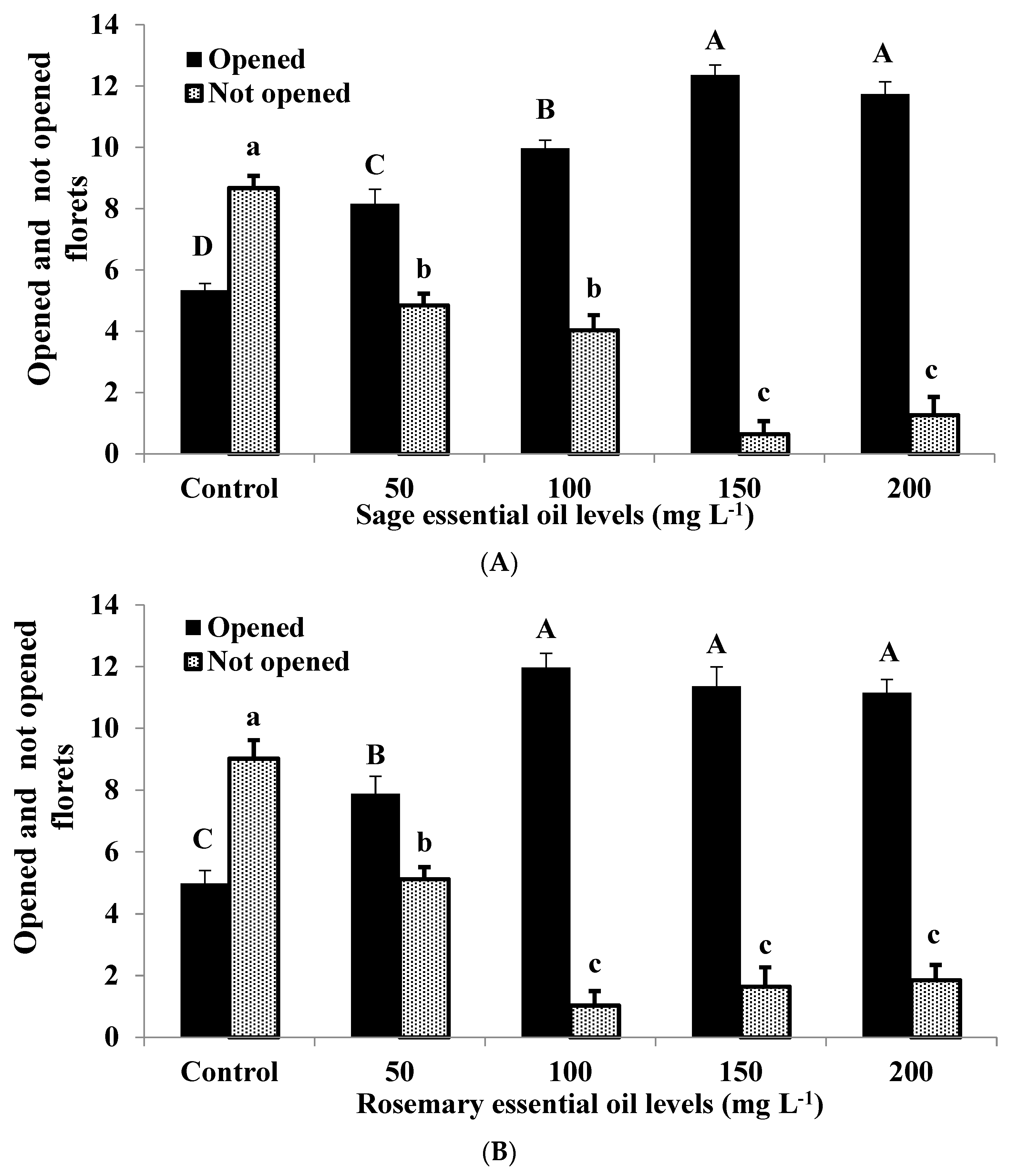
The number of opened and not opened florets in each gladiolus spike were counted, and the results show that all concentrations of sage and rosemary Eos markedly increased the number of opened florets and decreased the number of not opened florets on the spike compared to the control (Figure 2). The positive impacts of both Eos on floret opening were more observed with the sage essential oil at 100 mg L−1 (95.08% increase) or rosemary essential oil at 150 mg L−1 (92.06% increase).

3.5. Bacterial Counts

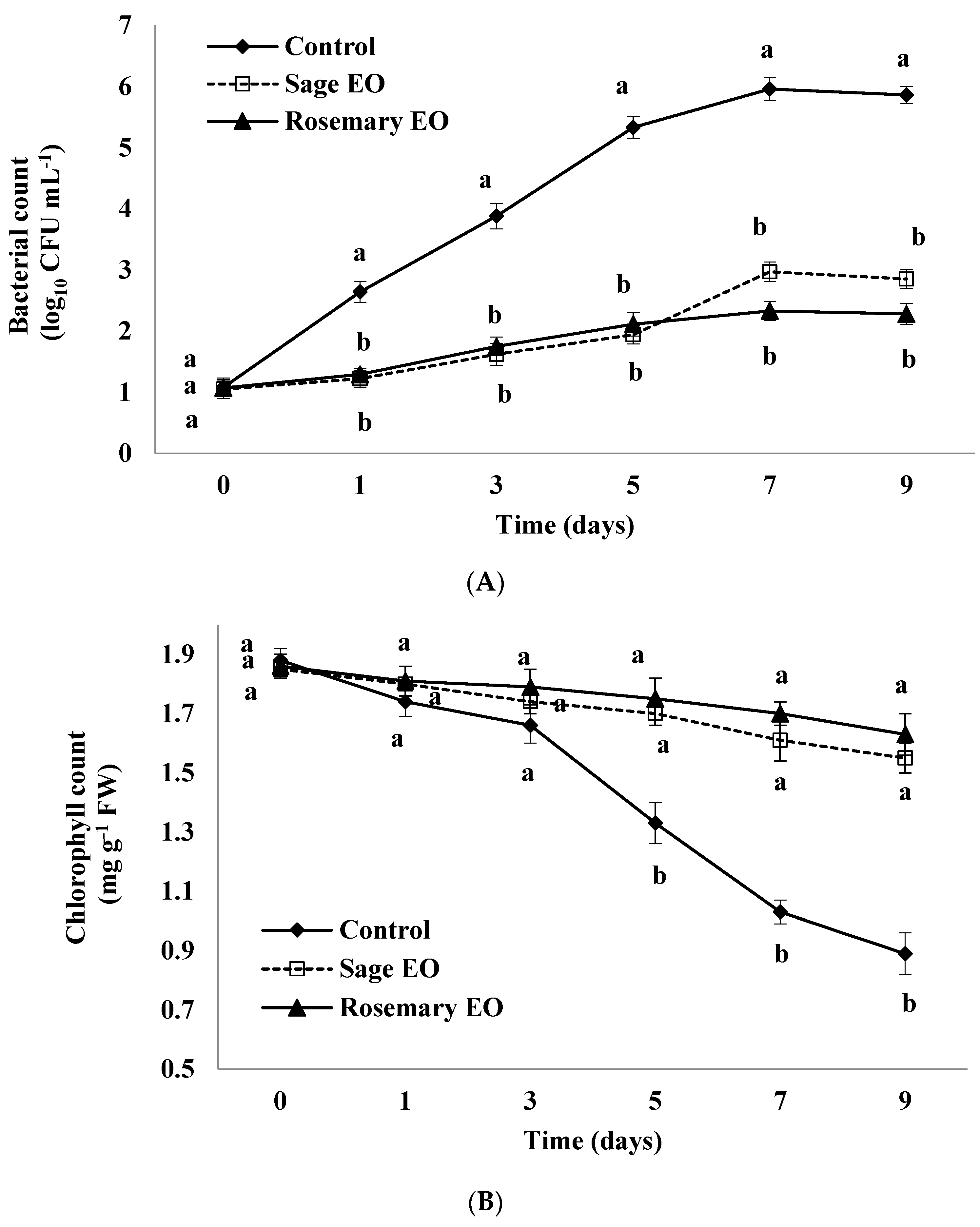
A sharp increase in the bacterial population in the vase solution of untreated spikes was observed throughout the vase life period and reached its highest value by day 9; however, both the sage and rosemary EO treatments significantly (p ≤ 0.05) prevented this increase (Figure 3A). Despite a small increase in bacterial population observed in the sage essential oil treatment compared to rosemary essential oil after day 5, there were no significant differences between both treatments in this context.

3.6. Chlorophyll Content

A gradual reduction in leaf chlorophyll content was observed in the treated and untreated spikes; however, the sage and rosemary EO treatments considerably preserved the chlorophyll contents compared to the control spikes. By day 7, when the vase life of the control was over, the chlorophyll content in the treated spikes was recorded at 56.31 and 65.04% higher than the control for sage and rosemary EO treatments, respectively (Figure 3B). The results also revealed that by day 7, 45.21% of premier chlorophyll content was lost in the control leaves, whereas this reduction was only 12.97% and 8.60% for the sage and rosemary EO treatments, respectively.

3.7. Hydrogen Peroxide (H2O2) Production

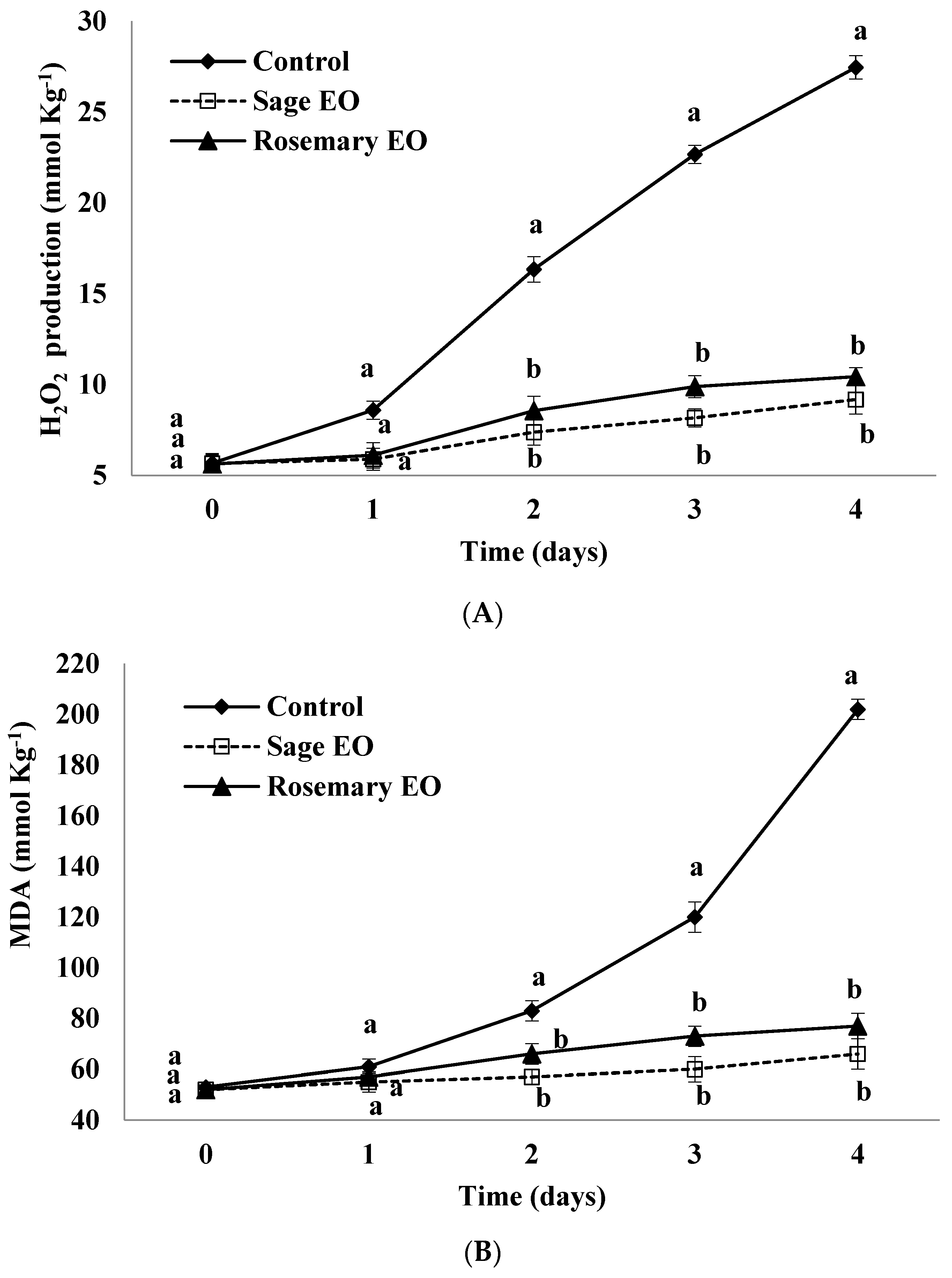
Despite a steady increase in H2O2 concentration shown during the floret life in the treated and control spikes, the production level of H2O2 sharply increased in the untreated spikes (Figure 4A). Contrary, the treatment of sage or rosemary Eos markedly decreased H2O2 overproduction relative to the control, which recorded the highest H2O2 production on day 4 (4.83-fold), which was higher than the initial production; however, the sage or rosemary EO treatments recorded only 1.62- and 1.85-fold, respectively.

3.8. Malondialdehyde (MDA) Content

A significant increase in the MDA content was observed in the control spikes; however, the sage or rosemary EO treatments significantly retarded the accumulation of MDA in floret tissues during the evaluation period. The control spikes recorded a 3.81-fold higher MDA content on day 4 relative to the initial value, while sage or rosemary EO-treated spikes recorded only 1.26- and 1.48-fold. Relative to the untreated spikes, the MDA content decreased by 67.32 and 61.88% on day 4 due to the application of sage or rosemary EO treatments, respectively (Figure 4B).

3.9. Membrane Stability Index (MSI)

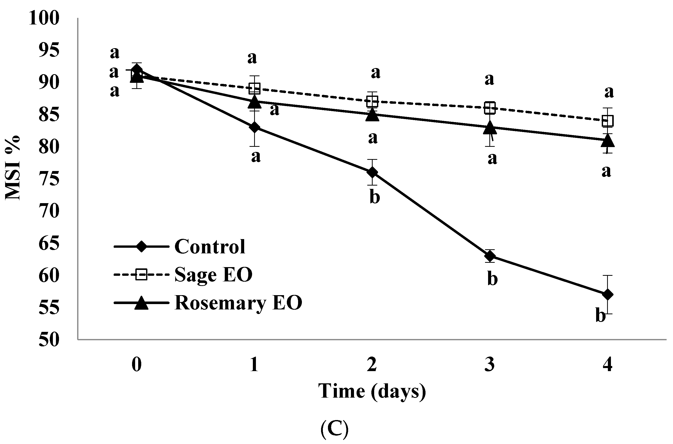
The membrane stability index clearly shows that the membrane stability of florets on the untreated spikes was markedly impaired throughout the estimation period, while the sage or rosemary EO treatments significantly preserved membrane stability (Figure 4C). On day 4, the MSI in florets on untreated spikes was 57% (35% lost) compared to 84 and 81% (7 and 10% lost) for the sage or rosemary EO treatments, respectively.

3.10. Total Phenol Content

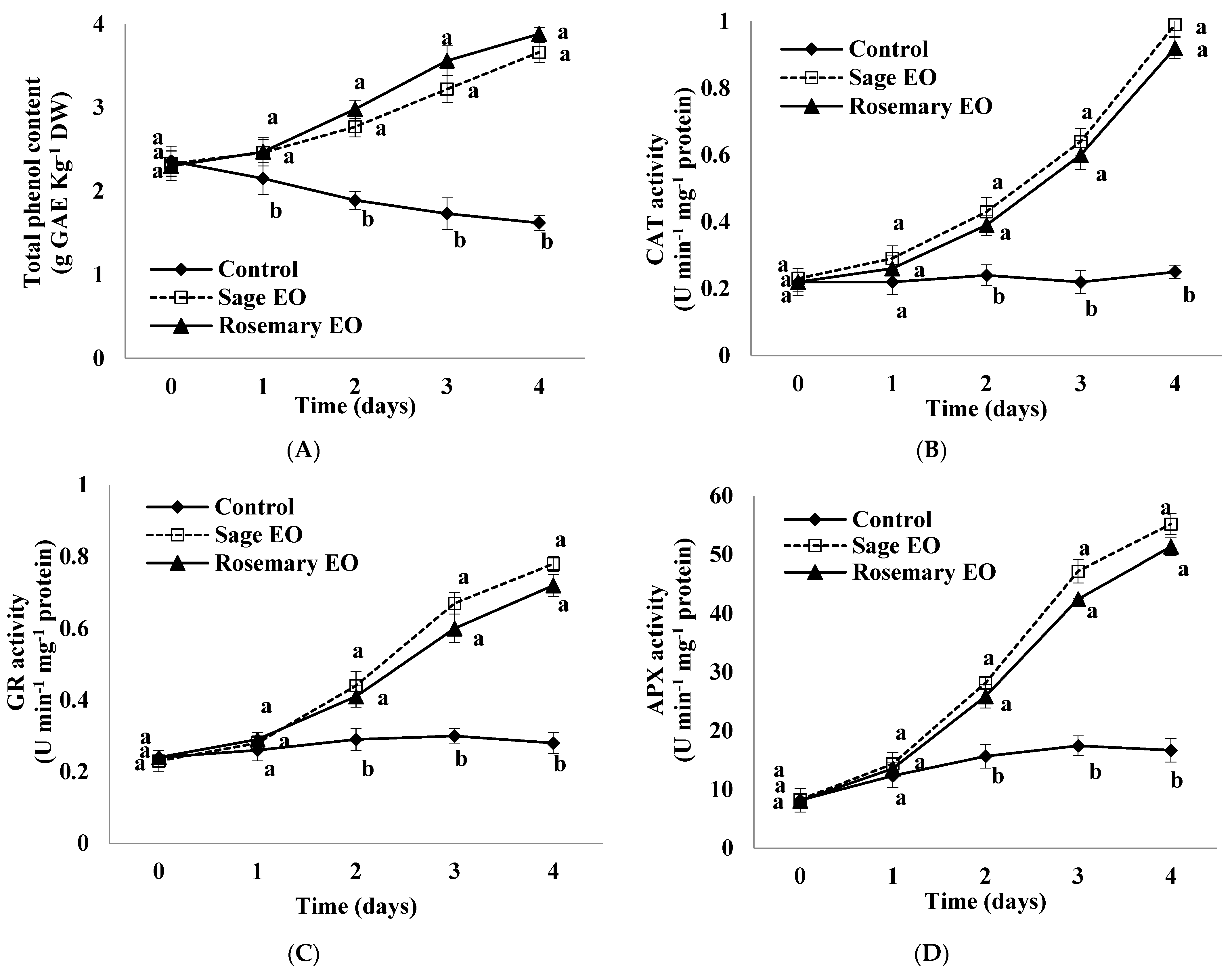
A slight reduction in total phenols of untreated spikes was detected during the floret life. Contrary, the sage or rosemary EO treatments significantly enhanced the phenol content through the evaluation period compared to the control (Figure 5A). On day 4, the total phenols in the sage or rosemary EO-treated florets were increased by 125.92 and 139.51% compared to the untreated florets, respectively.

3.11. Activities of Antioxidant Enzymes

Gladiolus florets of sage or rosemary EO-treated spikes exhibited a drastic increase in the activities of CAT, GR and APX enzymes relative to the control spikes throughout the assessment period (Figure 5B–D). A trivial increase in the activities of antioxidant enzymes was observed in the control florets until day 3 and then reduced; however, an excessive increase in CAT, GR and APX activities was detected in sage or rosemary EO-treated florets until day 4. Relative to the untreated florets, the activities of the CAT, GR and APX enzymes were 3.96-, 2.78- and 3.30-fold higher in the sage EO-treated spikes and 3.68-, 2.57- and 3.07-fold higher in the rosemary EO-treated ones, respectively.

3.12. Principal Component Analysis

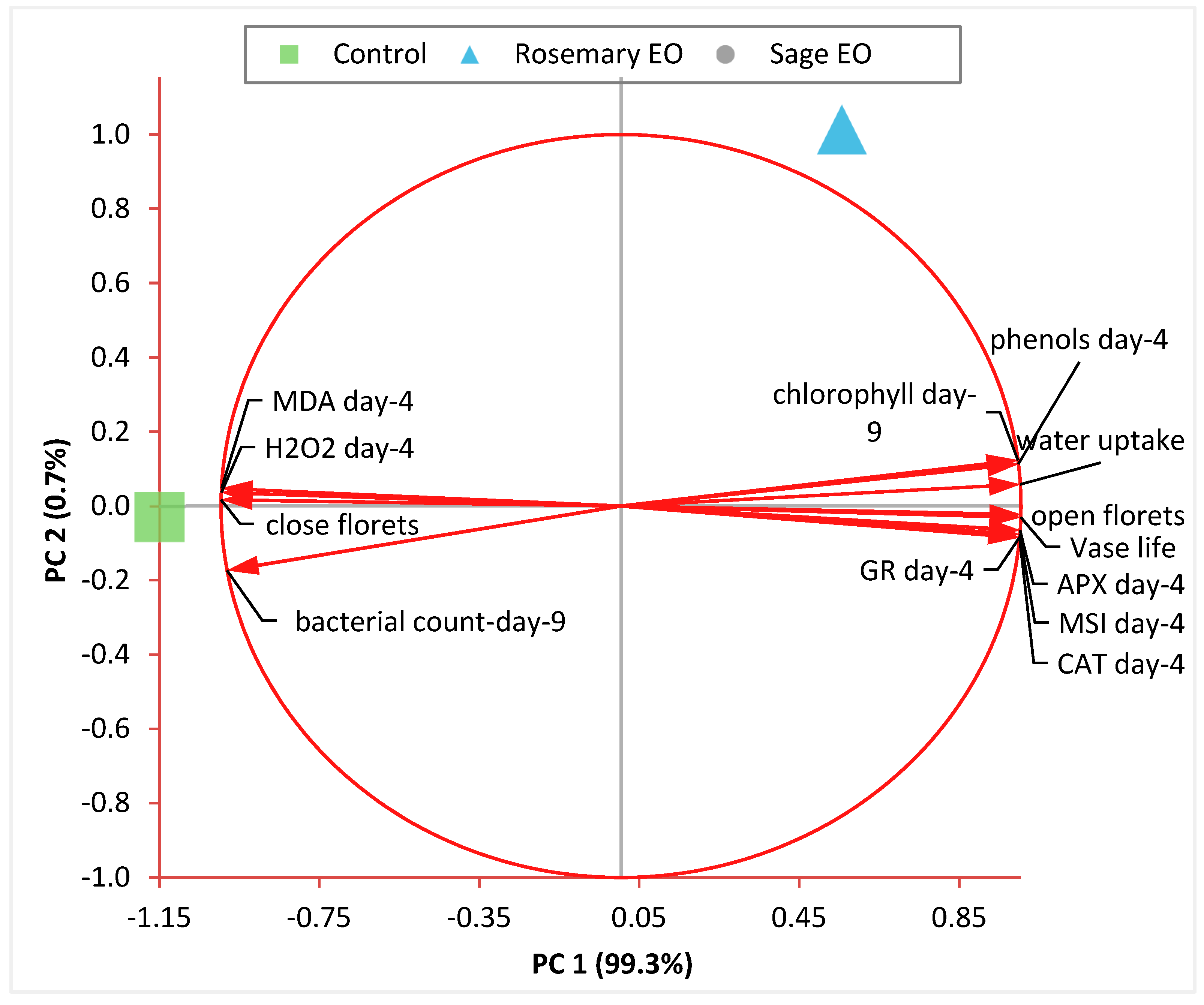
The results of the PCA biplots show the loading of various variables on the first two principal (PC1 and PC2) components (Figure 6). The long vectors revealed that all variables are strongly signified in the plot. The variables of the closed florets, bacterial count, MDA and H2O2 showed a negative correlation with PC1; however, a positive correlation was observed between the other variables with PC1. The biplots gave precious information about the correlations between variables. The vase life is positively correlated with water uptake, open florets, chlorophyll content, phenolic content and antioxidant enzyme activity, while it is negatively correlated with closed florets, bacterial count, MDA and H2O2. A positive correlation among the closed florets, bacterial count, MDA and H2O2 variables was detected. The same trend was obtained among the closed florets, bacterial count, MDA and H2O2 variables. Both PC1 and PC2 components successfully separated the impact of sage and rosemary EO treatments. The effect of both treatments appeared to group together; however, the control varied strongly.

4. Discussion

The results clearly showed the capability of sage or rosemary EOs to extend the vase life and preserve the postharvest quality of cut gladiolus spikes. This positive role could be attributed to their antimicrobial effect that mitigated the oxidative stress, and resulted in maintaining cellular water and chlorophyll contents, and hence membrane integrity in the treated spikes. The application of sage or rosemary EOs markedly increased the floret opening and water uptake and reduced the bacterial populations in the vase solution, thus extending the vase life. Increasing the water uptake maintained the freshness and increased the vase life of gladiolus spikes [11]. It has been reported that disturbing a plant’s water relations induced flower senescence while maintaining it is essential to prolong the flowers’ vase life [7]. The results of the current study have also indicated a strong positive correlation between vase life and water uptake, as well as open florets (Figure 6).
It is clearly noticeable that xylem occlusion by increased bacterial populations is the most important factor that decreases the water uptake in cut flower spikes [26,38,39]. This could justify the positive role of sage or rosemary EOs in decreasing the bacterial populations in vase solutions and thereby led to improved water relations and enhanced vase life of treated gladiolus spikes. The main active components of sage (1.8-Cineole, α-Thujone, β-Thujone, Camphor, Viridiflorol) and rosemary (α-Pinene, Camphene, 1.8-Cineole, Camphor, Borneol) EOs (Table 1) were likely the reason for their strong antimicrobial properties on various bacterial strains [40,41,42]. A strong negative correlation was clearly observed between the vase life and bacterial count (Figure 6). Furthermore, the presence of phenolic compounds in sage or rosemary EOs’ vase solutions could also be another reason for the limited bacterial population in the vase solution and thus might be linked with the prolonged vase life of cut spikes. Similar findings have been reported to extend the vase life of cut gladiolus spikes using natural leaf extracts rich in phenolic compounds [16]. Similarly, a positive role of various EOs in enhancing water relations and limiting bacterial growth has been reported in several plant species, such as using Shirazi thyme EO in gerbera [22], Salvia rosmarinus EO in carnations [23], Mentha piperita EO in gladioli [16], Mentha spicata EO in roses [24] and lemon grass EO in gladioli [25].
The chlorophyll content was markedly enhanced in leaves of sage- or rosemary-treated spikes compared to the untreated ones. It has been reported that a disruption in water relations via increasing the bacterial populations decreased the leaf chlorophyll content in cut gladiolus spikes [8]. Both a water deficit and induced oxidative stress consistently led to a reduced leaf chlorophyll content that could be attributed to the disorganization of the thylakoid membrane and activation of the chlorophyllase responsible for the degradation of chlorophyll, which eventually damages the photosynthetic apparatus [43,44]. The ability of EOs to improve the leaf chlorophyll content has also been reported in cut Davallia solida spikes [45] and has been revealed to delay leaf senescence, which is in accordance with the current data. Interestingly, the results have also revealed that the floret opening was improved in sage or rosemary EO-treated spikes, which could be attributed to the improved water relations in treated spikes required for flower development and eventually led to a prolonged vase life [8].
Moreover, the current data have also shown a positive role of sage or rosemary EOs in controlling the postharvest senescence in cut gladiolus spikes by reducing lipid peroxidation and preserving membrane stability. It is widely proved that exposing cut flowers to environmental stresses induced a generation of ROS that damages cellular macromolecules and leads to flower senescence [3], as indicated in the control spikes with high levels of H2O2 and MDA associated with cellular membrane damage, as previously reported in gladioli [8] and roses [3]. Unlike the control spikes, EO-treated ones exhibited a considerable reduction in their H2O2 and MDA levels, which can be justified by the fact of considerable amounts of phenol and antioxidant compounds in the EOs that enable gladiolus florets to alleviate oxidative damage. These results are in agreement with previous reports of using lemongrass essential oil in gladioli [25] and ginger essential oil in Davallia solida [45]. The content of MDA was markedly decreased in gladiolus florets in response to the application of sage or rosemary EOs. This is also obvious with reduced H2O2 production. It is very clear that EOs mitigated the oxidative stress in treated cut gladiolus spikes, as indicated by the reduced MDA levels—a sign of inhibited lipid peroxidation—and thereby enhanced membrane stability, recognized by the increased MSI, which are positively related to a prolonged vase life [3,8,46] as previously reported for the carnation cut flowers treated with Mentha spicata essential oil [47]. Furthermore, the PCA results exhibited informative correlations between the MDA and H2O2 levels.
The oxidative injuries apparently impacted the flower life; thus, encouraging the antioxidant system has been recognized as a step to alleviate these injuries in various reports [3,8]. The results indicated that the total phenol contents were markedly elevated in sage or rosemary EO-treated spikes. Phenols are known for their antimicrobial and antioxidant properties. In addition, EOs have a phenolic structure and contain several phenolic compounds, which may directly or indirectly affect the physiological and metabolic processes in such a way that they improve the interior phenolic content [18,19]. Enhancing the total phenols by EOs is consistent with reduced H2O2 and MDA contents, as phenols are identified to have a non-enzymatic antioxidant role [48]—the effect that may participate in senescence regulation of gladiolus cut spikes. A strong positive correlation between phenolic contents and vase life was observed (Figure 6). The positive impact of several EOs in increasing the phenolic content has also been reported in cut carnation spikes [49], which supports the current data. The current results have also shown improvement in the CAT, GR and APX activities in sage or rosemary EO-treated spikes, which were effectively scavenging the ROS produced by induced oxidative stress in cut spikes [50]. These enzymes could also contribute to the reduced MDA level in treated spikes and, therefore, repressed floret senescence. Furthermore, the PCA indicated a strong positive correlation between the CAT, GR and APX activities and vase life. It is proven that developing an effective antioxidant system can suppress the accumulation of ROS and repair oxidative injuries [3,50]. This observed impact of sage or rosemary EOs is probably responsible for maintaining a higher MSI and extending the vase life of treated spikes compared to the control ones. In reality, this study is considered the first to report the efficacy of sage or rosemary EOs on the induction of antioxidant machinery in “White Prosperity” cut gladioli. Similar observations have been reported in gladioli using Mentha piperita and lemon grass EOs [16,25] and also in carnation using Mentha spicata essential oil [47]. In addition, this study was performed on one species, and therefore, further studies on various species are recommended for them to be applicable on a commercial scale.

5. Conclusions

The current findings revealed the role of sage or rosemary EOs in preserving the postharvest quality and vase life of cut gladiolus spikes. A positive impact was ascribed via enhanced water relations and limited bacterial populations in a vase solution with an improved chlorophyll content and membrane stability due to induced antioxidant machinery. Sage EO at 150 mg L−1 or rosemary EO at 100 mg L−1 may be recommended as novel preservatives to prolong the vase life of cut gladioli during marketing. Further studies are recommended, particularly at a molecular level, to understand how essential oils influence the phenolic acid biosynthesis pathway encoding genes.

Author Contributions

Conceptualization, F.A.S.H. and M.M.M.; methodology, R.M.M. and M.M.M.; software, M.M.M.; validation, F.A.S.H., M.M.M. and R.M.M.; formal analysis, M.M.M.; investigation, R.M.M.; resources, M.M.M.; data curation, M.M.M.; writing—original draft preparation, F.A.S.H.; writing—review and editing, F.A.S.H.; visualization, M.M.M.; supervision, F.A.S.H. All authors have read and agreed to the published version of the manuscript.

Funding

This research received no external funding.

Data Availability Statement

Data are contained within the article.

Conflicts of Interest

The authors declare no conflicts of interest.

References

  1. Younis, A.; Akhtar, M.S.; Riaz, A.; Zulfiqar, F.; Qasim, M.; Farooq, A.; Tariq, U.; Ahsan, M.; Bhatti, Z.M. Improved cut flower and corm production by exogenous moringa leaf extract application on gladiolus cultivars. Acta Sci. Pol. Hortorum Cultus 2018, 17, 25–38. [Google Scholar] [CrossRef]
  2. Hassan, F.A.S.; Schmidt, G. Postharvest characteristics of cut carnations as the result of chemical treatments. Acta Agron. Hung. 2004, 52, 125–132. [Google Scholar] [CrossRef]
  3. Mazrou, R.M.; Hassan, S.; Yang, M.; Hassan, F.A.S. Melatonin preserves the postharvest quality of cut roses through enhancing the antioxidant system. Plants 2022, 11, 2713. [Google Scholar] [CrossRef] [PubMed]
  4. Bhattacharjee, S.; De, L.C. Post-Harvest Technology of Flowers and Ornamental Plants; Aavishkar Publishers: Jaipur, India, 2005; pp. 11–19. [Google Scholar]
  5. Darras, A. Overview of the dynamic role of specialty cut flowers in the international cut flower market. Horticulturae 2021, 7, 51. [Google Scholar] [CrossRef]
  6. Ezhilmathi, K.; Singh, V.P.; Arora, A.; Sairam, R.K. Effect of 5-sulfusalicylic acid on antioxidant activity in relation to vase life of gladiolus cut flowers. Plant Growth Regul. 2007, 51, 99–108. [Google Scholar] [CrossRef]
  7. Hassan, F.A.S.; Ali, E.F. Protective effects of 1-methylcyclopropene and salicylic acid on senescence regulation of gladiolus cut spikes. Sci. Hortic. 2014, 179, 146–152. [Google Scholar] [CrossRef]
  8. Hassan, F.A.S.; Fetouh, M.I. Does moringa leaf extract have preservative effect improving the longevity and postharvest quality of gladiolus cut spikes? Sci. Hortic. 2019, 250, 287–293. [Google Scholar] [CrossRef]
  9. Hu, H.; Li, P.; Shen, W. Preharvest application of hydrogen-rich water not only affects daylily bud yield but also contributes to the alleviation of bud browning. Sci. Hortic. 2021, 287, 110267. [Google Scholar] [CrossRef]
  10. Loubaud, M.; van Doorn, W.G. Wound induced and bacteria induced xylem blockage in roses, Astilbe, and Viburnum. Postharvest Biol. Technol. 2004, 32, 281–288. [Google Scholar] [CrossRef]
  11. Hatamzadeh, A.; Hatami, M.; Ghasemnezhad, M. Efficiency of salicylic acid delay petal senescence and extended quality of cut spikes of Gladiolus grandiflora cv ‘wing’s sensation’. Afric. J. Agric. Res. 2012, 7, 540–545. [Google Scholar]
  12. Hassan, F.A.S.; Mazrou, R.; Gaber, A.; Hassan, M. Moringa extract preserved the vase life of cut roses through maintaining water relations and enhancing antioxidant machinery. Postharvest Biol. Technol. 2020, 164, 111156. [Google Scholar] [CrossRef]
  13. Gupta, S.; Kumar, A. Effect of different levels of pulsing concentrations on vase life of gladiolus (Gladiolus grandiflorus L.). Int. J. Curr. Microbiol. App. Sci. 2018, 7, 330–334. [Google Scholar] [CrossRef]
  14. Knee, M. Selection of biocides for use in floral preservatives. Postharvest Biol. Technol. 2000, 18, 227–234. [Google Scholar] [CrossRef]
  15. Saeed, T.; Hassan, I.; Abbasi, N.; Jilani, G. Effect of gibberellic acid on the vase life and oxidative activities in senescing cut gladiolus flowers. Plant Growth Regul. 2014, 72, 89–95. [Google Scholar] [CrossRef]
  16. Akhtar, G.; Rajwana, I.A.; Sajjad, Y.; Shehzad, M.A.; Amin, M.; Razzaq, K.; Ullah, S.; Faried, H.N.; Farooq, A.; Samiullah. Do natural leaf extracts involve regulation at physiological and biochemical levels to extend vase life of gladiolus cut flowers? Sci. Hortic. 2021, 282, 110042. [Google Scholar] [CrossRef]
  17. Ramezanian, A.; Azadi, M.; Mostowfizadeh-Ghalamfarsa, R.; Saharkhiz, M.J. Effect of Zatariamultiflora Boiss and Thymus vulgaris L. essential oils on black rot of ‘Washington Navel’ orange fruit. Postharvest Biol. Technol. 2016, 112, 152–158. [Google Scholar] [CrossRef]
  18. Braga, P.C.; Culici, M.; Alferi, M.; Sasso, M. Thymol inhibits Candida albicans biofilm formation and mature biofilm. Int. J. Antimicrob. Agents 2008, 31, 472–477. [Google Scholar] [CrossRef] [PubMed]
  19. Solgi, M.; Ghorbanpour, M. Application of essential oils and their biological effects on extending the shelf-life and quality of horticultural crops. Trakia J. Sci. 2014, 12, 198–210. [Google Scholar]
  20. Zaouali, Y.; Bouzaine, T.; Boussaid, M. Essential oils composition in two Rosmarinus officinalis L. varieties and incidence for antimicrobial and antioxidant activities. Food Chem. Toxicol. 2010, 48, 3144–3152. [Google Scholar] [CrossRef]
  21. Bouaziz, M.; Yangui, T.; Sayadi, S.; Dhouib, A. Disinfectant properties of essential oils from Salvia officinalis L. cultivated in Tunisia. Food Chem. Toxicol. 2009, 47, 2755–2760. [Google Scholar] [CrossRef]
  22. Mallahi, T.; Ramezanian, A.; Saharkhiz, M.J.; Javanmardi, J.; Iraji, A. Antimicrobial activities of Asafoetida and Shirazi thyme essential oils improve the vase life of gerbera cut flowers. Acta Ecol. Sin. 2018, 38, 228–233. [Google Scholar] [CrossRef]
  23. Basiri, Y.; Zarei, H.; Mashayekhy, K.; Pahlavany, M.H. Effect of Rosemary extract on vase life and some qualitative characteristics of cut Carnation flowers (Dianthus caryophyllus cv. White librity). J. Stored Prod. Postharvest Res. 2011, 14, 261–265. [Google Scholar]
  24. Salmi, M.S.; Hoseini, M.F.; Heidari, M.; Daneshvar, M.H. Extending vase life of cut rose (Rosa hybrida L.) cv. Bacara by essential oils. Adv. Hortic. Sci. 2018, 32, 61–69. [Google Scholar]
  25. Thakur, M.; Verma, V.; Chandel, A.; Kumar, R.; Sharma, T.; Kumar, A.; Bhardwaj, S.; Kumar, R.; Bhargava, B. Lemon grass essential oil improve Gladiolus grandiforus postharvest life by modulating water relations microbial growth, biochemical activity, and gene expression. Sci. Rep. 2023, 13, 2630. [Google Scholar] [CrossRef]
  26. Balestra, G.M.; Agostini, R.; Bellincontro, A.; Mencarelli, F.; Varvaro, L. Bacterial populations related to gerbera (Gerbera jamesonii L.) stem break. Phytopathol. Mediterr. 2005, 44, 291–299. [Google Scholar]
  27. Metzner, H.; Rau, H.; Senger, H. Unter suchungen zur synchronisier barteit einzelner pigmentan angel mutanten von chlorela. Planta 1965, 65, 186. [Google Scholar] [CrossRef]
  28. Lichtenthaler, H.K. Chlorophylls and Carotenoids Pigments of Photosynthetic Biomembranes. In Methods in Enzymology; Colowick, S.P., Kaplan, N.O., Eds.; Academic Press: Cambridge, MA, USA, 1987; Volume 148, pp. 350–382. [Google Scholar]
  29. Patterson, B.D.; Macrae, E.A.; Ferguson, I.B. Estimation of hydrogen peroxide in plant extracts using titanium (IV). Anal. Chem. 1984, 134, 487–492. [Google Scholar] [CrossRef]
  30. Hodges, D.M.; Delong, J.M.; Forney, C.F.; Prange, R.K. Improving the thiobarbituric acidreactive-substances assay for estimating lipid peroxidation in plant tissue containing anthocyanin and other interfering compounds. Planta 1999, 207, 604–611. [Google Scholar] [CrossRef]
  31. Sairam, R.K.; Deshmukh, P.S.; Shukla, D.S. Tolerance to drought and temperature stress in relation to increased antioxidant enzyme activity in wheat. J. Agron. Crop Sci. 1997, 178, 171–177. [Google Scholar] [CrossRef]
  32. McDonald, S.; Prenzler, P.D.; Antolovich, M.; Robards, K. Phenolic content and antioxidant activity of olive extracts. Food Chem. 2001, 73, 73–84. [Google Scholar] [CrossRef]
  33. Bradford, M.M. A rapid and sensitive method for quantitation of micro quantities of protein utilizing the principle of protein-dye binding. Anal. Biochem. 1976, 72, 248–258. [Google Scholar] [CrossRef] [PubMed]
  34. Chandlee, J.M.; Scandalios, J.G. Analysis of variants affecting the catalase developmental program in maize scutellum. Theor. Appl. Genet. 1984, 69, 71–77. [Google Scholar] [CrossRef] [PubMed]
  35. Nakano, Y.; Asada, K. Hydrogen peroxide is scavenged by ascorbate-specific peroxidase in spinach chloroplasts. Plant Cell Physiol. 1981, 22, 867–880. [Google Scholar]
  36. Foyer, C.H.; Halliwell, B. The presence of glutathione and glutathione reductase in chloroplasts: A proposed role in ascorbic acid metabolism. Planta 1976, 133, 21–25. [Google Scholar] [CrossRef] [PubMed]
  37. Rao, M.V. Cellular detoxifying mechanisms determine the age dependent injury in tropical trees exposed to SO2. J. Plant Physiol. 1992, 140, 733–740. [Google Scholar] [CrossRef]
  38. Solgi, M.; Kafi, M.; Taghavi, T.S.; Naderi, R. Essential oils and silver nanoparticles (SNP) as novel agents to extend vase-life of gerbera (Gerbera jamesonii cv. ‘Dune’) flowers. Postharvest Biol. Technol. 2009, 53, 155–158. [Google Scholar] [CrossRef]
  39. Gun, S.; Uzun, L.; Tuysuz, M.; Erturk, O.; Ilhan, H.; Acıkgoz, M.; Ozturk, B. Nanofiber mats containing lavender oil and methyl jasmonate as an innovative treatment to extend vase life in cut rose flowers. Postharvest Biol. Technol. 2023, 201, 112343. [Google Scholar] [CrossRef]
  40. Raal, A.; Orav, A.; Arak, E. Composition of the essential oil of Salvia officinalis L. from various European countries. Nat. Prod. Res. 2007, 21, 406–411. [Google Scholar] [CrossRef] [PubMed]
  41. Özcan, B.; Birgul, E.M.; Coleri, A.; Yolcu, H.; Caliskan, M. In vitro antimicrobial and antioxidant activities of various extracts of Salvia microstegia (Boiss.) et. Bal. from Antakya, Turkey. Fresenius Environ. Bull. 2009, 18, 658–662. [Google Scholar]
  42. Günther, M.; Karygianni, L.; Argyropoulou, A.; Anderson, A.; Hellwig, E.; Skaltsounis, A.; Wittmer, A.; Vach, K.; Al-Ahmad, A. The antimicrobial effect of Rosmarinus officinalis extracts on oral initial adhesion ex vivo. Clin. Oral. Investig. 2022, 26, 4369–4380. [Google Scholar] [CrossRef]
  43. Rong-Hua, L.; Pei-Guo, G.; Baum, M.; Grando, S.; Ceccarelli, S. Evaluation of chlorophyll content and fluorescence parameters as indicators of drought tolerance in barley. Agric. Sci. China 2006, 5, 751–757. [Google Scholar]
  44. Hassan, F.A.S.; Ali, E.F.; Mostafa, N.Y.; Mazrou, R. Shelf-life extension of sweet basil leaves by edible coating with thyme volatile oil encapsulated chitosan nanoparticles. Int. J. Biol. Macromol. 2021, 177, 517–525. [Google Scholar] [CrossRef] [PubMed]
  45. Teerarak, M.; Laosinwattana, C. Essential oil from ginger as a novel agent in delaying senescence of cut fronds of the fern (Davallia solida (G. Forst.) Sw.). Postharvest Biol. Technol. 2019, 156, 110927. [Google Scholar] [CrossRef]
  46. Ali, E.F.; Issa, A.A.; Al-Yasi, H.M.; Hessini, K.; Hassan, F.A.S. The efficacies of 1-methylcyclopropene and chitosan nanoparticles in preserving the postharvest quality of Damask rose and their underlying biochemical and physiological mechanisms. Biology 2022, 11, 242. [Google Scholar] [CrossRef] [PubMed]
  47. Dehestani-Ardakani, M.; Gholamnezhada, J.; Alizadeha, S.; Meftahizadeha, H.; Ghorbanpour, M. Salicylic acid and herbal extracts prolong vase life and improve quality of carnation (Dianthus caryophyllus L.) flowers. S. Afr. J. Bot. 2022, 150, 1192–1204. [Google Scholar] [CrossRef]
  48. Gan, J.; Feng, Y.; He, Z.; Li, H.; Zhang, H. Correlations between antioxidant activity and alkaloids and phenols of maca (Lepidium meyenii). J. Food Qual. 2017, 3, 3185945. [Google Scholar] [CrossRef]
  49. Soliman, D.; El-Sayed, I. Study postharvest characteristics, chemical composition and antimicrobial activity of Dianthus caryophyllus L.; cut flowers using some essential oils. Ornam. Hortic. 2023, 29, 37–47. [Google Scholar] [CrossRef]
  50. Zhou, Q.; Ma, C.; Cheng, S.; Wei, B.; Liu, X.; Ji, S. Changes in antioxidative metabolism accompanying pitting development in stored blueberry fruit. Postharvest Biol. Technol. 2014, 88, 88–95. [Google Scholar] [CrossRef]
Figure 1. Vase life (A,B) and the average water uptake level until day 6 (C,D) of gladiolus cut spikes treated with sage or rosemary essential oil at 0, 50, 100, 150 and 200 mg L−1. Values are the mean ± SE (n = 6). Columns have different letters are significantly different from each other according to Tukey–Kramer’s multiple range test at p ≤ 0.05.
Horticulturae 10 00638 g001
Figure 2. Opened and not opened florets of gladiolus cut spikes at day 12 treated with sage (A) or rosemary (B) essential oil at 0, 50, 100, 150 and 200 mg L−1. Values are the mean ± SE (n = 6). Means have different letters are significantly different from each other according to Tukey–Kramer’s multiple range test at p ≤ 0.05.
Horticulturae 10 00638 g002
Figure 3. Bacterial count (A) and total chlorophyll content (B) of gladiolus cut spike leaves treated with sage essential oil (Sage EO) at 100 mg L−1 or rosemary essential oil (Rosemary EO) at 150 mg L−1. Values represent the mean ± SE (n = 6). Means have different letters are significantly different from each other according to Tukey–Kramer’s multiple range test at p ≤ 0.05.
Horticulturae 10 00638 g003
Figure 4. Hydrogen peroxide (A), malondialdehyde content (B) and membrane stability index (C) in the third floret from the base of gladiolus cut spikes treated with sage essential oil (Sage EO) at 100 mg L−1 or rosemary essential oil (Rosemary EO) at 150 mg L−1. Values are the mean ± SE (n = 6). Means have different letters and are significantly different from each other according to Tukey–Kramer’s multiple range test at p ≤ 0.05.
Horticulturae 10 00638 g004aHorticulturae 10 00638 g004b
Figure 5. Total phenol content (A) and the activity of catalase (B), glutathione reductase (C) and ascorbate peroxidase (APX) enzymes (D) in the third floret from the base of gladiolus cut spikes treated with sage essential oil (Sage EO) at 100 mg L−1 or rosemary essential oil (Rosemary EO) at 150 mg L−1. Values are the mean ± SE (n = 6). Means have different letters and are significantly different from each other according to Tukey–Kramer’s multiple range test at p ≤ 0.05.
Horticulturae 10 00638 g005
Figure 6. Biplot of the principal component analysis (PCA) for gladiolus cut spikes treated with sage essential oil (Sage EO) at 100 mg L−1 or rosemary essential oil (Rosemary EO) at 150 mg L−1. The red circle around vectors represents perfect correlation. The vectors (arrows) represent the variables, and the colored shapes represent sampling points under various treatments.
Horticulturae 10 00638 g006
Table 1. The composition of essential oils of sage and rosemary plants.
Salvia Essential OilRosemary Essential Oil
Oil ConstituentsRI *PercentageOil ConstituentsRIPercentage
α-Pinene9522.38α-Pinene102810.64
Camphene9632.66Camphene10506.82
β-Pinene9861.42β-Pinene11163.44
β-Myrcene9920.80β-Myrcene11701.63
p-Cymene10281.20α-Phellandrene12020.22
Limonene10361.38α-Terpinene12440.67
1.8-Cineole10447.88p-Cymene12782.85
α-Thujone110828.31Limonene12861.88
β-Thujone111012.44γ-Terpinene13221.22
Camphor114921.681.8-Cineole13.2637.54
Borneol11643.83Linalool13621.67
Terpinen-4-ol11780.81β-Caryophyllene16182.85
Bornyl acetate12763.66Camphor168613.59
Β-Caryophyllene13880.68Borneol17887.43
α-Humulene14482.34α-Terpineol18124.87
Viridiflorol15867.75Bornyl acetate18261.48
Total identified compounds99.22Total identified compounds98.80
* RI means retention index.
Disclaimer/Publisher’s Note: The statements, opinions and data contained in all publications are solely those of the individual author(s) and contributor(s) and not of MDPI and/or the editor(s). MDPI and/or the editor(s) disclaim responsibility for any injury to people or property resulting from any ideas, methods, instructions or products referred to in the content.