Article

In Vitro Shoot Regeneration and Callogenesis of Sechium compositum (Donn. Sm.) C. Jeffrey for Plant Conservation and Secondary Metabolites Product

by
Riviello-Flores María de la Luz
1,
Castillo-Martínez Carlos Román
2,
Cadena-Iñiguez Jorge
3,*,
Ruiz-Posadas Lucero del Mar
1,
Soto-Hernández Ramón Marcos
1,
Arévalo-Galarza Ma. de Lourdes
1 and
Castillo-Juárez Israel
4
1
Colegio de Postgraduados, Campus Montecillo, Km. 36.5, Carretera México-Texcoco, Montecillo, Texcoco 56230, Mexico
2
Instituto Nacional de Investigación Forestales, Agrícolas y Pecuarias, CENID-COMEF, Progreso Núm. 5, Barrio de Santa Catarina, Alcaldía Coyoacán, Ciudad de México 04010, Mexico
3
Colegio de Postgraduados, Campus San Luis Potosí, Salinas de Hidalgo, San Luis Potosí 78622, Mexico
4
Conahcyt-Instituto de Ciencias Básicas e Ingeniería, Universidad Autónoma del Estado de Hidalgo, Mineral de la Reforma, Hidalgo 42184, Mexico
*
Author to whom correspondence should be addressed.
Horticulturae 2024, 10(6), 537; https://doi.org/10.3390/horticulturae10060537
Submission received: 20 March 2024 / Revised: 8 May 2024 / Accepted: 10 May 2024 / Published: 21 May 2024

Abstract

:
Sechium compositum (Cucurbitaceae) is a wild species that is distributed in the Soconusco region, Chiapas, Mexico, and the border with Guatemala. This species has an intangible biochemical value resulting from the pharmacological relevance of its secondary metabolites. However, as a consequence of the lack of knowledge about its importance, it is being displaced from its habitat at an accelerated rate, incurring the risk of genetic loss. Therefore, an in vitro culture protocol with two experimental phases was evaluated to propagate, conserve, and regenerate this species. The first phases considered the shoot propagation, adding seven concentrations (0.1, 0.2, 0.4, 0.6, 0.8, 1.0, 1.2 mg mL−1) of 6-benzylaminopurine (BA) and thidiazuron (TDZ) and evaluating the number of buds and shoots and the shoot height. The best multiplication response was recorded with 0.1, 0.2, 0.4, and 1.0 mg L−1 of BA and 0.1 mg L−1 of TDZ, as well as the MS base culture medium. The validation of the results of the first phase (0.1 mg L−1 of BA) was compared with the MS in an independent experiment against the control (n = 50 repetitions), obtaining a height of 52 mm, 1.36 shoots, and 9.22 buds, suggesting that this concentration is adequate for the purpose, surpassing the MS control (MS culture medium alone). Of the total volume of roots obtained with packed bud structure in the previous experimental sample, it was reduced to 14% (n = 50). The second phase consisted of inducing callus formation from stem and leaf explants through the addition of 0.5, 1.0, and 2.0 mg L−1 of TDZ and 2,4-Dichlorophenoxyacetic acid (2,4-D) to the medium. Callus induction in S. compositum was better when using the stem in a medium with 2.0 mg L−1 of 2,4-D with a value of 97.8% around the explant. The addition of 500 mg L−1 of polyvinylpyrrolidone (PVP) is also suggested to reduce oxidation. This protocol represents a significant advance in the conservation, multiplication, and callus formation of S. compositum and contributes to its rescue and revaluation in the face of the danger of extinction.

1. Introduction

Agrobiodiversity includes local or native varieties that are tolerated, encouraged, and cultivated, as well as ecotypes in the process of domestication and their wild relatives [1,2]. Phytogenetic resources for food and agriculture, implicit in agrobiodiversity, play a fundamental role in human development, providing significant benefits [3] that contribute to the scientific, technological, socioeconomic, and cultural progress of megadiverse countries [4].
Some of these local varieties and their wild relatives have not yet been fully characterized, which creates a major limitation for the identification of new uses or the improvement of known ones. In many cases, the commercial success of a genotype can indirectly displace local varieties whose use or consumption are less popular—especially wild relatives, which have been rendered fragile, placed in conditions of erosion, or lost due to the lack of research about their potential uses [5,6,7].
The conservation of these phytogenetic resources requires the application of in situ and ex situ strategies [8]. The first focuses on the maintenance of plant species and traditional agricultural systems in their habitats of origin, where they have developed specific phenotypic and genetic characteristics. This effort entails the protection of natural areas, wild ecosystems, and other environments within their original context [8]. In this process, the lore of rural communities plays a crucial role, as their populations have been the guardians of these plants and their uses for generations [5,9,10].
Ex situ conservation is mainly carried out in germplasm banks (Veracruz, Mexico) or scientific collections [10,11] and consists of the preservation of orthodox and recalcitrant seeds [12,13] outside of natural habitats. Both approaches play a fundamental role in the protection and conservation of the genetic diversity of plant species, contributing to food security and global biodiversity.
In this context, chayote (Sechium P.Br.) (Cucurbitaceae) emerges as an important phytogenetic resource for Mexico (its center of origin and domestication). The genus includes ten species, of which only S. edule and S. tacaco are cultivated as food [14]. The other eight (S. chinantlense, S. compositum, S. hintonii, S. talamancense, S. panamense, S. pittieri, S. venosum, and S. vilosum) are wild species [15].
S. compositum is distributed in the Soconusco region, Chiapas, Mexico, and the border with Guatemala [14]. In 2011, the proximity of rural communities placed five S. compositum populations in the endangered category in Chiapas [16]. This limited geographical distribution and the lack of knowledge of any use among rural inhabitants have promoted its displacement by economically profitable crops, such as coffee, corn, and forage species [16]. It is a creeping, herbaceous, and climbing plant, with massive tuber-like roots, a smooth, almost glabrous stem, and a uniquely woody appearance when adult. The leaves are trisected when young and angled in the adult plant. It has tendrils, yellow pistillate flowers in the axil of the vine, and staminates on a rachis up to 50 cm long. Medium fruits (8–10 cm) are subglobose green, ovoid, generally without spines, and strongly bitter in flavor. Generally, the entire plant has a bitter taste. According to recent studies [17], both of the biological variants of S. edule and two identified morphotypes of S. compositum have a high content of secondary metabolites with pharmacological activity, specifically tetracyclic triterpenes, phenols, and flavonoids [18,19] with antileukemic [20] and antifungal [21] potential, which opens a window of opportunity for its revaluation and, therefore, contributing to its conservation.
The ex situ conservation of S. compositum faces significant challenges, given its climbing nature and recalcitrant seed, which hinder its preservation through traditional methods [22]. In response to these challenges, a viable alternative is its establishment under in vitro conditions. This approach not only allows for the preservation of the species but also its regeneration and possible reintegration into its original habitat [23,24]. Furthermore, in vitro preservation is a source of tissues that are valuable for bioprospecting research [18], as well as the induction of mutagenesis for future applications. This biotechnological technique has several applications, including the study of physiological aspects [25], clonal propagation [26], the production of secondary metabolites [27], plant regeneration [28,29], and obtaining disease-free varieties [23,30,31].
A significant number of research works have focused on the in vitro establishment, conservation, and regeneration of S. edule [22,32,33,34,35,36] in addition to clonal propagation, rooting, and acclimatization protocols [22,37,38]. However, unlike S. edule, other species of the genus, such as S. compositum, have not been studied to define explant-based callus formation protocols [39]. Callus formation is the basis of massive in vitro propagation through indirect organogenesis or indirect somatic embryogenesis [40,41]; however, no such protocol has been developed for wild species.
The objective was to develop an in vitro multiplication protocol from bud explants of S. compositum (Donn. Sm.) C. Jeffrey to obtain tissue for the regeneration of the species, thus contributing to its conservation, as well as a callus formation protocol from explants, such as leaves and stems, in order to have alternative material to obtain secondary metabolites in the future, such as cucurbitacins, phenolic acids, and flavonoids of interest.

2. Materials and Methods

The research was divided into two experimental phases. The first considered the shoot propagation through the addition of 0.1, 0.2, 0.4, 0.6, 0.8, 1.0, and 1.2 mgL−1 concentrations of the 6-benzylaminopurine (BA) PhytoTechnology Laboratories® B800, Shawnee Mission, KS, USA) and thidiazuron (TDZ) Sigma-Aldrich® P6186, San Luis, MO, USA) growth regulators in order to observe differences in the number of buds and shoots and the shoot height with the MS plant culture medium [42]. The second phase consisted of the induction of callus formation from stem and leaf explants, with the addition of 0.5, 1.0, and 2.0 mg L−1 concentrations of the TDZ and 2,4-D (2,4-Dichlorophenoxyacetic acid; Phytotechnology Laboratories® D295, Shawnee Mission, KS, USA) growth regulators to the medium.

2.1. Plant Material: Mother Plant Description

Sechium compositum (Donn. Sm.) C. Jeffrey, from Sechium P. Br. Germplasm Bank (19°08′48″ N and 97°57′00″ W) Veracruz, Mexico, is the original mother plant from which we obtained explants for research. The original growth conditions of the accession are high evergreen forest. However, since its collection (year 2005), it has been acclimatized to mountain cloud forest conditions: 1340 m.a.s.l., 19 °C average annual temperature, 85% relative humidity, and 2250 mm annual precipitation. The soils are nutrient-rich vitric luvisols with moderate fertility, coarse texture, fragments of volcanic glass, slightly acidic to acidic pH (4.3–6.5), abundance of organic matter, low levels of calcium, and high levels of iron, manganese, and zinc [43]. The plants receive relief irrigation in the months of January to May; and, from June to December, they receive only rain. Foliar fertilization (19N-19P-19K) is applied every 30 days, and 10 kg of vermicompost as organic matter is applied every six months per plant. The source of explants was taken from the last 50 cm of the vine of plagiotropic growth, which is in reproductive age.

2.2. Experimental Phase 1: Disinfection Procedure and Culture Establishment

Tips from plagiotropic stems, twenty centimeters long, were collected from the apical bud. The phenological phase of the mother plant was sexually mature and with fruits in production, which is reached at 12 months of age from sowing. The axillary buds were cut from the vine in the laboratory. The following disinfection procedure was applied. They were washed with soap (Axion®) and water, placed in 70% (v/v) alcohol for 1.0 min, and disinfected with sodium hypochlorite bleach (Cloralex®) at 20 °C, % (v/v) for 10 min while stirring. They were immediately washed with sterile distilled water in a laminar flow hood. Finally, they were established in MS medium supplemented with 1.5 mgL−1 of Plant Preservation Mixture (PPMTM, Plant Cell Technology, Inc.). The responsive buds were multiplied and used for the development of bioassays for the establishment, multiplication, and induction of callus.

2.2.1. In Vitro Base Culture Medium and General Maintenance Conditions

The base culture medium for the in vitro establishment, maintenance, and callus formation was the MS [42] medium with vitamins (PhytoTechnology Laboratories® M519, Shawnee Mission, KS, USA). They were gelled with 9 g L−1 of agar (PhytoTechnology Laboratories® A111, Shawnee Mission, KS, USA) supplemented with 30 g L−1 of D-sucrose (PhytoTechnology Laboratories® S391, Shawnee Mission, KS, USA); the pH was adjusted from 5.7 to 5.8 with 1.0 N sodium hydroxide (BAKER ANALYZED®, Rahway, NJ, USA) or 1.0 N hydrochloric acid (MERCK®, Rahway, NJ, USA). The media were sterilized in an automatic autoclave at 120 °C and 0.1 MPa pressure for 20 min. Then, 7 mL of medium was placed in 15 cm glass tubes with plastic lids; meanwhile, 25 mL of medium was placed in glass bottles with plastic lids. The incubation condition for all tests was 25 ± 1 °C using a 16 h light photoperiod with 3000 lux intensity.

2.2.2. In Vitro Multiplication

Two growth regulators were added to the MS base culture medium to induce shoot growth and in vitro shoot formation from buds of S. compositum: (1) 6-benzylaminopurine at 0.1, 0.2, 0.4, 0.6, 0.8, 1.0, and 1.2 mg L−1, and (2) thidiazuron in the same concentrations. The control consisted only of an MS culture medium (MS control). The 14 treatments plus the control were distributed in a completely randomized design with ten repetitions (n = 236). They were incubated at 25 ± 1 °C with a 16 h light photoperiod. The explants come from seedlings with at least 6 or 8 replantings. The evaluation of the variables was carried out after 3 months.
Registered variables were the length of the longest stem (mm), the number of shoots per tube, the number of buds per shoot, the presence or absence of roots, and the formation of structures (e.g., callus). Finally, to check the effect of the minimum concentration of BA, buds were placed on the MS medium supplemented with 0.1 mg L−1 of BA and compared with the MS base culture medium. The sample size was n = 50 repetitions per completely randomized treatment.

2.3. Experimental Phase 2: Callus Formation

Stem and leaf explants were taken from cultures established in vitro to obtain callus in vitro. Two growth regulators were added to the MS base culture medium, 2,4-D and TDZ, both with concentrations of 0.5, 1.0, and 2.0 mg L−1. The control was MS culture medium alone (MS control). And, one explant (Stem or leaf) was placed per tube. They were incubated at 25 ± 1 °C, with a light photoperiod of 16 h and an intensity of 3000 lux. A completely randomized design was applied. In total, 14 treatments were evaluated, with 18 repetitions per treatment (n = 252).
The evaluation was carried out 30 days after establishment while recording the fresh weight (g) of the callus, the level of callus formation (%) according to the scale described in Table 1, and diameter 1 and diameter 2, to estimate the callus volume.
The estimated callus volume per tube was calculated using the regression model, based on non-intrusive measurements reported by Ramos-Parra et al. [44]., with the following equation:
Estimated volume of callus = 1.019 + 0.044(d12h) + 0.106(d22h)
where ß’s = numbers that indicate the parameters of the models (1.019, 0.044, 0.106).
d1 = diameter 1, linear dimension parallel to the medium and largest horizon, considering that the container is in front of the observer.
d2 = diameter 2, dimension parallel to the medium and perpendicular to d1.
h = height (h), dimension perpendicular to d1, which was substituted in the model to obtain the estimated callus volume.

2.3.1. Prevention of Oxidation in Calli

Independent tests were carried out with activated charcoal and polyvinylpyrrolidone (PVP) to reduce the effect of oxidation on calli, to obtain calli that maintained normal growth characteristics, to increase the areas of potentially active calli, and to decrease brown-yellow areas.

Test with Activated Charcoal

One hundred twenty stems were placed on the medium with 2.0 mg L−1 of 2,4-D and 2.0 mg L−1 of activated charcoal (PhytoTechnology Laboratories® C325, Shawnee Mission, KS, USA). Callus formation was evaluated over 30 d.

Test with Polyvinylpyrrolidone (PVP) as Antioxidant Agent

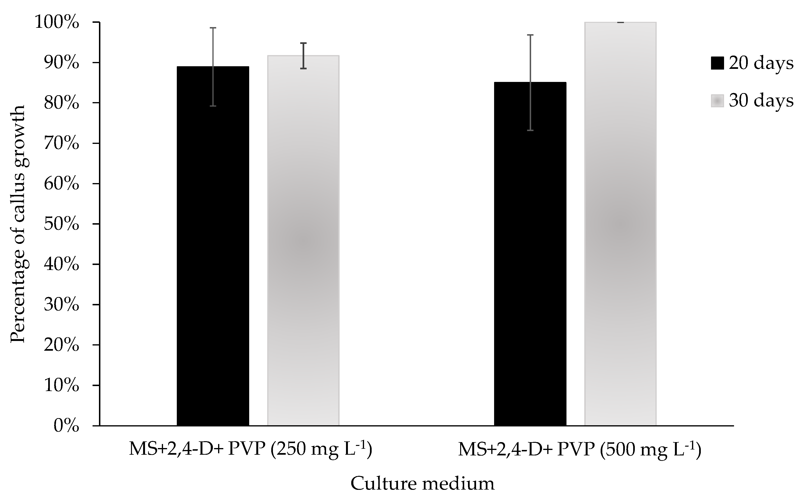
In this trial, stems were placed on the callus formation medium (2.0 mg L−1 of 2,4-D) with 250 mg L−1 and 500 mg L−1 concentrations of polyvinylpyrrolidone (PVP; Sigma-Aldrich® P2307, San Luis, MO, USA), and they were compared with the control medium without PVP. The development of each callus (%) in medium added with PVP was evaluated, taking into consideration the characteristics described in Table 1, under a completely randomized design with ten repetitions. In a parallel experiment, level 5 calli (Table 1) formed from stem were resown in a medium with 2.0 mg L−1 of 2,4-D, to which 250 mg L−1 and 500 mg L−1 of polyvinylpyrrolidone were added. The percentage of callus formation was evaluated considering various characteristics, such as growth of the mass, decrease in brown areas (sign of oxidation), and increase in light areas (potential active callus) (Table 2). Both experiments were established under a completely randomized design, with ten repetitions, and were evaluated at 20 and 30 d. The results were analyzed using the Kruskal–Wallis One-Way Analysis of Variance on Ranks test.

2.4. Feasibility of the Callus Formation Protocol

The repeatability of the callus formation protocol was checked by sowing 120 stems in an MS medium supplemented with 2.0 mg L−1 of the 2,4-D regulator. Callus formation, diameter 1, diameter 2, height, and estimated volume (per tube) were evaluated at 25 d, using the equation proposed [44]. Subsequently, 60 calli were divided into two fragments and resown in an MS medium supplemented with a 2.0 mg L−1 concentration of the 2,4-D regulator and 500 mg L−1 of PVP. At 30 d, callus formation, diameter 1, diameter 2, height, and estimated volume (per bottle) were evaluated following the equation proposed in [44].

2.5. Statistical Analysis

All treatments had a completely randomized experimental design. When the assumptions of normality and homogeneity were not met, the nonparametric Kruskal–Wallis test was applied. For the bud multiplication test, the following was applied. The Wilcoxon analysis of multiple range comparison of paired sides using the Dwass Method [45], Steel [46], and Critchlow–Fligner [47] (DSCF method) was used to determine the following variables: shoot height, number of buds, and number of shoots. For the callus and root formation variables’ analysis, the Mann–Whitney U Test (p = <0.001) was performed. The callus formation experiment was analyzed through the Wilcoxon analysis of multiple range comparison of paired sides using the Dwass Method [45], Steel [46], and Critchlow–Fligner [47] (DSCF method). In addition, a Mann–Whitney U Test comparative analysis (p = <0.001) was carried out separately for calli inducted from the stem explant and from the leaf explant. The efficiency of adding PVP to the medium to reduce oxidation in callus was determined using the Mann–Whitney U Test (p = <0.001). The data were analyzed in the SAS® 2022 On Demand for Academics statistical software, online, and using the SigmaPlot [14.0] software.

3. Results

3.1. Experimental Phase 1: In Vitro Multiplication

Under the comparison of ranges, the treatment of 1.0 mg L−1 of BA recorded the highest shoot height (64.46 ± 7.86 mm), exceeding the value of the control and other treatments (Table 3). The treatment of TDZ 0.1 mg L−1 demonstrated a significant increase in growth, with a 30.32 mm average value. Regarding the number of buds, similar results to the control were observed, with concentrations of 0.1, 0.2, 0.4, and 1.0 mg L−1 of BA and with 0.1 mg L−1 of TDZ all exhibiting optimal responses, with a range of 10 to 16 buds (Table 3).
The best treatments for the number of shoots were BA with concentrations of 0.1 mg L−1 and 1.0 mg L−1 and TDZ with a concentration of 0.1 mg L−1.
For callus formation, the treatments that showed differences compared to the control were BA of 0.6 mg L−1, 0.8 mg L−1, and 1.2 mg L−1 and TDZ from 0.2 mg L−1 to 1.2 mg L−1. The treatments that showed differences in root formation compared to the control were BA 0.8 mg L−1, 1.0 mg L−1, and 1.2 v and TDZ 0.8 mg L−1 (Figure 1).
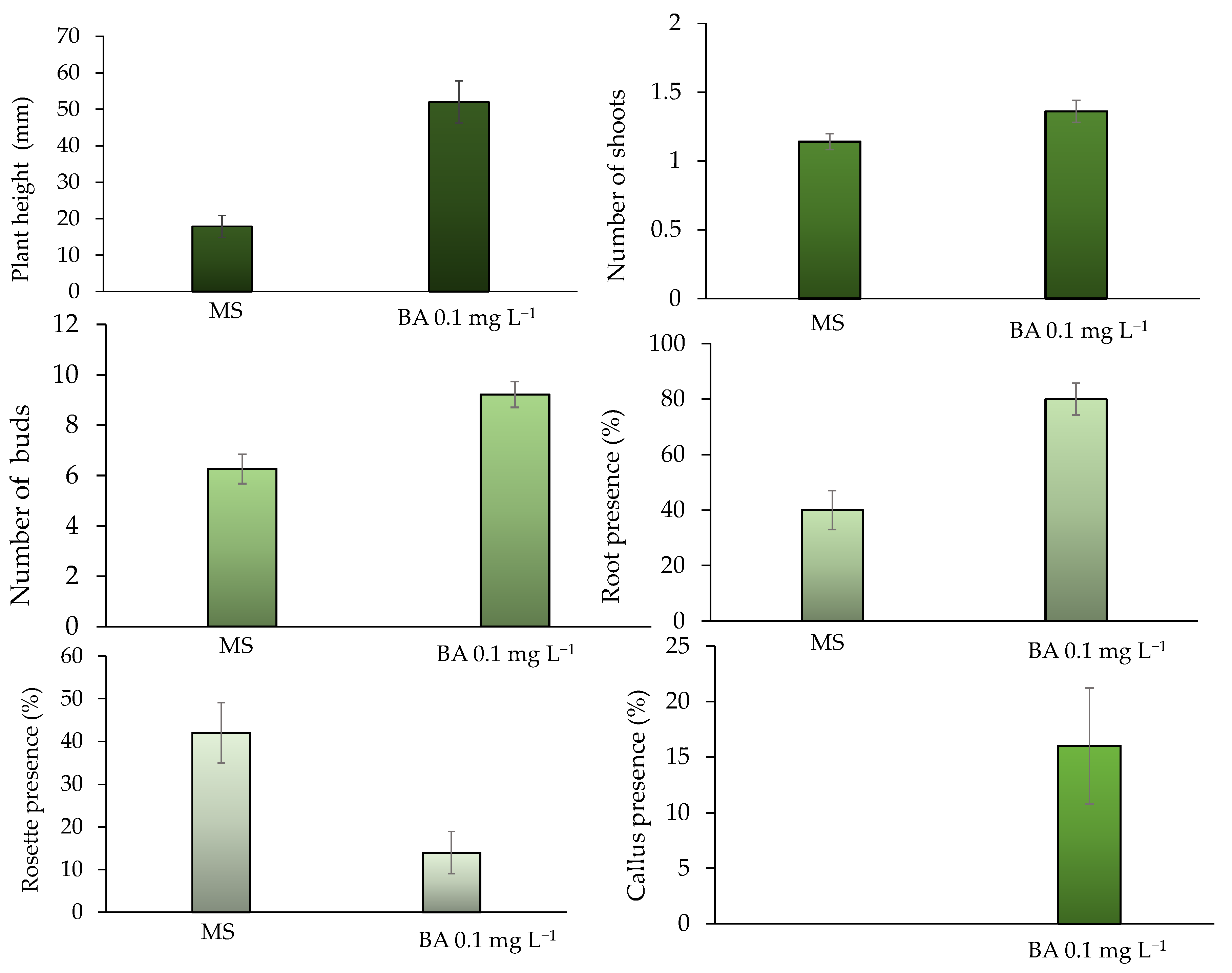
To validate the results of the first trial, 0.1 mg L−1 of BA—one of the concentrations that optimized the process—was compared with the MS control. According to the analysis of variance, there were differences in all the variables evaluated. The following average values were recorded for the 50 repetitions evaluated for each treatment with 0.1 mg L−1 of BA: a height of 52 mm, 1.36 shoots, and 9.22 buds. These figures were higher than those of the MS control (Figure 2).
Remarkably, root formation was observed in 50 representative replications, as 0.1 mg L−1 of BA generated twice the number of roots than the MS control (80%); even callus formation was observed (16%) without limiting its growth (Figure 2).
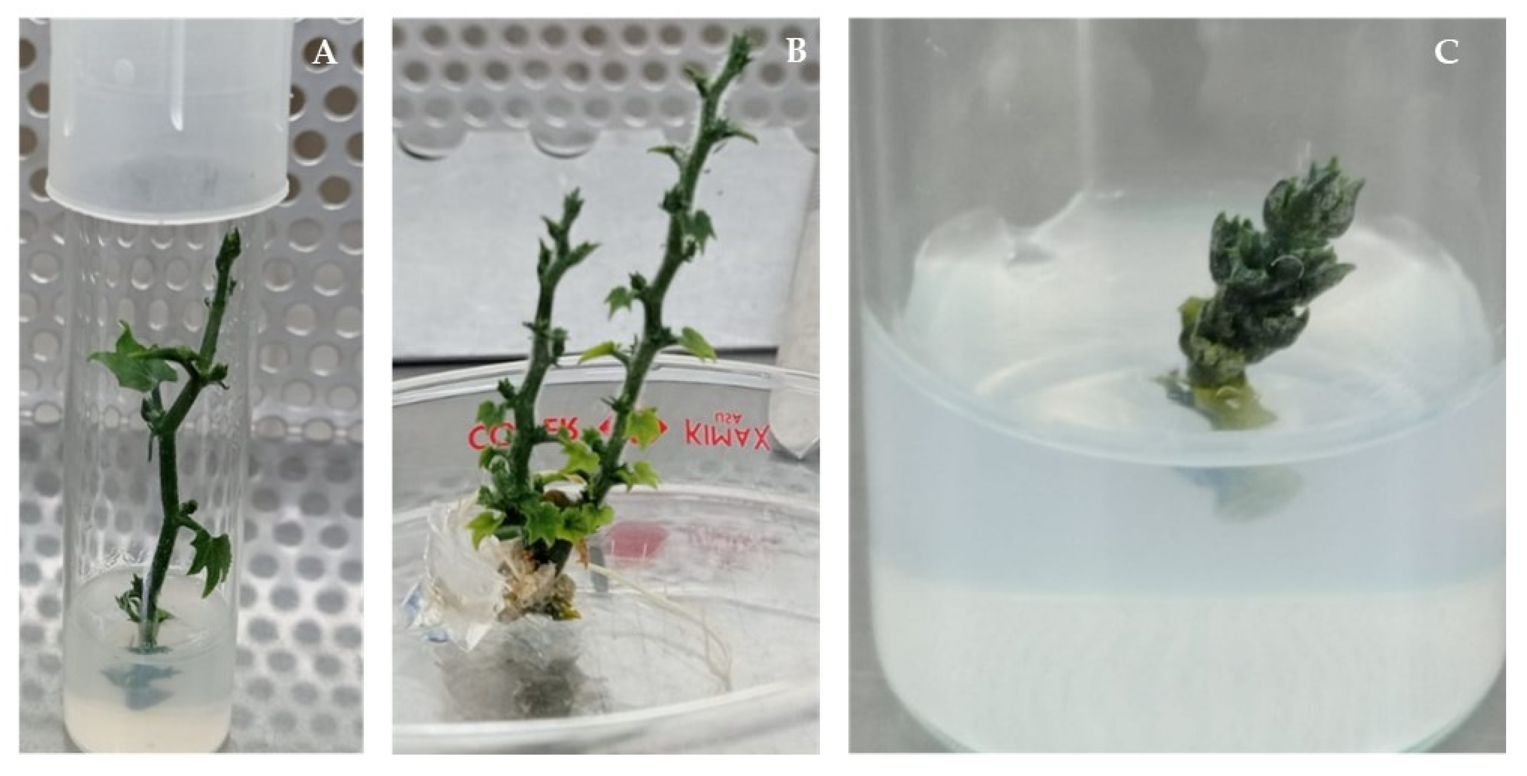
When the five reseedings of S. compositum are exceeded, a structure of packed buds (“rosettes”) is usually formed (Figure 3). Even when it has a significant number of buds, this structure is difficult to divide for their multiplication. Figure 2 indicates a significant decrease in the formation of the rosette-shaped structure (Figure 3C) when 0.1 mg L−1 of BA was added to the base culture medium, reducing it threefold compared to the MS control, in which 42% rosette formation was observed.

3.2. Experimental Phase 2: Callus Formation

In Table 4, it can be seen that there are differences between the treatments. Treatments 2,4-D 0.5–2.0 mg L−1 were the highest in the percentage of root formation (100%), weight, and volume, and all three presented a difference from the control. Treatment 2,4-D 2.0 mg L−1 showed the highest level of callus formation, with a mean of 4.89 ± 0.08. Table 4 represents a comparison between the explant (leaf or stem) against the control in the callus obtained from stems.
For calluses obtained from leaves at the variable “callus formation level”, treatments of 2,4-D 0.5, 1.0, and 2.0 mg L−1, as well as TDZ 2.0 mg L−1, were different from the control, with means of 2.83 ± 0.19, 2.67 ± 0.27, 2.72 ± 0.29, and 2.28 ± 0.14, respectively.
For the weight and volume variables, all treatments were different from the control. In root formation, treatments of 2,4-D 0.5, 1.0, and 2.0 mg L−1 were different from the control, with means of 88.9 ± 7.6, 55.6 ± 12.1, and 44.4 ± 12.1, respectively. Therefore, in the separate analysis of the explants (leaf and stem), compared to the control, the treatments with 2,4-D showed differences (p = <0.001). Furthermore, the concentration of this regulator can vary within a range of 0.5 to 2.0 mg L−1, highlighting that the highest concentration resulted in a notable improvement in callus development (Table 4).
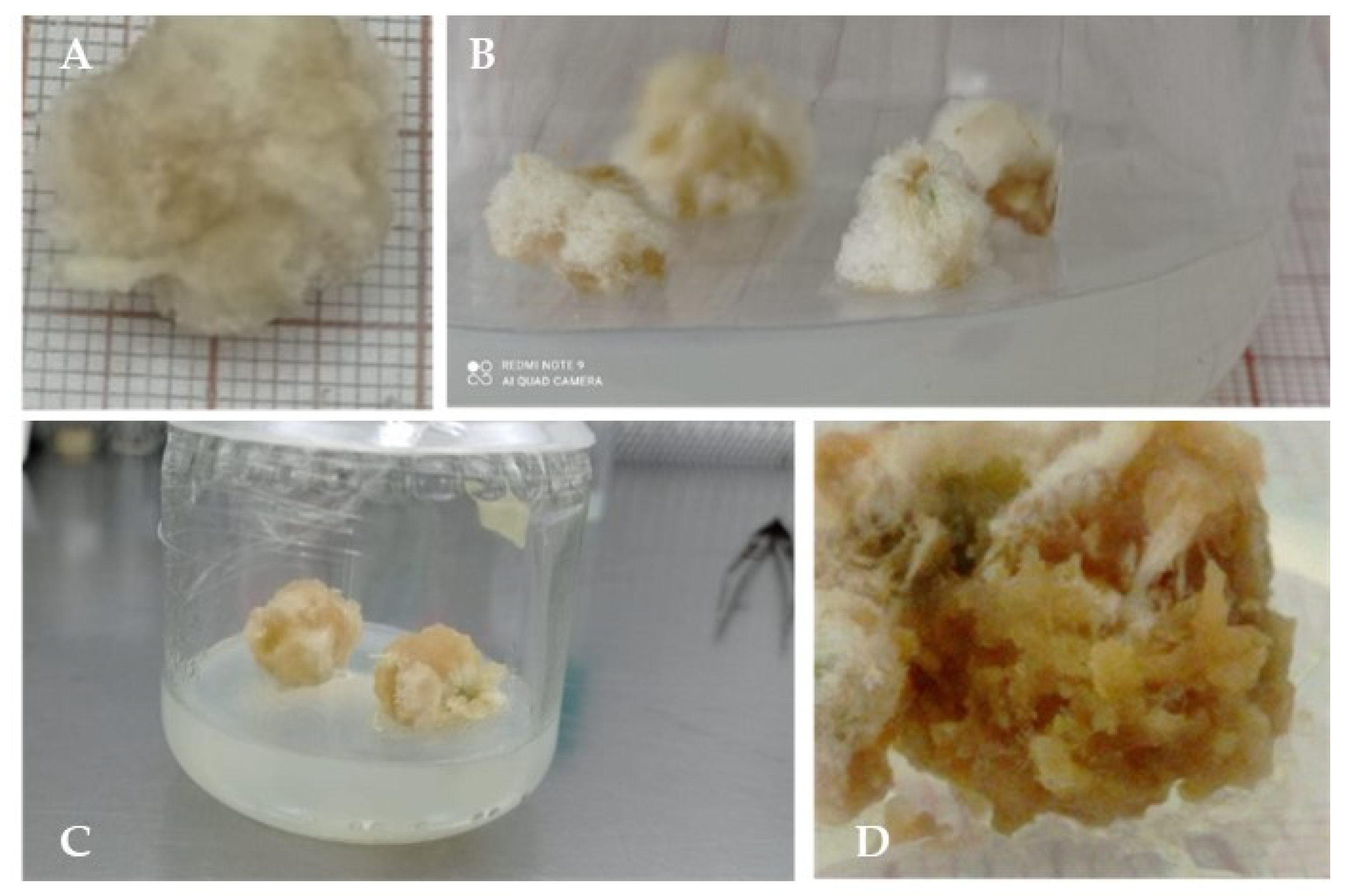
The callus formed was a compact mass with a white and cottony periphery, as well as some light yellow-green middle areas, especially in levels 3 and 4 (Figure 4D,E,I,J). The explants of level 5 calli are fully covered with a white layer (Figure 5A–C). After 40 d in the medium, an oxidation process began, during which the calli changed from light white yellow to brown-yellow (Figure 6D). This oxidation indicator lasted about 60 d, and then the tissue died.

3.2.1. Control of Callus Oxidation

Activated Charcoal

In this case, 120 repetitions of an experiment in which callus formation was induced from the stem in the callogenesis medium (2.0 mg L−1 of 2,4-D) with 2.0 mg L−1 of activated charcoal were evaluated. Under these conditions, the stems showed induction of callus formation at 7 d, with swelling of the explant at the ends where the cut was made (Figure 5A). After 15 days (Figure 5B), the callus development around the stem was more noticeable. However, after 30 d of evaluation (Figure 5C), 100% stopped their development with level 3 calli (Table 1) and generated roots; only one explant generated shoots and leaves (Figure 5D).

Application of Polyvinylpyrrolidone (PVP)

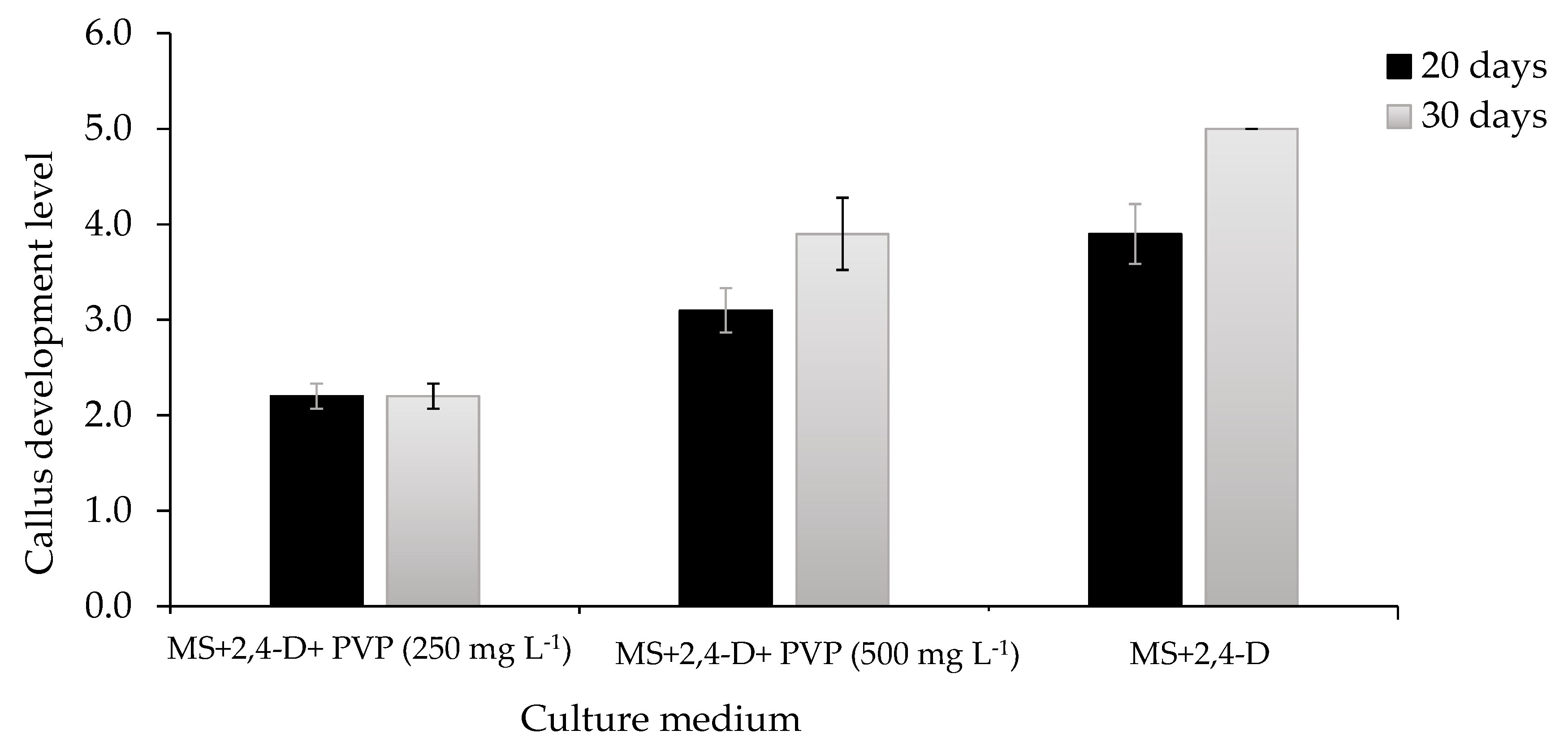
According to Figure 7, the level of development of calli with desirable characteristics (Figure 6A,C) at 20 and 30 d (Table 1) shows that the calli in the formation medium (control) reached level 5 (100%) at 30 d. Meanwhile, when 250 mg L−1 of PVP was added to the medium, the formation level reached 2.2 ± 0.13 (30%); for its part, the application of 500 mg L−1 of PVP to the medium resulted in a formation level of 3.9 ± 0.38 (72.5%). Therefore, adding PVP from the initial stage of callus formation should decrease callus induction from the stem by 27.5%. Both the control and the 500 mg L−1 concentration of PVP showed differences at 30 d (p = <0.001). The resowing of calli of average level (4.5) from a stem explant, in a medium with 2.0 mg L−1 of 2,4-D, to which two concentrations of polyvinylpyrrolidone (PVP 250 and 500 mg L−1) were added, did not register differences (p = >0.05) at 20 and 30 d (Figure 8). However, with 500 mg L−1, 100% calli with desired characteristics were obtained in a resown callus (Figure 6C), i.e., they presented mostly active zones, increase in mass, root formation, and transparent medium (Table 2). In that sense, adding PVP to the medium for resown calli helps to reduce the oxidation process.

3.3. Validation of the Callus Formation Protocol

As a result of the validation of the callus formation protocol, the 120 calli placed on the medium from a stem in a medium with a 2.0 mg L−1 concentration of the 2,4-D regulator recorded the following results at 25 d: average formation values of up to 70 ± 29.6%; diameter 1 of 7.74 ± 2.5 mm; diameter 2 of 8.41 ± 2.4 mm; height of 7.35 ± 2.2 mm; and a volume of 27.89 ± 19.5 mm3. Half of these calli were divided into two groups and resown for new callus on a medium that included 500 mg L−1 of PVP. A 30-day evaluation reported 100% callus development with active zones, increase in mass, root formation, and transparent medium.

4. Discussion

4.1. Experimental Phase 1: In Vitro Multiplication

Both regulators show a significant response in seedling growth, number of leaves, and number of shoots. The relatively low level of both (BA and TDZ) can effectively promote shoot formation and growth (Table 3). The same table shows that the lowest concentration of TDZ (0.1 mg L−1) favors the height of the shoots and the number of buds and shoots, with values of 37.25 ± 7.52, 12.2 ± 2.5, and 2.1 ± 0.4, respectively, and, when the dose increases, the values decrease. Based on the values obtained, BA was taken as a growth regulator with the possibility of enhancing the development of shoots. In this way, a second test was carried out with the lowest concentration of BA, thus increasing the number of repetitions.
BA has been the growth regulator par excellence for the optimization of the induction, multiplication, or regeneration stages through direct organogenesis in other species, and it has recorded more shoots and longer shoots than the MS control. Likewise, depending on the species or variety and the concentrations or combinations with other regulators, media supplemented with BA are significantly more potent [48,49,50].
Optimization treatments aimed at the in vitro growth of S. compositum must address several essential traits, including seedling size, number of buds, and induction of a greater number of shoots. These considerations are essential to ensuring adequate multiplication and root development, even in situations where callus formation is limited. In this study, treatments that can meet such conditions include 0.1 mg L−1 and 1.0 mg L−1 of BA and 0.1 mg L−1 of TDZ (Figure 1).
In addition, we observed that BA increases the formation of shoots and the number of buds. Important differences were recorded when comparing the MS control (MS culture medium by itself) and the MS base culture medium (MS medium supplemented with 0.1 mg L−1 of BA) that improved the micropropagation of S. compositum from buds.
Regarding the number of shoots, treatments BA 0.1 and 1.0 mg L−1 as well as TDZ 0.1 mg L−1 stood out, with an average of 2.1, 2.0, and 2.1 shoots, respectively. These data are considered indicative of the multiplication rate for each treatment (Table 3). The results highlight the positive influence of the treatments on shoot growth and multiplication. Overall, this difference in responses occurs because BA stimulates the formation of axillary buds, while the TDZ response is associated with the formation of adventitious buds [51].
Cytokinins can induce high proliferation and cell division; that is, they increase the multiplication rate of plants [52,53]. Likewise, they cause the elongation of the roots, in addition to an increase in the production of shoots [52,54].
Thasni et al. [55] evaluated the in vitro bud proliferation response in ivy gourd (Coccinia grandis (L.) Voigt.) cv. Nodal explants of Sulabha and found that the combination of MS + 1.0 mg L−1 BA was the best in terms of the number of days for bud initiation (5.50), the number of shoots per explant (1.75), the shoot length (5.71), and the response percentage (100). A similar result is shown by Chuengpanya et al. [56], who evaluated the effect of BA on the multiplication of in vitro shoots of Iris collettii Hook.f. and I. domestica (L.) Goldblatt & Mabb. And, they found that the bases of the leaves grown in MS medium and supplemented with 1 mg L−1 of BA presented the greatest shoot formation (3.13 shoots/explant and 2.31 cm length).
Little research has been conducted on in vitro cultures of S. compositum, as most of the knowledge on in vitro cultures of Sechium spp. has been generated with Sechium edule. For example, Mora et al. [36] recorded a more efficient propagation of S. edule plants by adding 0.1 mg L−1 of BA and 0.1 mg L−1 of gibberellic acid (GA3) to the MS base culture medium. Mora also identified that concentrations greater than 0.5 mg L−1 of BA induce callus formation. Thilagam et al. [57] observed that the association of different growth regulators with BA had significant advantages (e.g., greater shoot length) after four weeks of culture. The maximum number of nodes and shoot length was observed with 2.0 μM of BA; this number increased when they were subcultured in a culture medium with the same concentration.
BA evidently promotes the generation of shoots, growth, and stimulation of root formation in both S. edule and S. compositum, thus enhancing micropropagation. Cruz-Martinez et al. [22] induced the proliferation of axillary shoots from axenic nodal segments obtained from shoots germinated in vitro. Nodal segments were cultured in MS medium supplemented with 0.1 mg L−1 of 6-benzylaminopurine (BA), producing 8.0 ± 0.4 shoots per explant, with a 92% regeneration frequency. Furthermore, the regeneration of the shoot was optimized through the addition of 0.1 mg L−1 of BA and 0.05 mg L−1 of gibberellic acid (GA3) to the medium, obtaining 5.3 ± 1.9 shoots per average explant.
TDZ favored the formation of basal calli from 0.2 mg L−1 to 1.2 mg L−1. In the case of BA, treatments 0.6 mg L−1, 0.8 mg L−1, and 1.2 mg L−1 formed more calli than treatments that had the lowest concentrations (0.1, 0.2, 0.4, and 1.0 mg L−1). For example, in Castanea sativa, Mill Larson et al. [51] demonstrated that two of their TDZ and BA treatments recorded a higher (80%) formation of basal callus, which improved the establishment of the shoot. They also mention that the basal callus accumulates substances and hormones necessary for the in vitro response, which, according to Meier-Dinkel et al. [58], suggests a relationship between callus formation and the establishment of the shoot.
With the addition of BA to the medium, the “rosette” structure of packed buds (Figure 3) decreases. The formation of these structures may be caused by a loss of morphogenic competence in the mature material (morphological, biochemical, and molecular differences); consequently, the ontogenic age of the plant material causes the absence of elongation, as younger plant explants lead to a greater elongation rate of the axillary buds [51]. Therefore, it is suggested that BA acted as a rejuvenating agent for the material. Cytokinins, such as BA, promote the elimination of axillary bud dormancy and promote the formation of lateral shoots [59,60].

4.2. Experimental Phase 2: Callus Formation Protocol

Theoretically, high concentrations of auxins promote root formation, while high concentrations of cytokinins promote shoot regeneration. A balance between auxins and cytokinins leads to callus development [61]. However, multiple research works have reported that the use of 2,4-D induces in vitro callus formation from various explants, positioning it as one of the regulators that better promotes the callus formation, whether or not it is combined with cytokinins [62,63,64,65].
Moideen and Prabha [66]. concluded that the best callus formation response in Luffa (cucurbitacea) acutangular was observed in media treated with 2, 4–D + TDZ 2.0 mg L−1. Castillo-Martinez et al. [23] have reported that cytokinins (BA and TDZ) promote callus formation. Using in vitro stem segments of S. edule, they obtained 70% callus formation with 1.0 mg L−1 of the BA growth regulator, and 76% with 0.4 mg L−1 of TDZ, both at 30 days of evaluation. Compared with the first stage of this research, high concentrations of TDZ (0.2–1.2 mg L−1) and BA (0.5, 0.6, and 1.2 mg L−1) promoted the formation of basal calli on the development of the seedling.
Table 4 shows that the best callus formation treatment was based on a stem explant in a medium with 2.0 mg L−1 of 2,4-D (highest concentration), recording an average value of 4.89 (equivalent to 97.8% callus formation around the explant). The weight and volume variables indicated that the best treatments started from the stem explant and the medium enriched with 2,4-D, without differences between concentrations. The presence of a root is an indication that calli can become embryogenic, i.e., they can be the point of origin for the promotion of the regeneration of the seedling (indirect organogenesis) [40,67]; therefore, the best treatments started from the stem in a medium enriched with 2,4-D in the three concentrations evaluated.
In this sense, the success of callus formation of S. compositum depends on the type of growth regulator and the explant. The concentration did not show differences in callus fresh weight, diameter, height, volume, or root formation. Similar results were obtained by Soto-Contreras et al. [39], who evaluated the effect of 0.5, 1.0, and 1.5 mg L−1 concentrations of 2,4-D on the induction of callus formation from different vitroplant explants of S. edule. Their results show that with the highest concentration, the nodal segments recorded 100% formation of white calli and a fresh weight of 1.74 g, while the calli of the leaf segments were greenish and had a fresh weight of 0.35 g.
The addition of an antioxidant to in vitro culture is an alternative to reduce oxidation [68,69,70]. In this research, PVP shows better results compared to activated charcoal when added to the medium. After 30 d of evaluation of the MS medium supplemented with 2.0 mgL−1 of activated charcoal, 100% of calluses stopped their development at level 3 and generated roots, and only one explant generated shoots and leaves. Therefore, activated charcoal in a callus formation initiation process is not suitable for S. compositum.
May et al. [71] report that when they added activated charcoal to the best callus formation treatments, a negative effect was generated, such as slow cell proliferation of the Brosimum alicastrum callus.
Contrary to PVC, concentrations of 250 mgL−1 and 500 mgL−1 allowed for the conservation of the appropriate characteristics of the callus during its multiplication.
Likewise, Domínguez-Perales et al. [72] and other authors evaluated the effect of adding PVP to the culture medium as an antioxidant and observed that the treatment with 500 mg L−1 decreased the loss of explants of the guava variety CHRG (Psidium guajava L.), recording a higher frequency of explants and a lower level of necrosis through oxidation (7%). Cureño et al. [73] observed that the PVP treatment doubled the size of the callus of Taxus globosa Schlecht. The callus showed friability and 100% feasibility using fluorescein diacetate (FdA). They also observed that despite having an antioxidant, the calli of Taxus globosa induced with this medium slightly darkened in the subculture due to the presence of phenolic compounds.

5. Conclusions

The results show the feasibility of using growth regulators, such as BA, TDZ, and 2,4-D, for the in vitro multiplication and callus formation of S. compositum. These results do not exclude the possibility that the MS medium may continue to be the base culture medium for the maintenance and propagation of the shoot; however, supplementing it with 0.1 mg L−1 of BA will doubtlessly optimize the multiplication and regeneration of the shoot. For callus formation from stem explants, the best callus-inducing growth regulator was 2.0 mg L−1 of 2,4-D. The protocol for the induction, maintenance, and multiplication of calli was validated through the induction of calli from the stem in an MS medium supplemented with 2.0 mg L−1 of 2,4-D and resowing after 20 d in a medium with 2.0 mg L−1 of 2,4-D at + 500 mg L−1 of PVP. The success of callus maintenance consists of avoiding oxidation and proceeding to carry out frequent subcultures of the most active parts of the callus, approximately 20 days after sowing or resowing.

Author Contributions

Conceptualization, C.-I.J. and R.-F.M.d.l.L.; methodology, C.-I.J. and A.-G.M.d.L.; validation, R.-P.L.d.M., S.-H.R.M. and C.-J.I.; investigation, R.-F.M.d.l.L., C.-I.J. and C.-M.C.R.; resources, R.-P.L.d.M.; writing—original draft preparation, C.-I.J., C.-J.I. and A.-G.M.d.L.; writing—review and editing, C.-I.J. and R.-P.L.d.M. All authors have read and agreed to the published version of the manuscript.

Funding

This research received no external funding.

Data Availability Statement

The data presented in this study are available at http://www.biblio.colpos.mx/portal/, accessed on 12 April 2024. They are identified by the name of the primary author of this article.

Acknowledgments

To the Sechium edule germplasm bank in Mexico, from the Interdisciplinary Research Group on Sechium edule in Mexico, A.C. and the Biotechnology Laboratory, CENID-COMEF of INIFAP, Mexico.

Conflicts of Interest

The authors declare no conflicts of interest.

References

  1. Casas, A.; Vallejo, M. Agroecología y agrobiodiversidad. Crisis Ambient. México 2019, 103, 99–117. [Google Scholar]
  2. CDB Convenio de Diversidad Biológica. Available online: https://www.cbd.int/convention/text/default.shtml (accessed on 12 April 2024).
  3. Lobo, M.; Medina, C.I. Conservación de recursos genéticos de la agrobiodiversidad como apoyo al desarrollo de sistemas de producción sostenibles. Revista Corpoica. Cienc. Tecnol. Agropecu. 2009, 10, 33–42. [Google Scholar] [CrossRef]
  4. Cadena-Íñiguez, J.; Trejo-Téllez, B.I.; Morales-Flores, F.J.; Ruíz-Vera, V.M. Análisis de tratados internacionales relacionados con recursos genéticos y su congruencia con el marco jurídico mexicano. In Proceedings of the 22th International Congress on Project Engineering, Madrid, Spain, 11–13 July 2018; p. 97. [Google Scholar]
  5. Cadena-Iñiguez, J. El chayote (Sechium edule (Jacq.) Sw., importante recurso fitogenético mesoamericano. Agro Product. 2010, 3, 3307. [Google Scholar]
  6. Rodas, Y.C.R.; Galarza, M.L.A.; Hernández, M.S.; Íñiguez, J.C.; Solano, V.M.C. Gestión de un recurso fitogenético para producción de metabolitos secundarios en proyectos de diversificación económica rural. In Proceedings of the XXV Congreso Internacional de Ingeniería de Proyectos, Alcoy, Spain, 6–9 July 2021; p. 101. [Google Scholar]
  7. Salas, S.M.P.; Aguilar-Galván, F.; Sandoval, L.H. Plantas silvestres comestibles de la Barreta, Querétaro, México y su papel en la cultura alimentaria local. Rev. Etnobiología 2021, 19, 41–62. [Google Scholar]
  8. Organización de las Naciones Unidas para la Alimentación y la Agricultura. AGP-Conservación de los Recursos Fitogenéticos. Available online: https://www.fao.org/agriculture/crops/core-themes/theme/seeds-pgr/conservation/en (accessed on 19 December 2023).
  9. Cadena Iñiguez, J.; González Santos, R.; Cuevas Sánchez, J.; Riviello Flores, M.D.L.L.; Ruiz Posadas, L.D.M. La Conservación in situ de la Biodiversidad Agrícola, y la generación de proyectos en ejidos y comunidades de México. In Proceedings of the 22th International Congress on Project Engineering, Madrid, Spain, 7–9 July 2020. [Google Scholar]
  10. González-Santos, R.; Cadena-Iñiguez, J.; Morales-Flores, F.J.; Ruiz-Vera, V.M.; Pimentel-López, J.; Peña-Lomelí, A. Model for the conservation and sustainable use of plant genetic resources in México. Wulfenia J. 2015, 22, 333–353. [Google Scholar]
  11. Beeching, J.R.; Marmey, P.; Hughes, M.A.; Charrier, A. Evaluation of molecular approaches for determining genetic diversity in Cassava germplasm. In Proceedings of the 2nd International Scientific Meeting, The Cassava Biotechnology Network, Bogor, Indonesia, 22–26 August 1994; pp. 22–26. [Google Scholar]
  12. Organización de las Naciones Unidas para la Alimentación y la Agricultura. El segundo Informe Sobre el Estado de los Recursos Fitogenéticos para la Alimentación y la Agricultura en el Mundo. Available online: https://www.fao.org/documents/card/es?details=22fabd61-4b41-5bb3-b6be-d38e1516dccc/ (accessed on 23 November 2023).
  13. Vargas-Bermúdez, J.D. Conservación In situ de Plantas Nativas Herbáceas, Medicinales o Bioplaguicidas, Como Reservorio de Diversidad Genética y Cultural, Mariscal Sucre-Guayas. Ph.D. Thesis, Universidad Agraria Del Ecuador, Guayaquil, Ecuador, 2020. [Google Scholar]
  14. Barrera-Guzmán, L.A.; Cadena-Iñiguez, J.; Legaria-Solano, J.P.; Sahagún-Castellanos, J. Phylogenetics of the genus Sechium P. Brown: A review. Span. J. Agric. Res. 2021, 19, e07R01. [Google Scholar] [CrossRef]
  15. Avendaño-Arrazate, C.H.; Cadena Iñiguez, J.; Arévalo Galarza, M.L.C.; Campos Rojas, E.; Cisneros Solano, V.M.; Aguirre Medina, J.F. Las Variedades del Chayote Mexicano, Recurso Ancestral con Potencial de Comercialización; No. Libro 635.62 A8; Grupo Interdisciplinario de Investigación en Sechium edule en México (GISeM): Morelia, Mexico, 2010. [Google Scholar]
  16. Cadena-Iñiguez, J.; Arévalo, G.M. Volumen 2: Chayote. In GISeM Rescatando y Aprovechando los Recursos Fitogenéticos de Mesoamérica; Grupo Interdisciplinario de Investigación en Sechium edule en México, Colegio de Postgraduados: Texcoco, Mexico, 2011; 24p. [Google Scholar]
  17. Iñiguez-Luna, M.I.; Cadena-Iñiguez, J.; Soto-Hernández, R.M.; Morales-Flores, F.J.; Cortes-Cruz, M.; Watanabe, K.N. Natural bioactive compounds of Sechium spp. for therapeutic and nutraceutical supplements. Front. Plant Sci. 2021, 12, 772389. [Google Scholar] [CrossRef]
  18. Iñiguez-Luna, M.I.; Cadena-Iñiguez, J.; Soto-Hernández, R.M.; Morales-Flores, F.J.; Cortes-Cruz, M.; Watanabe, K.N.; Machida-Hirano, R.; Cadena-Zamudio, J.D. Bioprospecting of Sechium spp. varieties for the selection of characters with pharmacological activity. Sci. Rep. 2021, 11, 6185. [Google Scholar] [CrossRef]
  19. Rivera-Ponce, E.A.; Cadena-Iñiguez, J.; Cisneros-Solano, V.M.; Soto-Hernández, R.M.; San Miguel-Chávez, R.; García-Osorio, C.; Arévalo-Galarza, M.D.L. Composición Fitoquímica y uso Potencial del Jugo de Sechium compositum (Donn. Sm.) C. Jeffrey; Agro-Divulgación: Texcoco, Mexico, 2022; Volume 2. [Google Scholar]
  20. Aguiñiga Sánchez, I. Potencial Antileucémico In Vitro de Extractos de Cuatro Genotipos de Sechium spp. (Cucurbitaceae). Master’s Thesis, Colegio de Postgraduados, Montecillo, México, 2013. [Google Scholar]
  21. Gordillo Salinas, L.S. Actividad Antifúngica de Sechium compositum Contra Botrytis Cinerea y Colletotrichum Gloeosporioides en Condiciones In Vitro. Master’s Thesis, Colegio de Postgraduados, Montecillo, Meéxico, 2019. [Google Scholar]
  22. Cruz-Martínez, V.; Castellanos-Hernández, O.A.; Acevedo-Hernández, G.J.; Torres-Morán, M.I.; Gutiérrez-Lomelí, M.; Ruvalcaba-Ruiz, D.; Zurita, F.; Rodríguez-Sahagún, A. Genetic fidelity assessment in plants of Sechium edule regenerated via organogenesis. S. Afr. J. Bot. 2017, 112, 118–122. [Google Scholar] [CrossRef]
  23. Castillo-Martínez, C.; Cisneros-Solano, V.M.; Hernández-Marini, R.; Cadena-Iñiguez, J.; Avendaño-Arrazate, C.H. Conservación y Multiplicación de una Colección de Sechium spp.; Colegio de Postgraduados y Grupo Interdisciplinario de Investigación Sechium edule en México A.C.: Montecillo, Mexico, 2013. [Google Scholar]
  24. Cruz-Martínez, V.O.; Acevedo-Hernández, G.J.; Castellanos-Hernández, O.A.; Rodríguez-Sahagún, A. Regeneración de Sechium edule mediante organogénesis y uso de marcadores ramp para evaluar la fidelidad genética. Biotecnol. Sustentabilidad 2016, 1, 96. [Google Scholar]
  25. Wei, Y.; Li, Z.; Lv, L.; Yang, Q.; Cheng, Z.; Zhang, J.; Zhang, W.; Luan, Y.; Wu, A.; Li, W.; et al. Overexpression of MbICE3 increased the tolerance to cold and drought in lettuce (Lactuca sativa L.). In Vitro Cell. Dev. Biol.-Plant 2023, 59, 767–782. [Google Scholar] [CrossRef]
  26. Romo-Paz, F.d.J.; Orozco-Flores, J.D.; Delgado-Aceves, L.; Zamora-Natera, J.F.; Salcedo-Pérez, E.; Vargas-Ponce, O.; Portillo, L. Micropropagation of Physalis angulata L. and P. chenopodifolia Lam. (Solanaceae) via indirect organogenesis. In Vitro Cell. Dev. Biol.-Plant 2023, 59, 497–506. [Google Scholar] [CrossRef]
  27. Alihodzic, A.; Davis, J.; Roberts, C.; Henrie, S.; Bolyard, M. Production of secondary metabolites in regenerated Southern wormwood (Artemisia abrotanum L.) under various experimental conditions. In Vitro Cell. Dev. Biol.-Plant 2023, 59, 847–852. [Google Scholar] [CrossRef]
  28. Behera, S.; Kar, S.K.; Monalisa, K.; Mohapatra, S.; Meher, R.K.; Barik, D.P.; Panda, P.C.; Naik, P.K.; Naik, S.K. Assessment of genetic, biochemical fidelity, and therapeutic activity of in vitro regenerated Hedychium coronarium. In Vitro Cell. Dev. Biol.-Plant 2023, 59, 602–620. [Google Scholar] [CrossRef]
  29. Zhang, W.; Dai, W. In vitro plant regeneration of ‘Prelude’ red raspberry (Rubus idaeus L.). In Vitro Cell. Dev. Biol.-Plant 2023, 59, 461–466. [Google Scholar] [CrossRef]
  30. Gaspar, T.; Kevers, C.; Penel, C.; Greppin, H.; Reid, D.M.; Thorpe, T.A. Plant hormones and plant growth regulators in plant tissue culture. In Vitro Cell. Dev. Biol.-Plant 1996, 32, 272–289. [Google Scholar] [CrossRef]
  31. Kaur, K.; Dolker, D.; Behera, S.; Pati, P.K. Critical factors influencing in vitro propagation and modulation of important secondary metabolites in Withania somnifera (L.) dunal. Plant Cell Tissue Organ Cult. PCTOC 2022, 149, 41–60. [Google Scholar] [CrossRef] [PubMed]
  32. Alvarenga, S.; Morera, J. In vitro micropropagation of the chayote (Sechium edule Jacq. Sw.). Tecnol. Marcha 1992, 11, 31–42. [Google Scholar]
  33. Abdelnour, A.; Ramírez, C.; Engelmann, F. Micropropagación de chayote (Sechium edule Jacq. SW.) a partir de brotes vegetativos. Agron. Mesoam. 2002, 13, 147–151. [Google Scholar] [CrossRef]
  34. Abdelnour-Esquivel, A.; Bermudez, L.C.; Rivera, C.; Alvarenga-Venutolo, S. Cultivo de meristemas, termo y quimioterapia en chayote (Sechium edule Jacq. Sw.) para la erradicación del virus del mosaico del chayote (ChMV). In Manejo Integrado de Plagas y Agroecología; CATIE: Turrialba, Costa Rica, 2006; Volume 77. [Google Scholar]
  35. Del Ángel, O.; Vela, G.; Rodríguez, A.; Gómez, M.; García, H. Desinfección y regeneración eficiente de chayote in vitro (Sechium edule Jacq. Sw.). In Ciencias Agropecuarias Handbook T-II: Congreso Interdisciplinario de Cuerpos Académicos, ECORFAN; 2014; p. 23. Available online: https://www.coursehero.com/file/219916369/Dialnet-CienciasAgropecuariasHandbookTII-563076pdf/ (accessed on 12 April 2024).
  36. Mora, D.F.; Esquivel, A.A.; Venutolo, S.A. Micropropagación de fenotipos seleccionados de chayote. Tecnol. Marcha 1999, 13, 9–15. [Google Scholar]
  37. Abdelnour-Esquivel, A.; Brenes-Madriz, J.; Alvarenga-Venutolo, S. Guía Técnica Semilla de Chayote. 2015. Available online: https://repositoriotec.tec.ac.cr/bitstream/handle/2238/6768/gu%C3%ADa-tecnica-semillachayote-14%20SET-15.pdf?sequence=1&isAllowed=y (accessed on 12 April 2024).
  38. García, J.G.; Alvarado, E.S.; Bolaños, J.A. Efecto del AIA y el AIB sobre el enraizamiento in vitro de brotes de Sechium edule (Jacq.) Sw. Biotecnol. Veg. 2015, 15, 3–7. [Google Scholar]
  39. Soto-Contreras, A.; Núñez-Pastrana, R.; Rodríguez-Deméneghi, M.V.; Aguilar-Rivera, N.; Galindo-Tovar, M.E.; Ramírez-Mosqueda, M.A. Indirect organogenesis of Sechium edule (Jacq.) Swartz. In Vitro Cell. Dev. Biol.-Plant 2022, 58, 903–910. [Google Scholar] [CrossRef]
  40. Da Silva, R.F. Sistema de regeneración in vitro por embriogénesis somática indirecta en variedades venezolanas de arroz (Oryza sativa L.). Rev. Científica UDO Agrícola 2012, 12, 55–68. [Google Scholar]
  41. Riviello-Cogco, E.; Robledo-Paz, A.; Gutiérrez-Espinosa, M.A.; Suárez-Espinosa, J.; Mascorro-Gallardo, J.O. Maduración y germinación de embriones somáticos de Coffea arabica cv. colombia. Rev. Fitotec. Mex. 2021, 44, 161. [Google Scholar] [CrossRef]
  42. Classic Murashige, T.; Skoog, F. A revised medium for rapid growth and bioassays with tobacco tissue cultures. Physiol. Plant 1962, 15, 473–497. [Google Scholar] [CrossRef]
  43. Cadena Iñiguez, J.; Soto Hernández, M.; Arévalo Galarza, M.; Avendaño Arrazate, C.H.; Aguirre Medina, J.F.; Ruiz Posadas, L.D.M. Caracterización bioquímica de variedades domesticadas de chayote Sechium edule (Jacq.) Sw. comparadas con parientes silvestres. Rev. Chapingo Ser. Hortic. 2011, 17, 45–55. [Google Scholar] [CrossRef]
  44. Ramos-Parra, M.; Ulín-Montejo, F.; Aguilar-Nieto, J.A.; Solis-Trapala, I.L.; Fierro-Carbajal, J.B. Modelación y estimación del volumen de tejido vegetal in vitro de Strombocactus disciformis basada en mediciones no intrusivas. Rev. Chapingo. Ser. Hortic. 2010, 26, 195–203. [Google Scholar]
  45. Dwass, C. Some k-sample rank-order test. Contributions to probability and Statistics. In Contributions to Probability and Statistics; Stanford University Press: Redwood City, CA, USA, 1960; pp. 198–202. [Google Scholar]
  46. Steel, R. A rank sum test for comparing all pairs of treatments. Technometrics 1960, 2, 197–207. [Google Scholar] [CrossRef]
  47. Critchlow D y Fligner, M. On distribution-free multiple comparisons in the one-way analysis of variance. Commun. Statist.-Theory Meth. 1991, 20, 127–139. [Google Scholar]
  48. Castillo-Martínez, C.R.; Gutiérrez-Espinosa, M.; Buenrostro-Nava, M.T.; Cetina Alcalá, V.M.; Cadena Iñiguez, J. Regeneración de plantas de Paulownia elongata Steud: Por organogénesis directa. Rev. Mex. Cienc. For. 2012, 3, 41–49. [Google Scholar]
  49. Gil, A.I.; Ariza, C.A.; Castillo, L.M.; Salgado, L.E.; Banda Sánchez, L.; Vanegas, L.E. Induction of organogenesis in vitro with 6-benzylaminopurine in Cattleya trianae Linden & Rchb. f. Rev. UDCA Actual. Divulg. Científica 2019, 22. [Google Scholar] [CrossRef]
  50. Mora-Cruz, Y.; López-Peralta MC, G.; Hernández-Meneses, E.; Cruz-Huerta, N. Regeneración in vitro de plantas de prosthechea vitellina (lindley) we higging por organogenesis directa. Rev. Fitotec. Mex. 2023, 46, 33. [Google Scholar]
  51. Larson, C.G.; Hasbún, R.; Jofré, M.P.; Sánchez-Olate, M.; Ríos, D. Efecto del genotipo y fuente de citoquinina en la etapa de iniciación de cultivo in vitro de tejido adulto de Castanea sativa Mill. Gayana. Botánica 2017, 74, 30–40. [Google Scholar] [CrossRef]
  52. Alcantara-Cortes, J.S.; Acero Godoy, J.; Alcántara Cortés, J.D.; Sánchez Mora, R.M. Principales reguladores hormonales y sus interacciones en el crecimiento vegetal. Nova 2019, 17, 109–129. [Google Scholar] [CrossRef]
  53. Zúñiga Ortega, J.A. Aplicación de Reguladores de Crecimiento para Germinación In Vitro de Embriones Cigóticos de Quijo (Passiflora popenovvi Killip) bajo dos Temperaturas. Bachelor’s Thesis, UCE, Quito, Ecuador, 2023. [Google Scholar]
  54. Laguna-Ibarra, Y.; Cueva-López, J.; Tamariz-Angeles, C.; Olivera-Gonzales, P. Efecto de los reguladores de crecimiento vegetal en la multiplicación y enraizamiento in vitro de senecio calvus (asteraceae), planta medicinal altoandina, endémica del Perú. Rev. Investig. Altoandinas 2019, 21, 111–121. [Google Scholar] [CrossRef]
  55. Thasni, A.; Sarada, S.; Swapna, A. In vitro bud proliferation response in ivy gourd (Coccinia grandis (L.) Voigt.) cv. Sulabha nodal explants with cytokinin levels. J. Trop. Agric. 2022, 60. [Google Scholar]
  56. Chuengpanya, R.; Piyasangcharoen, T.; Thongteab, N.; Muangkroot, A.; Jenjittikul, T.; Chuenboonngarm, N. Effect of N6-benzyladenine on in vitro shoot multiplication of Iris collettii Hook. f. and I. domestica (L.) Goldblatt & Mabb. In Proceedings of the IX International Scientific and Practical Conference on Biotechnology as an Instrument for Plant Biodiversity Conservation, Bangkok, Thailand, 12–13 July 2021; Volume 1339, pp. 257–264. [Google Scholar]
  57. Thilagam, D.; Kumudini, B.S.; Manohar, S.H. Regeneration of Sechium edule (jacq) sw. From sterile in vitro nodal explants and assessment of clonalfidelity using issr and rapd markers. Int. J. Agric. 2016, 6, 285–292. [Google Scholar]
  58. Meier-Dinkel, A.; Becker, B.; Duckstein, D. Micropropagation and ex vitro rooting of several clones of late-flushing Quercus robur L. Ann. Des Sci. For. 1993, 50, 319–322. [Google Scholar] [CrossRef]
  59. Mao, J.; Niu, C.; Li, K.; Mobeen Tahir, M.; Khan, A.; Wang, H.; Li, S.; Liang, Y.; Li, G.; Yang, Z.; et al. Exogenous 6-benzyladenine application affects root morphology by altering hormone status and gene expression of developing lateral roots in Malus hupehensis. Plant Biol. 2020, 22, 1150–1159. [Google Scholar] [CrossRef]
  60. Dinçer, D. Determination of optimal plant growth regulators for breaking seed dormancy and micropropagation of Sorbus aucuparia L. Balt. For. 2023, 29, id679. [Google Scholar] [CrossRef]
  61. Hussain, A.; Ahmed Qarshi, I.; Nazir, H.; Ullah, I. Plant tissue culture: Current status and opportunities. Recent Adv. Plant Vitr. Cult. 2012, 6, 1–28. [Google Scholar] [CrossRef]
  62. Chen, X.; Ye, C.; Yang, H.; Ji, W.; Xu, Z.; Ye, S.; Wang, H.; Jin, S.; Yu, C.; Zhu, X. Callogenesis and plant regeneration in peony (Paeonia × suffruticosa) using flower petal explants. Horticulturae 2022, 8, 357. [Google Scholar] [CrossRef]
  63. Hernández-Amasifuen, A.D.; Cortez-Lázaro, A.A.; Argüelles-Curaca, A.; Díaz-Pillasca, H.B. Callogénesis in vitro de durazno (Prunus persica L.) var. Huayco rojo a partir de explantes foliares. Cienc. Tecnol. Agropecu. 2022, 23, e2032. [Google Scholar] [CrossRef]
  64. Sidek, N.; Nulit, R.; Kong, Y.C.; Yien, C.Y.S.; Sekeli, R.; EL-Barghathi, M.F. Callogenesis and somatic embryogenesis of Oryza sativa L.(cv. MARDI Siraj 297) under the influence of 2,4-dichlorophenoxyacetic acid and kinetin. AIMS Agric. Food 2022. [Google Scholar] [CrossRef]
  65. Zayova, E.; Nedev, T.; Stancheva, I. Callus via shoot organogenesis and plant regeneration of Stevia rebaudiana Bertoni. Proc. Bulg. Acad. Sci. 2022, 75, 620–628. [Google Scholar] [CrossRef]
  66. Moideen, R.S.; Prabha, L.A. In-Vitro Plant Regeneration of Luffa acutangular Roxb. Var Amara Lin.: An Important Medicinal Plant. Int. J. Sci. Res. 2013, 4, 23197064. [Google Scholar]
  67. Phillips, G.C.; Garda, M. Plant tissue culture media and practices: An overview. In Vitro Cell. Dev. Biol.-Plant 2019, 55, 242–257. [Google Scholar] [CrossRef]
  68. Salgado, J.; y Peñaranda, L. Modificaciones en medios de cultivo aplicadas en conservación y producción in-vitro de orquídeas. Rev. Colomb. Investig. Agroindustriales 2018, 6, 17–28. [Google Scholar]
  69. Verde DD, S.V.; de Souza Mendes, M.I.; da Silva Souza, A.; Pinto, C.R.; Nobre LV, C.; dos Santos Melo, J.E.; da Silva Ledo, C.A. Ácido ascórbico e polivinilpirrolidona no cultivo in vitro de Dioscorea spp. Res. Soc. Dev. 2021, 10, e10510917812. [Google Scholar] [CrossRef]
  70. Bouzroud, S.; El Maaiden, E.; Sobeh, M.; Devkota, K.P.; Boukcim, H.; Kouisni, L.; El Kharrassi, Y. Micropropagation of Opuntia and Other Cacti Species Through Axillary Shoot Proliferation: A Comprehensive Review. Front. Plant Sci. 2022, 13, 926653. [Google Scholar] [CrossRef]
  71. May, A.V.; Hau, J.A.; Sheseña, I.G.; Dzib, S.L.; Figueroa, J.L.; Zapata, E.E.; Benítez, J.E.; Galeazzis, A.P. 6-benzylaminopurine and 2,4-dichlorophenoxyacetic acid effect on callus genesis of Brosimum alicastrum. Agrociencia 2021, 55, 133–144. [Google Scholar]
  72. Domínguez-Perales, L.A.; Domínguez-Álvarez, J.L.; Cruz-Izquierdo, S.; Santacruz-Varela, A.; Barrientos-Priego, A.; Padilla-Ramírez, J.S.; Gutiérrez-Espinosa, M.A. Propagación in vitro de selecciones de guayabo (Psidium guajava L.). Rev. Fitotec. Mex. 2016, 39, 285–295. [Google Scholar] [CrossRef]
  73. Barrales-Cureño, H.J.; Soto-Hernández, R.M.; Ramos-Valdivia, A.C.; Trejo-Téllez, L.I.; Martínez-Vázquez, M.; Ramírez-Guzmán, M.E.; San Miguel-Chávez, R.; Luna-Palencia, G.R.; López-Upton, J. Extracción y cuantificación de taxoides por HPLC en hojas in situ y en callos inducidos in vitro de Taxus globosa Schlecht. Span. J. Rural. Dev. 2011, 2, 103–114. [Google Scholar]
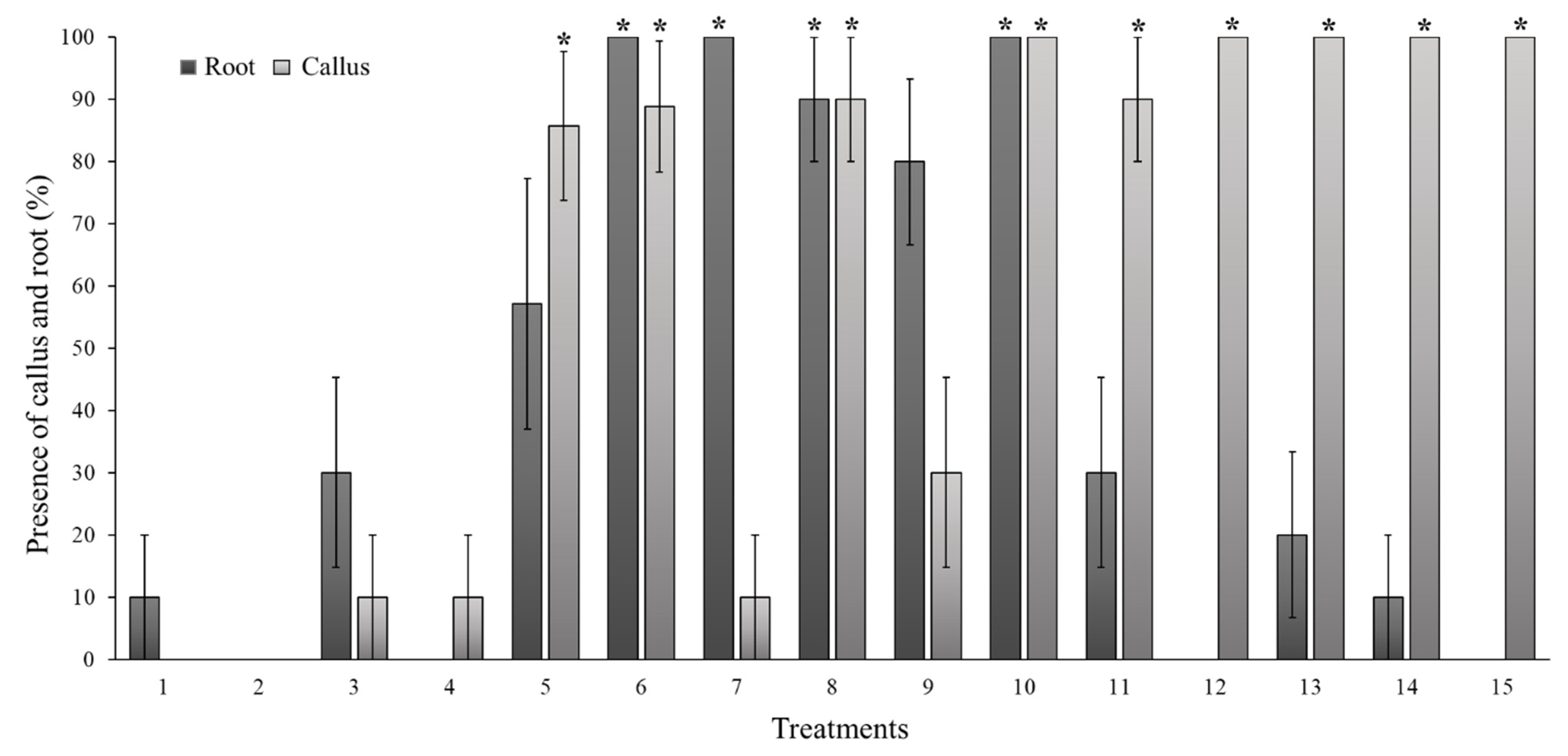
Figure 1. Effect of six concentrations of thidiazuron (TDZ) and 6-benzylaminopurine (BA), compared to the MS control, in which the presence of callus and root in shoot of Sechium compositum was evaluated. Means ± SE (standard error). The treatments are as follows. 1: MS control. 2: MS+BA (0.1 mg L−1). 3: MS+BA (0.2 mg L−1). 4: MS+BA (0.4 mg L−1). 5: MS+BA (0.6 mg L−1). 6: MS+BA (0.8 mg L−1). 7: MS+BA (1.0 mg L−1). 8: MS+BA (1.2 mg L−1). 9: MS+TDZ (0.1 mg L−1). 10: MS+TDZ (0.2 mg L−1). 11: MS+TDZ (0.4 mg L−1). 12: MS+TDZ (0.6 mg L−1). 13: MS+TDZ (0.8 mg L−1). 14: MS+TDZ (1.0 mg L−1). 15: MS+TDZ (1.2 mg L−1). Kruskal–Wallis One-Way Analysis of Variance on Ranks (p = <0.001); Mann–Whitney U Test (p = <0.001). * Differences between control and treatments (p < 0.05).
Horticulturae 10 00537 g001
Figure 2. Effect of 0.1 mg L−1 of BA compared to MS control. Average plant height (shoot), average number of shoots, buds, percentage of root, callus, and rosette structure of the in vitro shoot of Sechium compositum. Means ± SE (standard error) of n = 50 repetitions per treatment. Kruskal–Wallis One-Way Analysis of Variance on Ranks (p = <0.001); Mann–Whitney U Test (p = <0.001).
Horticulturae 10 00537 g002
Figure 3. Sechium compositum established in vitro. (A) +2-month-old seedling. (B) +2-month-old shoot with two shoots and formation of basal callus. (C) Shoot with packed buds (rosette structure).
Horticulturae 10 00537 g003
Figure 4. Representation of oxidation in Sechium compositum callus depending on the explant used for its formation. Oxidized callus obtained from leaf explant: (AC, FH). Callus obtained from stem explant: (D,E,I,J). All observed through a stereoscope.
Horticulturae 10 00537 g004
Figure 5. Calli obtained from stem of Sechium compositum placed on the medium (2.0 mg L−1 of 2,4-D) with 2.0 mg L−1 of activated charcoal. (A) Stem explant for callus formation observed 7 days after sowing (callus formation begins at the ends of the stem where the cut was made). (B) Callus observed 15 days after sowing (white callus begins to develop on the surface of the stem). (C) Callus observed 30 days after sowing (most of the calli formed roots, and their development stopped). (D) Single seedling regenerated at 30 d in the medium with activated charcoal from stem explant.
Horticulturae 10 00537 g005
Figure 6. Calli obtained from stem of Sechium compositum. (A) Callus with desired characteristics (white, with few yellow areas). (B) Callus culture was divided for multiplication and resown in medium with 2.0 mg L−1 of 2,4-D. (C) Calli resown in a medium with 2.0 mg L−1 of 2,4-D with PVP. (D) Oxidized callus.
Horticulturae 10 00537 g006
Figure 7. Effect of adding two concentrations of polyvinylpyrrolidone (PVP) to the medium with 2.0 mg L−1 of 2,4-D for the induction of callus from the stem of Sechium compositum. Callus development level at 20 and 30 days. Means ± standard error (SE). Comparative analysis with Mann–Whitney U Test (p = <0.001).
Horticulturae 10 00537 g007
Figure 8. Effect of the addition of two concentrations of polyvinylpyrrolidone (PVP) to the medium with 2.0 mg L−1 of 2,4-D on the growth of subcultured calli with desirable characteristics in Sechium compositum (reduction in oxidation, survival, and mass growth) evaluated at 20 and 30 days. Means ± standard error (SE). Kruskal–Wallis One-Way Analysis of Variance on Ranks (p = >0.05). There are no differences between the treatments.
Horticulturae 10 00537 g008
Table 1. Five-level scale used to determine callus formation from stem and leaf explants of Sechium compositum.
LevelCallus Formation Scale (%)Description
10There is no tissue response.
21–25The tissue swells (turgor) and begins to form a light-yellow callus at the ends.
326–50The ends surrounding tissue areas show a greater amount of white callus.
452–75A green tissue portion is observed at the top. The rest of the callus is white.
576–100The callus has completely covered the tissue, and there is an increase in the white mass, with a slight brown tone in small areas.
Table 2. Scale to determine the appropriate physical characteristics of the callus to multiply, 100% being the most ideal.
Callus Development (%)Description
0Brown callus and yellow medium are observed.
25The callus maintains a greater number of brown areas, and the mass does not increase. The medium looks slightly yellow.
50A greater percentage of potentially active callus is observed, and the medium turns a light yellow.
75A considerable decrease in brown areas is observed, along with a greater number of active areas in the callus and a transparent medium.
100The callus presents mostly or all active zones, its mass increases, and root formation is observed. The medium is transparent.
Table 3. Effect of seven concentrations of thidiazuron (TDZ) and 6-benzylaminopurine (BA) on the height and proliferation of buds and shoots of Sechium compositum. The evaluations were carried out 3 months after establishment.
Treatment (Growth Regulator)Concentration (mg L−1)Means Height Shoot (mm)Mean Number of BudsMean Number of Shoots
MS Control020.25 ± 6.10 ab10.3 ± 1.4 a1.0 ± 0 bc
BA0.110.02 ± 0.88 ab16.4 ± 1 a2.1 ± 0.3 a
0.231.76 ± 9.85 ab15.0 ± 1.9 a1.7 ± 0.2 ab
0.49.78 ± 1.31 ab11.7 ± 1.4 a1.6 ± 0.2 ab
0.628.76 ± 8.27 ab7.4 ± 1.6 ab1.4 ± 0.2 ab
0.88.56 ± 3.62 ab3.9 ± 1.9 ab0.7 ± 0.2 bc
1.064.46 ± 7.86 a12.4 ± 0.5 a2.0 ± 0.2 a
1.211.46 ± 2.92 ab4.1 ± 1.1 ab1.0 ± 0 bc
TDZ0.137.25 ± 7.52 a12.2 ± 2.5 a2.1 ± 0.4 a
0.230.32 ± 7.84 ab10.0 ± 3.1 ab1.4 ± 0.3 ab
0.416.46 ab5.6 ± 1.8 ab1.0 ± 0.3 ab
0.62.56 ± 0.7 c0.8 ± 0.2 bc0.7 ± 0.2 bc
0.83.25 ± 0.91 b0.9 ± 0.3 bc0.6 ± 0.2 bc
1.05.63 ± 1.76 b1.7 ± 0.6 bc1.1 ± 0.3 ab
1.22.13 ± 0.71 c0.5 ± 0.2 bc0.5 ± 0.2 bc
Means ± SE (standard error). Kruskal–Wallis Analysis Pr > Chisq < 0.0001. Means with the same letter (a, b, c) are not statistically different according to the Wilcoxon matched pairs signed rank test versus the Steel–Dwass–Critchlow–Fligner method (p < 0.05).
Table 4. Effect of three concentrations of thidiazuron (TDZ) and 2,4-dichlorophenoxyacetic acid (2,4-D) on the generation of callus from stem and leaf explants of Sechium compositum.
ExplantGrowth RegulatorConcentration (mg L−1)Mean Number of Callus Formation of Leaf and Stem ExplantsWeight (g)Ø 1 Mean Weight of Callus from Leaf and Stem Explants (mm)Ø 2 Mean Weight of Callus from Leaf and Stem Explants (mm).Mean Callus Height of Leaf and Stem Explants
(mm)
Mean Callus Volume of Leaf and Stem ExplantsMean Percentage of Root Formed from Callus of Leaf and Stem Explants
(%)
StemMS---1.70 ± 0.15 a0.15 ± 0.029 c7.15 ± 0.44 de 5.70 ± 0.51 cd 4.75 ± 0.51 c 14.06 ± 2.75 ce 50.0 ± 16.7 abc
2,4-D0.54.33 ± 0.11 b1.16 ± 0.068 a16.26 ± 0.30 a 13.48 ± 0.51 a 11.73 ± 0.31 a 149.11 ± 7.31 a 100.0 ± 0.0 a
1.04.72 ± 0.11 ab1.32 ± 0.064 a16.51 ± 0.30 a 12.95 ± 0.31 a 11.69 ± 0.28 a 152.05 ± 7.66 a 100.0 ± 0.0 a
2.04.89 ± 0.08 a1.16 ± 0.048 a16.08 ± 0.40 a 12.55 ± 0.25 a 11.12 ± 0.26 a 138.28 ± 8.14 a 100.0 ± 0.0 a
TDZ0.52.89 ± 0.08 c0.53 ± 0.039 b13.07 ± 0.43 b 10.68 ± 0.40 b 9.64 ± 0.31 b 80.82 ± 5.68 b 0.0 ± 0.0 ce
1.03.06 ± 0.06 c0.63 ± 0.026 b13.79 ± 0.30 b 10.08 ± 0.28 b 10.54 ± 0.37 b 96.43 ± 6.69 b 0.0 ± 0.0 ce
2.03.22 ± 0.10 c0.60 ± 0.026 b13.12 ± 0.36 b 10.40 ± 0.41 b 9.41 ± 0.31 b 79.01 ± 5.61 b 0.0 ± 0.0 ce
LeafMS---1.00 ± 0.00 e0.01 ± 0.001 d5.72 ± 0.05 e 4.61 ± 0.39 d 0.92 ± 0.11 d 2.45 ± 0.17 f 0.0 ± 0.0 ce
2,4-D0.52.83 ± 0.19 cd0.46 ± 0.068 bc11.48 ± 0.48 bc 9.04 ± 0.51 b 8.12 ± 0.55 bc 56.37 ± 7.05 bcd 88.90 ± 7.6 ab
1.02.67 ± 0.27 cde0.52 ± 0.098 bc12.07 ± 0.96 b 8.93 ± 0.74 b 6.78 ± 0.78 bc 63.73 ± 11.11 bc 55.6 ± 12.1 ab
2.02.72 ± 0.29 cde0.61 ± 0.128 bc11.61 ± 1.00 b 8.81 ± 0.91 b 7.52 ± 1.02 bc 70.47 ± 16.39 bc 44.40 ± 12.1 bcd
TDZ0.52.00 ± 0.08 e0.23 ± 0.024 c10.17 ± 0.51 cd 7.83 ± 0.40 c 7.21 ± 0.37 c 38.82 ± 3.93 cde 0.0 ± 0.0 ce
1.02.00 ± 0.11 de0.24 ± 0.027 c9.77 ± 0.59 cd 6.99 ± 0.39 c 6.42 ± 0.47 c 33.58 ± 4.65 cde 0.0 ± 0.0 ce
2.02.28 ± 0.14 de0.30 ± 0.032 c11.39 ± 0.54 c 8.06 ± 0.29 c 6.64 ± 0.45 c 45.15 ± 5.73 cd 0.0 ± 0.0 ce
Means ( X ¯ ) ± standard error (SE). Kruskal–Wallis Analysis Pr > Chisq < 0.0001. Means with the same letter (a, b, c, d, e) are not statistically different according to Wilcoxon matched pairs signed rank test versus the Steel–Dwass–Critchlow–Fligner Method (p < 0.05). Diameter (Ø).
Disclaimer/Publisher’s Note: The statements, opinions and data contained in all publications are solely those of the individual author(s) and contributor(s) and not of MDPI and/or the editor(s). MDPI and/or the editor(s) disclaim responsibility for any injury to people or property resulting from any ideas, methods, instructions or products referred to in the content.