Article

Full-Length Transcriptome Analysis and Characterization of DFRs Involved in the Formation of Anthocyanin in Allium wallichii

1
Pharmacy College, Guizhou University of Traditional Chinese Medicine, Guiyang 550025, China
2
Key Laboratory of State Forestry Administration on Biodiversity Conservation in Karst Mountain Area of Southwest of China, School of Life Science, Guizhou Normal University, Guiyang 550025, China
3
Yunfu Branch, Guangdong Laboratory for Lingnan Modern Agriculture, Yunfu 527300, China
*
Authors to whom correspondence should be addressed.
These authors contributed equally to this work.
Horticulturae 2024, 10(10), 1068; https://doi.org/10.3390/horticulturae10101068
Submission received: 2 August 2024 / Revised: 29 September 2024 / Accepted: 2 October 2024 / Published: 6 October 2024

Abstract

:
Allium wallichii is famous for its reddish-purple flowers, which can be utilized as cut flowers and garden landscaping. Flower color is mainly determined by flavonoids, betalains, carotenoids, as well as other pigments. However, there is no research on the color formation mechanism in A. wallichii, which restricts its genetic improvement and development of superior varieties. The flower of A. wallichii was collected for full-length transcriptome sequencing and metabolome analysis using PacBio SMART and UPLC-MS, respectively. A total of 45 anthocyanins were detected in its flower, and 75,778 transcripts of 107,208 non-redundant transcripts were annotated. Then, two AwDFRs were cloned and characterized using bioinformatics tools. Enzyme activity assays revealed that both AwDFR1 and AwDFR2 possessed DFR activity in vitro that only accepted DHQ and DHM as substrates, except for DHK. Finally, physiological results showed that AwDFR1 and AwDFR2 could restore the lacking phenotypes of Arabidopsis tt3 mutant and increase the content of anthoycanin in tobacco petals. The anthocyanins and transcriptome in A. wallichii were firstly reported, and AwDFR1 and AwDFR2 are key enzymes participating in the biosynthesis of anthocyanins. This research provides important guidance for future key gene mining, color improvement, and horticultural breeding in A. wallichii.

1. Introduction

Allium wallichii belonging to the family Lycoris is a famous flower which is well known for its ornamental value, with the color of deep-purple, reddish-purple, pink, and white. Especially in the southwest of China, it is widely distributed with rich resources. Owing to its unique ornamental value of reddish-purple flowers, it is also utilized as cut flowers and garden landscaping [1]. As one of the most important plant resources in karst landform, A. wallichii showed ornamental, medicinal, and edible values, and it has broad prospects for development and utilization. Flower color is mainly determined by flavonoids, betalains, carotenoids, as well as other pigments. Of them, flavonoids are found to be direct flower pigmentation and broadly distributed in different plant species [2,3]. In addition to pigmentation, flavonoids can also serve as signaling molecules and protectants against UV irradiation and stresses [4]. Furthermore, numerous clinical and animal studies have confirmed that flavonoids are health-promoting compounds because of their antioxidant, anti-inflammatory, anticancer, and cardio-protective effects against many human diseases [5,6].
The biosynthesis pathway of flavonoid has been elucidated extensively in various plants at different levels, in which anthocyanidins (cyanidin, pelargonidin, and delphinidin) are the first colored compounds [7,8]. Anthocyanidins are derived from colorless leucoanthocyanidins that are produced from the catalyzation by dihydrofavonol 4-reductase (DFR) [9]. DFR, a critical rate-limiting enzyme during flavonoid biosynthesis, catalyzes the NADPH-dependent reduction of dihydroquercetin (DHQ), dihydromyricetin (DHM), and dihydrokaempferol (DHK) to form leucoanthocyanidins [10]. Additionally, DFR can also catalyze reversely to generate dihydroflavonols [11]. The activity of DFR was identified firstly in cell suspension cultures of Pseudotsuga menziesii, and its coding gene was first isolated from corn in 1985 by O’Reilly et al. [12,13]. Subsequently, DFR genes have been cloned from numerous plant species including Medicago truncatula [14], Lotus japonicus [15], Camellia sinensis [16], Freesia hybrid [17], Ginkgo biloba [18], Scutellaria baicalensis [9], and so on. Meanwhile, their functions have also been characterized. For example, DFR-encoding genes from Ginkgo biloba and Fragaria species are involved primarily in the formation of anthocyanin pigments [18,19] and the lack of DFR activity in blue Angelonia angustifolia resulting in white flowers [20]. DFRs from Petunia and Cymbidium do not reduce DHK, effectively leading to the deficiency of pelargonidin-type anthocyanins [21,22]. Other DFR-like genes, such as OsDFR2 and AtDRL1, fulfill an essential role in pollen formation and male fertility [7]. Furthermore, the active site geometry of VvDFR has also been solved through crystal structure analysis [23]. As reported, the three dihydroflavonols (DHQ, DHK, and DHM) are also substrates of flavonol synthase (FLS) [24]. Thus, the competition between DFR and FLS is a critical regulatory point that controls the biosynthesis of anthocyanins and then affects the formation of flower color. Overall, these findings show that DFR is a key enzyme regulating anthocyanin biosynthesis.
However, the molecular mechanism underlying anthocyanins’ metabolism in A. wallichii flowers remains unknown. This is mainly due to the lack of genomic data. With the development of third-generation sequencing technology, full-length transcriptome sequencing has been a promising method of molecular biology. In order to investigate the molecular mechanism of anthocyanin formation in A. wallichii flowers, the full-length transcriptome of A. wallichii was analyzed. Some hub genes involved in anthocyanins formation were obtained. In addition, the types and content of anthocyanins at three different stages of flowers were detected using metabonomic techniques. Then, two AwDFR genes (AwDFR1 and AwDF2) were identified and cloned from flowers of A. wallichii through correlation analysis of gene expression levels and anthocyanins content at three different stages. After that, these two enzymes were heterologously expressed and purified for determining their catalytic activity in vitro. To clarify the functions of AwDFRs in plants, they were over-expressed in Arabidopsis tt3 mutant as well as tobacco and resulted in the promotion of anthocyanins in transgenic plants. All above findings highlighted the importance of AwDFR1 and AwDFR2 in anthocyanin biosynthesis, which not only provided a basis for the analysis of DFR gene but also supplied new candidate genes for anthocyanin metabolic engineering.

2. Materials and Methods

2.1. Experimental Materials

The flowers of A. wallichii were collected from Jiucaiping, Hezhang Country, Guiyang City, Guizhou Province, China (104.761° E, 26.993° N). Jiucaiping is the highest peak of Wumeng Mountain with an altitude of 2900.6 m. It belongs to a subtropical monsoon climate, with an average annual temperature of 20.8 °C and an average annual rainfall of 1890 mm. Wild-type Arabidopsis thaliana and T-DNA insertion mutant (AT5G42800) were cultivated at a constant temperature of 22 °C under 16 h/8 h (light/dark) illumination. T2 transgenic Arabidopsis seedlings cultured on 1/2 MS medium with 3% sucrose were sampled for RT-PCR and anthocyanin detection. K326 tobacco plants grown in 12 h/12 h (light/dark) at 22 °C were used for transformation. All above fresh samples were quick-frozen immediately and kept in a deep freezer until further utilization.
Dihydroquercetin (DHQ), dihydrokaempferol (DHK), dihydromyricetin (DHM), cyanindin, pelargonidin, delphinidin, and cyanidin 3-O-glucoside were purchased from Sigma (St. Louis, MO, USA). DHQ, DHK, and DHM were diluted as 10 mg/mL solutions in methanol, and cyanidin 3-O-glucoside for standard curve drawing was prepared as 1 mg/mL solutions in methanol.

2.2. Anthocyanins in Flowers of A. wallichii

The flowers at three different stages (stage 1, 0.8–1.0 cm long, pale pink; stage 2, 1.2–1.5 cm long, deep pink; stage 3, blooming flowers, pink) were collected, freeze-dried, and ground into powder. An amount of 50 mg of powder was extracted with 0.5 mL methanol:water:hydrochloric acid (500:500:1, v/v/v) twice by vortex for 5 min, ultrasound for 5 min, and centrifugation for 5 min. After filtrating through a 0.22 μm membrane filter, the supernatants were analyzed with a UPLC-ESI-MS system. The instrument was equipped with a ACQUITY BEH C18 column (1.7 μm, 2.1 mm × 100 mm) (Waters, Wilmington, NC, USA), and the solvent system was water (0.1% formic acid): methanol (0.1% formic acid). Linear ion trap (LIT) and triple quadrupole (QQQ) scans were acquired on a QTRAP® 6500+ LC-MS/MS System (Waters, Wilmington, NC, USA) equipped with an ESI Turbo Ion-Spray interface (Waters, Wilmington, NC, USA). The ESI source temperature, ion spray voltage (IS), and curtain gas (CUR) were 550 °C, 5500 V, and 35 psi, respectively. Anthocyanins were analyzed using scheduled multiple reaction monitoring (MRM). Data acquisitions were performed using Analyst software (version 1.6.3).

2.3. RNA Extraction, Library Construction, and Sequencing

The flowers at three different stages, roots, stems, and leaves were collected, each sample with three biological replicates. Total RNA was extracted using an RNA extraction kit (Biomarker, Beijing, China). RNA concentration, 28 S/18 S, and fragment size were checked by an Agilent 2100 Bioanalyzer (Beijing, China) to determine the RNA integrity. Purity of RNA was tested by using a UV spectrophotometer, NanoDrop™ (Thermo Fisher Scientific Inc., Shanghai, China). The first-strand cDNA were synthesized from purified RNA by PCR amplification using a SMARTer™ PCR cDNA Synthesis Kit (Takara, Dalian, China), followed by second-strand cDNA synthesis. Through end reparation, joint connection, and digestion with restriction endonuclease, all cDNA were mixed to construct the final library for sequencing. After quality inspection of the library, full-length transcriptome sequencing was performed using PacBio platform (Biomarker, Beijing, China).

2.4. Raw Data Processing and Full-Length Tanscriptome Analysis

The raw data were converted into circular consensus sequencing (CCS) reads, whose number, read bases, and mean read length were used for the assessment of raw data. Based on the presence of 3′ primer, 5′ primer, and Poly (A) tail, CCS reads were divided into full-length non-chimeric (FLNC) reads and non-full-length reads. The similar reads in FLNC were clustered into one consensus isoform using the IsoSeq module of SMRTLink software (version 11.0), producing high-quality isoforms (accuracy > 0.99) and low-quality isoforms. Finally, redundant transcripts were removed to obtain non-redundant transcripts using CD-HIT software (version 4.8.1) with a default threshold of 50%, and the integrity of transcriptome was assessed using BUSCO software (version 5.1.0) [25].
The transcripts with the length over 500 bp were analyzed using MISA software (version 2.1) to identify simple sequence repeats (SSRs), and the density distribution of different SSRs was counted. The coding region sequence and its corresponding amino acid sequence were predicted using TransDecoder software (version 5.5.0) [26], and the length distribution of open-reading region was also counted. LncRNAs were predicted using a Coding Potential Calculator (CPC), Coding-Non-Coding Index (CNCI), Coding Potential Assessment Tool (CPAT), and Protein family (Pfam). The target genes of LncRNA were also predicted using LncTar (version 1.0) [27].

2.5. Cloning and Sequence Analysis of AwDFRs

The coding sequences (CDS) of AwDFR1 and AwDFR2 were searched in the transcriptome database, and primers were designed using Primer 5.0 (Table S1). Total RNA from A. wallichii flowers was isolated using an RNA pure Plant Kit (CWBIO, Taizhou, China). Then, 1 µg of high-quality RNA was reversed to cDNA using M-MLV reverse transcriptase (Takara, Beijing, China) and oligo (dT). After that, PCR amplification was conducted using flower cDNA as template, with an annealing temperature of 54 °C and 56 °C, respectively. The amplification products were gel-purified and ligated into pMD18-T vectors (Takara, Japan) for sequencing to confirm the nucleic acid order of AwDFR1 and AwDFR2.
The multi-alignment of different DFR sequences was carried out with DNAMAN 6.0. A phylogenetic tree was built using Mega 6.0 with 2000 bootstrap replicates, and motif analysis was performed using MEME Suite 5.5.5.

2.6. Relative Expression of AwDFRs in Different Tissues of A. wallichii

The total RNA was isolated from different vegetative tissues and flowers (stage 1–3) of A. wallichii. Then, equal concentration of RNA (500 ng) was used for synthesizing cDNA as mentioned above. Gene-specific primers were designed according to sequence information and are shown in Table S1. Real-time PCR reaction was performed on a BioRad CFX96 Real-Time PCR System using TransStart R Green qPCR SuperMix (TRANSGEN, Beijing, China) with the conditions reported previously [28]. The relative transcript levels of AwDFRs were normalized to the transcription abundance of AwActin through using 2−ΔΔCt method. Expression analyses were conducted with three biological replicates, and melting curve analysis as well as agarose gel electrophoresis were employed to affirm the gene-specific amplification.

2.7. Assay of AwDFRs Activities

The ORFs of AwDFR1 and AwDFR2 were subcloned into BamH I and Xho I sites of the protein expression vector (pET32a) using primers listed in Table S1. After verification by sequencing, the pET32a empty vector, pET32a-AwDFR1, and pET32a-AwDFR2 were all transformed into Escherichia coli BL21 (DE3) for expressing protein. The procedures of protein-inducible expression and purification were the same as those described previously [29]. In brief, positive colonies were inoculated into LB broth and cultured at 37 °C till the OD600 reached 0.6, at which point 0.2 and 0.6 mM of IPTG were added to induce protein expression at 15 °C for 24 and 36 h, respectively. The purifications of recombinant AwDFRs protein were conducted on a Ni-NTA column (TransGen, Beijing, China) at 4 °C according to the instructions. Purity was then evaluated via SDS-PAGE.
To identify the catalytic activity of AwDFR1 and AwDFR2, enzymatic assays were performed in a 500 µL reaction volume containing 100 mM of TrisHCl buffer at pH 7.0, 50 µL of NADPH (20 mM), 10 µL of substrates (10 mg/mL), and 40 µg of AwDFRs crude protein. After incubation at 30 °C for 1 h, 300 µL of n-butanol:HCl (95:5, v/v) was added. Then, an n-butanol layer was transferred to a new 1.5 mL tube and cultivated at 95 °C for 1 h to produce anthocyanidin. After evaporation by nitrogen gas, the residue was dissolved in 100 µL of methanol and used for HPLC detection. The reaction products of AwDFRs were tested by a Shimadzu HPLC system with ACCHROM XUnion C18 column (Wenling, China)at detection wavelength of 520 nm. The elution system was composed of 5% formic acid (solvent A) and methanol (solvent B), and 50 µL samples were eluted based on the program described previously [28].

2.8. Anthocyanin Analysis of Transformed Arabidopsis and Tobacco

The full-length ORFs of AwDFR1 and AwDFR2 were amplified through PCR, and cloned into the pBI121 expression vector containing CaMV35S promoter using Xba I and BamH I sites. The resulting recombinant vectors were introduced into Agrobacterium tumefaciens GV3101 competent cells. Agrobacterium-mediated Arabidopsis transformation was executed using the floral dipping method [30]. Meanwhile, transformation of tobacco was also performed following an existing protocol previously reported [31]. Selection of Arabidopsis and tobacco transgenic plants were carried out on Murashige and Skoog medium containing 50 mg/L of kanamycin. T2 Arabidopsis transgenic seedlings which were grown on 1/2 MS medium supplemented with 3% sucrose for 7 days were photographed and then collected for further analysis. For transgenic tobacco, T1 fully expanded flowers were observed and harvested for later detection. Expressions of AwDFR1 and AwDFR2 were identified by RT-PCR using Arabidopsis actin and NtTub genes as internal controls.
To analyze the anthocyanins in transgenic Arabidopsis seedlings and tobacco flowers, 0.2 g fresh samples were powdered in liquid nitrogen and extracted with 1 mL of extraction solution (75%H2O:24%MeOH:1%HCl) at a low temperature for 14 h. Following centrifugation at 12,000 rpm for 5 min, the supernatant was filtered using 0.22 µm microporous membrane and quantified via an HPLC system according to the method described in “Assay of AwDFRs Activities”. Total anthocyanin contents were then computed through external standard curve of cyanidin 3-O-glucoside standard in three biological replicates [32].

3. Results

3.1. UPLC-MS Analysis of Anthocyanins in A. wallichii Flower

The flowers A. wallichii at three different developmental stages, named 1, 2, and 3, were collected and applied for UPLC-MS detection. A total of 45 anthocyanins were detected using UPLC-ESI-MS, including 7 cyanidins, 10 delphinidins, 3 malvidins, 8 pelargonidins, 8 peonidins, and 9 petunidins, respectively. The top 13 anthocyanins with higher content are listed in Table 1. The results showed that the color of A. wallichii was mainly determined by cyanidin-3,5-O-diglucoside, cyanidin-3-O-glucoside, peonidin-3,5-O-diglucoside, and Peonidin-3-O-(6-O-malonyl-beta-D-glucoside). The anthocyanin with the highest content was cyanidin-3,5-O-diglucoside.

3.2. PacBio SMART Sequencing and Data Analysis

The PacBio library was established with cDNA from flowers, leaves, and roots of A. wallichii, and full-length transcriptome sequencing was performed using PacBio SMART technology. A total of 109.4 Gb of raw data were obtained. Based on the full passes ≥3 and the sequence accuracy >0.9, CCSs were extracted and polished. The number, read bases, and mean read length of CCSs were 624,955 reads, 933,525,572 bp, and 1493 bp, respectively (Figure 1A). Next, 499,912 reads of full-length non-chemiric (FLNC) reads were determined according to whether the poly A tail signal and the 5′ and 3′ cDNA primers existed in CCSs. Then, the similar sequences of FLNCs were clustered to obtain the consensus isoforms with an average length of 1435 bp and number of 157,508 reads (Figure 1B). Of these sequences, 157,486 reads were high-quality isoforms (with the accuracy >0.99). Finally, 107,208 non-redundant transcripts were obtained after merging sequences with higher similarity. Taking OrthoDB as a reference database, a total of 1044 genes, including 533 single-copy genes and 500 duplicated copy genes, were identified using BUSCO assessment (Figure S1).
For SSR prediction, 62,867 transcripts with a length larger than 500 bp were analyzed with MISA software. Of these sequences, 14,459 transcripts were SSRs containing sequences, and a total of 19,414 SSRs were identified, including Mono-nucleotide repeats (p1, 11,372), Di-nucleotide repeats (p2, 3370), Tri-mono-nucleotide (p3, 2606), Tetra-nucleotide (p4, 185), Penta-nucleotide (p5, 22), and Hexa-nucleotide (p6, 111). Meanwhile, compound SSR without overlapping position (c, 1695) and compound SSR with overlapping position (c*, 53) were also identified (Figure 2A). For CDS prediction, 85,518 CDSs were obtained, of which 51,340 were complete CDSs. For lncRNA analysis, 33,215, 55,589, 28,224, and 56,060 lncRNAs were identified using CNCI, CPC, Pfam, and CPAT tools, respectively (Figure 2B), resulting a total of 28,224 lncRNAs that were simultaneously detected. For TF analysis, a total of 5168 TFs were obtained, including 153 MYB-related TFs, 140 C3Hs, 133 bHLHs, 131 bZIP, 127 AP2/ERF-ERF, 121 C2H2, 81 MYBs, and so on (Figure 2C).

3.3. Functional Annotation of Non-Redundant Transcripts

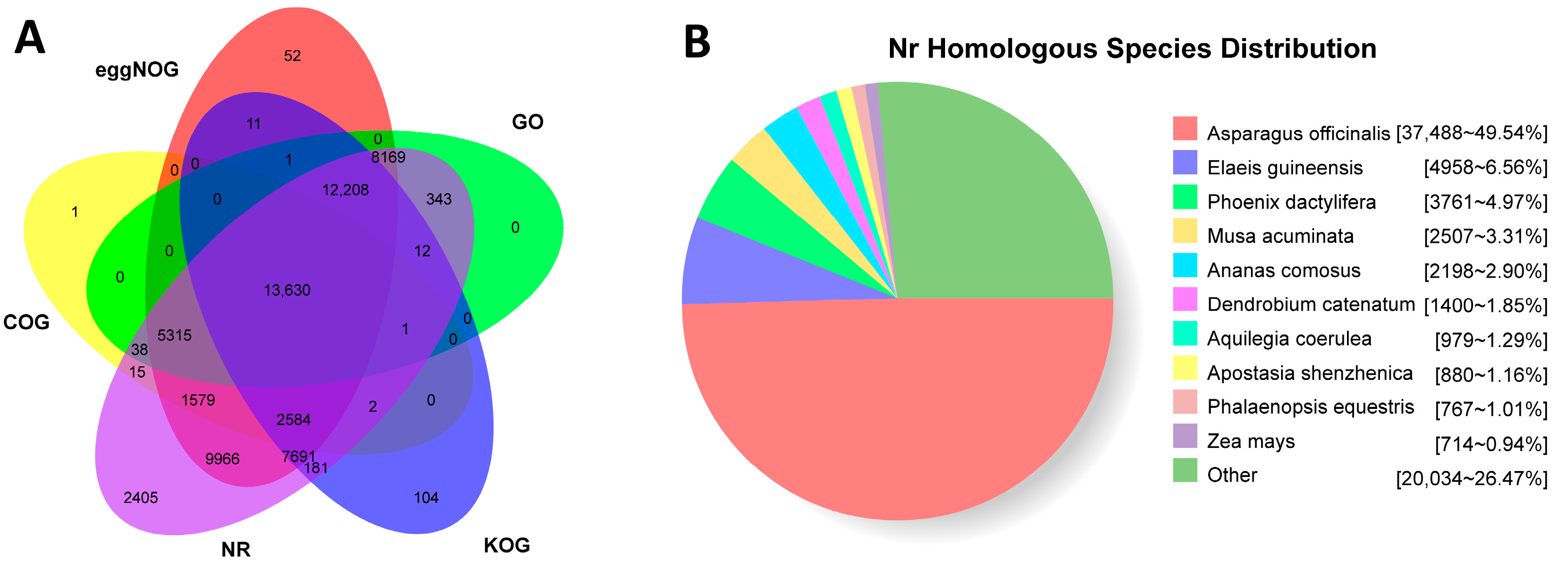
In total, 107,208 non-redundant transcripts were blasted with public databases, and 13,630 transcripts were annotated simultaneously in the GO, COG, eggNOG, KOG, and NR databases (Figure 3A). The highest number of transcripts, 75,778, were annotated by the NR database, while the COG database annotated the lowest number of 27,590. A total of 76,798 transcripts were functionally annotated. However, up to 16,447 transcripts were function unknown. And Asparagus officinalis showed the highest sequence homology with A. wallichii as high as 49.53% (Figure 3B).
For GO annotation, a total of 46,848 transcripts were annotated and assigned into three categories, namely biological process, molecular function, and cellular component (Figure 4). In the biological process, all transcripts were divided into 21 subcategories, including “metabolic process” (24,629), “cellular process” (22,814), “single-organism process” (15,704), and so on. In the molecular function, all transcripts were distributed into 14 subgroups, most of which belonged to “catalytic activity” (25,276) and “binding” (21,858). In the cellular component, all transcripts were annotated into 15 subclasses, which contained “cell part” (22,509), “cell” (22,494), “membrane” (16,223), and “organelle” (16,102), etc. Meanwhile, orthologous classifications were performed through COG, eggNOG, and KOG annotation, and all transcripts were divided into 25 functional categories (Figure 5). Without regard to “function unknown” and “general function only” groups, the group with the highest quality in all three databases was “posttranslational modification, protein turnover, chaperones” (6410 in eggNOG, 5092 in KOG, and 3125 in COG), followed by “signal transduction mechanisms” (4165/3420) and “carbohydrate transport and metabolism” (3668/2435) in eggNOG and KOG. But the followed groups in COG were “carbohydrate transport and metabolism” (2943) and “translation, ribosomal structure and biogenesis” (2690). For KEGG annotation, 127 pathways were successfully annotated. The top five pathways were “carbon metabolism” (1507), “biosynthesis of amino acids” (1432), “protein processing in endoplasmic reticulum” (1233), “starch and sucrose metabolism” (1177), and “ribosome” (1157).

3.4. Identification and Expression Level of AwDFRs

Based on transcriptome and metabolome information of A. wallichii, four putative AwDFR genes were screened and identified. The cDNA sequences of AwDFR1, AwDFR2, AwDFR3, and AwDFR4 were cloned and found to have the ORFs of 1155 bp, 1149 bp, 765 bp, and 1002 bp, respectively. After sequence alignment, we found the ORFs of AwDFR3 were significantly shorter than the typical lengths of DFRs, leading to a deletion of the NADPH binding domain that plays an important role in DFR activity, while the amino acids sequence of AwDFR4 is part of AwDFR1, and only one amino acid is different at 3′ ends. Therefore, based on the unusual ORF lengths and variations in the conserved motifs, AwDFR1 and AwDFR2 were subjected to further analysis. Analysis of sequence alignment revealed that both AwDFR1 and AwDFR2 genes contained the DFR characteristic domains such as NADPH-binding domain and substrate-binding domain (Figure 6). Previous reports showed that DFRs can be classified into three categories (Asp type, Asn type, and neither Asp nor Asn type) according to the conservation of amino acid at the 134th position. AwDFR1 and AwDFR2, like most DFR proteins, were Asn-type DFR, implying their ability to catalyze DHK, DHQ, and DHM to produce corresponding leucoanthocyanidins.
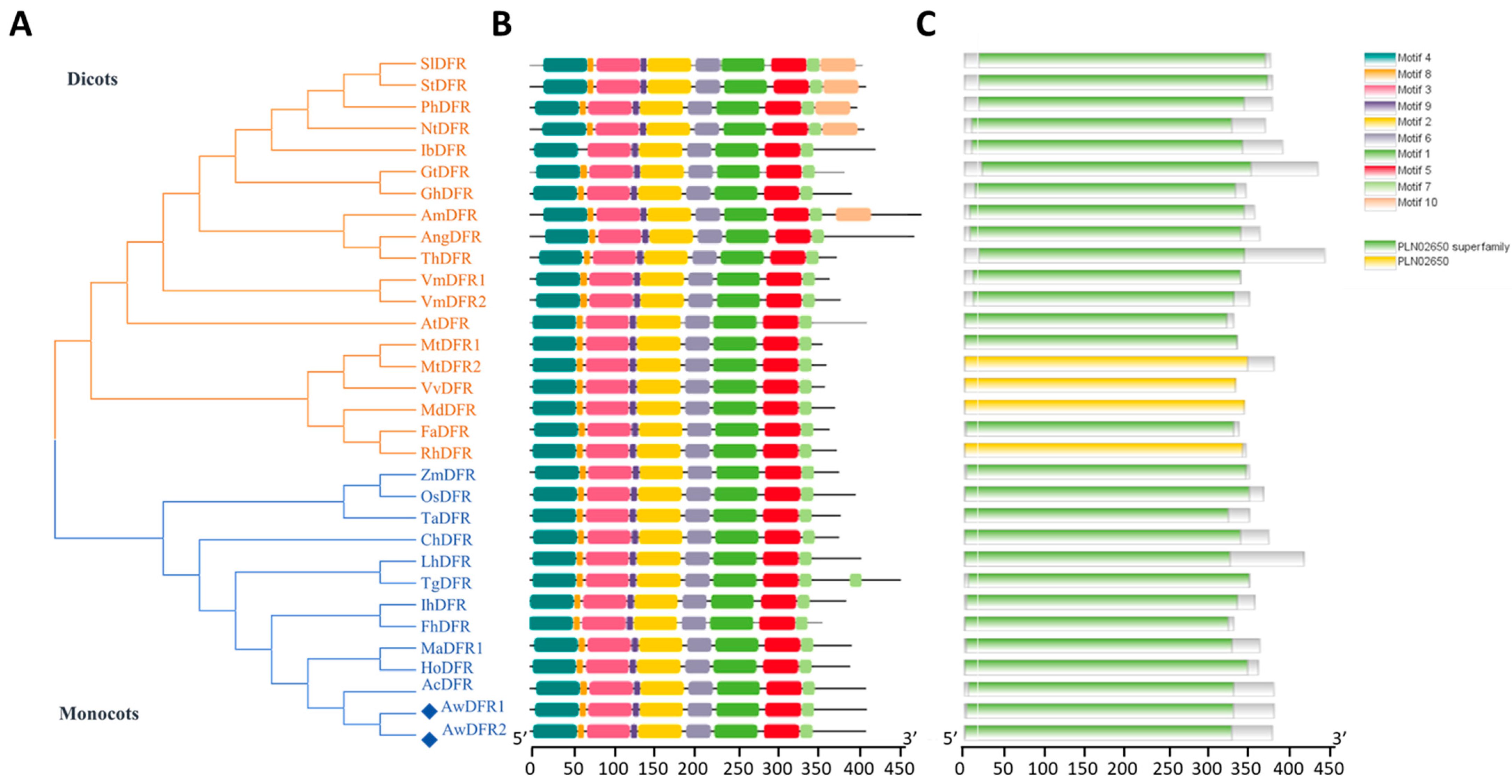
To better understand the homology of AwDFR1 and AwDFR2 with other known DFRs, a phylogenetic tree was built. AwDFRs were clustered into the monocotyledon group and possessed the closest genetic relationship with Allium cepa DFR (Figure 7A). Meanwhile, MEME analysis displayed that DFRs retained abundant conserved motifs during the evolutionary process, and conserved motifs of different DFR proteins mainly include motif 4, motif 8, motif 3, motif 9, motif 2, motif 6, motif 1, motif 5, motif 7, and motif 10 (Figure 7B). Among these motifs, motif 4 and motif 2 correspond to the NADPH-binding domain and substrate-binding domain, respectively; thus, the existence of these two motifs is crucial for DFR function. As shown in Figure 7C, all DFR proteins have PLN02650, which is the symbol of dihydroflavonol-4-reductase, and this indicates that motif 10 does not change the function of DFR protein and its loss may be one of the reasons for functional differentiation of DFR.
AwDFR1 and AwDFR2 transcript levels in different tissues as well as in the developing flowers of different stages were measured by real-time PCR (Figure 8A). Both genes were differentially expressed in distinct tissues, with the minimal expression levels in root, and the highest expression levels in petal and scape, respectively. During flower development, transcript abundance values of AwDFR1 and AwDFR2 were at a low level at stage 2, then significant increases were observed at stage 3 (Figure 8B). As the expression levels of AwDFR1 and AwDFR2 were both relatively high in leaves, scapes, and petals, this indicates their probable involvement in the biosynthesis of not only anthocyanin but also the other flavonoid in the detected tissues of A. wallichii.

3.5. Enzyme Activity Analysis of Recombinant AwDFRs

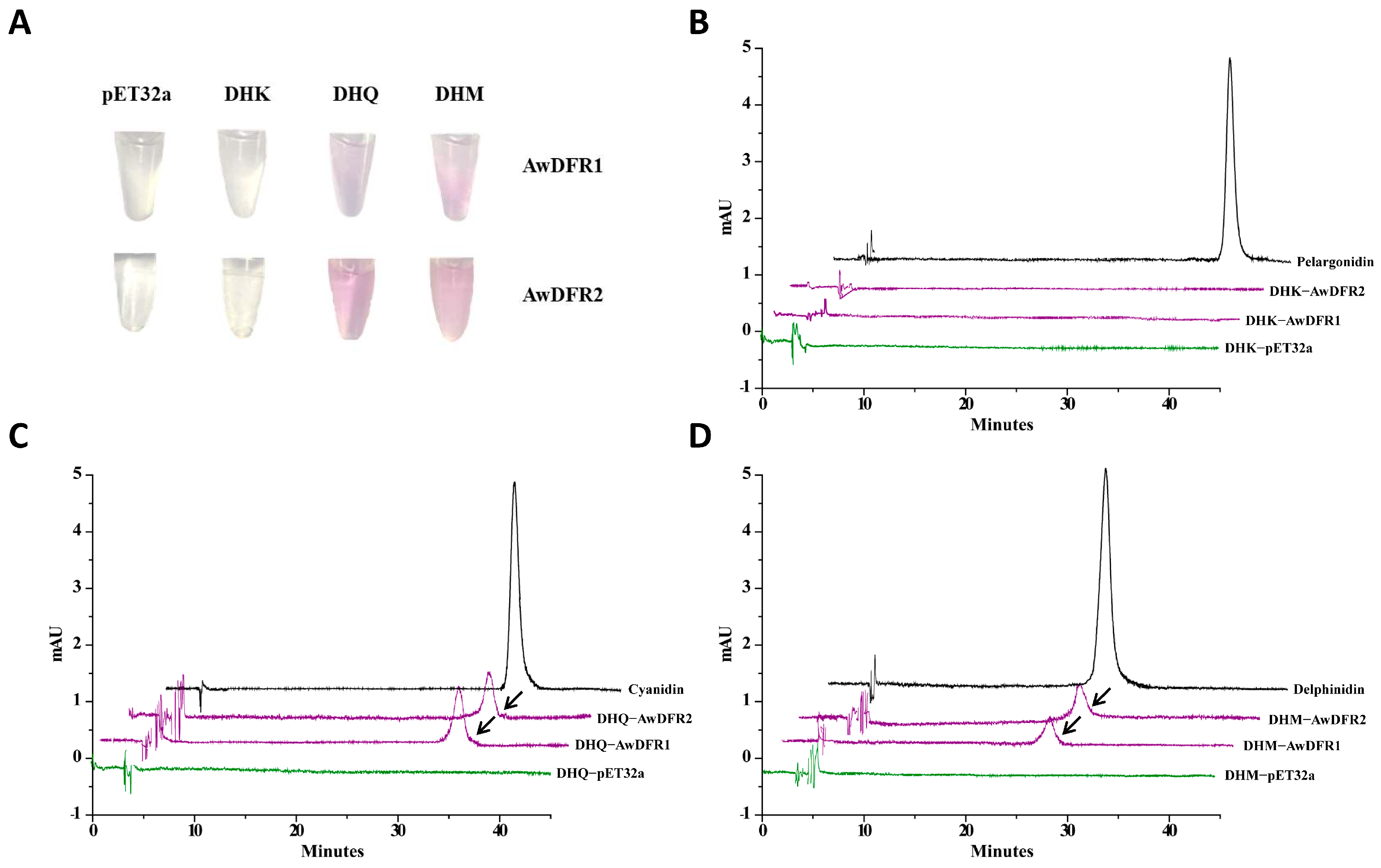
To determine the enzymatic properties of AwDFR1 and AwDFR2, pET32a vectors carrying these two genes were constructed and inductively expressed in E. coli BL21 (DE3). Denaturing SDS-PAGE electrophoretogram exhibited that clear target bands of recombinant AwDFRs protein with approximately 60 kDa (corresponding to the 18 kDa of Trx.Tag-His.Tag-S.Tag fused to the 42 kDa AwDFRs gene products) were obtained (Figure S2A,B). Leucoanthocyanidins, the nature products of DFR, are highly unstable and difficult to detect via HPLC directly. Therefore, we choose to test the stable corresponding anthocyanidins that stem from leucoanthocyanidins under acidic condition and 95 °C high temperature (Figure S2C). As the results present in Figure 9, red products were observed in the reactions of recombinant AwDFR1 and AwDFR2 using DHQ and DHM as substrates, and their chromatograms of enzymatic products were identical with the corresponding authentic anthocyanidins. However, when dihydrokaempferol was used as substrate, no red products were perceived, indicating the substrate specificity of AwDFR1 and AwDFR2. Meanwhile, no products were produced in the culture of protein from empty vector control.

3.6. Transgenic Confirmation of AwDFRs in Arabidopsis

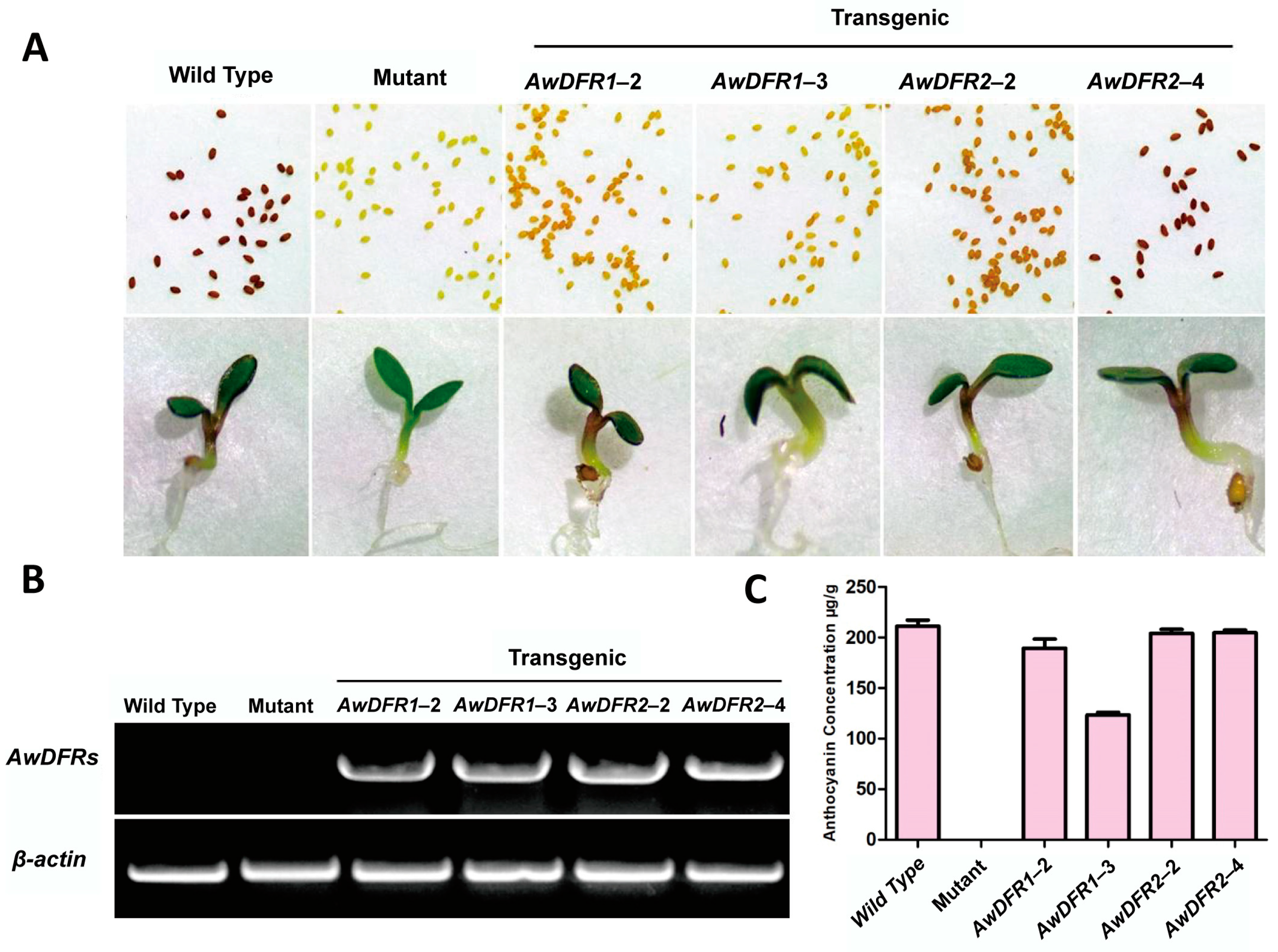
Arabidopsis tt3 plants resulted from the DFR mutation were generated through T-DNA insertion. This mutant exhibits a pale yellow seed coat and anthocyanin absence in hypocotyls. Therefore, tt3 is a suitable model plant to determine whether AwDFRs are the plausible candidate genes that associated with anthocyanins and proanthocyanidins deposition in vivo. The A. tumefaciens strain GV3101 carrying pBI121-AwDFR1 and pBI121-AwDFR2 were used for A. thaliana transformation via the flower dip method. After overexpression, visible phenotypic differences could be seen in the AwDFR1 and AwDFR2 transgenic seedlings and seed coats compared to tt3 plants. As shown in Figure 10A, AwDFR1 and AwDFR2 not only recovered the purple coloration in petiole but also restored proanthocyanidin synthesis in seed coat relative to tt3 mutant. Meanwhile, RT-PCR results demonstrated that phenotype changes in transgenic plants were due to the expression of AwDFRs (Figure 10B). Next, anthocyanins were extracted from seedlings of AwDFRs transgenic lines, tt3 mutant and wild type, and contents of anthocyanins were detected by HPLC. Compared with tt3 mutant, the impaired anthocyanins were fully complemented in AwDFRs transgenic lines, yielding similar anthocyanin profile of WT (Figure 10C and Figure S3). These data suggest that AwDFR1 and AwDFR2 took part in the biosynthesis of anthocyanins and proanthocyanidins, which was consistent with their catalytic functions in vitro.

3.7. Transgenic Confirmation of AwDFRs in Tobacco

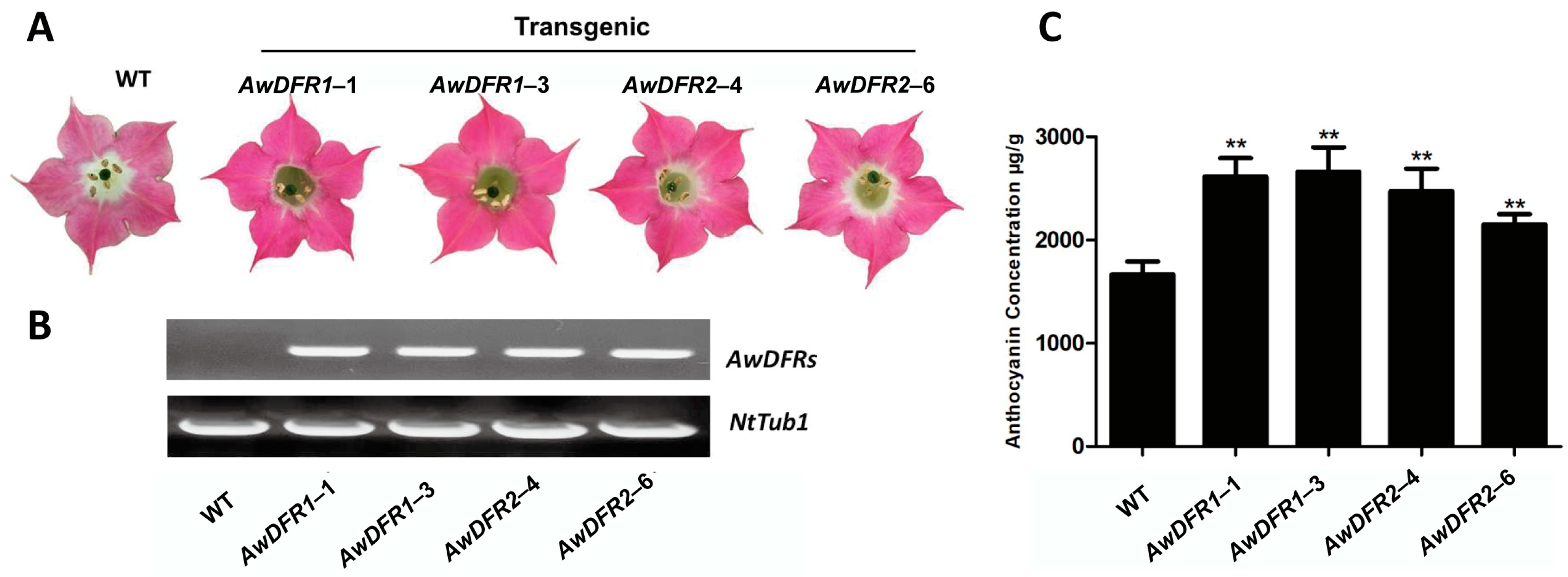
In order to confirm AwDFRs function in anthocyanin biosynthesis on flower color, binary vectors harboring AwDFR1 and AwDFR2 were transformed into tobacco. A total of 20 independent transgenic lines were generated, and four of them with dark pink flowers were chosen for later analysis. Compared to WT, flowers of transgenic plants exhibited more pigmentation (Figure 11A), and RT-PCR displayed that all transgenic lines expressed relatively high levels of AwDFRs transcripts (Figure 11B). Then, anthocyanins were extracted from the petals of AwDFR transgenic plants and measured by HPLC. The results showed that anthocyanin levels in AwDFR overexpression lines were all significantly higher than that in the control (Figure 11C), implying that the enzymes encoded by AwDFR1 and AwDFR2 actually increased anthocyanin accumulation in flowers.

4. Discussion

Allium is a perennial bulbous herbaceous plant in the botanical family Amaryllidaceae, with a wide variety of species. Allium wallichii with purple flowers is not only used as cut flowers and garden landscaping but also has important pharmacological components. In the flower of A. wallichii, there are up to 45 types of anthocyanins, including cyanidin, pelargonidin, and peonidin. Most anthocyanins are glycosylation modification products of anthocyanidins, named anthocyanins. With the development of sequencing technology, the genomes of some Allium plants have been successfully assembled, including Allium sativum L. [33] and Allium cepa L. [34]. However, no genomic information was reported in A. wallichii. As we know, only a full-length transcriptome of purple garlic (Allium sativum L.) was generated using PacBio and Illumina platform. This sequencing produced 22.56 Gb of clean data, generating 454,698 CCSs, which contained 379,206 FLNCs and 36,571 high-quality consensus reads. Here, a PacBio SMART library was constructed and applied for full-length transcriptome sequencing, which generating 109.4 Gb of raw data. Then, 624,955 CCSs were obtained, including 499,912 FLNCs and 157,508 high-quality consensus reads. Finally, 107,208 non-redundant transcripts were obtained and submitted for functional annotation. This provides rich genetic resources for flower color formation.
As a pivotal structural gene in the pathway of flavonoid, DFR controls the flux of flavonol, proanthocyanins, and anthocyanins in plants [35]. For elucidating the role of DFR on anthocyanin accumulation in A. wallichii flowers, the CDSs of AwDFR1 and AwDFR2 were successfully cloned. Multiple-sequence alignment discovered that AwDFR1 and AwDFR2 had the conserved NADPH-binding and substrate-binding domains, suggesting that these two enzymes belong to the DFR family (Figure 6). Furthermore, both AwDFR1 and AwDFR2 were Asn-type DFR, which indicates their ability to catalyze DHQ, DHK, and DHM like DFR from Zanthoxylum bungeanum [36]. Phylogenetic and motif analysis showed that the motif compositions of AwDFRs and most DFRs are conserved, although their sequences and molecular weight vary greatly, indicating that the function of DFR is relatively conservative (Figure 7).
In recent years, numerous studies have shown that DFR expression is positively related to anthocyanin and proanthocyanidin accumulation. In peonies, the DFR gene showed the highest expression in petals accumulated high anthocyanin, and similar results were also reported in gentian, which indicate that the DFR gene regulates petal color formation in plants [37,38]. But in R. delavayi, abundant expression of RdDFR1 was observed in leaf, implying its role as a participant in proanthocyanidin biosynthesis [39]. Shimada et al. studied the expression of DFR from Lotus japonicus and found that the DFR transcript level was correlated with its catalytic activity [15]. Two DFR genes from A. wallichii were identified, and their expressions were investigated in this work. AwDFR1 was strongly expressed in the petals which accumulate the most anthocyanin, suggesting that it may be the major gene determining anthocyanin accumulation in A. wallichii. Meanwhile, both AwDFR1 and AwDFR2 exhibited relatively high expression in leaves and scapes, which is not consistent with anthocyanin phenotype. These results imply that AwDFR1 and AwDFR2 genes are not flower-specific and can be involved in other flavonoids biosynthesis in A. wallichii, such as proanthocyanindin (Figure 8B).
According to the kinds of amino acid at position 134, AwDFR1 and AwDFR2 belong to Asn-type DFRs, which indicate their ability to catalyze DHQ, DHK, and DHM. But in biochemical analysis, AwDFR1 and AwDFR2 only accepted DHQ and DHM as substrates (Figure 4) like DFR from Freesia hybrid and Agapanthus praecox ssp. orientalis [17,40]. In contrast, an Asn-type DFR of purple-fleshed potato could only convert DHK to leucopelargonidin [41]. Furthermore, Liu et al. demonstrated that mutation at position 134 of MaDFR did not change its substrate specificity [42]. Several analyses revealed that the 26-amino-acid region might have crucial effects on DFR substrate specificity [21]. Therefore, although many studies have looked into the substrate specificity of DFR, its catalytic mechanism and the key amino acid residues at active site are not completely understood. Further detailed studies combining site-directed mutation and domain swapping with homologous modeling are needed to uncover the mechanism of DFR substrate specificity.
DFR reduces dihydroflavonols to form leucoanthocyanins in the flavonoid biosynthesis pathway. Then, under the action of ANS, leucoanthocyanins are converted into anthocyanins. For investigating the function of AwDFR1 and AwDFR2 participating in anthocyanin synthesis, they were introduced into Arabidopsis tt3 mutant. The results displayed that overexpression of these two genes successfully restore the deficient phenotypes of tt3 mutant (Figure 10A), confirming AwDFR1 and AwDFR2 as DFRs taking part in anthocyanin and proanthocyanidin biosynthesis in vivo. Similarly, ectopic expression of DFR from Chrysanthemum and Purple Sweet Potato in tt3 mutant had also recovered the biosynthesis of anthocyanin in seedlings and proanthocyanidin in seed coat, which suggests that DFRs are functionally exchangeable among diverse plant species [43,44]. In addition, the seed coat color of AwDFR2 transgenic plants was deeper than that of AwDFR1, which was consistent with its relatively high expression in leaves and scapes, indicating its role mainly on proanthocyanidin synthesis.
To confirm the potential function of AwDFR1 and AwDFR2 on flower color formation, these two genes were also overexpressed in tobacco. As present in Figure 11, corollas of transgenic tobacco were darker than that of control, and this was congruent with anthocyanin accumulation levels. Likewise, similar results leading to dark pink corollas in transgenic tobacco had also been reported in Ginkgo biloba, Populus trichocarpa, and cranberry [18,45], which identify the role of DFR as a control point in anthocyanin biosynthesis pathway. Together with the data mentioned above, we discovered that DFRs from diverse plants displayed semblable functions in transgenic tobacco but played different roles in the host plants. Therefore, we proposed that the function of DFR in plants not only depended on the level and patterns of its transcription in plants but also effected by protein level competition among enzymes participated in flavonoid synthesis.

5. Conclusions

In this study, full-length transcriptome and anthocyanins metabolome of A. wallichii were analyzed, resulting in 107,208 non-redundant transcripts and 45 types of anthocyanins. Based on the transcriptome data, we successfully cloned and characterized the AwDFR genes (AwDFR1 and AwDFR2) from A. wallichii and demonstrated their biological functions in vitro and in vivo. Enzyme activity assays revealed that both AwDFR1 and AwDFR2 possessed DFR activity in vitro that converted dihydroflavonols into corresponding leucoanthocyanins. Physiological roles of AwDFR1 and AwDFR2 were studied in Arabidopsis tt3 mutant, and these two genes were both shown to restore the lacking phenotypes of tt3 mutant. Transferring AwDFR1 and AwDFR2 into tobacco could increase the content of anthocyanin in petals, suggesting that these two genes are credible candidates for petal coloration. In conclusion, AwDFR1 and AwDFR2 belong to the DFR superfamily and are key enzymes participated in the biosynthesis of anthocyanins.

Supplementary Materials

The following supporting information can be downloaded at: https://www.mdpi.com/article/10.3390/horticulturae10101068/s1, Figure S1: BUSCO assessment of non-redundant transcriptome; Figure S2: The purified recombinant protein and reaction mechanism. (A) The recombinant AwDFR1. (B) The recombinant AwDFR2. (C) The reaction mechanism of DFRs. Lane 1 means total soluble protein from E. coli (BL21), lane 2 means total soluble protein from E. coli expressing pET-32a (+) vector, lane 3 means total soluble protein from E. coli expressing AwDFRs prior to induction by IPTG, lane 4 means purified AwDFRs; Figure S3: HPLC analysis of anthocyanin in transgenic A. thaliana; Figure S4: Content of PA in transgenic A. thaliana seeds. Table S1: List of primers used in this study.

Author Contributions

Conceptualization and writing—original draft preparation, Z.J. and L.L.; data curation and visualization, L.L. and H.S.; writing—review and editing, Y.Z. and W.Z.; funding acquisition and investigation, W.S. and Y.P. All authors have read and agreed to the published version of the manuscript.

Funding

This research was funded by the Research and Demonstration on Key technologies for conservation and Innovative utilization of germplasm resources of important Southern medicine in Guangdong Province ([2021]163), grants from Guizhou Science and Technology Department (ZK [2023]270), and grants from department of education of Guizhou Province ([2022]044).

Data Availability Statement

The datasets supporting the conclusions and description of a complete protocol can be found within the manuscript and its Supplementary Materials. The datasets used and/or analyzed during the current study are available from the corresponding author on reasonable request.

Conflicts of Interest

The authors declare no conflicts of interest.

References

  1. Zhao, J.; Huang, M.; Ni, R.; Peng, T.; Chen, X.; Nan, H. The complete chloroplast genome of Allium wallichii Kunth (Amaryllidaceae). Mitochondrial DNA Part B Resour. 2023, 8, 1054–1058. [Google Scholar] [CrossRef]
  2. Koes, R.; Verweij, W.; Quattrocchio, F. Flavonoids: A colourful model for the regulation and evolution of biochemical pathways. Trends Plant Sci. 2005, 10, 236–242. [Google Scholar] [CrossRef]
  3. Winkel-Shirley, B. Flavonoid biosynthesis. A colourful model for genetics, biochemistry, cell biology, and biotechnology. Plant Physiol. 2001, 126, 485–493. [Google Scholar] [CrossRef]
  4. Chen, X.; Liu, W.; Huang, X.; Fu, H.; Wang, Q.; Wang, Y.; Cao, J. Arg-type dihydroflavonol 4-reductase genes from the fern Dryopteris erythrosora play important roles in the biosynthesis of anthocyanins. PLoS ONE 2020, 15, e0232090. [Google Scholar] [CrossRef] [PubMed]
  5. Tsuda, T. Recent progress in anti-obesity and anti-diabetes effect of berries. Antioxidants 2016, 5, 13. [Google Scholar] [CrossRef] [PubMed]
  6. Koning, R.A.; Grayson, I.; Weckbecker, C. The effects of bilberries, blackcurrants and their constituent anthocyanins on heart health in humans. Agro Food Industry Hi-Tech. 2015, 26, 16–20. [Google Scholar]
  7. Ruan, H.; Shi, X.; Gao, L.; Rashid, A.; Li, Y.; Lei, T.; Dai, X.; Xia, T.; Wang, Y. Functional analysis of the dihydroflavonol 4-reductase family of Camellia sinensis: Exploiting key amino acids to reconstruct reduction activity. Hortic. Res. 2022, 9, uhac098. [Google Scholar] [CrossRef]
  8. Saito, K.; Yonekura-Sakakibara, K.; Nakabayashi, R.; Higashi, Y.; Yamazaki, M.; Tohge, T.; Fernie, A.R. The flavonoid biosynthetic pathway in Arabidopsis: Structural and genetic diversity. Plant Physiol. Biochem. 2013, 72, 21–34. [Google Scholar] [CrossRef]
  9. Wang, J.; Wang, Y.; Wang, S.; Zhang, F.; Niu, Y.; Wang, D. Cloning and temporal-spatial expression analysis of dfr gene from Scutellaria baicalensis with different colors. Sheng Wu Gong. Cheng Xue Bao 2021, 37, 1312–1323. [Google Scholar] [PubMed]
  10. Feng, X.; Zhang, Y.; Wang, H.; Tian, Z.; Xin, S.; Zhu, P. The dihydroflavonol 4-reductase BoDFR1 drives anthocyanin accumulation in pink-leaved ornamental kale. Theor. Appl. Genet. 2021, 134, 159–169. [Google Scholar] [CrossRef] [PubMed]
  11. Halbwirth, H.; Kahl, S.; Jager, W.; Reznicek, G.; Forkmann, G.; Stich, K. Synthesis of (C-14)-labeled 5-deoxyflavonoids and their application in the study of dihydroflavonol/leucoanthocyanidin interconversion by dihydroflavonol 4-reductase. Plant Sci. 2006, 170, 587–595. [Google Scholar] [CrossRef]
  12. Stafford, H.A.; Lester, H.H. Enzymic and nonenzymic reduction of (+)-dihydroquercetin to its 3,4,-diol. Plant Physiol. 1982, 70, 695–698. [Google Scholar] [CrossRef]
  13. O’Reilly, C.; Shepherd, N.S.; Pereira, A.; Schwarz-Sommer, Z.; Bertram, I.; Robertson, D.S.; Peterson, P.; Saedler, H. Molecular cloning of the a1 locus of Zea mays using the transposable elements En and Mu1. EMBO J. 1985, 4, 877–882. [Google Scholar] [CrossRef]
  14. Xie, D.Y.; Jackson, L.A.; Cooper, J.D.; Ferreira, D.; Paiva, N.L. Molecular and biochemical analysis of two cDNA clones encoding dihydroflavonol-4-reductase from Medicago truncatula. Plant Physiol. 2004, 134, 979–994. [Google Scholar] [CrossRef] [PubMed]
  15. Shimada, N.; Sasaki, R.; Sato, S.; Kaneko, T.; Tabata, S.; Aoki, T. A comprehensive analysis of six dihydroflavonol 4-reductases encoded by a gene cluster of the Lotus japonicus genome. J. Exp. Bot. 2005, 56, 2573–2585. [Google Scholar] [CrossRef]
  16. Singh, K.; Kumar, S.; Yadav, S.K.; Ahuja, P.S. Characterization of dihydroflavonol 4-reductase cDNA in tea [Camellia sinensis (L.) O. Kuntze]. Plant Biotechnol. Rep. 2009, 3, 95–101. [Google Scholar] [CrossRef]
  17. Li, Y.Q.; Liu, X.; Cai, X.; Shan, X.; Gao, R.; Yang, S.; Han, T.; Wang, S.; Wang, L.; Gao, X. Dihydroflavonol 4-reductase genes from Freesia hybrida play important and partially overlapping roles in the biosynthesis of flavonoids. Front. Plant Sci. 2017, 8, 428. [Google Scholar] [CrossRef]
  18. Ni, J.; Ruan, R.; Wang, L.; Jiang, Z.; Gu, X.; Chen, L.; Xu, M. Functional and correlation analyses of dihydroflavonol-4-reductase genes indicate their roles in regulating anthocyanin changes in Ginkgo biloba. Ind. Crop. Prod. 2020, 152, 112546. [Google Scholar] [CrossRef]
  19. Miosic, S.; Thill, J.; Milosevic, M.; Gosch, C.; Pober, S.; Molitor, C.; Halbwirth, H. Dihydroflavonol 4-reductase genes encode enzymes with contrasting substrate specificity and show divergent gene expression profiles in Fragaria species. PLoS ONE 2014, 9, e112707. [Google Scholar] [CrossRef] [PubMed]
  20. Gosch, C.; Nagesh, K.M.; Thill, J.; Miosic, S.; Plaschil, S.; Milosevic, M.; Halbwirth, H. Isolation of dihydroflavonol 4-reductase cDNA clones from Angelonia x angustifolia and heterologous expression as GST fusion protein in Escherichia coli. PLoS ONE 2014, 9, e107755. [Google Scholar] [CrossRef]
  21. Johnson, E.T.; Ryu, S.; Yi, H.; Shin, B.; Cheong, H.; Choi, G. Alteration of a single amino acid changes the substrate specificity of dihydrof lavonol 4-reductase. Plant J. 2001, 25, 325–333. [Google Scholar] [CrossRef]
  22. Johnson, E.T.; Yi, H.; Shin, B.; Oh, B.J.; Cheong, H.; Choi, G. Cymbidium hybrida dihydroflavonol 4-reductase does not efficiently reduce dihydrokaempferol to produce orange pelargonidin-type anthocyanins. Plant J. 1999, 19, 81–85. [Google Scholar] [CrossRef] [PubMed]
  23. Petit, P.; Granier, T.; d’Estaintot, B.L.; Manigand, C.; Bathany, K.; Schmitter, J.M.; Lauvergeat, V.; Hamdi, S.; Gallois, B. Crystal structure of grape dihydroflavonol 4-reductase, a key enzyme in flavonoid biosynthesis. J. Mol. Biol. 2007, 368, 1345–1357. [Google Scholar] [CrossRef] [PubMed]
  24. Roslan, N.D.; Yusop, J.M.; Baharum, S.N.; Othman, R.; Mohamed-Hussein, Z.A.; Ismail, I.; Zainal, Z. Flavonoid biosynthesis genes putatively identified in the aromatic plant Polygonum minus via expressed sequences tag (EST) analysis. Int. J. Mol. Sci. 2012, 13, 2692–2706. [Google Scholar] [CrossRef]
  25. Li, W.Z.; Godzik, A. Cd-hit: A fast program for clustering and comparing large sets of protein or nucleotide sequences. Bioinformatics 2006, 22, 1658–1659. [Google Scholar] [CrossRef]
  26. Felipe, A.S.; Robert, M.W.; Panagiotis, I.; Evgenia, V.K.; Evgeny, M.Z. BUSCO: Assessing genome assembly and annotation completeness with single-copy orthologs. Bioinformatics 2015, 31, 3210–3212. [Google Scholar]
  27. Li, J.; Ma, W.; Zeng, P.; Wang, J.; Geng, B.; Yang, J.; Cui, Q. LncTar: A tool for predicting the RNA targets of long noncoding RNAs. Brief. Bioinform. 2015, 16, 806. [Google Scholar] [CrossRef]
  28. Sun, W.; Sun, S.; Xu, H.; Wang, Y.; Chen, Y.; Xu, X.; Yi, Y.; Ju, Z. Characterization of Two Key Flavonoid 3-O-Glycosyltransferases Involved in the Formation of Flower Color in Rhododendron Delavayi. Front. Plant Sci. 2022, 13, 863482. [Google Scholar] [CrossRef] [PubMed]
  29. Sun, W.; Zhou, N.; Feng, C.; Sun, S.; Tang, M.; Tang, X.; Ju, Z.; Yi, Y. Functional analysis of a dihydroflavonol 4-reductase gene in Ophiorrhiza japonica (OjDFR1) reveals its role in the regulation of anthocyanin. PeerJ 2021, 9, e12323. [Google Scholar] [CrossRef] [PubMed]
  30. Clough, S.J.; Bent, A.F. Floral dip: A simplified method for agrobacterium mediated transformation of Arabidopsis thaliana. Plant J. 1998, 16, 735–743. [Google Scholar] [CrossRef] [PubMed]
  31. Sparkes, I.A.; Runions, J.; Kearns, A.; Hawes, C. Rapid, transient expression of fluorescent fusionproteins in tobacco plants and generation of stably transformed plants. Nat. Protoc. 2006, 4, 2019–2025. [Google Scholar] [CrossRef] [PubMed]
  32. Fanali, C.; Dugo, L.; D’Orazio, G.; Lirangi, M.; Dacha, M.; Dugo, P.; Luigi, M. Analysis of anthocyanins in commercial fruit juices by using nano-liquid chromatography- electrospray-mass spectrometry and high-performance liquid chromatography with UV-visdetector. J. Separ. Sci. 2001, 34, 150–159. [Google Scholar] [CrossRef]
  33. Finkers, R.; van Kaauwen, M.; Ament, K.; Burger-Meijer, K.; Egging, R.; Huits, H.; Kodde, L.; Kroon, L.; Shigyo, M.; Sato, S.; et al. Insights from the first genome assembly of onion (Allium cepa). G3-Genes Genomes Genet. 2021, 11, 243. [Google Scholar] [CrossRef] [PubMed]
  34. Sun, X.; Zhu, S.; Li, N.; Cheng, Y.; Zhao, J.; Qiao, X.; Lu, L.; Liu, S.; Wang, Y.; Liu, C.; et al. A chromosome-level genome assembly of garlic (Allium sativum) provides insights into genome evolution and allicin biosynthesis. Mol. Plant 2020, 13, 1328–1339. [Google Scholar] [CrossRef] [PubMed]
  35. Lei, T.; Huang, J.; Ruan, H.; Qian, W.; Fang, Z.; Gu, C.; Zhang, N.; Liang, Y.; Wang, Z.; Gao, L.; et al. Competition between FLS and DFR regulates the distribution of flavonols and proanthocyanidins in Rubus chingii Hu. Front. Plant Sci. 2023, 14, 1134993. [Google Scholar] [CrossRef] [PubMed]
  36. Zhao, A.G.; Ding, R.W.; Wang, C.; Chen, C.; Wang, D.M. Insights into the catalytic and regulatory mechanisms of dihydroflavonol 4-reductase, a key enzyme of anthocyanin synthesis in Zanthoxylum bungeanum. Tree Physiol. 2023, 43, 169–184. [Google Scholar]
  37. Zhao, D.; Tao, J.; Han, C.; Ge, J. Flower color diversity revealed by differential expression of flavonoid biosynthetic genes and flavonoid accumulation in herbaceous peony (Paeonia lactiflora Pall.). Mol. Biol. Rep. 2012, 39, 11263–11275. [Google Scholar] [CrossRef] [PubMed]
  38. Nakatsuka, T.; Nishihara, M.; Mishiba, K.; Yamamura, S. Temporal expression of flavonoid biosynthesis-related genes regulates flower pigmentation in gentian plants. Plant Sci. 2005, 168, 1309–1318. [Google Scholar] [CrossRef]
  39. Sun, W.; Zhou, N.; Wang, Y.; Sun, S.; Zhang, Y.; Ju, Z.; Yi, Y. Characterization and functional analysis of RdDFR1 regulation on flower color formation in Rhododendron delavayi. Plant Physiol. Biochem. 2021, 169, 203–210. [Google Scholar] [CrossRef]
  40. Mori, S.; Otani, M.; Kobayashi, H.; Nakano, M. Isolation and characterization of the dihydroflavonol 4-reductase gene in the monocotyledonous ornamental Agapanthus praecox ssp. orientalis (Leighton) Leighton. Sci. Hortic. 2014, 166, 24–30. [Google Scholar] [CrossRef]
  41. Liu, X.; Xiang, M.; Fan, Y.; Yang, C.; Zeng, L.; Zhang, Q.; Chen, M.; Liao, Z. A rootpreferential DFR-like gene encoding dihydrokaempferol reductase involved in anthocyanin biosynthesis of purple-fleshed sweet potato. Front. Plant Sci. 2017, 8, 279. [Google Scholar]
  42. Liu, H.; Lou, Q.; Ma, J. Cloning and functional characterization of dihydroflavonol 4-reductase gene involved in anthocyanidin biosynthesis of grape hyacinth. Int. J. Mol. Sci. 2019, 20, 4743. [Google Scholar] [CrossRef] [PubMed]
  43. Forkmann, G.; Ruhnau, B. Distinct substrate specificity of dihydroflavonol 4-reductase from flowers of Petunia hybrida. Z. Für Naturforschung C 1987, 42, 1146–1148. [Google Scholar] [CrossRef]
  44. Wang, H.; Fan, W.; Li, H.; Yang, J.; Huang, J.; Zhang, P. Functional characterization of Dihydroflavonol-4-reductase in anthocyanin biosynthesis of purple sweet potato underlies the direct evidence of anthocyanins function against abiotic stresses. PLoS ONE 2013, 8, e78484. [Google Scholar] [CrossRef] [PubMed]
  45. Huang, Y.; Gou, J.Q.; Jia, Z.C.; Yang, L.; Sun, Y.M.; Xiao, X.Y.; Song, F.; Luo, K.M. Molecular cloning and characterization of two genes encoding dihydroflavonol 4- reductase from Populus trichocarpa. PLoS ONE 2012, 7, e30364. [Google Scholar] [CrossRef]
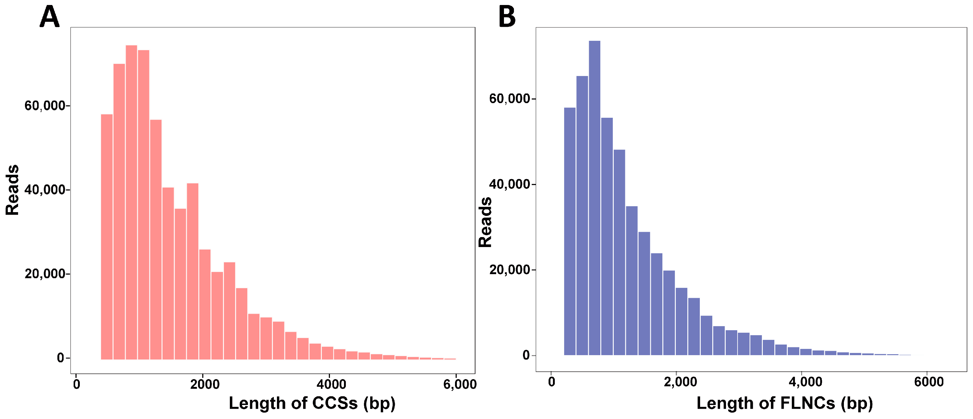
Figure 1. Length distribution of CCSs (A) and FLNCs (B).
Horticulturae 10 01068 g001
Figure 2. (A) The types and density of SSRs. c, compound SSR without overlapping position; c*, compound SSR with overlapping position; p1, mono-nucleotide repeats; p2, di-nucleotide repeats; p3, tri-mono-nucleotide; p4, tetra-nucleotide; p5, penta-nucleotide; p6, hexa-nucleotide. (B) LncRNAs prediction results with CNCI, CPC, Pfam, and CPAT. (C) Number of transcription factors.
Horticulturae 10 01068 g002
Figure 3. Functional annotation statistics (A) and NR annotation (B).
Horticulturae 10 01068 g003
Figure 4. GO annotation of non-redundant transcripts.
Horticulturae 10 01068 g004
Figure 5. COG, eggNOG, and KOG annotation of non-redundant transcripts.
Horticulturae 10 01068 g005
Figure 6. Multiple-sequence alignment of AwDFRs. The red box represents the NADP-binding domain, the green box represents the substrate-binding domain, and the black triangles indicate the important amino acid residue for substrate specificity of DFR.
Horticulturae 10 01068 g006
Figure 7. Phylogenetic tree (A), motif (B), and domain (C) analysis of AwDFRs. Plant species and GenBank accession numbers are as follows: SlDFR (Solanum lycopersicum, CAA79154.1), StDFR (Solanum tuberosum, AF449422), PhDFR (Petunia hybrida, AAF60298.1), NtDFR (Nicotiana tabacum, NP_001312559.1), IbDFR (Ipomoea batatas, HQ441167), AaDFR (Angelonia angustifolia, KJ817183), AmDFR (Aegle marmelos, X15536), AnDFR (Angelonia angustifolia, AIR09398.1), ThDFR (Torenia hybrida, AB012924), GtDFR (Gentiana triflora, D85185), GhDFR (Gerbera hybrid, Z17221), VmDFR1 (Vaccinium macrocarpon, AAL89714.1), VmDFR2 (Vaccinium macrocarpon, AAL89715.1), AtDFR (Arabidopsis thaliana, AB033294), MtDFR1 (Medicago truncatula, AY389346), MtDFR2 (Medicago truncatula, AAR27015.1), VvDFR (Vitis vinifera, Y11749.1), MdDFR (Malus domestica, AAO39816), FaDFR (Fragaria x ananassa, AF029685), RhDFR (Rosa hybrida, D85102), ZmDFR (Zea mays, Y16040), OsDFR (Oryza sativa, AB003495), TaDFR (Triticum aestivum, AB162139.1), CmDFR (Cymbidium hybrid, AF017451), LhDFR (Lilium hybrid, AB058641), TgDFR (Tulipa gesneriana, BAH98155.1), IhDFR (Iris x hollandica, BAF93856.1), FhDFR (Freesia hybrid, KU132389), HoDFR (Hyacinthus orientalis, AFP58815.1), AcDFR (Allium cepa, AY221250.2), MaDFR1 (Muscari aucheri, KJ619964).
Horticulturae 10 01068 g007
Figure 8. The different tissues of A. wallichii (A) and expression profiles of AwDFR1 and AwDFR2 in different tissues of A. wallichii (B). Ro, roots; Le, leaves; Sc, scapes; Pe, petals; 1, stage 1; 2, stage 2; 3, stage 3. a–c indicate very significant difference at the 0.01 level.
Horticulturae 10 01068 g008
Figure 9. Biochemical assays of recombinant AwDFR1 and AwDFR2. (A) The reaction results of recombinant AwDFR1 and AwDFR2 using DHK, DHQ, and DHM as substrates. (B) HPLC analysis of recombinant AwDFR1 and AwDFR2 using DHK as substrates. (C) HPLC analysis of recombinant AwDFR1 and AwDFR2 using DHQ as substrates. (D) HPLC analysis of recombinant AwDFR1 and AwDFR2 using DHM as substrates.
Horticulturae 10 01068 g009
Figure 10. Complementation of the pigmentation of DFR mutant seedlings with AwDFR1 and AwDFR2. (A) Phenotypes of wild-type, mutant, and transgenic Arabidopsis seedlings. (B) Expression confirmation of AwDFR1 and AwDFR2 in transgenic Arabidopsis. (C) The anthocyanin concentration in transgenic Arabidopsis.
Horticulturae 10 01068 g010
Figure 11. The function of AwDFR1 and AwDFR2 on anthocyanin accumulation in transgenic tobacco flowers. (A) The flowers color of WT and transgenic tobacco. (B) Expression confirmation of AwDFR1 and AwDFR2 in the flowers of transgenic tobacco. (C) The anthocyanin concentration in the flowers of transgenic tobacco. Asterisks indicate significant differences between means of wild-type and transgenic plants calculated by Student’s t-test (** represents p < 0.01).
Horticulturae 10 01068 g011
Table 1. The anthocyanins with higher quantities (≥1.0 μg/g) in the flowers of A. wallichii.
NumberAnthocyaninsContent of Anthocyanins (μg/g)
Stage 1Stage 2Stage 3
1Cyanidin-3-O-rutinoside4.18774.32554.2816
2Cyanidin-3-O-(6-O-p-coumaroyl)-glucoside1.10553.04403.5191
3Cyanidin-3-O-(6-O-malonyl-beta-D-glucoside)11.904714.568528.1693
4Cyanidin-3-O-glucoside15.441949.980541.9956
5Cyanidin-3,5-O-diglucoside68.857860.860542.7762
6Pelargonidin-3,5-O-diglucoside3.27113.68931.6777
7Pelargonidin-3-O-(6-O-malonyl-beta-D-glucoside)1.30952.32133.8407
8Pelargonidin-3-O-glucoside1.23884.40713.0832
9Peonidin-3-O-glucoside1.18262.64453.8103
10Peonidin-3-O-(6-O-malonyl-beta-D-glucoside)14.241522.066829.4758
11Peonidin-3,5-O-diglucoside24.333320.050112.6799
12Petunidin-3-O-glucoside1.12581.13500.9218
13Petunidin-3-O-(6-O-malonyl-beta-D-glucoside)1.27072.23781.4461
Disclaimer/Publisher’s Note: The statements, opinions and data contained in all publications are solely those of the individual author(s) and contributor(s) and not of MDPI and/or the editor(s). MDPI and/or the editor(s) disclaim responsibility for any injury to people or property resulting from any ideas, methods, instructions or products referred to in the content.