Article

Early Chilling Injury Symptom Development and Recovery of Mature Green Banana: Involvement of Ethylene under Different Severities of Chilling Stress

by
Lan-Yen Chang
1,2,* and
Jeffrey K. Brecht
2
1
Department of Horticulture, National Chung-Hsing University, Taichung 402202, Taiwan
2
Horticultural Sciences Department, University of Florida, Gainesville, FL 32611, USA
*
Author to whom correspondence should be addressed.
Horticulturae 2024, 10(10), 1050; https://doi.org/10.3390/horticulturae10101050
Submission received: 5 August 2024 / Revised: 16 September 2024 / Accepted: 27 September 2024 / Published: 1 October 2024
(This article belongs to the Section Postharvest Biology, Quality, Safety, and Technology)

Abstract

:
The involvement of stress ethylene in early chilling injury (CI) symptom development of mature-green (MG) banana fruit was examined using the ethylene action inhibitor, 1-methyclopropene (1-MCP). MG banana fruit pretreated with 0 or 50 μg L−1 1-MCP were stored at two chilling temperatures, 5 °C or 10 °C, for 0, 1, 12, or 72 h (different CI stresses), then transferred to 20 °C (rewarming) for 2 d. Irreversible CI symptoms that developed during 72 h storage at 5 or 10 °C included vascular browning and epidermal color parameters (L*, chroma, and hue angle). Some CI symptoms drastically intensified during exposure to 5 or 10 °C for 72 h, even being exacerbated after rewarming. Fruit in the other treatments suffered milder CI, and the recovery response progressed from slight and reversible to severe and irreversible with longer durations of exposure to lower temperatures. The effect of 1-MCP pretreatment was to attenuate the effect of chilling in terms of the CI symptom development (i.e., the browning of sub-epidermal tissues and the increasing of electrolyte efflux) and to promote recovery after rewarming, especially for the fruit stored at 5 °C, indicating the potential involvement of ethylene in membrane structural alterations under CI stress.

1. Introduction

Temperature is an important environmental factor affecting most biological reactions in living creatures. Low postharvest temperatures reduce metabolic activity and thus extend shelf life, but some crops originating from tropical or subtropical regions have abnormal responses to low, but nonfreezing temperatures (0 to 15 °C) that limit the utility of low temperatures for shelf-life extension [1]. The physiological damage that develops in response to such temperatures is termed chilling injury (CI) [2]. Some CI symptoms can be reversible when the temperature returns to the non-chilling range if the duration of chilling exposure is brief, but some dysfunctions may be irreversible under more prolonged chilling exposure [3]. The CI severity is usually cumulative as the duration of low temperature exposure increases or when the temperature is lower [3]. This relationship of exposure time, temperature, and symptom development and recovery has been described for fruits of lemon (Citrus limon L. cv. Eureka) [4], avocado (Persea americana Mill., cv. “Hass”) [5], and banana (Musa acuminata Colla., AAA Group, “Cavendish” type) [6].
The primary CI responses are considered to be related to initial membrane structural alterations or the activity of membrane-associated proteins/enzymes once encountering chilling temperature [7]. Before irreversible damages (secondary CI responses) occur, there is a lag period during which tissues can overcome the effect of chilling exposure, even at the injurious level, by recovering their function during rewarming (Figure 1). Increased electrolyte efflux (EE) or abnormal respiration/photosynthetic reactions (the electron transport chain in the membranes of mitochondria or chloroplasts) have been reported to be reversible in mildly injured plant tissues upon transfer from a chilling to a non-chilling environment [8,9,10,11,12]. The dynamic balance between CI damage and recovery from the injured status may be impaired when a specific injury threshold is reached. This will result in the appearance of visual CI symptoms, e.g., necrosis, pitting, internal or surface browning, changes of texture in pulp, increased susceptibility to microbes, or mechanical damage [7,13]. Moreover, the development of CI symptoms may be suppressed or reduced by the same low temperature that causes the injury, requiring a return to warmer, non-chilling temperature for the CI symptoms to develop, as observed and reported for cucumber (Cucumis sativus L.) [14] and peach [Prunus persica (L.) Batsch] [15,16]. Therefore, the physiological condition of chilling-sensitive tissues shortly after their transfer to non-chilling temperature will reflect their actual CI status.
Ethylene is a stress hormone, and its production varies internally (e.g., for different crops or physiological maturities) and externally (e.g., in response to environmental stresses) [17]. Ethylene production that is induced by external stresses (e.g., wounding, flooding, etc.) is regarded as stress ethylene [17,18,19]. The association of ethylene with CI is controversial. Increased ethylene production with extended CI storage has been observed in zucchini (Cucurbita pepo L.) [20]; some fruits produce a significant amount of ethylene upon rewarming from the chilling temperature, with greater ethylene production occurring upon rewarming as chilling storage temperatures decline and exposure durations increase (e.g., cucumber) [14]. The application of exogenous ethylene in avocado intensified CI symptoms [21]. But the same application alleviated CI in cantaloupe melon (Cucumis melo L.) [22], peaches and nectarines [23], and papayas (Carica papaya L.) and tomatoes (Solanum lycopersicum L.) [24], possibly due to ethylene stimulation of ripening, which is another factor influencing CI sensitivity as shown for the declining CI sensitivity of maturing and ripening banana fruit [25,26,27].
The fruit of banana (AAA group, Cavendish type), which originated and is mainly produced in tropical areas, are strictly maintained between 13.5 and 14 °C during commercial shipments to avoid CI [28]. Classic banana CI symptoms are observed firstly in the peel. The visible, irreversible CI symptoms include, in order of appearance, sub-epidermal discoloration [referred to as vascular browning (VB) or underpeel discoloration], dull peel appearance or peel surface browning, delayed or abnormal ripening or failure to ripen (i.e., inhibition of conversion of starch to sugar, development of flavor, carotenoid biosynthesis, and softening), and finally, pulp browning [29,30,31,32,33,34,35,36]. However, banana fruit are ethylene sensitive and about 0.015 to 0.5 µL L−1 ethylene can trigger the onset of the ripening process in mature-green (MG) fruit at ripening-conducive (i.e., non-chilling) temperatures [37,38]. Ethylene is produced when plants encounter various types of stresses [39]. However, the possible involvement of endogenous, stress-induced ethylene in the development of CI of banana fruit has not been well elucidated.
Less development of VB was observed in 1-MCP-treated MG banana peel in response to chilling [38]. Since 1-MCP is an inhibitor of ethylene action, this suggests possible ethylene involvement in early CI symptom development. The difference in VB severity between control and 1-MCP-treated fruit was first observed when the VB was just beginning to become visible, before the later appearing peel discoloration [38]. Ethylene is known to activate production of the enzyme phenylalanine ammonia-lyase (PAL) in plants, which catalyzes the rate limiting step in the biosynthesis of phenolics, which include the compounds responsible for banana VB [40]. Ethylene is also well known to promote plant membrane breakdown, leading to leakiness and water-soaked tissue in crops [41,42,43]. Increased tissue browning and EE across membranes are both symptoms of CI in many susceptible plants [44,45]. We hypothesize that the stress ethylene produced in response to the low temperatures that cause CI in susceptible plant species is responsible for at least some of the observed CI symptoms. Using MG banana fruit, the objectives of this work were (1) to determine the reversible and irreversible CI symptoms in the early chilling period and to evaluate the injury and recovery responses to 5 °C and 10 °C exposure, during storage and after rewarming; and (2) to examine the physiological changes induced by low temperature and their association with ethylene by using 1-MCP to remove ethylene action as a factor.

2. Materials and Methods

2.1. Sample Preparation

Mature-green banana fruit from Guatemala (Chiquita Brands International Sarl, Etoy, Switzerland) were shipped to a local distribution center (DC) in Florida within about 4 d after harvest. Fruit, without the ethylene treatment normally used to promote banana ripening, were received from a downstream local retailer within 24 h of delivery to the DC. The fruit were transferred from the retailer to the Postharvest Laboratory, University of Florida, immediately after receipt by the retailer. All shipping and handling and any brief storage at the lab prior to beginning the experiments were at 14 °C.
To reduce maturity variation, only banana hands with a* values < −14 and hue angle < 105° on the CIE L*a*b* scale performed similar maturity during the ripening trial in a preliminary test, and therefore only banana fingers with the color index were selected from a group representing twice the number required for each experiment [38]. Selected clusters were divided into individual fruit (fingers) at stage 2 (on a 1 to 7 scale from dark green to yellow with brown spots) [36], discarding fruit with different size, color, or defects. The fingers were randomized and then washed and sanitized using 100 µL L−1 peroxyacetic acid immersion for 3 min and allowed to air dry before further treatment.

2.2. Experimental Procedure

The preparation of aqueous 1-MCP solution was based on previous studies [46,47]. In our previous research, the minimum 1-MCP concentration for successfully delaying ripening on MG banana was found to be 25 μg L−1 [48], but 50 μg L−1 1-MCP solution was used here to achieve consistent performance. The solution was prepared at 23 °C with 1-MCP powder (AF×RD-380, AgroFresh, Inc., Rohm and Haas, Philadelphia, PA, USA) at 0 or 50 μg L−1 (active ingredient), and the powder was suspended in 10 L distilled water in a 20 L plastic bucket with stirring for 1 min. The solution was used between 10 min and 45 min after preparation to assure the accuracy of the 1-MCP concentration. Banana fingers were immersed in the water or 1-MCP suspension for 60 s at 23 °C, followed by air drying, and then stored at 14 °C and 95% RH overnight. The two treatments were handled, treated, and dried separately, first the 0 μg L−1 1-MCP and then the 50 μg L−1 1-MCP, and stored in separate refrigerated rooms in order to avoid the possibility of 1-MCP cross-contamination.
The CI storage duration was determined by conducting a preliminary test on the development of early CI symptoms. Based on our observation in that test that banana fruit produced the highest rates of CO2 and ethylene after 72 h at 5 °C, we decided to investigate the events of CI development leading up to that point. In the preliminary test, gray skin was first observed (~5% gray peel area) on fruit during storage after 3 d at 10 °C or 1 d at 5 °C and on rewarmed fruit previously stored for 24 h at 10 °C or 5 h at 5 °C. Vascular browning was observed at the very beginning (after 1 h) of storage at either 5 °C or 10 °C, with less than 5% VB apparent. Rewarmed fruit showed more drastic VB in treatments after 1 h storage at 5 °C or 12 h storage at 10 °C. Therefore, after treatment with 0 or 50 μg L−1 aqueous 1-MCP in the experiment reported here, fruit were stored at 5 °C (severe CI) or 10 °C (mild CI) with 95% relative humidity (RH) for 0, 1, 12, or 72 h and rewarmed at 20 °C and 95% RH for evaluation of CI development and recovery. The temperature of banana sub-epidermal and internal tissues (1 mm under the peel surface and at the central pulp) was monitored during storage using T-type thermocouples (24 gauge) (Omega Engineering, Stamford, CT, USA). Analyses were performed after chilling storage for 0, 1, 12, or 72 h and after rewarming at 20 °C for 1 and 2 d.

2.3. Respiration Rate and Ethylene Production

Three replicates of three banana fingers from each treatment were placed in 1.75 L glass containers equipped with septa and sealed for 1 h at 20 °C or 1.5 h at 5 °C or 10 °C. A 3.0 mL headspace sample was withdrawn for CO2 and ethylene measurements using a Varian CP-3800 gas chromatograph (Varian Inc., Walnut Creek, CA, USA) equipped with a thermal conductivity detector (TCD) and a pulse discharge helium ionization detector (PDHID). With an automated sample-loop and valve system, a 1.0 mL portion of the injected sample for ethylene determination passed through Hayesep Q Ultimetal (1 m × 3.18 mm) [particle size, 149–177 µm (80/100 mesh)] and Hayesep Q Ultimetal Ultimetal (1 m × 3.18 mm) [particle size, 149–177 µm (80/100 mesh)] columns (Varian) coupled in series to the PDHID. Another 1.0 mL of the injected sample for CO2 determination passed through Hayesep Q Ultimetal (1 m × 3.18 mm) [particle size, 149–177 µm (80/100 mesh)] and Molsieve 13 (1.5 m × 3.18 mm) [particle size, 149–177 µm (80/100 mesh)] columns (Varian) coupled in series to the TCD. The carrier gas (helium) flow rate was 0.25 mL s−1. The injector and columns were operated at 220 °C and 50 °C, respectively. The PDHID was operated at 120 °C and the TCD was operated at 130 °C.

2.4. Evaluation of Peel Color and Vascular Browning

Six banana fingers were randomly selected and evaluated at each sampling time. Peel color was measured on two sides at the equatorial point along the longitudinal axis using a Minolta Chroma meter (CR-400, Minolta Camera Co. Ltd.,Tokyo, Japan) with an 8 mm aperture and D65 light source using the CIE L*, a*, b* color space. The CIE color space parameters a* and b* were used to calculate the hue angle (h = arc tan(b*/a*) and chroma [C = sqrt(a*2 + b*2)].
Chilling injury severity in terms of VB in banana fruit was scored visually on another random set of three banana fingers per treatment. Vascular browning in the sub-epidermal layer was examined by percentage area of browning after removing the epidermal layer from the middle portion (ca. 3 × 5 cm2) using a potato peeler.

2.5. Chlorophyll Fluorescence

The photosystem II (PSII) function of chlorophyll was determined with a modulated chlorophyll fluorometer OS5p+ (Opti-Science Ltd., Hudson, NY, USA). For the dark-adapted test of maximum quantum yield (photochemical efficiency of PSII), the fruit (same 6 fingers used for color measurement per treatment) were placed in continuous darkness for at least 30 min, and the equatorial region of each finger was evaluated in the dark without light disruption while collecting data of Fv/Fm. For the light-adapted test of the quantum yield of PSII, fruit were exposed to illumination from a full spectrum LED light fixture (40 W, 5000 K, 4100 lumens, Sunco Lighting, Valencia, CA, USA) for at least 30 min, and the equatorial region of the sample exposed to light was evaluated using Y(II) mode.

2.6. Peel Malondialdehyde (MDA) Content and Electrolyte Efflux (EE)

Peel tissues from the same 3 fruit used for evaluating VB were prepared in three forms from the equatorial portion: (1) the entire peel (epicarp plus mesocarp) was collected by peeling it off from the whole finger and carefully discarding the endocarp (pulp) tissues; (2) the epidermis layer of the peel was acquired using a potato peeler for a thin slice (<0.5 mm); and (3) the 2 mm thick sub-epidermal layer (containing vascular bundles and latex tubes) was obtained at the same position after removing the epidermis tissue. The sub-epidermal layer tissues were only collected for samples at 5 °C. A portion of the peel tissue samples were immediately transferred to −30 °C until malondialdehyde (MDA) measurements were performed (within 1 month).
Malondialdehyde measurement was performed as described by [49] on the three forms of peel tissues. Frozen peel tissue (2.0 g) was homogenized in 10 mL of 100 g L−1 trichloroacetic acid and centrifuged at 10,000× gn for 20 min. A 1 mL aliquot of the resulting supernatant was collected and mixed with 3 mL of 5 g L−1 thiobarbituric acid. The resulting mixture was boiled for 15 min, cooled in an ice bath, and centrifuged at 12,000× gn for 15 min. The clear supernatant was collected and used to measure absorbance at 450, 532, and 600 nm. The MDA concentration was calculated according to the formula: 6.453 × (A532 − A600) − 0.563 × A450. The concentration of MDA on a fresh weight basis was calculated and expressed as mmol kg−1.
Electrolyte efflux of banana peel tissue was evaluated using fresh peel tissues prepared from the same banana fruit that were used to collect samples for MDA measurements. Whole peel tissue evaluation was conducted using three peel discs per treatment excised from the equatorial region of the same three fruit per treatment that were used for MDA measurements. The peel discs were excised using a 9 mm diameter (No. 5) cork borer. To obtain epidermis and sub-epidermal tissue samples of the same weight, the three peel discs were trimmed with a scalpel knife into multiple 0.5 × 0.5 cm squares. All peel samples were rinsed with distilled water, followed by drying with paper towels, and then immersed in 10 mL of a 0.7 M mannitol solution at 23 °C for 4 h with gentle shaking [50]. The conductivity of the mannitol bathing solution after 4 h incubation was measured using a YSI-31A conductivity meter (Model 3403, Yellow Springs, OH, USA). Total electrolyte content was determined after freezing the tissue samples in mannitol solution at −20 °C for at least 24 h. Frozen samples were thawed, boiled for 30 min, cooled to 23 °C, and the conductivity of the bathing solution was re-measured. Electrolyte efflux was expressed as a percentage of total tissue electrolyte content.

2.7. Statistical Analysis

There was a factorial arrangement of treatments, and a completely randomized design was used. Data were subjected to repeated measures analysis of variance (RM-ANOVA) using JMP statistical software (Version 8, SAS Institute, Cary, NC, USA). One-factor RM-ANOVA was applied on the data measured during CI storage at each temperature. A two-factor RM-ANOVA was applied to the data measured during rewarming after storage at each temperature. Fisher’s least significant differences (LSD, p ≤ 0.05) were determined to compare differences between treatment means following identification of a significant ANOVA effect. Data are presented as the mean ± standard error of the mean (SEM). The experiment reported was conducted twice, and one set of representative results is presented.

3. Results

Banana sub-epidermal temperatures dropped immediately from 14 °C to 5 °C or 10 °C upon exposure to the lower temperatures. For bananas stored at 5 °C, the temperature detected beneath the banana peel (ca. 5 mm depth) declined from 14 °C to 8 °C after 1 h and to 5 °C after 3.5 h. Pulp temperature (ca. 15 mm depth) was 9 °C after 1 h and 5 °C after 4 h. For 10 °C storage, the temperature beneath the peel took 1 h to decline from 14 °C to 11.5 °C and 2.5 h to reach 10 °C; the pulp temperature took 1 h to reach 12 °C and 3 h to reach 10 °C. The temperatures of individual fruit at each temperature did not differ significantly among air control and 1-MCP treatments.

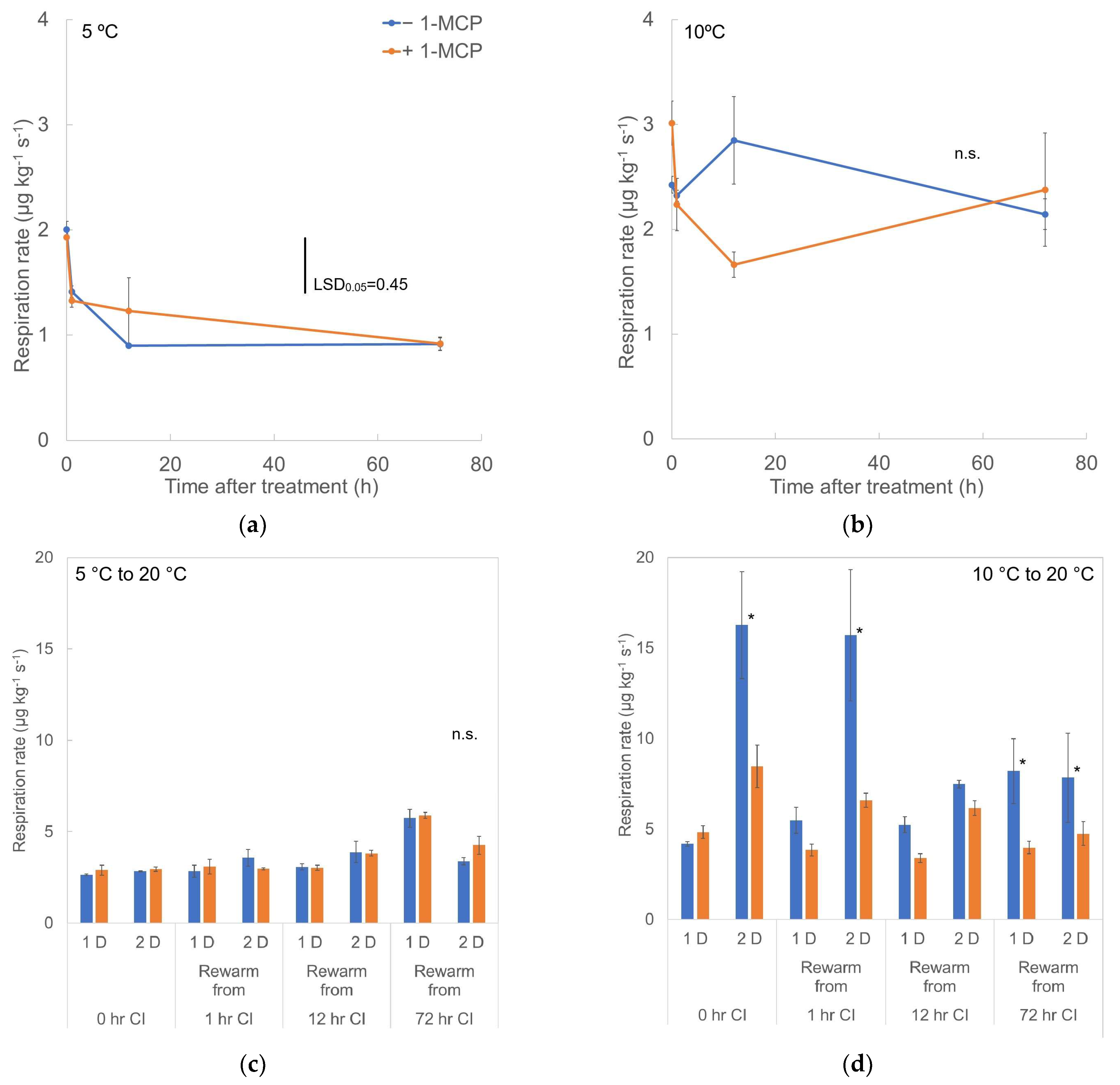
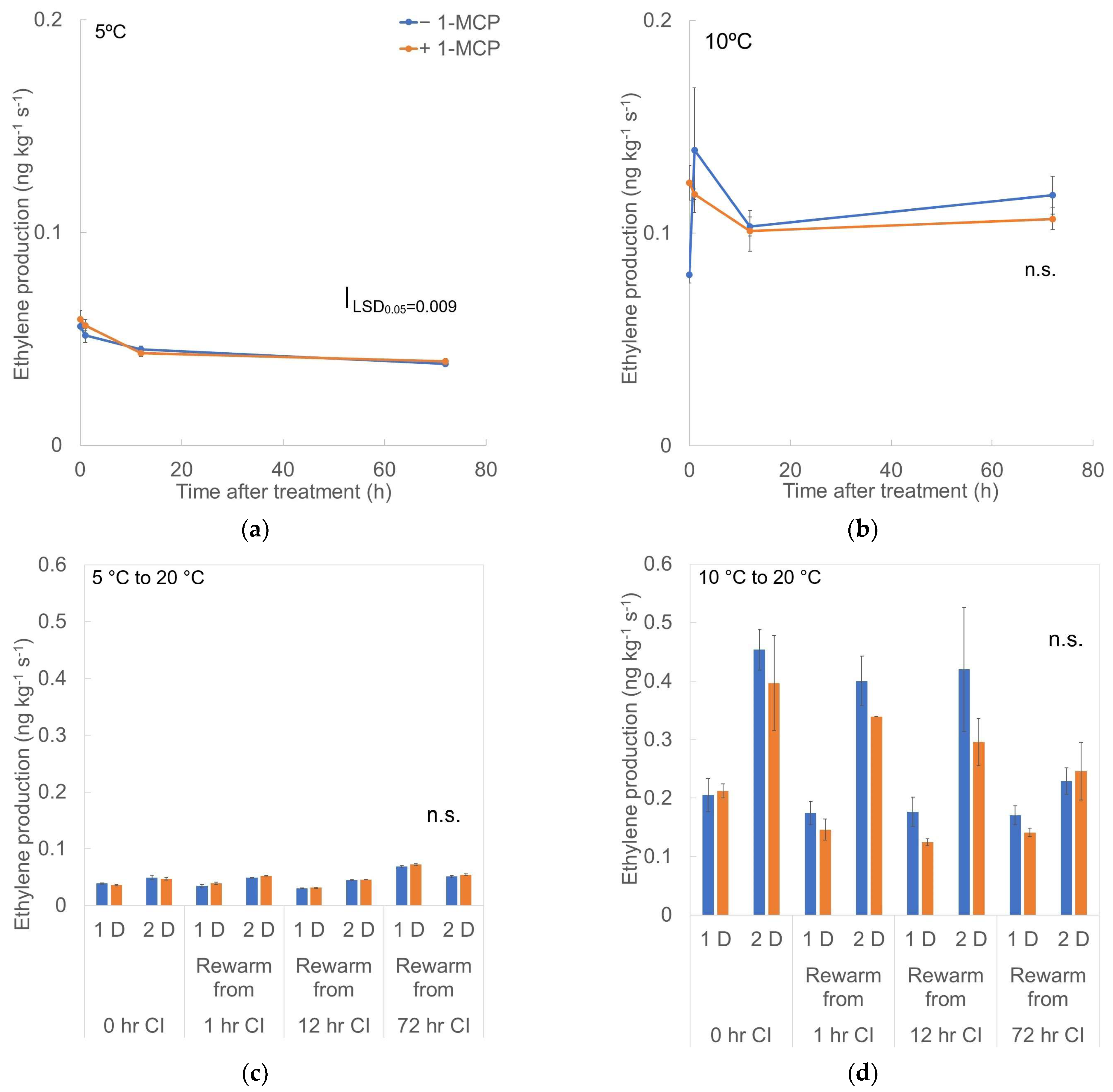
3.1. Respiration Rate and Ethylene Production

Respiration rate of fruit (+/−1-MCP) stored at 5 °C or 10 °C (Figure 2) showed decreasing rates, dropping from 2–3 µg kg−1 s−1 (when conditioned at 14 °C) to 1 µg kg−1 s−1 at 5 °C or 2 µg kg−1 s−1 at 10 °C. Fruit stored at 5 °C for 0, 1, or 12 h had a relatively low respiration rate of around 3 µg kg−1 s−1, while the respiration of fruit stored at 5 °C for 72 h had risen to 5 µg kg−1 s−1 after 1 d of rewarming to 20 °C and then dropped to 3.5–4 µg kg−1 s−1 on the second day at 20 °C (Figure 2). For fruit stored at 10 °C (Figure 2), 0 and 1 h CI storage resulted in similar patterns of fruit respiration at around 5 µg kg−1 s−1 after rewarming to 20 °C for 1 d, and the control (−1-MCP) fruit had a higher respiration rate (15 µg kg−1 s−1) on the next day. For 10 °C storage for 12 h, the respiration rate stayed around 5 µg kg−1 s−1 with control (−1-MCP) fruit having a slightly higher value than +1-MCP fruit; the +/−1-MCP treatments subsequently increased to 6−7 µg kg−1 s−1 as the storage duration at 10 °C continued. The fruit from 10 °C storage for 72 h had a respiration rate of 8 µg kg−1 s−1 in the control treatment, but 4 µg kg−1 s−1 in the +1-MCP treatment.
In terms of ethylene production, the fruit produced less ethylene after CI storage treatment (Figure 3). Fruit stored at 5 °C dropped from an initial ethylene production rate of 0.05 ng kg−1 s−1 to 0.04 ng kg−1 s−1, while the ethylene production rate of fruit at 10 °C declined to 0.1 ng kg−1 s−1. After rewarming, the ethylene production of fruit in 5 °C storage for 0, 1, or 12 h remained around 0.3 ng kg−1 s−1, while storage at 5 °C for 72 h resulted in a slightly increased rate of 0.4 ng kg−1 s−1, decreasing to 0.3 ng kg−1 s−1 on the next day. Fruit stored at 10 °C for 0, 1, or 12 h had similar ethylene production patterns that stayed around 0.2 ng kg−1 s−1 after the first day of rewarming to 20 °C then jumped to 0.4 ng kg−1 s−1 the next day. For 72 h 10 °C storage, the control (−1-MCP) fruit produced more ethylene (8 ng kg−1 s−1) than the +1-MCP treatment during the rewarming period.

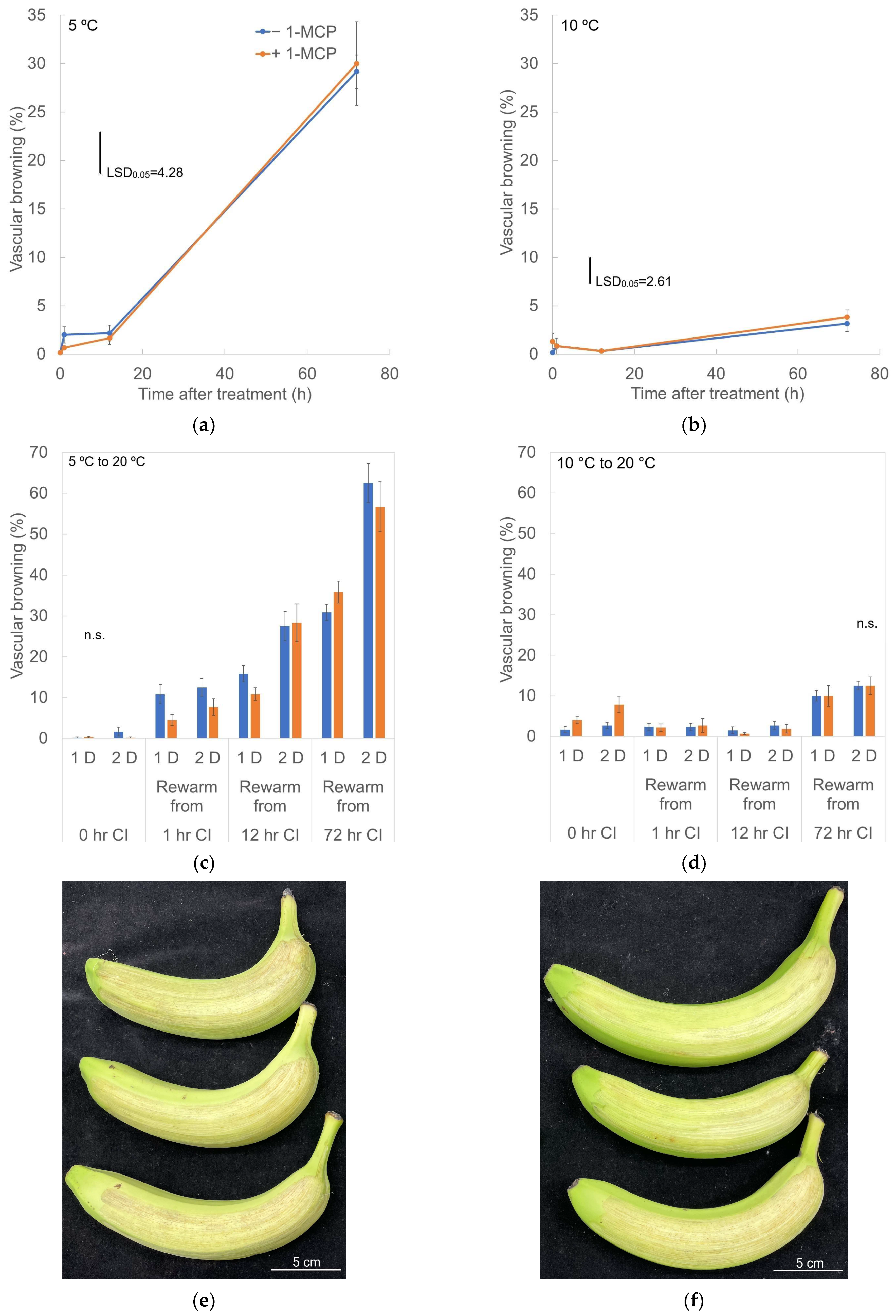
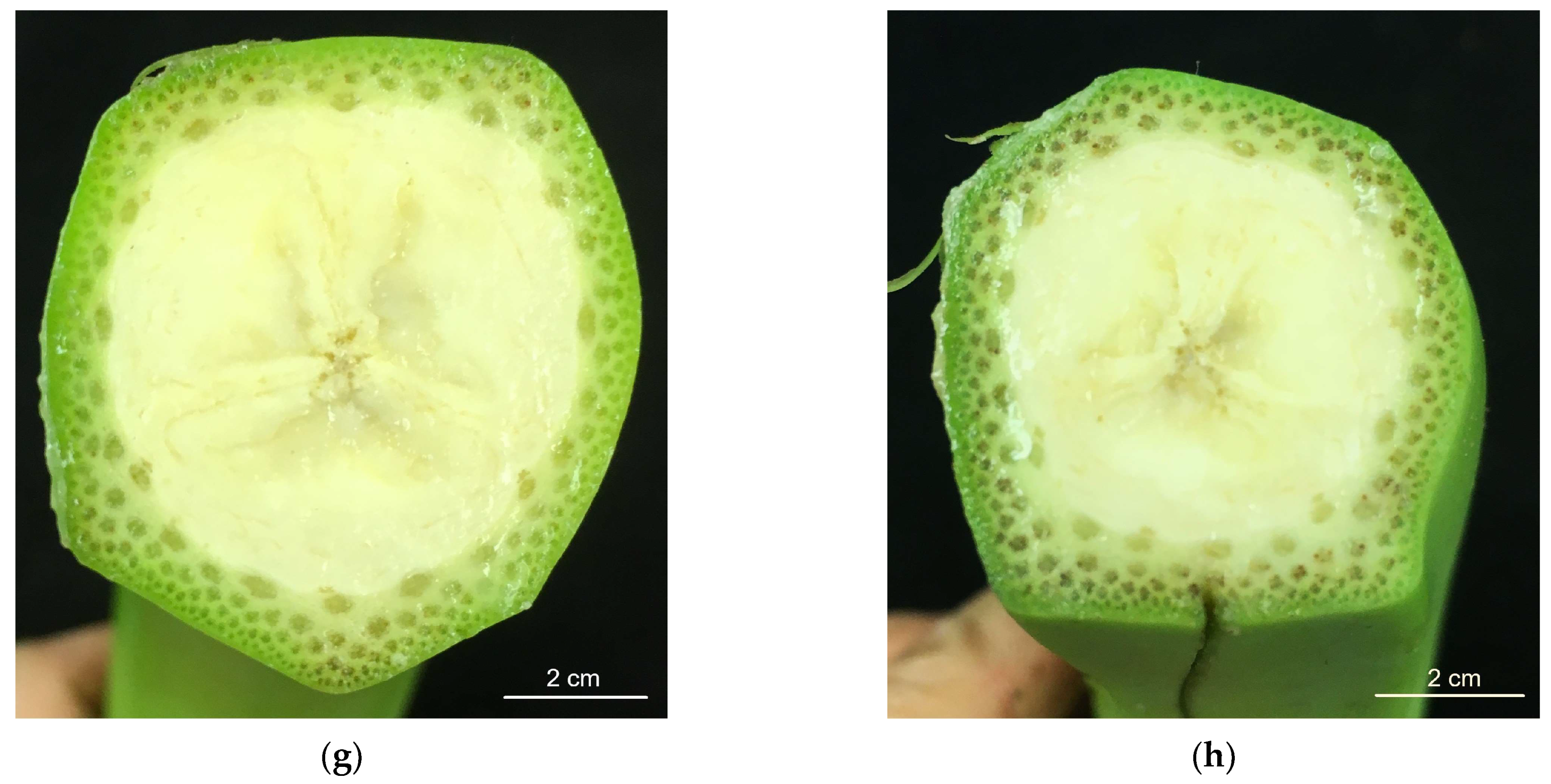
3.2. Vascular Browning (VB)
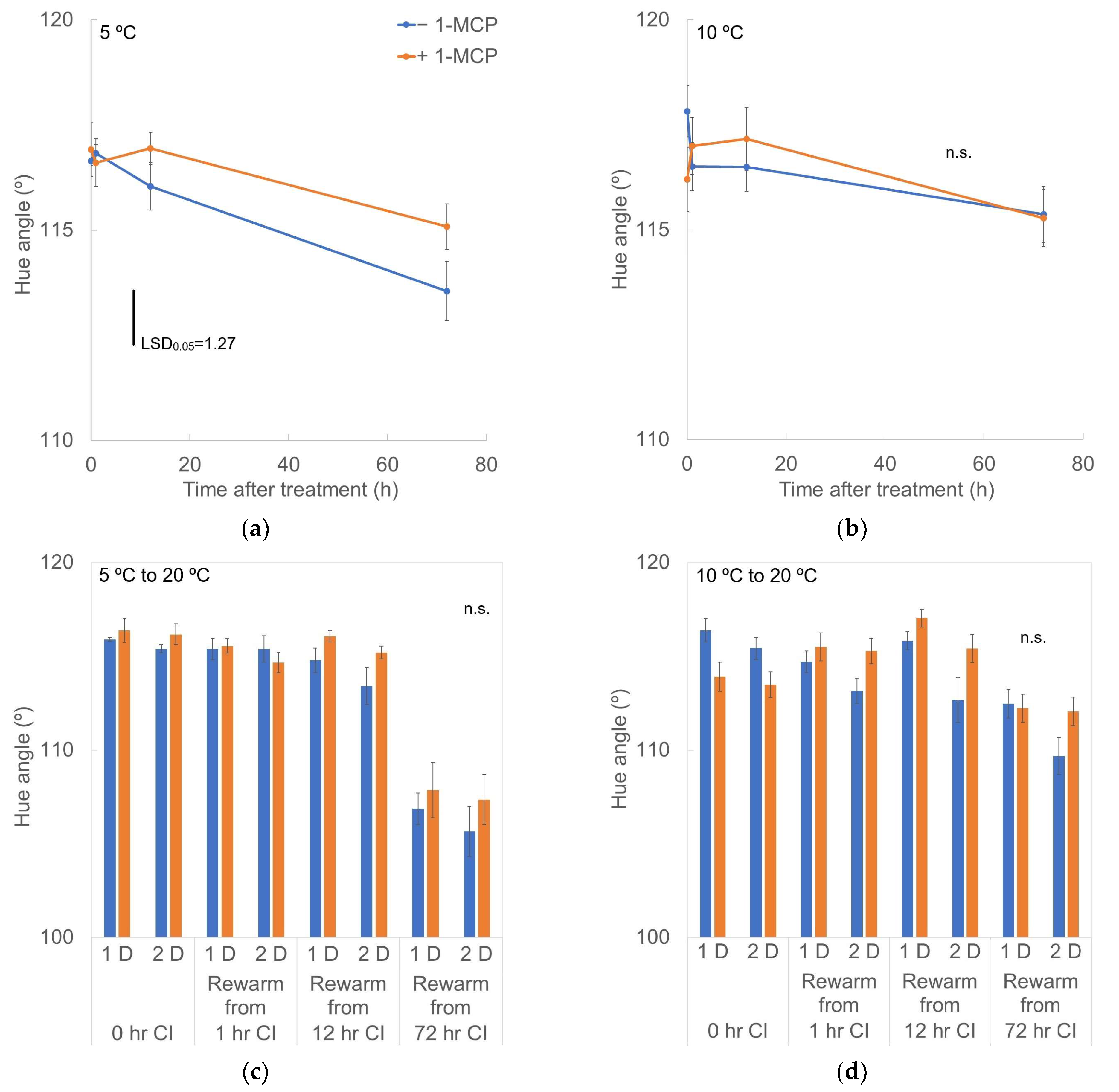
Fruit developed more severe VB at 5 °C than at 10 °C (Figure 4). Control fruit (−1-MCP) stored at 5 °C showed VB slightly earlier than in the +1-MCP treatment but reached 30% VB after 72 h of CI storage. Fruit at 10 °C (+/−1-MCP) showed mild VB during 72 h storage. However, more severe VB developed during the rewarming period in fruit stored more than 1 h at 5 °C or for 72 h at 10 °C. On the first day of rewarming, control fruit stored at 5 °C for 1 h or 12 h showed more severe VB than +1-MCP fruit. But for more severe CI stress conditions, the development of VB did not show a 1-MCP effect. For 10 °C storage, only the fruit stored for 72 h exhibited increased VB during rewarming, without a 1-MCP effect.

3.3. External Color

Lightness (L*) decreased during both 5 °C and 10 °C storage (Figure 5) with no significant difference. Rewarmed fruit from 5 °C storage showed an obvious decrease in L* in the 12 and 72 h storage treatments. The 1-MCP-treated fruit had a relatively lower L* level than the control (−1-MCP). Fruit rewarmed after 10 °C storage had a higher and constant L* level of around 58–62. For the 0 h storage at 10 °C, 1-MCP treatment had a slightly higher L* value. Treatments of 1, 12, or 72 h at 10 °C developed increased L* values in the control after rewarming.
Chroma (C*) decreased as CI storage proceeded as presented in Figure 6, from 40 to 35 at 5 °C and to 39 at 10 °C. After rewarming, fruit from 5 °C had lower C*, especially for the 72 h treatment at 10 °C. A 1-MCP effect was not obvious in the 0, 1, and 12 h at 5 °C storage treatments, and only the control fruit from the 72 h CI storage had a higher C* value. The C* of fruit rewarmed after 10 °C storage stayed around 40–42, except that the C* of fruit stored for 72 h at 10 °C dropped to 37–40. Control fruit (−1-MCP) had relatively higher C* values than +1-MCP fruit.
The hue angle (H*) of the 1-MCP-treated fruit slowly decreased during 5 °C storage (Figure 7), but the H* of the fruit without 1-MCP decreased faster than the 1-MCP-treated fruit—from 116° to 113°. The H* of fruit at 10 °C declined only slightly to 115° after 72 h with no 1-MCP effect (Figure 7). The H* declined during rewarming following prolonged storage at 5 °C (Figure 7), but 1-MCP-treated fruit retained higher H* than the control during the rewarming period.

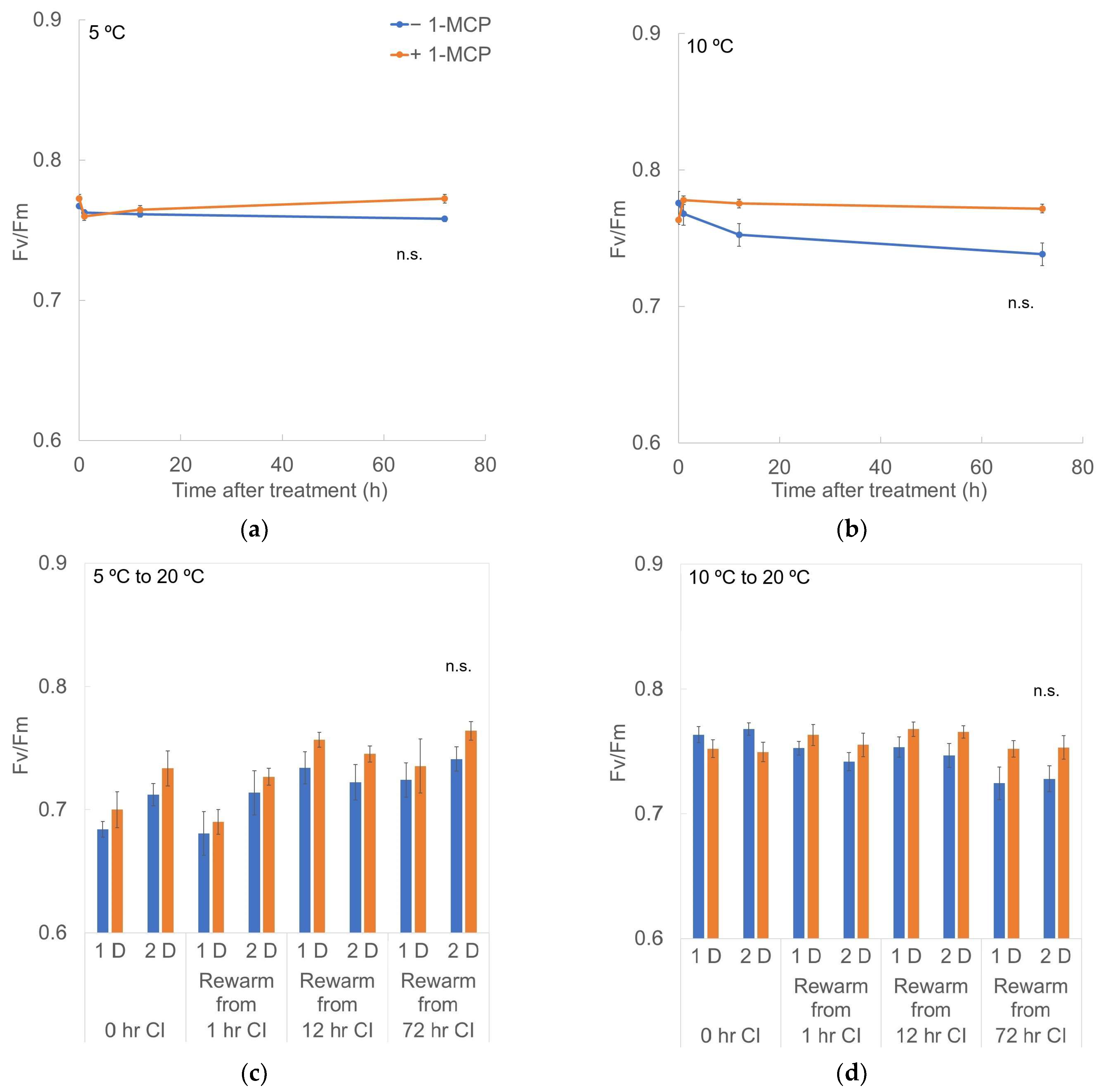
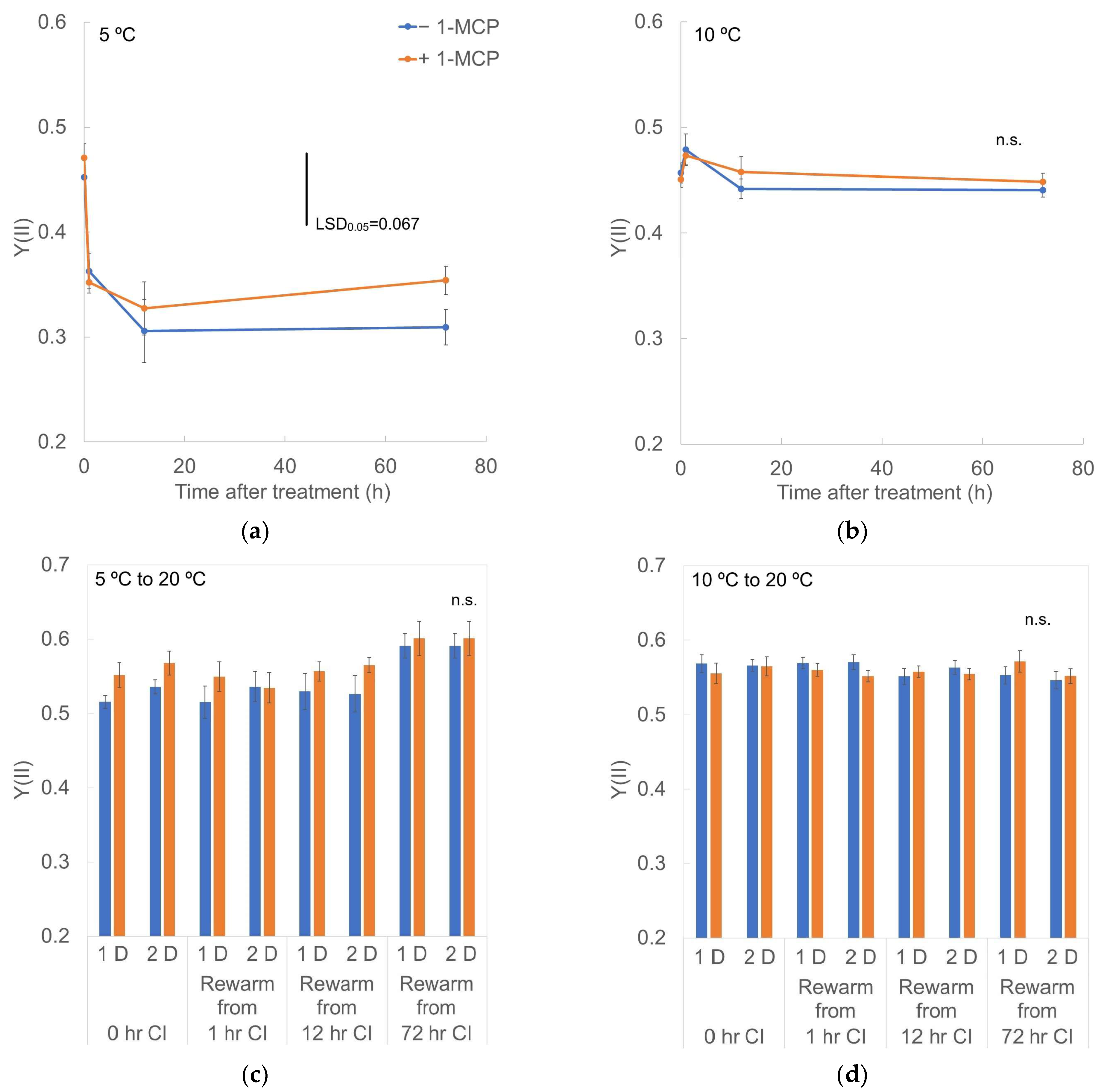
3.4. Chlorophyll Fluorescence
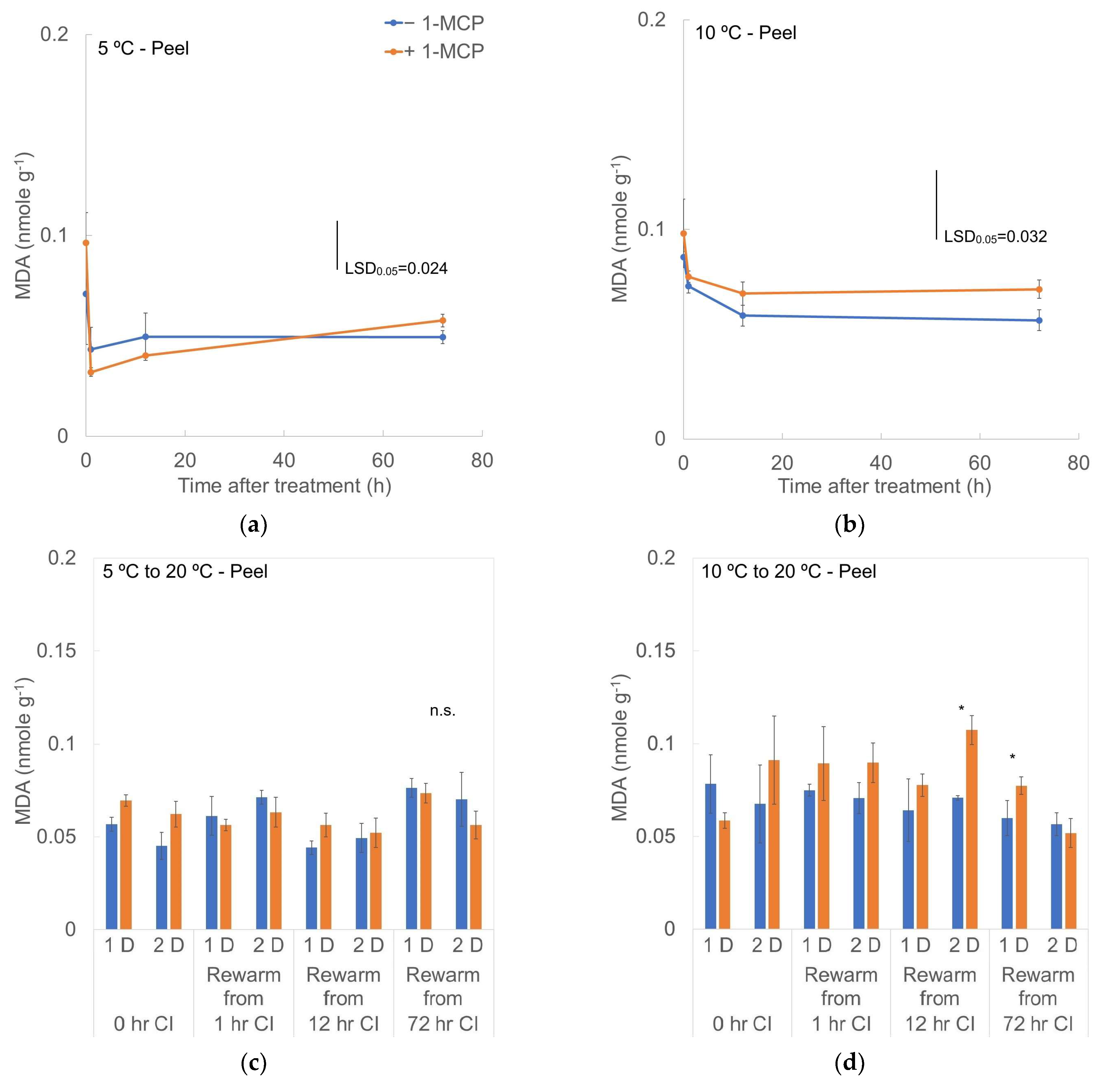
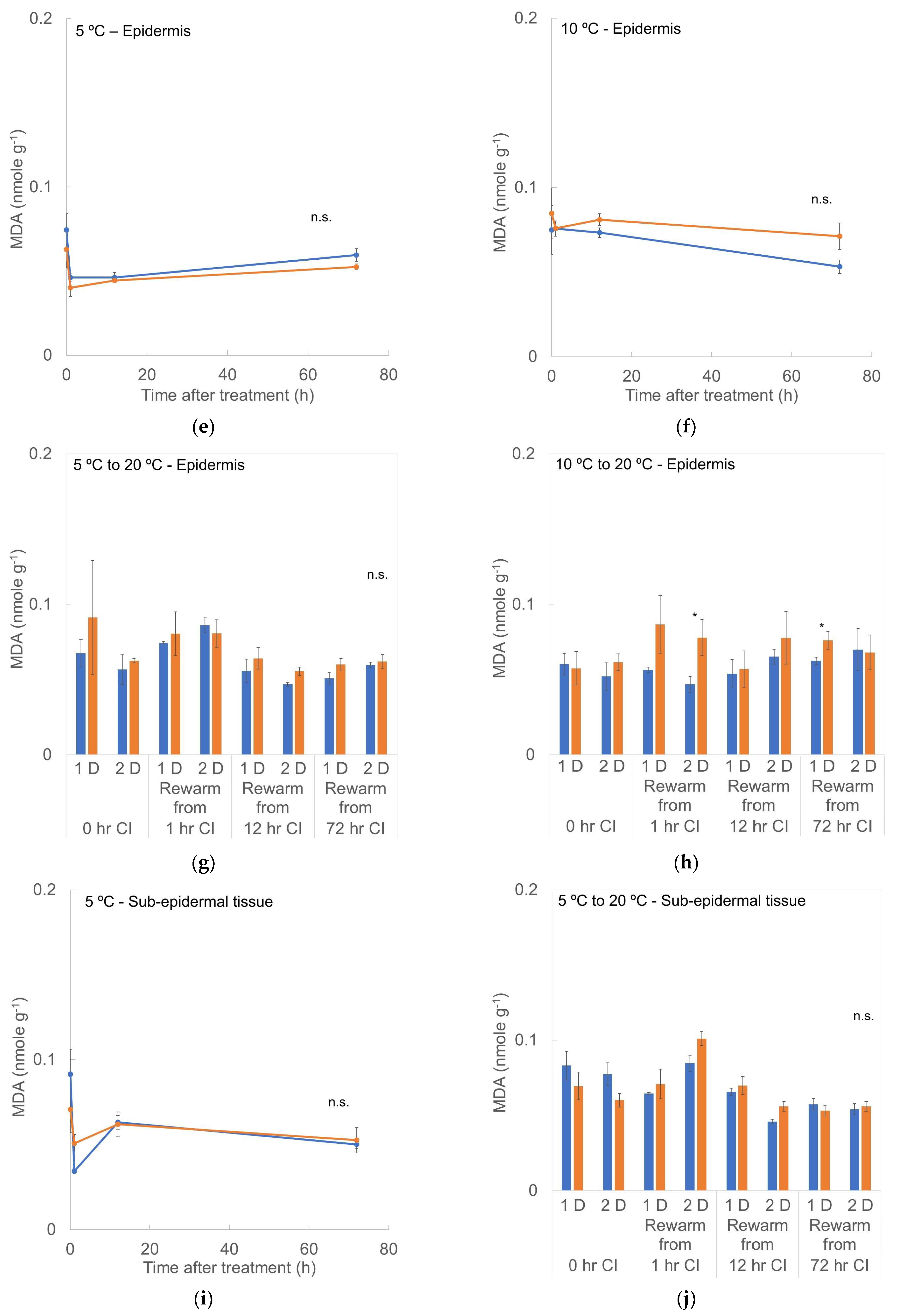
The Fv/Fm was higher for +1-MCP fruit at 5 or 10 °C without a significant temperature effect (Figure 8). For the fruit stored at 5 °C, the Fv/Fm values were higher after rewarming for 1-MCP-treated fruit (Figure 8). Fruit with 0 or 1 h CI storage at either temperature had similar patterns, with increased Fv/Fm on the second rewarming day. Treatments with 12 or 72 h storage durations had slightly higher Fv/Fm than the 0 and 1 h treatments (0.73−0.75). Unstored control (−1-MCP) fruit had a higher Fv/Fm value than unstored +1-MCP fruit. In the treatments involving storage at 10 °C, there were higher Fv/Fm values for the +1-MCP fruit than for the −1-MCP fruit. The Fv/Fm was lowest in fruit stored for 72 h at 10 °C compared with the other treatments (+/−1-MCP) involving storage for 1 or 12 h.
The Y(II) values of fruit stored at 5 °C decreased significantly during storage, from 0.45 to 0.3, compared with fruit from the 10 °C treatments, which remained at 0.45 (Figure 9). The 1-MCP-treated fruit had slightly higher Y(II) values than the control during the shorter storage times at both temperatures, with the Y(II) values of fruit stored for 0, 1, or 12 h remaining around 0.5 for the control and 0.55 for the 1-MCP treatments. Longer storage at 5 °C (for 72 h) +/− prior 1-MCP treatment resulted in the Y(II) rising to 0.6 during rewarming. For fruit stored at 10 °C for 72 h +/− prior 1-MCP treatment, the Y(II) barely changed (to 0.56) during rewarming to 20 °C.

3.5. MDA Content

MDA content in the whole peel at 5 or 10 °C is illustrated in Figure 10a,b. After transferring the fruit from 14 °C (the conditioning temperature) to 5 °C, MDA decreased from 0.07–0.09 nmole g−1 to 0.03–0.04 nmole g−1. As the storage duration at 5 °C was extended, MDA content rose back to 0.05 nmole g−1, but with no 1-MCP effect. For fruit stored at 10 °C, the patterns of the changes in MDA content were similar to those at 5 °C. The MDA decreased from 0.09 nmole g−1 to 0.055–0.07 nmole g−1 after 12 h at 10 °C and then remained constant up to 72 h. The MDA content of fruit transferred from 5 °C to 20 °C remained around 0.05 nmole g−1, except there was higher MDA content (0.07 nmole g−1) in fruit from the 72 h CI storage during the first d of rewarming; this slightly decreased to 0.06 nmole g−1 over the next day [Figure 10c]. With regard to the 10 °C storage treatments [Figure 10d], the fruit had slightly higher MDA contents compared with the 5 °C storage. Though the 1-MCP treatments followed by different chilling storage durations consistently had higher MDA levels than the corresponding −MCP controls, there was no significant difference.
The results for MDA content in epidermis tissue during chilling storage [Figure 10e,f] were similar to the effects on the whole peel. The epidermal MDA content decreased from 0.07–0.08 nmole g−1 to 0.04–0.05 nmole g−1 at 5 °C and to 0.06–0.07 nmole g−1 at 10 °C, but there was no 1-MCP effect. During rewarming [Figure 10g], the MDA level increased slightly to around 0.05–0.07 nmole g−1 for the 5 °C storage treatment. For the 10 °C treatments [Figure 10h], the MDA increased to around 0.05–0.1 nmole g−1 during rewarming. There was no 1-MCP effect observed in epidermis tissues.
The MDA content of sub-epidermal tissues during 5 °C storage [Figure 10i] dropped from 0.07–0.09 nmole g−1 to 0.05–0.06 nmole g−1. After 2 d at 20 °C, there were slightly higher MDA levels in the control fruit than in the 1-MCP treatment without prior cold storage, while for fruit stored for 1 or 12 h at 5 °C, the MDA content was higher for the 1-MCP treatments than the controls, but the same was not true for 72 h at 5 °C [Figure 10j].

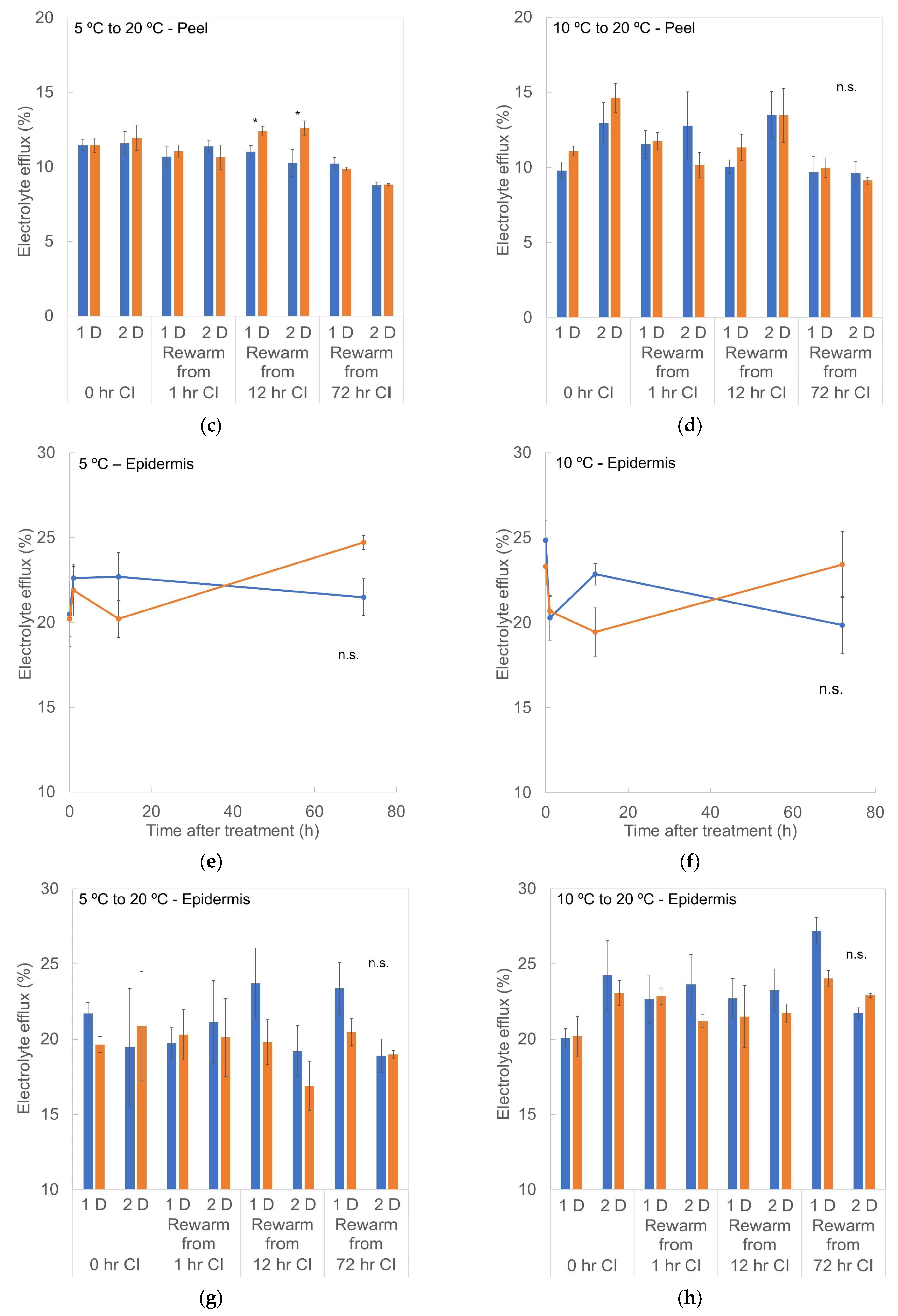
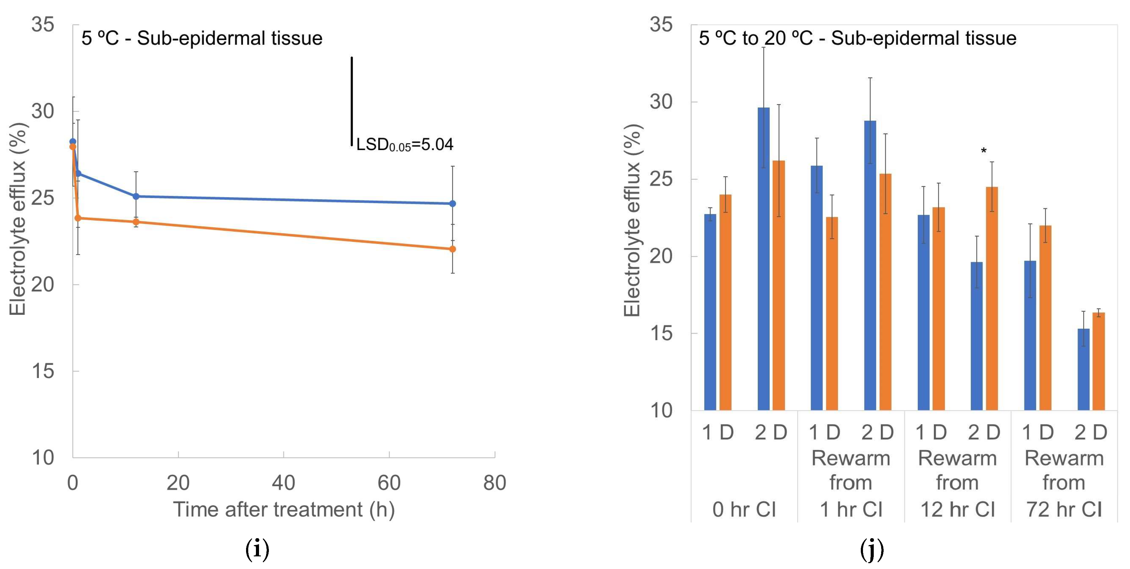
3.6. Electrolyte Efflux (EE)

Electrolyte efflux of the whole peel samples stored at 5 or 10 °C is shown in Figure 11a,b. As the duration of the low temperature storage increased, the EE also increased. The EE of peel from fruit at 5 °C increased to 13%, while for the fruit at 10 °C, it rose to 12%. After rewarming from 5 °C to 20 °C [Figure 11c], the EE decreased to 9–10%, and only the storage for 12 h had a higher value in the peel of 1-MCP-treated fruit. For fruit rewarmed from 10 °C [Figure 11d], storage for 0, 1, or 12 h resulted in higher EE after 2 d rewarming, but for the 72 h storage time, the whole peel EE remained at around 10% EE.
Epidermis tissues had higher EE than whole peel tissue [Figure 11e,f], but there were no significant treatment differences during the low temperature storage. After rewarming from 5 °C to 20 °C [Figure 11g], the EE stayed around 20% for fruit stored at that temperature for 0 or 1 h. Storage at 5 °C for 12 or 72 h resulted in higher EE values in the control fruit epidermis (23%, −1-MCP) than in the +1-MCP (20%) after 1 d rewarming. When rewarmed, the EE values of the epidermis tissue from the +/−1-MCP treatments both decreased by the second day at 20 °C. Epidermis tissue of rewarmed fruit previously stored at 10 °C for 0 or 1 h had 20–23% EE after rewarming for 1 d, and control fruit (−1-MCP) had a higher EE value than +1-MCP fruit the next day [Figure 11h]. Epidermis tissue from control fruit (−1-MCP) stored at 10 °C for 12 or 72 h had higher EE than +1-MCP fruit epidermis, but the EE of epidermal tissue from the 72 h at 10 °C storage treatments (+/−1-MCP) was much higher than for shorter 10 °C storage durations, but the EE decreased to the same level as the rest of the treatments by the second day after transfer to 20 °C.
Sub-epidermal tissues had higher EE than the whole peel tissue as well [Figure 11i], and the sub-epidermal tissues from −1-MCP fruit had higher EE values than that of the +1-MCP fruit throughout both low-temperature storages. After rewarming to 20 °C [Figure 11j], after 0 or 1 h at 5 °C, there was higher EE in the control (−1-MCP) than with +1-MCP, and the EE increased by the second day at 20 °C. Sub-epidermal tissue from the fruit from the 12 and 72 h storage durations at 5 °C had lower EE values in the control (−1-MCP) than with +1-MCP, and the EE in the control after 12 h or 72 h at 5 °C and the 1-MCP-treated fruit for 72 h CI storage at 5 °C had decreased by the second day at 20 °C.

4. Discussion

The severity of CI is intensified by the duration and temperature of cold storage, the type of plant organ, the variety, the physiological maturity, and the environmental factors during plant growth [3,7,51]. In this study, the origin, the exporter, and the retailer of our sample fruit were the same in repeated experiments. Pre-selection in our lab was conducted on each banana cluster by colorimeter readings once receiving fruit cartons to eliminate outliers and to confirm the initial maturity/ripeness stage and that the color parameters were within a narrow range. However, slight variations among each batch were shown in the CI responses we encountered in this research. With the same experimental procedure, the magnitude of VB that developed during up to 72 h of 5 °C or 10 °C storage and after rewarming to 20 °C and holding for 2 d was not at the same level, which suggests the CI sensitivity was not identical. Other than physiological maturity variation that we were unable to detect via appearance, preharvest or postharvest accumulated CI stress before the reception at the local retailer [3,52] or the seasonal temperature variation in the production area [51] could be potential causes of this variation in CI response. As with other tropical crops, mango, for example, CI symptoms developed from dark scald-like discoloration, pitting or sunken lesion in the peel, and even breakdown of the flesh or tissue [53,54]. However, the suppression of ethylene biosynthesis resulted in increased CI, which was also observed in “Kensington Pride” mango [55]. Though banana and mango both developed the browning in exocarp tissues during CI storage, their response to ethylene on CI symptoms development could be different.
Respiration rate is regarded as an indication of the overall metabolic activity of a living organism. Decreased respiration rate was associated with the actual temperature of fruit after transferring from 14 °C to 5 or 10 °C. Similar responses were observed in ethylene production, Fv/Fm, Y(II), and MDA accumulation, indicating that the enzyme activities involved in those parameters are temperature sensitive. After transferring banana fruit from 5 °C or 10 °C storage to 20 °C, those parameters increased, but the extent of recovery was determined by the prior level of CI stress experienced. With regard to respiration rate, rewarmed fruit from 0 or 1 h at 10 °C had a similar response, reflecting the normal function. The rates of CO2 production of control fruit (−1-MCP) from the 12 and 72 h at 10 °C storage treatments were slightly affected. Treatment for 72 h at 5 °C resulted in a CO2 production burst during the first day of rewarming to 20 °C and then slightly lower CO2 production during the next day, which was also as previously reported [6].
In terms of ethylene production, the recovery of fruit stored at 10 °C for 0, 1, or 12 h was back to normal, but it was reduced for fruit stored for 72 h at 10 °C. Upon transfer from 5 °C to 20 °C, the ethylene response was similar to that of respiration rate, with a burst of ethylene production observed for fruit that had been stored for 72 h at 5 °C. The recovery from storage at 10 °C for 12 h appeared to be complete, as the ethylene production and respiration rate were only slightly affected, especially for the control fruit, indicating that the CI effect was minimal under that mild CI stress (10 °C for 12 h).
Vascular browning is often regarded as the integrative, irreversible result of the membrane disruption that occurs during chilling storage of banana fruit, resulting in the release of polyphenol oxidase (PPO), the oxidation of most dopamine, and the polymerization of melanins [37,40,52,56,57]. It has been demonstrated that laticifer cells closely associated with the phloem in banana peel are the location of early VB development [52]. Chilled bananas showed a collapse of phloem tissue [58]. In our study, we observed a few brown spots randomly distributed in cross sections of the sub-epidermal layers of the peel at the beginning of chilling. The spots subsequently developed more, joined with other spots on the same vascular bundles, and became streaks as the CI stress accumulated. Incidence of VB below 5% of the peel cross-sectional area was observed in conjunction with the milder CI stress treatments (10 °C for 1–72 h; 5 °C for 1–12 h), appearing as random brown spots. Fruit stored at 5 °C for 72 h showed multiple darker brown streaks randomly distributed in the sub-epidermal area. After rewarming, the VB continued to develop in parallel with the disruption level resulting from various levels of CI stress that had been applied.
Color changes related to graying of the external peel surface during the CI storage and after rewarming were in accordance with the VB development. Graying was first detectable when the VB became apparent (at circa 5−10% VB incidence visible in peel cross sections). As suggested by [59], colorimeter measurements are potentially sensitive in detecting banana CI, reporting that L* and chroma values showed a high correlation to peel damage. The results of our study also support the significant differences in chroma and hue angle that develop during CI storage, along with the differences in L*, chroma, and hue angle that developed after removal from the chilling temperature storage, especially under the more severe CI stress levels. The dull appearance of chilled banana fruit appears to be related to loss of lightness (L*) and color purity (chroma). As VB developed in the sub-epidermal layer of the MG bananas, the external color became a mixture of green and brown (resulting in the gray appearance), and the color purity decreased.
It has been reported for “Dwarf Cavendish” bananas that exposure to 8.8 °C for 7 d, 5.5 °C for 48−60 h, or 2.7 °C for 36 h resulted in equivalent CI damage by visual evaluation [60]. In our observation, VB in the early stages of CI development was reduced by prior 1-MCP treatment in that fewer brown streaks (3% VB) were found on +1-MCP-treated fruit stored at 13 °C for 10 d than in control fruit (−1-MCP; 10% VB). The equivalent CI severity in the present study would be the treatment held at 5 °C for 1 h, for which VB also developed faster in the control fruit (−1-MCP), with the effect persisting even after rewarming.
Development of VB in banana fruit during chilling storage has been reported to be a consequence of upstream control by PAL and PPO [40]. The potential involvement of ethylene with tissue browning in various crops [21,56,61,62] is something that needs further evaluation with regard to CI symptom development. Moreover, in our repeated trials using the same procedures, equivalent CI symptom development in control (−1-MCP) fruit was observed for treatments ranging from 5 °C for 1−12 h to 10 °C for 72 h, which indicates that the chilling sensitivity varied from batch to batch. In the present report, the VB that developed in response to the 5 °C for 12 h chilling exposure and the 10 °C for 72 h chilling exposure showed similarly affected VB areas before rewarming, but the actual CI level was apparently greater for the fruit transferred from 5 °C for 12 h, as evidenced by the CI symptom severity of the rewarmed fruit.
Chlorophyll fluorescence measured in light-adapted [yield (II), Y(II)] and dark-adapted (Fv/Fm) tissues containing chloroplasts indicates the operating efficiency of photosystem II (PSII) under real-time environmental conditions and the photochemical efficiency of PSII, respectively. Photosynthetic organs are chilling sensitive [11], as shown by the alteration in performance and activity after transfer to low temperature and the reported effects on photosynthetic pigment content [63], PSII photochemical efficiency [64], and even disorganization of chloroplasts [10]. Y(II) is a sensitive indicator of the early stages of CI [65]. We observed decreased Fv/Fm and more drastic decline of Y(II) after transferring bananas from the non-chilling to chilling temperature, and the subsequent recovery from the chilling exposures was to the original non-chilled level, indicating that exposure to 5 or 10 °C for up to 72 h was not injurious to the chloroplasts. With 1-MCP treatments, there were consistently higher Fv/Fm and Y(II) values than the controls during chilling storage and after rewarming. Moreover, for the most severe CI stress (5 °C for 72 h), the Fv/Fm and Y(II) increased only slightly in the +1-MCP fruit compared to the −1-MCP control. This indicates the possibility of better photosynthetic efficiency, especially on the electron chain transport of PSII with reduced or absent ethylene action.
The integrity of the cell membranes enables the various biochemical and physiological functions to operate in an orderly manner in the different compartments. Membrane alterations induced by chilling temperatures are believed to be affected by the lipid composition of membranes [2] or the deleterious action of reactive oxygen species (ROS) [66]. Increased EE and peroxidation of unsaturated fatty acids under CI stress led to the destabilization of the cell membrane [35]. Increased ROS (e.g., O2) levels [63] and abnormal energy and other metabolism [67] at chilling temperatures have been observed in chilled crops.
Evaluations of membrane permeability and peroxidation levels as determined by measurements of EE and MDA content are often used as biomarkers in CI investigations [66,68]. It was further indicated that the lipid peroxidation that occurs before irreversible CI damage is reached could be a potential CI indicator like increased ethylene evolution and visual CI symptoms [69]. However, MDA was found here to decrease in the whole peel, epidermal tissues, and sub-epidermal tissues at the beginning (within the first h) of chilling exposure as the temperature transitioned from 14 °C to 5 °C or 10 °C. There was then a slight increase in MDA as 5 °C storage progressed, which corresponded to the VB development over longer CI durations. After rewarming, the MDA content of whole peel and sub-epidermal tissues from fruit held at 5 °C for 1, 12, or 72 h increased. Still, the MDA decreased during rewarming, indicating the reversal of CI-related membrane damage upon transfer to the non-chilling temperature. There were no significant differences in MDA levels among the 10 °C treatments, including no 1-MCP effect on MDA accumulation.
With respect to EE, the whole peel showed elevated EE during chilling storage at 5 °C or 10 °C, suggesting increased membrane permeability in response to CI. However, the EE value after rewarming recovered to at least the original (non-chilled) level. More interestingly, a much lower EE value was observed after rewarming for the most severe CI treatments of 5 °C or 10 °C for 72 h, suggesting more active membrane repair in severe CI tissues during rewarming. A 1-MCP effect on EE was observed in the whole peel and sub-epidermal tissues in that the +1-MCP EE value was consistently lower than the control (−1-MCP) during chilling storage. After rewarming, the EE changes in the sub-epidermal tissues of non-chilled or milder CI treatments were slightly higher, but two of the more severe CI exposures (5 °C for 12 or 72 h) had higher EE after rewarming in the +1-MCP fruit instead. Along with the previously noted observation of chlorophyll fluorescence recovery, this suggests a beneficial effect of 1-MCP on the maintenance of membrane systems during chilling exposure or adaptation to low-temperature conditions. In other words, it appears that 1-MCP (i.e., inhibition of ethylene action) overcame the negative effects of chilling-induced stress ethylene on the banana membrane systems. Moreover, CI symptoms development in relation to the changes of membrane permeability and ethylene production shared some similarities to the process of ripening and even senescence. The response to 1-MCP treatment in our study suggested alleviation of CI stress on membranes. 6-Benzylaminopurine, which modulated the delay of senescence and ripening processes in mango fruit, also showed inhibition of ethylene production and repression of lipid catabolism [70]. This may suggest that CI triggers “one specific type” of the senescence process.
Irreversible CI-related changes during 72 h of storage at 5 or 10 °C included VB and the related drastic deterioration of lightness, chroma, and hue angle values with longer chilling duration (5 or 10 °C for 72 h). Other physiological processes such as respiration, ethylene production, Fv/Fm, Y(II), EE, and MDA production were potentially reversible and were likely related to the low temperature-induced slowing of fruit metabolism. Respiration rate, ethylene production, and Y(II) responded quickly to temperature changes. But the recovery of those processes upon return to a non-chilling temperature depended on the chilling sensitivity of each parameter. Respiration rate and ethylene production recovered in most treatments of this study, but the CI responses were shown by their hyper-recovery reaction (over-recovery) on the first rewarming day for the most severe treatment (5 °C for 72 h). The Y(II) and Fv/Fm returned to their initial, non-chilled levels on the first rewarming day. It may be suggested that the chloroplasts show less CI susceptibility compared with other CI symptoms for the levels of CI stress applied in this research.

5. Conclusions

In this study, we evaluated the CI symptom development of MG banana fruit and its apparent association with the stress ethylene that is produced in response to different amounts of chilling stress. Visible CI symptoms [i.e., VB, along with loss of lightness (L*) and color purity (chroma)] were irreversible. Other potential CI responses such as elevated respiration and ethylene production, Fv/Fm, Y(II), EE, and MDA were mostly recoverable (reversible) except after the most severe CI treatment of 5 °C for 72 h. Our results confirm that CI responses are reversible until some CI stress threshold is reached that differs for each CI symptom. Use of 1-MCP showed that without the involvement of stress ethylene, the banana fruit showed better CI tolerance in terms of VB, peel graying, Fv/Fm and Y(II) declines, and EE increase in the whole peel and peel sub-epidermal layers. The hypothesized potential role of ethylene in membrane alteration under CI stress was confirmed. Moreover, the genetic mechanism of CI with regard to the ethylene effect as shown via 1-MCP would need further advancement to identify molecular markers for future banana breeding efforts to speed up CI-resistance trait selection.

Author Contributions

Conceptualization, J.K.B. and L.-Y.C.; methodology, J.K.B. and L.-Y.C.; software, L.-Y.C.; validation, J.K.B. and L.-Y.C.; formal analysis, L.-Y.C.; investigation, L.-Y.C.; resources, J.K.B. and L.-Y.C.; data curation, L.-Y.C.; writing—original draft preparation, L.-Y.C.; writing—review and editing, J.K.B.; visualization, L.-Y.C.; supervision, J.K.B.; project administration, J.K.B.; funding acquisition, J.K.B. All authors have read and agreed to the published version of the manuscript.

Funding

This research was funded by It’s Fresh Ltd., Burntwood, UK, Award AGR DTD 9-12-2017.

Data Availability Statement

The raw data supporting the conclusions of this article will be made available by the authors on request.

Acknowledgments

We thank Donald J. Huber for helpful discussions during the conceptualization and planning of this research and assistance with the aqueous 1-MCP application technique.

Conflicts of Interest

The authors declare no conflicts of interest.

References

  1. Kader, A.A. Postharvest Biology and Technology: An Overview. In Postharvest Technology of Horticultural Crops, 3rd ed.; Kader, A.A., Ed.; University of California Agriculture & Natural Resources: Oakland, CA, USA, 2002; pp. 39–47. [Google Scholar]
  2. Lyons, J.M. Chilling injury in plants. Annu. Rev. Plant Physiol. 1973, 24, 445–466. [Google Scholar] [CrossRef]
  3. Saltveit, M.E., Jr.; Morris, L.L. Overview on Chilling Injury of Horticultural Crops. In Chilling Injury of Horticultural Crops; Wang, C.Y., Ed.; CRC Press: Boca Raton, FL, USA, 1990; pp. 3–16. [Google Scholar]
  4. Eaks, I.L. Effect of chilling on respiration and volatiles of California lemon fruit. J. Am. Soc. Hortic. Sci. 1980, 105, 865–869. [Google Scholar] [CrossRef]
  5. Eaks, I.L. Effects of chilling on respiration and ethylene production of ‘Hass’ avocado fruit at 20 °C. HortScience 1983, 18, 235–237. [Google Scholar] [CrossRef]
  6. Trejo-Márquez, M.A.; Vendrell, M. Physiological response to chilling temperature of banana fruit (Musa acuminata cv. Dwarf cavendish). Acta Hortic. 2010, 864, 387–393. [Google Scholar] [CrossRef]
  7. Sevillano, L.; Sanchez-Ballesta, M.T.; Romojaro, F.; Flores, F.B. Physiological, hormonal and molecular mechanisms regulating chilling injury in horticultural species. Postharvest technologies applied to reduce its impact. J. Sci. Food Agric. 2009, 89, 555–573. [Google Scholar] [CrossRef]
  8. Aroca, R.; Irigoyen, J.J.; Sanchez-Diaz, M. Photosynthetic characteristics and protective mechanisms against oxidative stress during chilling and subsequent recovery in two maize varieties differing in chilling sensitivity. Plant Sci. 2001, 161, 719–726. [Google Scholar] [CrossRef]
  9. Cabrera, R.M.; Saltveit, M.E.; Owens, K. Sensitivity of cucumber lines to chilling injury. HortScience 1990, 24, 1174. [Google Scholar] [CrossRef]
  10. Kratsch, H.A.; Wise, R.R. The ultrastructure of chilling stress. Plant Cell Environ. 2000, 23, 337–350. [Google Scholar] [CrossRef]
  11. Smillie, R.M.; Nott, R.; Hetherington, S.E.; Öquist, G. Chilling injury and recovery in detached and attached leaves measured by chlorophyll fluorescence. Physiol. Plant. 1987, 69, 419–428. [Google Scholar] [CrossRef]
  12. Prasad, T.K.; Anderson, M.D.; Stewart, C.R. Acclimation, hydrogen peroxide, and abscisic acid protect mitochondria against irreversible chilling injury in maize seedlings. Plant Physiol. 1994, 105, 619–627. [Google Scholar] [CrossRef]
  13. Lurie, S.; Laamin, M.; Zand, L.; Fallik, E. Heat treatments to decrease chilling injury in tomato fruit. Effects on lipids, pericarp lesions and fungal growth. Physiol. Plant. 1997, 100, 297–302. [Google Scholar] [CrossRef]
  14. Andersen, C.R.; Kent, M.W. Storage of cucumber fruit at chilling temperatures. HortScience 1982, 17, 527. [Google Scholar]
  15. Anderson, R.E. The influence of storage temperatures and warming during storage on peach and nectarine fruit quality. J. Am. Soc. Hortic. Sci. 1979, 104, 459–461. [Google Scholar] [CrossRef]
  16. Crisosto, C.H.; Mitchell, F.G.; Ju, Z. Susceptibility to chilling injury of peach, nectarine, and plum cultivars grown in California. HortScience 1999, 34, 1116–1118. [Google Scholar] [CrossRef]
  17. Groen, S.C.; Whiteman, N.K. The Evolution of Ethylene Signaling in Plant Chemical Ecology. J. Chem. Ecol. 2014, 40, 700–716. [Google Scholar] [CrossRef]
  18. Hyodo, H. Stress/Wound Ethylene. In The Plant Hormone Ethylene; Mattoo, A.K., Ed.; CRC Press: Boca Raton, FL, USA, 1991; pp. 43–63. [Google Scholar]
  19. Grichko, V.P.; Glick, B.R. Ethylene and flooding stress in plants. Plant Physiol. Biochem. 2001, 39, 1–9. [Google Scholar] [CrossRef]
  20. Megías, Z.; Martínez, C.; Manzano, S.; García, A.; del Mar Rebolloso-Fuentes, M.; Valenzuela, J.L.; Garrido, D.; Jamilena, M. Ethylene biosynthesis and signaling elements involved in chilling injury and other postharvest quality traits in the non-climacteric fruit of zucchini (Cucurbita pepo). Postharvest Biol. Technol. 2016, 113, 48–57. [Google Scholar] [CrossRef]
  21. Pesis, E.; Ackerman, M.; Ben-Arie, R.; Feygenberg, O.; Feng, X.; Apelbaum, A.; Goren, R.; Prusky, D. Ethylene involvement in chilling injury symptoms of avocado during cold storage. Postharvest Biol. Technol. 2002, 24, 171–181. [Google Scholar] [CrossRef]
  22. Ben-Amor, M.; Flores, B.; Latche, A.; Bouzayen, M.; Pech, J.C.; Fomojaro, F. Inhibition of ethylene biosynthesis by antisense ACC oxidase RNA prevents chilling injury in Charentais cantaloupe melons. Plant Cell Environ. 2002, 22, 1579–1586. [Google Scholar] [CrossRef]
  23. Palou, L.; Crisosto, C.H.; Garner, D.; Basinal, L.M. Effect of continuous exposure to exogenous ethylene during cold storage on postharvest decay development and quality attributes of stone fruits and table grapes. Postharvest Biol. Technol. 2003, 27, 243–254. [Google Scholar] [CrossRef]
  24. Wang, C.Y. Approaches to reduce chilling injury of fruits and vegetables. Hortic. Rev. 1993, 15, 63–95. [Google Scholar]
  25. Li, T.; Yun, Z.; Zhang, D.; Yang, C.; Zhu, H.; Jiang, Y.; Duan, X. Proteomics analysis of differentially expressed proteins involved in ethylene-induced chilling tolerance in harvested banana fruit. Front. Plant Sci. 2015, 6, 845. [Google Scholar] [CrossRef] [PubMed]
  26. Yin, Q.; Qin, W.; Zhou, Z.; Wu, A.M.; Deng, W.; Li, Z.; Shan, W.; Chen, J.Y.; Kuang, J.F.; Lu, W.J. Banana MaNAC1 activates secondary cell wall cellulose biosynthesis to enhance chilling resistance in fruit. Plant Biotechnol. J. 2024, 22, 413–426. [Google Scholar] [CrossRef]
  27. Zhou, Z.; Yang, Y.; Shan, W.; Zhang, H.; Wei, W.; Kuang, J.; Chen, J.; Lu, W. Ethylene attenuates chilling injury of banana fruit via the MabHLH060/183 module in controlling phosphatidic acid formation genes. Postharvest Biol. Technol. 2022, 183, 111724. [Google Scholar] [CrossRef]
  28. Marriott, J.; Palmer, J.K. Bananas—Physiology and biochemistry of storage and ripening for optimum quality. Crit. Rev. Food Sci. Nutr. 1980, 13, 41–88. [Google Scholar] [CrossRef]
  29. De Vasconcelos Facundo, H.V.; dos Santos Garruti, D.; dos Sants Dias, C.T.; Cordenunsi, B.R.; Lajolo, F.M. Influence of different banana cultivars on volatile compounds during ripening in cold storage. Food Res. Int. 2012, 49, 626–633. [Google Scholar] [CrossRef]
  30. De Vasconcelos Facundo, H.V.; Gurak, P.D.; Mercadante, A.Z.; Lajolo, F.M.; Cordenunsi, B.R. Storage at low temperature differentially affects the colour and carotenoid composition of two cultivars of banana. Food Chem. 2015, 170, 102–109. [Google Scholar] [CrossRef]
  31. Grierson, E.B.; Grierson, W.; Soule, J. Chilling injury in tropical fruit. I. Bananas (Musa paradisiaca var. sapientum cv. Lacatan). Proc. Trop. Reg. Am. Soc. Hortic. Sci. 1967, 11, 82–94. [Google Scholar]
  32. Jiang, Y.; Joyce, D.C.; Jiang, W.; Lu, W. Effects of chilling temperatures on ethylene binding by banana fruit. Plant Growth Regul. 2004, 43, 109–115. [Google Scholar] [CrossRef]
  33. Kader, A.A.; Sommer, N.F.; Arpaia, M.L. Postharvest Handling Systems: Tropical Fruits. In Postharvest Technology of Horticultural Crops; Kader, A.A., Ed.; University of California, Agriculture and Natural Resources: Davis, CA, USA, 2002; pp. 385–398. [Google Scholar]
  34. Mootoo, A.; Murr, D.P. Quality attributes of banana fruit as affected by chilling and non-chilling temperatures. In Proceedings of the 21st Annual Meeting of Caribbean Food Crops Society, Port-of-Spain, Trinidad and Tobago, 8–13 September 1985. [Google Scholar]
  35. Ratule, A.; Osman, M.T.; Ahmad, S.H.; Saari, N. Development of chilling injury of ‘Berangan’ banana (Musa cv. ‘Berangan’ (AAA)) during storage at low temperature. J. Food Agric Environ. 2006, 4, 128–134. [Google Scholar]
  36. Von Loesecke, H.W. Bananas, 2nd ed.; Interscience: New York, NY, USA, 1950; pp. 52–66. [Google Scholar]
  37. Chang, W.H.; Hwang, Y.J. Effect of ethylene treatment on the ripening, polyphenoloxydase activity and water soluble tannin content of Taiwan northern banana at different maturity stages and the stability of banana polyphenoloxydase. Acta Hortic. 1990, 275, 603–610. [Google Scholar] [CrossRef]
  38. Chang, L.Y.; Sargent, S.A.; Kim, J.; Brecht, J.K. Delaying ripening using 1-MCP reveals chilling injury symptom development at the putative chilling threshold temperature for mature green banana. Front. Plant Sci. 2022, 13, 966789. [Google Scholar] [CrossRef] [PubMed]
  39. Chen, H.; Bullock, D.A., Jr.; Alonso, J.M.; Stepanova, A.N. To Fight or to Grow: The Balancing Role of Ethylene in Plant Abiotic Stress Responses. Plants 2022, 11, 33. [Google Scholar] [CrossRef]
  40. Nguyen, T.B.T.; Ketsa, S.; van Doorn, W.G. Relationship between browning and the activities of polyphenol oxidase and phenylalanine ammonia lyase in banana peel during low temperature storage. Postharvest Biol. Technol. 2003, 30, 187–193. [Google Scholar] [CrossRef]
  41. Jeong, J.; Brecht, J.; Huber, D.; Sargent, S. Influence of 1-methylcyclopropene (1-MCP) on the shelf life and deterioration of fresh-cut cantaloupe. HortScience 2004, 39, 816. [Google Scholar] [CrossRef]
  42. Natalini, A.; Martinez-Diaz, V.; Ferrante, A.; Pardossi, A. Ethylene sensitivity regulates the wounding response in wild type and never ripe tomatoes. J. Hortic. Sci. Biotechnol. 2017, 92, 591–597. [Google Scholar] [CrossRef]
  43. Thompson, J.E.; Mayak, S.; Shinitzky, M.; Halevy, A.H. Acceleration of membrane senescence in cut carnation flowers by treatment with ethylene. Plant Physiol. 1982, 69, 859–863. [Google Scholar] [CrossRef]
  44. Murata, T. Relation of Chilling Stress to Membrane Permeability. In Chilling Injury of Horticultural Crops; Wang, C.Y., Ed.; CRC Press: Boca Raton, FL, USA, 1990; pp. 202–210. [Google Scholar]
  45. Mao, L.; Karakurt, Y.; Huber, D.J. Incidence of water-soaking and phospholipid catabolism in ripe watermelon (Citrullus lanatus) fruit: Induction by ethylene and prophylactic effects of 1-methylcyclopropene. Postharvest Biol. Technol. 2004, 33, 1–9. [Google Scholar] [CrossRef]
  46. Choi, S.T.; Huber, D.J. Influence of aqueous 1-methylcyclopropene concentration, immersion duration, and solution longevity on the postharvest ripening of breaker-turning tomato (Solanum lycopersicum L.) fruit. Postharvest Biol. Technol. 2008, 49, 147–154. [Google Scholar] [CrossRef]
  47. Hagan, L.L.; Johnson, P.N.T.; Sargent, S.A.; Huber, D.J.; Berry, A. 1-Methylcyclopropene treatment and storage condition delay the ripening of plantain fruit while maintaining sensory characteristics of ampesi, the boiled food product. Int. Food Res. J. 2017, 24, 630–636. [Google Scholar]
  48. Chang, L.Y.; Brecht, J.K. Responses of 1-methylcyclopropene (1-MCP)-treated banana fruit to pre—And post—Treatment ethylene exposure. Sci. Hortic. 2023, 309, 111636. [Google Scholar] [CrossRef]
  49. Huang, H.; Jing, G.; Gua, L.; Zhang, D.; Yang, B.; Duan, X.; Ashraf, M.; Jiang, Y. Effect of oxalic acid on ripening attributes of banana fruit during storage. Postharvest Biol. Technol. 2013, 84, 22–27. [Google Scholar] [CrossRef]
  50. Villalta, A.M.; Sargent, S.A. Response of Beit Alpha-type cucumbers (Cucumis sativus L., ‘Manar’) to continuous ethylene exposure. Proc. Fla. State Hortic. Soc. 2004, 117, 368–372. [Google Scholar]
  51. Bugaud, C.; Joannès-Dumec, C.; Louisor, J.; Tixier, P.; Salmon, F. Preharvest temperature affects chilling injury in dessert bananas during storage. J. Sci. Food Agric. 2008, 96, 2384–2390. [Google Scholar] [CrossRef] [PubMed]
  52. Harvey, B.V. Mild Chilling Injury of Banana (Cavendish cv. Williams) and Its Control in the Field. Ph.D. Thesis, University of Adelaide, Adelaide, Australia, 2005. [Google Scholar]
  53. Lizada, M.C. Postharvest physiology of the mango—A review. Acta Hortic. 1991, 291, 437–453. [Google Scholar] [CrossRef]
  54. Phakawatmongkol, W.; Ketsa, S.; Van Doorn, W.G. Variation in fruit chilling injury among mango cultivars. Postharvest Biol. Technol. 2004, 32, 115–118. [Google Scholar] [CrossRef]
  55. Suresh Nair, S.; Zora Singh, Z.; Tan, S.C. Chilling injury in relation to ethylene biosynthesis in ‘Kensington Pride’ mango fruit. J. Hortic. Sci. Biotechnol. 2015, 79, 82–90. [Google Scholar] [CrossRef]
  56. Castaner, M.; Gil, M.I.; Artes, F.; Tomas-Barberan, F.A. Inhibition of browning of harvested head lettuce. J. Food Sci. 1996, 61, 314–316. [Google Scholar] [CrossRef]
  57. Griffiths, L.A. Detection and identification of polyphenol oxidase substrate of banana. Nature 1959, 184, 58–59. [Google Scholar] [CrossRef]
  58. Luengwilai, K.; Beckles, D.M.; Siriphanich, J. Postharvest internal browning of pineapple fruit originates at the phloem. J. Plant Physiol. 2016, 202, 121–133. [Google Scholar] [CrossRef]
  59. Hewage, K.S.; Wainwright, H.; Wijeratnam, R.S.W. Quantitative assessment of chilling injury in bananas using a colorimeter. J. Hortic. Sci. 1996, 71, 135–139. [Google Scholar] [CrossRef]
  60. Dalal, V.B.; Nagaraja, N.; Thomas, P.; Amla, B.L. Some aspects of storage of dwarf Cavendish bananas at refrigerated temperature for export. Indian Food Pack. 1969, 23, 34. [Google Scholar]
  61. Couture, R.; Cantwell, M.I.; Ke, D.; Saltveit, M.E., Jr. Physiological attributes related to quality attributes and storage life of minimally processed lettuce. HortScience 1993, 28, 723–725. [Google Scholar] [CrossRef]
  62. Jin, P.; Shang, H.; Chen, J.; Zhu, H.; Zhao, Y.; Zheng, Y. Effect of 1-methylcyclo-propene on chilling injury and quality of peach fruit during cold storage. J. Food Sci. 2011, 76, S485–S491. [Google Scholar] [CrossRef] [PubMed]
  63. Huang, H.; Jian, Q.; Jiang, Y.; Duan, X.; Qu, H. Enhanced chilling tolerance of banana fruit treated with malic acid prior to low-temperature storage. Postharvest Biol. Technol. 2016, 111, 209–213. [Google Scholar] [CrossRef]
  64. Chaiprasart, P.; Gemma, H.; Iwahori, S. Changes in chlorophyll fluorescence and enzyme activity for scavenging of free radicals in banana fruits stored at low temperature. Jpn. J. Trop. Agric. 2001, 45, 181–191. [Google Scholar]
  65. Dong, Z.; Men, Y.; Liu, Z.; Ji, J. Application of chlorophyll fluorescence imaging technique in analysis and detection of chilling injury of tomato seedlings. Comput. Electron. Agric. 2020, 168, 105109. [Google Scholar] [CrossRef]
  66. Shewfelt, R.L.; Rosario, B.A. The role of lipid peroxidation in storage disorders of fresh fruits and vegetables. HortScience 2000, 35, 575–579. [Google Scholar] [CrossRef]
  67. Murata, T. Physiological and biochemical studies of chilling injury in bananas. Physiol. Plant. 1969, 22, 401–411. [Google Scholar] [CrossRef]
  68. Gemma, H.; Matsuyama, Y.; Wang, H. Ripening characteristics and chilling injury of banana fruit. I. Effect of storage temperature on respiration, ethylene production and membrane permeability of peel and pulp tissues. Jpn. J. Trop. Agric. 1994, 38, 216–220. [Google Scholar]
  69. Parkin, K.L.; Kuo, S.J. Chilling-induced lipid degradation in cucumber (Cucumis sativa L. cv Hybrid C) fruit. Plant Physiol. 1989, 90, 1049–1056. [Google Scholar] [CrossRef] [PubMed]
  70. Zhang, Y.; Gao, Z.; Hu, M.; Pan, Y.; Xu, X.; Zhang, Z. Delay of ripening and senescence in mango fruit by 6-benzylaminopurine is associated with inhibition of ethylene biosynthesis and membrane lipid catabolism. Postharvest Biol. Technol. 2022, 185, 111797. [Google Scholar] [CrossRef]
Figure 1. Scheme of the properties of hypothetical CI response and CI symptoms. With increasing duration of exposure and lower temperature, chilling injury progresses from mild (reversible) to moderate then severe (irreversible) symptoms.
Horticulturae 10 01050 g001
Figure 2. Respiration rate for banana fruit initially treated with 0 (−MCP, blue) or 50 µL L−1 (+MCP, orange) aqueous 1-MCP for 60 s at 23 °C, conditioned at 14 °C overnight, and stored at 5 °C (a) or 10 °C (b) for 0, 1, 12, or 72 h, then transferred to 20 °C for 1 d and 2 d (c,d). Vertical bars are standard errors of means (n = 3). Fisher’s least significant differences (LSD, p ≤ 0.05) value indicated the level of statistical significance (p > 0.05) among factorial treatments. Asterisks denote statistical differences relative to control based on t-test at each time point (* p < 0.05). n.s. indicates no statistical significance for all treatments.
Horticulturae 10 01050 g002
Figure 3. Ethylene production for banana fruit initially treated with 0 (−MCP, blue) or 50 µL L−1 (+MCP, orange) aqueous 1-MCP for 60 s at 23 °C, conditioned at 14 °C overnight and stored at 5 °C (a) or 10 °C (b) for 0, 1, 12, or 72 h, then transferred to 20 °C for 1 d and 2 d (c,d). Vertical bars are standard errors of means (n = 3). Fisher’s least significant differences (LSD, p ≤ 0.05) value indicated the level of statistical significance (p > 0.05) among factorial treatments. n.s. indicates no statistical significance for all treatments.
Horticulturae 10 01050 g003
Figure 4. Vascular browning for banana fruit initially treated with 0 (−MCP, blue) or 50 µL L−1 (+MCP, orange) aqueous 1-MCP for 60 s at 23 °C, conditioned at 14 °C overnight and stored at 5 °C (a) or 10 °C (b) for 0, 1, 12, or 72 h, then transferred to 20 °C for 1 d and 2 d (c,d). Appearance of peeled banana on 1 h 5 °C CI storage without 1-MCP treatment (e) and with 1-MCP treatment (f). Horizontal dissections of MG banana of 12 h CI treatment without 1-MCP (g) and 72 h CI treatment without 1-MCP (h). Vertical bars are standard errors of means (n = 6). Fisher’s least significant differences (LSD, p ≤ 0.05) value indicated the level of statistical significance (p > 0.05) among factorial treatments. n.s. indicates no statistical significance for all treatments.
Horticulturae 10 01050 g004aHorticulturae 10 01050 g004b
Figure 5. Lightness (L*) changes for banana fruit initially treated with 0 (−MCP, blue) or 50 µL L−1 (+MCP, orange) aqueous 1-MCP for 60 s at 23 °C, conditioned at 14 °C overnight and stored at 5 °C (a) or 10 °C (b) and transferred to 20 °C for 1 d and 2 d (c,d), respectively. Appearance on banana of control (0 h CI without 1-MCP) (e) and 72 h 5 °C CI storage without 1-MCP (f). Vertical bars are standard errors of means (n = 6). Fisher’s least significant differences (LSD, p ≤ 0.05) value indicated the level of statistical significance (p > 0.05) among factorial treatments. n.s. indicates no statistical significance for all treatments.
Horticulturae 10 01050 g005aHorticulturae 10 01050 g005b
Figure 6. Chroma changes for banana fruit initially treated with 0 (−MCP, blue) or 50 µL L−1 (+MCP, orange) aqueous 1-MCP for 60 s at 23 °C, conditioned at 14 °C overnight and stored at 5 °C (a) or 10 °C (b) and transferred to 20 °C for 1 d and 2 d (c,d), respectively. Vertical bars are standard errors of means (n = 6). Fisher’s least significant differences (LSD, p ≤ 0.05) value indicated the level of statistical significance (p > 0.05) among factorial treatments. Asterisks denote statistical differences relative to control based on t-test at each time point (* p < 0.05). n.s. indicates no statistical significance for all treatments.
Horticulturae 10 01050 g006
Figure 7. Hue angle changes for banana fruit initially treated with 0 (−MCP, blue) or 50 µL L−1 (+MCP, orange) aqueous 1-MCP for 60 s at 23 °C, conditioned at 14 °C overnight and stored at 5 °C (a) or 10 °C (b) and transferred to 20 °C for 1 d and 2 d (c,d), respectively. Vertical bars are standard errors of means (n = 6). Fisher’s least significant differences (LSD, p ≤ 0.05) value indicated the level of statistical significance (p > 0.05) among factorial treatments. n.s. indicates no statistical significance for all treatments.
Horticulturae 10 01050 g007
Figure 8. Changes in dark-adapted status photosystem II chlorophyll fluorescence (Fv/Fm) for fruit initially treated with 0 (−MCP, blue) or 50 µL L−1 (+MCP, orange) aqueous 1-MCP for 60 s at 23 °C, conditioned at 14 °C overnight and stored at 5 °C (a) or 10 °C (b), then transferred to 20 °C for 1 d and 2 d (c,d), respectively. Vertical bars are standard errors of means (n = 6). Fisher’s least significant differences (LSD, p ≤ 0.05) value indicated the level of statistical significance (p > 0.05) among factorial treatments. n.s. indicates no statistical significance for all treatments.
Horticulturae 10 01050 g008
Figure 9. Changes in light-adapted status photosystem II chlorophyll fluorescence, quantum yield [Yield (II)] of fruit initially treated with 0 (−MCP, blue) or 50 µL L−1 (+MCP, orange) aqueous 1-MCP for 60 s at 23 °C, conditioned at 14 °C overnight and stored at 5 °C (a) or 10 °C (b), then transferred to 20 °C for 1 d and 2 d (c,d), respectively. Vertical bars are standard errors of means (n = 6). Fisher’s least significant differences (LSD, p ≤ 0.05) value indicated the level of statistical significance (p > 0.05) among factorial treatments. n.s. indicates no statistical significance for all treatments.
Horticulturae 10 01050 g009
Figure 10. MDA content for banana fruit peel tissues (peel, epidermis layer, sub-epidermal layer) initially treated with 0 (–MCP, blue) or 50 µL L−1 (+MCP, orange) aqueous 1-MCP for 60 s at 23 °C, conditioned at 14 °C overnight and stored at 5 °C (a,e,i) or 10 °C (b,f) for 0, 1, 12, or 72 h, then transferred to 20 °C for 1 d and 2 d (c,d,g,h,j). Vertical bars are standard errors of means (n = 3). Fisher’s least significant differences (LSD, p ≤ 0.05) value indicated the level of statistical significance (p > 0.05) among factorial treatments. Asterisks denote statistical differences relative to control based on t-test at each time point (* p < 0.05). n.s. indicates no statistical significance for all treatments.
Horticulturae 10 01050 g010aHorticulturae 10 01050 g010b
Figure 11. Electrolyte efflux for banana fruit peel tissues (peel, epidermis layer, sub-epidermal layer) initially treated with 0 (−1-MCP, blue) or 50 µL L−1 (+MCP, orange) aqueous 1-MCP for 60 s at 23 °C, conditioned at 14 °C overnight and stored at 5 °C (a,e,i) or 10 °C (b,f) for 0, 1, 12, or 72 h and transferred to 20 °C for 1 d and 2 d (c,d,g,h,j). Vertical bars are standard errors of means (n = 3). Fisher’s least significant differences (LSD, p ≤ 0.05) value indicated the level of statistical significance (p > 0.05) among factorial treatments. Asterisks denote statistical differences relative to control based on t-test at each time point (* p < 0.05). n.s. indicates no statistical significance for all treatments.
Horticulturae 10 01050 g011aHorticulturae 10 01050 g011bHorticulturae 10 01050 g011c
Disclaimer/Publisher’s Note: The statements, opinions and data contained in all publications are solely those of the individual author(s) and contributor(s) and not of MDPI and/or the editor(s). MDPI and/or the editor(s) disclaim responsibility for any injury to people or property resulting from any ideas, methods, instructions or products referred to in the content.

