Article

Isolation and Molecular Identification of the Pure Culture of Morchella Collected from Türkiye and Its Characteristics

by
Mustafa Kemal Soylu
1,2
1
Mushroom Research Centre, Atatürk Horticultural Central Research Institute, 77100 Yalova, Türkiye
2
Department of Horticulture, Faculty of Agriculture, University of Uşak, 64000 Uşak, Türkiye
Horticulturae 2024, 10(10), 1020; https://doi.org/10.3390/horticulturae10101020
Submission received: 17 July 2024 / Revised: 30 August 2024 / Accepted: 3 September 2024 / Published: 25 September 2024
(This article belongs to the Special Issue Morel Crops: Cultivation, Breeding and Their Processing Innovation)

Abstract

:
True morels (Morchella spp.) are highly valuable and medicinal mushrooms. Saprophytic morels have been cultivated, especially in China and some Western countries, over the last few decades. Türkiye has a rich potential in terms of wild morel diversity, with nearly 40 Morchella species in its genetic pool, though only 22 of these have been identified molecularly. It has high economic value worldwide, and Türkiye exports morels worth approximately 2 million $ annually. There is also significant interest in morel mushroom cultivation in Türkiye. In this study, 40 Morchella strains were collected and isolated from different regions of Türkiye and analyzed based on the Internal Transcribed Spacer (ITS) of the nuclear ribosomal DNA (rDNA) region. A phylogenetic dendrogram was drawn. The isolates of M. importuna, M. exima, M. exuberans, M. dunali, M. tridentina, M. crassipes, and M. esculenta were identified based on the ITS rDNA region. However, the identification of isolates 849-Kg027 and 966-Kg142 could not be determined clearly, and the isolates of M. vulgarius and M. spongiola were not distinct based on the ITS analysis. The macro-morphological features of the mycelia were investigated. Mycelia colors ranged from off-white to pale gray in the juvenile stage, orange to pale brown during early pigmentation, and pale brown to dark brown in the senescence stage. M. crassipes, M. exuberans, and 966-Kg142 formed lighter-colored mycelia, whereas M. dunali and M. vulgarius exhibited the darkest mycelial pigments. Sclerotia formation was compact, pale yellow to yellow, and abundant. In conclusion, molecular identification of Turkish morel cultures was performed, and cultural characteristics along with morphological differences were examined. The cultures have been deposited for further study in the Mushroom Gene Bank at the Atatürk Central Horticultural Research Institute.

1. Introduction

True morels (Morchella spp.) belong to the family Morchellaceae, class Pezizomycetes, in the Ascomycota phylum. They are highly valuable and popular due to their flavor, medicinal value, and economic significance [1,2]. True morels have been used as traditional medicine in China for thousands of years and are rich in protein, fatty acids, vitamins, minerals, organic acids, and fiber [3]. Moreover, morel mushrooms are a dietary food with low carbohydrates (36.8–80.5 g/100 g), fats (2.3–12.0 g/100 g), and calories (355.6–386.5 kcal/100 g dried morels) [4,5,6,7,8]. They are also rich in bioactive compounds such as polysaccharides, phenolics, tocopherols, and ergosterols, which have antioxidative, anti-inflammatory, immune-protective, gut health-preserving, anti-cancer, and cardiovascular disease-preventive properties [9,10,11,12,13,14,15,16,17,18]. Additionally, morels have neuroprotective [19] and hepatoprotective [20] effects.
True morels are one of most highly prized mushrooms. Wild morels are picked and exported mostly in China, India, Pakistan, Türkiye (Turkey), and North America. Annual wild fresh mushroom export in Türkiye is approximately USD 6.2 million, of which 34.4% consists of true morels and the average price of dried morels is 115–190 $ in Türkiye [21].
According to the fungarium index lists (http://www.indexfungorum.org/names/names.asp, accessed on 4 July 2023), there are 358 Morchella taxa, including species, subspecies, and varieties. Insufficient microscopic features, high variation in color and shape of the ascocarp during development [22], and ecological and climatic factors make the identification of Morchella species challenging [11,23,24,25]. Morphologically, Morchella is categorized into black morels, yellow morels, and semi-free capped morels. With the development of the Genealogical Concordance Phylogenetic Species Recognition (GCPSR) [26], they are divided into three clades: the Elata Clade (black morels), the Esculenta Clade (yellow morels), and the Rufobrunnea Clade (blushing morels) [11,27,28,29,30,31,32,33,34]. A total of 65 phylogenetically distinct species have been identified, with 34 in the Far East, 27 in Europe, and 21 in North America, making the genus Morchella widespread globally [11].
Türkiye hosts all three Morchella clades: Elata, Esculenta, and Rufobrunnea [28]. The country has a notably high diversity of Morchella species, with approximately twice as many species found in Türkiye compared to other regions in Europe [28]. Nearly 40 species have been identified morphologically in Türkiye [35,36], and 22 of these have been verified molecularly [37]. Notable species described in Türkiye include Morchella anatolica Işıloğlu, Spooner, Allı, and Solak, M. fekeensis H.H. Doğan, Taşkın, and Büyükalaca, M. mediterraneensis Taşkın, Büyükalaca, and H.H. Doğan, M. magnispora Büyükalaca, H.H. Doğan, and Taşkın, and M. conifericola Taşkın, Büyükalaca, and H.H. Doğan [38,39].
The shortening of the wild morel mushroom picking season and the potential accumulation of heavy metals in fresh ascomata have led to the development of biotechnological methods for cultivating this mushroom species under controlled conditions. Li et al. reported that the first work on morel cultivation was conducted by Roze in France in 1882. Ower achieved the first successful indoor morel production in 1982 and obtained three patents between 1986 and 1989. Masaphy produced M. rufobrunnea under soil-free controlled conditions in laboratory settings, but this did not transition to industrial production. Bionic morel production has been commercialized in Yunnan Province, China; however, this technique has been limited due to wood consumption. The most significant development in morel production has been the discovery of the exogenous nutrition bag technique in China [40]. Currently, M. importuna, M. sextelata, and M. eximia are the most commonly commercially produced species in China [41].
Strain selection and suitable species are crucial for successful indoor and outdoor cultivation [25,42]. Global warming, excessive mushroom picking, and immature morel harvesting have negatively impacted wild morel populations recently. It is important to collect and protect wild mushroom resources to maintain genetic diversity.
This study aims to evaluate wild morels collected from various parts of Türkiye, identify the cultures based on ITS sequences, investigate the morphological characteristics of these cultures, and conserve pure cultures in a mushroom gene bank for future morel cultivation and breeding studies.

2. Material and Methods

2.1. Collection of the Specimens

Specimens of Morchella examined here were collected from various locations in Türkiye between 2021 and 2022, based on data from the checklist of macrofungi in Türkiye [35,36], local mushroom hunters, domestic morel sellers, and forest fire area statistics for Türkiye in 2021 [43]. Macro-morphological features of the ascomata (shape, color, dimensions of the ascomycota and stipe) were examined in ten specimens of each species and location, habitat, dominant plants, ecology, and GPS coordinates were noted. Fresh specimens were transported to the laboratory in an icebox.

2.2. Isolation the Cultures

Isolation of the morels was conducted at the Mushroom Research Center laboratory in the Atatürk Central Horticulture Research Institute. From the mushroom samples brought to the laboratory, fresh and fleshy mushrooms with different morphological characteristics were numbered. Isolates were obtained from ascomata using two methods. Initially, tissue sections from fresh mushrooms were inoculated onto media containing Potato Dextrose Agar (PDA) (Difco™, Becton Dickinson, Franklin Lakes, NJ, USA, 39 g/L) and Complete Yeast Medium (CYM) (containing peptone 2 g, glucose 20 g, potassium dihydrogen phosphate 0.05 g, magnesium sulfate 0.05 g, dipotassium phosphate 0.05 g, potassium nitrate 1.5 g, agar 20 g) under sterile conditions and incubated at 20 °C to promote mycelial growth. For ascomata from which isolates were not obtained by this method, isolates were obtained by germinating ascospores. To this end, ascomata were placed on sterile Petri dishes and kept at 30 °C for 5–6 h in an incubator to facilitate the release of ascospores. The ascospores were then pipetted into sterile antibiotic-infused water in test tubes. The next day, after observing the ascospores under a light microscope, they were transferred to nutrient media containing ¼ PDA and incubated at 20 °C to promote spore germination and mycelial development. Mycelium characteristics and sclerotia formation were observed for each strain on PDA. The cultures were preserved through periodic subculturing in tubes [44] and in liquid nitrogen using 10% glycerol [45] for molecular analysis and further research.

2.3. DNA Extraction of the Cultures

Morchella cultures were grown in 200 mL flasks containing 30 mL of sterile Potato Dextrose Broth (PDB) media (Difco™ 24 g/L) for 20–25 days. The liquid mycelia were filtered through a micro-perforated cloth strainer (Miracloth, Calbiochem®, Avontor VWR, Bridgeport, PA, USA). The mycelia were rinsed twice with sterile water and then squeezed out. The mycelia were dried at 40 °C in an oven, placed in 10 mL sterile plastic tubes, and stored at 4 °C until use. Dried mycelium samples were ground in a mortar using liquid nitrogen. DNA extraction was performed using the Gnematrix® Plant and Fungi Kit (Eurx, Gdansk, Poland), following the manufacturer’s instructions. DNA solutions were stored at −20 °C until use.

2.4. PCR Amplification and Sequencing

The internal transcribed spacer (ITS) region of the rDNA gene [46] was used for PCR reactions in this study. PCR amplification was conducted using a T100 thermal cycler (BIO-RAD, Hercules, CA, USA). ITS-5 (forward primer: GGAAGTAAAAGTCGTAAC AAGG) and ITS-4 (reverse primer: TCCTCCGCTTATTGATATGC) were used for all 36 samples in this study. The PCR mix was prepared in a 50 µL volume, containing 25 µL of 2x Taq Master Mix (Biomatik, Wilmington, DE, USA), 6 µL of each primer (10 µM each), 7 µL of dH2O, and 6 µL of DNA sample (20 ng/µL). The PCR program included an initial denaturation step at 94 °C for 5 min, followed by 30 cycles of denaturation at 94 °C for 30 s, annealing at 50 °C for 60 s, and extension at 72 °C for 90 s, with a final extension at 72 °C for 10 min. PCR products and a 1 kb DNA ladder (Biomatik, USA) were separated on a 1% agarose gel stained with Ezwiew Gel Stain (Biomatik, USA) and observed under UV illumination (Vilber, E-Box, Marne-la-Vallée, France). Subsequently, the PCR products were purified and sequenced by BMlabosis, Ankara, Türkiye.

2.5. Phylogenetic Relationship Analysis

Sequence chromatograms were viewed with BioEdit software and manually edited. Sequences were aligned using the ClustalX 2.1 algorithm, and a phylogenetic tree was constructed in MEGA 7.0. Statistical support was obtained by bootstrapping with 1000 replicates.

2.6. Culture Characteristics and Colony Morphology

M. exima, M. impotuna, M. dunali, M. triditina, M. exuberans, M. esculenta, M. crassipes, M. vulgarius, and Kg142 (unidentified) species were examined. Morchella isolates were inoculated onto Petri dishes (90 mm) containing PDA. Mycelium growth, mycelial texture, density, and media color, as well as sclerotium features, including color and density, were observed and examined at 7, 14, and 21 days after inoculation.

3. Results

3.1. Collection of the Specimens and Isolation of the Cultures

Wild Morchella ascocarps were collected in spring (April and May) from various locations in Türkiye. Information on the origin, habitat, and GPS coordinates of the collected Morchella species is provided in Table 1. The collected Morchella samples were primarily black (Elata clade) and yellow (Esculenta) morels. Forty cultures of Morchella were isolated from fresh mushroom caps and by germination of ascospores on PDA and CYM media. All isolated cultures were stored in liquid nitrogen vapor with 10% glycerol as a cryoprotectant in the Mushroom Genetic Bank of the Atatürk Central Horticultural Research Institute.

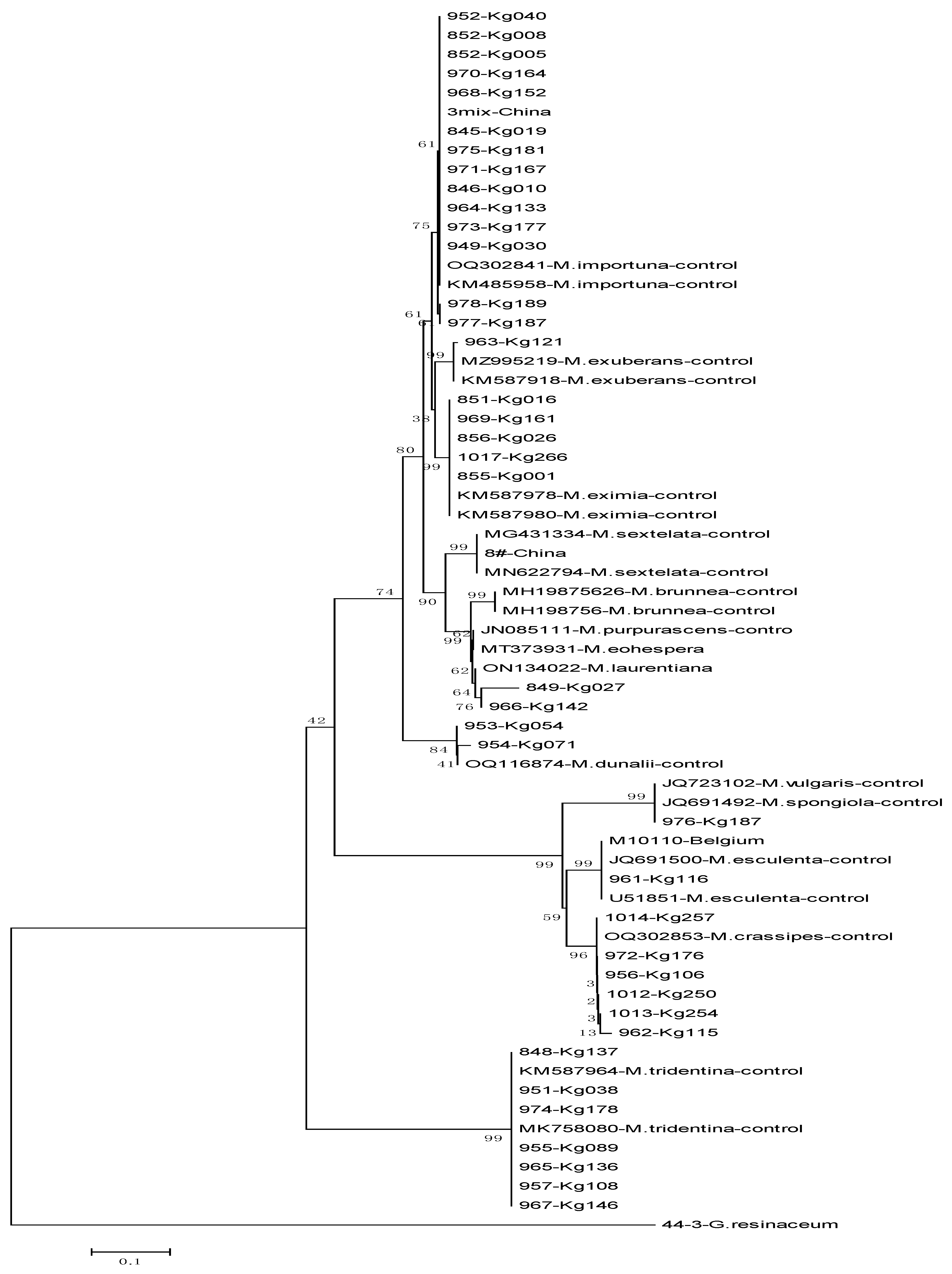
3.2. Molecular Identification of Morchella Isolates

Turkish wild morels collected and isolated in this study were identified as belonging to eight mushroom species, including the Elata and Esculenta subclades, based on the ITS region of DNA. The constructed phylogenetic tree is shown in Figure 1. Morchella crassipes (67% bootstrap support, BS), M. esculenta, and M. vulgaris (99% BS) were in the Esculenta clade, while M. tridentina (100% BS), M. exuberans (97% BS), M. eximia (98% BS), M. importuna (66% BS), and M. dunali (89% BS) were in the Elata clade.
The following identifications were made based on the ITS region of DNA:
  • M. crassipes: K115-GBK962, Kg250-1012, Kg106-956, Kg176-972, Kg254-1013, Kg257-1014
  • M. esculenta: GBK961-Kg116, M10110
  • M. vulgaris: GBK976, which showed maximum similarity with M. vulgaris and the M. spongiola clade
  • M. tridentina (formerly M. frustrata): 848-Kg137, Kg088-951, Kg089-955, Kg108-957, Kg136-965, Kg146-967, Kg178-974
  • M. exuberans: K121-GBK963
  • M. eximia: GBK851-Kg016, 855-Kg001, 856, 969-Kg161, 1017-Kg266
  • M. importuna: GBK845-Kg019, 846-Kg010, 852-Kg008, 949-Kg030, 952-Kg0140, 964-Kg133, 968-Kg152, 970-Kg164, 971-Kg167, 973-Kg177, 975-Kg181, 977, 978-Kg189
  • M. dunali: GBK953-Kg054, 954-Kg71
  • GBK849-Kg027 and 966-Kg142 could not be clearly identified but were closely related to M. laurentiana, M. eohespera, and M. purperescens clades.

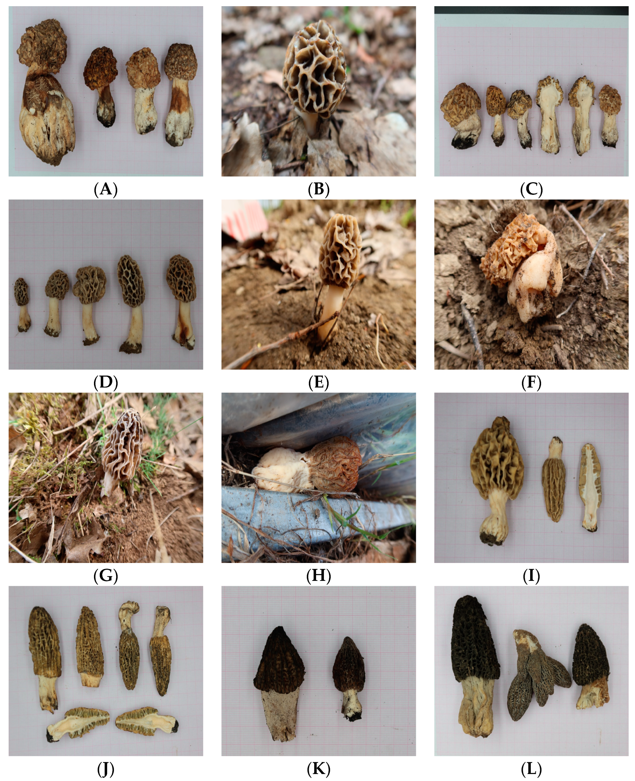
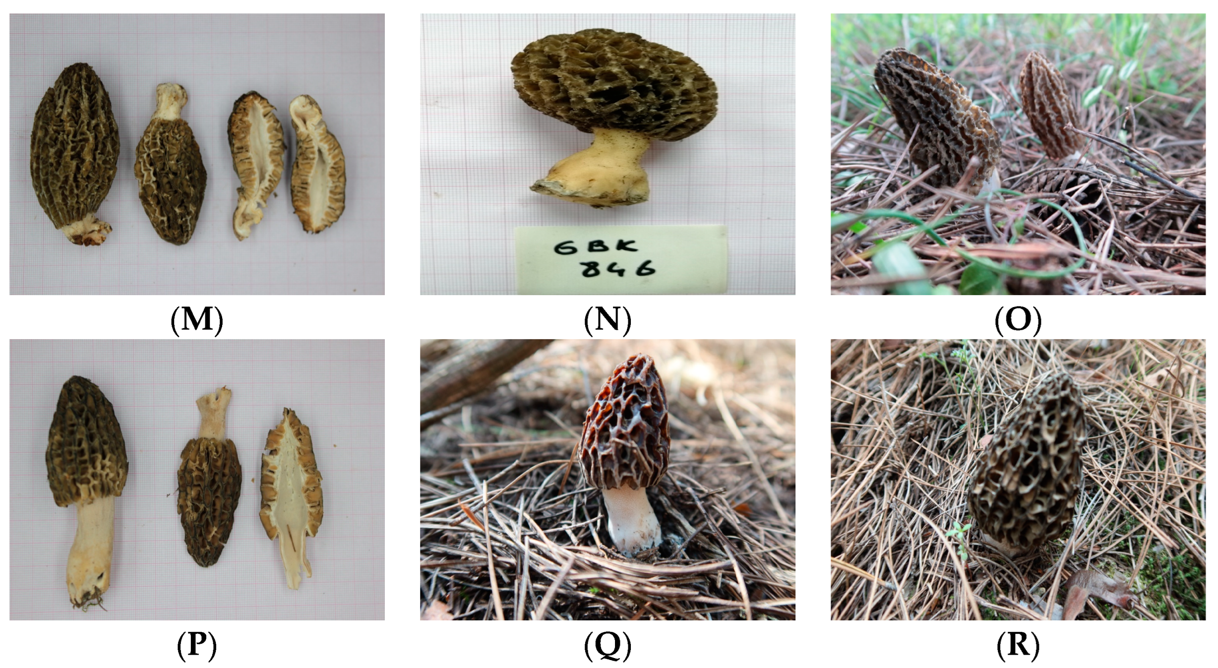
3.3. Macro-Morphologic Characters, Habitat, and Distribution

Morchella crassipes has a long and larger stem, with beige to brown necrosis on the stems in some specimens (Figure 2A–C). The ascocarps vary in color from yellow-pale brown, green beige to olive yellow, rusty, maize yellow, to gray beige. They have conical, cylindrical, or oval shapes. The pits are irregular and deep, with ridge edges usually lighter in color than the pits. The ascocarps are spongy and attached to the stem, measuring 4–16 cm long × 3–7 cm wide. The stipe is off-white to yellowish or cream-colored, with beige to brown necrosis in some specimens. It is hollow inside, straight, with a club-shaped base, flat, and inflated, with general dimensions of 3–10 cm long × 1.5–4 cm thick. Habitat and distribution: wild poplar, bunt pine forests and oak forests; altitude ranges from 328 m to 827 m. Locations: Ankara, Bursa, Manisa.
M. esculenta has ascocarps measuring 4–7.5 × 1.5–2.5 cm, and colors ranging from green beige to olive yellow (Figure 2D,E). They are ellipsoid to round in shape, with irregular deep pits and yellow to orange ridge edges. The stipe is attached, hollow, flat (glabrous), and off-white to cream in color, measuring 4–7.5 cm long and 1.5–3 cm thick, sometimes ochraceous-stained with age. Habitat and distribution: wild poplar forests in Bursa, at an altitude of 308 m.
M. vulgaris/M. spongiola have ascocarps that are graye yellow to maize yellow in color and cylindrical in shape (Figure 2F). The pits are irregular and deep, with ridge edges usually lighter in color than the pits. The ascocarps are spongy and attached to the stem, measuring 4–9 cm long × 3–4 cm wide. The stipe is off-white to yellowish, hollow inside, and straight with a club-shaped base, and is inflated; general dimensions are 3–7 cm long × 1.5–3.5 cm thick. Habitat and distribution: burned pine forests in Manisa, at an altitude of 341 m.
M. tridentina (synonym M. frustrata) has ascocarps with dimensions of 4.5–11.5 × 2.5–5 cm. The hymenophore measures 2–7 × 1.5–5 cm. The stipe is 2.5–5.5 × 1.5–3.5 cm, sometimes ochraceous-stained with age. Habitat and distribution: poplar, pine, stone pine, oak forests, and strawberry greenhouse beds (Figure 2G–I). Altitude: 77–616 m. Locations: İzmir, Çanakkale, Bursa.
The ascocarps of M. eximia are light brown to dark brown. They are cylindrical to conic in shape, measuring 4.5–9 cm × 2.5–3.5 cm (Figure 2J,K). The stalks are off-white to gray in color, hollow, and measure 1.5–3 cm × 1.5–2.5 cm. Habitat and distribution: burned pine forests and pine forests. Locations: İzmir, Muğla, Kastamonu, Ankara. Altitude: 320–1154 m.
The ascocarps of the examined M. exuberans are gray when young and black-brown when mature, and cylindrical to conical in shape (Figure 2L). The pits are irregular and deep. The ascocarps are spongy and attached to the stem, measuring 8–14 cm long × 5–6.5 cm wide. The stipe is off-white to yellowish, hollow inside, straight with a club-shaped base, and inflated; general dimensions are 2.5–5.5 cm long × 4–4.5 cm thick. Habitat and distribution: burned pine forests. Location: Bursa-Osmaneli.
The ascocarps of M. importuna are light brown to dark brown in color, with irregular pits. They are cylindrical to conic in shape (Figure 2M–O), measuring 6–12 cm long × 2.5–5.5 cm wide. The stipe is 3–5.5 cm long × 1.5–3 cm wide, and can be white, off-white, or gray in color, straight or irregular, with a club-shaped base. Habitat and distribution: dominant plants include fruit trees (apple), burned pine forests, pine forests, and black tea waste under Tilia sp. Locations include Yalova, Bursa, Çanakkale, Bilecik, Manisa, and İzmir, at altitudes ranging from 17 to 1353 m.
The ascocarps of M. dunali are 4–8 cm long × 2.5–4 cm wide, conical, cylindrical, or ovoid, and grayish-brown or olivaceous-gray in color. They are attached to the stipe with a sinus and feature longitudinal primary ridges that are thick and crowded. The pits are pinkish-purple and turn black at maturity. The stipe is short, 2–3 cm long × 1–2 cm wide, cylindrical, typically enlarged at the base, hollow, and white to cream in color, sometimes ochraceous-stained (Figure 2Q,R). Habitat and distribution: the examined ascocarps were collected under Pinus sp. forest in İzmir, at altitudes ranging from 325 to 507 m.
The ascocarps of the examined GBK849-Kg027 and 966-Kg142 (not clearly identified but close to M. purpurescens in the phylogenetic tree) measure 8–10 × 3–4 cm. They have irregular, deep, pale brown pits with darkened black ridge edges and are attached to the stipe with a sinus. The stipe measures 5–6 × 1.5–2 cm, is off-white to gray in color, straight, and occasionally ochraceous-stained at the base (Figure 2P). Habitat and distribution: collected under Pinus pinea and Pinus sp. from İzmir and Çanakkale at an altitude of 327 m.

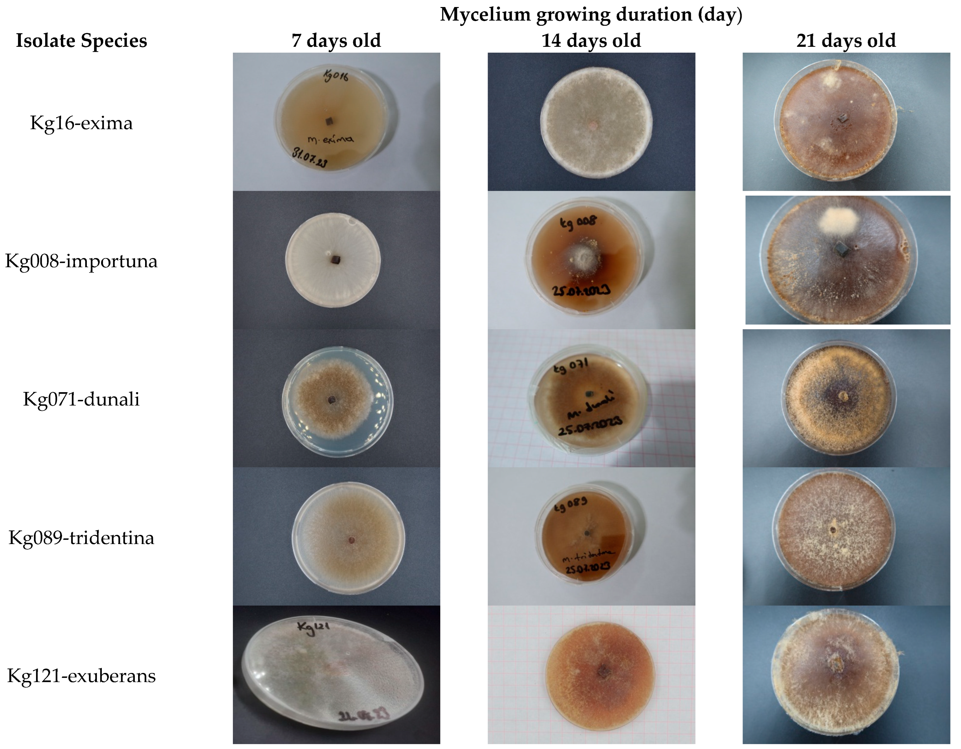
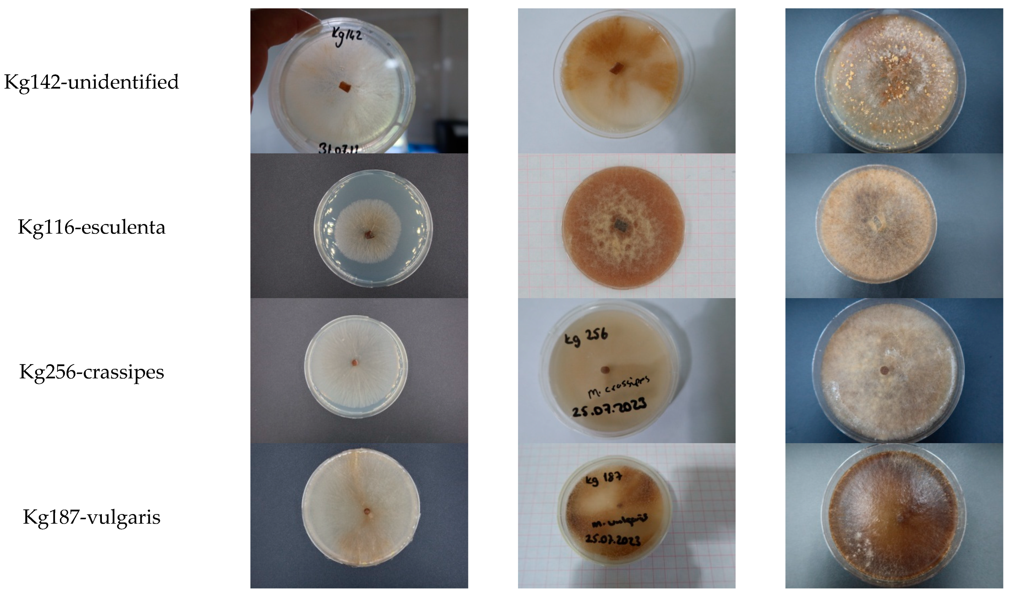
3.4. Cultural Characters of the Morchella

Morphological features of the cultures were investigated. Morel cultures primarily exhibited orange to brown colors (Figure 3). Detailed characteristics of the cultures obtained in this study are provided below.
The culture of M. crassipes showed slow growth, with the mycelium covering the entire plate in 10–11 days. The mycelium was of low density, off-white to pale yellow in color, and the surface mycelium was mainly hyaline with slight pigmentation. In the senescence phase, the color shifted to orange, and there were few or no sclerotia present, with a distinctive distribution.
The mycelium of M. esculenta initially developed as hyaline and off-white. After 5–6 days, it started to form pigments, resulting in a brown appearance by day 10. The mycelium covered the plates in 7 to 8 days. The isolates showed high mycelial density with aerial mycelium at the colony margins. Few or no sclerotia were observed.
The mycelium of M. vulgaris/M. spongiola was initially whitish to off-white and then secreted light brown pigments, turning brown in appearance by day 10. Growth was radial, with surface and submerged mycelium on the plates. The mycelium was abundant and of high density, with numerous sclerotia around the inoculum and edges of the plates. The sclerotia were compact and pale yellow, and mitospores were observed on the surface of the plates.
The mycelium of M. tridentina (synonym M. frustrada) grows aerially on the surface of PDA, initially appearing off-white to light grayish in color during the juvenile phase. In six days, it covers the entire Petri dish and then turns orange to light brown with pigmentation in 10 to 15 days. In the senescent phase (15 days old), it develops abundant, pale yellow, compact sclerotia near the inoculum and at the periphery of the plates.
The mycelium of M. exuberans covers the entire plate in 6 days, showing high-density aerial growth. Initially, the mycelium is white to off-white, then turns orange and light brown with aging. Sclerotia development is rapid, completing in 10–15 days. In the senescent phase (15 days old), numerous yellow, compact sclerotia are distributed around the periphery of the plates.
The culture of M. exima grows submerged and covers the plate in 7 days. The mycelium is off-white to grayish hyaline in the juvenile phase, turning orange to pale brown with aging (15 days old). It produces abundant, compact sclerotia at the periphery of the plates.
The culture of M. importuna shows ordinary growth, covering the plates in 6 days. The surface mycelium is mainly hyaline, off-white to pale grayish during the juvenile phase, with fine, regular density. Abundant sclerotia develop with aging (15–20 days old). Pigmentation begins after 6 days, shifting from orange to pale brown and then brown. White to pale yellow, compact sclerotia are distributed at the edge of the plates.
The Kg142 unidentified isolates colonized the Petri dishes in 5 days. The mycelium grows on the surface of the media, initially off-white to gray in color. Pigmentation begins in 6 to 7 days, with the appearance changing to orange and then pale brown. The color of the isolate on PDA is not homogeneous, showing striations. Sclerotia formation starts between 15 and 20 days, with the sclerotia being yellow, compact, and abundant, exuding, and being distributed around the periphery of the plates.
The culture of M. dunali exhibited sparse mycelium growth, covering the plates in 10 days. Initially, the mycelium is off-white to grayish in color. It then secretes pale brown pigments, turning brown in appearance during the senescent phase (15–20 days old). The cultures develop pale yellow, abundant, and compact sclerotia distributed around the periphery of the plates.

4. Discussion

Morchella species are classified into three groups based on phylogenetic analysis: esculenta (yellow morels), elata (black morels), and rufobrunnea Clade (blushing morels) [29]. The morels belonging to the esculenta and elata subclades were collected in the current study. However, M. anatolica, which belongs to the rufobrunnea Clade, was not found in this study but is reported to exist in Türkiye [38].
In this study, wild morels were collected from various locations in Türkiye, and their isolates were obtained. Molecular identifications of the isolates were performed based on the ITS rDNA. According to the results of the molecular phylogenetic analysis, eight Morchella species were identified: M. crassipes, M. esculenta, M. vulgarius/spongiola, M. tridentina, M. exuberans, M. exima, M. importuna, and M. dunali. Isolates 849-Kg027 and 966-Kg142 could not be clearly identified but are closely related to M. purpureescens, M. laurentiana, and M. eohespera. The ITS rDNA gene alone is insufficient for the complete identification of all Morchella species [30]. Similarly, Keskinkılıç [37] analyzed the HT508, HT709, HT712, and HT713 Morchella genotypes using ITS rDNA and 28S rDNA gene regions and found that M. purpureescens was grouped with M. eohespera and M. laurentiana in their phylogenetic tree. M. purpureescens has previously been identified by molecular analysis in Türkiye [27,28]. However, M. laurentiana and M. eohespera have not yet been identified in Türkiye. M. purpureescens is characterized by purplish or pinkish ascomata [25], whereas our examined specimens were pale brown, suggesting that 849-Kg027 and 966-Kg142 might not be M. purpureescens. A multilocus phylogenetic analysis is recommended for definitive molecular diagnosis of these genotypes. The molecular identification of these genotypes can be precisely achieved using the Genealogical Concordance Phylogenetic Species Recognition (GCPSR) method. O’Donnell et al. [29] successfully classified morels using GCPSR. Similarly, genotype 976-Kg187 was found in the same cluster as M. vulgaris and M. spongiola. This genotype should also be analyzed by combining different gene regions.
Since morel cultivation is targeted in the later stages of this study, the collection activities were primarily aimed at identifying species that are cultivatable. In this study, we focused on collecting saprophytic morels. Cultivated Morchella species include M. importuna, M. exima, M. sextelata, M. rufobrunnea [47], M. conica [48], M. esculenta, M. exuberans, M. owneri, M. tomentosa, Mel-13, and Mel-21 [3,40,49]. Türkiye has a very rich morel diversity, nearly twice that of Europe [28]. The number of morel species in Türkiye is close to 40 [50,51]. Of these species, 22 have been identified molecularly [28,37].
M. exuberans typically has large, robust ascocarps and turns dark brown when mature; it is often found in conifer-burnt pine sites. Taşkın et al. [27] previously reported this species as Mel-9 from Türkiye. Similarly, Loizides et al. [52] and Andrew N. Miller et al. [53] identified M. exuberans as a fire-adapted morel. The morels collected from burned areas in this study included M. crassipes, M. vulgarius, M. exuberans, M. exima, and M. importuna. Hobbie et al. [54] reported that post-fire Morchella species are saprophytes that utilize old radiocarbon rather than carbon obtained from photosynthesis in the same year. M. tomentosa, M. sextelata, M. eximia, and Mel-8 have also been reported as fire-adapted species. M. exuberans and M. importuna are considered facultative fire-adaptive species [55].
Saprophytic fungi can be cultivated without mycorrhizal hosts [42]. M. tridentina and M. frustrata are synonymous species [25]. M. tridentina was found under pine, poplar, and oak forests, and, intriguingly, also in a strawberry greenhouse. This species can be considered saprophytic. The M. crassipes specimens was collected from the burnt area in Nallıhan, Ankara, and showed distinct beige to brown necroses on the stem. These necroses had not been reported in previous studies and may be attributed to ecological factors rather than genetic differences.
Since PDA and MEA media are the most suitable for the mycelial growth rate for Morchella spp. [56], PDA was used and incubated at 20 °C for the mycelial growth of Morchella spp. in ongoing experiments. In this study, Morchella cultures mostly covered the Petri dishes within 5–7 days, except for Morchella dunali and M. crassipes, which colonized in 10–11 days on PDA. While mycelial colors were off-white to pale gray in the juvenile stage, pigment formation started 5–6 days after inoculation, turning orange to pale brown. The M. crassipes and Kg142 isolate formed lighter-colored pigments compared to other species. Unlike the other species, the Kg142 isolate formed prominent, compact, and numerous sclerotia. M. exuberans, on the other hand, was distinctive for developing white aerial mycelia in the form of arcs on the edge of the Petri dish, which turned yellow in the senescence stage. M. dunali and M. vulgaris formed the darkest mycelial pigments. Additionally, intense yellow sclerotial formation was observed, resembling cinnamon sprinkled on the dark brown mycelia of M. dunali.
He et al. [57] reported that the rapid development of vegetative mycelia in the first few days is due to increased ROS levels or oxidative stress [58], which may directly or indirectly stimulate necrosis formation. Sclerotial formation and size are influenced by genetic factors as well as different nitrogen and carbon sources, light, temperature, pH, humidity, organic acids, phenolic compounds, and osmotic potential [59,60]. Mycelia of M. vulgaris, M. crassipes, and the Kg142 isolate were scarce and of low density, and were submerged. Exudates in Petri dishes were not observed, whereas Wang et al. [61] reported that exudates are a common feature of ascomycota during sclerotial development [62]. Dehydration, cell thickness, polymerization of soluble compounds, and moisture in tissues affect the intensity of exudate formation [63]. Sanz-Rocha et al. [64] reported that the mycelial morphology of Morchella isolates is influenced by media and incubation temperature. On PDA medium, much more sclerotial formation was observed in most isolates. Sclerotia can be located in the center of the cone or in different regions. On PDA medium, the order of sclerotial formation was M. eximia > M. importuna > M. tridentina. Intense aerial mycelial formation was observed in M. eximia. Most isolates showed better mycelial growth at 20–24 °C [65], although some isolates performed best in terms of mycelial growth and sclerotial production at 28 °C [64].
In summary, previous studies, such as those by Taşkın et al. [27,28] and Keskinkılıç et al. [37] have collected morel mushrooms from various regions in Türkiye and performed molecular analyses on them, but they did not isolate the cultures. The author has successfully isolated the collected materials, which provides a foundation for creating a culture collection gene pool for future breeding and production efforts. Additionally, this study has examined the macro-morphological development of the cultures. Previous morphological studies on cultures were primarily limited to the M. esculenta species [66,67]. Recent research has explored the mitosporic features of M. importuna, M. exima, M. crassipes, and M. galilliaea [68], mycelium development and sclerotial characteristics in M. tridentina, M. andinensis, and M. exima [64], the morphological and microscopic characteristics of Morchella elata in juvenile and senescent stages [57], and the mycelium characteristics of M. conica [59]. Furthermore, the author compared the morphological development of black clade morels such as M. exuberans, M. dunali, M. importuna, and M. exima with yellow morels including M. esculenta, M. crassipes, M. spongiola/vulgaris, and M. tridentina. It was observed that black morels produce darker pigments while yellow morels produce lighter pigments. This study marks the first establishment of a morel mushroom culture collection in Türkiye. The cultures obtained from this study will serve as valuable material for future breeding and mushroom production research.

5. Conclusions

This study investigated true morels (Morchella) collected and isolated from different locations in Türkiye, focusing on their molecular identification and macro-morphological characteristics. Isolates belonging to M. importuna, M. exima, M. exuberans, M. dunali, and M. tridentina species in the black clade, as well as M. crassipes and M. esculenta in the yellow clade, were unambiguously identified through the ITS-based DNA barcoding study. However, the identification of isolates 849-Kg027 and 966-Kg142 could not be clearly determined. Some isolates were placed in the same clade as M. vulgaris and M. spongiola and could not be differentiated from them. In future studies, the definitive identification of these species should be achieved through multilocus analysis. There are no specific morphological features that unambiguously distinguish the isolates belonging to different species. More detailed studies of both the micro- and macro-morphological features of these isolates are needed.

Funding

This research was funded by the collaboration project between Gyeonsangbuk-do Agricultural Research and Extension Services of Republic of Korea and General Directorate of Agricultural Research and Policy of Republic of Türkiye. Project title: ‘Genetic Resources Evaluation and Breeding of the Medicinal Mushrooms’.

Data Availability Statement

The original contributions presented in the study are included in the article, further inquiries can be directed to the corresponding author.

Acknowledgments

I would like to give a special thanks to Younghyun RYU, Gyeonsangbuk-do Agricultural Research and Extension Services of Republic of Korea for its valuable support during this work.

Conflicts of Interest

No potential conflicts of interest are reported by the author.

References

  1. Hibbett, D.S.; Binder, M.; Bischoff, J.F.; Blackwell, M.; Cannon, P.F.; Eriksson, O.E.; Huhndorf, S.; James, T.; Kirk, P.M.; Lücking, R.; et al. A higher-level phylogenetic classification of the fungi. Mycol. Res. 2007, 111, 509–547. [Google Scholar] [CrossRef] [PubMed]
  2. Sa, W.; Qiao, J.; Gao, Q.; Li, Z.; Shang, Q. DNA Barcoding and Species Classification of Morchella. Genes 2022, 13, 1806. [Google Scholar] [CrossRef]
  3. Li, Y.; Chen, H.; Zhang, X. Cultivation, nutritional value, bioactive compounds of morels, and their health benefits: A systematic review. Front. Nutr. 2023, 10, 1159029. [Google Scholar] [CrossRef] [PubMed] [PubMed Central]
  4. Magrati, T.P.; Tripathee, H.P.; Devkota, K.P. Nutritional Analysis of Morchella conica and its Role on Rural Livelihood. Nepal J. Sci. Technol. 2011, 12, 119–126. [Google Scholar] [CrossRef]
  5. Beluhan, S.; Ranogajec, A. Chemical composition and non-volatile components of Croatian wild edible mushrooms. Food Chem. 2011, 124, 1076–1082. [Google Scholar] [CrossRef]
  6. Heleno, S.A.; Stojković, D.; Barros, L.; Glamočlija, J.; Soković, M.; Martins, A.; Queiroz, M.J.R.; Ferreira, I.C. A comparative study of chemical composition, antioxidant and antimicrobial properties of Morchella esculenta (L.) Pers. from Portugal and Serbia. Food Res. Int. 2013, 51, 236–243. [Google Scholar] [CrossRef]
  7. Vieira, V.; Fernandes, Â.; Barros, L.; Glamočlija, J.; Ćirić, A.; Stojković, D.; Martins, A.; Soković, M.; Ferreira, I.C. Wild Morchella conica Pers. from different origins: A comparative study of nutritional and bioactive properties. J. Sci. Food Agric. 2016, 96, 90–98. [Google Scholar] [CrossRef]
  8. Altaf, U.; Lalotra, P.; Sharma, Y.P. Nutritional and mineral composition of four wild edible mushrooms from Jammu and Kashmir, India. Indian Phytopathol. 2020, 73, 313–320. [Google Scholar] [CrossRef]
  9. Gürsoy, N.; Sarikurkcu, C.; Cengiz, M.; Solak, M.H. Antioxidant activities, metal contents, total phenolics and flavonoids of seven Morchella species. Food Chem. Toxicol. 2009, 47, 2381–2388. [Google Scholar] [CrossRef]
  10. Ooi, V.E.; Liu, F. Immunomodulation and anti-cancer activity of polysaccharide protein complexes. Curr. Med. Chem. 2000, 7, 715–729. [Google Scholar] [CrossRef]
  11. Du, X.; Zhao, Q.; Yang, Z.L. A review on research advances, issues, and perspectives of morels. Mycology 2015, 6, 78–85. [Google Scholar] [CrossRef]
  12. Tietel, Z.; Masaphy, S. True morels (Morchella)—nutritional and phytochemical composition, health benefits and flavor: A review. Crit. Rev. Food Sci. Nutr. 2018, 58, 1888–1901. [Google Scholar] [CrossRef]
  13. Akyüz, M.; Kireççi, A.D.O.; Gökce, Z.; Kirbag, S.; Yılmaz, O. Biochemical constituents and antioxidant activities of some mushrooms from Turkey: Agaricus spp., Pleurotus spp., Morchella esculenta and Terfezia boudieri. Istanb. J. Pharm. 2019, 49, 1–7. [Google Scholar]
  14. Li, Y.; Li, T.; Liu, R.H. Perspectives of b-glucans for health promotion. Nov. Approaches Drug Des. Dev. 2020, 5, 58–62. [Google Scholar] [CrossRef]
  15. Zhang, Z.; Shi, M.; Zheng, H.; Ren, R.; Zhang, S.; Ma, X. Structural characterization and biological activities of a new polysaccharide isolated from Morchella Sextelata. Glycoconj. J. 2022, 39, 369–380. [Google Scholar] [CrossRef] [PubMed]
  16. Taşkin, H.; Süfer, Ö.; Attar, S.H.; Bozok, F.; Baktemur, G.; Büyükalaca, S.; Kafkas, N.E. Total phenolics, antioxidant activities and fatty acid profiles of six Morchella species. J. Food Sci. Technol. 2021, 58, 692–700. [Google Scholar] [CrossRef]
  17. Chen, Q.; Che, C.; Yang, S.; Ding, P.; Si, M.; Yang, G. Anti-inflammatory effects of extracellular vesicles from Morchella on LPS-stimulated RAW2647 cells via the ROS-mediated p38 MAPK signaling pathway. Mol. Cell Biochem. 2022, 478, 317–327. [Google Scholar] [CrossRef] [PubMed]
  18. Li, F.; Wang, K.; Dong, X.; Xu, H. Structure, conformation and immunomodulatory activity of a polysaccharide from Morchella sextelata. Int. J. Food Sci. Technol. 2022, 57, 4628–4637. [Google Scholar] [CrossRef]
  19. Xiong, C.; Li, Q.; Chen, C.; Chen, Z.; Huang, W. Neuroprotective effect of crude polysaccharide isolated from the fruiting bodies of Morchella importuna against H2O2-induced PCl2 cell cytotoxicity by reducing oxidative stress. Biomed. Pharmacother. 2016, 83, 569–576. [Google Scholar] [CrossRef] [PubMed]
  20. Nitha, B.; Fijesh, P.V.; Janardhanan, K.K. Hepatoprotective activity of cultured mycelium of morel mushroom, Morchella esculenta. Exp. Toxicol. Pathol. 2013, 65, 105–112. [Google Scholar] [CrossRef]
  21. Öztürk, M.; Soylu, M.K.; Temel, M.; Pezikoğlu, F.; Mısır Bilen, G. Turkey’s Place in the World Mushroom Foreign Trade. IJAAES Internat. J. Anatolia Agricul. Eng. 2019, 1, 102–107. Available online: https://dergipark.org.tr/tr/pub/uazimder/issue/51011/666017 (accessed on 2 September 2024).
  22. Du, X.H.; Zhao, Q.; Yang, Z.L. Diversity, evolutionary history and cultivation of morels: A review. Mycosystema 2014, 33, 183–197. [Google Scholar]
  23. Kuo, M.; Dewsbury, D.R.; O’Donnell, K.; Carter, M.C.; Rehner, S.A.; Moore, J.D.; Moncalvo, J.M.; Canfield, S.A.; Stephenson, S.L.; Methven, A.S.; et al. Taxonomic revision of true morels (Morchella) in Canada and the United States. Mycologia 2012, 104, 1159–1177. [Google Scholar] [CrossRef]
  24. Mortimer, P.E.; Karunarathna, S.C.; Li, Q.H.; Gui, H.; Yang, X.Q.; Yang, X.F.; He, J.; Ye, L.; Guo, J.Y.; Li, H.L.; et al. Prized edible Asian mushrooms: Ecology, conservation and sustainability. Fungal Divers. 2012, 56, 31–47. [Google Scholar] [CrossRef]
  25. Richard, F.; Bellanger, J.M.; Clowez, P.; Hansen, K.; O’Donnell, K.; Urban, A.; Sauve, M.; Courtecuisse, R.; Moreau, P.A. True morels (Morchella, Pezizales) of Europe and North America: Evolutionary relationships inferred from multilocus data and a unified taxonomy. Mycologia 2015, 107, 359–382. [Google Scholar] [CrossRef] [PubMed]
  26. Taylor, J.W.; Jacobsen, D.J.; Kroken, S.; Kasuga, T.; Geiser, D.M.; Hibbett, D.S.; Fisher, M.C. Phylogenetic species recognition and species concepts in fungi. Fungal Genet. Biol. 2000, 31, 21–32. [Google Scholar] [CrossRef] [PubMed]
  27. Taskin, H.; Büyükalaca, S.; Doğan, H.H.; Rehner, S.A.; O’Donnell, K. A multigene molecular phylogenetic assessment of true morels (Morchella) in Turkey. Fungal Genet. Biol. 2010, 47, 672–682. [Google Scholar] [CrossRef]
  28. Taşkın, H.; Büyükalaca, S.; Hansen, K.; O’Donnell, K. Multilocus phylogenetic analysis of true morels (Morchella) reveals high levels of endemics in Turkey relative to other regions of Europe. Mycologia 2012, 104, 446–461. [Google Scholar] [CrossRef]
  29. O’Donnell, K.; Rooney, A.P.; Mills, G.L.; Kuo, M.; Weber, N.S.; Rehner, S.A. Phylogeny and historical biogeography of true morels (Morchella) reveals an early Cretaceous origin and high continental endemism and provincialism in the Holarctic. Fungal Genet. Biol. 2011, 48, 252–265. [Google Scholar] [CrossRef] [PubMed]
  30. Du, X.H.; Zhao, Q.; O’Donnell, K.; Rooney, A.P.; Yang, Z.L. Multigene molecular phylogenetics reveals true morels (Morchella) are especially species-rich in China. Fungal Genet. Biol. 2012, 49, 455–469. [Google Scholar] [CrossRef]
  31. Zeng, N.K.; Tang, L.P.; Li, Y.C.; Tolgor, B.; Zhu, X.T.; Zhao, Q.; Yang, Z.L. The genus Phylloporus (Boletaceae, Boletales) from China: Morphological and multilocus DNA sequence analyses. Fungal Divers. 2013, 58, 73–101. [Google Scholar] [CrossRef]
  32. Elliott, T.F.; Bougher, N.L.; O’Donnell, K.; Trappe, J.M. Morchella australiana sp. nov., an apparent Australian endemic from New South Wales and Victoria. Mycologia 2014, 106, 113–118. [Google Scholar] [CrossRef] [PubMed]
  33. Pildain, M.B.; Visnavsky, S.B.; Barroetave~na, C. Phylogenetic diversity of true morels (Morchella), the main edible non-timber product from native Patagonian forests of Argentina. Fungal Biol. 2014, 118, 755–763. [Google Scholar] [CrossRef]
  34. Voitk, A.; Burzynski, M.; O’Donnell, K.; Voitk, M.; Marceau, A. Mel-36-preliminary description of a new morel species. Omphalina 2014, 5, 7–10. [Google Scholar]
  35. Sesli, E.; Denchev, C.M. Checklists of the myxomycetous, larger ascomycetes, and larger basidiomycetes in Turkey, 6th ed. Mycotaxon 2014, 1–136. Available online: http://www.mycotaxon.com/resources/checklists/sesli-v106-checklist.pdf (accessed on 2 September 2024).
  36. Solak, M.H.; Işıloğlu, M.; Kalmış, E.; Allı, H. Macrofungi of Turkey, Checklist; Üniversiteliler Ofset: İzmir, Turkey, 2015; Volume 2. [Google Scholar]
  37. Keskinkılıç, İ. Contribution to Genetic Diversıty of Morchella (morel) Genus in Türkiye Using Molecular Methods. Master’s Thesis, Department of Biotechnology, Instıtute of Natural and Applied Sciences, University of Çukurova, Adana, Turkey, 2019; 71p. [Google Scholar]
  38. Işıloğlu, M.; Allı, H.; Spooner, B.M.; Solak, M.H. Morchella anatolica (Ascomycota), a new species from southwestern Anatolia, Turkey. Mycologia 2010, 102, 455–458. [Google Scholar] [CrossRef]
  39. Taşkın, H.; Doğan, H.H.; Büyükalaca, S.; Clowez, P.; Arthur, P.A.; O’Donnell, K. Four new morel (Morchella) species in the Elata Subclade (sect. Distantes) from Turkey. Mycotaxon 2016, 131, 467–482. [Google Scholar] [CrossRef]
  40. Liu, Q.; Ma, H.; Zhang, Y.; Dong, C. Artificial cultivation of true morels: Current state, issues and perspective. Crit. Rev. Biotechnol. 2017, 38, 259–271. [Google Scholar] [CrossRef]
  41. Xu, Y.; Tang, J.; Wang, Y.; He, X.; Tan, H.; Yu, Y.; Chen, Y.; Peng, W. Large-scale commercial cultivation of morels: Current state and perspectives. Appl. Microbiol. Biotechnol. 2022, 106, 4401–4412. [Google Scholar] [CrossRef]
  42. Ge, S. Studies on Molecular Identification, Biological Characteristics and Indoor Cultivation of Morchella spp. Master’s Thesis, Department of Plant Pathology and Environmental Microbiology of the Pennsylvania State University, University Park, PA, USA, 2019; 166p. [Google Scholar]
  43. Anonymous. Forest Fire Statistics of General Directorate of Forestry of Agriculture and Forestry Ministry of Republic of Türkiye. 2021. Available online: https://www.ogm.gov.tr/tr/e-kutuphane-sitesi/Istatistikler/Ormanc%C4%B1l%C4%B1k%20%C4%B0statistikleri/Ormanc%C4%B1l%C4%B1k%20%C4%B0statistikleri%202021.zip (accessed on 2 September 2024).
  44. Chang, S.T.; Miles, P.G. Mushrooms: Cultivation, Nutritional Value, Medicinal Effect, and Environmental Impact, 2nd ed.; CRC Press: Boca Raton, FL, USA, 2004. [Google Scholar]
  45. Nakasone, K.K.; Peterson, S.W.; Jong, S.C. Preservation and distribution of fungal cultures. In Biodiversity of Fungi: Inventory and Monitoring Methods; Elsevier: Amsterdam, The Netherlands; Academic Press: Cambridge, CA, USA, 2004; pp. 37–47. [Google Scholar]
  46. White, T.J.; Bruns, T.; Lee, S.; Taylor, J. Amplification and Direct Sequencing of Fungal Ribosomal RNA Genes for Phylogenetics; Academic Press: Cambridge, CA, USA, 1990; pp. 315–322. [Google Scholar]
  47. Masaphy, S. Biotechnology of morel mushrooms: Successful fruiting body formation and development in a soilless system. Biotechnol. Lett. 2010, 32, 1523–1527. [Google Scholar] [CrossRef]
  48. Zhao, Q.; Xu, Z.Z.; Cheng, Y.H.; Qi, S.W.; Hou, Z.J. Bionic cultivation of Morchella conica. Southwest China J. Agric. Sci. 2009, 22, 1690–1693. [Google Scholar]
  49. Zhao, Q.; Lv, M.; Li, L.; Huang, W.; Zhang, Y.; Hao, Z. “Temptation” and “Trap” of Morel industry in China. J. Fungal Res. 2021, 19, 232–237. [Google Scholar] [CrossRef]
  50. Sesli, E.; Asan, A.; Selçuk, F.; Akata, I.; Akgül, H.; Aktaş, S.; Allı, H.; Demirel, K.; Doğan, H.H.; Kaya, A.; et al. Türkiye Mantarları Listesi. Ali Nihat Gökyiğit Vakfi Yayin. İstanbul 2020, 1177. [Google Scholar]
  51. Solak, M.H.; Türkoğlu, A. Macrofungi of Turkey (Checklist Volume III); Kanyılmaz Matbaacılık, Bornova-İZMİR: İzmir, Turkey, 2022. [Google Scholar]
  52. Loizides, M.; Alvarado, P.; Clowez, P.; Moreau, P.A.; Romero de la Osa, L.; Palazón, A. Morchella tridentina, M. rufobrunnea, and M.kakiicolor: A study of three poorly known Mediterranean morels, with nomenclatural updates in section Distantes. Mycol. Prog. 2015, 14, 13. [Google Scholar] [CrossRef]
  53. Miller, A.N.; Raudabaugh, D.B.; Iturriaga, T.; Matheny, P.B.; Petersen, R.H.; Hughes, K.W.; Gube, M.; Powers, R.A.; James, T.Y.; O’donnell, K. First report of the post-fire morel Morchella exuberans in eastern North America. Mycologia 2017, 109, 710–714. [Google Scholar] [CrossRef]
  54. Hobbie, E.A.; Rice, S.F.; Weber, N.S.; Smith, J.E. Isotopic evidence indicates saprotrophy in post-fire Morchella in Oregon and Alaska. Mycologia 2016, 108, 638–645. [Google Scholar] [CrossRef]
  55. Pilz, D.; Rebecca, M.L.; Susan, A.; Villarreal-Ruiz, L.; Berch, S.; Wurtz, T.L.; Parks, C.G.; McFarlane, E.; Baker, B.; Molina, R. Ecology and Management of Morels Harvested from the Forests of Western North America; US Department of Agriculture, Forest Service, Pacific Northwest Research Station: Portland, OR, USA, 2007. [CrossRef]
  56. Kalmis, E.; Kalyoncu, F. Mycelial growth rate of some morels (Morchella spp.) in four different microbiological media. Am. Eurasian J. Agric. Environ. Sci. 2008, 3, 861–864. [Google Scholar]
  57. He, P.; Cai, Y.; Liu, S.; Han, L.; Huang, L.; Liu, W. Morphological and ultrastructural examination of senescence in Morchella elata. Micron 2015, 78, 79–84. [Google Scholar] [CrossRef]
  58. He, P.; Liu, W.; Cai, Y.; Ma, B.; Chen, L.; Wu, X. Effects of oxidative stress on mycelial growth and sclerotial metamorphosis of Morchella crassipes. Hubei Agric. Sci. 2014, 53, 3085–3089. [Google Scholar]
  59. Güler, P.; Özkaya, E.G. Morphological development of Morchella conica mycelium on different agar media. J. Environ. Biol. 2009, 30, 601–604. [Google Scholar]
  60. Rollins, J.A.; Dickman, M.B. Increase in endogenous and exogenous cyclic AMP levels inhibits sclerotial development in Sclerotinia sclerotiorum. Appl. Environ. Microbiol. Biotechnol 1998, 64, 2539–2544. [Google Scholar] [CrossRef]
  61. Wang, D.; Fu, J.; Zhou, R.; Li, Z.; Xie, Y.; Liu, X.; Han, Y. Formation of sclerotia in Sclerotinia ginseng and composition of the sclerotial exudate. PeerJ 2018, 6, e6009. [Google Scholar] [CrossRef] [PubMed] [PubMed Central]
  62. Liang, Y.; Strelkov, S.E.; Kav, N.N.V. The proteome of liquid Sclerotial exudates from Sclerotinia sclerotiorum. J. Proteome Res. 2010, 9, 3290–3298. [Google Scholar] [CrossRef] [PubMed]
  63. Willetts, H.J. The survival of fungal sclerotia under adverse environmental conditions. Biol. Rev. 1971, 46, 387–407. [Google Scholar] [CrossRef]
  64. Sanz-Rocha, M.; Gerding, M.; Quezada, T.; Vargas, M.; Chávez, D.; Machuca, Á. Molecular and cultural characterization of Morchella spp. from disturbed environments of central-southern Chile. Fungal Biol. 2023, 127, 938–948. [Google Scholar] [CrossRef] [PubMed]
  65. Kalyoncu, F.; Oskay, M.; Kalyoncu, M. The effects of some environmental parameters on mycelial growth of six Morchella species. J. Pure Appl. Microbiol. 2009, 3, 467–472. [Google Scholar]
  66. Amir, R.; Levanon, D.; Hadar, Y.; Chet, I. Morphology and physiology of Morchella esculenta during sclerotial formation. Mycol. Res. 1993, 97, 683–689. [Google Scholar] [CrossRef]
  67. Kanwal, N.; William, K.; Sultana, K. In vitro propagation of Morchella esculenta and study of its life cycle. J. Bioresour. Manag. 2016, 3, 6. [Google Scholar]
  68. Liu, W.; He, P.; Zhang, J.; Wu, L.; Er, L.; Shi, X.; Gu, Z.; Yu, F.; Pérez-Moreno, J. Ultrastructure and physiological characterization of Morchella mitospores and their relevance in the understanding of the morel life cycle. Microorganisms 2023, 11, 345. [Google Scholar] [CrossRef]
Figure 1. Maximum likelihood phylogenetic tree based on ITS rDNA gene of Morchella specimens of the Elata and Esculenta clades, with specimens and isolates from Türkiye collected in this study. The control sequences considered in the phylogenetic tree were obtained from NCBI Nucleotide database.
Horticulturae 10 01020 g001
Figure 2. (A) GBK1012. M. crassipes with large stalk with negrosis. (B) Specimen GBK 956 without negrosis on stipe. (C) GBAK1013 hollow stalk of M. crassipes. (D) GBK961. M. esculenta with medium stalk. (E) Specimen GBK 961 in poplar area. (F) GBK976. M. vulgarius-spongiola in fired pine forest. (G) GBK974. M. tridentina in oak forest. (H) GK 951. M. tridentina in strawberry greenhouse (I) GBK965. young and mature ascocarp of M. tridentina. (J) GBK969. Light brown young and mature ascocarp of M. exima in pine forest. (K) GBK1017. Dark brown ascocarp of M. exima in fired pine forest. (L) GBK963. Young and mature ascocarp of M. exuberans. (M) GBK968. Ascocarp of M. importuna in fired pine forest. (N) GBK846. Ascocarp of M. importuna under Tilia sp. at black tea waste compost. (O) GBK949. M. importuna in pine forest. (P) Ascocarp of GBK966 in pine forest. (Q) GBK953. Ascocarp of M. dunali in pine forest. (R) GBK954. Ascocarp of M. dunali in pine forest.
Horticulturae 10 01020 g002aHorticulturae 10 01020 g002b
Figure 3. Morphological characteristics of mycelium and sclerotia of Morchella species after 7, 14, and 21 days of mycelia growing duration.
Horticulturae 10 01020 g003aHorticulturae 10 01020 g003b
Table 1. Collected specimen and cultures used in this study and origin and habitat information.
SpeciesAccession and İsolate NoOriginDominant PlantGPS Coordinates
M. crassipes962-Kg115Bursa-İnegöl-KurşunluPopulus sp.40°00′044″ N-29°40′041″ E-487 m
1012-Kg250Ankara-NallıhanFired Pine forest40°06′054″ N-30°53′008″ E-369 m
956-Kg106Bursa-İnegöl-Close to quarry mineMix Populus-Quercus40°01′074″ N-29°37′908″ E
972-Kg176Manisa-Akhisar-YayakırıldıkFired Pine forest38°52′181″ N-028°04′325″ E-328 m
1013-Kg254Ankara-NallıhanFired Pine forest40°11′051″ N-31°27′025″ E-805 m
1014-Kg257Ankara-NallıhanFired Pine forest40°11′051″ N-31°27′057″ E-827 m
M. esculenta961-Kg116Bursa-İnegöl-CerrahPopulus sp.40°04′563″ N-029°27′711″ E-308 m
M. vulgarius/
M. spongiola
976-Kg187Manisa-OsmancalıFired Pine forest38°47′028″ N-27°161′86″ E-341 m
M. tridentina967-Kg146Çanakkale-Bayramiç-KaykılarPinus sp.39°50′031″ N-26°44′015E-389 m
951-Kg038İzmir-MordoğanStrawberry greenhouse38°30491″ N-26°36′54″ E-77 m
955-Kg089Bursa-İnegöl-ÇitliPopulus sp.40°01.072 N-29°37.140 E-373 m
957-Kg108Bursa-İnegöl-ÇitliPopulus sp.40°01′072″N-29°37′140″ E-373 m
965-Kg136Çanakkale-BayramiçPinus sp.39°45′051″ N-26°37′008″ E-262 m
974-Kg178Manisa-Salihli-İğdecikQuercus sp.38°34′321″ N-028°22′525″ E-616 m
848-Kg137İzmir-Bergama-GöbellerPinus pinea39°242′67 80″ N-27°025′39 30″ E
M. exuberans963-Kg121Bilecik-Osmaneli-DereyörükFired Pine forest40°18.051 N-29°51.025 E-269 m
M. exima851-Kg016İzmir-BalçovaPinus sp.38°22′042″ N-27°02′037″ E-320 m
969-Kg161Muğla-KöyceğizPinus sp.37°00′052″ N-28°42′026″ E-469 m
856-Kg026Kastamonu-Taşköprü-AslanlıFired Pine-poplar forest41°328′7600″ N-34°52′6970″ E
1017-Kg266Ankara-NallıhanFired Pine forest40°15′055″ N-31°31′003″ E-1154 m
855-Kg001Kastamonu-Taşköprü-Dedeağaç-DumanFired Pine forest41°358′27 60″ N-34°190′6300″ E
M. sponigoal/
M. vulgaris
976-Kg187Manisa-OsmancalıFired Pine forest38°47′028″ N-27°161′86″ E-341 m
M. importuna978-Kg189Muğla-MilasFruit orchard37°13′005″ N-28°28′007″ E-1353 m
977-Kg188Bursa-İnegölFruit orchard39°59′045″ N-29°41′035″ E-936 m
968-Kg152Çanakkale-Bayramiç-HacıköyFired Pine forest39°53′029″ N-26°37′002″ E-205 m
846-Kg010Yalova-ÇınarcıkTilia sp. Waste of black tea40°38′043″ N-29°07′011″ E-17 m
971-Kg167Manisa-Akhisar-YayakırıldıkFired Pine forest38°52′181″ K-028°04′325″ D-328 m
975-Kg181Manisa-OsmancalıFired Pine forest38°47′028″ N-027°161′86″ E-341 m
852-Kg005Manisa-MuradiyeFired Pine forest38°715′4420″ N-27°25′15720″ E
852-Kg008Manisa-MuradiyeFired Pine forest38°715′4420″ N-27°25′15720″ E
970-Kg164Çanakkale-Merkez-KızılkeçiliPinus sp.40°11′040″ N-26°34′029″ E-219 m
964-Kg133Bilecik-Osmaneli-DereyörükFired Pine forest40°18′050″ N-29°51′024″ E-274 m
845-Kg019Yalova-ÇınarcıkTilia sp. Waste of black tea40°38′041″ N-29°07′007″ E-21 m
949-Kg030İzmir-Tırazlı-KarabağlarPinus sp.38°21′484″ N-27°03′101″ E
952-Kg040İzmir-Menderes-ÇileFired pine forest38°02′308″ N-27°10′796″ E-163 m
973-Kg177Manisa-Akhisar-YayakırıldıkFired Pine forest38°52′180″ K-028°04′326″ D-330 m
Unidentified849-Kg027İzmir-BergamaPinus pinea39°242′6780″ N-27°025′3930″ E
966-Kg142Çanakkale-Bayramiç-KaykılarPinus sp.39°50′016″ N-26°43′053″ E-327 m
M. dunali953-Kg054İzmir-Menderes-ÇilePinus sp.38°01′664″ N-27°09′474″ E-325 m
954-Kg071İzmir-Menderes-KaracadağPinus sp.38°05′151″ N-27°07′785″ E-507 m
M. esculentaM10110Belgium-Mycelia companyCommercial strain
8#ChinaCommercial strain
M. importuna3 mixChinaCommercial strain
Disclaimer/Publisher’s Note: The statements, opinions and data contained in all publications are solely those of the individual author(s) and contributor(s) and not of MDPI and/or the editor(s). MDPI and/or the editor(s) disclaim responsibility for any injury to people or property resulting from any ideas, methods, instructions or products referred to in the content.

