Article

Characterization of Biofilm Formation and Bacterial Resistance to Benzalkonium Chloride under Contrasting Cultivation Conditions

Faculty of Biology, University of Latvia, 1 Jelgavas Str., LV-1004 Riga, Latvia
*
Author to whom correspondence should be addressed.
Fermentation 2023, 9(8), 699; https://doi.org/10.3390/fermentation9080699
Submission received: 22 June 2023 / Revised: 12 July 2023 / Accepted: 19 July 2023 / Published: 25 July 2023
(This article belongs to the Section Industrial Fermentation)

Abstract

:
Benzalkonium chloride (BAC) is one of the most commonly used quaternary ammonium compounds in the pharmaceutical, cosmetic, and food industries. The aim of our study was to compare the physiological responses of Escherichia coli MSCL 332, Pseudomonas putida MCCL 650, and Staphylococcus epidermidis MSCL 333 on 50 mg/L BAC in rich and poor medium (100% and 5% tryptone soya broth (TSB)) in the temperature range from 8 °C to 37 °C, under static and shaking conditions. A high-throughput, 96-well microplate method was used to compare a broad range of cultivation conditions. The effect of BAC on growth, biofilm formation activity, and dehydrogenase and fluorescein diacetate hydrolysis activity was evaluated. Addition of BAC to 100% TSB inhibited biofilm formation at 37 °C by 2.4, 1.8, and 1.6 times for E. coli, P. putida, and S. epidermidis, respectively. In turn, BAC stimulated biofilm formation in E. coli in 5% TSB at 37 °C and 100% TSB at 8 °C, i.e., 1.4 and 1.3 times, respectively. Statistical optimization of broth composition with emphasis on biofilm formation and further testing under experimental conditions was performed with P. putida.

1. Introduction

A significantly increased tolerance of biofilms to disinfectants and antibiotics represents a serious problem worldwide [1]. Biofilms are complex, dynamic microbial communities that colonize and grow on surfaces where the close proximity of cells to each other facilitates cell-to-cell communication, increasing the opportunity for nutrient and genetic exchange. In the context of drinking water distribution systems, biofilm formation is associated with many risks, particularly health issues related to the occurrence of pathogens, corrosion of pipes, odor, the taste of drinking water, and others [2,3]. Cells in biofilms are also known to be more resistant to treatment than their planktonic forms [4]. Prevention and treatment of bacterial biofilms have been an area of active research for the past two decades [5]. In this respect, the global antiseptic and disinfectant market will expand at a 21% compound annual growth rate in the next decade, i.e., by 2031 [6].
Benzalkonium chloride (BAC) is one of the most commonly used quaternary ammonium compounds (QACs) in the pharmaceutical, cosmetic, and food industries. The mechanism by which QACs kill bacterial cells can vary on the basis of the chemical structure of these compounds. Typically, QACs kill bacteria by penetrating their plasmatic membrane and altering the phospholipid bilayer, which leads to membrane rupture and the eventual release of intracellular contents from the cell [7]. BAC is a broad-spectrum disinfectant lethal to Gram-positive and -negative bacteria and lipophilic viruses, in addition to being fungi- and algistatic [8]. Biocide exposure may induce cross-resistance to clinically relevant antibiotics. Thus, the generation of antibiotic resistance in 6 out of 84 possible combinations of bacteria, biocides, and antibiotics has been reported by Henly et al. [9]. BAC exposure led to one observed case of cross-resistance toward ciprofloxacin [9]. For the food industry, the recommended BAC concentration range is 200–1000 μg/mL [10].
However, the antibacterial effect of BAC is known to be species-specific and is dependent on the composition of BAC-containing preparations and testing conditions [4,11,12]. The impact of BAC on activated sludge has been by Chen et al. [13]. Long-term exposure to BAC (>2.0 mg BAC/g sludge) reduced microbial community diversity and enriched the community for BAC-resistant microbes [13]. Comparative studies on the effect of antimicrobials (AM) on biofilm vs. planktonic cells have been performed with different cultures. In a study on Escherichia coli, sublethal concentrations of BAC significantly increased exopolysaccharide (EPS) production during biofilm development and promoted swimming motility in tested isolates [12]. Biofilms of Staphylococcus aureus were studied as exceptionally drug-resistant microbial communities, which extrude antibiotics and transport cell signaling molecules via highly energetic efflux pumps [14]. Turchi et al. reported a 60% reduced susceptibility towards BAC of staphylococci isolated from ovine bulk-tank milk (mostly S. epidermidis). These results evidence that biocide resistance genes could confer unspecific additional resistance mechanisms against a broad range of AM [15]. By using an adaptive laboratory evolution method, a comparison of different biocides in their induction of cross-resistance in gut-derived Escherichia coli showed that evolution in the BAC and other biocides had the greatest impact on antibiotic susceptibility, while hydrogen peroxide and povidone-iodine had the least [16].
Among the methodological approaches for testing the AM susceptibility of planktonic cells and biofilms, high-throughput microwell microplate methods are frequently used [16,17,18,19]. The AM activity of biocides can be estimated by a broad spectrum of methodical approaches, e.g., as a log-reduction in previously washed planktonic cells exposed to different AM concentrations over time, as well as cell recovery upon restoration of optimum growth conditions. Among the indicators used for the evaluation of cell viability are the following: culturability, membrane integrity, metabolic activity (resazurin), cellular energy (ATP), and cell structure and morphology [20]. Therefore, the role of biofilms has been a major aetiological factor in many infections. Bacteria growing within a biofilm are extremely resilient to standard AM, making biofilm-associated infections challenging to treat. Understanding how and why biofilms form can lead to improvements in the prevention and management of biofilm-associated infections [21]. There remains a knowledge gap regarding the mechanisms of biofilm formation and the physiological state of immobilized cells under various conditions. The thermal response of biological systems under environmental stressors is poorly understood [22]. The effect of such environmental factors as temperature and nutrient composition under BAC stress could highlight new aspects of biofilm formation and infection risk.
The aim of this study was to compare the effect of BAC on the growth of planktonic cells, biofilm formation, and enzyme activity of Escherichia coli MSCL 332, Pseudomonas putida MSCL 650, and S. epidermidis MSCL 333 under contrasting cultivation conditions. The experiments were performed with 5% nutrient-poor and 100% complete tryptone soya broth (TSB) at 8 °C, 23 °C, and 37 °C, in static and shaking mode, in the presence or absence of BAC. Additional experiments were performed with a culture of P. putida to test its resistance to BAC under optimized conditions. The physiological activity of bacterial cultures was evaluated by fluorescein diacetate (FDA) hydrolysis and dehydrogenase (DHA) activity.

2. Materials and Methods

Three bacterial cultures, i.e., E. coli MSCL 332, P. putida MSCL 650, and S. epidermidis MSCL 333, were obtained from the Microbial Strain Collection of Latvia (MSCL), University of Latvia. The stock culture of E. coli and P. putida were maintained on Plate Count Agar (PCA) (GranuCult®, Darmstadt, Germany) with the following composition (g/L): enzymatic digest of casein 5.0; yeast extract 2.5; D(+) glucose 1.0; agar-agar 14.0. The stock culture of S. epidermidis was maintained on R2A agar (Thermo Fisher Scientific, Waltham, MA, USA) with the following composition, g/L: yeast extract 0.5; proteose peptone 0.5; casamino acids 0.5; dextrose 0.5; soluble starch 0.5 sodium pyruvate 0.3; agar 15.0. For further experiments, 24 h-old cultures grown on the respective solidified media at 28 °C were used.

2.1. Cultivation Conditions

Experiment A. Growth of three bacterial cultures in 5% and 100% TSB supplemented with 50 mg/L BAC. Cultivation of three bacterial strains was carried out in 96-well polystyrene flat-bottom plates with a maximum well volume of 360 μL (Corning®, Corning, NY, USA) at 8 °C, 23 °C, and 37 °C under static conditions. Additional plates were prepared for cultivation at 23 °C with shaking. Shaking conditions were provided by a Mini-Shaker Multi Bio 3D rotary shaker (Biosan, Riga, Latvia). For these experiments, the shaking conditions Orbital 100, Reciprocal 330′ (15 s), and Vibro 5′ (5 s) were chosen. Trypto-Casein–Soy Broth (TSB) (Bio-Rad, Marnes-la-Coquette, France) was used at 100% and 5%. The composition of 100% TSB was as follows (g/L): tryptone 17.0 g; papaic digest of soybean meal 3.0 g; glucose 2.5; K2HPO4 2.5; NaCl 5.0. The pH value of the ready-to-use media at 25 °C was 7.3 ± 0.2. The final volume of the culture was 300 μL, including 30 μL inoculum with 107 CFU/mL (CFU—colony forming units). Concentrated sterile BAC solution was added to a final concentration of 50 mg/L in the sets with BAC according to the experimental setup. Each variant was tested in triplicate. After 5 days’ of cultivation, the planktonic culture and biofilm were tested.
Experiment B. Optimization of the broth composition for biofilm formation in P. putida MSCL 650. The broth contained the basal mineral medium, as well as various concentrations of Mg2+ and Ca2+ ions, yeast extract (YE) (Biolife, Monza, Italy), glucose (Glu) (Enola, Riga, Latvia), and tryptone (Try) (Biolife, Italy). The concentration range of Mg2+ (MgSO4) and Ca2+ (CaCl2) ions was 0–10 mM and 0–1 mM, respectively. The concentration range of Glu, Try, and YE was 0–2.5 g/L, 0–17 g/L, and 0–2.5 g/L, respectively. All these ingredients were prepared as 10 times concentrated stock solutions. The basal mineral medium composition was as follows (g/L): NH4Cl 0.19; CH3COONa 1.17; NaCl 4; NaH2PO4·12H2O 4.37; Na2HPO4·2H2O 1.83; and trace element solution 3 mL. The trace element solution contained (g/L): MnSO4·H2O 3.36; ZnSO4·7H2O 3; H3BO3 1.12; and FeSO4·7H2O 0.3. The basal mineral medium was prepared as a two-times concentrated stock solution. All media compounds were aseptically filtered using 0.22 µm syringe cellulose acetate filters (STARLAB International GmbH, Hamburg, Germany) before use. The inoculum was obtained from a 24 h-old culture grown on PCA and then rinsed twice by centrifugation at 10,000 rpm (Microspin 12 High-speed Mini-centrifuge, Biosan, Latvia) and resuspended in sterile deionized water. The final volume of the culture was 1000 μL, including 50 μL inoculum with 107 CFU/mL. Each variant was performed in four replications. After 5 days of cultivation, the planktonic culture and biofilm were tested.
Statistical optimization of the broth variables for biofilm formation of P. putida MSCL 650 using Response Surface Methodology. A response surface central composite design (CCD) was applied to specify the optimal concentration of five variables, i.e., Mg2+, Ca2+, YE, Try, and Glu, for biofilm formation under the tested conditions, i.e., at 8 °C, 23 °C, and 37 °C. Statistical Software (Minitab, LLC, State College, PA, USA) was used for the statistical optimization of biofilm formation by P. putida MSCL650. It comprised 24 runs at three levels. The model was validated by ANOVA and response surface plots to ensure efficiency [23].
Experiment C. Validation of the statistically optimized broth composition for biofilm formation by P. putida MSCL 650 culture. The cultivation of P. putida was performed as indicated above in the previous section, using the concentrations of Ca2+, Mg2+, YE, Glu, and Try, as determined by CCD (Table 1). The final volume of the culture was 1000 μL, including 50 μL inoculum with 107 CFU/mL. Each variant was performed in eight replications. Additional sets were prepared with 50, 100, 150, and 250 mg/L BAC.
In all experiments, the plates were wrapped with parafilm to prevent water from evaporating during cultivation.

2.2. Microbiological and Biochemical Testing of Bacterial Activity

Changes in culture growth were measured by the optical density (OD620). The optical density for testing different parameters of bacterial activity, as described in Section 2.3, was measured using the Tecan Infinite F50 microplate reader (Tecan, Mannedorf, Switzerland).

2.2.1. Crystal Violet Assay for Biofilm Quantification

Biofilm formation was measured spectrophotometrically using crystal violet as a staining agent, as described by Peterson et al. [24]. Bacterial cultures were incubated for 5 days at 8 °C, 23 °C, and 37 °C. After 5 days of incubation, the medium with planktonic cells was removed, and the plates were rinsed twice with deionized water. After airdrying, 150 μL of 0.1% w/v crystal violet was added to each well and stained for 10 min at 23 °C. The plates were then rinsed with distilled water 3 times and air dried. A 200 μL volume of 95% ethanol was added to each well and incubated for 10 min at 23 °C. The resulting solution was pipetted twice and transferred to a new 96-well plate, and the OD620 was measured.

2.2.2. Fluorescein Diacetate Hydrolysis Activity (FDA)

The obtained microbial suspension (150 μL) was transferred to a 96-well plate. Each well was supplemented with 150 μL fluorescein diacetate (FDA) reaction mixture (4 mg FDA, 2 mL acetone, 48 mL 60 mM phosphate buffer, pH 7.6), and the plate was incubated for 60 min at 23 °C. After incubation, 150 μL of the reaction mixture with the cell suspension was transferred to another plate, and 150 μL acetone was added to stop the reaction. The formation of hydrolyzed FDA was determined spectrophotometrically at OD492 [25].

2.2.3. Dehydrogenase Activity (DHA)

The obtained microbial suspension (150 μL) was transferred to a 96-well plate. To each well, 150 μL DHA reaction mixture (Trizma 1.97 g, 2-p-iodo-3-nitrophenyl-5-phenyltetrazolium chloride (INT) 0.1 g and glucose 25 mg, deionized water 50 mL) was added, and the plate was incubated for 24 h at 23 °C. The DHA activity was determined spectrophotometrically at OD492 [26].

2.3. Microscopy Study

Samples were analyzed using a Leica DM RA-2 confocal laser scanning microscope (Wetzlar, Germany) equipped with a TCS-SL confocal scanning head. Propidium iodide (PI) was excited at a 488 nm band, and fluorescence was detected between 600 nm and 640 nm.

2.4. Statistical Analysis

The data presented in the figures are expressed as the mean value ± standard deviation. Differences between treatments were assessed by one-way analysis of variance (ANOVA) in Microsoft Excel, Office365. Standardized effects on biofilm growth at three levels of five variable compounds used in the broth were analyzed using CCD setup in MiniTab, showing the effects as Pareto charts.

3. Results

3.1. The Growth of Planktonic Cultures and Biofilm Formation under Various Cultivation Conditions

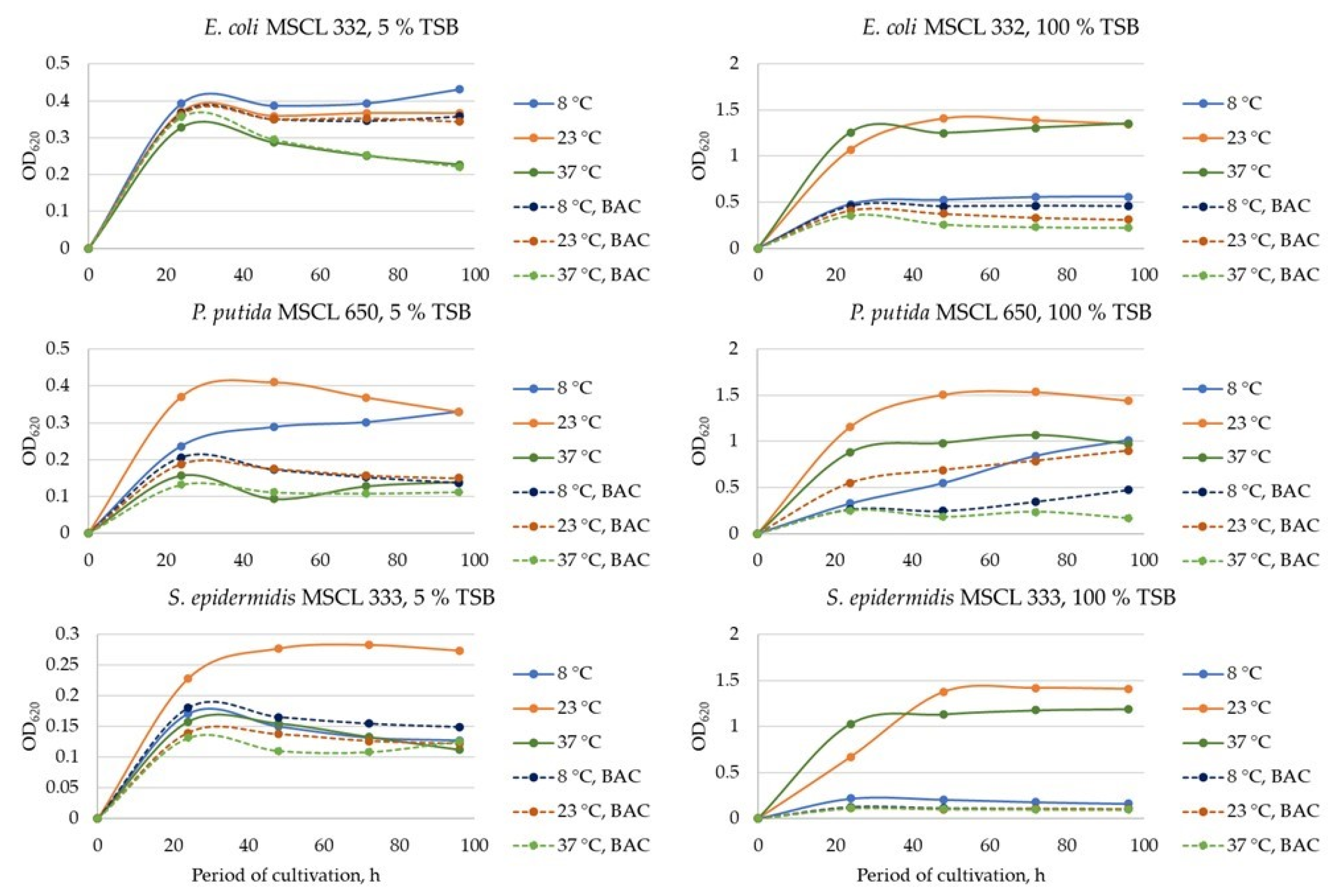
The growth of E. coli MSCL 332, P. putida MSCL 650, and S. epidermidis MSCL 333 under the tested conditions was monitored for 96 h. All three cultures achieved their maximum turbidity after 24 h of cultivation (Figure A1 (see Appendix A)). To reveal the conditions favorable for biofilm formation, the growth intensity of three bacterial cultures after 24 h of cultivation and their biofilm formation after 96 h of cultivation was compared at 8 °C, 23 °C, and 37 °C, in 5% and 100% TSB, as well as in the presence and absence of 50 mg/L BAC (Figure 1). As shown in Figure 1, the highest growth intensity for all three tested cultures was detected after 24 h of cultivation without BAC in 100% TSB at 23 and 37 °C.
Along with thermal regimes, the growth medium composition plays a crucial role in a bacterial culture’s growth activity and resistance to antimicrobials. In our study, the best growth of all tested bacteria was observed in 100% TSB as compared to 5% TSB, which was quite predictable. The bacterial growth in 5% TSB differed in temperature dependence; particularly, the OD620 of P. putida MSCL 650 grown at 23 °C was 2.45 and 1.61 times higher than that at 37 °C and 8 °C, respectively (Figure 1B). The same trend was also detected for S. epidermidis MSCL 333, i.e., the OD620 at 23 °C was 1.44 and 1.35 times higher than that at 37 and 8 °C, respectively (Figure 1C). The concentration of planktonic cells of E. coli MSCL 332 grown in a 5% TSB broth for 24 h at 8 °C, 23 °C, and 37 °C did not show considerable differences (Figure 1A).
Quantification of biofilm in the tested sets was performed to compare the biofilm formation activity in terms of dependence on temperature and availability of nutrients. In the sets with 100% TSB without BAC, the most favorable temperature for biofilm formation in E. coli MSCL 332 and S. epidermidis MSCL 333 cultures was shown to be 37 and 23 °C, respectively. For P. putida MSCL 650, all three tested thermal regimes showed a similar intensity of biofilm formation. In turn, in the sets with 5% TSB, the biofilm of P. putida MSCL 650 and S. epidermidis MSCL 333 was grown to a similar extent at all three temperatures tested, while for biofilm formation in the E. coli MSCL 332 culture, temperatures of 8 °C and 23 °C were more favorable than 37 °C (Figure 1D–F).
The addition of BAC to 100% TSB considerably inhibited the growth of all three cultures, especially at 23 and 37 °C; nevertheless, a comparison of the growth of P. putida MSCL 650 with 50 mg/L BAC at different temperatures showed cultivation at 23 °C to be the most favorable condition to resist BAC. The presence of BAC in 100% TSB broth inhibited the growth of planktonic P. putida MSCL 650 cells by 1.3, 2.1, and 3.5 times at 8 °C, 23 °C and 37 °C, respectively (Figure 1A–C). The inhibition effect of BAC was 1.7, 5.9, and 9.1 times, respectively, in the sets with S. epidermidis MSCL 333 and 1.0, 2.7, and 3.8 times, respectively, in those with E. coli MSCL 332 (Figure 1A–C). The effect of BAC on biofilm formation was negligible in 5% TSB, whereas in 100% TSB, the inhibition or stimulation of biofilm formation was detected. Thus, an inhibitory effect of BAC on biofilm formation was shown for E. coli MSCL 332, P. putida MSCL 650, and S. epidermidis MSCL 333 at 37 °C, i.e., 2.4, 1.8, and 1.6 times, respectively. Biofilm formation in the S. epidermidis MSCL 333 culture in the presence of BAC was also inhibited at 8 °C and 23 °C, i.e., 1.7 and 6.9 times, respectively. Unexpected stimulation of biofilm formation in the presence of BAC was detected in the E. coli MSCL 332 culture grown in 5% TSB at 37 °C (p = 0.0001) and 100% TSB at 8 °C (0.001), i.e., 1.4 and 1.3 times, respectively (Figure 1D–F).
In order to bring the experimental conditions closer to those of drinking water distribution systems, the microorganisms were not only grown in a 5% nutrient-poor medium but also subjected to a regular shaking regime, thus reproducing the water flow/turbulence in a wastewater treatment process. The effects on the growth of planktonic cultures and biofilm formation under static and shaking conditions are presented in Figure A2. The shaking conditions stimulated the growth of planktonic cells in all three bacterial cultures tested in the sets without BAC. In the cultures of S. epidermidis MSCL 333, a slight increase in the culture growth was detected also in the presence of BAC (Figure A2A–C (see Appendix A)). Comparison of shaking and static conditions in terms of biofilm formation did not reveal any notice = able changes, except E. coli MSCL 332 in a 5% TSB, where the shaking significantly (p < 0.05) retarded biofilm formation (Figure A2D–F (see Appendix A)).

3.2. Enzyme Activity of Bacterial Cultures Grown in Different Cultivation Conditions

The measurement of the activity of different enzymatic groups in bacterial cultures supports an in-depth investigation of cell physiological response to adverse conditions. Two enzymatic groups, which are known to reflect different important physiological functions in bacterial cells, i.e., FDA hydrolysis and DHA activity, were tested for planktonic cells. The differences in the FDA hydrolysis activity in the three tested bacterial cultures and their dependence on cultivation conditions are summarised in Figure 2. In particular, E. coli MSCL 332 culture showed a rather low FDA activity over three temperatures and two medium concentrations, except in the set incubated at 8 °C in a 100% TSB without BAC (OD620 = 0.37). The addition of 50 mg/L BAC to the medium inhibited FDA hydrolysis activity, compared with the corresponding conditions without BAC, except the set incubated in a 100% TSB at 23 °C with shaking (Figure 2A,D). The highest FDA hydrolysis activity (OD620 = 0.44) of P. putida MSCL 650 was found in 100% medium at 8 °C (without BAC) and at 23 °C (with BAC). The addition of BAC to 100% TSB resulted in enhanced FDA hydrolysis activity in the P. putida MSCL 650 culture incubated at 23 °C, i.e., OD620 = 0.26 and OD620 = 0.44 in the medium without BAC and with BAC, respectively (Figure 2B,E). A similar trend, i.e., stimulation of the FDA hydrolysis activity by BAC at 23 °C, was also detected for the S. epidermidis MSCL 333 culture, but in 5% TSB without shaking (Figure 2C,F).
The DHA activity of E. coli MSCL 332 peaked after 24 h of incubation, with the highest value being at 23 °C in 100% TSB without shaking, followed by 8 °C in 100% TSB with OD620 values of 2.22 and 1.86, respectively (Figure A3A,D (see Appendix A)). The DHA activity of P. putida MSCL 650 in 100% TSB increased gradually with increasing temperature; in particular, OD620 of 0.89, 1.49, and 1.64 were found after incubation at 8 °C, 23 °C and 37 °C, respectively. Shaking conditions increased the DHA activity of P. putida MSCL 650 slightly at 23 °C, as compared with that under static conditions (Figure A3B,E (see Appendix A)). The DHA activity of S. epidermidis MSCL 333 was detected only after growth in 100% TSB at 23 °C and 37 °C, where the OD620 values reached 1.06 and 2.38, respectively. Shaking conditions in 100% TSB at 23 °C stimulated DHA activity 1.37 times, compared with static conditions (Figure A3C,F (see Appendix A)). The addition of 50 mg/L BAC completely inhibited the growth of the tested bacterial cultures in 100% TSB, except P. putida MSCL 650 grown at 23 °C, where the DHA activity was 3.32 times lower than in the set without BAC, i.e., OD620 = 0.45 and OD620 = 1.49, respectively. All three cultures did not exhibit DHA activity after growth in 5% TSB (Figure A3 (see Appendix A)).

3.3. Statistical Optimization of the Media Variables for P. putida MSCL 650

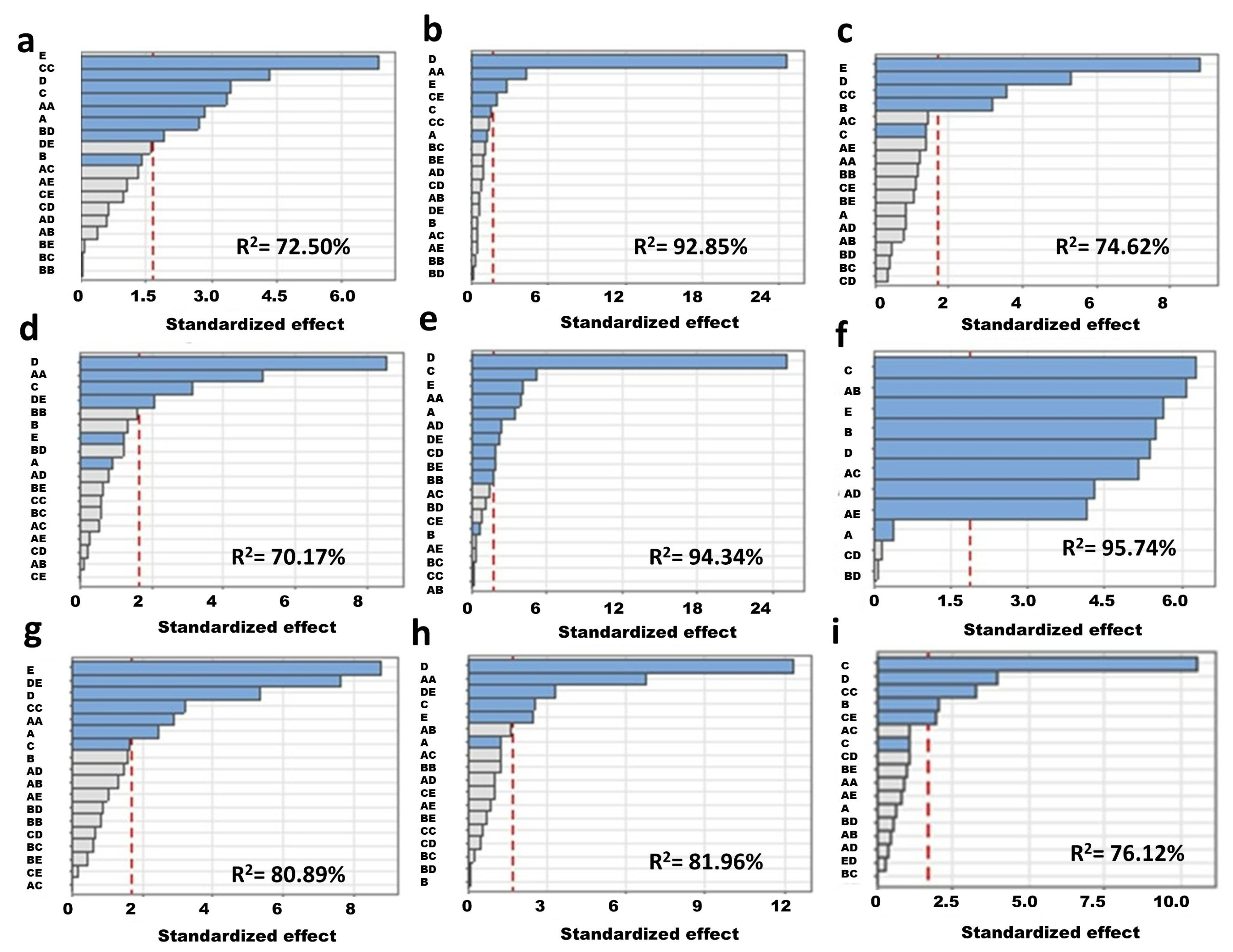
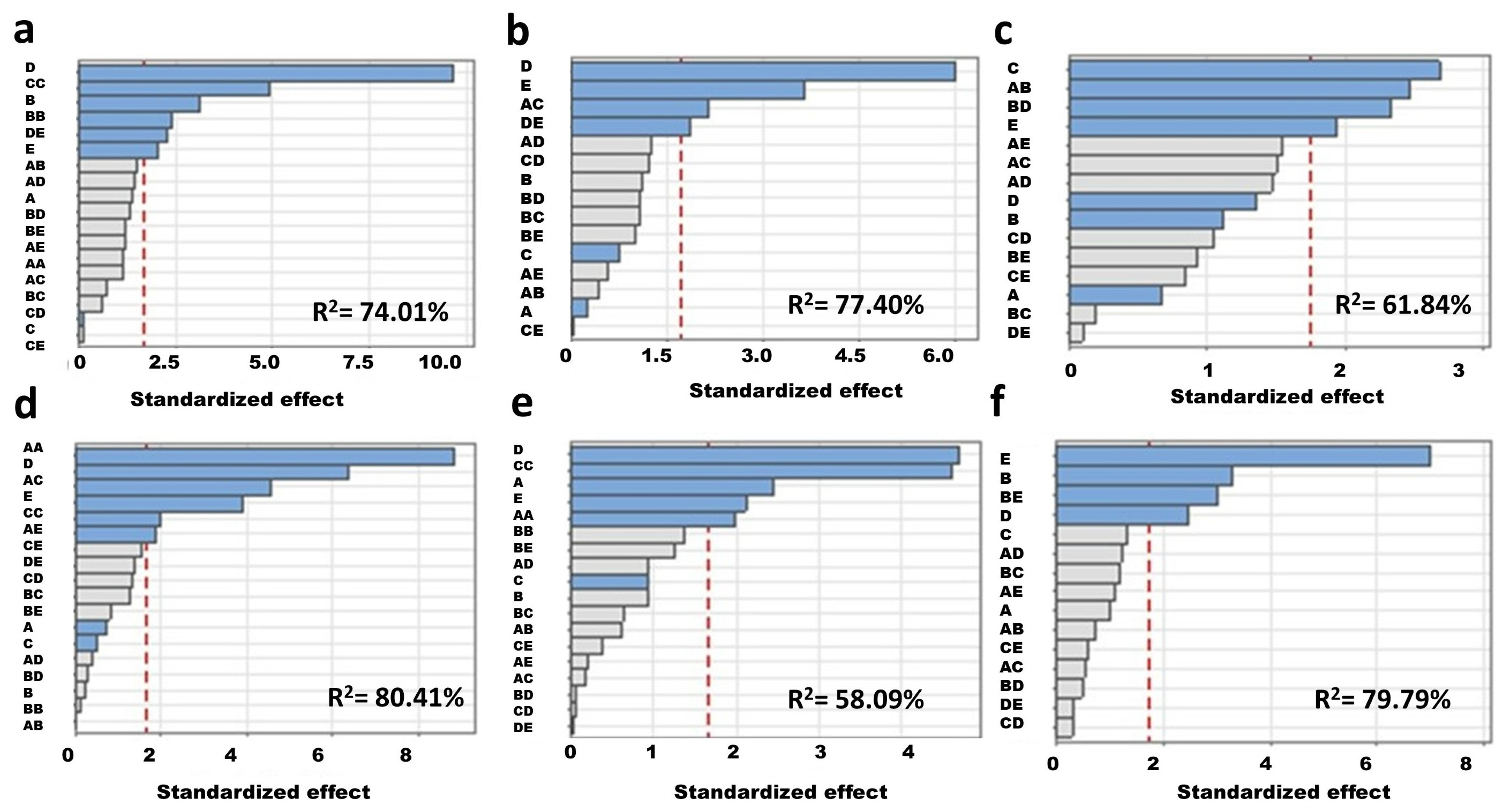
As shown in Figure 1, the biofilm formation activity of the tested bacterial cultures was quite similar at different temperatures but sensitive to nutrient availability. In this respect, optimization of the medium would bring new insight into the bacterial potential of biofilm formation and its resistance to antimicrobials. The next series of experiments were performed with P. putida MSCL 650 in order to analyze the role of five nutrient ingredients, i.e., Ca2+, Mg2+, YE, Try, and Glu, in biofilm formation and its FDA hydrolysis activity. Besides the quantification of biofilm, other outputs were measured, i.e., the growth of planktonic biomass and its FDA hydrolysis and DHA activity. The Pareto charts separate significant factors with the greatest effect on the biofilm quantity and its FDA hydrolysis activity. As shown in Figure 3a,b, in the sets cultivated at 8 °C and 23 °C, the average response of biofilm quantity to tryptone was found to be the highest among the variables. Biofilm formation at 37 °C was dependent mostly on Mg2+ ions (Figure 3c). The FDA hydrolysis activity of immobilized cells was dependent on YE, Try, and Glu in the sets cultivated at 8 °C, 23 °C, and 37 °C, respectively (Figure 3d–f). In turn, for planktonic cells, tryptone was found to be the most significant variable in the growth media composition at 23 °C for three outputs, i.e., biomass, FDA hydrolysis, and DHA activity (Figure A4b,e,h (see Appendix A)). For the sets cultivated at 4 °C, tryptone was significant only for FDA hydrolysis activity, while glucose influenced the biomass and DHA activity (Figure A4a,d,g (see Appendix A)). Glucose also played the most significant role in biomass and DHA activity at 37 °C, Mg2+, for FDA hydrolysis activity (Figure A4c,f,i (see Appendix A)). The optimization results for broth compounds obtained from the Minitab statistical software revealed temperature-specific compositions for different parameters of P. putida MSCL 650 activities. The results for the optimized broth compositions for biofilm formation activity are summarized in Table 1. The surface plots show the predicted optimal levels of two variables, i.e., glucose vs. tryptone (Figure 4A–C) and Ca2+ vs. tryptone (Figure 4D–F), for biofilm formation activity by P. putida MSCL 650. As shown in Figure 4, the biofilm formation activity at 8 °C was dependent on these variables to a greater extent than that at 23 °C and 37 °C.

3.4. Validation of the Optimized Broth Composition

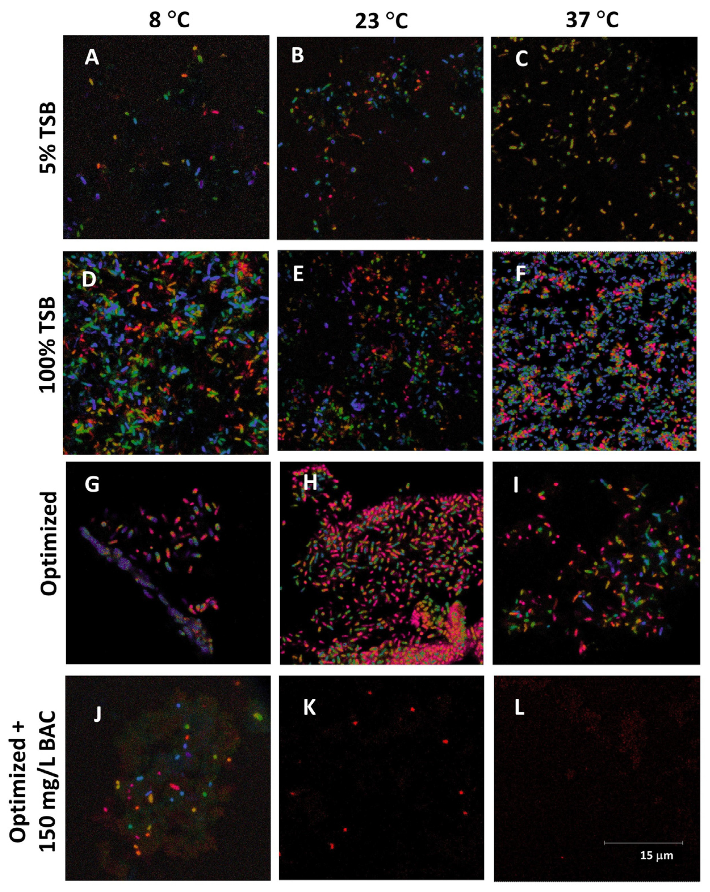
The final step of this study was to test the efficiency of the optimized broth composition for biofilm formation activity by P. putida MSCL 650. The biofilm was quantified after 6 days’ cultivation in 5% and 100% TSB, as well as the modified broth, which has been optimized by CCD. As shown in Figure 5, the optimized broth stimulated biofilm formation, as compared to 5% and 100% TSB, was higher by 133% and 110% at 8 °C and by 378% and 386% at 23 °C, respectively. Interestingly, the culture of P. putida MSCL 650 at 37 °C showed a negative trend in biofilm formation in the optimized broth compared with 5% and 100% TSB and was lower by 34% and 38%, respectively (Figure 5). The biofilm that developed under contrasting nutrient conditions was also tested for bacterial resistance to BAC. The biofilm of P. putida MSCL 650, obtained in 5% and 100% TSB, was inhibited by 50 mg/L BAC at 8 °C and 23 °C, while no effect was found at 37 °C. A further increase in the BAC concentration up to 150 mg/L unexpectedly and gradually increased the amount of biofilm in the sets with optimized broth at all three thermal regimes tested (Figure 5). The planktonic cells of P. putida MSCL 650 were inhibited in the presence of 50–150 mg/L BAC, compared with the medium without BAC. The planktonic cells after 6 days’ cultivation also differed morphologically. The confocal laser scanning micrographs in Figure 6 demonstrate the differences in cell length of P. putida MSCL 650 in dependence on cultivation conditions.
In particular, the average cell length was 1.2 ± 0.3 μm, 1.0 ± 0.3 μm, and 1.2 ± 0.4 μm, respectively, after growth in 5% TSB at 8 °C, 23 ° C and 37 ° C ; 1.4 ± 0.4 μm, 0.7 ± 0.3 μm and 0.7 ± 0.3 μm, respectively, after growth in 100% TSB; and 1.6 ± 0.3 μm, 1.6 ± 0.6 μm, and 1.3 ± 0.7 μm, respectively after growth in the optimized broth. After cultivation in the optimized broth with 150 mg/L BAC, the cell length at 8 ° C and 23 ° C was 1.1 ± 0.5 μm and 0.8 ± 0.2 μm, respectively. The culture after cultivation at 37 °C with BAC was not representative of cell length measurements.

4. Discussion

The results of the present study provide new data on bacterial physiology under different cultivation conditions, i.e., the effect of temperature, nutrient availability, and species-specific characteristics on biofilm formation and resistance to antimicrobials. The first part of this study focused on a comparison of the physiological response of E. coli MSCL 332, P. putida MSCL 650, and S. epidermidis MSCL 333 to variations in temperature and nutrient availability. The highest growth intensity for all three tested cultures was detected after 24 h of cultivation without BAC in 100% TSB at 23 and 37 ° C (Figure 1). The optimum temperature for E. coli is comparatively high, i.e., the mean values for 32 strains of E. coli were found to be in the range from 40.2 to 41.2 ° C [27]; however, these thermal conditions are not relevant to modeling the bacterial activity in the context of wastewater treatment and other environmental processes. In the studies on the response of E. coli to 25 and 37 ° C , differences in phenotypical, biochemical, and morphological characteristics of immobilized and planktonic E. coli cells were shown [28]. Furthermore, the shift in incubation temperature of E. coli from 23 to 37 ° C elicited the expression of stress response genes, thus benefiting the survival of the bacterium within the host [29]. Regarding thermal conditions for the optimum growth and metabolic activity of P. putida, numerous comparative studies showed the highest bacterial activity in the temperature range from 25 to 30 ° C , while more precise data are dependent on the cultivation conditions and experimental setup. In particular, the maximum nitrate removal activity of P. putida was found at 25 ° C [30]; the phenol degradation by P. putida was nine times longer at 10 ° C , compared to the corresponding values at 30 ° C [31]; the production of arginine deiminase by P. putida was higher at 25 °C, compared with 37 ° C [32]; and the optimum growth of P. putida isolated from Antarctic soils was at 16–28 ° C [33]. The optimum temperature for the growth of S. epidermidis in culture was reported to be 37 ° C [34]. These data are in concordance with our recent study [35]. The response of S. epidermidis and other staphylococci to low temperatures was studied earlier by Onyango et al. [36]. After eight weeks of cold stress (4 °C), substantial species-specific alterations in the amino acid composition and increasing proportions of small colony variant phenotypes were observed [36].
The P. putida MCSL 650 strain demonstrated comparatively high resistance to BAC (Figure 1). Pseudomonas spp. strains can naturally withstand the highest concentrations of BACs. Pseudomonas aeruginosa can survive at up to 1200 mg/L BACs without a previous adaptation to it [7]. In the study with planktonic P. fluorescens cells, the death threshold for culturability, membrane integrity, metabolic activity (resazurin), and cellular energy (ATP) was found at 160 mg/L BAC when the cell recovery ability was tested in a nutrient media after BAC exposure assay [20]. Adaptation of P. fluorescens to BAC was shown at 10 mg/L BAC [37].
Unexpected stimulation of biofilm formation in the presence of BAC was detected in the E. coli culture grown in 5% TSB at 37 ° C and 100% TSB at 8 ° C . However, other studies showed a different trend. Thus, as reported previously by Forbes et al. [38], adaptation to BAC at 0.1 or 1.0 mg/L resulted in significantly greater reductions in biofilm formation. The response of E. coli to antimicrobials at different temperatures (i.e., from 22 ° C to 46 ° C ) has recently been studied by Cruz-Loya et al. [22]. The authors reported that antibiotic stress often results in considerable changes in the optimal temperature for the growth of E. coli [22]. On the other hand, enhanced resistance of E. coli O157:H7 antimicrobial treatment at a low temperature was recently reported; in particular, bacteria grown at 4 ° C or 10 ° C were less susceptible to acidic electrolytic water than those cultured at 37 ° C [39].
The resistance of immobilized bacterial cells to QACs was compared in many studies and was shown to be higher than that of planktonic cells. This effect can be explained by the following properties of biofilms: extracellular matrix, composition of the medium, and production of resistance genes in biofilms. The Gram-positive bacterium S. aureus was more sensitive to the studied QACs than Gram-negative bacteria [18,40]. Our results are in good agreement with these data. It is important to note that batch-fed conditions can significantly increase the resistance of bacterial biofilm (e.g., E. coli) to BAC [41]. Below-optimal often leads to a decrease in enzyme activity and slower microbial metabolism. Interestingly, biofilm formation by the tested cultures at 8 ° C was almost as intense as at 23 ° C and 37 ° C , even in the sets with 5% TSB. Moreover, the addition of BAC does not always result in inhibition of biofilm growth (Figure 1D–F). Biofilm development at 8 °C with and without added BAC is important in the context of drinking water supply systems. Biofilm formation in pipes is a significant problem for the drinking water industry as a potential source of bacterial contamination, including pathogens and corrosion of pipes, which often occurs due to the production of different acids [42].
Characterization of the bacterial physiological response to environmental stresses implies the use of different testing parameters, particularly enzyme activity. In this respect, the choice of FDA hydrolysis and DHA assays for that purpose can be considered relevant. The FDA assay relies on the cleavage of fluorescein diacetate by metabolically active bacteria, producing the green fluorescent compound fluorescein. This assay has been applied for the determination of the antimicrobial effect of antibiotics for E. coli, S. aureus, and P. aeruginosa [43]. In the earlier studies with E. coli, the FDA assay showed a positive correlation with the plate counting method [44]. Other authors reported that the FDA hydrolysis assay as a means of detecting active bacteria may be limited to environments rich in eucaryotes and Gram-positive cells [45]. With regard to the measurement of metabolic activity in bacterial biofilms, a comparison of three assays, i.e., the crystal violet assay, ATP-luminescence, and FDA hydrolysis, showed that the crystal violet assay would be more appropriate for cases when the biofilm needs to be quantified irrespective of bacterial cell viability, and the FDA assay for cases when only the viable bacterial cells in the biofilm need to be quantified [46].
The use of DHA activity as a valuable criterion to detect the response of E. coli, P. putida, and S. epidermidis to the changing conditions/growth phase has been reported earlier [47,48,49]. The inhibition of DHA activity in pure Staphylococcus and Pseudomonas species has been used for assessing the antimicrobial effect of plant extracts [50]. The toxicity of aliphatic and aromatic alcohols for P. putida was measured by inhibition of the DHA activity [51]. DHA activity was also used for the evaluation of the potential impact of BACs on the microbial activity of sludge [13,52], as well as the biofilm of P. aeruginosa [53]. Regarding the DHA response to BAC in our study, only P. putida MSCL 650 cultivated in the presence of 50 mg/L BAC at 23 °C showed DHA activity (Figure A3C,E (see Appendix A)). Other enzyme groups have been tested in this study, e.g., quinone reductase (QR). The addition of BAC to 100% TSB resulted in a comparatively high QR activity in the culture of E. coli grown at 8 ° C and P. putida grown at 23 ° C .
Thus, the results discussed above bring new insight into the variability of physiological characteristics of three bacterial cultures in terms of dependence on temperature and the concentration of TSB. Although TSB is considered to be an appropriate medium for the cultivation of E. coli, Pseudomonas spp., and Staphylococcus spp. [54,55,56], other combinations of nutrients could notably influence the potential of bacteria to form biofilm and resist antimicrobials. In our study, it was hypothesized that changes in the proportions of TSB compounds, i.e., tryptone, glucose, Mg2+, and Ca2+, as well as replacement of soya peptone by yeast extract, could reveal novel effects on bacterial growth at different temperatures in the presence of BAC. In this respect, statistical optimization of broth composition with an emphasis on biofilm formation was undertaken. Optimization and further testing were performed with P. putida MSCL 650. The addition of 100 and 150 mg/L BAC to the optimized broth resulted in an unexpected gradual increase in biofilm formation activity. This phenomenon should be investigated in further studies.

5. Conclusions

In summary, the following conclusions are drawn:
-
Planktonic cells of three tested bacterial cultures responded positively to increasing temperature nutrients, while biofilm formation was species-specific and was stimulated by increased temperatures (E. coli, S. epidermidis) and 100% TSB (P. putida).
-
Statistical optimization of broth composition for biofilm formation by P. putida showed the stimulatory effect of tryptone and glucose at the highest concentrations tested in this study, i.e., 17 g/L and 2.5 g/L, respectively. The optimized broth composition was temperature specific.
-
A stimulation effect of 150 mg/L BAC on the biofilm formation activity of P. putida in a 100% TSB and optimized broth need further investigation.
-
The optimized broth composition for biofilm formation by P. putida can be applied to different environmental biotechnological processes where immobilized cells are used. Special attention should be paid to the low-temperature processes.

Author Contributions

Conceptualization, L.Ž. and O.M.; methodology, L.Ž., A.D. and O.M.; software, A.D.; writing—original draft preparation, L.Ž. and O.M.; microscopy visualization, T.S. All authors have read and agreed to the published version of the manuscript.

Funding

This research was funded by the SAM 8.2.2. third round project “Strengthening the capacity of the doctoral program of the University of Latvia in the framework of the new doctoral program model”, VPP-EM-BIOMEDICĪNA-2022/1-001 (Y3-VPP32f-ZR-N-090) “State research project in the field of biomedicine, medical technologies and pharmacy”, Y5-AZ20-ZF-N-270 Optimization of biotechnological processes for effective utilization of renewable resources.

Institutional Review Board Statement

Not applicable.

Informed Consent Statement

Not applicable.

Data Availability Statement

Not applicable.

Conflicts of Interest

The authors declare no conflict of interest.

Appendix A

Figure A1. Growth of E. coli MSCL 332, P. putida MSCL 650 and S. epidermidis MSCL 333 in 5% and 100% TSB at different temperatures, with or without 50 mg/L benzalkonium chloride. Experiment A in Materials and Methods.
Fermentation 09 00699 g0a1
Figure A2. Growth (24 h) (AC) and biofilm formation (96 h) (DF) in E. coli, P. putida and S. epidermidis in 5% and 100% TSB at 23 ° C , with or without 50 mg/L benzalkonium chloride. Experiment A in Materials and Methods.
Fermentation 09 00699 g0a2
Figure A3. The dehydrogenase (DHA) activity of planktonic E. coli, P. putida and S. epidermidis after 24 h incubation in 5% and 100% tryptone soya broth under different conditions. Experiment A in Materials and Methods. (AC) Cultivation at 8 ° C , 23 ° C and 37 ° C ; (DF) cultivation at 23 ° C .
Fermentation 09 00699 g0a3
Figure A4. Pareto chart of standardized effects of factors on planktonic cells of P. putida. Factors: A—yeast extract; B—Ca2+ ions; C—Mg2+ ions; D—tryptone; E—glucose. (ac) OD620 values; (df) the FDA hydrolysis activity; (gi) the DHA activity. (a,d,g) 8 ° C ; (b,e,h) 23 ° C ; (c,f,i) 37 ° C . The R2 value of the variation in the activity of planktonic cells is explained by the model. The red line is the effect size at the 0.10 level of significance. Blue bars represent the terms that are in the model. Gray bars represent non-significant terms that were removed from the model. Experiment B in Materials and Methods.
Fermentation 09 00699 g0a4

References

  1. Vickery, K. Special Issue: Microbial biofilms in healthcare: Formation, prevention and treatment. Materials 2019, 12, 2001. [Google Scholar] [CrossRef] [PubMed] [Green Version]
  2. Liu, S.; Gunawan, C.; Barraud, N.; Rice, S.A.; Harry, E.J.; Amal, R. Understanding, monitoring, and controlling biofilm growth in drinking water distribution systems. Environ. Sci. Technol. 2016, 50, 8954–8976. [Google Scholar] [CrossRef]
  3. Servais, P.; Laurent, P.; Randon, G. Comparison of the bacterial dynamics in various French distribution systems. Aqua London 1995, 44, 10–17. [Google Scholar]
  4. Giorgi, F.; Curran, J.M.; Patterson, E.A. Real-time monitoring of the dynamics and interactions of bacteria and the early-stage formation of biofilms. Sci. Rep. 2022, 12, 18146. [Google Scholar] [CrossRef]
  5. Suresh, M.K.; Biswas, R.; Biswas, L. An update on recent developments in the prevention and treatment of Staphylococcus aureus biofilms. Int. J. Med. Microbiol. 2019, 309, 1–12. [Google Scholar] [CrossRef] [PubMed]
  6. Global Demand for Disinfectants and Antiseptics Predicted to Increase. 2021. Available online: http://www.issa.com/media/news/global-demand-for-disinfectants-and-antiseptics-predicted-to-increase (accessed on 22 June 2023).
  7. Ioannou, C.J.; Hanlon, G.W.; Denyer, S.P. Action of disinfectant quaternary ammonium compounds against Staphylococcus aureus. Antimicrob. Agents Chemother. 2007, 51, 296–306. [Google Scholar] [CrossRef] [Green Version]
  8. Basiry, D.; Heravi, N.E.; Uluseker, C.; Kaster, K.M.; Kommedal, R.; Pala-Ozkok, I. The effect of disinfectants and antiseptics on co- and cross-selection of resistance to antibiotics in aquatic environments and wastewater treatment plants. Front. Microbiol. 2022, 13, 1050558. [Google Scholar] [CrossRef] [PubMed]
  9. Henly, E.L.; Dowling, J.A.R.; Maingay, J.B.; Lacey, M.M.; Smith, T.J.; Forbes, S. Biocide exposure induces changes in susceptibility, pathogenicity, and biofilm formation in uropathogenic Escherichia coli. Antimicrob. Agents Chemother. 2019, 63, e01892-18. [Google Scholar] [CrossRef] [Green Version]
  10. Møretrø, T.; Schirmer, B.C.T.; Heir, E.; Fagerlund, A.; Hjemli, P.; Langsrud, S. Tolerance to quaternary ammonium compound disinfectants may enhance growth of Listeria monocytogenes in the food industry. Int. J. Food Microbiol. 2017, 241, 215–224. [Google Scholar] [CrossRef] [Green Version]
  11. Khelissa, S.; Gharsallaoui, A.; Fadel, A.; Barras, A.; Jama, C.; Jbilou, F.; Chihib, N.-E. Microencapsulation of benzalkonium chloride enhanced its antibacterial and antibiofilm activities against Listeria monocytogenes and Escherichia coli. J. Appl. Microbiol. 2021, 131, 1136–1146. [Google Scholar] [CrossRef]
  12. Yu, T.; Ma, M.; Sun, Y.; Xu, X.; Qiu, S.; Yin, J.; Chen, L. The effect of sublethal concentrations of benzalkonium chloride on the LuxS/AI-2 quorum sensing system, biofilm formation and motility of Escherichia coli. Int. J. Food Microbiol. 2021, 353, 109313. [Google Scholar] [CrossRef] [PubMed]
  13. Chen, M.; Zhang, X.; Wang, Z.; Liu, M.; Wang, L.; Wu, Z. Impacts of quaternary ammonium compounds on membrane bioreactor performance: Acute and chronic responses of microorganisms. Water Res. 2018, 134, 153–161. [Google Scholar] [CrossRef] [PubMed]
  14. Kaur, B.; Gupta, J.; Sharma, S.; Sharma, D.; Sharma, S. Focused review on dual inhibition of quorum sensing and efflux pumps: A potential way to combat multi drug resistant Staphylococcus aureus infections. Int. J. Biol. Macromol. 2021, 190, 33–43. [Google Scholar] [CrossRef]
  15. Turchi, B.; Bertellomi, F.; Marzoli, F.; Cerri, D.; Tola, S.; Azara, E.; Longheu, C.M.; Tassi, R.; Schiavo, M.; Ciliza, G.; et al. Coagulase negative staphylococci from ovine milk: Genotypic and phenotypic characterization of susceptibility to antibiotics, disinfectants and biofilm production. Small Rumin. Res. 2020, 183, 106030. [Google Scholar] [CrossRef]
  16. Merchel Piovesan Pereira, B.; Wang, X.; Tagkopoulos, I. Biocide-induced emergence of antibiotic resistance in Escherichia coli. Front. Microbiol. 2021, 12, 640923. [Google Scholar] [CrossRef]
  17. Doucet, A.N.; Slipski, C.J.; Golding, G.R.; Mulvey, M.R.; Bay, D.C. Generation of greater bacterial biofilm biomass using PCR-Plate deep well microplate devices. J. Vis. Exp. 2022, 182, e63069. [Google Scholar] [CrossRef]
  18. Nunez, C.; Kostoulias, X.; Peleg, A.; Short, F.; Qu, Y. A comprehensive comparison of biofilm formation and capsule production for bacterial survival on hospital surfaces. Biofilm 2023, 5, 100105. [Google Scholar] [CrossRef]
  19. Sapozhnikov, S.V.; Sabirova, A.E.; Shtyrlin, N.V.; Druk, A.Y.; Agafonova, M.N.; Chirkova, M.N.; Kazakova, R.R.; Grishaev, D.Y.; Nikishova, T.V.; Krylova, E.S.; et al. Design, synthesis, antibacterial activity and toxicity of novel quaternary ammonium compounds based on pyridoxine and fatty acids. Eur. J. Med. Chem. 2021, 211, 113100. [Google Scholar] [CrossRef]
  20. Barros, A.C.; Melo, L.F.; Pereira, A. Pseudomonas fluorescens cells’ recovery after exposure to BAC and DBNPA biocides. Antibiotics 2022, 11, 1042. [Google Scholar] [CrossRef]
  21. Rowson, C.; Townsend, R. Biofilms: Prevention and treatment. Br. J. Hosp. Med. 2016, 77, 699–703. [Google Scholar] [CrossRef] [PubMed]
  22. Cruz-Loya, M.; Tekin, E.; Kang, T.M.; Cardona, N.; Lozano-Huntelman, N.; Rodriguez-Verdugo, A.; Savage Van, M.; Yen, P.J. Antibiotics shift the temperature response curve of Escherichia coli growth. mSystems 2021, 6, 2–21. [Google Scholar] [CrossRef]
  23. Amir, M.; Bano, N.; Baker, A.; Zia, Q.; Banawas, S.; Zaheer, M.R.; Shariq, M.; Nawaz, M.S.; Khan, M.F.; Azad, Z.R.A.A.; et al. Isolation and optimization of extracellular PHB depolymerase producer Aeromonas caviae Kuk1-(34) for sustainable solid waste management of biodegradable polymers. PLoS ONE 2022, 17, e0264207. [Google Scholar] [CrossRef] [PubMed]
  24. Peterson, S.B.; Irie, Y.; Borlee, B.R.; Murakami, K.; Harrison, J.J.; Colvin, K.M.; Parsek, M.R. Different methods for culturing biofilms in vitro. In Biofilm Infections; Springer: New York, NY, USA, 2011; pp. 251–266. [Google Scholar] [CrossRef]
  25. Chen, W.; Hotink, H.; Schmithenner, A.F.; Tuovinen, O.H. The role of microbial activity in suppression of damping-off caused by Pythium ultimum. Phytopathology 1988, 78, 314–322. [Google Scholar] [CrossRef]
  26. Camiña, F.; Trasar-Cepeda, C.; Gil-Sotres, F.; Leirós, C. Measurement of dehydrogenase activity in acid soils rich in organic matter. Soil Biol. Biochem. 1998, 30, 1005–1011. [Google Scholar] [CrossRef]
  27. Gonthier, A.; Guérin-Faublée, V.; Tilly, B.; Delignette-Muller, M.L. Optimal growth temperature of O157 and non-O157 Escherichia coli strains. Lett. Appl. Microbiol. 2001, 33, 352–356. [Google Scholar] [CrossRef] [Green Version]
  28. Kikuchi, T.; Mizunoe, Y.; Takade, A.; Naito, S.; Yoshida, S.I. Curli fibers are required for development of biofilm architecture in Escherichia coli K-12 and enhance bacterial adherence to human uroepithelial cells. Microbiol. Immunol. 2005, 49, 875–884. [Google Scholar] [CrossRef]
  29. Kanegusuku, A.G.; Stankovic, I.N.; Cote-Hammarlof, P.A.; Yong, P.H.; White-Ziegler, C.A. A shift to human body temperature (37 °C) rapidly reprograms multiple adaptive responses in Escherichia coli that would facilitate niche survival and colonization. J. Bacteriol. 2021, 203, e0036321. [Google Scholar] [CrossRef]
  30. El-Sesy, M.E.; Ibrahim, S.S. Application of central composite design approach for optimization nitrate removal from aqueous solution by immobilized Pseudomonas putida. Water Sci. Technol. 2021, 83, 2931–2946. [Google Scholar] [CrossRef] [PubMed]
  31. Nowak, A.; Żur-Pińska, J.; Piński, A.; Pacek, G.; Mrozik, A. Adaptation of phenol-degrading Pseudomonas putida KB3 to suboptimal growth condition: A focus on degradative rate, membrane properties and expression of xylE and cfaB genes. Ecotoxicol. Environ. Saf. 2021, 221, 112431. [Google Scholar] [CrossRef]
  32. Patil, M.D.; Shinde, K.D.; Patel, G.; Chisti, Y.; Banerjee, U.C. Use of response surface method for maximizing the production of arginine deiminase by Pseudomonas putida. Biotechnol. Rep. 2016, 10, 29–37. [Google Scholar] [CrossRef] [Green Version]
  33. Holovan, V.; Andriichuk, O.; Budzanivska, I.; Zelena, P.; Kondratiuk, T.; Shevchenko, O. Bacteriophages and their microbial hosts in terrestrial biotopes of Antarctica. Antarct. Sci. 2022, 34, 120–136. [Google Scholar] [CrossRef]
  34. Baidamshina, D.R.; Koroleva, V.A.; Olsannikova, S.S.; Trizna, E.Y.; Bogachev, M.I.; Artyukhov, V.G.; Holyavka, M.G.; Kayumov, A.R. Biochemical properties and anti-biofilm activity of chitosan-immobilized papain. Mar. Drugs 2021, 19, 197. [Google Scholar] [CrossRef] [PubMed]
  35. Vibornijs, V.; Liepins, J.; Selga, T.; Bankovskis, V.; Cosemans, P.; Muter, O. Comparison of the antibacterial effect of a copper-coated surface on Staphylococcus epidermidis and Pseudomonas putida in different physiological states. IOP Conf. Ser. Mater. Sci. Eng. 2021, 1186, 012004. [Google Scholar] [CrossRef]
  36. Onyango, L.A.; Dunstan, R.H.; Gottfries, J.; von Eiff, C.; Roberts, T.K. Effect of low temperature on growth and ultra-structure of Staphylococcus spp. PLoS ONE 2012, 7, e29031. [Google Scholar] [CrossRef] [Green Version]
  37. Dynes, J.J.; Lawrence, J.R.; Korber, D.R.; Swerhone, G.D.W.; Leppard, G.G.; Hitchcock, A.P. Morphological and biochemical changes in Pseudomonas fluorescens biofilms induced by sub-inhibitory exposure to antimicrobial agents. Can. J. Microbiol. 2009, 55, 163–178. [Google Scholar] [CrossRef]
  38. Forbes, S.; Morgan, N.; Humohreys, G.J.; Amezquita, A.; Mistry, H.; McBain, A.J. Loss of function in Escherichia coli exposed to environmentally relevant concentrations of benzalkonium chloride. Appl. Environ. Microbiol. 2019, 85, e02417-18. [Google Scholar] [CrossRef] [Green Version]
  39. Yu, M.; Jiang, C.; Meng, Y.; Wang, F.; Qian, J.; Fei, F.; Yin, Z.; Zhao, W.; Zhao, Y.; Liu, H. Effect of low temperature on the resistance of Listeria monocytogenes and Escherichia coli O157:H7 to acid electrolyzed water. Food Res. Int. 2023, 168, 112776. [Google Scholar] [CrossRef] [PubMed]
  40. Frolov, N.; Detusheva, E.; Fursova, N.; Ostashevskaya, I.; Vereshchagin, A. Microbiological Evaluation of Novel Bis-Quaternary Ammonium Compounds: Clinical Strains, Biofilms, and Resistance Study. Pharmaceuticals 2022, 15, 514. [Google Scholar] [CrossRef]
  41. Kocot, A.M.; Wróblewska, B.; Cabo, M.L. Operational culture conditions determinate benzalkonium chloride resistance in L. monocytogenes-E. coli dual species biofilms. Int. J. Food Microbiol. 2021, 360, 109441. [Google Scholar] [CrossRef]
  42. Fu, Y.; Peng, H.; Liu, J.; Nguyen, T.H.; Hashmi, M.Z.; Shen, C. Occurrence and quantification of culturable and viable but non-culturable (VBNC) pathogens in biofilm on different pipes from a metropolitan drinking water distribution system. Sci. Total Environ. 2021, 764, 142851. [Google Scholar] [CrossRef]
  43. Wanandy, S.; Brouwer, N.; Liu, Q.; Mahon, A.; Cork, S.; Karuso, P.; Vemulpad, S.; Jamie, J. Optimisation of the fluorescein diacetate antibacterial assay. J. Microbiol. Methods 2005, 60, 21–30. [Google Scholar] [CrossRef] [PubMed]
  44. Jayapal, V.; Sharmila, K.M.; Selvibai, G.; Thyagarajan, S.P.; Shanmugasundaram Subramanian, N.S. Fluorescein diacetate and ethidium bromide staining to determine the viability of Mycobacterium smegmatis and Escherichia coli. Lepr. Rev. 1991, 62, 310–314. [Google Scholar] [PubMed]
  45. Chrzanowski, T.H.; Crotty, R.D.; Hubbard, J.G.; Welch, R.P. Applicability of the fluorescein diacetate method of detecting active bacteria in freshwater. Microb. Ecol. 1984, 10, 179–185. [Google Scholar] [CrossRef]
  46. Koreňová, J.; Lopašovská, J.; Kuchta, T. Comparison of three microtitre plate-based methods for quantification of biofilm formation ability of bacteria contaminating food technologies. J. Food Nutr. Res. 2008, 47, 100–104. [Google Scholar]
  47. Dmitrieva, M.V.; Zolotukhina, E.V.; Gerasimova, E.V.; Terent’ev, A.A.; Dobrovol’skii, Y.A. Dehydrogenase and electrochemical activity of Escherichia coli extracts. Appl. Biochem. Microbiol. 2017, 53, 458–463. [Google Scholar] [CrossRef]
  48. Uribe-Alvarez, C.; Chiquete-Felix, N.; Contreras-Zentella, M.; Guerro-Castilo, S.; Pena, A.; Uribe-Carvajal, S. Staphylococcus epidermidis: Metabolic adaptation and biofilm formation in response to different oxygen concentrations. Pathog. Dis. 2018, 74, ftv111. [Google Scholar] [CrossRef] [Green Version]
  49. Michalska, J.; Piński, A.; Zur, J.; Mrozik, A. Analysis of the bioaugmentation potential of Pseudomonas putida OR45a and Pseudomonas putida KB3 in the sequencing batch reactors fed with the phenolic landfill leachate. Water 2020, 12, 906. [Google Scholar] [CrossRef] [Green Version]
  50. Alisi, C.S.; Nwanyanwu, C.E.; Akujobi, C.O.; Ibegbulem, C.O. Inhibition of dehydrogenase activity in pathogenic bacteria isolates by aqueous extracts of Musa paradisiaca (var sapientum). Afr. J. Biotechnol. 2008, 7, 1821–1825. [Google Scholar] [CrossRef]
  51. Gül, Ş.; Öztürk, D. Determination of structure-toxicity relationship of amphiprotic compounds by means of the inhibition of the dehydrogenase activity of Pseudomonas putida. Turk. J. Chem. 1998, 22, 341–349. [Google Scholar]
  52. An, H.; Ren, J.; Ma, J.; Li, Z.; Liu, Y.; Liu, X. Effect of benzalkonium chloride on microbial activity of activated sludge. Chin. J. Environ. Eng. 2020, 14, 2701–2709. [Google Scholar] [CrossRef]
  53. Huang, Z.; Qi, Z.; Ding, X.; Liu, C. N-chlorosuccinimide enhancing the antimicrobial effect of benzalkonium chloride on biofilm Pseudomonas aeruginosa and its interaction mechanisms. J. Environ. Sci. Health Part A Toxic Hazard. Subst. Environ. Eng. 2022, 57, 593–600. [Google Scholar] [CrossRef]
  54. Hepburn, N.F.; MacRae, M.; Johnston, M.; Mooney, J.; Ogden, I.D. Optimizing enrichment conditions for the isolation of Escherichia coli O157 in soils by immunomagnetic separation. Lett. Appl. Microbiol. 2022, 34, 365–369. [Google Scholar] [CrossRef] [PubMed]
  55. Abd El-Al, M.S.; Saeed, A.M.; Hammad, A.A.; Swailam, H.M.; Abouzeid, M.A. Gamma-irradiation induced effects on histamine-forming bacteria isolated from the chilled mackerel fish. Egypt. J. Aquat. Biol. Fish. 2022, 26, 869–883. [Google Scholar] [CrossRef]
  56. Ma, D.; Jiang, Y.; Ahmed, S.; Qin, W.; Liu, Y. Physical and antimicrobial properties of edible films containing Lactococcus lactis. Int. J. Biol. Macromol. 2019, 141, 378–386. [Google Scholar] [CrossRef] [PubMed]
Figure 1. Planktonic growth (24 h) (AC) and biofilm formation (5 days) (DF) of three bacterial cultures—E. coli MSCL 332, P. putida MSCL 650, and S. epidermidis MSCL 333—in 5% and 100% tryptone soya broth, at different temperatures, with and without 50 mg/L benzalkonium chloride (BAC). Experiment A in Materials and Methods. Error bars indicate standard deviation (95% confidence interval).
Fermentation 09 00699 g001
Figure 2. The FDA hydrolysis activity of planktonic E. coli MSCL 332, P. putida MSCL 650, and S. epidermidis MSCL 333 after a 48 h incubation in 5% and 100% TBS, in the presence and absence of 50 mg/L BAC, at 8 ° C , 23 ° C and 37 ° C . (AC) static conditions; (DF) static vs. shaking conditions at 23 °C. Experiment A in Materials and Methods. Error bars, as in Figure 1.
Fermentation 09 00699 g002
Figure 3. Pareto chart of standardized effects of factors on biofilm of P. putida MSCL 650. Factors: A—yeast extract; B—Ca2+ ions; C—Mg2+ ions; D—tryptone; E—glucose. (ac) The relative number of immobilized cells; (df) the FDA hydrolysis activity of immobilized cells. (a,d) 8 ° C ; (b,e) 23 ° C ; (c,f) 37 ° C . The R2 value of the variation in biofilm quantification explained by the model. The red line is the effect size at the 0.10 level of significance. Blue bars represent the terms that are in the model. Gray bars represent non-significant terms that were removed from the model. Experiment B in Materials and Methods.
Fermentation 09 00699 g003
Figure 4. Response surface plots of biofilm formation activity by P. putida MSCL 650 as affected by tryptone and glucose (AC) and tryptone and Ca2+ (DF). The scale of Y axis (i.e., Biofilm) corresponds to the optical density at 620 nm. Experiment B in Materials and Methods.
Fermentation 09 00699 g004
Figure 5. The biofilm formation activity of P. putida MSCL 650 after 6 days’ cultivation in 5% and 100% tryptone soya broth, as well as optimized broth at 8 ° C , 23 ° C , and 37 ° C with addition of BAC (0; 50; 100; 150 mg/L). Experiment C in Materials and Methods. Error bars, as in Figure 1.
Fermentation 09 00699 g005
Figure 6. Confocal laser scanning micrographs of planktonic cells of P. putida MSCL 650 after 6 days’ cultivation in 5% TSB (AC), 100% TSB (DF), optimized broth (GI), and optimized broth with 150 mg/L BAC (JL) at 8 ° C (A,D,G,J), 23 ° C (B,E,H,K), and 37 ° C (C,F,I,L). Experiment C in Materials and Methods. Cells were stained with propidium iodide (PI) at concentration of 20 mM. The excitation wavelength was 488 nm, and emission was observed at 600–640 nm. The field of view was scanned in three dimensions, using 20 optical sections separated in depth by 10 μm. Objective, 100×; NA, 1.25. Color-coded 3D projection. Blue corresponds to the top, green corresponds to the middle, and red corresponds to the bottom of the specimen. Bar 15 μm.
Fermentation 09 00699 g006
Table 1. Concentrations of five variables in the broth found by CCD for biofilm formation by P. putida MSCL 650.
TemperatureCa2+, mMMg2+, mMYeast Extract, g/LTryptone, g/LGlucose, g/L
8 °C1.001.952.5017.002.50
23 °C1.0010.000.0017.002.50
37 °C1.000.002.503.400.50
Disclaimer/Publisher’s Note: The statements, opinions and data contained in all publications are solely those of the individual author(s) and contributor(s) and not of MDPI and/or the editor(s). MDPI and/or the editor(s) disclaim responsibility for any injury to people or property resulting from any ideas, methods, instructions or products referred to in the content.