Communication

Cloning, Expression, and Characterization of Family A DNA Polymerase from Massilia aurea

by
Aleksandra A. Kuznetsova
1,
Ksenia S. Bedritskikh
2,
Anatoly A. Bulygin
1,2 and
Nikita A. Kuznetsov
1,2,*
1
Institute of Chemical Biology and Fundamental Medicine (ICBFM), Siberian Branch of Russian Academy of Sciences (SB RAS), 8 Prospekt Akad. Lavrentyeva, Novosibirsk 630090, Russia
2
Department of Natural Sciences, Novosibirsk State University, 2 Pirogova Str., Novosibirsk 630090, Russia
*
Author to whom correspondence should be addressed.
Fermentation 2023, 9(7), 650; https://doi.org/10.3390/fermentation9070650
Submission received: 20 June 2023 / Revised: 4 July 2023 / Accepted: 7 July 2023 / Published: 10 July 2023
(This article belongs to the Special Issue Fermentation: Screening, Enzyme Induction and Production)

Abstract

:
Mau DNA polymerase is a family A DNA polymerase isolated from Massilia aurea. In this study, a recombinant plasmid, His6-tagged Mau-pET28c, was constructed. His-tagged Mau was expressed in Escherichia coli Rosseta 2 (DE3) competent cells and, after optimization of purification conditions, was successfully isolated via a two-step purification system by Ni2+-chelating affinity chromatography followed by heparin affinity chromatography. The biochemical properties of Mau DNA polymerase were investigated next. This polymerase showed maximal polymerase activity at 30 °C, pH 8.4–8.8, 2–10 mM MgCl2, and 10–40 mM KCl. Kinetic parameters of correct and incorrect dNTP incorporation as well as DNA-binding affinity were determined too. KdNTPd,app values were found to be 16 µM for correct dNTP and 200–500 µM for incorrect dNTP. The kinetic parameter kcat turned out to be 0.2 s−1 for correct dNTP incorporation and an order of magnitude less for incorrect dNTP incorporation. It was demonstrated that Mau DNA polymerase has 5′→3′ and 3′→5′ exonuclease activities associated with the main activity.

1. Introduction

DNA polymerases are a class of enzymes responsible for DNA replication. Polymerases differ in size, structure, the need for auxiliary proteins, and their role in DNA replication and repair. Their specific properties differ too, including processivity, fidelity, and selectivity toward incorporated nucleotides [1,2,3,4,5,6,7]. In addition to their fundamental role in the maintenance of genome integrity during replication and repair, DNA polymerases are widely used for in vitro DNA manipulations, including DNA cloning, sequencing, labeling, mutagenesis, and other purposes [8,9,10]. Thermostable DNA polymerases are especially important for molecular-biological techniques related to polymerase chain reaction (PCR). In practice, polymerases of families A and B are used in PCR [11,12]. PCR is a powerful and sensitive method for DNA amplification and has found abundant applications in various fields such as molecular biology, diagnostics, forensics, and food safety control [13].
There is a constant search for new enzymes for genetic technologies that may possess improved properties as compared to the existing ones. Modification of existing polymerases is also widely employed [14]. Currently, the modifications intended to optimize polymerases are mostly based on the improvement of reaction buffers, the use of PCR enhancers, and site-directed mutagenesis of the enzymes. Mutations can lead to enzymes with higher thermal stability and resistance to inhibitors [15,16,17,18].
At present, DNA polymerases from families A and B are actively used in PCR. The best-known representative of family A is Taq polymerase from Thermus aquaticus, and the best-known representative of family B is Pfu polymerase from Pyrococcus furiosus. By genetic engineering methods, various mutants of these enzymes have been obtained to improve their catalytic properties [16,19,20]. Despite promising results, there are still limitations to PCR: the product yield, the length of DNA that can be amplified, polymerase processivity, and the fidelity of the amplification. Ultimately, the duration of PCR depends solely on the kinetic properties of the enzyme. In practice, researchers often encounter difficulties when trying to amplify DNA found in natural materials. The presence of enzyme-inhibiting substances in biological samples reduces or blocks the capacity for PCR amplification in contrast to pure nucleic-acid solutions. Furthermore, DNA can contain sequences that form strong secondary structures that can block amplification [18]. At the moment, there is no “universal” DNA polymerase capable of overcoming such obstacles.
Finding new thermostable DNA polymerases is a nontrivial task. At present, there is no full understanding of the reasons for an enzyme’s thermostability; therefore, the search for thermostable DNA polymerases is performed in thermophilic organisms whose optimal habitat temperature is not lower than 50–60 °C. Nonetheless, organisms that live at elevated temperatures, up to 80 °C, have polymerases that are functional at such temperatures, but their stability significantly declines at higher temperatures. On the other hand, it can be hypothesized that an enzyme in some microorganisms can remain stable at temperatures higher than the optimal growth temperature of this organism.
In our previous work [21], we conducted a comparative sequence analysis of family A DNA polymerases from the bacteria available in the Collection of Extremophilic Microorganisms and Type Cultures (CEMTC, Novosibirsk, Russia). We selected 47 microorganisms, which were collected from a medium with a high temperature (>50 °C). Prior to that, it generated a “functional framework” containing 62 residues, which, according to the 3D structure of the KlenTaq polymerase fragment, directly interact with DNA, dNTP, and metal ions and, thereby, are potentially important for the polymerase activity. A pairwise comparison of all sequences using the “functional framework” revealed that DNA polymerases share > 70% similarity to the functional framework of Thermus enzymes [21]. It is noteworthy that only one of the DNA polymerases of these microorganisms has been characterized, namely the B. licheniformis DNA polymerase [22].
Therefore, DNA polymerase from Massilia aurea, which has 79.8% similarity to the “functional framework” of Thermus enzymes [21], was selected in the present study for characterization of enzyme properties. M. aurea is a gram-negative, motile, rod-shaped bacterium able to form yellow-pigmented colonies; it has been isolated from the tap water system of Seville (Spain) and has been characterized via a polyphasic taxonomic approach [23]. The specimen of Massilia aurea stored in the CEMTC has been isolated from thermal water (74 °C) collected in the caldera of the Uzon volcano (Kamchatka, Russia). In this study, the gene encoding a full-length DNA polymerase from M. aurea (Mau DNA polymerase) was cloned and expressed in Escherichia coli. We report the purification protocol and biochemical properties of the enzyme.

2. Materials and Methods

2.1. Bacterial Strain

An M. aurea strain was obtained from the CEMTC of the ICBFM SB RAS http://www.niboch.nsc.ru/doku.php/emtc_collection (accessed on 5 December 2022).

2.2. Construction of a Plasmid Encoding Mau DNA Polymerase

The full-length Mau DNA polymerase gene (UniProt ID A0A422QJZ7) was amplified using genomic DNA and two primers (the N-terminal primer: P5_Mau_NdeI, 5′-GTCCATATGACCCTGCTGCTCGTTGACGGTTCC-3′, and the C-terminal primer: P3_Mau_BamHI, 5′-GCGGATCCTCAGTGCGCCTCTTCCCAGTTCTTGCC-3′; the underlined sequences denote the NdeI site within the N-terminal primer and the BamHI site within the C-terminal primer). DNA amplification was carried out in a 20-microliter reaction mixture: Pfu DNA polymerase buffer, 1.25 mM dNTP, 20 pmol of each primer (P5_Mau_NdeI and P3_Mau_BamHI), 20 ng of Mau genomic DNA, and 1 U of Pfu DNA polymerase (SibEnzyme Ltd., Novosibirsk, Russia). The PCR program consisted of one initial denaturation step at 94 °C for 4 min, followed by 30 cycles of 98 °C for 40 s, 55 °C for 40 s, and 72 °C for 5 min; the final extension was performed at 72 °C for 10 min. The amplified 2.7-kbp PCR product was then digested with FauNDI (an isoschizomer of NdeI restriction enzyme, SibEnzyme Ltd., Novosibirsk, Russia) and BamHI (SibEnzyme Ltd., Novosibirsk, Russia), purified from a 0.8% low-melting agarose gel using the Cleanup S-Cap Kit (Evrogen, Moscow, Russia), and ligated into the pET-28c expression vector that had been digested with the same enzymes. The ligation mixture was then used to transform E. coli strain Emax. Clones with the correct construct were selected through restriction enzyme analysis and sequencing.

2.3. Purification of Mau DNA Polymerase

E. coli strain Rosseta 2 (DE3) was transformed with pET28c-Mau and grown overnight at 37 °C in 15 mL of LB Broth containing 50 mg/mL kanamycin as a starter culture. The starter was added to 1 L of LB Broth containing 50 mg/mL kanamycin, and the cells were grown with shaking at 37 °C to an optical density at 600 nm (OD600) of 0.6–0.8. Overexpression of the Mau DNA polymerase gene was induced by the addition of isopropyl-β-D-thiogalactopyranoside (IPTG) to a final concentration of 0.2 mM, followed by an additional 3 h of growth at 20 °C. The cells were harvested by centrifugation at 4000× g for 20 min at 4 °C and resuspended in 20 mM HEPES-KOH buffer (pH 7.8) containing 40 mM NaCl. The cells were disrupted by means of a French press, and cellular debris was removed by centrifugation at 40,000× g for 40 min at 4 °C. To the resulting supernatant, a solution of NaCl and imidazole was added at concentrations of 500 and 20 mM, respectively. This solution was mixed with 1 mL of the Ni SepharoseTM High Performance resin (Amersham Biosciences, Uppsala, Sweden) and stirred for 1 h at 4 °C. The enzyme was eluted with 5 mL of a buffer consisting of 20 mM HEPES-NaOH pH 7.8, 500 mM NaCl, and 600 mM imidazole. The obtained enzyme-containing fraction was diluted to a final NaCl concentration of 40 mM and applied to a HiTrap-Heparin™ column (Amersham Biosciences, Uppsala, Sweden) at a flow rate of 0.4 mL/min. The chromatography was performed in a buffer composed of 20 mM HEPES-NaOH pH 7.8 and a linear 40 → 1000 mM gradient of NaCl; the OD280 of the solution was recorded. The purity of the Mau protein was determined by gel electrophoresis. Fractions containing the Mau DNA polymerase were collected, and a glycerol solution was added to attain a 50% final concentration of glycerol. The enzyme was stored at −20 °C. The enzyme concentration was calculated from the OD280 of the protein and a molar extinction coefficient of 85970 M−1·cm−1.

2.4. DNA Substrates

DNA substrates S-I, S-II, and S-III were utilized for polymerase and exonuclease activity assays (Table 1). For the polymerase activity assay, substrates S-I and S-II were used. For 3′→5′ and 5′→3′ exonuclease activity assays, substrates S-I and S-III were employed, respectively. A 44-mer DNA template was annealed to a corresponding primer as described previously [24].

2.5. A DNA Polymerase Activity Assay

Pre-steady-state burst and single-turnover experiments were conducted to examine the transient kinetics of the incorporation of a correct/incorrect nucleotide into a primer-template duplex. To obtain kinetic curves for the accumulation of polymerization products, equal volumes of 50 nM substrate S-I, dNTP (correct ddTTP at 5–80 µM, incorrect dNTP at 300–1500 µM), and 100 nM Mau DNA polymerase in a buffer (50 mM Tris-HCl pH 8.8, 2 mM MgCl2, and 20 mM KCl) were rapidly mixed at 30 °C. Aliquots of 10 µL were taken from the reaction mixture at different time points. The enzymatic reaction was quenched with an equal volume of stop buffer (95% formamide, 10 mM EDTA, 10 mM NaOH, 2 μM competitor oligodeoxynucleotide Template-trap, and 0.05% of the xylene cyanol indicator dye). Primer-template duplexes were denatured by heating at 100 °C for 10 min, followed by cooling on ice. A competitor, which had the same sequence as that of the fully extended primer but lacked the fluorophore, prevented substantial rehybridization of the fluorescent products to the template. Separation of the products by electrophoresis was carried out in a 15% polyacrylamide gel under denaturing conditions (7 M urea) in vertical Protean II xi and Mini-PROTEAN units (Bio-Rad Laboratories, Inc., Hercules, CA, USA) at a voltage of 200–300 V and 50 °C. The gel was visualized by means of an E-Box CX.5 TS gel documentation system (Vilber Lourman, Collégien, France). The degree of substrate transformation was determined in the Gel-Pro Analyzer 4.0 software (Media Cybernetics, Rockville, MD, USA). The degree of transformation was computed as the ratio of the sum of peak areas of cleavage products to the sum of peak areas of the products and of a starting oligodeoxyribonucleotide. The expected error in the determination of the degree of transformation, as a rule, did not exceed 20%.
For finding optimal conditions for polymerase activity, 50 nM substrate S-I, 400 µM dNTP, and 100 nM enzyme were used in various reactions, and the reaction time was 10 s. To find the optimal MgCl2 concentration, a buffer (50 mM Tris-HCl pH 8.8, 20 mM KCl) was utilized in which the Mg2+ concentration was varied from 0 to 10 mM. To determine the optimal KCl concentration, a buffer (50 mM Tris-HCl pH 8.8, 2 mM MgCl2) was used in which the KCl concentration varied from 0 to 300 mM. To find the optimal pH of the reaction, a buffer was employed in which the pH varied from 7.6 to 9.0 (Tris-HCl) and from 9.2 to 10.6 (Gly-NaOH). To determine the optimal temperature for the enzymatic activity, a buffer consisting of 50 mM Tris-HCl (pH 8.8), 20 mM KCl, and 2 mM MgCl2 was employed.

2.6. An Exonuclease Activity Assay

The 3′→5′ exonuclease activity assay was carried out using S-I in a 60 µL reaction mixture containing 50 mM Tris-HCl (pH 8.8), 2 mM MgCl2, and 20 mM KCl at 30 °C for 60 min in the absence of dNTPs. The 5′→3′ exonuclease activity assay was performed by means of S-III in a 60 µL reaction mixture containing 50 mM Tris-HCl (pH 8.8), 2 mM MgCl2, and 20 mM KCl at 30 °C for 60 min in the presence of dNTPs. The kinetic curves of exonuclease activities were built similarly to those of polymerase activities.

2.7. Thermal Stability Analysis

The thermal stability of the polymerases was tested by heating samples in QuantStudio 5 (Applied Biosystems, Life Technologies, Carlsbad, CA, USA) in the presence of ProteOrange Protein Gel Stain (Lumiprobe RUS Ltd., Moscow, Russia) and measuring any increase in fluorescence. Thermal melting experiments were conducted in a 20-microliter reaction mixture composed of 10 mM Na2HPO4, 1.8 mM NaH2PO4 (pH 7.5), 5× ProteOrange (by the addition of a 5000× stock solution in dimethyl sulfoxide), and 6.4 μM Mau DNA polymerase. Excitation and emission took place at 470 and 555 nm, respectively. The temperature was increased from 25 to 90 °C at a rate of 0.015 °C/s. Data analysis was carried out in QuantStudioTM Design&Analysis software v1.5.2, and the melting profiles are presented.

2.8. Data Analysis

Pre-steady-state kinetic data were analyzed via nonlinear regression. Equations were generated with OriginLab 15.0 software (OriginLab Corp., Northampton, MA, USA). Data points obtained during the burst experiment were fitted to the burst equation [25]:
P = A × 1 e k o b s × t + k s s × t
where A is the amplitude of the burst, which reflects the actual concentration of the active form of the enzyme; kobs is the observed first-order rate constant for dNTP incorporation; and kss is the observed steady-state rate constant [25,26].
Data from single-turnover experiments were fitted to a single-exponential equation that measures the rate of dNTP incorporation (kobs) per given dNTP concentration ([dNTP]). These results could then be used to determine Kd, the dissociation constant for binding of dNTP to the [enzyme•primer/template] binary complex, and kpol, the maximum rate of chemical catalysis. This was performed by fitting the data to the following quadratic equation:
k o b s = k c a t × [ d N T P ] K d , a p p d N T P + [ d N T P ]
From Equation (2), the following kinetic constants were calculated: kcat, the maximum rate of dNTP incorporation, and KdNTPd,app, the equilibrium dissociation constant for the interaction of dNTP with the E•DNA complex [26,27].

2.9. The Estimation of DNA-Binding Affinity

Reactions for determining the DNA (S-I) concentration dependence of the Mau DNA polymerase activity were carried out via the following procedure: Mau DNA polymerase (100 nM) was preincubated with various concentrations of S-I (10 nM 5′-FAM-labeled S-I and 0–2000 nM cold S-I). Polymerization was initiated by the addition of 400 µM dNTP and allowed to proceed at 30 °C for 10 s. The extent of product formation was measured and fitted to the following quadratic equation:
E · S = K d D N A + E 0 +   S 0 ( K d D N A + E 0 +   S 0 ) 2 4 × E 0 ×   S 0 2
where E0 and S0 are initial enzyme and S-I concentrations, and KDNAd represents the equilibrium dissociation constant for the binding of the enzyme to S-I [26].

3. Results

3.1. Expression and Purification of Mau DNA Polymerase

The Mau DNA polymerase gene was amplified by PCR and inserted at the NdeI and BamHI sites of vector pET-28c to facilitate enzyme overexpression and purification. E. coli Rosseta 2 (DE3) cells harboring pET-28c-Mau were obtained, harvested, and then homogenized by means of a French Press. Purification of the Mau DNA polymerase was performed through a combination of a HisTrapTM HP Ni2+ affinity column (GE Healthcare, Chalfont Saint Giles, UK) and a HiTrapTM Heparin HP column (GE Healthcare, UK). The purification of the enzyme was monitored by SDS-PAGE, which revealed a 100-kDa major protein band consistent with the 102.7-kDa molecular mass calculated from the 941 amino acid sequence.

3.2. Characterization of Mau DNA Polymerase

This enzyme’s polymerase activity manifested pH dependence in the pH range of 7.6–10.6. The following buffers were used: 50 mM Tris-HCl (pH 7.6–9.0) and 50 mM glycine-NaOH (pH 9.2–10.6). The enzymatic activity was highest at pH 8.4–8.8 (Figure 1a). The dependence of Mau DNA polymerase activity on temperature was investigated in the range of 20–55 °C. In these experiments, the optimal temperature for the Mau DNA polymerase was found to be 30 °C (Figure 1b). The DNA polymerase proved to be highly dependent on KCl, with maximal activity at 10–40 mM (Figure 1c). The optimal MgCl2 concentration for Mau DNA polymerase activity was in the range of 2–10 mM, and there was no detectable activity in the absence of MgCl2 (Figure 1d).

3.3. Thermal Stability of Mau DNA Polymerase

The stability of Mau DNA polymerase was investigated by differential scanning fluorimetry (DSF). The protein was subjected to a steady increase in temperature in the presence of a dye (ProteOrange Protein Gel Stain). This method is based on the ability of certain fluorescent dyes to bind to a protein’s hydrophobic regions. As thermally induced unfolding takes place, the dye can bind to exposed hydrophobic regions, resulting in an increase in fluorescence. The fluorescent signal can be measured on a real-time PCR machine. DSF has helped to successfully measure the thermal stability of family B, Bst, and other DNA polymerases [28,29]. The first-derivative curve of the melting profile is presented in Figure 2. Mau DNA polymerase showed a melting transition with a Tm of 48.5 °C.

3.4. Kinetic Analysis of Mau DNA Polymerase Activity

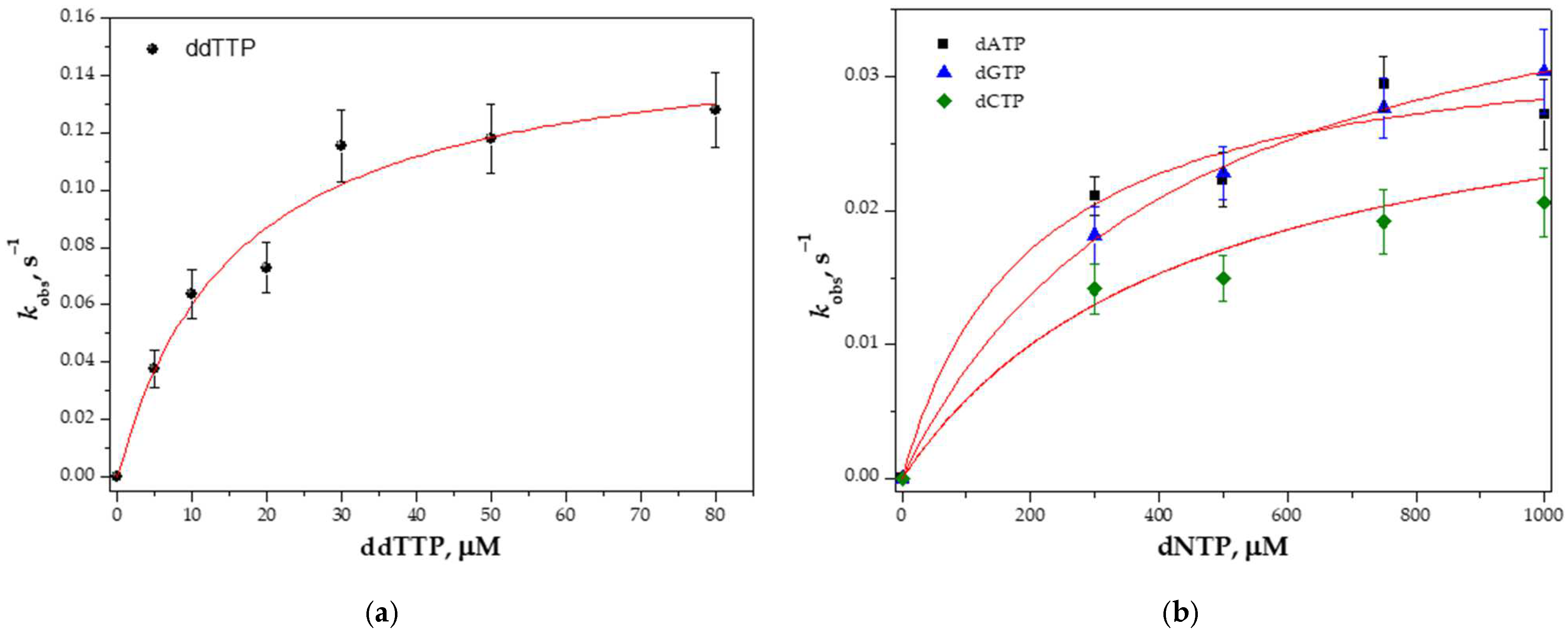
The maximum rate of dNTP incorporation (kcat) and dNTP-binding affinity (KdNTPd,app) can be determined by analyzing the dependence of the reaction rate (kobs) on dNTP concentration. At first, the rate of product formation corresponding to correct dTTP incorporation was determined. ddTTP was used to prevent the contribution of possible subsequent incorporations (after the first polymerization event) of dTTP as an incorrect nucleotide. The reaction condition used likely enabled both a single round and multiple rounds of ddTTP incorporation. The obtained kinetic curves at different concentrations of ddTTP (5–80 µM) were approximated by means of Equation (1), corresponding to a single-turnover burst reaction.
The rate of product formation corresponding to incorrect dNTP incorporation was determined in the same manner. In this case, dNTP concentration was varied in the range of 300–1500 µM, and the obtained kinetic curves were approximated using a one-exponential equation. The dependence of kobs values on dNTP concentration was fitted to a hyperbolic equation (Equation (2); Figure 3); the kinetic parameters kcat and KdNTPd,app are listed in Table 2.
The kcat/Kd,appdNTP parameter denotes the efficacy of dNTP incorporation. It can be concluded that Mau DNA polymerase can incorporate an incorrect dNTP with two orders of magnitude lower efficiency than a correct one.

3.5. Primer-Template Binding

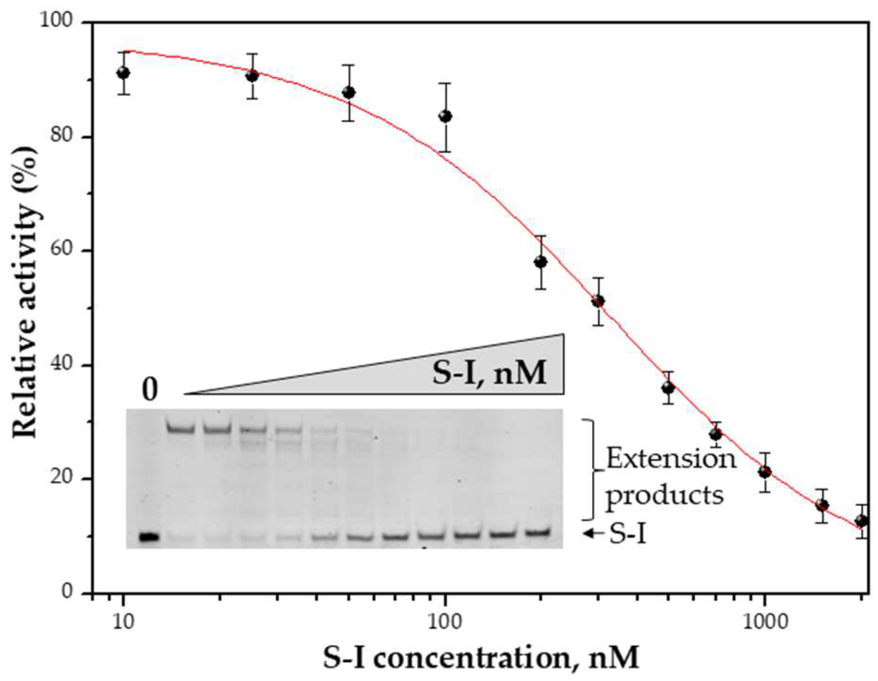
The affinity (KdDNA) of Mau DNA polymerase for a primer-template duplex was estimated as described in ref. [26]. With the help of 100 nM Mau DNA polymerase and 400 µM dNTP, the product formation was assessed, corresponding to a constant reaction time of 10 s in the presence of various concentrations of S-I (Figure 4). Mau DNA polymerase was found to bind the primer-template duplex with a KdDNA of 280 ± 50 nM.

3.6. Exonuclease Activity of Mau DNA Polymerase

Family A DNA polymerases are known to have 5′→3′ exonuclease and 3′→5′ exonuclease activities associated with the main activity. The 5′→3′ exonuclease activity allows DNA polymerases to remove RNA primers; besides, DNA polymerases can use nick-translated DNA as a template owing to this activity [30]. The 3′→5′ proofreading exonuclease activity is responsible for the correction of mismatched dNTPs and is ultimately responsible for the fidelity of DNA polymerases. Some members of this family are devoid of proofreading activity. The reason may be a loss of the active-site proofreading domain (for example, in bacteria of the genera Rickettsia and Thermus [31]) or a deletion of several catalytic amino acid residues (for example, in Geobacillus stearothermophilus [32]).
Template-primer duplexes S-II and S-III, which have a two-nucleotide gap (Table 1), were utilized for the detection of Mau DNA polymerase’s 5′→3′ exonuclease activity. S-II allows us to examine the primer elongation process (FAM is located at the 5′ end of the primer), whereas S-III enables us to observe the 5′→3′ exonuclease reaction (FAM is located at the 3′ end of the flanking oligonucleotide). Analysis of the reaction products (Figure 5a) indicated that during the polymerase reaction, the flanking oligonucleotide is digested, indicating the presence of 5′→3′ exonuclease activity in Mau DNA polymerase.
For the detection of Mau DNA polymerase’s 3′→5′ exonuclease activity, template-primer duplex S-I was chosen. Incubation of Mau DNA polymerase with S-I in the absence of dNTPs led to primer degradation, pointing to the presence of 3′→5′ exonuclease activity (Figure 5b).

4. Discussion

In this study, a gene encoding a full-length DNA polymerase from M. aurea was cloned and expressed in E. coli. Optimal conditions for the polymerase reaction of Mau DNA polymerase were found. We present here a detailed kinetic characterization of Mau DNA polymerase. Together, the obtained data showed that Mau DNA polymerase has biochemical properties typical of family A DNA polymerase (Table 3). Such Kd,appDNA, was estimated at 280 nM (Table 3), which is higher than that of other DNA polymerases. Nonetheless, it should be noted that our method of DNA-binding affinity determination allows us to obtain only an approximate value of DNA-binding affinity owing to the dependence of the product amount on reaction time (Equation (3)). Time points corresponding mainly to a single-turnover burst reaction should be used. As one can see in Figure 4, completed products were obtained at the lowest tested S-I concentration, showing that single-turnover burst conditions were not the case for Mau DNA polymerase in this reaction. Nevertheless, this experiment enabled us to estimate only the upper limit of Kd,appDNA. KdNTPd,app values were found to be 16 µM for correct dNTP and 200–500 µM for incorrect dNTP and are in excellent agreement with literature data (Table 3). The kinetic parameter kcat turned out to be 0.2 s−1 for correct dNTP incorporation and an order of magnitude less for incorrect dNTP incorporation.
Summarizing, finding new thermostable DNA polymerases for biotechnological applications is an important part of fundamental research. Our attention was focused on the DNA polymerase from one of the thermal strains, which could contain thermostable enzymes. Despite the thermal origin of the strain M. aurea, the properties of the tested DNA polymerase do not allow it to be referred to as a thermostable enzyme. Obtained data revealed that Mau DNA polymerase has an optimal temperature for enzyme activity in the area of 30 °C. Therefore, this enzyme could be used in “room-temperature” applications as an analog of Klenow polymerase. Nevertheless, the appearance of another well-characterized DNA polymerase allows for the expansion of the range of enzymes for various applications and fundamental research.

Author Contributions

Conceptualization, A.A.K. and N.A.K.; methodology, A.A.K. and K.S.B.; software, A.A.B.; validation, K.S.B. and A.A.K.; formal analysis, K.S.B. and A.A.K.; investigation, K.S.B.; data curation, N.A.K.; writing—original draft preparation, A.A.K.; writing—review and editing, N.A.K.; visualization, K.S.B. and A.A.K.; supervision, A.A.K. and N.A.K.; project administration, N.A.K.; funding acquisition, N.A.K. All authors have read and agreed to the published version of the manuscript.

Funding

This research was funded by the Ministry of Science and Higher Education under agreement No. 075-15-2021-1085.

Institutional Review Board Statement

Not applicable.

Informed Consent Statement

Not applicable.

Data Availability Statement

Raw experimental data are available from N.A.K. upon request.

Conflicts of Interest

The authors declare no conflict of interest.

References

  1. Johansson, E.; Dixon, N. Replicative DNA Polymerases. Cold Spring Harb. Perspect. Biol. 2013, 5, a012799. [Google Scholar] [CrossRef] [Green Version]
  2. Jain, R.; Aggarwal, A.K.; Rechkoblit, O. Eukaryotic DNA Polymerases. Curr. Opin. Struct. Biol. 2018, 53, 77–87. [Google Scholar] [CrossRef]
  3. Lange, S.S.; Takata, K.; Wood, R.D. DNA Polymerases and Cancer. Nat. Rev. Cancer 2011, 11, 96–110. [Google Scholar] [CrossRef] [Green Version]
  4. Henrikus, S.S.; van Oijen, A.M.; Robinson, A. Specialised DNA Polymerases in Escherichia coli: Roles within Multiple Pathways. Curr. Genet. 2018, 64, 1189–1196. [Google Scholar] [CrossRef] [Green Version]
  5. Kuznetsova, A.A.; Fedorova, O.S.; Kuznetsov, N.A. Structural and Molecular Kinetic Features of Activities of DNA Polymerases. Int. J. Mol. Sci. 2022, 23, 6373. [Google Scholar] [CrossRef]
  6. Delagoutte, E. DNA Polymerases: Mechanistic Insight from Biochemical and Biophysical Studies. Front. Biosci. 2012, 17, 509. [Google Scholar] [CrossRef] [Green Version]
  7. Ling, J.A.; Frevert, Z.; Washington, M.T. Recent Advances in Understanding the Structures of Translesion Synthesis DNA Polymerases. Genes 2022, 13, 915. [Google Scholar] [CrossRef]
  8. Aschenbrenner, J.; Marx, A. DNA Polymerases and Biotechnological Applications. Curr. Opin. Biotechnol. 2017, 48, 187–195. [Google Scholar] [CrossRef]
  9. Ishino, S.; Ishino, Y. DNA Polymerases as Useful Reagents for Biotechnology—The History of Developmental Research in the Field. Front. Microbiol. 2014, 5, 465. [Google Scholar] [CrossRef] [Green Version]
  10. Houlihan, G.; Arangundy-Franklin, S.; Holliger, P. Engineering and Application of Polymerases for Synthetic Genetics. Curr. Opin. Biotechnol. 2017, 48, 168–179. [Google Scholar] [CrossRef]
  11. Terpe, K. Overview of Thermostable DNA Polymerases for Classical PCR Applications: From Molecular and Biochemical Fundamentals to Commercial Systems. Appl. Microbiol. Biotechnol. 2013, 97, 10243–10254. [Google Scholar] [CrossRef]
  12. Zhang, L.; Kang, M.; Xu, J.; Huang, Y. Archaeal DNA Polymerases in Biotechnology. Appl. Microbiol. Biotechnol. 2015, 99, 6585–6597. [Google Scholar] [CrossRef]
  13. Zhu, H.; Zhang, H.; Xu, Y.; Laššáková, S.; Korabečná, M.; Neužil, P. PCR Past, Present and Future. Biotechniques 2020, 69, 317–325. [Google Scholar] [CrossRef]
  14. Coulther, T.A.; Stern, H.R.; Beuning, P.J. Engineering Polymerases for New Functions. Trends Biotechnol. 2019, 37, 1091–1103. [Google Scholar] [CrossRef]
  15. Karunanathie, H.; Kee, P.S.; Ng, S.F.; Kennedy, M.A.; Chua, E.W. PCR Enhancers: Types, Mechanisms, and Applications in Long-Range PCR. Biochimie 2022, 197, 130–143. [Google Scholar] [CrossRef]
  16. Wang, Y.; Prosen, D.E.; Mei, L.; Sullivan, J.C.; Finney, M.; Vander Horn, P.B. A Novel Strategy to Engineer DNA Polymerases for Enhanced Processivity and Improved Performance in Vitro. Nucleic Acids Res. 2004, 32, 1197–1207. [Google Scholar] [CrossRef]
  17. Śpibida, M.; Krawczyk, B.; Olszewski, M.; Kur, J. Modified DNA Polymerases for PCR Troubleshooting. J. Appl. Genet. 2017, 58, 133–142. [Google Scholar] [CrossRef] [Green Version]
  18. Sidstedt, M.; Rådström, P.; Hedman, J. PCR Inhibition in QPCR, DPCR and MPS—Mechanisms and Solutions. Anal. Bioanal. Chem. 2020, 412, 2009–2023. [Google Scholar] [CrossRef] [Green Version]
  19. Dietrich, J.; Schmitt, P.; Zieger, M.; Preve, B.; Rolland, J.L.; Chaabihi, H.; Gueguen, Y. PCR Performance of the Highly Thermostable Proof-Reading B-Type DNA Polymerase from Pyrococcus Abyssi. FEMS Microbiol. Lett. 2002, 217, 89–94. [Google Scholar] [CrossRef]
  20. Cline, J.; Braman, J.C.; Hogrefe, H.H. PCR Fidelity of Pfu DNA Polymerase and Other Thermostable DNA Polymerases. Nucleic Acids Res. 1996, 24, 3546–3551. [Google Scholar] [CrossRef] [Green Version]
  21. Bulygin, A.A.; Kuznetsova, A.A.; Fedorova, O.S.; Kuznetsov, N.A. Comparative Analysis of Family A DNA-Polymerases as a Searching Tool for Enzymes with New Properties. Mol. Biol. 2023, 57, 182–192. [Google Scholar] [CrossRef]
  22. Stenesh, J.; McGowan, G.R. DNA Polymerase from Mesophilic and Thermophilic Bacteria. Biochim. Biophys. Acta Nucleic Acids Protein Synth. 1977, 475, 32–41. [Google Scholar] [CrossRef]
  23. Gallego, V.; Sánchez-Porro, C.; García, M.T.; Ventosa, A. Massilia Aurea Sp. Nov., Isolated from Drinking Water. Int. J. Syst. Evol. Microbiol. 2006, 56, 2449–2453. [Google Scholar] [CrossRef]
  24. Malboeuf, C.M.; Isaacs, S.J.; Tran, N.H.; Kim, B. Thermal Effects on Reverse Transcription: Improvement of Accuracy and Processivity in CDNA Synthesis. Biotechniques 2001, 30, 1074–1084. [Google Scholar] [CrossRef] [Green Version]
  25. Johnson, K.A. Rapid Quench Kinetic Analysis of Polymerases, Adenosinetriphosphatases, and Enzyme Intermediates. Methods Enzymol. 1995, 249, 38–61. [Google Scholar] [CrossRef]
  26. Weiss, K.K.; Chen, R.; Skasko, M.; Reynolds, H.M.; Lee, K.; Bambara, R.A.; Mansky, L.M.; Kim, B. A Role for DNTP Binding of Human Immunodeficiency Virus Type 1 Reverse Transcriptase in Viral Mutagenesis. Biochemistry 2004, 43, 4490–4500. [Google Scholar] [CrossRef]
  27. Skasko, M.; Weiss, K.K.; Reynolds, H.M.; Jamburuthugoda, V.; Lee, K.; Kim, B. Mechanistic Differences in RNA-Dependent DNA Polymerization and Fidelity between Murine Leukemia Virus and HIV-1 Reverse Transcriptases. J. Biol. Chem. 2005, 280, 12190–12200. [Google Scholar] [CrossRef] [Green Version]
  28. Elshawadfy, A.M.; Keith, B.J.; Ee Ooi, H.; Kinsman, T.; Heslop, P.; Connolly, B.A. DNA Polymerase Hybrids Derived from the Family-B Enzymes of Pyrococcus Furiosus and Thermococcus Kodakarensis: Improving Performance in the Polymerase Chain Reaction. Front. Microbiol. 2014, 5, 224. [Google Scholar] [CrossRef]
  29. Oscorbin, I.P.; Belousova, E.A.; Boyarskikh, U.A.; Zakabunin, A.I.; Khrapov, E.A.; Filipenko, M.L. Derivatives of Bst-like Gss-Polymerase with Improved Processivity and Inhibitor Tolerance. Nucleic Acids Res. 2017, 45, 9595–9610. [Google Scholar] [CrossRef] [Green Version]
  30. Rigby, P.W.J.; Dieckmann, M.; Rhodes, C.; Berg, P. Labeling Deoxyribonucleic Acid to High Specific Activity in Vitro by Nick Translation with DNA Polymerase I. J. Mol. Biol. 1977, 113, 237–251. [Google Scholar] [CrossRef]
  31. Huang, Y.-P.; Downie, J.A.; Ito, J. Primary Structure of the DNA Polymerase I Gene of an α-Proteobacterium, Rhizobium Leguminosarum, and Comparison with Other Family A DNA Polymerases. Curr. Microbiol. 1999, 38, 355–359. [Google Scholar] [CrossRef]
  32. Sellmann, E.; Schröder, K.L.; Knoblich, I.M.; Westermann, P. Purification and Characterization of DNA Polymerases from Bacillus Species. J. Bacteriol. 1992, 174, 4350–4355. [Google Scholar] [CrossRef] [Green Version]
  33. Setlow, P. DNA Polymerase I from Escherichia coli. Methods Enzymol. 1974, 29, 3–12. [Google Scholar] [CrossRef]
  34. McClure, W.; Jovin, T. The Steady State Kinetic Parameters and Non-Processivity of Escherichia coli Deoxyribonucleic Acid Polymerase I. J. Biol. Chem. 1975, 250, 4073–4080. [Google Scholar] [CrossRef]
  35. Lowe, L.G.; Guengerich, F.P. Steady-State and Pre-Steady-State Kinetic Analysis of DNTP Insertion Opposite 8-Oxo-7,8-Dihydroguanine by Escherichia coli Polymerases I Exo- and II Exo-. Biochemistry 1996, 35, 9840–9849. [Google Scholar] [CrossRef]
  36. Driscoll, M.D.; Rentergent, J.; Hay, S. A Quantitative Fluorescence-Based Steady-State Assay of DNA Polymerase. FEBS J. 2014, 281, 2042–2050. [Google Scholar] [CrossRef] [Green Version]
  37. Polesky, A.H.; Steitz, T.A.; Grindley, N.D.; Joyce, C.M. Identification of Residues Critical for the Polymerase Activity of the Klenow Fragment of DNA Polymerase I from Escherichia coli. J. Biol. Chem. 1990, 265, 14579–14591. [Google Scholar] [CrossRef]
  38. Spratt, T. Structure of the Hydrogen Bonding Complex of O6-Methylguanine with Cytosine and Thymine during DNA Replication. Nucleic Acids Res. 1997, 25, 3354–3361. [Google Scholar] [CrossRef] [Green Version]
  39. Gillin, F.D.; Nossal, N.G. T4 DNA Polymerase Has a Lower Apparent Km for Deoxynucleoside Triphosphates Complementary Rather than Noncomplementary to the Template. Biochem. Biophys. Res. Commun. 1975, 64, 457–464. [Google Scholar] [CrossRef]
  40. Wu, P.; Nossal, N.; Benkovic, S.J. Kinetic Characterization of a Bacteriophage T4 Antimutator DNA Polymerase. Biochemistry 1998, 37, 14748–14755. [Google Scholar] [CrossRef]
  41. Kong, H.; Kucera, R.B.; Jack, W.E. Characterization of a DNA Polymerase from the Hyperthermophile Archaea Thermococcus Litoralis. Vent DNA Polymerase, Steady State Kinetics, Thermal Stability, Processivity, Strand Displacement, and Exonuclease Activities. J. Biol. Chem. 1993, 268, 1965–1975. [Google Scholar] [CrossRef]
  42. Tang, F.; Liu, S.; Li, Q.-Y.; Yuan, J.; Li, L.; Wang, Y.; Yuan, B.-F.; Feng, Y.-Q. Location Analysis of 8-Oxo-7,8-Dihydroguanine in DNA by Polymerase-Mediated Differential Coding. Chem. Sci. 2019, 10, 4272–4281. [Google Scholar] [CrossRef] [Green Version]
  43. Patel, S.S.; Wong, I.; Johnson, K.A. Pre-Steady-State Kinetic Analysis of Processive DNA Replication Including Complete Characterization of an Exonuclease-Deficient Mutant. Biochemistry 1991, 30, 511–525. [Google Scholar] [CrossRef]
  44. Boosalis, M.S.; Petruska, J.; Goodman, M.F. DNA Polymerase Insertion Fidelity. Gel Assay for Site-Specific Kinetics. J. Biol. Chem. 1987, 262, 14689–14696. [Google Scholar] [CrossRef]
  45. Takata, K.; Shimizu, T.; Iwai, S.; Wood, R.D. Human DNA Polymerase N (POLN) Is a Low Fidelity Enzyme Capable of Error-Free Bypass of 5S-Thymine Glycol. J. Biol. Chem. 2006, 281, 23445–23455. [Google Scholar] [CrossRef] [Green Version]
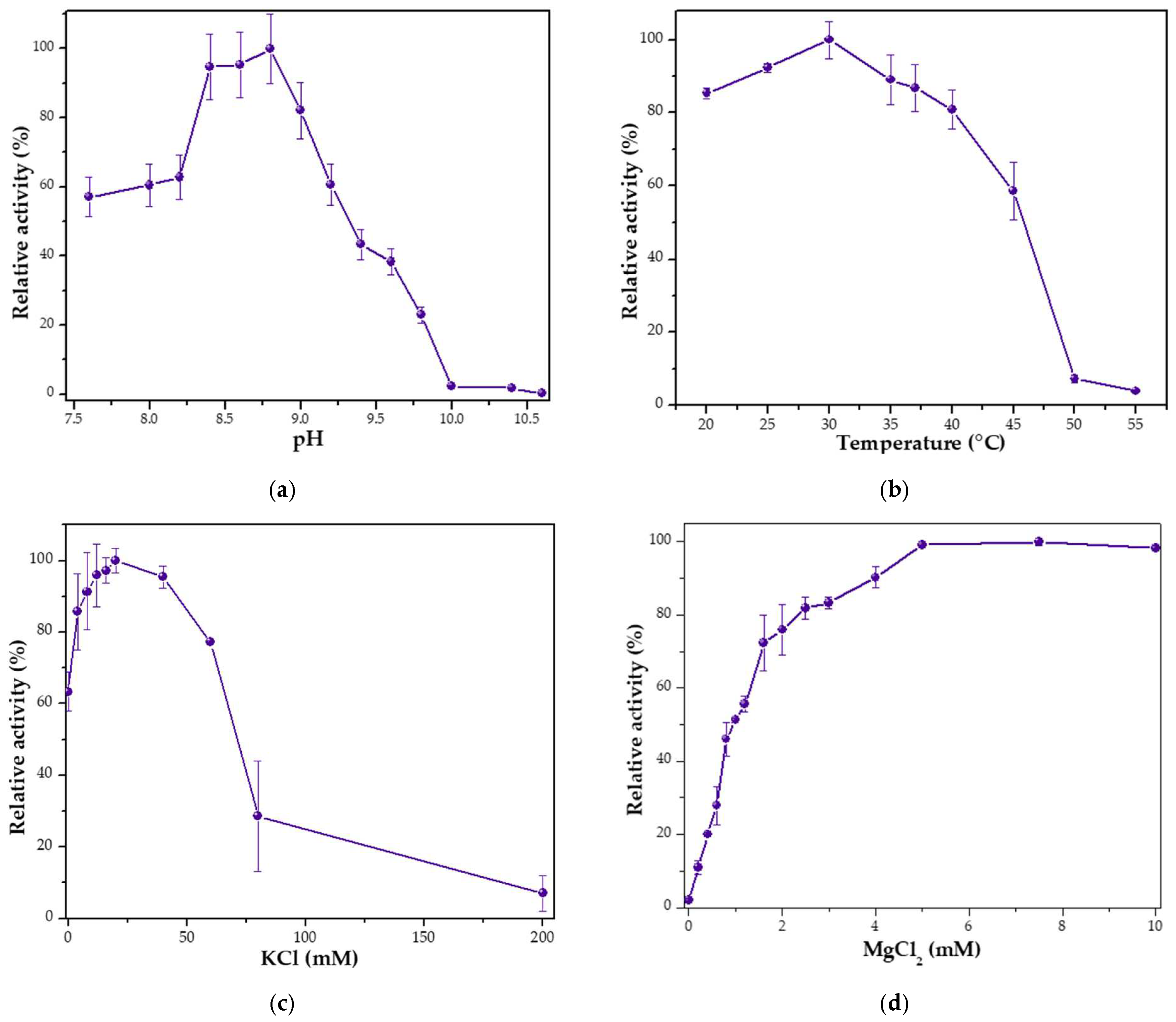
Figure 1. Characterization of Mau DNA polymerase. (a) The effect of pH on its polymerase activity; (b) The influence of temperature on polymerase activity; (c) The impact of KCl on the polymerase activity; (d) The effect of MgCl2 on the polymerase activity.
Fermentation 09 00650 g001
Figure 2. The first derivative of the DSF profile of Mau DNA polymerase is depicted, with dF/dT indicating the change in fluorescence (relative units). Each experiment was conducted in triplicate; a typical curve is presented.
Fermentation 09 00650 g002
Figure 3. The dependence of kobs values on dNTP concentration. (a) The kobs values for correct ddTTP incorporation; (b) The kobs values for incorrect dATP/dGTP/dCTP incorporation. The kobs values were determined at different dNTP concentrations with 100 nM Mau DNA polymerase and 50 nM S-I. The kcat and KdNTPd,app values were calculated by fitting these results to Equation (2) and are listed in Table 2.
Fermentation 09 00650 g003
Figure 4. The estimation of DNA-binding affinity. The extent of product formation was measured at different S-I concentrations using 100 nM Mau DNA polymerase with 400 µM dNTP and fitted to Equation (3). The inset shows gel electrophoretic analysis of primer strand extension.
Fermentation 09 00650 g004
Figure 5. The detection of Mau DNA polymerase’s 5′→3′ and 3′→5′ exonuclease activities. (a) The presence of 5′→3′ exonuclease activity in Mau DNA polymerase was investigated with template-primer duplexes S-II and S-III in the presence of dNTPs. (b) The presence of 3′→5′ exonuclease activity in Mau DNA polymerase was assayed with template-primer duplex S-I in the absence of dNTPs.
Fermentation 09 00650 g005
Table 1. Oligodeoxyribonucleotides and DNA duplexes that are used as substrates.
Short NameSequence
S-IFAM-5′CGGCCCCCAGATGAGTCGAGCAGC3′
    3′GCCGGGGGTCTACTCAGCTCGTCGATAGTTCCGTTTTACAGAGG5′
S-IIFAM-5′CGGCCCCCAGATGAGTCGAGCAGC3′5′TCAAGGCAAAATGTCTCC3′
    3′GCCGGGGGTCTACTCAGCTCGTCGATAGTTCCGTTTTACAGAGG5′
S-III5′CGGCCCCCAGATGAGTCGAGCAGC3′5′TCAAGGCAAAATGTCTCC3′-FAM
3′GCCGGGGGTCTACTCAGCTCGTCGATAGTTCCGTTTTACAGAGG5′
Template-trap  5′CGGCCCCCAGATGAGTCGAGCAGCTATCAAGGCAAAATGTCTCC3′
Table 2. The kinetic parameters kcat and KdNTPd,app, determined for Mau DNA polymerase (mean ± SD).
dNTPkcat (s−1)Kd,appdNTP (µM)kcat/Kd,appdNTP (µM−1·s−1)
dTTP (corr.)0.20 ± 0.0716 ± 4(1.0 ± 0.5) × 10−2
dATP (incorr.)0.034 ± 0.004200 ± 100(1.0 ± 0.5) × 10−4
dCTP (incorr.)0.030 ± 0.004500 ± 100(0.6 ± 0.2) × 10−4
dGTP (incorr.)0.044 ± 0.003430 ± 50(1.0 ± 0.1) × 10−4
Table 3. Kinetic parameters kcat, KdNTPd,app, and Kd,appDNA of family A DNA polymerases.
DNA PolymeraseKd,appDNA, nMKd,appdNTP, µMkcat, s−1Ref.
Pol I (E. coli)5
1–2
12–147
8.3
[33]
[34]
KF (E. coli)

8
1.1
2.8–8.0
2.3–3.2
0.014–0.042
0.37
0.93–1.55
2.0–2.8
[35]
[36]
[37]
[38]
T4 (E. coli phage T4)
70
6–17
20

[39]
[40]
Taq (T. aquaticus)1.0–1.8

(corr.) 14–17
(incorr.) 6–12
0.99


[41]

[36]
Tth (T. thermophiles)

(corr.) 2.5
(incorr.) 230–280
0.022


[42]

[38]
Bsu (B. subtilis)(corr.) 1.8
(incorr.) 180
[42]
T717.8180.24 [43]
Pol ɑ (Drosophila)3.72.2 [44]
Pol η (Human)7.8–8.2 [45]
Mau (M. aurea)<280(corr.) 16
(incorr.) 200–500
(corr.) 0.2
(incorr.) 0.03–0.044
this study
Disclaimer/Publisher’s Note: The statements, opinions and data contained in all publications are solely those of the individual author(s) and contributor(s) and not of MDPI and/or the editor(s). MDPI and/or the editor(s) disclaim responsibility for any injury to people or property resulting from any ideas, methods, instructions or products referred to in the content.