Abstract
l-Lysine, an essential amino acid for humans and mammals, is widely used in the food, feed, medicine, and cosmetics industries. In this study, a lysine over-producing Escherichia coli mutant was isolated using a fluorescence-based screen and an E. coli strain lacking five of the six L-lysine tRNA-UUU genes. Firstly, an l-lysine codon-rich protein was fused with a green fluorescent protein (all AAG codons were replaced with AAA), yielding a rare codon-rich screening marker positively correlated with l-lysine content. After association and room temperature plasma (ARTP) mutagenesis and induced fluorescent protein expression culture, mutant strains with strong fluorescence were sorted using flow cytometry. The fermentation performance of the high-yielding l-lysine strains were evaluated, which resulted in 16 of the 29 mutant strains showing increased L-lysine yields compared with those of the wild-type strains and a screening efficiency of up to 55.2%. Following a 48 h fermentation, the production of l-lysine (14.8 g/L) and biomass by E. coli QD01ΔtRNA L2 were 12.1 and 4.5% higher than those of the wild-type strain. The screening strategy for high-yielding strains based on the artificial rare cryptosystem established in this study will provide an efficient, accurate, and simple method for screening other amino-acid-producing microorganisms.
1. Introduction
l-Lysine, a basic essential amino acid for humans and mammals, plays important roles in multiple physiological and biochemical processes, such as protein, fat, and energy metabolism. It has a wide range of applications in the food, feed, medicine, and cosmetics industries. In the livestock sector, it is used as a feed additive that promotes the growth performance and protein deposition in pigs, poultry, and fish [1,2]. Additionally, l-lysine has potential value as a feedstock for producing active pharmaceutical constituents and other high-value chemicals [3].
The industrial production of l-lysine is mainly dependent on microbial fermentation [4]. According to the Global Amino Acid Market Size Insight, l-lysine will account for approximately 34% of the global amino acid market by 2020 [5]. The global production of l-lysine is approximately 2,200,000 tons/year, and it continues to grow annually at a rate of approximately 7%, making it one of the leading biotechnological products [6]. Corynebacterium glutamicum and its mutant strains were the microorganisms of choice used in the early industrial production of l-lysine [7,8]. However, given its rapid growth rate and high l-lysine production capacity, Escherichia coli has emerged as a more promising strain for the industrial production of l-lysine.
Strategies have recently been proposed for the screening of high-yielding amino-acid-producing bacterial strains using genes rich in rare codons. Owing to degeneracy, different codons can encode the same amino acid, and the decoding rate of synonymous codons varies widely, depending mainly on the abundance of homologous tRNAs [9,10]. Common codons are recognized by rich tRNAs and are translated more efficiently than rare codons, particularly at low amino acid concentrations. However, the transport efficiency of rare transcripts is close to zero, while that of ordinary tRNA remains high for only a few minutes [11,12]. Thus, instead of “codon optimization”, we can replace synonymous common codons with rare codons and knock out a proportion of the corresponding tRNAs to reduce the expression of alloproteins in the host. Additionally, if genes with rare codons can be expressed normally, the corresponding amino acid content can be maintained at a high level, and these strains are likely to be high-amino-acid-yielding strains (Figure 1) [13].
Figure 1.
Rare codon, tRNA, and L-lysine abundance influence the rate of translation. When the l-lysine codon AAG is replaced with the artificial rare codon AAA, there is a reduction in the expression of the green fluorescent fusion protein. The low abundance of rare tRNAs retards the expression of this protein. In response to an increase in the concentration of amino acids in the cell, tRNAs that recognize rare codons bind to the corresponding amino acid via the action of amino tRNA synthase, and the expression of genes containing the rare codon is restored.
Although traditional mutagenesis breeding methods are still used to screen industrial microorganisms, inefficient screening, strain instability, and food safety issues remain [14,15]. Genetically engineered strains can retain the advantages of wild-type strains and still have the capacity to produce substantially higher yields of l-lysine. A common genetic engineering strategy entails modifying the rate-limiting steps of key enzymes involved in the l-lysine biosynthesis pathway [16]. The microbial synthesis of l-lysine is mainly mediated via two pathways, the aminoheptadaric and diaminoheptadaric acid synthetic pathways [17,18], and is influenced by the decomposition and transport of l-lysine, enzyme activity, gene expression levels, precursor substances, and intermediate metabolites.
Biosensor-based high-throughput screening methods have been widely used to screen for mutant strains characterized by high amino acid yields [19,20]. For example, Binder et al. developed a biosensor capable of detecting l-lysine metabolites in C. glutamicum [21]. This biosensor is based on the lysG transcriptional regulator of C. glutamicum, which activates the lysE promoter to drive the transcription of the enhanced yellow fluorescent protein. However, the drawback of using this device is that it responds to changes in l-lysine concentration with a lag. Therefore, developing biosensors with specific and generalized responses to intracellular l-lysine for high-throughput screening of l-lysine-producing strains should be a target for future research [22]. High-throughput fluorescence-activated cell sorting (FACS) is receiving increasing attention as a novel and powerful tool for screening target strains that enable high-throughput screening of large libraries [23].
In our previous research, we used eGFP as a screening marker in C. glutamicum [24]. However, this approach is not applicable for use with E. coli. Therefore, as a novel screening marker for this bacterium, we linked the l-lysine codon-rich L21r with a staygold fluorescent protein gene to generate an L21r-staygold fluorescent fusion protein. In the E. coli genome, the two codons encoding l-lysine are AAA and AAG, the frequency of the former of which (35.3 per thousand nucleotides) is approximately three-fold higher than that of the latter (12.4 per thousand nucleotides) (https://www.genscript.com/tools/codon-frequency-table, accessed on 16 August 2023). In this study, a five-tRNA-UUU gene knockout E. coli strain was constructed through a homologous recombination method, and the expression intensity of the l-lysine tRNA-UUU genes of codon AAA was reduced. Then, an l-lysine artificial rare codon, AAA, was constructed in E. coli QD01ΔtRNA. Meanwhile, by fusing the l-lysine codon-rich protein with a green fluorescent protein (all AAG codons in the fusion protein were replaced with AAA), we generated a rare codon-rich screening marker that was positively correlated with the l-lysine content. After constant temperature and atmospheric pressure plasma mutagenesis and induced fluorescent protein expression culture, mutant strains with strong fluorescence were sorted by flow cytometry, and we assessed the fermentation performance of the high-yielding l-lysine strains thus obtained.
2. Materials and Methods
2.1. Strains, Plasmids, and Culture Media
The strains and plasmids used in this study are listed in Table 1. LB, LBG, CgXII, EPO, LBHISG, and fermentation media were used in the experiment (Tables S1–S6). The LB medium comprised LB medium + 0.05% sodium pyruvate, and the CgXII medium comprised CgXII medium + 0.05% sodium pyruvate.
Table 1.
The strains and plasmids used in this study.
2.2. Construction of l-Lysine Codon-Rich Fluorescent Fusion Protein Screening Vectors
We used the NCBI database to search for the nucleotide sequences of staygold fluorescent protein genes (GenBank: LC601652.1) [25]. The l-lysine codon-rich genes asr (GenBank: EIE7460467.1), fkpA (GenBank: WP000838250.1), s19 (GenBank: WP205529307.1), CsbD (GenBank: WP205529307.1), rplx (GenBank: WP000729185.1), and L21 (GenBank: WP205529307.1) were obtained from the genomes of E. coli and C. glutamicum, the l-lysine contents of which were 18, 11, 14, 14, 10, and 19%, respectively. In all cases, the AAG l-lysine codons were replaced with the rare codon AAA, and the corresponding asrr-staygold, fkpAr-staygold, s19r-staygold, CsbDr-staygold, rplxr-staygold, and L21r-staygold fluorescent fusion protein gene fragments, along with egfpr, were generated via gene synthesis by Nanjing GenScript Biotechnology Co., Ltd. (Nanjing, China). These sequences were subsequently inserted into the NdeI and BamHI cleavage sites of the linearized vector pET-22b(+) to generate the rare codon-rich screening vectors pET-22b(+)-asrr-staygold, pET-22b(+)-fkpAr-staygold, pET-22b(+)-s19r-staygold, pET-22b(+)-CsbDr-staygold, pET-22b(+)-rplxr-staygold, pET-22b(+)-L21r-staygold, and pET-22b(+)-egfpr, respectively.
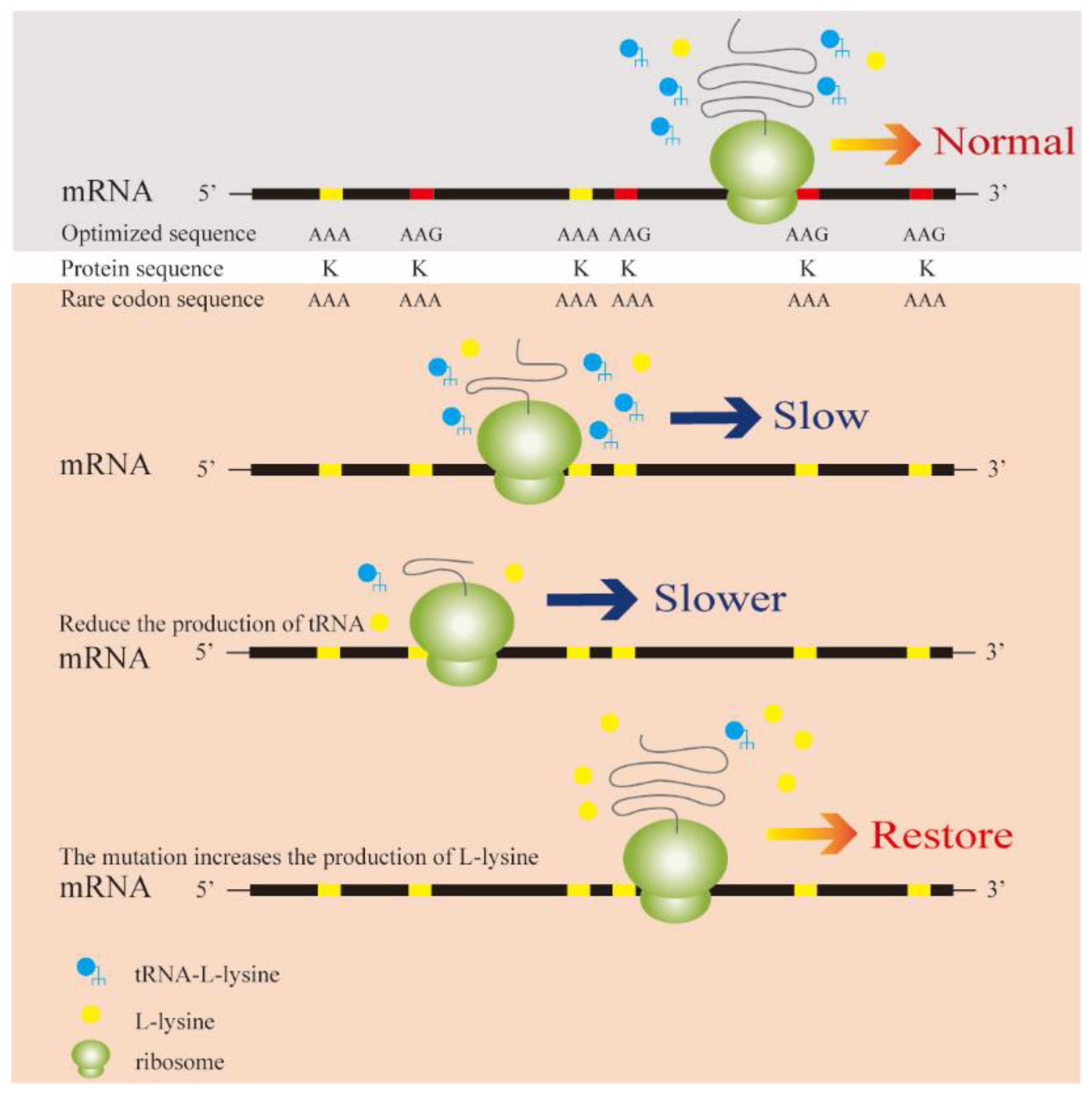
2.3. Construction of an Artificial Rare l-Lysine Codon in E. coli
Using the E. coli QD01 genome as a template, we used the primers t-Lys-F1/t-Lys-R1 and t-Lys-F2/t-LysR to amplify the upstream and downstream homologous arm tRNA-L and tRNA-R fragments, respectively. Using the pKD13 plasmid as a template, the primer pair Kanr-F/Kanr-R were used to amplify the Kanr fragment containing FRT sites and resistance markers. Following digestion with XhoI and BamHI, the tRNA-L, tRNA-R, and Kanr fragments were seamlessly cloned into the cut pET-28a(+) plasmid. The obtained recombinant plasmid pET-28a(+)-tRNA was used to transform E. coli DH5α receptor cells, whereas the pKD46 plasmid was used to transform E. coli QD01 cells. Using pET-28a(+)-tRNA as a template, the knockout fragment was amplified using the primer pair t-Lys-F1/t-Lys-R2 and used to transform E. coli QD01/pKD46 receptor cells. Bacteria expressing the recombinant plasmids were selected for knockout DNA sequencing to further confirm whether the knockout box was successfully transferred and that the mutation was excluded. Single bacterial colonies were selected and screened on plates containing ampicillin and kanamycin or only kanamycin for verification. Strains growing only on the kanamycin plates were selected, which were recombinant strains in which the pKD46 plasmid was successfully eliminated. Plasmid pCP20 was used to transform receptive recombinant cells, and the correct strain was verified as the recombinant E. coli QD01ΔtRNA.
2.4. Transformation and Expression of l-Lysine Codon-Rich Screening Vectors
E. coli QD01 and E. coli QD01ΔtRNA receptors were prepared as described in our previous study [26]. The plasmids pET-22b(+)-asrr-staygold, pET-22b(+)-fkpAr-staygold, pET-22b(+)-s19r-staygold, pET-22b(+)-CsbDr-staygold, pET-22b(+)-rplxr-staygold, and pET-22b(+)-L21r-staygold were used to transform the pre-prepared E. coli QD01 receptor cells, thereby generating the strains E. coli asrr, E. coli fkpAr, E. coli s19r, E. coli CsbD r, E. coli rplxr, and E. coli L21r, respectively. Similarly, the plasmids pET-22b(+)-rplxr-staygold and pET-22b(+)-L21r-staygold were used to transform pre-prepared E. coli QD01ΔtRNA receptor cells to generate the E. coli ΔtRNA rplxr and E. coli ΔtRNA L21r strains, respectively.
2.5. Correlation Analysis of Fluorescence Intensity and Lysine Content in Recombinant Strains of E. coli QD01
The recombinant strains E. coli asrr, E. coli fkpAr, E. coli s19r, E. coli CsbDr, E. coli rplxr, E. coli L21r, E. coli ΔtRNA rplxr, and E. coli ΔtRNA L21r were cultured for 12 h in LB medium. The activated bacterial suspensions were inoculated into 250 mL conical bottles containing 50 mL of CgXII medium supplemented with ampicillin at a final concentration of 100 μg/mL and were cultured at 37 °C rotating at 200 r/min until reaching an OD600 of approximately 1.0. The suspensions were then collected in 50 mL centrifuge tubes and centrifuged twice, each time discarding the supernatants. The remaining bacterial pellets were subsequently resuspended and re-inoculated into 50 mL of CgXII medium containing ampicillin at a final concentration of 100 g/mL and l-lysine hydrochloride at final concentrations of 0, 1, 2, 3, or 4 g/L; each concentration was assessed in triplicate. To induce fluorescent protein expression, IPTG was added to the suspensions to a final concentration of 0.5 μM, and the cultures were incubated at 25 °C and 200 r/min for 14 h. Following induction, fluorescence intensities were measured at excitation and detection wavelengths of 488 and 535 nm, respectively, and correlations between the expression of fluorescent proteins and the concentration of l-lysine were determined.
2.6. Atmospheric Pressure and Room Temperature Plasma Mutagenesis
The recombinant strain E. coli ΔtRNA L21r was streaked onto solid LB medium containing 100 μg/mL ampicillin and cultured overnight in a constant-temperature incubator at 37 °C for 12–16 h until single colonies were observed. Single colonies were selected and used to inoculate LB medium containing 100 μg/mL ampicillin. The suspension was then cultured at 37 °C and 200 r/min until reaching an OD600 of approximately 1.0. Ten microliter aliquots of the bacterial suspension were evenly coated on small stainless-steel plates and exposed to ARTP for 20, 40, 60, 80, 100, 120, 140, 160, or 180 s. The ARTP parameters were as follows: incident power, 120 W; gas volume, 10 SLM; and helium pressure, 120 MPa. Following mutagenesis, the stainless-steel discs were placed in 1 mL of LB medium containing 100 μg/mL ampicillin and eluted for 1 min. Thereafter, 100 μL aliquots of bacterial suspension were withdrawn and uniformly coated on LB plates containing 100 μg/mL ampicillin and incubated at 37 °C. The cells were cultured at 200 r/min for 12–16 h until single colonies had grown. The mortality rate for each treatment period was calculated, and the best ARTP mutant with a mortality rate > 90% was selected.
2.7. Flow Cytometric Cell Sorting of Mutant Strains
Mutant strains with a mortality rate greater than 90% were selected and used to inoculate 50 mL of LB medium. The cells were cultured at 30 °C and 200 r/min until reaching an OD600 of approximately 1.0. The bacterial suspensions were collected and transferred to 50 mL centrifuge tubes and centrifuged twice. The resulting supernatants were discarded, and the remaining bacterial pellets were weighed and subsequently used to inoculate 50 mL of CgXII medium. IPTG was then added to a final concentration of 0.5 mM, and expression was induced at 25 °C and 200 r/min for 12 h. To screen for high-yielding l-lysine strains, 1 mL of bacterial suspension was withdrawn, and the cultured cells were cleaned, re-suspended in 0.1% PBS buffer, and diluted to an OD600 of approximately 1.0. Ultraviolet-mutated strains characterized by strong green fluorescence were sorted using FACS (MoFlo XDP; Beckman Coulter, Brea, CA, USA), and strain classification was determined at excitation and fluorescence detection wavelengths of 488 and 535 nm, respectively. The sample pressure used was 60 psi, and the nozzle diameter was set to 70 μm. In selecting the mutant library, a gate accounting for 0.01% of the total cells was set up based on a preliminary analysis of the mutant library, and cells with high fluorescent protein expression were collected and further cultured in wells of a 96-well plate containing 200 μL of LB medium.
2.8. Fermentation Analysis of Mutant Strains
Aliquots (1 mL) of a fermentation medium were added to each well of pre-sterilized 96-deep-well plates. The bacterial suspensions thus obtained were used as seed suspensions, which were re-transferred to the wells of the deep-well plates at an inoculum of 10% and further cultured in a micro-well plate incubator at 37 °C and 600 r/min for 48 h. Following fermentation, 100 μL of the fermentation suspension was centrifuged at 5000 r/min for 5 min, and the supernatant was diluted to an l-lysine concentration of between 0.1 and 1 g/L. The concentration of l-lysine was determined using a biosensor analyzer.
The strains characterized by the highest l-lysine yield and E. coli QD01 were used to inoculate LB medium and cultured overnight for 14 h. A total of 100 mL of fermentation medium was added to 500 mL conical bottles incorporating a baffle plate and sterilized at 115 °C for 30 min. The seed suspensions were then transferred to the fermentation medium at an inoculum of 10%, and three parallel samples were incubated at 37 °C and 200 r/min for 48 h. During the fermentation process, the pH was adjusted to 6.5–7.0 using aqueous ammonia, and the residual sugar was maintained at 5–10 g/L by adding glucose. At 12 h intervals, 1 mL aliquots of the fermentation broth were collected to determine OD600 values and the contents of l-lysine.
3. Results
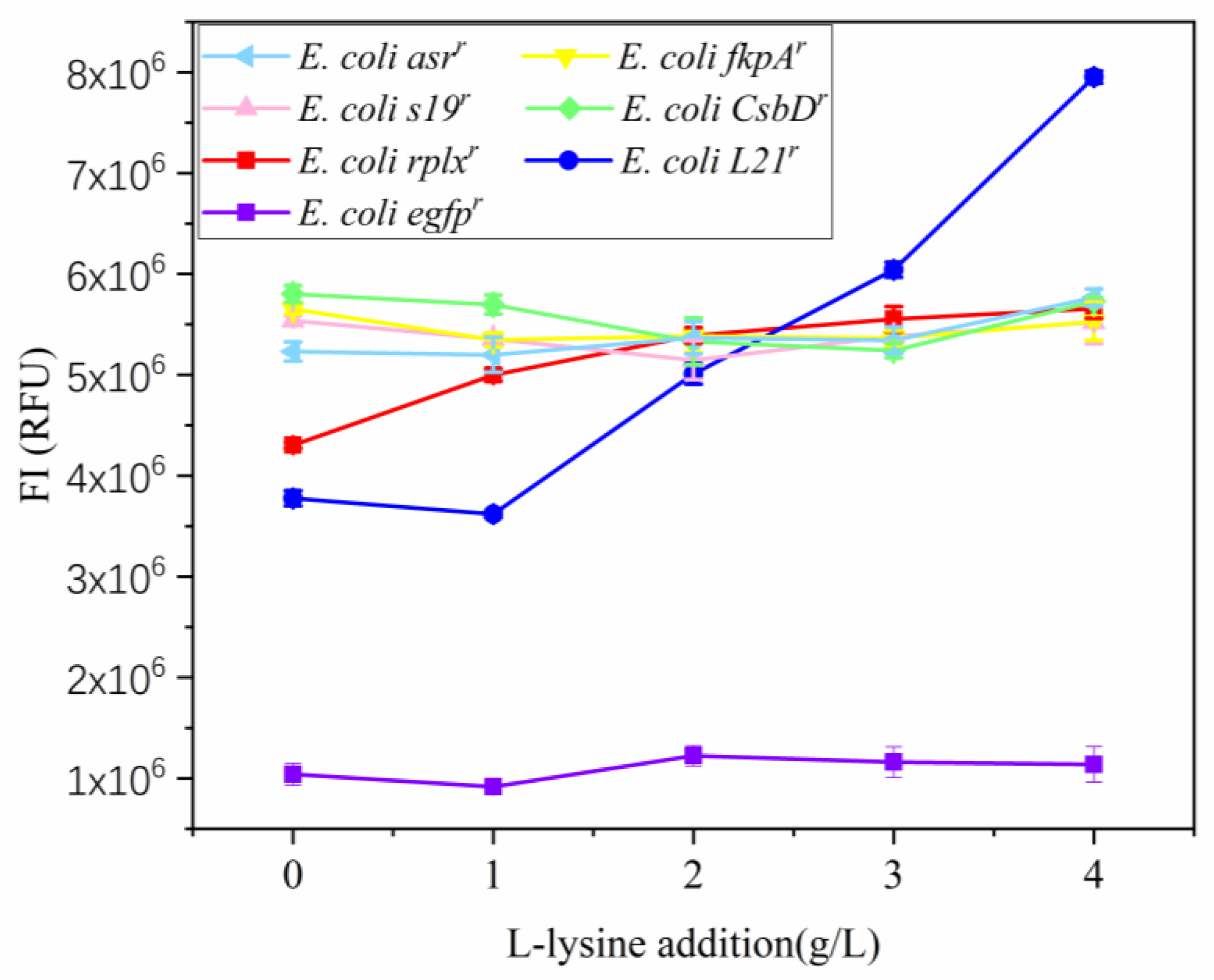
3.1. Construction and Expression of Lysine-Rich Fluorescent Fusion Protein Expression Vectors
The fluorescent fusion protein expression vectors pET-22b(+)-asrr-staygold, pET-22b(+)-fkpAr-staygold, pET-22b(+)-s19r-staygold, pET-22b(+)-CsbDr-staygold, pET-22b(+)-rplxr-staygold, and pET-22b(+)-L21r-staygold containing the l-lysine rare codon AAA were used to transform E. coli QD01. According to the test results obtained using the SpectraMax i3x Multi-mode Detection Platform, we obtained correlation coefficient (R2) values of 0.866, 0.3521, 0.0100, 0.0026, and 0.3944 for asr-staygold, fkpA-staygold, s19-staygold, CsbD-staygold, and egfpr, respectively, indicating no significant correlation with l-lysine concentration in E. coli QD01. Contrastingly, the fluorescence fusion proteins Staygold-rplxr (R2 = 0.92439) and Staygold-L21r (R2 = 0.91061) were found to show a positive correlation with l-lysine concentration (Figure 2).
Figure 2.
Fluorescence intensity of green fluorescent fusion proteins. According to the results obtained using the SpectraMax i3x Multi-Mode Detection Platform, egfpr, asr-staygold, fkpA-staygold, s19-staygold, and CsbD-staygold showed no correlation with l-lysine concentration in E. coli QD01. Contrastingly, Staygold-rplxr and Staygold-L21r showed a positive correlation with l-lysine concentration.
3.2. tRNA Knockout in E. coli QD01
To reduce the background fluorescence intensity, we constructed the tRNA knockout strain E. coli QD01ΔtRNA. Following linearization of the enzyme-digested pET-28a(+), a ClonExpress II One Step Cloning kit was used to seamlessly link the knockout frames tRNA-L (500 bp), tRNA-R (500 bp), and Kanr (1324 bp) fragments, and positive recombinant transformants were picked using a sterile inoculation loop. Colony PCR was performed using the primer pair t-Lys-F1/t-Lys-R2. The results revealed that the upstream homologous arm tRNA-L, downstream homologous arm tRNA-R fragment, and the Kanr fragment containing FRT sites and resistance markers were successfully inserted into the plasmid pET-28a(+) to generate pET-28a(+)-tRNA (Supplementary Figure S1A). To verify results obtained for the ampicillin-resistant, kanamycin-resistant, and non-resistant LB plates, the strains growing only on the non-resistant LB plates were selected, and colony PCR was performed using the primer pair t-Lys-F1/t-Lys-R2. The results indicated that the Kanr gene fragments of 1324 bp were successfully deleted. Accordingly, we confirmed the successful construction of the recombinant E. coli QD01ΔtRNA (Supplementary Figure S1B).
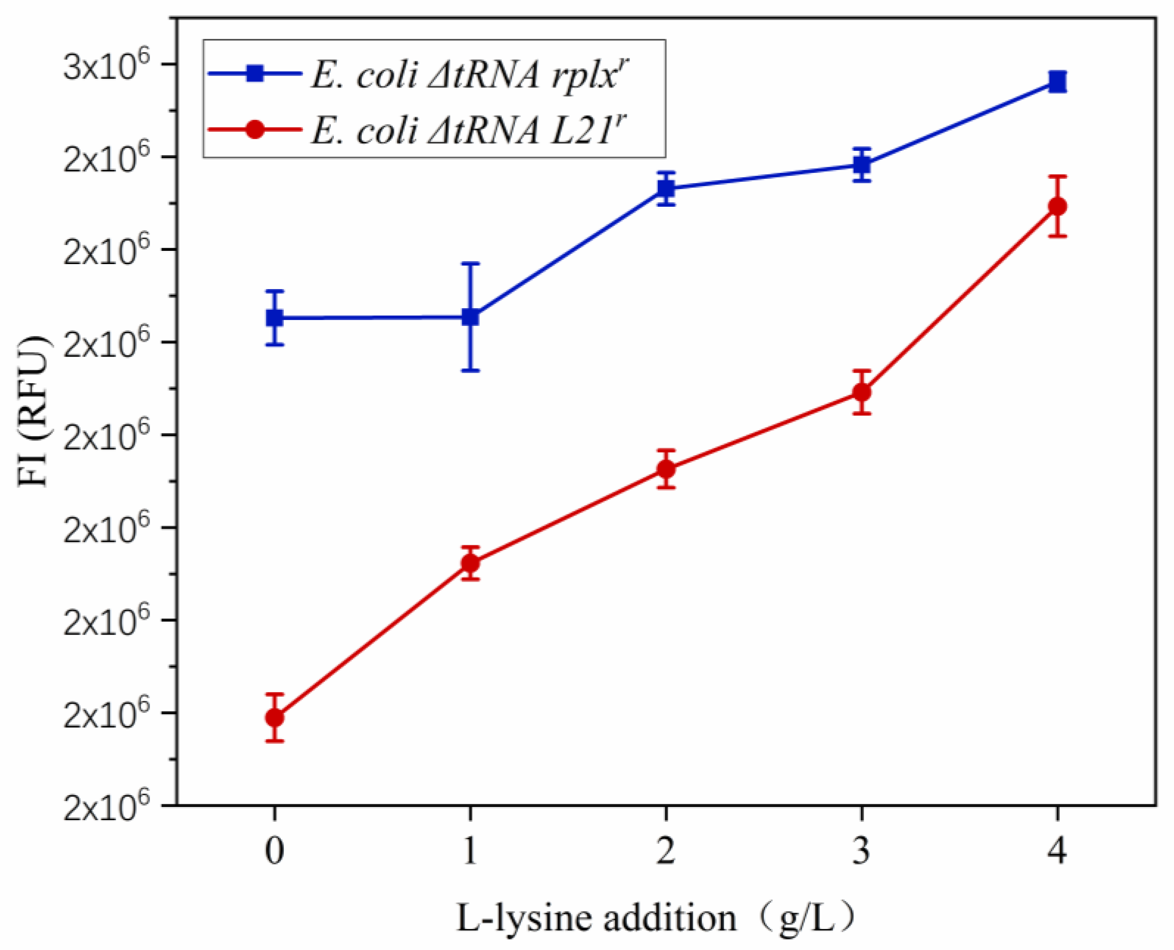
3.3. Construction of a Rare Codon-Rich Marker Screening System for E. coli QD01ΔtRNA
Samples of normally growing recombinant E. coli ΔtRNA rplxr and E. coli ΔtRNA L21r were obtained using a sterile inoculation loop, and the primer pairs staygoldr-F/rplxr-R and staygoldr-F/L21r-R were, respectively, used for PCR verification. The electrophoresis results revealed that E. coli QD01ΔtRNA receptor cells had been successfully transformed using the recombinant plasmids pET-22b(+)-rplxr-staygold and pET-22b(+)-L21r-staygold (Supplementary Figure S1C).
The expression of the fluorescent fusion proteins Staygold-rplxr and Staygold-L21r in E. coli ΔtRNA rplxr and E. coli ΔtRNA L21r revealed that the intensity of fluorescence gradually increased with an increase in l-lysine supplementation (Figure 3). Following tRNA knockout, the correlation between fluorescence intensity and l-lysine addition changed from E. coli rplxr (R2 = 0.92439) and E. coli L21r (R2 = 0.91061) to E. coli ΔtRNA rplxr (R2 = 0.96531) and E. coli ΔtRNA L21r (R2 = 0.97071), respectively. These findings indicate that the fluorescent fusion proteins rplxr-staygold and L21r-staygold, containing the rare codon AAA of l-lysine, could be expressed in E. coli QD01ΔtRNA and that the intensity of fluorescence was positively correlated with the concentration of l-lysine. However, we established that, of these two fusion proteins, the positive correlation between the fluorescence intensity of fluorescent fusion protein L21r-staygold and l-lysine concentration was higher than that of rplxr-staygold, and that the background fluorescence was lower, with the fluorescence intensity value decreasing from 5.7 × 10⁶ to 2.3 × 10⁶. Consequently, we selected the fluorescent protein L21r-staygold as a screening marker for flow cytometry sorting.
Figure 3.
Fluorescence intensity of green fluorescent fusion proteins. The fluorescent fusion proteins rplxr-staygold and L21r-staygold could be expressed in E. coli QD01ΔtRNA, and we found that the intensity of fluorescence was positively correlated with l-lysine concentration. Of the two proteins, the positive correlation between the fluorescence intensity of fluorescent fusion protein L21r-staygold and l-lysine concentration was higher, and the background fluorescence was lower.
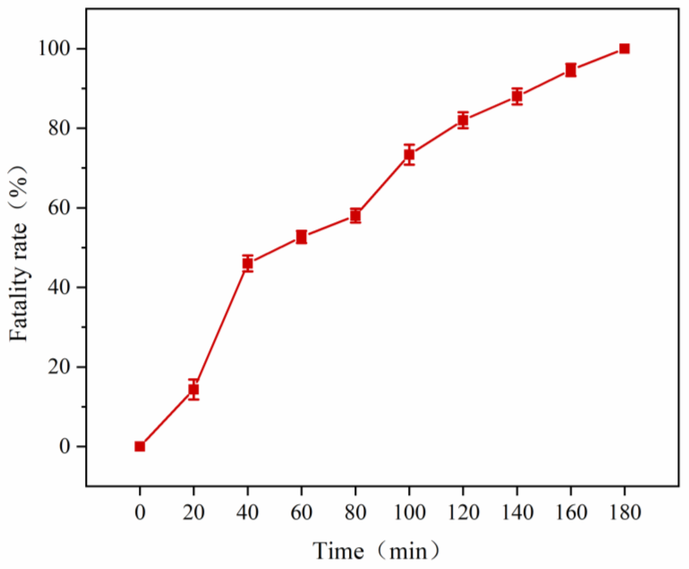
3.4. Mutant Library Construction Using ARTP
As the flow cytometry sorting system can perform ultra-high-speed sorting and purification of specific cells, we used a flow cytometer to sort 2 mL aliquots of a suspension of the E. coli ΔtRNA L21r (1 × 10⁸ cfu/mL) mutant strain mutated using ARTP for 160 s (Figure 4). The ratio of the total number of screened cells to the total number of cells was approximately 1:100,000. The analysis and screening results shown in Figure 5 indicate that that the cells in Region A have a strong fluorescence. Single cells with strong fluorescence signals were sorted and inoculated into the wells of a 96-well plate containing LB medium.
Figure 4.
Atmospheric pressure and room temperature plasma (ARTP) mutagenesis mortality curve. A time of 160 s was selected as the most effective mutagenesis time.
Figure 5.
Flow cytometry analysis results. The ordinate FITC shows the fluorescence intensity of staygold-L21r. The darker the color, the more densely clustered the cells. The cells in Region A are characterized by strong fluorescence. Single cells with strong fluorescence signals were sorted and inoculated into a 96-well plate containing LB medium.
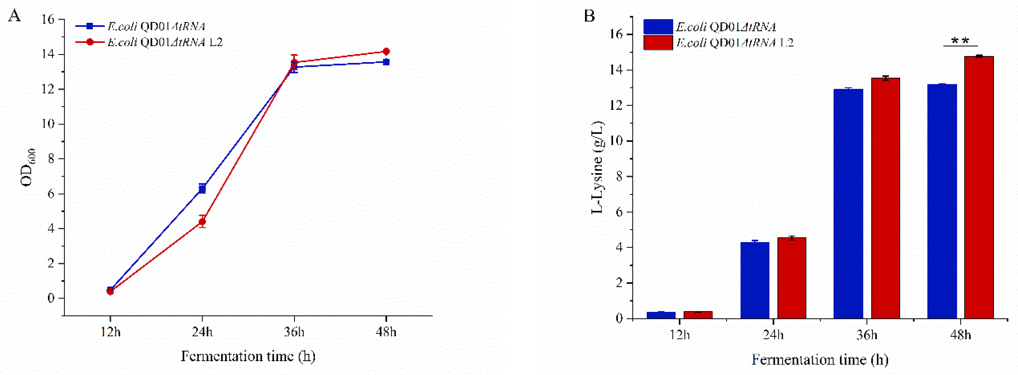
3.5. Fermentation of E. coli ΔtRNA L21r Mutants
Using the sorting results, we selected 29 mutant strains of E. coli L21r (Figure 6). Among the 29 mutant strains, we screened 16 with elevated l-lysine yields, representing a screening efficiency of 55.2%. The highest yielding l-lysine group among the three parallel fermentation yields of the E. coli QD01ΔtRNA strain (1.73 g/L) was E. coli QD01ΔtRNA L2 (Figure 6).
Figure 6.
The l-lysine produced by the wild-type and mutated strains. Twenty-nine mutant strains of E. coli ΔtRNA L21r were selected in the wells of 96-well plates. The dashed blue line indicates the highest yielding l-lysine group among three parallel fermentation yields of the E. coli QD01ΔtRNA strain (1.73 g/L). Among the 29 mutant strains, we identified 16 with elevated l-lysine yields. The red strain E. coli QD01ΔtRNA L2 showed the strongest fluorescence intensity.
The E. coli QD01ΔtRNA L2 strain with the highest fluorescence intensity was cultured in a 250 mL shaking flask containing 50 mL of LB medium. During the initial stage of fermentation, the OD600 of E. coli QD01ΔtRNA L2 was lower than that of E. coli QD01ΔtRNA, whereas at 48 h, the OD600 of E. coli QD01ΔtRNA was lower than that of E. coli QD01ΔtRNA L2, with a 4.5% increase in OD600 (Figure 7A). After fermentation for 48 h, the l-lysine yield of the wild-type E. coli QD01ΔtRNA was 13.2 g/L, whereas that of the engineered mutant E. coli QD01ΔtRNA L2 was 14.8 g/L, representing a 12.1% increase (Figure 7B). The l-lysine yield of the mutant strain was significantly higher than that of the wild-type strain (p < 0.01). We also compared the OD600 values and l-lysine production of E. coli QD01 and E. coli QD01ΔtRNA (Supplementary Figure S3), which indicated that these two strains had comparable OD600 values, and we detected no significant difference between the strains with respect to l-lysine production (p > 0.05).
Figure 7.
OD600 values and l-lysine production of E. coli QD01ΔtRNA L2 and E. coli QD01ΔtRNA (fermentation medium). (A) The OD600 of E. coli QD01ΔtRNA was lower than that of E. coli QD01ΔtRNA L2, which showed a 4.5% increase in OD600. (B) After fermentation for 48 h, the l-lysine yield of the wild-type strain E. coli QD01ΔtRNA was 13.2 g/L, whereas that of the engineered mutant E. coli QD01ΔtRNA L2 was 14.8 g/L, which represents an increase of 12.1% (** p > 0.05).
4. Discussion
Most target metabolites are not associated with readily identifiable phenotypes. When traditional methods such as chromatography and mass spectrometry are used to analyze multiple genetic variables, the screening process is often inefficient. Typically, the concentrations of chemicals are sensed by a diverse array of molecular constituents, including allosteric enzymes, transcription factors, and ribose switches. Artificial biosensors developed by exploiting these molecular systems can react with chemical signals, which are thereby converted to readily detectable signals.
Currently, the high-throughput screening methods applied to E. coli are mainly based on biosensors and FACS (Table 2), the latter of which has received increasing attention as a novel and powerful tool for screening target strains. FACS can be used to detect fluorescence signals [27,28], thereby enabling high-throughput screening of large-volume libraries. Previously, we constructed the pET-22b(+)-egfpr vector and used this to transform E. coli QD01ΔtRNA [13], the screening efficiency of which was only 13.8% (Supplementary Figure S2). However, in the present study, we substituted the screening marker with a fluorescent fusion protein rich in an artificial l-lysine rare codon, which elevated the screening efficiency to 55.2%. Previous studies have generally used a single fluorescent protein as a screening marker; however, in this study, we used lysine-rich staygold to establish a screening marker that was positively correlated with the content of l-lysine in cells. Following plasma mutagenesis and the induction of fluorescent protein expression at a normal temperature and pressure, the mutant strains showing strong fluorescence were sorted by flow cytometry, and we evaluated the fermentation performance of the strains showing high yields of l-lysine.
Table 2.
The high-throughput screening method used in this study.
Previously, Zhang et al. constructed an artificial cryptosystem, using which, they found that the growth of E. coli was stunted when artificial rare codons were present in the absence of the corresponding “artificial tRNAs” [35]. Therefore, it is hypothesized that the lack of tRNAs capable of recognizing “artificial codons” leads to the slower translation of ribosomal proteins within cells, thereby affecting the growth of E. coli. This indicates that by regulating the cryptosystem, it would be possible to control the rate of microbial protein synthesis. Subsequently, a strategy for screening amino-acid-overproducing bacteria using rare codons was proposed. By increasing the number of rare codons in gene sequences, Huo et al., for the first time, raised the amino acid concentration “threshold” required for protein translation, thereby establishing a screening system for strains producing high yields of l-leucine, l-arginine, and l-serine using a synthetic GFP rich in rare natural codons, and thus demonstrating that this approach is effective in E. coli. This system has similarly been demonstrated to be applicable for use in glutamic-acid-producing bacteria [36]. Inspired by these developments, we knocked out five l-lysine tRNA-UUUs in E. coli QD01 using RED gene recombination technology, reduced the expression efficiency of the AAA codon corresponding to tRNA-UUU, and constructed a rare artificial cryptosystem. A fluorescent protein expression vector containing rare lysine codons was subsequently used to transform E. coli QD01ΔtRNA, a mutation library was constructed based on ARTP, and mutant strains with strong fluorescence signals were screened via flow cytometry. The mutant strains obtained were subsequently cultured to determine the l-lysine production.
5. Conclusions
In this study, we reduced the expression of l-lysine tRNA-UUU in E. coli QD01 using RED gene recombination technology and constructed an artificial l-lysine rare codon (AAA). By constructing a fluorescent green fusion protein containing this AAA codon, we developed a screening marker, the expression of which was shown to be positively correlated with the l-lysine content in cells. Following constant temperature and atmospheric pressure plasma mutagenesis and induced fluorescent protein expression culture, we used flow cytometry to sort the mutant strains characterized by strong fluorescence and evaluated the fermentation performance of the l-lysine high-yielding strains obtained. The FACS method can make an important contribution to developing high-yielding l-lysine strains. The screening strategy for amino high-yielding strains based on the artificial rare cryptosystem established in this study provides an efficient, accurate, and simple method that could be used to screen for other amino-acid-producing microorganisms.
Supplementary Materials
The following supporting information can be downloaded at https://www.mdpi.com/article/10.3390/fermentation9100899/s1, Tables S1–S6: 6 medium formulations in the study. Table S7: Primers list. Figure S1: Gel electrophoresis of PCR products. Figure S2: The l-lysine produced by the original strain and the mutated strains. Figure S3: OD600 values and l-lysine production of E. coli QD01 and E. coli QD01ΔtRNA (fermentation medium).
Author Contributions
J.W. and C.Y. designed the experiments. H.L. performed the investigation. L.Y., R.W. and P.L. analyzed the results. B.D. contributed reagents and materials. N.L. drafted the manuscript. All authors have read and agreed to the published version of the manuscript.
Funding
This work was supported by the Focus on Research and Development Plan in Shandong Province (2021ZDSYS10, 2020CXGC010603, 2022CXGC020206), Taishan Scholar Foundation of Shandong Province (tscx202306067), Innovation Fund for Small- and Medium-sized Technology Innovation Capacity Enhancement Project of Shandong Province (2023TS1047), Key innovation Project of Qilu University of Technology (Shandong Academy of Sciences) (2022JBZ01-06), and National Natural Science Foundation of China (31801527).
Institutional Review Board Statement
Not applicable.
Informed Consent Statement
Not applicable.
Data Availability Statement
Not applicable.
Acknowledgments
We would like to thank the State Key Laboratory of Bio-based Materials and Green Papermaking, Qilu University of Technology, for its help and support and the Taishan industry-leading talent funding.
Conflicts of Interest
The authors declare that the research was conducted in the absence of any commercial or financial relationships that could be construed as a potential conflict of interest.
References
- Eggeling, L.; Bott, M. A giant market and a powerful metabolism: L-lysine provided by Corynebacterium glutamicum. Appl. Microbiol. Biotechnol. 2015, 99, 3387–3394. [Google Scholar] [CrossRef] [PubMed]
- Nguyen, L.; Davis, D.A. Comparison of crystalline lysine and intact lysine used as a supplement in practical diets of channel catfish (Ictalurus punctatus) and Nile tilapia (Oreochromis niloticus). Aquaculture 2016, 464, 331–339. [Google Scholar] [CrossRef]
- Felix, F.K.D.; Letti, L.A.; Pereira, G.V.D.; Bonfim, P.G.B.; Soccol, V.T.; Soccol, C.R. L-lysine production improvement: A review of the state of the art and patent landscape focusing on strain development and fermentation technologies. Crit. Rev. Biotechnol. 2019, 39, 1031–1055. [Google Scholar] [CrossRef]
- Xu, J.Z.; Yang, H.K.; Liu, L.M.; Wang, Y.Y.; Zhang, W.G. Rational modification of Corynebacterium glutamicum dihydrodipicolinate reductase to switch the nucleotide-cofactor specificity for increasing L-lysine production. Biotechnol. Bioeng. 2018, 115, 1764–1777. [Google Scholar] [CrossRef] [PubMed]
- Suvachan, A.; Lal, R.; Nampoothiri, K.M.; Sindhu, R.; Bhaskar, T.; Binod, P.; Pandey, A.; Yasarla, R. Valorization of paper industry rejects by combined thermo-chemical pretreatment and biological conversion to L-lysine. Environ. Technol. Innov. 2021, 24, 101882. [Google Scholar] [CrossRef]
- Wang, J.P.; Gao, C.; Chen, X.L.; Liu, L.M. Chapter one—Expanding the lysine industry: Biotechnological production of L-lysine and its derivatives. Adv. Appl. Microbiol. 2021, 115, 1–33. [Google Scholar] [CrossRef]
- Zhang, J.; Zhang, S.X.; Yang, X.N.; Qiu, L.; Gao, B.; Li, R.; Chen, J. Reactive extraction of amino acids mixture in hydrolysate from cottonseed meal with di(2-ethylhexyl) phosphoric acid. J. Chem. Technol. Biotechnol. 2016, 91, 483–489. [Google Scholar] [CrossRef]
- Xiao, J.; Wang, D.T.; Wang, L.; Jiang, Y.J.; Xue, L.; Sui, S.S.; Wang, J.; Guo, C.; Wang, R.; Wang, J.; et al. Increasingl-lysine production in Corynebacterium glutamicum by engineering amino acid transporters. Amino Acids 2020, 52, 1363–1374. [Google Scholar] [CrossRef]
- Yu, C.H.; Dang, Y.K.; Zhou, Z.P.; Wu, C.; Zhao, F.Z.; Sachs, M.S.; Liu, Y. Codon Usage Influences the Local Rate of Translation Elongation to Regulate Co-translational Protein Folding. Mol. Cell 2015, 59, 744–754. [Google Scholar] [CrossRef]
- Guimaraes, J.C.; Mittal, N.; Gnann, A.; Jedlinski, D.; Riba, A.; Buczak, K.; Schmidt, A.; Zavolan, M. A rare codon-based translational program of cell proliferation. Genome Biol. 2020, 21, 44. [Google Scholar] [CrossRef]
- Dittmar, K.A.; Sorensen, M.A.; Elf, J.; Ehrenberg, M.; Pan, T. Selective charging of tRNA isoacceptors induced by amino-acid starvation. Embo Rep. 2005, 6, 151–157. [Google Scholar] [CrossRef] [PubMed]
- Subramaniam, A.R.; Zid, B.M.; O’Shea, E.K. An Integrated Approach Reveals Regulatory Controls on Bacterial Translation Elongation. Cell 2014, 159, 1200–1211. [Google Scholar] [CrossRef] [PubMed]
- Zheng, B.; Ma, X.Y.; Wang, N.; Ding, T.T.; Guo, L.W.; Zhang, X.R.; Yang, Y.; Li, C.; Huo, Y.-X. Utilization of rare codon-rich markers for screening amino acid overproducers. Nat. Commun. 2018, 9, 3616. [Google Scholar] [CrossRef] [PubMed]
- Schrumpf, B.; Eggeling, L.; Sahm, H.J.A.M. Isolation and prominent characteristics of an L-lysine hyperproducing strain of Corynebacterium glutamicum. Appl. Microbiol. Biotechnol. 1992, 37, 566–571. [Google Scholar] [CrossRef]
- Javed, A.; Jamil, A.; Rezaei-Zarchi, S. Optimization and hyper-expressed production of lysine through chemical mutagenesis of Brevibacterium flavum by N-nitroso-N-ethylurea. Afr. J. Microbiol. Res. 2011, 5, 5230–5238. [Google Scholar] [CrossRef]
- Holatko, J.; Elisakova, V.; Prouza, M.; Sobotka, M.; Nesvera, J.; Patek, M. Metabolic engineering of the L-valine biosynthesis pathway in Corynebacterium glutamicum using promoter activity modulation. J. Biotechnol. 2009, 139, 203–210. [Google Scholar] [CrossRef]
- Xu, H.Y.; Andi, B.; Qian, J.H.; West, A.H.; Cook, P.F. The alpha-aminoadipate pathway for lysine biosynthesis in fungi. Cell Biochem. Biophys. 2006, 46, 43–64. [Google Scholar] [CrossRef]
- Liu, N.; Zhang, T.T.; Rao, Z.M.; Zhang, W.G.; Xu, J.Z. Reconstruction of the Diaminopimelic Acid Pathway to Promote L-lysine Production in Corynebacterium glutamicum. Int. J. Mol. Sci. 2021, 22, 9065. [Google Scholar] [CrossRef]
- Chen, W.; Zhang, S.; Jiang, P.X.; Yao, J.; He, Y.Z.; Chen, L.C.; Gui, X.; Dong, Z.; Tang, S.-Y. Design of an ectoine-responsive AraC mutant and its application in metabolic engineering of ectoine biosynthesis. Metab. Eng. 2015, 30, 149–155. [Google Scholar] [CrossRef]
- Cress, B.F.; Trantas, E.A.; Ververidis, F.; Linhardt, R.J.; Koffas, M.A.G. Sensitive cells: Enabling tools for static and dynamic control of microbial metabolic pathways. Curr. Opin. Biotechnol. 2015, 36, 205–214. [Google Scholar] [CrossRef]
- Binder, S.; Schendzielorz, G.; Stabler, N.; Krumbach, K.; Hoffmann, K.; Bott, M.; Eggeling, L. A high-throughput approach to identify genomic variants of bacterial metabolite producers at the single-cell level. Genome Biol. 2012, 13, R40. [Google Scholar] [CrossRef] [PubMed]
- Cazier, A.P.; Blazeck, J. Advances in promoter engineering: Novel applications and predefined transcriptional control. Biotechnol. J. 2021, 16, 2100239. [Google Scholar] [CrossRef]
- Della Corte, D.; van Beek, H.L.; Syberg, F.; Schallmey, M.; Tobola, F.; Cormann, K.U.; Schlicker, C.; Baumann, P.T.; Krumbach, K.; Sokolowsky, S.; et al. Engineering and application of a biosensor with focused ligand specificity. Nat. Commun. 2020, 11, 4851. [Google Scholar] [CrossRef] [PubMed]
- Yang, C.P.; Peng, Z.H.; Yang, L.; Du, B.W.; Guo, C.Z.; Sui, S.S.; Wang, J.; Li, J.; Wang, J.; Li, N. Design and application of artificial rare L-lysine codons in Corynebacterium glutamicum. Front. Bioeng. Biotechnol. 2023, 11, 1194511. [Google Scholar] [CrossRef] [PubMed]
- Hirano, M.; Ando, R.; Shimozono, S.; Sugiyama, M.; Takeda, N.; Kurokawa, H.; Deguchi, R.; Endo, K.; Haga, K.; Takai-Todaka, R.; et al. A highly photostable and bright green fluorescent protein. Nat. Biotechnol. 2022, 40, 1132. [Google Scholar] [CrossRef]
- Yang, Y.Q.; Yu, Q.L.; Wang, M.; Zhao, R.; Liu, H.W.; Xun, L.Y.; Xia, Y. Escherichia coli BW25113 Competent Cells Prepared Using a Simple Chemical Method Have Unmatched Transformation and Cloning Efficiencies. Front. Microbiol. 2022, 13, 838698. [Google Scholar] [CrossRef]
- East, A.K.; Mauchline, T.H.; Poole, P.S. Chapter 5 Biosensors for Ligand Detection. Adv. Appl. Microbiol. 2008, 64, 137–166. [Google Scholar] [CrossRef]
- Schallmey, M.; Frunzke, J.; Eggeling, L.; Marienhagen, J. Looking for the pick of the bunch: High-throughput screening of producing microorganisms with biosensors. Curr. Opin. Biotechnol. 2014, 26, 148–154. [Google Scholar] [CrossRef]
- Niu, F.X.; He, X.; Huang, Y.B.; Liu, J.Z. Biosensor-Guided Atmospheric and Room-Temperature Plasma Mutagenesis and Shuffling for High-Level Production of Shikimic Acid from Sucrose in Escherichia coli. J. Agric. Food Chem. 2020, 68, 11765–11773. [Google Scholar] [CrossRef]
- Shen, Y.P.; Pan, Y.Y.; Niu, F.X.; Liao, Y.L.; Huang, M.T.; Liu, J.Z. Biosensor-assisted evolution for high-level production of 4-hydroxyphe- nylacetic acid in Escherichia coli. Metab. Eng. 2022, 70, 1–11. [Google Scholar] [CrossRef]
- Zhu, Y.; Li, Y.; Xu, Y.; Zhang, J.; Ma, L.L.; Qi, Q.S.; Wang, Q. Development of bifunctional biosensors for sensing and dynamic control of glycolysis flux in metabolic engineering. Metab. Eng. 2021, 68, 142–151. [Google Scholar] [CrossRef] [PubMed]
- Dietrich, J.A.; Shis, D.L.; Alikhani, A.; Keasling, J.D. Transcription Factor-Based Screens and Synthetic Selections for Microbial Small-Molecule Biosynthesis. Acs Synth. Biol. 2013, 2, 47–58. [Google Scholar] [CrossRef] [PubMed]
- Wu, T.; Chen, Z.Y.; Guo, S.Y.; Zhang, C.Y.; Huo, Y.X. Engineering Transcription Factor BmoR Mutants for Constructing Multifunctional Alcohol Biosensors. Acs Synth. Biol. 2022, 11, 1251–1260. [Google Scholar] [CrossRef] [PubMed]
- Zhou, S.H.; Liu, P.R.; Chen, J.; Du, G.C.; Li, H.Z.; Zhou, J.W. Characterization of mutants of a tyrosine ammonia-lyase from Rhodotorula glutinis. Appl. Microbiol. Biotechnol. 2016, 100, 10443–10452. [Google Scholar] [CrossRef]
- Zhang, Y.; Ptacin, J.L.; Fischer, E.C.; Aerni, H.R.; Caffaro, C.E.; Jose, K.S.; Feldman, A.W.; Turner, C.R.; Romesberg, F.E. A semi-synthetic organism that stores and retrieves increased genetic information. Nature 2017, 551, 644. [Google Scholar] [CrossRef] [PubMed]
- Huo, Y.X.; Zheng, B.; Wang, N.; Yang, Y.P.; Liang, X.X.; Ma, X.Y. Identifying Amino Acid Overproducers Using Rare-Codon-Rich Markers. Jove-J. Vis. Exp. 2019, 148, e59331. [Google Scholar] [CrossRef]
Disclaimer/Publisher’s Note: The statements, opinions and data contained in all publications are solely those of the individual author(s) and contributor(s) and not of MDPI and/or the editor(s). MDPI and/or the editor(s) disclaim responsibility for any injury to people or property resulting from any ideas, methods, instructions or products referred to in the content. |
© 2023 by the authors. Licensee MDPI, Basel, Switzerland. This article is an open access article distributed under the terms and conditions of the Creative Commons Attribution (CC BY) license (https://creativecommons.org/licenses/by/4.0/).


