Review

Historical Aspects of Restriction Endonucleases as Intelligent Scissors for Genetic Engineering

by
Irina V. Alekseeva
1 and
Nikita A. Kuznetsov
1,2,*
1
Institute of Chemical Biology and Fundamental Medicine, Siberian Branch of Russian Academy of Sciences, Novosibirsk 630090, Russia
2
Department of Natural Sciences, Novosibirsk State University, Novosibirsk 630090, Russia
*
Author to whom correspondence should be addressed.
Fermentation 2023, 9(10), 874; https://doi.org/10.3390/fermentation9100874
Submission received: 24 August 2023 / Revised: 21 September 2023 / Accepted: 21 September 2023 / Published: 28 September 2023
(This article belongs to the Special Issue Applications of Enzymes in Biosynthesis)

Abstract

:
Restriction endonucleases are a component of restriction–modification systems, where the main biological function is to protect bacterial cells from incoming foreign DNA molecules. There are four main types of restriction enzymes (types I, II, III, and IV), which differ in protein composition, cofactor requirements, and mode of action. The most studied are representatives of type II, which specifically recognize DNA sequences of 4–8 bp and catalyze DNA cleavage within these sequences or not far from them. The exceptional precision of type II enzymes has made them indispensable for DNA manipulations. Although hundreds of DNA restriction enzymes are currently known, there is still a need for enzymes that recognize new DNA targets. For this reason, the discovery of new natural restriction endonucleases and rational design of their properties (to obtain enzymes with high specificity for a unique nucleotide sequence at a restriction site and without nonspecific activity) will expand the list of enzymes for use in biotechnology and genetic engineering. This review briefly touches upon the main types of restriction endonucleases, their classification, nomenclature, and typical properties, and it concisely describes approaches to the construction of enzymes with altered properties.

1. Introduction

The advent of such a research field as genetic engineering ~50 years ago was associated with the emergence of the first enzymes that could help to obtain desired DNA fragments. With the development of molecular biology, genetics, biotechnology, and other related sciences, increasingly more “molecular tools” have arisen that allow for direct manipulation of the genetic material of any organism, thereby now leading to the appearance of one of the most rapidly evolving areas of research: genetic technologies. In fact, for manipulations with nucleic acid molecules, there is currently a complete toolbox of enzymes that perform functions of a molecular “scalpel”, “scissors”, and “sewing needle”. By means of these specialized “tools”, it is possible to “cut” DNA at a strictly defined site; cut a fragment out of it; or, conversely, insert a new one, and then “mend” the molecular gap.
There are several classes of enzymes that can introduce a site-specific break into DNA. For this procedure, as a rule, restriction endonucleases are used, which are considered classic enzymes for obtaining DNA fragments. At present, their list is rapidly replenished with natural and artificial site-specific nucleases from the following families: zinc finger nucleases, transcription activator-like effector nucleases (TALENs), and clustered regularly interspaced short palindromic repeats (CRISPR/Cas) [1]. Interestingly, the biological function of restriction endonucleases from restriction–modification systems and, for example, endonucleases from the CRISPR/Cas system can be considered as an ancestor of innate and adaptive immunity, respectively [2]. This biological specialization probably leads to differences in the practical applications of these classes of enzymes. Indeed, restriction endonucleases are commonly used as scissors for DNA cleavage at a strictly defined site to create engineered DNA fragments with predetermined sequences of ends. In contrast, natural and artificial site-specific nucleases are widely accepted because of their diversity and enormous potential for targeted genomic modifications in eukaryotes and other animals. In this context, the use of these site-specific nucleases is mainly associated with genome editing tools, which, for example, allow for producing various genetically modified animal models for studying human diseases [3,4], performing crop improvement [5], and developing applications in microbial biotechnology [6]. Despite the qualities of CRISPR/Cas systems, one of the most significant hurdles that stall CRISPR/Cas adoption in therapeutic and clinical applications on a larger scale is its propensity to generate off-target effects [7,8]. Therefore, the displacement of restriction endonucleases, as tools for genetic material manipulation, by site-specific nucleases does not occur due to the simplicity and efficiency of classical restriction endonucleases to perform their tasks.
The discovery of restriction endonucleases was started in the 1960s. The first experiments demonstrating the possibilities of restriction enzymes were conducted by Kathleen Danna and Daniel Nathans from Johns Hopkins University (Baltimore) and were published in 1971 [9]. That article showed for the first time that a restriction enzyme called “endonuclease R” (discovered by Hamilton Smith and Kent Wilcox [10]) can be employed to obtain specific fragments of simian virus 40 (SV40) DNA. Moreover, those authors showed that the resultant fragments can be effectively separated from each other by electrophoresis. These results were the first example of the preparation of individual nucleic acid fragments with predefined terminal regions. That groundbreaking study laid the foundation for modern molecular biological practices, where restriction enzymes are crucial tools, and at present, obtaining DNA fragments by means of restriction enzymes is a routine task.
Restriction endonucleases are a component of restriction–modification (RM) systems, where the main biological function is to protect bacterial cells from incoming foreign DNA molecules [11]. There are four main types of restriction enzymes (types I, II, III, and IV), which differ in enzyme composition, cofactor requirements, and mode of action [12]. The most investigated are representatives of type II, which specifically recognize DNA sequences of 4–8 bp and catalyze DNA cleavage within these sequences or at a short distance from them. The exceptional precision of type II enzymes [13,14] has made them indispensable tools for DNA manipulations. It should be noted that ~5000 restriction endonucleases of all types have been characterized to date, but among them, there are almost 4900 type II enzymes [15,16]. At the same time, 625 commercially available type II enzymes recognize only 239 different specific sites in DNA. Because the list of available specific sites is relatively short, both an extensive search for new variants of restriction enzymes in natural sources and attempts to artificially change the specificity of known enzymes are carried out incessantly. Although hundreds of DNA restriction enzymes are currently known, there is a demand for enzymes that recognize new DNA targets; the discovery of new restriction enzymes that are highly specific for a unique nucleotide sequence at a “restriction site” and do not exert nonspecific action will expand the arsenal of enzymes for biotechnology or replace existing ones with more effective ones [17].
This review briefly examines the main classes of restriction endonucleases and their classification by type, nomenclature, and typical properties. It also concisely describes approaches to the creation of enzymes possessing altered properties.

2. Diversity of Restriction Endonucleases and Their Function in Vivo

Originally, restriction endonucleases were found in bacterial genomes and plasmids, but they are also present in archaea and some viruses [18,19,20]. It is known that every fourth tested bacterial species has more than one restriction enzyme gene [21]. Of note, such pathogens as Neisseria gonorrhoeae and Helicobacter pylori are the richest sources of restriction enzymes [22,23]. Some strains of these microorganisms may possess more than a dozen endonucleases of the RM system, although some of them are not actively expressed [24,25].
The main biological function of these enzymes is to protect the cell from foreign DNA. This is achieved through cleavage of any DNA that is recognized as foreign by the absence of characteristic marker modifications—which are cytosine methylated at position N4 or C5 and adenine methylated at N6—located in a specific nucleotide context called a recognition site. From the restriction activity, host cell DNA is protected by the action of methyltransferases, which recognize the same sequence as a restriction enzyme does, and methylate cytosine or adenosine in complementary strands of the restriction site. After the methylation, host DNA is no longer a substrate for the restriction enzyme. Given that both strands of host DNA are methylated, even after replication, the resulting hemimethylated host DNA is still protected and is not cleaved by the endonuclease.
It has been suggested that restriction enzymes can also perform additional functions, such as the maintenance of species identity among bacteria [22] and the creation of genetic variability [26]. Additionally, the role of the RM system in some cases may be considered parasitic toward the host cell. When there is dysregulation between the cellular lifetime of restriction endonucleases and methyltransferases, the degradation of the host DNA by its own enzymes and the death of the host cell could happen. A small number of phages can be methylated by the host cell’s enzymes before the cleavage of phage DNA, and this process allows these phage variants to proliferate. It should also be pointed out that long recognition sites of restriction enzymes are usually rare in phage genomes, which are small. More evidence in favor of the parasitic theory is that restriction endonucleases usually have a longer lifetime than their corresponding methyltransferases, and this difference can be fatal to the host if a methyltransferase does not perform its function properly. In summary of the above, it has been suggested that modifications made by the RM system apparently act as a third party toward a bacteriophage–bacterium pair because, on the one hand, these modifications protect the host cell from the invasion by foreign biomaterial, and on the other hand, they can create conditions leading to the host’s death [27,28].

3. Nomenclature and Classification of Restriction Endonucleases

Recombinant restriction enzymes are usually named according to the nomenclature proposed by Smith and Nathans [29], which involves writing the name of a restriction enzyme in italics, with three letters derived from the first letter of the genus and the first two letters of the species of the microorganism from which the enzyme was obtained. An additional letter without italics may be used to indicate a strain. This is followed by a Roman numeral indicating the first, second, etc., enzyme found in the microorganism.
Restriction endonucleases are classified by their structure, recognition site, cleavage site, and reaction cofactors [12]. Type I restriction endonucleases consist of three different subunits, each of which is responsible for methylation, restriction, and sequence recognition [30]. For the functioning of enzymes of this type, Mg2+ ions and S-adenosylmethionine (AdoMet) are required (the latter as a source of a methyl group). They usually interact with two recognition sites, and movement along the DNA strand is accompanied by hydrolysis of ATP; DNA breakage then occurs, as a rule, approximately in the middle between the two sites.
Type II enzymes cleave DNA at specific positions near or within their recognition site, which consists of 4–8 bp [21]. Most of them are homodimeric or tetrameric enzymes, and their catalysis requires only Mg2+ ions. Because of their ability to perform precise site-specific DNA cleavage, these enzymes have found many applications in DNA analysis and gene cloning. Nonetheless, type II enzymes are not a single family of proteins but a collection of unrelated proteins that differ greatly in amino acid sequence from one another [23]. Such dissimilarities cause these enzymes to differ in the details of the process of recognition of a specific site. Nevertheless, most of them share similarities in the structural organization of the catalytic core and evidently have a common DNA cleavage mechanism, suggesting that they originated from a common ancestor [31].
The differences in properties among type II enzymes allow for dividing this vast class into subtypes (Table 1). In some cases, however, for the purposes of nomenclature, some enzymes can be assigned to more than one subtype. This is because subtyping criteria can be based both on the cleaved sequence and on, e.g., the structure of the enzymes themselves; therefore, not all subtypes are mutually exclusive. Subtype IIS enzymes, originally designated as enzymes with a cleavage site shifted relative to their recognition sequence [32], will keep that affiliation, but a new subtype, IIA, is now defined, which includes all restriction endonucleases of type II that recognize asymmetric sequences. Furthermore, a new subtype IIP will be employed to designate enzymes that recognize symmetrical sequences (palindromes).
Thus, the predominant criterion for classifying an enzyme as type II is that it produces a specific set of fragmentation products and cleaves either within its recognition sequence or nearby at a fixed site or with known and limited variability.
Type III restriction enzymes consist of only two subunits, one of which is responsible for DNA recognition and modification and the other for DNA cleavage [33,34]. Catalytic activity requires ATP and Mg2+ ions and is stimulated by AdoMet. The enzyme interacts with two recognition sites, moves along DNA owing to ATP hydrolysis, and introduces a break into the DNA next to one of the recognition sites.
Type IV restriction enzymes cleave modified DNA [35]. As such, these enzymes are not a part of the RM system and have low DNA sequence selectivity but are able to recognize modified DNA that contains such nitrogenous bases as C-5-methylcytosine (m5C), N4-methylcytosine (m4C), N6-methyladenine (m6A), 5-hydroxymethyluracil (hm5U), 5-hydroxymethylcytosine (hm5C), and its derivatives with attached sugar residues, for example, 5-glucosyl-hydroxymethylcytosine (ghm5C). Additionally, it has been shown that these enzymes can recognize phosphothioate derivatives of the internucleotide phosphate group [36,37,38].
Most recognition sites are four, six, or eight bases long and are palindromes. Nonetheless, some enzymes can specifically recognize and catalyze DNA breaks at sites containing (at some positions) not a strictly defined base but a set of bases up to any of the four possible letters (Table 2). In the case of such ambiguity, when a nucleotide substitution appears in the recognition site at some position, a partially palindromic or asymmetric sequence is formed. For example, the recognition site of StyI is listed as CCWWGG. Therefore, substrate sequences for StyI can be palindromic (CCTAGG or CCATGG) or partially palindromic (CCTTGG or CCAAGG) [39].
In conjunction with attempts to create artificial restriction endonucleases with altered recognition sites, the variation in natural recognition sites of some enzymes still raises questions. Of particular interest are situations where “allowed” nucleotides can be purine or pyrimidine or when only one nucleotide is not allowed.
It should be mentioned that RM systems that recognize the same nucleotide sequence can form different sets of methylation products and may differ in sensitivity to methylated-DNA hydrolysis. Nonetheless, when many enzymes from different biological sources recognize the same DNA sequence, such enzymes are called isoschizomers. Furthermore, the category in which two enzymes recognize the same DNA sequence but cleave it at different positions is called neoschizomers.
An important finding from cloning and sequence comparisons is that there is virtually no amino acid sequence homology between an endonuclease and methyltransferase that recognizes the same DNA sequence. Additionally, even restriction isoschizomers may share little or no homology, even in terms of the amino acid residues involved in cleavage site recognition. For example, enzymes HhaII and HinfI isolated from strains of Haemophilus recognize the GANTC sequence and cleave it between G and A. In contrast, their amino acid sequences are only 19% identical [11].

4. Genomic Organization

Genes of a restriction endonuclease and corresponding methyltransferase are located next to each other in genomic DNA and can be transcribed in a convergent, divergent, or sequential manner. It is thought that methylation must occur before restriction activity in order to protect host DNA. One approach that bacteria utilize to limit their ability to self-cleave is to greatly reduce the number of restriction sites in their genome. Usually, methyltransferase expression precedes endonuclease expression. One way this can happen is when an open reading frame is located upstream of an endonuclease gene and encodes a control protein (C protein) in some RM systems. This C protein positively regulates the endonuclease gene and enables methyltransferase expression to precede endonuclease expression [40]. Genes for such a C protein are often present in situations where methyltransferase and endonuclease genes show convergently oriented transcription. By means of cloned RM systems with disrupted C protein genes for BamHI, SmaI, PvuII, and EcoRV, various C protein variants have been tested where genes are located in a separate plasmid. Restriction activity of BamHI was found to be equally stimulated by the gene of SmaI-C and the gene of BamHI-C but an order of magnitude less stimulated by the gene of PvuII-C. The gene of EcoRV-C did not induce the stimulation. The gene of BamHI-C stimulated the restriction activity of PvuII to the same extent as the gene of PvuII-C did [41]. It is possible that the ability of some C-protein genes to stimulate the expression of alternative endonucleases has evolutionary significance for RM systems.
All type II restriction endonucleases where crystal structures have been determined share a common catalytic sequence motif: PD…D/EXK [31]. Nonetheless, this consensus sequence involves substantial variation, which, in some cases, makes it difficult to locate catalytic amino acid residues by amino acid sequence analysis alone; accordingly, the function of residues is confirmed by mutational analysis [42,43,44]. Among methyltransferases, common motifs have been found for 30 different 6-methyladenine-, 4-methylcytosine-, and 5-methylcytosine-producing enzymes [45]. This fact seems to indicate that methyltransferase genes are more conserved than restriction endonuclease genes.

5. Mechanism of Specific-Site Recognition and Catalysis

The generalized mechanism of site-specific DNA cleavage by restriction enzymes includes several steps (Figure 1). In the first step, the enzyme binds to DNA in a nonspecific manner, which usually involves interaction only with the phosphate backbone. Then, there is a search for the recognition site by movement along the DNA owing to linear diffusion. It has been determined that the EcoRV enzyme can scan 2 × 10⁶ bp at a rate of 1.7 × 10⁶ bp/s per binding event [46].
When a specific recognition site is found, a network of hydrogen bonds (usually 15–20) involving nitrogenous bases of the recognition site is formed in the DNA-binding center, and this process ensures the specificity of the enzyme [24]. Nevertheless, it must be noted that the sequence flanking the recognition site can also influence the specific binding. For instance, for BamHI, the binding increases 5400-fold as the length of the oligonucleotide is extended from 10 to 14 bp and varies by up to 30-fold depending on more or less suitable flanking triplets [47].
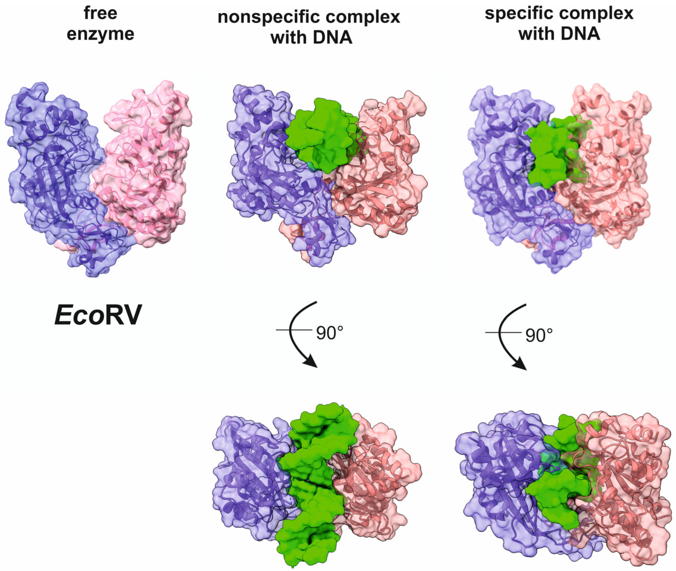
As a specific complex is forming, conformational changes and structural rearrangements take place both in the enzyme molecule and in DNA (Figure 2). Crystal structures of complexes of the EcoRV endonuclease with DNA enable an analysis of stages of protein-induced DNA bending. EcoRV is a homodimeric type II restriction endonuclease and cleaves DNA with the formation of blunt ends in the central part of its recognition site GAT↓ATC while bending DNA within this region by ~50° toward the major groove [48,49,50]. Meanwhile, the DNA remains mainly in the canonical B-form on both sides of the cleavage site. A comparison of crystal structures between the enzyme not bound to the ligand and the enzyme bound to DNA reveals considerable rearrangements of a quaternary structure after the binding, primarily involving the rotation of the DNA-binding domain and catalytic domain by 25°, leading to a change in the mutual arrangement of subunits in the dimer. In addition, a 12° difference has been found in the rotation angle between these domains in different structures [51,52]. Thus, during the formation of the catalytic complex, there are substantial rearrangements of the quaternary structure of the enzyme that induce strong DNA bending.
After the assembly of the specific complex between the enzyme and DNA, in the presence of Mg2+ ions, a phosphodiester bond is cleaved in the 2′-ribose phosphate backbone of both DNA strands. The outcome is either blunt ends or single-stranded 3′ or 5′ overhangs 1–4 bases long: so-called sticky ends. It should be pointed out that enzymes with completely different recognition sites can produce matching overhangs that are suitable for ligation. For example, restriction endonucleases NarI, MspI, AcyI, TaqI, ClaI, Csp45I, HpaII, and AccI generate a single-stranded 5′-CG end, even though these enzymes have different recognition sites.
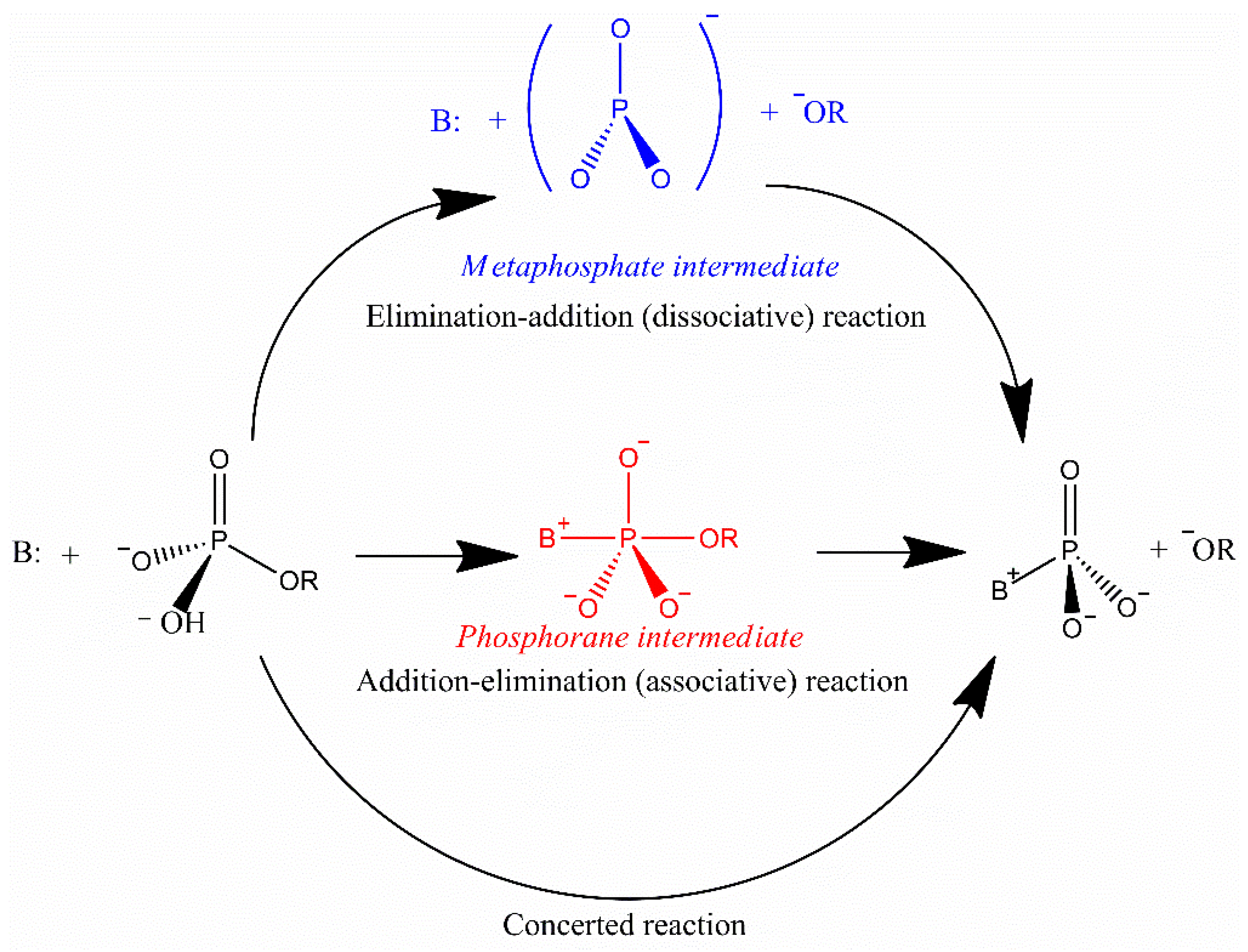
The catalytic reaction of phosphodiester bond cleavage by restriction endonucleases is usually described as a transfer of a phosphoryl residue to water [53]. Such a transfer can potentially be mediated by three alternative mechanisms. The dissociative mechanism includes the emergence of a metaphosphate intermediate; an SN2-type reaction proceeds in the coordinated (concerted) mechanism, including simultaneous breakage of one bond and the formation of a new bond; a pentavalent phosphorane intermediate arises in the associative mechanism (Figure 3). Nonetheless, the actual mechanism that is implemented by restriction endonucleases is still a matter of debate [21,23].

6. Practical Application of Type II Restriction Endonucleases

The type II enzymes are most commonly used for molecular cloning and are applied in biotechnology via a combination of desired enzymatic properties. The most common type II restriction endonucleases are homodimers, in most cases consisting of monomeric subunits with a molecular weight of 25–35 kDa; require Mg2+ ions for the catalysis; and cleave DNA at a certain site within palindromes, partial palindromes, or interrupted palindromes. The monomers of type II restriction endonucleases are not active on their own.
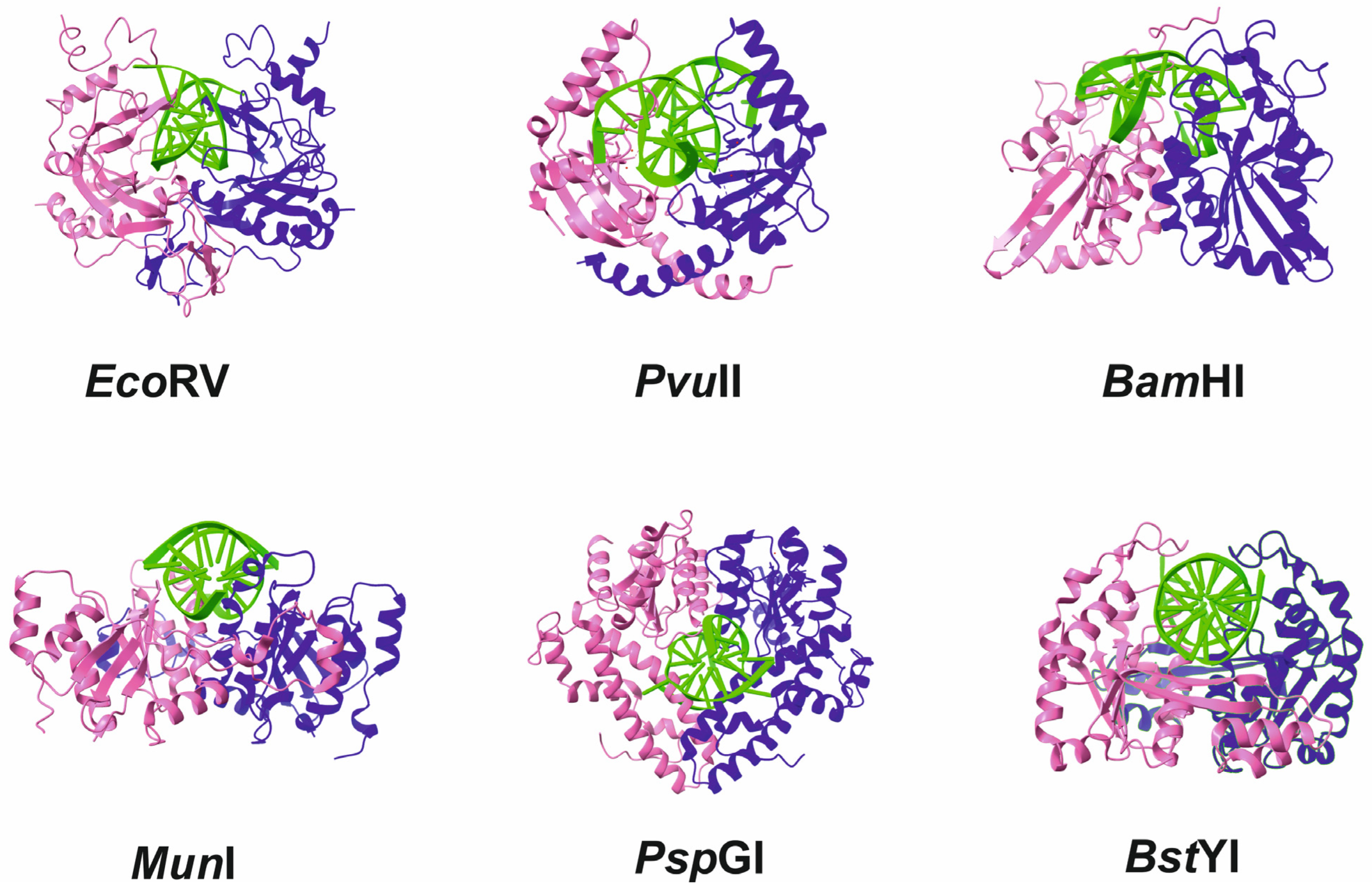
Crystal structures determined for type II enzymes share a common core consisting of five β-folds surrounded on each side by an α-helix, similar to enzymes MutH and α-exonuclease [54,55,56]. Despite their different primary sequences, type II restriction endonucleases have similar three-dimensional structure (Figure 4): a U-shaped dimeric holoenzyme, in which each of the identical subunits is arranged in such a way that domains that contribute to recognition and catalysis are located on the sides while bridging (connecting) domains are at the bottom.
Nevertheless, type II enzymes are further categorized (Table 1) based on differences in the cleavage site (cutting within or at a fixed distance from the recognition site), on their ability to noncooperatively cleave a single recognition site or a cooperative cleavage mechanism that requires simultaneous binding to multiple sites for increased cleavage efficiency, and on their domain organization, i.e., the differing arrangement of protein domains participating in identification of the recognition site and its cleavage and ensuring sensitivity to DNA methylation [21,23,57].
Furthermore, type II enzymes can be classified by additional structural homology among enzymes that produce 5′ sticky ends that are near the DNA major groove and enzymes that produce 3′ sticky or blunt ends that are near the minor groove of DNA. Crystal structures of type II endonucleases producing 5′ sticky ends suggest that these enzymes employ an α-helix to search for a specific site. On the contrary, restriction enzymes that form 3′ sticky or blunt ends use a β-fold region to recognize the restriction site. There are also differences in the polarity of β-sheets between these two groups [58]. Two enzymes can serve as an example (Figure 5): BamHI recognizes the GGATCC sequence, whereas BglII recognizes the closely related AGATCT site. Both generate the same 5′ overhang of four nucleotides: GATC. Nonetheless, enzyme–DNA contacts and the degree of DNA bending in the specific complex differ considerably between the two enzymes [59].
Below, we examined more detailed information about the subtypes of type II restriction endonucleases, which allows us to conclude that some of these enzymes are promising from the standpoint of their possible practical use in various molecular biological applications.

6.1. Endonucleases of Subtype IIA

Subtype IIA enzymes, such as SapI (GCTCTTC 1/4), recognize asymmetric sequences and catalyze DNA breakage within or at a certain distance from the recognition sequence. The restriction site recognition and cleavage functions of subtype IIA enzymes are usually localized to different domains. The recognition domain individually binds to DNA, while the catalytic domain dimerizes with an identical domain from another molecule, resulting in the formation of a catalytic complex [60,61]. It should be noted that there are much fewer enzymes of this subtype (which recognize asymmetric DNA sequences) than enzymes of the IIP subtype (which recognize symmetrical sequences).

6.2. Endonucleases of Subtype IIB

Type IIB enzymes cleave DNA outside their recognition sites and introduce a break on both sides of their recognition sequence, thereby releasing a small DNA fragment containing the recognition sequence [62]. The recognition sites of these enzymes usually consist of two specific sequences that are separated from each other by a DNA segment of fixed length. For example, the BcgI enzyme [63] recognizes the sequence
5′-10(N)-CGA-(N)6-TGC-(N)12↓-3′
3′-↓(N)12-GCT-(N)6-ACG-(N)10↓-5′
and cuts both DNA strands on both sides of this sequence at a distance of 10 and 12 nucleotides, thus leaving behind a 34-nucleotide fragment in both strands. Methyltransferase BcgI carries out methylation of the adenine residues highlighted in red in the above sequence. BcgI is inactive when bound to only one site of the two [64]; just as most type IIB enzymes [62,65], this enzyme needs two DNA recognition sites. After binding to the recognition sites, BcgI creates two double-strand breaks at each site before dissociating from end products, thereby hydrolyzing a total of eight phosphodiester bonds within one complex. It is noteworthy that in the course of the reaction, partially cleaved products almost do not accumulate [64].
BcgI, just as many but not all subtype IIB enzymes [62], consists of two subunits: subunit A (71.6 kDa, which catalyzes both methylase and restriction reactions) and subunit B (39.2 kDa, which recognizes the target sequence) [63,66]. Similarly to subtype IIG systems, which have both activities on the same subunit, BcgI requires AdoMet as a cofactor not only to perform methylation but also to exert the restriction enzyme action. Additionally, the enzyme needs Mg2+ [67]. The active form of enzyme BcgI is a hexamer composed of two identical trimers of A2B composition [66]. Some subtype IIB enzymes contain a single subunit that performs all three functions at once [68,69].

6.3. Endonucleases of Subtype IIC

Subtype IIC enzymes have a monosubunit composition and possess both endonuclease and methyltransferase activities. Most of them contain an N-terminal endonuclease domain followed by a methyltransferase domain. Most subtype IIC enzymes bind to their target sequences as a monomer. Meanwhile, these enzymes can recognize asymmetric sequences and catalyze breakage only on one side of the target sequence at a certain distance, for example, one turn of the DNA helix in the case of Tth111II (CAARCA 11/9), one and a half turns for Eco57I (CTGAAG 16/14), or even two turns for MmeI (TCCRAC 20/18). Additionally, the distance can vary by ±1–2 base pairs. It is believed that “accessibility” between the recognition sequence and the cleavage site depends on physical spatial (linear) distance and not on the number of base pairs in between, and this distance can depend on DNA topology, ionic conditions, and nucleotide sequence.

6.4. Endonucleases of Subtype IIE

These endonucleases differ from other type II members as follows: for efficient cleavage of DNA, they must simultaneously bind two copies of their palindromic recognition site in DNA, with one copy being the target for cleavage and the other an allosteric effector. As a consequence, these enzymes cleave only half of their recognition sites. That is, each enzyme dimer binds a recognition sequence in its catalytic site and the second one in the allosteric site [70]; hence, optimal efficiency of DNA cleavage is implemented when the ratio of recognition sites to enzyme dimers is 2–4.
Type IIE enzymes can slowly cleave one DNA region, while another region of the same sequence in the same or another DNA molecule is resistant to cleavage [71]. It is thought that flanking DNA sequences influence the cleavage kinetics of various recognition sites. Furthermore, type IIE enzymes can be categorized into two subclasses based on the type of action during the binding to the effector sequence, which can be an oligonucleotide, a linear phage, or supercoiled DNA. In enzymes of the first subclass, K (e.g., NarI, HpaII, and SacII), binding of activator DNA leads to more efficient binding of the cleavage site (because of a reduction in Michaelis constant Km) but does not alter maximum cleavage rate Vmax. This nature of the dependence indicates that the binding of the effector causes conformational rearrangements that enhance the affinity of the enzyme for the substrate. In class V (e.g., NaeI and BspMI), the effector binding raises the maximum cleavage rate Vmax without changing Km, indicating that the elevated catalytic activity is not related to the enzyme’s substrate affinity [71].
On the one hand, incomplete cleavage of DNA substrates by type IIE enzymes can complicate the interpretation of the obtained results and subsequent practical application of these enzymes. On the other hand, the ability to activate the enzyme by the addition of an effector enables researchers to carry out a controlled “switching on” of the enzymatic activity at the right time. The role of such activators can be played, for example, by oligonucleotides containing the recognition site of EcoRII (↓CCATGG), which is not cleaved due to specific methylation or the presence of modified nucleotide analogs but can bind to an allosteric site and stimulate cleavage of a target site in a pBR322 plasmid [72]. In a similar approach [73], an oligonucleotide is used that contains phosphorothioate in the hydrolyzable bond within the recognition site of NaeI. Complete cleavage of the target substrate, in this case, is achieved without the addition of an activator oligonucleotide because sulfur prevents the hydrolysis of such substrates by restriction endonuclease NaeI. The same strategy has been successfully utilized for the NarI enzyme, indicating the usefulness of this approach for enzymes of both subclasses V and K. Currently, some of these type IIE enzymes are commercially available in kits with activating oligonucleotides premixed in reaction buffer (e.g., Turbo™ NaeI and Turbo™ NarI from Promega). The presence of an oligonucleotide does not affect either ligation or labeling of random primers, and one-step purification yields DNA with sticky ends [74].
Of note, subtype IIE enzymes have been used to explain potential relations between such DNA-processing enzymes as endonuclease, topoisomerase, and recombinase [75,76]. It has been demonstrated that the NaeI enzyme contains in the N-terminal region a small motif (TD...DCK) that is similar to a motif in the N-terminal region of human DNA ligase I: 39TLDQLYDGQR48 (10 amino acid residues [58]). The leucine at position 43 within NaeI likely performs the same functions as the lysine—in the ligase motif—that takes part in the production of an adenylated intermediate and is required for ligation. It has been shown that the NaeI mutant containing substitution L43K possesses type I topoisomerase activity, i.e., aside from DNA cleavage, the enzyme promotes DNA unwinding and subsequent ligation of the termini.

6.5. Endonucleases of Subtype IIF

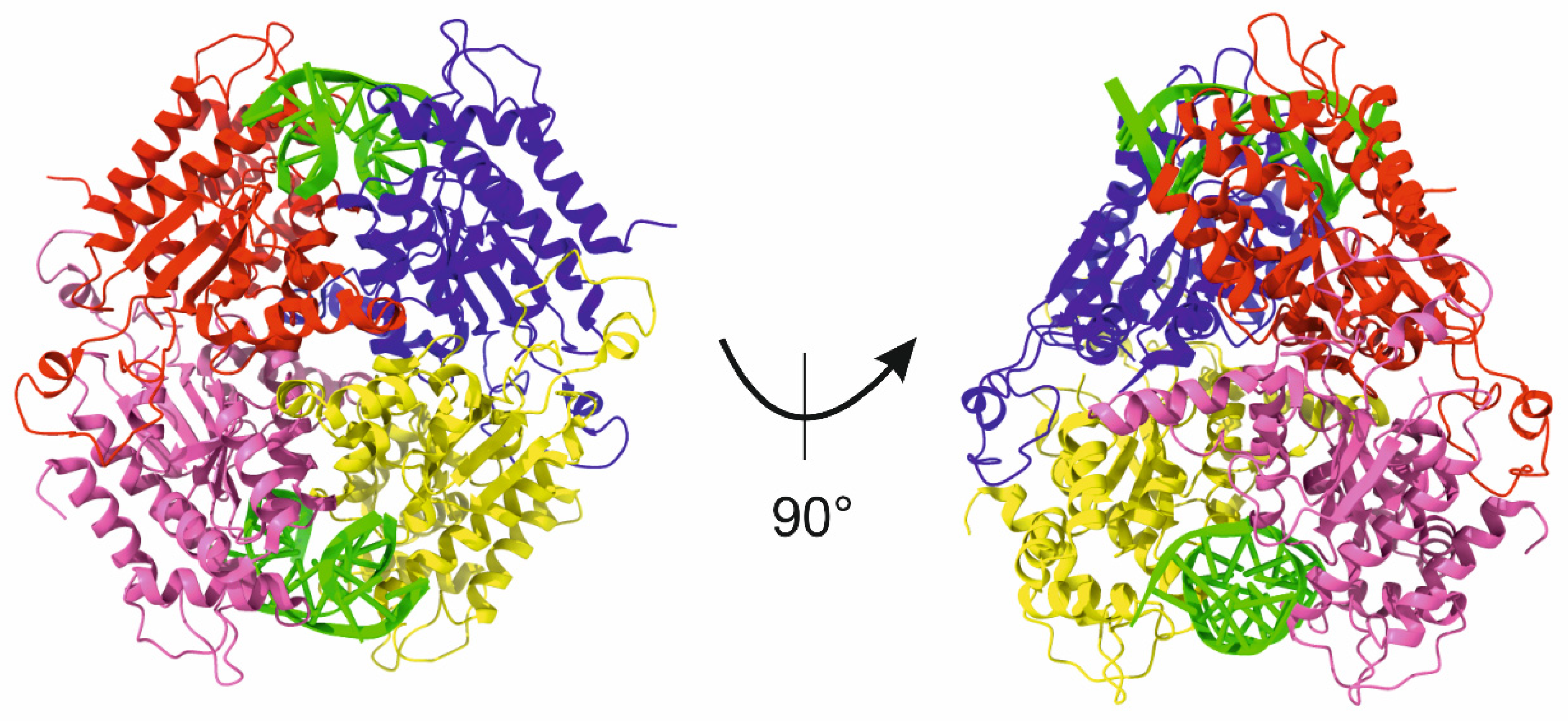
A distinctive feature of subtype IIF restriction endonucleases is that these enzymes form a homotetramer composed of a complex of two dimers in a back-to-back orientation, while a DNA molecule must be bound by two catalytic sites for cleavage to proceed (Figure 6). Representatives of this subtype of enzymes are SfiI [77], AatII [78], Cfr10I [74], and NgoMIV [79].
Because enzymes of subtype IIF also need two copies of the recognition site for DNA cleavage (just as enzymes of subtype IIE do), hydrolysis of the last few sites in the reaction can be problematic even with an excess of the enzyme relative to the substrate. In this regard, for SfiI, it is reported that the protein homotetramer should interact with two intact recognition sites containing cleavable phosphodiester bonds (in contrast to the activator that is acceptable for type IIE enzymes and contains nonhydrolyzable phosphorothioate [80]), resulting in cleavage of both recognition sites during one turnover of the enzyme [74,81]. Another observation regarding the interrupted palindrome recognized by SfiI (GGCCNNNN↓NGGCC) is a significant difference in the cleavage rate depending on the context of the internal degenerate sequence. For instance, sequence AAAC↓A is hydrolyzed 70 times more efficiently than AACA↓A. The authors of ref. [82] hypothesize that the rigidity of a DNA molecule (resulting from the context of this sequence) induces additional stress on the DNA double helix after it is bent by the enzyme, thereby improving catalysis efficiency.

6.6. Endonucleases of Subtype IIG

These endonucleases have previously been classified as type IV [83,84], but later, their affiliation was revised and switched to type II because the presence of Mg2+ ions is a necessary and sufficient condition for DNA cleavage, whereas AdoMet exerts a stimulatory effect on the activity of the enzyme [31]. In general, enzymatic properties of restriction enzymes of this subclass are also common among type II endonucleases. Cleavage outside nonpalindromic recognition sites may prevent the cleavage reaction from reaching 100% completion, just as in the case of subtypes IIE and IIF. Enzymes of the IIG subtype function as a monomer with all types of activity: site recognition, cleavage, and methylation [84].

6.7. Endonucleases of Subtype IIH

Such endonucleases are a hybrid subtype, also known as type 1½, incorporating features of type I and II enzymes [21]. At present, however, the assignment of enzymes to this subtype is rare and is not in demand.

6.8. Endonucleases of Subtype IIM

Subtype IIM enzymes are sensitive to methylation of the recognition sequence. The best-known example is the DpnI enzyme (Gm6A↓TC), which is widely used in techniques for the cleavage of methylated template DNA after PCR amplification. These enzymes act as monomers consisting of an N-terminal catalytic domain and a C-terminal domain responsible for allosteric activation. Both domains bind to DNA depending on the sequence and its methylation and catalyze DNA cleavage within one enzymatic cycle [85]. It remains unclear which structural features of these enzymes give rise to the absolute sensitivity to methylation in the recognition sequence. It can be theorized that methyl groups cause structural alterations, for example, by influencing the conformation of side chains of amino acid residues in the DNA-binding site, e.g., arginine and lysine, and thus switch these residues from a conformation that prevents binding to a conformation that is compatible with (and allows) the binding and the formation of a catalytically active complex.

6.9. Endonucleases of Subtype IIP

Subtype IIP enzymes are the most common and diverse of all type II restriction endonucleases [86]. They recognize symmetrical sequences 4–8 bp long and cleave DNA within this sequence (e.g., EcoRI: G↓AATTC) or less commonly at its boundaries (for example, EcoRII: ↓CCWGG). In most cases, these enzymes function as homodimers or homotetramers. Hundreds of enzymes of the IIP subtype are currently known, and apparently, the number of these enzymes is much larger, thereby enabling a search for DNA restriction enzymes with new recognition sites to expand the opportunities afforded by these enzymes in biotechnological and genetic engineering applications.

6.10. Endonucleases of Subtype IIS

Such endonucleases are monomeric proteins with a molecular weight of 45–110 kDa; they require the presence of Mg2+ for the catalysis, recognize nonpalindromic sequences, and cleave at least one of the two strands outside the recognition site. The bulk of available structural information about these endonucleases is based on the crystal structure of one of the members of this subtype, FokI, bound to DNA (Figure 7).
Subtype IIS enzymes comprise two domains: the N-terminal portion contains the DNA recognition domain, and the C-terminal part contains the catalytic site. Binding of the N-terminal DNA-binding domain to recognition site 5′-GGATG-3′ in double-stranded DNA results in activation of the catalytic domain and DNA cleavage at two sites (9 nucleotides downstream of the recognition site on the forward strand and 13 nucleotides downstream of the recognition site on the reverse strand) thereby giving rise to two sticky ends, with the overhangs of 4 nucleotides.
Structural data obtained for complexes of FokI with a 20 bp DNA fragment containing the recognition site (5′-GGATG-3′) have revealed additional distinctive features of this enzyme. For example, upon binding to its recognition site and in the presence of Mg2+, the catalytic domain becomes catalytically active through a series of intramolecular rearrangements [87]. Because each monomer contains only one catalytic domain, for cleaving both strands of DNA, the next step involves temporary dimerization of catalytic domains from the two monomers at the cleavage site. Structural similarity to the catalytic and bridging domains of homodimeric type II enzyme BamHI further confirms this model [61]. It has also been reported that the second molecule of FokI must be bound to DNA in order to cleave both strands of DNA. Currently, it is not known how the second monomer of FokI is oriented relative to DNA. If it is parallel to the first one, then this arrangement may lead to a protein–protein interaction and stabilization; if it is antiparallel, then this may cause a symmetrical orientation of the protein molecules in the dimer [88]. Evidently, a combination of structural reorganization processes during the activation of catalytic DNA cleavage is important for maintaining high accuracy of the positioning at which the phosphodiester bond hydrolysis takes place.
Recently, a new member of this subtype of endonucleases, PaqCI, has been described [89]. It was shown that despite PaqCI and FokI sharing similar structural mechanisms of DNA cleavage, these enzymes have considerable differences in their domain organization and quaternary architecture. Another member of this subtype, KpnK, can be considered a recently described enzyme, which was found in pathogenic Klebsiella pneumonia [90]. This enzyme is a monomer in solution, nicks double-stranded DNA, recognizes degenerate sequence, and catalyzes the degradation of DNA into smaller products after the initial cleavage at preferred sites.

6.11. Endonucleases of Subtype IIT

These enzymes function as heterodimers (for example, BbvCI, Bpu10I, BtsI, BsrDI, and BspD6I) consisting of two different subunits, although there are representatives (e.g., Mva1269I, BtsCI, AciI, BsrI, BssSI, and BsrBI) consisting of one subunit containing both catalytic domains [31]. The defining characteristic of type IIT restriction endonucleases is that catalysis requires the presence of both α and β subunits. It has been hypothesized that the active form of the BslI enzyme is the α2β2 heterotetramer, although heterodimers and oligomers also exist in solution [91]. Additionally, it has been shown that the bond between the subunits is rather weak, thereby leading to their easy separation from each other during purification and requiring restoration of the functional dimer for the manifestation of the activity [92]. This feature makes it possible to clone the genes of the α and β subunits and to prepare desired amounts of the recombinant proteins separately in the absence of methyltransferase without a toxic effect on the producer cells. These enzymes can have either a nonpalindromic recognition site (e.g., Bpu10I: CC↓TNAGC) or a palindromic sequence (e.g., BslI: CCNNNNN↓NNGG), which, together with their enzymatic properties, makes these enzymes attractive for practical use.

7. Off-Target Activity of Restriction Endonucleases

Although endonucleases form a nonspecific complex at the first stage of binding to DNA, they show very high catalytic selectivity with respect to their restriction site as compared to sites that differ even by one base pair. Nevertheless, a decrease in the specificity under nonoptimal reaction conditions is some enzymes’ inherent property, commonly referred to as “star activity.” Depending on the enzyme, the star activity manifests itself under certain reaction conditions: an excess of the enzyme over the recognition site, nonoptimal pH of the incubation medium, replacement of Mg2+ ions by some other divalent metal ions, or the presence of organic solvents (e.g., glycerol, dimethyl sulfoxide, or ethanol) [83]. For example, under optimal conditions, EcoRI cleaves its recognition site (5′-GAATTC-3′) at a rate 105 times faster as compared to a sequence differing by only one letter (5′-GATTTC-3′) [93].
At alkaline pH, there is a higher concentration of OH ions, the presence of which can reduce the need for the activated water that forms in the catalytic center as an attacking nucleophile [83]. On the other hand, pH and ionic strength can change the overall affinity of a protein rather than directly affect the specific/nonspecific balance or catalysis efficiency [94].
All restriction endonucleases require the presence of Mg2+ ions for their activity. Nonetheless, these enzymes can use surrogate metal ions, usually Mn2+ but sometimes Ca2+, Fe2+, Co2+, Ni2+, or Zn2+. DNA cleavage in the presence of these ions usually proceeds more slowly and less selectively [95]. It should be mentioned that a water molecule coordinated by the Mn2+ ion can be a more efficient proton donor required for the leaving 3′-OH group as compared to the coordination with the Mg2+ ion. For EcoRV, it is reported that the cleavage rate of a specific site (5′-GATATC-3′) is higher in the presence of Mg2+ than in the presence of Mn2+. By contrast, the cleavage of a site that differs by only one base pair proceeds 106 times faster in the presence of Mn2+ than Mg2+ [96].

8. Single-Stranded DNA Cleavage

The great interest in the ability of enzymes to cleave single-stranded DNA as well as single-strand in double-stranded DNA is due to the fact that nucleic acids, as encoding information for all forms of life, are excellent biomarkers for the identification of various targets such as bacteria, viruses, and other cells.
Most restriction endonucleases cleave only double-stranded DNA, but some enzymes can cleave single-stranded DNA, albeit at a much slower rate. There are a few approaches for single-stranded DNA cleavage. In some cases, for example, MspI [97] or HhaI and CfoI [98], direct cleavage of single-stranded DNA has been reported. Another example is the GGCC-specific restriction endonuclease BspRI, which performed kinetically separated acts of complementary strands cleavage, which leads to the formation of single-strand breaks in DNA [99]. In other cases, for example, for MnlI [100], it was shown that cleavage of single-stranded DNA proceeds in the presence of Ni2+ and some other transition metal ions, whereas Mg2+ ions are required for the cleavage of double-stranded DNA. Moreover, nicking endonucleases [101,102,103] become increasingly attractive for their application in single-strand cleavage of double-stranded DNA [104,105,106,107], and the list of these enzymes is regularly updated with new natural and artificial members [90,108,109]. Indeed, nicking endonucleases have been used in DNA optical mapping by nicking, nick translation of the nicked sites with fluorescently labeled dNTP, and religation of the nicked strand [110,111]. Large chromosome rearrangement of cancer cells could be “visualized” under a high-resolution fluorescent microscope when the abnormal DNA nicking pattern images are compared with the wild type [111,112]. These enzymes are also used in isothermal DNA amplifications, such as strand-displacement DNA amplification [113,114], nicking enzyme-assisted amplification [115,116], random whole genome amplification [117], and open chromatin profiling [118]. Recently, nicking enzyme-assisted reaction technology has been applied to the detection of COVID-19 viral RNA by reverse transcription, DNA nicking, and isothermal DNA amplification [119].
However, in most cases, to cleave the target single-stranded sequence, a double-stranded structure is formed, which can be readily cleaved by classical restriction enzymes [120,121,122,123]. One approach applied to cleave single-stranded DNA involves an oligonucleotide adapter and a restriction endonuclease with a shifted recognition site relative to the cleavage site (e.g., FokI). The oligonucleotide adapter is a hairpin containing a duplex portion carrying the enzyme’s recognition site and a single-stranded segment responsible for the formation of a duplex with the target sequence to be cleaved. The restriction endonuclease, having identified its recognition site in the double-stranded region of the oligonucleotide adapter, is expected to introduce a break into the region formed by the “oligonucleotide–single-stranded DNA” hybrid, as illustrated in Figure 8a. After the cleavage, the single-stranded-DNA–bound oligonucleotide fragment can be heat-denatured. On the other hand, this method cannot be used to cleave single-stranded RNA [95].
A similar approach to the cutting of single-stranded DNA can be implemented with the XcmI enzyme, which recognizes the longest known degenerate sequence (nine nucleotides: 5′-CCANNNNN/NNNNTGG-3′). The oligonucleotide adapter, in this case, contains two hairpins and a single-stranded region connecting them, which is complementary to the target sequence. The complementary single-stranded DNA that hybridizes to this single-stranded region (generating a duplex structure) will be cleaved, as presented in Figure 8b [124].
In addition to the biotechnology approaches that produce single-strand breaks, argonaute proteins could be mentioned [125,126]. These enzymes are key players in RNA interference and related pathways in eukaryotes, but the properties and functions of these proteins in archaeal and bacterial species have just started to emerge [127]. Nevertheless, some enzymes could act as RNA- or DNA-guided DNA nucleases at physiological temperatures. It was shown that these enzymes could be programmed with small (~18–20 nt) DNA or RNA guides to cleave single-stranded DNA with high specificity [128,129,130,131,132,133,134,135,136,137].

9. Changing the Specificity of Enzymes by Protein Engineering

Despite the availability of a large amount of information about amino acid sequences and structural data, the construction of modified restriction enzymes has led to a decrease in specificity and/or in the rate of cleavage. Additionally, the introduction of mutations that alter the recognition site for the restriction enzyme may be lethal to the host without a corresponding change in methyltransferase activity. Indeed, the successes of rational or semi-rational specificity engineering are quite rare. This results in part from the close spatial proximity of catalytic sites and specificity-determining regions, which makes enzyme activity vulnerable to changes intended to alter specificity, and in part from many interdependent degrees of freedom of the substrate DNA, which make detailed modeling difficult [138,139,140,141,142,143,144].
Nonetheless, there has been some success in the field of the creation of mutant restriction enzymes capable of recognizing and using various modifications in recognition sites. For the EcoRV enzyme, mutants containing substitutions N188Q and T94V have been obtained. EcoRV N188Q has been shown to cleave recognition sites containing uracil instead of thymine by >2 orders of magnitude more efficiently than the wild-type enzyme does [145]. EcoRV T94V cleaves recognition sites with methylphosphonate in one of the positions in the phosphate backbone three orders of magnitude more efficiently than the wild-type enzyme does [146]. Additionally, EcoRV mutants A181K and A181E preferentially cleave sites containing a purine or thymine 5′ to the recognition site [142]. A directed-evolution approach has helped to construct EcoRV mutants N97T/S183A/T222S and K104N/A181T, which 20-fold and 7-fold preferentially use GC- and AT-rich flanking regions, respectively [144]. Heterodimers of EcoRV containing a catalytically inactive subunit created by site-directed mutagenesis act as a site-specific nickase by cleaving only one strand in DNA [147].
A significant effort was made to alter BamHI recognition, which allowed for discovering a mutant form that preferentially cut at the same sequence but required that the adenine base be methylated [148]. Janulaitis and coworkers obtained a variant of R.Eco57I with a relaxed specificity by inactivating the endonuclease activity with a single substitution in the nuclease domain, error-prone PCR mutagenesis, and selection of variants with a relaxed methylation specificity [149]. In the final step, the endonuclease activity of the modified enzyme was restored by reversing the substitution in the nuclease domain, leading to an enzyme with a new, relaxed specificity. Morgan and coworkers conducted a comparative analysis of amino acid sequences in a relatively large group of close homologs of R.MmeI to create enzymes that exhibit conversion of specificity [150,151].
In the recent report [152], authors present the results of a directed evolution approach to the engineering of a dimeric, blunt-end-cutting restriction enzyme NlaIV (GGN/NCC). The obtained variants cleaved target sites with an up to 100-fold kcat/KM preference for AT or TA (GGW/WCC) over GC or CG (GGS/SCC) in the central dinucleotide step, compared to the only ~17-fold preference of the wild-type enzyme.

10. Fusion Proteins

Hybrid enzymes can be constructed via the fusion of recognition and cleavage domains from different proteins. One such example is an enzyme created from the recognition domain of subtype IIS restriction endonuclease AlwI and the catalytic domain of nickase N.BstNBI. The resulting chimeric N.AlwI enzyme cuts only one strand of DNA at four nucleotides downstream of the recognition site of AlwI (GGATC) [153].
It was subtype IIS restriction enzymes that were chosen to create chimeric proteins, and restriction endonuclease FokI was the first to be used. FokI recognizes nonpalindromic pentadeoxyribonucleotide 5′-GGATG-3′ and cleaves the DNA duplex at a distance of 9/13 nucleotides downstream of the recognition site. This fact implies the presence of two separate protein domains in FokI: one for specific DNA sequence recognition and the other for the catalytic activity of the endonuclease. The modular nature of the FokI restriction endonuclease suggested that it might be possible to create chimeric restriction enzymes with novel specificity by fusing other DNA-binding proteins to the cleavage domain of FokI. In 1994, the first “chimeric” restriction endonuclease came onto the scene and contained the DNA-binding domain of the Ubx protein from Drosophila and the catalytic domain of FokI [154]. Afterward, based on the catalytic domain of FokI, constructs were created containing the “zinc finger” domain of eukaryotic transcription factor Sp1 [155] and 147 N-terminal amino acid residues of yeast transcription factor Gal4 [156]. Notably, the FokI–Sp1 “chimera” can cleave a hybrid DNA/RNA duplex. Another example of fusion proteins is the study [157], which showed the possibility of the generation of type II enzymes with predetermined specificities. Authors engineered a functional type IIB endonuclease, having previously undescribed DNA specificity by swapping putative target recognition domains between the type IIB enzymes AloI, PpiI, and TstI. In the study [39], authors cloned and expressed variants of the BsaXI enzyme. By rearrangement of target recognition domains (TRDs) and conserved regions (CRs), they examined the activity of variants and demonstrated that the S subunit of a Type IIB system could be manipulated to create new specificities. Over the years, this field has rapidly evolved and currently represents a set of approaches and methods for editing DNA in the genome of live cells [158,159,160].

11. Conclusions

Restriction endonucleases, which are some of the key tools for gene sequence manipulations in a variety of molecular biological techniques, continue to be the subject of active research on the mechanism of action, in vivo function, and evolutionary origins. Restriction endonucleases are still attractive models for investigating specific DNA–protein interactions in complicated genetic processes in which enzymes interact simultaneously with several DNA regions, as well as in models for studying the mechanisms of the search for a specific recognition site with a certain nucleotide sequence in the presence of a huge excess of nonspecific DNA. Along with this research, attempts to find new natural restriction enzymes and rational design of their properties continue to give enzymes with high specificity for a unique nucleotide sequence within a restriction site and devoid of nonspecific activity. Taken together, these approaches can expand the possibilities of both the study and—in the future—modification of any genome.

Author Contributions

N.A.K. conceived of the idea for this project; N.A.K. and I.V.A. contributed equally to drafting and editing the manuscript. All authors have read and agreed to the published version of the manuscript.

Funding

The work was supported by the Ministry of Science and Higher Education of the Russian Federation, agreement No. 075-15-2021-1085.

Conflicts of Interest

The authors declare no conflict of interest.

References

  1. Gaj, T.; Gersbach, C.A.; Barbas, C.F. ZFN, TALEN, and CRISPR/Cas-Based Methods for Genome Engineering. Trends Biotechnol. 2013, 31, 397–405. [Google Scholar] [CrossRef] [PubMed]
  2. Maguin, P.; Varble, A.; Modell, J.W.; Marraffini, L.A. Cleavage of Viral DNA by Restriction Endonucleases Stimulates the Type II CRISPR-Cas Immune Response. Mol. Cell 2022, 82, 907–919.e7. [Google Scholar] [CrossRef]
  3. Phan, H.T.L.; Kim, K.; Lee, H.; Seong, J.K. Progress in and Prospects of Genome Editing Tools for Human Disease Model Development and Therapeutic Applications. Genes 2023, 14, 483. [Google Scholar] [CrossRef] [PubMed]
  4. Li, T.; Yang, Y.; Qi, H.; Cui, W.; Zhang, L.; Fu, X.; He, X.; Liu, M.; Li, P.; Yu, T. CRISPR/Cas9 Therapeutics: Progress and Prospects. Signal Transduct. Target. Ther. 2023, 8, 36. [Google Scholar] [CrossRef] [PubMed]
  5. Arora, L.; Narula, A. Gene Editing and Crop Improvement Using CRISPR-Cas9 System. Front. Plant Sci. 2017, 8, 1932. [Google Scholar] [CrossRef] [PubMed]
  6. Ding, W.; Zhang, Y.; Shi, S. Development and Application of CRISPR/Cas in Microbial Biotechnology. Front. Bioeng. Biotechnol. 2020, 8, 711. [Google Scholar] [CrossRef] [PubMed]
  7. Loureiro, A.; da Silva, G. CRISPR-Cas: Converting A Bacterial Defence Mechanism into A State-of-the-Art Genetic Manipulation Tool. Antibiotics 2019, 8, 18. [Google Scholar] [CrossRef] [PubMed]
  8. Naeem, M.; Majeed, S.; Hoque, M.Z.; Ahmad, I. Latest Developed Strategies to Minimize the Off-Target Effects in CRISPR-Cas-Mediated Genome Editing. Cells 2020, 9, 1608. [Google Scholar] [CrossRef]
  9. Danna, K.; Nathans, D. Specific Cleavage of Simian Virus 40 DNA by Restriction Endonuclease of Hemophilus Influenzae. Proc. Natl. Acad. Sci. USA 1971, 68, 2913–2917. [Google Scholar] [CrossRef]
  10. Smith, H.O.; Welcox, K.W. A Restriction Enzyme from Hemophilus Influenzae. I. Purification and General Properties. J. Mol. Biol. 1970, 51, 379–391. [Google Scholar] [CrossRef]
  11. Wilson, G.G.; Murray, N.E. Restriction and Modification Systems. Annu. Rev. Genet. 1991, 25, 585–627. [Google Scholar] [CrossRef] [PubMed]
  12. Roberts, R.J. A Nomenclature for Restriction Enzymes, DNA Methyltransferases, Homing Endonucleases and Their Genes. Nucleic Acids Res. 2003, 31, 1805–1812. [Google Scholar] [CrossRef] [PubMed]
  13. Thielking, V.; Alves, J.; Fliess, A.; Maass, G.; Pingoud, A. Accuracy of the EcoRI Restriction Endonuclease: Binding and Cleavage Studies with Oligodeoxynucleotide Substrates Containing Degenerate Recognition Sequences. Biochemistry 1990, 29, 4682–4691. [Google Scholar] [CrossRef] [PubMed]
  14. Alves, J.; Selent, U.; Wolfes, H. Accuracy of the EcoRV Restriction Endonuclease: Binding and Cleavage Studies with Oligodeoxynucleotide Substrates Containing Degenerate Recognition Sequences. Biochemistry 1995, 34, 11191–11197. [Google Scholar] [CrossRef] [PubMed]
  15. Roberts, R.J.; Vincze, T.; Posfai, J.; Macelis, D. REBASE—A Database for DNA Restriction and Modification: Enzymes, Genes and Genomes. Nucleic Acids Res. 2015, 43, D298–D299. [Google Scholar] [CrossRef] [PubMed]
  16. Roberts, R.J.; Vincze, T.; Posfai, J.; Macelis, D. REBASE: A Database for DNA Restriction and Modification: Enzymes, Genes and Genomes. Nucleic Acids Res. 2023, 51, D629–D630. [Google Scholar] [CrossRef] [PubMed]
  17. Di Felice, F.; Micheli, G.; Camilloni, G. Restriction Enzymes and Their Use in Molecular Biology: An Overview. J. Biosci. 2019, 44, 38. [Google Scholar] [CrossRef]
  18. Bickle, T.A.; Krüger, D.H. Biology of DNA Restriction. Microbiol. Rev. 1993, 57, 434–450. [Google Scholar] [CrossRef]
  19. Raleigh, E.A.; Brooks, J.E. Restriction Modification Systems: Where They Are and What They Do. In Bacterial Genomes; Springer US: Boston, MA, USA, 1998; pp. 78–92. [Google Scholar]
  20. Van Etten, J.L. Unusual Life Style of Giant Chlorella Viruses. Annu. Rev. Genet. 2003, 37, 153–195. [Google Scholar] [CrossRef]
  21. Pingoud, A.; Wilson, G.G.; Wende, W. Type II Restriction Endonucleases—A Historical Perspective and More. Nucleic Acids Res. 2014, 42, 7489–7527. [Google Scholar] [CrossRef]
  22. Jeltsch, A. Maintenance of Species Identity and Controlling Speciation of Bacteria: A New Function for Restriction/Modification Systems? Gene 2003, 317, 13–16. [Google Scholar] [CrossRef] [PubMed]
  23. Pingoud, A.; Fuxreiter, M.; Pingoud, V.; Wende, W. Type II Restriction Endonucleases: Structure and Mechanism. Cell. Mol. Life Sci. 2005, 62, 685–707. [Google Scholar] [CrossRef] [PubMed]
  24. Stein, D.C.; Gunn, J.S.; Radlinska, M.; Piekarowicz, A. Restriction and Modification Systems of Neisseria Gonorrhoeae. Gene 1995, 157, 19–22. [Google Scholar] [CrossRef] [PubMed]
  25. Lin, L.F.; Posfai, J.; Roberts, R.J.; Kong, H. Comparative Genomics of the Restriction-Modification Systems in Helicobacter Pylori. Proc. Natl. Acad. Sci. USA 2001, 98, 2740–2745. [Google Scholar] [CrossRef] [PubMed]
  26. Arber, W. Evolution of Prokaryotic Genomes. Curr. Top. Microbiol. Immunol. 2002, 264, 1–14. [Google Scholar] [PubMed]
  27. Rocha, E.P.C.; Danchin, A.; Viari, A. Evolutionary Role of Restriction/Modification Systems as Revealed by Comparative Genome Analysis. Genome Res. 2001, 11, 946–958. [Google Scholar] [CrossRef] [PubMed]
  28. Nobusato, A.; Uchiyama, I.; Kobayashi, I. Diversity of Restriction–Modification Gene Homologues in Helicobacter Pylori. Gene 2000, 259, 89–98. [Google Scholar] [CrossRef]
  29. Smith, H.O.; Nathans, D. A Suggested Nomenclature for Bacterial Host Modification and Restriction Systems and Their Enzymes. J. Mol. Biol. 1973, 81, 419–423. [Google Scholar] [CrossRef]
  30. Loenen, W.A.M.; Dryden, D.T.F.; Raleigh, E.A.; Wilson, G.G. Type I Restriction Enzymes and Their Relatives. Nucleic Acids Res. 2014, 42, 20–44. [Google Scholar] [CrossRef]
  31. Pingoud, A.; Jeltsch, A. Structure and Function of Type II Restriction Endonucleases. Nucleic Acids Res. 2001, 29, 3705–3727. [Google Scholar] [CrossRef]
  32. Szybalski, W.; Blumenthal, R.M.; Brooks, J.E.; Hattman, S.; Raleigh, E.A. Nomenclature for Bacterial Genes Coding for Class-II Restriction Endonucleases and Modification Methyltransferases. Gene 1988, 74, 277–280. [Google Scholar] [CrossRef] [PubMed]
  33. Rao, D.N.; Dryden, D.T.F.; Bheemanaik, S. Type III Restriction-Modification Enzymes: A Historical Perspective. Nucleic Acids Res. 2014, 42, 45–55. [Google Scholar] [CrossRef] [PubMed]
  34. Butterer, A.; Pernstich, C.; Smith, R.M.; Sobott, F.; Szczelkun, M.D.; Tóth, J. Type III Restriction Endonucleases Are Heterotrimeric: Comprising One Helicase–Nuclease Subunit and a Dimeric Methyltransferase That Binds Only One Specific DNA. Nucleic Acids Res. 2014, 42, 5139–5150. [Google Scholar] [CrossRef] [PubMed]
  35. Loenen, W.A.M.; Raleigh, E.A. The Other Face of Restriction: Modification-Dependent Enzymes. Nucleic Acids Res. 2014, 42, 56–69. [Google Scholar] [CrossRef] [PubMed]
  36. Zhou, X.; He, X.; Liang, J.; Li, A.; Xu, T.; Kieser, T.; Helmann, J.D.; Deng, Z. A Novel DNA Modification by Sulphur. Mol. Microbiol. 2005, 57, 1428–1438. [Google Scholar] [CrossRef] [PubMed]
  37. Ou, H.-Y.; He, X.; Shao, Y.; Tai, C.; Rajakumar, K.; Deng, Z. DndDB: A Database Focused on Phosphorothioation of the DNA Backbone. PLoS ONE 2009, 4, e5132. [Google Scholar] [CrossRef] [PubMed]
  38. Xu, T.; Liang, J.; Chen, S.; Wang, L.; He, X.; You, D.; Wang, Z.; Li, A.; Xu, Z.; Zhou, X.; et al. DNA Phosphorothioation in Streptomyces Lividans: Mutational Analysis of the Dnd Locus. BMC Microbiol. 2009, 9, 41. [Google Scholar] [CrossRef]
  39. Gidwani, S.; Heiter, D.; Xu, S. Expression and Purification of BsaXI Restriction Endonuclease and Engineering New Specificity from BsaXI Specificity Subunit. Front. Microbiol. 2022, 13, 888435. [Google Scholar] [CrossRef]
  40. Tao, T.; Bourne, J.C.; Blumenthal, R.M. A Family of Regulatory Genes Associated with Type II Restriction-Modification Systems. J. Bacteriol. 1991, 173, 1367–1375. [Google Scholar] [CrossRef]
  41. Ives, C.L.; Sohail, A.; Brooks, J.E. The Regulatory C Proteins from Different Restriction-Modification Systems Can Cross-Complement. J. Bacteriol. 1995, 177, 6313–6315. [Google Scholar] [CrossRef]
  42. Stahl, F.; Wende, W.; Jeltsch, A.; Pingoud, A. The Mechanism of DNA Cleavage by the Type II Restriction Enzyme EcoRV: Asp36 Is Not Directly Involved in DNA Cleavage but Serves to Couple Indirect Readout to Catalysis. Biol. Chem. Hoppe Seyler 1998, 379, 467–474. [Google Scholar] [CrossRef] [PubMed]
  43. Thielking, V.; Selent, U.; Koehler, E.; Wolfes, H.; Pieper, U.; Geiger, R.; Urbanke, C.; Winkler, F.K.; Pingoud, A. Site-Directed Mutagenesis Studies with EcoRV Restriction Endonuclease to Identify Regions Involved in Recognition and Catalysis. Biochemistry 1991, 30, 6416–6422. [Google Scholar] [CrossRef] [PubMed]
  44. Anderson, J.E. Restriction Endonucleases and Modification Methylases. Curr. Opin. Struct. Biol. 1993, 3, 24–30. [Google Scholar] [CrossRef]
  45. Smith, H.O.; Annau, T.M.; Chandrasegaran, S. Finding Sequence Motifs in Groups of Functionally Related Proteins. Proc. Natl. Acad. Sci. USA 1990, 87, 826–830. [Google Scholar] [CrossRef] [PubMed]
  46. Jeltsch, A.; Pingoud, A. Kinetic Characterization of Linear Diffusion of the Restriction Endonuclease EcoRV on DNA. Biochemistry 1998, 37, 2160–2169. [Google Scholar] [CrossRef] [PubMed]
  47. Engler, L.E.; Sapienza, P.; Dorner, L.F.; Kucera, R.; Schildkraut, I.; Jen-Jacobson, L. The Energetics of the Interaction of BamHI Endonuclease with Its Recognition Site GGATCC11Edited by R. Ebright. J. Mol. Biol. 2001, 307, 619–636. [Google Scholar] [CrossRef] [PubMed]
  48. Horton, N.C.; Perona, J.J. Crystallographic Snapshots along a Protein-Induced DNA-Bending Pathway. Proc. Natl. Acad. Sci. USA 2000, 97, 5729–5734. [Google Scholar] [CrossRef]
  49. Schildkraut, I.; Banner, C.D.B.; Rhodes, C.S.; Parekh, S. The Cleavage Site for the Restriction Endonuclease EcoRV Is 5′-GAT/ATC-3′. Gene 1984, 27, 327–329. [Google Scholar] [CrossRef]
  50. Winkler, F.K.; Banner, D.W.; Oefner, C.; Tsernoglou, D.; Brown, R.S.; Heathman, S.P.; Bryan, R.K.; Martin, P.D.; Petratos, K.; Wilson, K.S. The Crystal Structure of EcoRV Endonuclease and of Its Complexes with Cognate and Non-Cognate DNA Fragments. EMBO J. 1993, 12, 1781–1795. [Google Scholar] [CrossRef]
  51. Kostrewa, D.; Winkler, F.K. Mg2+ Binding to the Active Site of EcoRV Endonuclease: A Crystallographic Study of Complexes with Substrate and Product DNA at 2-.ANG. Resolution. Biochemistry 1995, 34, 683–696. [Google Scholar] [CrossRef]
  52. Perona, J.J.; Martin, A.M. Conformational Transitions and Structural Deformability of EcoRV Endonuclease Revealed by Crystallographic Analysis. J. Mol. Biol. 1997, 273, 207–225. [Google Scholar] [CrossRef] [PubMed]
  53. Lassila, J.K.; Zalatan, J.G.; Herschlag, D. Biological Phosphoryl-Transfer Reactions: Understanding Mechanism and Catalysis. Annu. Rev. Biochem. 2011, 80, 669–702. [Google Scholar] [CrossRef] [PubMed]
  54. Kovall, R.A.; Matthews, B.W. Type II Restriction Endonucleases: Structural, Functional and Evolutionary Relationships. Curr. Opin. Chem. Biol. 1999, 3, 578–583. [Google Scholar] [CrossRef] [PubMed]
  55. Kovall, R.A.; Matthews, B.W. Structural, Functional, and Evolutionary Relationships between λ-Exonuclease and the Type II Restriction Endonucleases. Proc. Natl. Acad. Sci. USA 1998, 95, 7893–7897. [Google Scholar] [CrossRef] [PubMed]
  56. Kubareva, E.A.; Thole, H.; Karyagina, A.S.; Oretskaya, T.S.; Pingoud, A.; Pingoud, V. Identification of a Base-Specific Contact between the Restriction Endonuclease Ssoll and Its Recognition Sequence by Photocross-Linking. Nucleic Acids Res. 2000, 28, 1085–1091. [Google Scholar] [CrossRef] [PubMed]
  57. Mucke, M. Diversity of Type II Restriction Endonucleases That Require Two DNA Recognition Sites. Nucleic Acids Res. 2003, 31, 6079–6084. [Google Scholar] [CrossRef] [PubMed]
  58. Huai, Q. Crystal Structure of NaeI–an Evolutionary Bridge between DNA Endonuclease and Topoisomerase. EMBO J. 2000, 19, 3110–3118. [Google Scholar] [CrossRef] [PubMed]
  59. Lukacs, C.M.; Kucera, R.; Schildkraut, I.; Aggarwal, A.K. Understanding the Immutability of Restriction Enzymes: Crystal Structure of Bg/II and Its DNA Substrate at 1.5 Å Resolution. Nat. Struct. Biol. 2000, 7, 134–140. [Google Scholar] [CrossRef]
  60. Wah, D.A.; Bitinaite, J.; Schildkraut, I.; Aggarwal, A.K. Structure of FokI Has Implications for DNA Cleavage. Proc. Natl. Acad. Sci. USA 1998, 95, 10564–10569. [Google Scholar] [CrossRef]
  61. Bitinaite, J.; Wah, D.A.; Aggarwal, A.K.; Schildkraut, I. Foki Dimerization Is Required for Dna Cleavage. Proc. Natl. Acad. Sci. USA 1998, 95, 10570–10575. [Google Scholar] [CrossRef]
  62. Marshall, J.J.T.; Halford, S.E. The Type IIB Restriction Endonucleases. Biochem. Soc. Trans. 2010, 38, 410–416. [Google Scholar] [CrossRef] [PubMed]
  63. Kong, H. Analyzing the Functional Organization of a Novel Restriction Modification System, the BcgI System. J. Mol. Biol. 1998, 279, 823–832. [Google Scholar] [CrossRef] [PubMed]
  64. Marshall, J.J.T.; Smith, R.M.; Ganguly, S.; Halford, S.E. Concerted Action at Eight Phosphodiester Bonds by the BcgI Restriction Endonuclease. Nucleic Acids Res. 2011, 39, 7630–7640. [Google Scholar] [CrossRef] [PubMed]
  65. Marshall, J.J.T.; Gowers, D.M.; Halford, S.E. Restriction Endonucleases That Bridge and Excise Two Recognition Sites from DNA. J. Mol. Biol. 2007, 367, 419–431. [Google Scholar] [CrossRef] [PubMed]
  66. Kong, H.; Roemer, S.E.; Waite-Rees, P.A.; Benner, J.S.; Wilson, G.G.; Nwankwo, D.O. Characterization of BcgI, a New Kind of Restriction-Modification System. J. Biol. Chem. 1994, 269, 683–690. [Google Scholar] [CrossRef] [PubMed]
  67. Kong, H.; Morgan, R.D.; Maunus, R.E.; Schildkraut, I. A Unique Restriction Endonuclease, Bcgl, from Bacillus Coagulans. Nucleic Acids Res. 1993, 21, 987–991. [Google Scholar] [CrossRef] [PubMed]
  68. Piekarowicz, A.; Golaszewska, M.; Sunday, A.O.; Siwińska, M.; Stein, D.C. The HaeIV Restriction Modification System of Haemophilus Aegyptius Is Encoded by a Single Polypeptide. J. Mol. Biol. 1999, 293, 1055–1065. [Google Scholar] [CrossRef] [PubMed]
  69. Česnavičiene, E.; Petrušyte, M.; Kazlauskiene, R.; Maneliene, Z.; Timinskas, A.; Lubys, A.; Janulaitis, A. Characterization of AloI, a Restriction-Modification System of a New Type. J. Mol. Biol. 2001, 314, 205–216. [Google Scholar] [CrossRef]
  70. Reuter, M.; Kupper, D.; Meisel, A.; Schroeder, C.; Krüger, D.H. Cooperative Binding Properties of Restriction Endonuclease EcoRII with DNA Recognition Sites. J. Biol. Chem. 1998, 273, 8294–8300. [Google Scholar] [CrossRef]
  71. Oiler, A.R.; Vanden Broek, W.; Conrad, M.; Topal, M.D. Ability of DNA and Spermidine to Affect the Activity of Restriction Endonucleases from Several Bacterial Species. Biochemistry 1991, 30, 2543–2549. [Google Scholar] [CrossRef]
  72. Pein, C.-d.; Reuter, M.; Meisel, A.; Cech, D.; Krüger, D.H. Activation of Restriction Endonuclease EcoRII Does Not Depend on the Cleavage of Stimulator DNA. Nucleic Acids Res. 1991, 19, 5139–5142. [Google Scholar] [CrossRef] [PubMed]
  73. Conrad, M.; Topal, M.D. Modified DNA Fragments Activate Nael Cleavage of Refractory DNA Sites. Nucleic Acids Res. 1992, 20, 5127–5130. [Google Scholar] [CrossRef] [PubMed]
  74. Embleton, M.L.; Siksnys, V.; Halford, S.E. DNA Cleavage Reactions by Type II Restriction Enzymes That Require Two Copies of Their Recognition Sites. J. Mol. Biol. 2001, 311, 503–514. [Google Scholar] [CrossRef] [PubMed]
  75. Jo, K.; Topal, M.D. Step-Wise DNA Relaxation and Decatenation by NaeI-43K. Nucleic Acids Res. 1998, 26, 2380–2384. [Google Scholar] [CrossRef] [PubMed]
  76. Jo, K.; Topal, M.D. DNA Topoisomerase and Recombinase Activities in Nae I Restriction Endonuclease. Science 1995, 67, 1817–1820. [Google Scholar] [CrossRef] [PubMed]
  77. Wentzell, L.M.; Nobbs, T.J.; Halford, S.E. The SfiI Restriction Endonuclease Makes a Four-Strand DNA Break at Two Copies of Its Recognition Sequence. J. Mol. Biol. 1995, 248, 581–595. [Google Scholar] [CrossRef] [PubMed]
  78. Nwankwo, D.O.; Maunus, R.E.; Xu, S. Cloning and Expression of AatII Restriction-Modification System in Escherichia coli. Gene 1997, 185, 105–109. [Google Scholar] [CrossRef]
  79. Deibert, M.; Grazulis, S.; Sasnauskas, G.; Siksnys, V.; Huber, R. Structure of the Tetrameric Restriction Endonuclease NgoMIV in Complex with Cleaved DNA. Nat. Struct. Biol. 2000, 7, 792–799. [Google Scholar] [CrossRef]
  80. Eckstein, F. Phosphorothioate Oligodeoxynucleotides: What Is Their Origin and What Is Unique about Them? Antisense Nucleic Acid. Drug Dev. 2000, 10, 117–121. [Google Scholar] [CrossRef]
  81. Nobbs, T.J.; Halford, S.E. DNA Cleavage at Two Recognition Sites by the SfiI Restriction Endonuclease: Salt Dependence of Cis and Trans Interactions between Distant DNA Sites. J. Mol. Biol. 1995, 252, 399–411. [Google Scholar] [CrossRef]
  82. Williams, S.A.; Halford, S.E. Sfil Endonuclease Activity Is Strongly Influenced by the Non-Specific Sequence in the Middle of Its Recognition Site. Nucleic Acids Res. 2001, 29, 1476–1483. [Google Scholar] [CrossRef] [PubMed]
  83. Pingoud, A.; Jeltsch, A. Recognition and Cleavage of DNA by Type-II Restriction Endonucleases. Eur. J. Biochem. 1997, 246, 1–22. [Google Scholar] [CrossRef] [PubMed]
  84. Janulaitis, A.; Petrusyte, M.; Maneliene, Z.; Klimasauskas, S.; Butkus, V. Purification and Properties of the Eco57lrestriction Endonuclease and Methylas—Prototypes of a New Class (Type IV). Nucleic Acids Res. 1992, 20, 6043–6049. [Google Scholar] [CrossRef] [PubMed]
  85. Siwek, W.; Czapinska, H.; Bochtler, M.; Bujnicki, J.M.; Skowronek, K. Crystal Structure and Mechanism of Action of the N6-Methyladenine-Dependent Type IIM Restriction Endonuclease R.DpnI. Nucleic Acids Res. 2012, 40, 7563–7572. [Google Scholar] [CrossRef] [PubMed]
  86. Gowers, D.M. One Recognition Sequence, Seven Restriction Enzymes, Five Reaction Mechanisms. Nucleic Acids Res. 2004, 32, 3469–3479. [Google Scholar] [CrossRef]
  87. Wah, D.A.; Hirsch, J.A.; Dorner, L.F.; Schildkraut, I.; Aggarwal, A.K. Structure of the multimodular endonuclease Fok I bound to DNA. Nature 1997, 388, 97–100. [Google Scholar] [CrossRef] [PubMed]
  88. Vanamee, É.S.; Santagata, S.; Aggarwal, A.K. FokI Requires Two Specific DNA Sites for Cleavage. J. Mol. Biol. 2001, 309, 69–78. [Google Scholar] [CrossRef] [PubMed]
  89. Kennedy, M.A.; Hosford, C.J.; Azumaya, C.M.; Luyten, Y.A.; Chen, M.; Morgan, R.D.; Stoddard, B.L. Structures, Activity and Mechanism of the Type IIS Restriction Endonuclease PaqCI. Nucleic Acids Res. 2023, 51, 4467–4487. [Google Scholar] [CrossRef]
  90. Santoshi, M.; Engleng, B.; Eligar, S.M.; Ratnakar, I.S.; Nagamalleshwari, E.; Nagaraja, V. Identification and Characterization of a New HNH Restriction Endonuclease with Unusual Properties. Appl. Microbiol. Biotechnol. 2023. [Google Scholar] [CrossRef]
  91. Hsieh, P.C.; Xiao, J.P.; O’Loane, D.; Xu, S.Y. Cloning, Expression, and Purification of a Thermostable Nonhomodimeric Restriction Enzyme, BslI. J. Bacteriol. 2000, 182, 949–955. [Google Scholar] [CrossRef]
  92. Stankevicius, K.; Lubys, A.; Timinskas, A.; Vaitkevicius, D.; Janulaitis, A. Cloning and Analysis of the Four Genes Coding for Bpu10I Restriction-Modification Enzymes. Nucleic Acids Res. 1998, 26, 1084–1091. [Google Scholar] [CrossRef] [PubMed]
  93. Lesser, D.R.; Kurpiewski, M.R.; Jen-Jacobson, L. The Energetic Basis of Specificity in the Eco RI Endonuclease—DNA Interaction. Science 1990, 250, 776–780. [Google Scholar] [CrossRef] [PubMed]
  94. Sidorova, N.Y.; Rau, D.C. Linkage of EcoRI Dissociation from Its Specific DNA Recognition Site to Water Activity, Salt Concentration, and PH: Separating Their Roles in Specific and Non-Specific Binding. J. Mol. Biol. 2001, 310, 801–816. [Google Scholar] [CrossRef] [PubMed]
  95. Szybalski, W.; Kim, S.C.; Hasan, N.; Podhajska, A.J. Class-IIS Restriction Enzymes—A Review. Gene 1991, 100, 13–26. [Google Scholar] [CrossRef] [PubMed]
  96. Vermote, C.L.M.; Halford, S.E. EcoRV Restriction Endonuclease: Communication between Catalytic Metal Ions and DNA Recognition. Biochemistry 1992, 31, 6082–6089. [Google Scholar] [CrossRef] [PubMed]
  97. Yoo, O.J.; Agarwal, K.L. Cleavage of Single Strand Oligonucleotides and Bacteriophage ΦX174 DNA by Msp I Endonuclease. J. Biol. Chem. 1980, 255, 10559–10562. [Google Scholar] [CrossRef] [PubMed]
  98. Reckmann, B.; Krauss, G. The Cleavage of Single-Stranded DNA by the Isoschizomeric Restriction Endonuclease HhaI and CfoI. Biochim. Biophys. Acta Gene Struct. Expr. 1987, 908, 90–96. [Google Scholar] [CrossRef]
  99. Raskó, T.; Dér, A.; Klement, É.; Ślaska-Kiss, K.; Pósfai, E.; Medzihradszky, K.F.; Marshak, D.R.; Roberts, R.J.; Kiss, A. BspRI Restriction Endonuclease: Cloning, Expression in Escherichia Coli and Sequential Cleavage Mechanism. Nucleic Acids Res. 2010, 38, 7155–7166. [Google Scholar] [CrossRef]
  100. Kriukiene, E.; Lubiene, J.; Lagunavicius, A.; Lubys, A. MnlI—The Member of H-N-H Subtype of Type IIS Restriction Endonucleases. Biochim. Biophys. Acta Proteins Proteom. 2005, 1751, 194–204. [Google Scholar] [CrossRef]
  101. Zheleznaya, L.A.; Kachalova, G.S.; Artyukh, R.I.; Yunusova, A.K.; Perevyazova, T.A.; Matvienko, N.I. Nicking Endonucleases. Biochemistry 2009, 74, 1457–1466. [Google Scholar] [CrossRef]
  102. Xu, S. Sequence-Specific DNA Nicking Endonucleases. Biomol. Concepts 2015, 6, 253–267. [Google Scholar] [CrossRef] [PubMed]
  103. Chan, S.-H.; Stoddard, B.L.; Xu, S.-Y. Natural and Engineered Nicking Endonucleases—From Cleavage Mechanism to Engineering of Strand-Specificity. Nucleic Acids Res. 2011, 39, 1–18. [Google Scholar] [CrossRef] [PubMed]
  104. Cao, S.; Tang, X.; Chen, T.; Chen, G. Types and Applications of Nicking Enzyme-Combined Isothermal Amplification. Int. J. Mol. Sci. 2022, 23, 4620. [Google Scholar] [CrossRef] [PubMed]
  105. Shi, C.; Liu, Q.; Zhou, M.; Zhao, H.; Yang, T.; Ma, C. Nicking Endonuclease-Mediated Isothermal Exponential Amplification for Double-Stranded DNA Detection. Sens. Actuators B Chem. 2016, 222, 221–225. [Google Scholar] [CrossRef]
  106. Kiesling, T.; Cox, K.; Davidson, E.A.; Dretchen, K.; Grater, G.; Hibbard, S.; Lasken, R.S.; Leshin, J.; Skowronski, E.; Danielsen, M. Sequence Specific Detection of DNA Using Nicking Endonuclease Signal Amplification (NESA). Nucleic Acids Res. 2007, 35, e117. [Google Scholar] [CrossRef]
  107. Zheng, G.; Dai, J.; Wang, H.; Li, L.; Yuan, D.; Bai, S.; Song, X.; Zhao, Y. A Hairpin-Mediated Nicking Enzymatic Signal Amplification for Nucleic Acids Detection. Talanta 2021, 225, 121991. [Google Scholar] [CrossRef]
  108. Xu, S. Engineering Infrequent DNA Nicking Endonuclease by Fusion of a BamHI Cleavage-Deficient Mutant and a DNA Nicking Domain. Front. Microbiol. 2022, 12, 787073. [Google Scholar] [CrossRef]
  109. Xu, S.; Gupta, Y.K. Natural Zinc Ribbon HNH Endonucleases and Engineered Zinc Finger Nicking Endonuclease. Nucleic Acids Res. 2013, 41, 378–390. [Google Scholar] [CrossRef]
  110. Zhang, P.; Too, P.H.-M.; Samuelson, J.C.; Chan, S.-H.; Vincze, T.; Doucette, S.; Bäckström, S.; Potamousis, K.D.; Schramm, T.M.; Forrest, D.; et al. Engineering BspQI Nicking Enzymes and Application of N.BspQI in DNA Labeling and Production of Single-Strand DNA. Protein Expr. Purif. 2010, 69, 226–234. [Google Scholar] [CrossRef]
  111. Jeffet, J.; Margalit, S.; Michaeli, Y.; Ebenstein, Y. Single-Molecule Optical Genome Mapping in Nanochannels: Multidisciplinarity at the Nanoscale. Essays Biochem. 2021, 65, 51–66. [Google Scholar] [CrossRef]
  112. English, A.C.; Salerno, W.J.; Hampton, O.A.; Gonzaga-Jauregui, C.; Ambreth, S.; Ritter, D.I.; Beck, C.R.; Davis, C.F.; Dahdouli, M.; Ma, S.; et al. Assessing Structural Variation in a Personal Genome—Towards a Human Reference Diploid Genome. BMC Genom. 2015, 16, 286. [Google Scholar] [CrossRef] [PubMed]
  113. Zeng, L.; Mukama, O.; Lu, X.; Cao, S.; Lin, D. Strand Displacement Amplification for Multiplex Detection of Nucleic Acids. In Modulating Gene Expression—Abridging the RNAi and CRISPR-Cas9 Technologies; IntechOpen: London, UK, 2019. [Google Scholar]
  114. Qian, J.; Ferguson, T.M.; Shinde, D.N.; Ramírez-Borrero, A.J.; Hintze, A.; Adami, C.; Niemz, A. Sequence Dependence of Isothermal DNA Amplification via EXPAR. Nucleic Acids Res. 2012, 40, e87. [Google Scholar] [CrossRef] [PubMed]
  115. Van Ness, J.; Van Ness, L.K.; Galas, D.J. Isothermal Reactions for the Amplification of Oligonucleotides. Proc. Natl. Acad. Sci. USA 2003, 100, 4504–4509. [Google Scholar] [CrossRef] [PubMed]
  116. Qian, C.; Wang, R.; Wu, H.; Ji, F.; Wu, J. Nicking Enzyme-Assisted Amplification (NEAA) Technology and Its Applications: A Review. Anal. Chim. Acta 2019, 1050, 1–15. [Google Scholar] [CrossRef] [PubMed]
  117. Chan, S.-H. Cloning of CviPII Nicking and Modification System from Chlorella Virus NYs-1 and Application of Nt.CviPII in Random DNA Amplification. Nucleic Acids Res. 2004, 32, 6187–6199. [Google Scholar] [CrossRef] [PubMed]
  118. Ponnaluri, V.K.C.; Zhang, G.; Estève, P.-O.; Spracklin, G.; Sian, S.; Xu, S.; Benoukraf, T.; Pradhan, S. NicE-Seq: High Resolution Open Chromatin Profiling. Genome Biol. 2017, 18, 122. [Google Scholar] [CrossRef] [PubMed]
  119. James, A.; Alawneh, J. COVID-19 Infection Diagnosis: Potential Impact of Isothermal Amplification Technology to Reduce Community Transmission of SARS-CoV-2. Diagnostics 2020, 10, 399. [Google Scholar] [CrossRef]
  120. Blakesley, R.W.; Dodgson, J.B.; Nes, I.F.; Wells, R.D. Duplex Regions in “single-Stranded” PhiX174 DNA Are Cleaved by a Restriction Endonuclease from Haemophilus Aegyptius. J. Biol. Chem. 1977, 252, 7300–7306. [Google Scholar] [CrossRef]
  121. Smith, M.; Smith, K.; Olstein, A.; Oleinikov, A.; Ghindilis, A. Restriction Endonuclease-Based Assays for DNA Detection and Isothermal Exponential Signal Amplification. Sensors 2020, 20, 3873. [Google Scholar] [CrossRef]
  122. Ghindilis, A.L.; Smith, M.W.; Simon, H.M.; Seoudi, I.A.; Yazvenko, N.S.; Murray, I.A.; Fu, X.; Smith, K.; Jen-Jacobson, L.; Xu, S. Restriction Cascade Exponential Amplification (RCEA) Assay with an Attomolar Detection Limit: A Novel, Highly Specific, Isothermal Alternative to QPCR. Sci. Rep. 2015, 5, 7737. [Google Scholar] [CrossRef]
  123. Ghindilis, A.L.; Chesnokov, O.; Ngasala, B.; Smith, M.W.; Smith, K.; Mårtensson, A.; Oleinikov, A.V. Detection of Sub-Microscopic Blood Levels of Plasmodium Falciparum Using Tandem Oligonucleotide Repeat Cascade Amplification (TORCA) Assay with an Attomolar Detection Limit. Sci. Rep. 2019, 9, 2901. [Google Scholar] [CrossRef] [PubMed]
  124. Pang-Chui, S.; Yu-Keung, M. XcmI as a Universal Restriction Enzyme for Single-Stranded DNA. Gene 1993, 133, 85–89. [Google Scholar] [CrossRef] [PubMed]
  125. Huang, S.; Wang, K.; Mayo, S.L. Genome Manipulation by Guide-Directed Argonaute Cleavage. Nucleic Acids Res. 2023, 51, 4078–4085. [Google Scholar] [CrossRef] [PubMed]
  126. Kropocheva, E.V.; Lisitskaya, L.A.; Agapov, A.A.; Musabirov, A.A.; Kulbachinskiy, A.V.; Esyunina, D.M. Prokaryotic Argonaute Proteins as a Tool for Biotechnology. Mol. Biol. 2022, 56, 854–873. [Google Scholar] [CrossRef] [PubMed]
  127. Ryazansky, S.; Kulbachinskiy, A.; Aravin, A.A. The Expanded Universe of Prokaryotic Argonaute Proteins. mBio 2018, 9, e01935-18. [Google Scholar] [CrossRef] [PubMed]
  128. Esyunina, D.; Okhtienko, A.; Olina, A.; Panteleev, V.; Prostova, M.; Aravin, A.A.; Kulbachinskiy, A. Specific Targeting of Plasmids with Argonaute Enables Genome Editing. Nucleic Acids Res. 2023, 51, 4086–4099. [Google Scholar] [CrossRef] [PubMed]
  129. Lisitskaya, L.; Shin, Y.; Agapov, A.; Olina, A.; Kropocheva, E.; Ryazansky, S.; Aravin, A.A.; Esyunina, D.; Murakami, K.S.; Kulbachinskiy, A. Programmable RNA Targeting by Bacterial Argonaute Nucleases with Unconventional Guide Binding and Cleavage Specificity. Nat. Commun. 2022, 13, 4624. [Google Scholar] [CrossRef]
  130. Kuzmenko, A.; Yudin, D.; Ryazansky, S.; Kulbachinskiy, A.; Aravin, A.A. Programmable DNA Cleavage by Ago Nucleases from Mesophilic Bacteria Clostridium Butyricum and Limnothrix Rosea. Nucleic Acids Res. 2019, 47, 5822–5836. [Google Scholar] [CrossRef]
  131. Zander, A.; Willkomm, S.; Ofer, S.; van Wolferen, M.; Egert, L.; Buchmeier, S.; Stöckl, S.; Tinnefeld, P.; Schneider, S.; Klingl, A.; et al. Guide-Independent DNA Cleavage by Archaeal Argonaute from Methanocaldococcus Jannaschii. Nat. Microbiol. 2017, 2, 17034. [Google Scholar] [CrossRef]
  132. Kropocheva, E.; Kuzmenko, A.; Aravin, A.A.; Esyunina, D.; Kulbachinskiy, A. A Programmable PAgo Nuclease with Universal Guide and Target Specificity from the Mesophilic Bacterium Kurthia massiliensis. Nucleic Acids Res. 2021, 49, 4054–4065. [Google Scholar] [CrossRef]
  133. Liu, Y.; Li, W.; Jiang, X.; Wang, Y.; Zhang, Z.; Liu, Q.; He, R.; Chen, Q.; Yang, J.; Wang, L.; et al. A Programmable Omnipotent Argonaute Nuclease from Mesophilic Bacteria Kurthia massiliensis. Nucleic Acids Res. 2021, 49, 1597–1608. [Google Scholar] [CrossRef]
  134. Hegge, J.W.; Swarts, D.C.; Chandradoss, S.D.; Cui, T.J.; Kneppers, J.; Jinek, M.; Joo, C.; van der Oost, J. DNA-Guided DNA Cleavage at Moderate Temperatures by Clostridium butyricum Argonaute. Nucleic Acids Res. 2019, 47, 5809–5821. [Google Scholar] [CrossRef] [PubMed]
  135. Swarts, D.C.; Hegge, J.W.; Hinojo, I.; Shiimori, M.; Ellis, M.A.; Dumrongkulraksa, J.; Terns, R.M.; Terns, M.P.; van der Oost, J. Argonaute of the Archaeon Pyrococcus furiosus Is a DNA-Guided Nuclease That Targets Cognate DNA. Nucleic Acids Res. 2015, 43, 5120–5129. [Google Scholar] [CrossRef]
  136. Olina, A.; Kuzmenko, A.; Ninova, M.; Aravin, A.A.; Kulbachinskiy, A.; Esyunina, D. Genome-Wide DNA Sampling by Ago Nuclease from the Cyanobacterium Synechococcus elongatus. RNA Biol. 2020, 17, 677–688. [Google Scholar] [CrossRef] [PubMed]
  137. Swarts, D.C.; Jore, M.M.; Westra, E.R.; Zhu, Y.; Janssen, J.H.; Snijders, A.P.; Wang, Y.; Patel, D.J.; Berenguer, J.; Brouns, S.J.J.; et al. DNA-Guided DNA Interference by a Prokaryotic Argonaute. Nature 2014, 507, 258–261. [Google Scholar] [CrossRef] [PubMed]
  138. Pingoud, V.; Sudina, A.; Geyer, H.; Bujnicki, J.M.; Lurz, R.; Lüder, G.; Morgan, R.; Kubareva, E.; Pingoud, A. Specificity Changes in the Evolution of Type II Restriction Endonucleases. J. Biol. Chem. 2005, 280, 4289–4298. [Google Scholar] [CrossRef]
  139. Chan, S.-H.; Bao, Y.; Ciszak, E.; Laget, S.; Xu, S.-Y. Catalytic Domain of Restriction Endonuclease BmrI as a Cleavage Module for Engineering Endonucleases with Novel Substrate Specificities. Nucleic Acids Res. 2007, 35, 6238–6248. [Google Scholar] [CrossRef] [PubMed]
  140. Samuelson, J.C.; Xu, S. Directed Evolution of Restriction Endonuclease BstYI to Achieve Increased Substrate Specificity. J. Mol. Biol. 2002, 319, 673–683. [Google Scholar] [CrossRef]
  141. Lanio, T.; Jeltsch, A.; Pingoud, A. On the Possibilities and Limitations of Rational Protein Design to Expand the Specificity of Restriction Enzymes: A Case Study Employing EcoRV as the Target. Protein Eng. Des. Sel. 2000, 13, 275–281. [Google Scholar] [CrossRef]
  142. Schottler, S.; Wenz, C.; Lanio, T.; Jeltsch, A.; Pingoud, A. Protein Engineering of the Restriction Endonuclease EcoRV. Structure-Guided Design of Enzyme Variants That Recognize the Base Pairs Flanking the Recognition Site. Eur. J. Biochem. 1998, 258, 184–191. [Google Scholar] [CrossRef]
  143. Skowronek, K.; Boniecki, M.J.; Kluge, B.; Bujnicki, J.M. Rational Engineering of Sequence Specificity in R.MwoI Restriction Endonuclease. Nucleic Acids Res. 2012, 40, 8579–8592. [Google Scholar] [CrossRef] [PubMed]
  144. Lanio, T.; Jeltsch, A.; Pingoud, A. Towards the Design of Rare Cutting Restriction Endonucleases: Using Directed Evolution to Generate Variants of EcoRV Differing in Their Substrate Specificity by Two Orders of Magnitude. J. Mol. Biol. 1998, 283, 59–69. [Google Scholar] [CrossRef] [PubMed]
  145. Wenz, C.; Selent, U.; Wende, W.; Jeltsch, A.; Wolfes, H.; Pingoud, A. Protein Engineering of the Restriction Endonuclease EcoRV. Replacement of an Amino Acid Residue in the DNA Binding Site Leads to an Altered Selectivity towards Unmodified and Modified Substrates. Biochim. Biophys. Acta Gene Struct. Expr. 1994, 1219, 73–80. [Google Scholar] [CrossRef]
  146. Lanio, T.; Selent, U.; Wenz, C.; Wende, W.; Schulz, A.; Adiraj, M.; Katti, S.B.; Pingoud, A. EcoRV-T94V: A Mutant Restriction Endonuclease with an Altered Substrate Specificity towards Modified Oligodeoxynucleotides. Protein Eng. 1996, 9, 1005–1010. [Google Scholar] [CrossRef] [PubMed]
  147. Stahl, F.; Wende, W.; Jeltsch, A.; Pingoud, A. Introduction of Asymmetry in the Naturally Symmetric Restriction Endonuclease EcoRV to Investigate Intersubunit Communication in the Homodimeric Protein. Proc. Natl. Acad. Sci. USA 1996, 93, 6175–6180. [Google Scholar] [CrossRef] [PubMed]
  148. Dorner, L.F.; Bitinaite, J.; Whitaker, R.D.; Schildkraut, I. Genetic Analysis of the Base-Specific Contacts of Bam HI Restriction Endonuclease 1 1Edited by J. A. Wells. J. Mol. Biol. 1999, 285, 1515–1523. [Google Scholar] [CrossRef] [PubMed]
  149. Rimseliene, R.; Maneliene, Z.; Lubys, A.; Janulaitis, A. Engineering of Restriction Endonucleases: Using Methylation Activity of the Bifunctional Endonuclease Eco57I to Select the Mutant with a Novel Sequence Specificity. J. Mol. Biol. 2003, 327, 383–391. [Google Scholar] [CrossRef] [PubMed]
  150. Morgan, R.D.; Dwinell, E.A.; Bhatia, T.K.; Lang, E.M.; Luyten, Y.A. The MmeI Family: Type II Restriction–Modification Enzymes That Employ Single-Strand Modification for Host Protection. Nucleic Acids Res. 2009, 37, 5208–5221. [Google Scholar] [CrossRef]
  151. Morgan, R.D.; Luyten, Y.A. Rational Engineering of Type II Restriction Endonuclease DNA Binding and Cleavage Specificity. Nucleic Acids Res. 2009, 37, 5222–5233. [Google Scholar] [CrossRef]
  152. Czapinska, H.; Siwek, W.; Szczepanowski, R.H.; Bujnicki, J.M.; Bochtler, M.; Skowronek, K.J. Crystal Structure and Directed Evolution of Specificity of NlaIV Restriction Endonuclease. J. Mol. Biol. 2019, 431, 2082–2094. [Google Scholar] [CrossRef]
  153. Xu, Y.; Lunnen, K.D.; Kong, H. Engineering a Nicking Endonuclease N. AlwI by Domain Swapping. Proc. Natl. Acad. Sci. USA 2001, 98, 12990–12995. [Google Scholar] [CrossRef] [PubMed]
  154. Chandrasegaran, S. Chimeric Restriction Endonuclease. Proc. Natl. Acad. Sci. USA 1994, 91, 883–887. [Google Scholar] [CrossRef]
  155. Kim, Y.G.; Shi, Y.; Berg, J.M.; Chandrasegaran, S. Site-Specific Cleavage of DNA-RNA Hybrids by Zinc Finger/FokI Cleavage Domain Fusions. Gene 1997, 203, 43–49. [Google Scholar] [CrossRef] [PubMed]
  156. Kim, Y.G.; Durgesha, M.; Chandrasegaran, S.; Smith, J. Chimeric Restriction Enzyme: Gal4 Fusion to Fok Cleavage Domain. Biol. Chem. Hoppe Seyler 1998, 379, 489–496. [Google Scholar] [CrossRef] [PubMed]
  157. Jurėnaitė-Urbanavičienė, S.; Šerkšnaitė, J.; Kriukienė, E.; Giedrienė, J.; Venclovas, Č.; Lubys, A. Generation of DNA Cleavage Specificities of Type II Restriction Endonucleases by Reassortment of Target Recognition Domains. Proc. Natl. Acad. Sci. USA 2007, 104, 10358–10363. [Google Scholar] [CrossRef] [PubMed]
  158. Ain, Q.U.; Chung, J.Y.; Kim, Y.-H. Current and Future Delivery Systems for Engineered Nucleases: ZFN, TALEN and RGEN. J. Control. Release 2015, 205, 120–127. [Google Scholar] [CrossRef] [PubMed]
  159. Wani, A.K.; Akhtar, N.; Singh, R.; Prakash, A.; Raza, S.H.A.; Cavalu, S.; Chopra, C.; Madkour, M.; Elolimy, A.; Hashem, N.M. Genome Centric Engineering Using ZFNs, TALENs and CRISPR-Cas9 Systems for Trait Improvement and Disease Control in Animals. Vet. Res. Commun. 2023, 47, 1–16. [Google Scholar] [CrossRef]
  160. Yanik, M.; Alzubi, J.; Lahaye, T.; Cathomen, T.; Pingoud, A.; Wende, W. TALE-PvuII Fusion Proteins—Novel Tools for Gene Targeting. PLoS ONE 2013, 8, e82539. [Google Scholar] [CrossRef]
Figure 1. The mechanism of site-specific DNA cleavage by type II restriction enzymes. kon: substrate-binding rate constant; koff: substrate dissociation rate constant; kbend: the rate constant of specific-complex formation (this constant takes into account the conformational changes that occur during adjustments of the enzyme and DNA molecules); kcat: the rate constant of catalytic DNA cleavage; kdiss: the dissociation rate constant of the enzyme–product complex.
Fermentation 09 00874 g001
Figure 2. A comparison of crystal structures between the free enzyme (Protein Data Bank [PDB] ID: 1RVE) and nonspecific (PDB ID: 2RVE) and specific (PDB ID: 4RVE) complexes of the EcoRV enzyme with DNA. Two identical EcoRV subunits are shown in purple and reddish; DNA is green. The changes in enzyme conformation in the nonspecific and specific complexes are shown.
Fermentation 09 00874 g002
Figure 3. Possible mechanisms of the phosphoryl transfer reaction: a cleavage-addition reaction through the formation of a metaphosphate intermediate, an addition-cleavage reaction via emergence of a phosphorane intermediate, and a concerted reaction with simultaneous bond formation and cleavage.
Fermentation 09 00874 g003
Figure 4. A comparison of structural organization among type II restriction endonucleases. A similar organization of the enzyme-DNA complex is shown: a U-shaped restriction endonuclease with DNA located between two enzyme subunits. Two subunits are shown in magenta and dark blue; DNA is green.
Fermentation 09 00874 g004
Figure 5. Distinctive features of recognition of specific sites by enzymes BamHI and BglII, both creating a 5′ sticky end of four nucleotides (GATC). The enzymes are shown in blue; the DNA-binding site is marked by magenta; DNA is green.
Fermentation 09 00874 g005
Figure 6. Characteristic tetrameric structure of the complex of subtype IIF enzyme NgoMIV with DNA. Monomers of NgoMIV subunits are blue, red, yellow, and magenta. DNA is highlighted in green.
Fermentation 09 00874 g006
Figure 7. Restriction endonuclease FokI, found in Flavobacterium okeanokoites, is a subtype IIS restriction endonuclease composed of an N-terminal DNA-binding domain (blue) and a non-sequence-specific DNA cleavage domain at the C terminus (red). DNA are shown in green color.
Fermentation 09 00874 g007
Figure 8. Use of an oligonucleotide adapter to cleave single-stranded DNA by means of (a) the FokI or (b) XcmI restriction endonuclease. Cleavage sites are shown by arrows.
Fermentation 09 00874 g008
Table 1. Subtypes of type II restriction endonucleases.
SubtypeDefining Feature
AAsymmetric recognition sequence.
BCleaves both sides of the recognition site on both strands.
CSymmetric or asymmetric recognition site. Functions of endonuclease and methyltransferase in one polypeptide.
ETwo recognition sites: one cleavable, one effector site.
FTwo recognition sites: coordinated cleavage of both sites.
GSymmetric or asymmetric recognition site. Stimulation of the activity by AdoMet.
HSymmetric or asymmetric recognition site. Gene structure similar to that of type I restriction enzymes
MSubtype IIP or IIA, but the recognition site must be methylated.
PSymmetric recognition sequence and cleavage product.
SAsymmetric recognition sequence and cleavage product.
TSymmetric or asymmetric recognition site. The restriction enzyme functions as a heterodimer or heterotetramer.
Table 2. One-letter codes for combinations of nucleotides in a recognition site of restriction endonucleases.
Any NucleotideThree of the FourTwo of the Four
N = A, C, G, or TB = not A
(C, G, or T)
D = not C
(A, G, or T)
V = not T
(A, C, or G)
H = not G
(A, C, or T)
Y = C or T
S = G or C
M = A or C
W = A or T
R = A or G
K = G or T
Disclaimer/Publisher’s Note: The statements, opinions and data contained in all publications are solely those of the individual author(s) and contributor(s) and not of MDPI and/or the editor(s). MDPI and/or the editor(s) disclaim responsibility for any injury to people or property resulting from any ideas, methods, instructions or products referred to in the content.