Article

Effect of Lactobacillus plantarum Fermentation on Metabolites in Lotus Leaf Based on Ultra-High-Performance Liquid Chromatography–High-Resolution Mass Spectrometry

1
Tourism College of Zhejiang, Hangzhou 310000, China
2
School of Light Industry and Food, Zhongkai Agricultural Engineering College, Guangzhou 510000, China
3
City College of Huizhou, Huizhou 516000, China
*
Authors to whom correspondence should be addressed.
Fermentation 2022, 8(11), 599; https://doi.org/10.3390/fermentation8110599
Submission received: 6 October 2022 / Revised: 22 October 2022 / Accepted: 27 October 2022 / Published: 2 November 2022
(This article belongs to the Section Microbial Metabolism, Physiology & Genetics)

Abstract

:
The lotus leaf is a raw material commonly used in slimming herbal products, but the deep processing technology is insufficient. Lactic acid bacteria (LAB) fermentation is an effective method to improve the efficacy of plant materials. In this study, ultra-high-performance liquid chromatography–high-resolution mass spectrometry (UHPLC–HR-MS) was used to explore the differential metabolites of a lotus leaf aqueous extract before and after fermentation. Information about the metabolites in the water extract of lotus leaves before and after fermentation was collected in positive- and negative-ion modes, and the metabolites identified before and after fermentation were screened by multivariate statistical analysis. A total of 91 different metabolites were obtained. They included flavonoids, alkaloids, phenylpropanoids, organic acids and derivatives, terpenoids, fatty acids and fatty acyls, phenols, amino acid derivatives and others. Compared with the metabolites’ levels before fermentation, the relative contents of 68 metabolites were upregulated after fermentation, and the relative contents of 23 metabolites were downregulated. A Kyoto Encyclopedia of Genes and Genomes (KEGG) pathway analysis identified 25 metabolic pathways, of which flavone and flavonol biosynthesis, citrate cycle and flavonoid biosynthesis were the main metabolic pathways. The results of this study can provide a basis for further research and the development of products containing lotus leaves fermented by LAB.

1. Introduction

Nelumbo nucifera Gaertn (N. nucifera) is a perennial aquatic plant that grows in Asian countries such as China, South Korea, India and Japan. Its leaves, the lotus leaves, have been used as herbs and food in Asia for more than 2000 years [1,2]. It has been reported that N. nucifera has a variety of biological activities, including anti-inflammatory, antioxidation, anti-obesity and anticancer effects [3,4,5]. It is a classic medicine for weight loss. Its clinical efficacy in lipid lowering and weight loss has been widely recognized. The lotus leaves contain a variety of active ingredients for weight loss, mainly alkaloids and flavonoids, as well as polyphenols and organic acids [2].
LAB fermentation refers to the process by which, under certain conditions, LAB use the nutrients in raw materials to produce, metabolize and synthesize substances that may be interesting for human use. This process can often enhance some characteristics of the substrate or provide the product with new characteristics. As a medicinal and food homologous plant, lotus leaf extracts have the effects of weight loss [6], antioxidation [7], bacteriostasis [8] and others. Hwang et al. [9] studied the fermentation broth of lotus leaves naturally fermented for 0–180 days. The results showed that the contents and kinds of organic acids, fatty acids, alkaloids and phenols were significantly higher than before fermentation. In particular, the contents of total phenols and total flavonoids were greatly increased, and the antioxidant activity was significantly improved. Shukla et al. [10] added the Meju starter culture to lotus leaves and showed a significant increase in the inhibition rate of tyrosinase and α-glucosidase activities and an enhancement of the antibacterial activity against Bacillus cereus. Fermentation can increase the nutritional and medicinal value of lotus leaves and contribute to the development and utilization of lotus leaf resources.
The planting area of lotus leaves in China is very vast, and its output is large, but most people only use lotus leaves as medicine, and the utilization of lotus leaf resources is insufficient [2]. Currently, research on the extraction and identification of active components of lotus leaves and their efficacy is relatively complete, but there are still few studies on lotus leaf fermentation. With the improvement of quality of life, people are paying increasing attention to nutrition and health care products. As a medicinal food, it is necessary to study the effect of lotus leaf fermentation on the transformation of active substances, lotus leaves’ functional activity and the development of lotus leaf-related fermentation products suitable for a wide population to strengthen the development and application of lotus leaves.
Metabonomics is a new research approach based on the qualitative and quantitative analysis of the low-molecular-weight metabolites produced by an organism or cell in a specific physiological period. Usually, two methods are used in metabonomics research. One method, called metabolite fingerprinting, uses liquid chromatography–mass spectrometry (LC–MS) to compare the respective metabolites in different samples and identify all the metabolites. Another method is metabolic profile analysis, which is focused on a specific metabolic pathway which is studied in its details. As a preliminary study, this paper used the comprehensive quantitative and qualitative techniques of non-targeted metabonomics to analyze small molecular substances [11]. In order to characterize the changes of metabolites caused by fermentation more comprehensively and with high quality, UHPLC–HR-MS was used. With this technique, we analyzed water extracts of lotus leaves before and after fermentation with Lactobacillus plantarum G3, in order to identify significant differences in metabolites and metabolic pathways. This study provides basic data for the future study of the L. plantarum fermentation of lotus leaves.

2. Materials and Methods

2.1. Chemicals

Methanol and acetonitrile were purchased from the CNW company, Germany. Formic acid was obtained from Sigma Aldrich (Shanghai) Trading Co., Ltd., China. Ultra-pure water was purchased from the Watsons Company (Guangzhou, China). We added 0.1% formic acid w to ultra-pure water to prepare the mobile phase A solution and 0.1% formic acid to acetonitrile to prepare the mobile phase B solution. We purchased 2-Chloro-L-phenylalanine, used as an internal standard, from Shanghai Hengbai Biotechnology Co., Ltd., Shanghai, China.

2.2. Preparation and Fermentation of a Water Extract from Lotus Leaves

Dry lotus leaves were purchased from Hangzhou Qingchunbao Health Products Co., Ltd., Hangzhou, China. The dried lotus leaves were produced in Hangzhou, Zhejiang Province, China. Fresh leaves were heated and dried in a pot, rolled into shape, and sealed for preservation. The dried lotus leaves were ground and mixed evenly with purified water at a ratio of 1:10 (g/mL). The supernatant was the lotus leaf water extract (LL), which was extracted in a 95 °C water bath for 20 min and centrifuged at 5000× g for 5 min. In the lotus leaf water extract, L. plantarum G3 was inoculated at 3% (v/v) and cultured at 37 °C for 24 h to obtain a fermented lotus leaf water extract (FLL). The prepared LL and FLL were stored at −80 °C.

2.3. Metabolite Extraction

The samples were thawed on ice. After 30 s of vortexing, the samples were centrifuged at 13,800× g for 15 min at 4 °C. Then, 300 μL of supernatant was transferred to a fresh tube, and 1000 μL of extracted solution containing 10 μg/mL of internal standard was added. Then, the samples were sonicated for 5 min in an ice-water bath. After 1 h at −40 °C, the samples were centrifuged at 13,800× g for 15 min at 4 °C. The supernatant was carefully filtered through a 0.22 μm microporous membrane, and 50 μL from each sample was pooled as QC samples. The samples were stored at −80 °C until UHPLC–MS analysis.

2.4. LC–MS/MS Conditions

LC–MS/MS analysis was performed on an UHPLC system (Vanquish, Thermo Fisher Scientific) with a Waters UPLC BEH C18 column (1.7 μm 2.1 × 100 mm). The flow rate was set at 0.5 mL/min, and the sample injection volume was set at 5 μL. The mobile phase consisted of 0.1% formic acid in water (A) and 0.1% formic acid in acetonitrile (B). The multistep linear elution gradient program was as follows: 0–11 min, 85–25% A; 11–12 min, 25–2% A; 12–14 min, 2–2% A; 14–14.1 min, 2–85% A; 14.1–16 min, 85–85% A.
An Orbitrap Exploris 120 mass spectrometer coupled with Xcalibur software was employed to obtain the MS and MS/MS data based on the IDA acquisition mode. During each acquisition cycle, the mass range was from 100 to 1500, the top four molecules of every cycle were screened, and the corresponding MS/MS data were further acquired. Sheath gas flow rate: 35 Arb, Aux gas flow rate: 15 Arb, Ion Transfer Tube Temp: 350 °C, Vaporizer Temp: 350 °C, Full ms resolution: 60,000, MS/MS resolution: 15,000, Collision energy: 16/32/48 in NCE mode, Spray Voltage: 5.5 kV (positive) or −4 kV (negative). Detection and analysis were provided by SHANGHAI BIOTREE BIOTECH CO., LTD., Shanghai, China.

2.5. Statistical Analysis

The mass spectrometry data were imported into XCMS software. Retention time correction, peak recognition, peak extraction, peak integral, peak alignment and other operations were carried out. The substances corresponding to the peaks provided by MS/MS were identified by using the self-built second-level mass spectrometry database (Anin-house MS2 database (Biotreedb)) and the corresponding splitting law matching method.
The final dataset containing peak number, sample name and normalized peak area information was imported into the SIMCA16.0.2 software package (Sartorius Stedim Data Analytics AB, Umea, Sweden) for multivariate analysis. Data were scaled and logarithmically transformed to minimize the impact of both noise and high variance of the variables. After these transformations, principal component analysis (PCA), an unsupervised analysis that reduces the dimensions of the data, was carried out to visualize the distribution and the grouping of the samples. The 95% confidence interval in the PCA score plot was used as the threshold to identify potential outliers in the dataset.
To visualize group separation and find metabolites whose levels were significantly changed, supervised orthogonal projections to latent structures-discriminate analysis (OPLS-DA) was applied. Then, 7-fold cross validation was performed to calculate the R2 and Q2 values. R2 indicates how well the variation of a variable is explained, and Q2 indicates how well a variable can be predicted. To check the robustness and predictive ability of the OPLS-DA model, 200 permutations were further conducted. Afterward, the R2 and Q2 intercept values were obtained. Here, the intercept value of Q2 represents the robustness of the model, the risk of overfitting and the reliability of the model, which are better the smaller their values are.
Furthermore, the value of variable importance in the projection (VIP) of the first principal component in OPLS-DA was obtained. It summarizes the contribution of each variable to the model. Metabolites with VIP > 1 and p < 0.05 (Student’s t test) were considered metabolites whose levels were significantly changed. In addition, commercial databases, including KEGG (http://www.genome.jp/kegg/, accessed on 20 August 2022.) and MetaboAnalyst (http://www.metaboanalyst.ca/, accessed on 21 August 2022.), were used for pathway enrichment analysis.
All samples were analyzed three times. The results are presented as the means ± SEs, and the differences among the different samples were analyzed using a t test. Values of p < 0.05 or p < 0.01 were considered statistically significant.

3. Results and Discussion

3.1. UHPLC–HR-MS Metabolic Profile Analysis

In our study, we used advanced UHPLC–HR-MS analysis techniques to identify potential biomarkers in an aqueous extract of lotus leaves fermented by L. plantarum G3. The water extracts of lotus leaves before and after fermentation were analyzed in positive-e and negative-ion modes, and the total ion current (TIC) shown in Figure 1 was obtained. The retention time, peak area and peak number of the TIC peaks were different in positive- and negative-ion modes, so the original data needed to be further analyzed.

3.2. Multivariate Statistical Analysis

To reduce the dimensionality and better visualize the data and follow-up analysis, multivariate statistical analysis techniques, including PCA and the OPLS-DA model, were introduced into the processed metabolite list for further analysis. After data correction, the PCA model in unsupervised mode was applied to the lotus leaf samples before and after fermentation. PCA can reflect the variability between and within samples and reveal the distribution trend among samples. In our study, all samples in each PCA score plot fell within a 95% confidence ellipse (Figure 2A, B). As shown in the figure, the samples before and after fermentation were clearly separated, especially by the difference in the first principal component. Additionally, the quality control samples were located in the middle of the two groups and clustered closely (Supplemental Figure S1), indicating that the experiment had good stability and reproducibility. The distribution results showed that there were significant differences in metabolites between different samples, which could be effectively separated.
To more accurately identify the differences in metabolites [12], we implemented supervised classification through the OPLS-DA model to monitor the changes in metabolites with fermentation. The score plot of the OPLS-DA multivariate method (Figure 2C, D) showed a high degree of differentiation between the sample groups, and there was a clear separation between the two groups. In addition, cross-validation and response permutation tests (RPTs) showed that no overfitting occurred in the OPLS-DA model (Supplementary Figure S2). Thus, the OPLS-DA model appeared effective and with good predictive ability, indicating that it could be used to explore the differences in metabolites in lotus leaves extracts before and after fermentation.

3.3. Analysis of Differential Metabolites

The higher the VIP value in OPLS-DA, the greater the contribution of variables to the grouping. Generally, metabolites with VIP values >1 are considered differential metabolites [13]. This study was further developed according to the screening principle considering a VIP value > 1, a Student’s t test p value < 0.05, and a fold change greater than 1.2 or less than 0.83. Finally, 91 differential metabolites (68 upregulated and 23 downregulated) were identified. The specific information for each metabolite is shown in Table S1. They mainly included the following categories: alkaloids, phenylpropanoids, terpenoids, fatty acids and fatty acyls, flavonoids, phenols, amino acid derivatives, etc.
We visualized the results of the screening of differential metabolites in the form of a volcano plot (Figure 3). Each point in the volcanic map represents a metabolite. The abscissa represents the multiple changes in the group for each substance (logarithm based on 2). The ordinate represents the p value of the Student’s t test (negative logarithm based on 10). The size of the scatter represents the VIP value of the OPLS-DA model. The larger the scatter is, the greater the VIP value. The scatter color represents the final screening result. The significantly upregulated metabolites are shown in red; the significantly downregulated metabolites are shown in blue, and the metabolites whose levels were not significantly different are shown in gray.
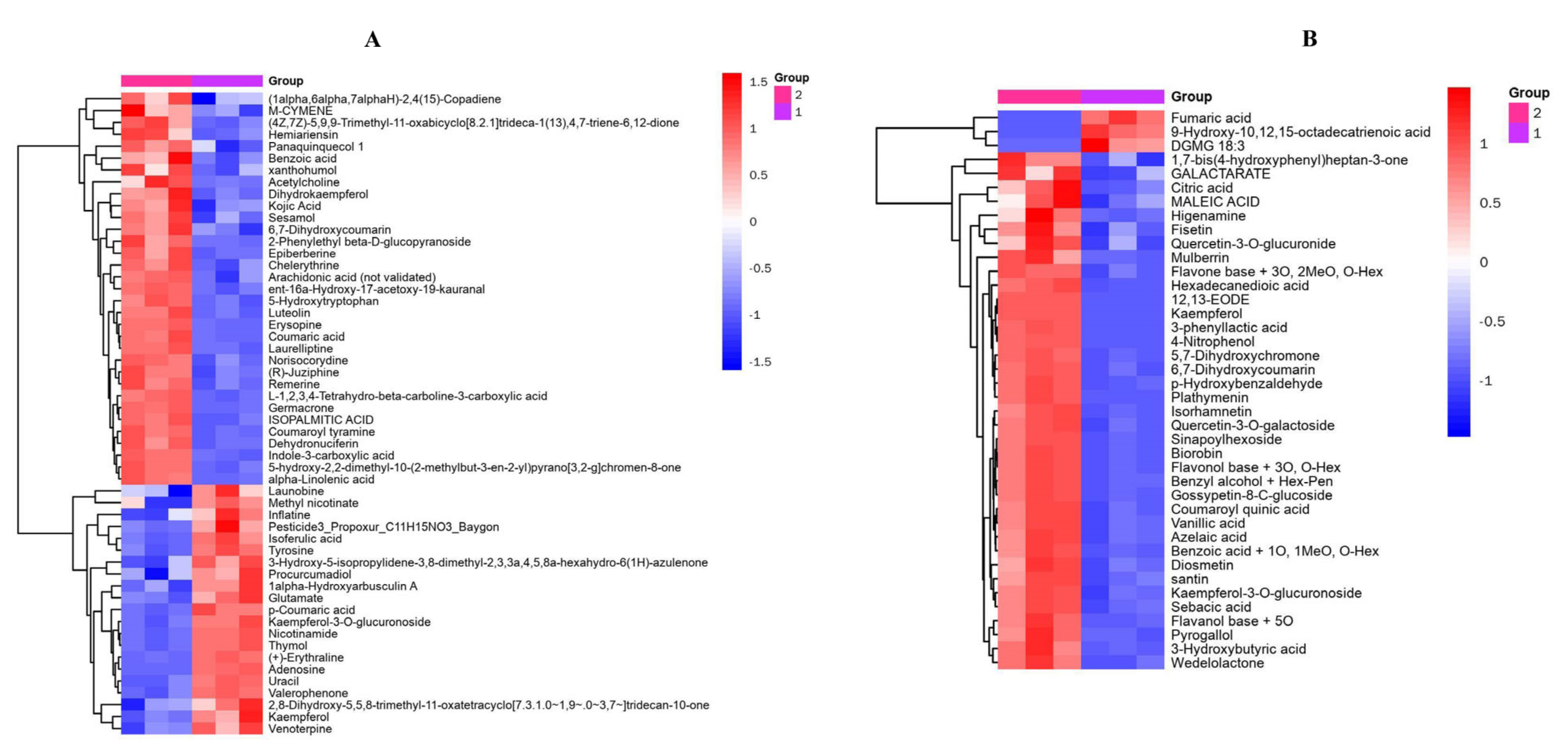
At the same time, we carried out a hierarchical cluster analysis (HCA) to reveal the diversity of the metabolite profiles in different experimental samples. As shown in Figure 4, the abscissa and ordinate represent the sample groups and differential metabolites, respectively. The colors from blue to red indicate the abundance of a metabolite from low to high. The resulting heatmap shows that two main clusters clearly appeared for the two groups, indicating that the intensity of the differential markers varied between the groups.
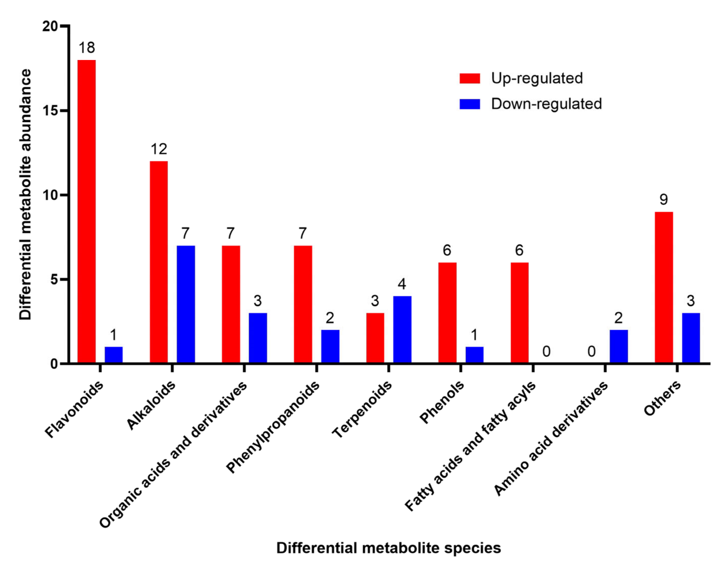
In addition, to better understand the changes in major differential metabolites induced in lotus leaves by fermentation, we sequenced the major differential metabolites detected by VIP labeling (Figure 5). The main differential metabolites in fermented lotus leaves were flavonoids, alkaloids, organic acids and derivatives, phenylpropanoids, terpenoids, phenols, fatty acids and fatty acyls, amino acid derivatives and others. The metabolic differences were mainly reflected in the types and abundance of the metabolites, which was consistent with the HCA results.
In our study, flavonoids, alkaloids, organic acids and phenylpropanoids were the four most abundant secondary metabolites in fermented lotus leaves (Table S1).

3.3.1. Flavonoid Compounds

It was reported that flavonoids are an important active substance present in lotus leaf, with anti-inflammatory, antioxidation and anti-obesity properties [5,14,15]. In this study, L. plantarum G3 fermentation resulted in significant changes in 19 flavonoids in lotus leaves. Specifically, 18 flavonoids (kaempferol, platymenin, 5,7-dihydroxychromone, isorhamnetin, luteolin, mercberrin, flavon base + 5°, biorobin, gossypetin-8-C-glucoside, flavonol base + 3°, O-hex, quercetin-3-O-galactoside, diosmetin, santin, flavone base + 3°, 2MeO, O-hex, fisetin, quercetin-3-O-glucuronide, xanthohumol and dihydrokaempferol) were significantly upregulated, while one (kaempferol-3-O-glucuronoside) was significantly downregulated.
Among these metabolites, kaempferol showed the largest differential expression (5.97 times). Due to its anti-inflammatory properties, kaempferol can be used to treat a variety of diseases caused by acute and chronic inflammation [16] and has beneficial effects on cancer, liver injury, obesity and diabetes [17,18,19]. In this study, the relative content of quercetin-3-O-galactoside was the highest, and fermentation increased it to 1.97 times. Liu et al. [20] showed that quercetin-3-O-galactoside inhibited renal inflammation by regulating macrophage polarization in type 2 diabetic mice. Xanthohumol is a prenylated flavonoid compound unique to hops [21]. It has beneficial effects on body weight, blood lipids, glucose metabolism and other metabolic syndromes [22,23]. However, this compound has not been reported in lotus leaves [24,25]. In our study, xanthohumol was significantly upregulated, after fermentation induced a 1.30-fold increase in its content. Kaempferol-3-O-glucuronoside was significantly downregulated by 0.76-fold by fermentation in this study. The change in flavonoids content may be related to the abundant glycosidase expression in L. plantarum.

3.3.2. Alkaloid Compounds

Alkaloids are important functional active substances present in lotus leaves. In this study, 19 differential metabolites of alkaloids were detected, of which 12 (laurelliptine, dehydronuciferin, 5-hydroxytryptophan, indole-3-carboxylic acid, L-1,2,3,4-tetrahydro-beta-carboline-3-carboxylic acid, norisocorydine, epiberberine, remerine, erysopine, chelerythrine, (R)-juziphine, and higenamine) were upregulated, and 7 (methyl nicotinate, inflatine, launobine, venoterpine, (+)-erythraline, nicotinamide, and adenosine) were downregulated. Among them, the relative content and differential expression fold (2.42) of higenamine were the highest. Higenamine has been shown to have antithrombotic, anti-apoptotic, antioxidant, anti-inflammatory, and immunomodulatory effects [26]. However, higenamine is a β2 agonist, and professional athletes should avoid eating higenamine-containing plants during competitions [27]. Epiberberine is a natural protoberberine from Coptidis Rhizoma that has anti-fatty liver, anti-hyperlipidemic and anticancer activities [28,29]. This compound has not been previously reported in lotus leaves. In our study, epiberberine was significantly upregulated by 1.50-fold after fermentation. (+)-Erythraline is an alkaloid in the seeds of Erythrina [30], which has not been reported in lotus leaves. In this study, fermentation significantly reduced it by 0.20 times.

3.3.3. Organic Acids and Their Derivatives

A total of 10 organic acids and their derivatives were identified as differentially expressed metabolites, of which 7 (citric acid, 3-hydroxybutyric acid, vanillic acid, maleic acid, kojic acid, 3-phenyllactic acid, benzoic acid + 1°, 1MeO, O-hex) were upregulated and 3 (fumaric acid, 9-hydroxy-10,12,15-octadecatrienoic acid, digalactosyl monoacylglycerol 18:3) were downregulated. Among them, 3-phenyllactic acid (PLA) is an organic acid widely found in LAB-fermented foods that has broad and effective antibacterial and antifungal activity [31], but it has not been reported in lotus leaves. In this study, PLA level increased up to 2.92 × 10⁵-fold after fermentation. Consistent with the results reported by Gerez et al. [32], L. plantarum 1081, L. plantarum 778, L. plantarum 1073 and Rodriguez et al. [33], L. plantarum CECT-221 can produce relatively high levels of PLA. In contrast, fumaric acid was significantly downregulated by 0.08 times after fermentation.

3.3.4. Phenylpropanoid Compounds

A total of nine phenylpropanoids were identified as differential metabolites, of which seven (coumaric acid, 5-hydroxy-2,2-dimethyl-10-(2-methylbut-3-en-2-yl) pyrano[3,2-g] chromen-8-one, hemiariensin, 6,7-dihydroxycoumarin, sinapoylhexoside, coumaroyl quinic acid, and wedelolactone) were upregulated, and two (isoferulic acid, p-coumaric acid) were downregulated. Among them, 6,7-dihydroxycoumarin has strong antioxidant activity [34], and its relative content and differential expression fold (2.50) were the highest. p-Coumaric acid has a wide range of biological activities, including antioxidation, anticancer, antibacterial, antiviral and antiarthritis activities, and has a mitigating effect on diabetes, obesity, hyperlipidemia and gout [35]. However, in this study, it was found that p-coumaric acid was significantly downregulated by 0.66-fold after fermentation.

3.4. Metabolic Pathway Analysis

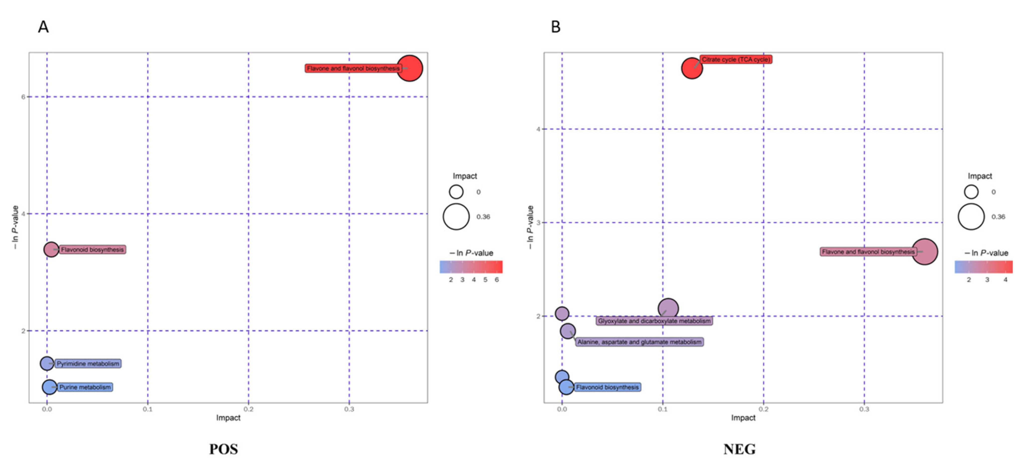
Metabolite pathway enrichment analysis is a useful tool to elucidate the mechanism of metabolic changes in different experimental samples. We analyzed the metabolic pathway enrichment for different metabolites based on the KEGG pathway database. Before and after fermentation, a total of 25 metabolic pathways were identified, involving 61 different metabolites (Table S2). These metabolites were mainly flavonoids, phenols, organic acids and their derivatives, amino acids, nucleosides and nucleotides. Through a comprehensive analysis of the pathways where the differential metabolites are located (including enrichment analysis and topology analysis), we found seven key pathways with the highest correlation with the differences in metabolites (Figure 6), namely, flavone and flavonol biosynthesis, citrate cycle, flavonoid biosynthesis, glyoxylate and dicarboxylate metabolism, alanine, aspartate and glutamate metabolism, pyrimidine metabolism, purine metabolism.

4. Conclusions

In this study, an advanced analytical technique, UHPLC–HR-MS, was used to study the metabolic changes in an aqueous extract of lotus leaves fermented by L. plantarum G3. PCA, OPLS-DA and HCA models were used to analyze the samples before and after fermentation. The significant differences in metabolites between the samples were mainly in the type and abundance of the metabolites. A total of 91 differential metabolites were identified in FLL. They mainly included flavonoids, alkaloids, phenylpropanoids, organic acids and derivatives, terpenoids, fatty acids and fatty acyls, phenols, and amino acid derivatives. KEGG pathway analysis identified 25 metabolic pathways, of which flavone and flavonol biosynthesis, citrate cycle and flavonoid biosynthesis were the main metabolic pathways. In this study, changes in the main metabolites in the water extract of lotus leaves fermented by L. plantarum G3 were described in detail; therefore, this study provides a basis for future research on lotus leaves fermented by L. plantarum.

Supplementary Materials

The following supporting information can be downloaded at: https://www.mdpi.com/article/10.3390/fermentation8110599/s1, Figure S1. Principal component analysis (PCA). Figure S2. Cross-Validation and Response Permutation Test (RPT). Table S1. Analysis of differential metabolites. Table S2. Metabolic pathway analysis.

Author Contributions

Conceptualization, Y.W.; Investigation, B.L., Z.L. and Y.W.; Data curation, B.L.; Funding acquisition, Z.L. and Y.W.; Methodology, B.L.; Project administration, Y.W.; Supervision, Z.L.; Writing—original draft, Y.W.; Writing—review and editing, B.L. All authors have read and agreed to the published version of the manuscript.

Funding

This research was funded by the Research Project of Zhejiang Federation of Social Sciences (2021N40) and the bidding project of the Key Research Base of Humanities and Social Sciences in Universities of Guangdong Province (20KYKT19).

Institutional Review Board Statement

Not applicable.

Informed Consent Statement

Not applicable.

Data Availability Statement

The data are available from the corresponding author.

Conflicts of Interest

The authors declare no conflict of interest.

References

  1. Yoo, J.H.; Park, E.J.; Kim, S.H.; Lee, H.J. Gastroprotective Effects of Fermented Lotus Root against Ethanol/HCl-Induced Gastric Mucosal Acute Toxicity in Rats. Nutrients 2020, 12, 808. [Google Scholar] [CrossRef] [Green Version]
  2. Zheng, H.; Han, L.; Shi, W.; Fang, X.; Hong, Y.; Cao, Y. Research Advances in Lotus Leaf as Chinese Dietary Herbal Medicine. Am. J. Chin. Med. 2022, 50, 1423–1445. [Google Scholar] [CrossRef] [PubMed]
  3. Paudel, K.R.; Panth, N. Phytochemical Profile and Biological Activity of Nelumbo nucifera. Evid. -Based Complement. Altern. Med. 2015, 2015, 789124. [Google Scholar] [CrossRef] [PubMed] [Green Version]
  4. Bishayee, A.; Patel, P.A.; Sharma, P.; Thoutireddy, S.; Das, N. Lotus (Nelumbo nucifera Gaertn.) and Its Bioactive Phytocompounds: A Tribute to Cancer Prevention and Intervention. Cancers 2022, 14, 529. [Google Scholar] [CrossRef] [PubMed]
  5. Chen, G.; Zhu, M.; Guo, M. Research advances in traditional and modern use of Nelumbo nucifera: Phytochemicals, health promoting activities and beyond. Crit. Rev. Food Sci. Nutr. 2019, 59, S189–S209. [Google Scholar] [CrossRef] [PubMed]
  6. Wang, Y.; Yao, W.; Li, B.; Qian, S.; Wei, B.; Gong, S.; Wang, J.; Liu, M.; Wei, M. Nuciferine modulates the gut microbiota and prevents obesity in high-fat diet-fed rats. Exp. Mol. Med. 2020, 52, 1959–1975. [Google Scholar] [CrossRef]
  7. Li, C.; He, Y.; Yang, Y.; Gou, Y.; Li, S.; Wang, R.; Zeng, S.; Zhao, X. Antioxidant and Inflammatory Effects of Nelumbo nucifera Gaertn. Leaves. Oxidative Med. Cell Longev. 2021, 2021, 8375961. [Google Scholar] [CrossRef]
  8. Hammi, K.M.; Essid, R.; Khadraoui, N.; Ksouri, R.; Majdoub, H.; Tabbene, O. Antimicrobial, antioxidant and antileishmanial activities of Ziziphus lotus leaves. Arch. Microbiol. 2022, 204, 119. [Google Scholar] [CrossRef]
  9. Hwang, D.; Charchoghlyan, H.; Lee, J.S.; Kim, M. Bioactive compounds and antioxidant activities of the Korean lotus leaf (Nelumbo nucifera) condiment: Volatile and nonvolatile metabolite profiling during fermentation. Int. J. Food Sci. Technol. 2015, 50, 1988–1995. [Google Scholar] [CrossRef]
  10. Shukla, S.; Park, J.; Park, J.H.; Lee, J.S.; Kim, M. Development of novel Meju starter culture using plant extracts with reduced Bacillus cereus counts and enhanced functional properties. Sci. Rep. 2017, 7, 11409. [Google Scholar] [CrossRef]
  11. Gao, P.; Xu, G. Mass-spectrometry-based microbial metabolomics: Recent developments and applications. Anal. Bioanal. Chem. 2015, 407, 669–680. [Google Scholar] [CrossRef] [PubMed]
  12. Huang, B.M.; Zha, Q.L.; Chen, T.B.; Xiao, S.Y.; Xie, Y.; Luo, P.; Wang, Y.P.; Liu, L.; Zhou, H. Discovery of markers for discriminating the age of cultivated ginseng by using UHPLC-QTOF/MS coupled with OPLS-DA. Phytomedicine 2018, 45, 8–17. [Google Scholar] [CrossRef] [PubMed]
  13. Ai, J.; Wu, Q.; Battino, M.; Bai, W.; Tian, L. Using untargeted metabolomics to profile the changes in roselle (Hibiscus sabdariffa L.) anthocyanins during wine fermentation. Food Chem. 2021, 364, 130425. [Google Scholar] [CrossRef] [PubMed]
  14. Buchholz, T.; Melzig, M.F. Polyphenolic Compounds as Pancreatic Lipase Inhibitors. Planta Med. 2015, 81, 771–783. [Google Scholar] [CrossRef] [PubMed] [Green Version]
  15. Tungmunnithum, D.; Drouet, S.; Hano, C. Phytochemical Diversity and Antioxidant Potential of Natural Populations of Nelumbo nucifera Gaertn. throughout the Floristic Regions in Thailand. Molecules 2022, 27, 681. [Google Scholar] [CrossRef] [PubMed]
  16. Ren, J.; Lu, Y.; Qian, Y.; Chen, B.; Wu, T.; Ji, G. Recent progress regarding kaempferol for the treatment of various diseases. Exp. Ther. Med. 2019, 18, 2759–2776. [Google Scholar] [CrossRef] [PubMed] [Green Version]
  17. Imran, M.; Salehi, B.; Sharifi-Rad, J.; Aslam Gondal, T.; Saeed, F.; Imran, A.; Shahbaz, M.; Tsouh Fokou, P.V.; Umair Arshad, M.; Khan, H.; et al. Kaempferol: A Key Emphasis to Its Anticancer Potential. Molecules 2019, 24, 2277. [Google Scholar] [CrossRef] [PubMed] [Green Version]
  18. Bian, Y.; Lei, J.; Zhong, J.; Wang, B.; Wan, Y.; Li, J.; Liao, C.; He, Y.; Liu, Z.; Ito, K.; et al. Kaempferol reduces obesity, prevents intestinal inflammation, and modulates gut microbiota in high-fat diet mice. J. Nutr. Biochem. 2022, 99, 108840. [Google Scholar] [CrossRef]
  19. Zhou, B.; Jiang, Z.; Li, X.; Zhang, X. Kaempferol’s Protective Effect on Ethanol-Induced Mouse Primary Hepatocytes Injury Involved in the Synchronous Inhibition of SP1, Hsp70 and CYP2E1. Am. J. Chin. Med. 2018, 46, 1093–1110. [Google Scholar] [CrossRef]
  20. Liu, J.; Zhang, Y.; Sheng, H.; Liang, C.; Liu, H.; Moran Guerrero, J.A.; Lu, Z.; Mao, W.; Dai, Z.; Liu, X.; et al. Hyperoside Suppresses Renal Inflammation by Regulating Macrophage Polarization in Mice with Type 2 Diabetes Mellitus. Front. Immunol. 2021, 12, 733808. [Google Scholar] [CrossRef]
  21. Liu, M.; Hansen, P.E.; Wang, G.; Qiu, L.; Dong, J.; Yin, H.; Qian, Z.; Yang, M.; Miao, J. Pharmacological profile of xanthohumol, a prenylated flavonoid from hops (Humulus lupulus). Molecules 2015, 20, 754–779. [Google Scholar] [CrossRef] [PubMed]
  22. Girisa, S.; Saikia, Q.; Bordoloi, D.; Banik, K.; Monisha, J.; Daimary, U.D.; Verma, E.; Ahn, K.S.; Kunnumakkara, A.B. Xanthohumol from Hop: Hope for cancer prevention and treatment. IUBMB Life 2021, 73, 1016–1044. [Google Scholar] [CrossRef] [PubMed]
  23. Neumann, H.F.; Frank, J.; Venturelli, S.; Egert, S. Bioavailability and Cardiometabolic Effects of Xanthohumol: Evidence from Animal and Human Studies. Mol. Nutr. Food Res. 2022, 66, e2100831. [Google Scholar] [CrossRef] [PubMed]
  24. Jia, X.B.; Zhang, Q.; Xu, L.; Yao, W.J.; Wei, L. Lotus leaf flavonoids induce apoptosis of human lung cancer A549 cells through the ROS/p38 MAPK pathway. Biol. Res. 2021, 54, 7. [Google Scholar] [CrossRef]
  25. Zhou, G.; Feng, X.; Tao, A. Explore the Lipid-Lowering and Weight-Reducing Mechanism of Lotus Leaf Based on Network Pharmacology and Molecular Docking. Evid. -Based Complement. Altern. Med. 2021, 2021, 1464027. [Google Scholar] [CrossRef]
  26. Zhang, N.; Lian, Z.; Peng, X.; Li, Z.; Zhu, H. Applications of Higenamine in pharmacology and medicine. J. Ethnopharmacol. 2017, 196, 242–252. [Google Scholar] [CrossRef]
  27. Rangelov Kozhuharov, V.; Ivanov, K.; Ivanova, S. Higenamine in Plants as a Source of Unintentional Doping. Plants 2022, 11, 354. [Google Scholar] [CrossRef]
  28. Liu, L.; Li, J.; He, Y. Multifunctional epiberberine mediates multi-therapeutic effects. Fitoterapia 2020, 147, 104771. [Google Scholar] [CrossRef]
  29. Zou, Z.Y.; Hu, Y.R.; Ma, H.; Feng, M.; Li, X.G.; Ye, X.L. Epiberberine reduces serum cholesterol in diet-induced dyslipidemia Syrian golden hamsters via network pathways involving cholesterol metabolism. Eur. J. Pharmacol. 2016, 774, 1–9. [Google Scholar] [CrossRef]
  30. Zarev, Y.; Foubert, K.; Cos, P.; Maes, L.; Elgorashi, E.; Apers, S.; Ionkova, I.; Pieters, L. HPLC-DAD-SPE-NMR isolation of tetracyclic spiro-alkaloids with antiplasmodial activity from the seeds of Erythrina latissima. Nat. Prod. Res. 2020, 34, 1037–1040. [Google Scholar] [CrossRef]
  31. Mu, W.; Yu, S.; Zhu, L.; Zhang, T.; Jiang, B. Recent research on 3-phenyllactic acid, a broad-spectrum antimicrobial compound. Appl. Microbiol. Biotechnol. 2012, 95, 1155–1163. [Google Scholar] [CrossRef] [PubMed]
  32. Gerez, C.L.; Carbajo, M.S.; Rollan, G.; Torres Leal, G.; Font de Valdez, G. Inhibition of citrus fungal pathogens by using lactic acid bacteria. J. Food Sci. 2010, 75, M354–M359. [Google Scholar] [CrossRef] [PubMed]
  33. Rodríguez, N.; Salgado, J.M.; Cortés, S.; Domínguez, J.M. Antimicrobial activity of d-3-phenyllactic acid produced by fed-batch process against Salmonella enterica. Food Control 2012, 25, 274–284. [Google Scholar] [CrossRef]
  34. Marques, E.S.; Salles, D.B.; Maistro, E.L. Assessment of the genotoxic/clastogenic potential of coumarin derivative 6,7-dihydroxycoumarin (aesculetin) in multiple mouse organs. Toxicol. Rep. 2015, 2, 268–274. [Google Scholar] [CrossRef] [Green Version]
  35. Pei, K.; Ou, J.; Huang, J.; Ou, S. p-Coumaric acid and its conjugates: Dietary sources, pharmacokinetic properties and biological activities. J. Sci. Food Agric. 2016, 96, 2952–2962. [Google Scholar] [CrossRef]
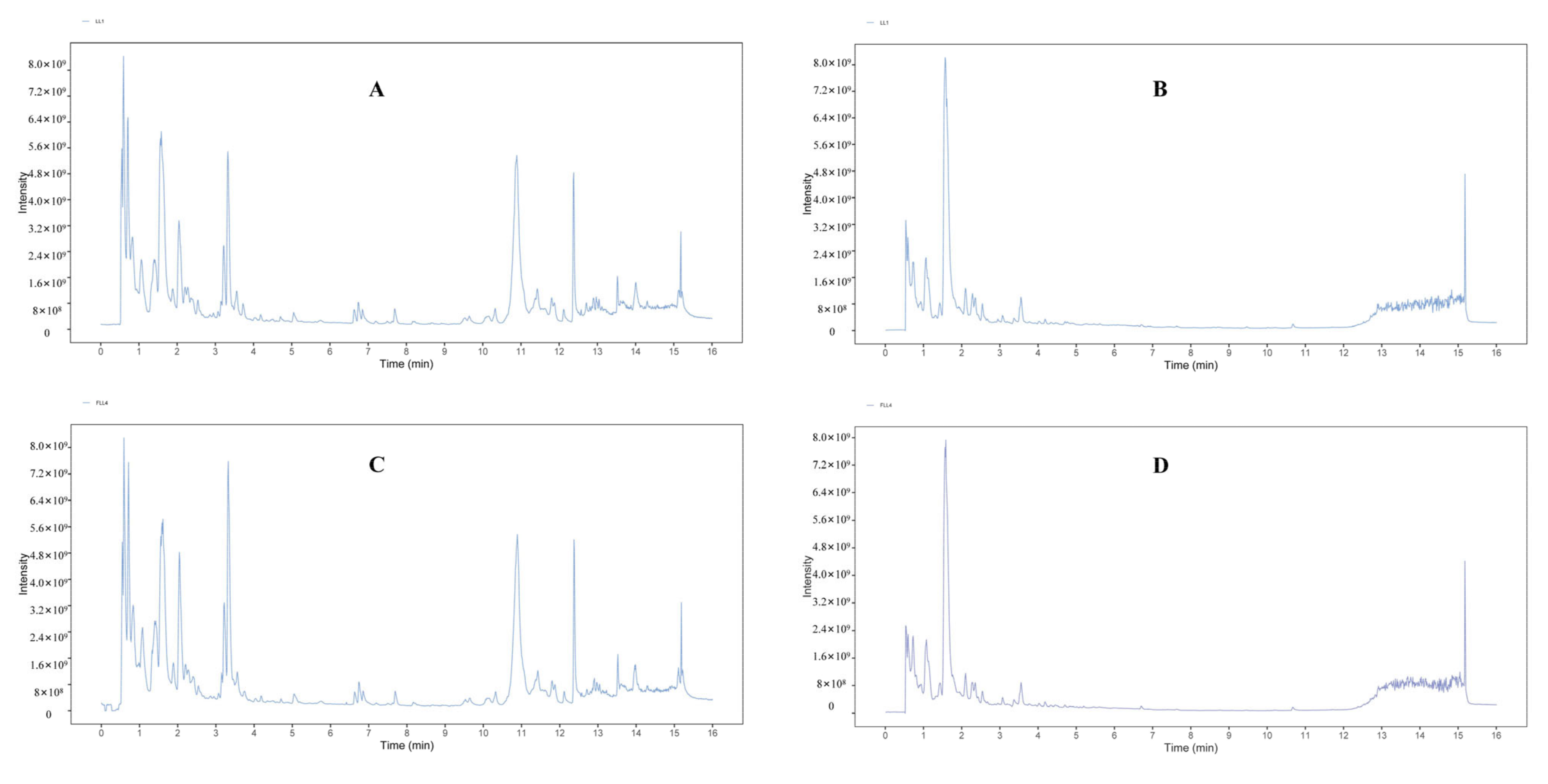
Figure 1. Total ion current diagram of random single samples of LL and FLL. (A) Positive-ion mode analysis of LL, (B) negative-ion mode analysis of LL, (C) positive-ion mode analysis of FLL, and (D) negative-ion mode analysis of FLL.
Fermentation 08 00599 g001
Figure 2. PCA and OPLS-DA score plots of the samples before and after fermentation (n = 3). Remarks: 1, red circle (LL); 2, blue square (FLL). (A) PCA score plot in positive-ion mode; (B) PCA score plot in negative-ion mode; (C) OPLS-DA score plot in positive-ion mode; (D) Score plot of OPLS-DA in negative-ion mode.
Fermentation 08 00599 g002
Figure 3. Volcano plot for the FLL vs. the LL. (A) Positive-ion mode; (B) Negative-ion mode. Gray indicates that there is no significant difference, red means significantly up-regulated, blue indicates significant down-regulation.
Fermentation 08 00599 g003
Figure 4. Heatmap of hierarchical clustering analysis for the FLL group (2, blue square) vs. the LL group (1, red circle). (A) Positive-ion mode; (B) Negative-ion mode.
Fermentation 08 00599 g004
Figure 5. Analysis of differential metabolites in fermented lotus leaves (FLL) compared with those present in pre-fermented lotus leaves (LL). The labels on the bar chart indicate the number of metabolites for each kind of compounds.
Fermentation 08 00599 g005
Figure 6. Enrichment analysis of pathways associated with the differentially expressed metabolites. (A) Positive-ion mode; (B) Negative-ion mode. The size of the bubbles represents the size of the impact factor of the pathway in the topology analysis. The larger the size is, the greater the impact factor. The color represents the p value (negative natural logarithm, namely, ln (p)). The darker the color, the smaller the p value, and the more significant the enrichment degree.
Fermentation 08 00599 g006
Publisher’s Note: MDPI stays neutral with regard to jurisdictional claims in published maps and institutional affiliations.