Article

Development of a Whole-Cell Bioprocess for Ursodeoxycholic Acid Production from Lithocholic Acid Using Fusarium equiseti HG18


1
MOE International Joint Research Laboratory on Synthetic Biology and Medicines, School of Biology and Biological Engineering, South China University of Technology, Guangzhou 510006, China
2
Zhuhai Institute of Modern Industrial Innovation, South China University of Technology, Zhuhai 519100, China
*
Author to whom correspondence should be addressed.
Fermentation 2026, 12(9), 437; https://doi.org/10.3390/fermentation12090437
Submission received: 28 July 2026 / Revised: 12 September 2026 / Accepted: 14 September 2026 / Published: 16 September 2026
(This article belongs to the Special Issue New Research on Fungal Secondary Metabolites, 3rd Edition)

Abstract

Ursodeoxycholic acid (UDCA) is the first-line therapy for primary biliary cholangitis and an important active constituent of bear bile. Whole-cell microbial conversion of inexpensive lithocholic acid (LCA) offers a promising alternative to conventional chemical synthesis owing to its high regioselectivity and mild reaction conditions, yet the fermentation process of Fusarium equiseti HG18 (CCTCC M2023160), a natural fungal catalyst for LCA 7β-hydroxylation, has not been systematically engineered for scalable production. This study developed a whole-cell process for UDCA production by F. equiseti HG18. Systematic optimization by single-factor experiments, Plackett–Burman design and Box–Behnken response surface methodology raised the shake-flask UDCA titer from 0.19 to 0.59 mg mL−1. Scale-up in a 3 L stirred-tank bioreactor shortened the fermentation time from 144 h in shake-flask cultivation to 96 h in the bioreactor under optimized operating conditions (pH 8.5, aeration 2.5 L min−1, and agitation 200 rpm). Experiments across a range of initial LCA loadings revealed a progressive decline in UDCA molar yield at elevated substrate concentrations, consistent with substrate-related inhibitory effects, and this loading-dependent behavior was used to design a two-stage fed-batch feeding strategy. Under the optimized condition (2.0 mg mL−1 LCA fed at 0 and 48 h), UDCA titer reached 1.71 mg mL−1, corresponding to a volumetric productivity of 17.8 mg L−1 h−1 and a UDCA molar yield of 41%. This study establishes a laboratory-scale whole-cell bioprocess for UDCA production from LCA using a wild-type fungal catalyst, integrating statistical medium optimization, bioreactor process development, and substrate-feeding strategies. The findings provide a practical framework for improving fungal whole-cell steroid biotransformation and highlight the potential of wild-type fungal platforms for scalable biocatalytic production.

1. Introduction

Ursodeoxycholic acid (UDCA) is an amphiphilic C24 bile acid synthesized in trace amounts from cholesterol in humans and enriched in bear bile, from which it was historically obtained via live bile drainagea practice now largely banned on animal-welfare grounds [1]. Since its 1997 approval by the U.S. Food and Drug Administration as the first-line agent for primary biliary cholangitis (PBC) [2], the clinical indications of UDCA have expanded to intrahepatic cholestasis of pregnancy [3] and, more recently, to the prevention of symptomatic gallstones after bariatric surgery [4], with additional preclinical evidence supporting neuroprotective [5], intestinal-barrier [6], cardioprotective [7] and chemopreventive activities [8]. The combination of growing clinical demand and restricted bear-bile supply has therefore made the development of sustainable, scalable and animal-free UDCA manufacturing a priority for both pharmaceutical production and industrial biotechnology.
Industrial UDCA manufacturing currently relies on the following three families of routes: chemical synthesis from bile acid precursors such as chenodeoxycholic acid (CDCA) or cholic acid (CA), free-enzyme or chemoenzymatic synthesis, and whole-cell biocatalysis. Chemical synthesis is technologically mature and has long supported commercial supply, but it requires 5–7 reaction steps, uses toxic reagents such as hydrazine hydrate, CrO3 and pyridine, delivers an overall yield of only 30%, and generates substantial waste [9,10,11]. Biocatalytic routes have advanced rapidly in recent years. Refined by cofactor-preference engineering, directed evolution and multienzyme co-expression, 7α/7β-hydroxysteroid dehydrogenase (HSDH) cascades can convert 100 mmol L−1 CDCA to UDCA near-quantitatively at the 2 L scale within 2 h [12,13,14,15], and multi-kilogram and immobilized continuous processes have reduced the environmental factor (E-factor) below 10 [16,17,18,19]; engineered whole-cell P450 systems, exemplified by the CYP107D1 (OleP) triple mutant (F84Q/S240A/V291G) reported by Grobe et al. [20], achieve 60% molar conversion of lithocholic acid (LCA) to UDCA in recombinant Escherichia coli strain. Enzymatic cascades nevertheless depend on purified enzymes, external cofactor regeneration and multi-reactor operation, whereas engineered P450 hosts require regulatory approval as genetically modified organisms and rely on complex heterologous redox-partner engineering. Each existing route therefore retains intrinsic limitations with respect to substrate availability, catalytic complexity, process sustainability, or industrial scalability.
Fungal whole-cell biocatalysis is an attractive alternative because of its excellent regio- and stereoselectivity toward LCA 7β-hydroxylation, yet its industrial development has been limited. Fungal C-7 hydroxylation of LCA has been studied for more than four decades. Sawada et al. (1982) screened F. equiseti M41 from 609 soil isolates and converted 1 g L−1 LCA to 350 mg L−1 UDCA within 112 h [21]. The M41 culture conditions were subsequently optimized by Kulprecha et al., who also reported a promoting effect of K+ on the biotransformation, and further strain screening and product identification were contributed by Sun and Fa [22]. Gibberella zeae VKM F-2600, reported by Kollerov et al. [23], achieved 90% molar conversion at 1 g L−1 LCA but its performance decreased sharply at higher substrate loadings; a UV/ketoconazole-selected mutant, G. zeae M23, was subsequently obtained by Kollerov and Donova and produced 3.52 g L−1 UDCA from 4 g L−1 LCA, the highest fungal titer reported to date [24]. All of these studies, however, were conducted only on a shake-flask scale.
Cytochrome P450 monooxygenases from filamentous fungi are well-known for their diverse and regioselective steroid hydroxylation capabilities [25,26]. Specifically, the cytochrome P450 monooxygenase P450FE has recently been identified as the enzyme responsible for LCA 7β-hydroxylation in the F. equiseti HG18 [27], establishing the molecular basis for microbial UDCA biosynthesis in this system. Heterologous expression of P450FE in Pichia pastoris, however, gave a UDCA yield of only 5.2% [27]. While the P450FE enzyme has been characterized at the molecular level, its application in a scalable bioprocess has not been demonstrated. Whole-cell fungal systems offer advantages in terms of cofactor regeneration, single-step conversion, and simpler downstream processing pending mature enzyme engineering. Nevertheless, the fermentation process of F. equiseti HG18 has not been developed beyond shake-flask cultivation: medium composition and induction conditions have not been jointly optimized, stirred-tank behavior has not been characterized, and substrate-feeding strategies for improved biotransformation performance have not been evaluated.
In the present study, a whole-cell fermentation process for LCA-to-UDCA conversion by the F. equiseti HG18 was therefore developed by combining statistical medium optimization, 3 L stirred-tank scale-up and a fed-batch strategy informed by the substrate-loading behavior of the biotransformation.

2. Materials and Methods

2.1. Strains, Media, and Reagents

F. equiseti HG18, previously isolated and identified as an LCA-to-UDCA converting strain, has been deposited at the China Center for Type Culture Collection (CCTCC M2023160) and was maintained on potato dextrose agar (PDA) plate at 4 °C. Colony and conidial morphology, internal transcribed spacer (ITS) and translation elongation factor 1-α (TEF-1α) gene dual-locus phylogenetic analyses [28,29,30], and supporting PCR evidence are provided in Supplementary Figure S1 and Table S1. Phylogenetic analyses based on both ITS and TEF-1α sequences placed strain HG18 within the F. equiseti clade, supporting its identification as F. equiseti.
Compositions of the media (g L−1) prepared for this study include PDA medium: potato 200 (decoction), glucose 20, agar 20, vitamin B1 0.05; seed medium: dextrin 20, L-asparagine 15, KH2PO4 3, MgSO4·7H2O 0.5; basal fermentation medium: dextrin 20, L-asparagine 15, KH2PO4 3, MgSO4·7H2O 0.5, FeSO4·7H2O 0.02, initial pH 7.0; optimized seed medium: dextrin 20, peptone 15, KH2PO4 3, MgSO4·7H2O 0.5; initial pH 6.0; and optimized fermentation medium: dextrin 25, peptone 18, yeast extract 10.5, KH2PO4 3, MgSO4·7H2O 0.5, FeSO4·7H2O 0.02, KCl 0.59 mol L−1, initial pH 7.6. The initial pH of the media was adjusted before autoclaving. Heat-labile components like vitamin B1 were sterilized separately by membrane filtration and added aseptically after the media had cooled.
Lithocholic acid (LCA, purity > 97%) and ursodeoxycholic acid (UDCA, purity > 99%) were purchased from Aladdin Bio-Chem Technology Co., Ltd. (Shanghai, China). All other chemicals provided by Damao Chemical Reagent Factory (Tianjin, China) were of analytical grade.

2.2. Preparation and Addition of LCA

LCA exhibits extremely low aqueous solubility (about 18.83 μg L−1 at room temperature). Prior to cosolvent optimization, LCA was introduced using 1% (v/v) DMSO. Following cosolvent screening (Section 3.4), LCA stock solutions were prepared in alkaline potassium phosphate buffer (pH 10.0) and supplemented into sterilized fermentation media to achieve target working concentrations, giving a final buffer concentration of 1% (v/v). Under these conditions, LCA formed visually homogeneous dispersions without macroscopic precipitation at concentrations < 4.0 mg mL−1, whereas visible solid particles were observed at concentrations ≥ 4.0 mg mL−1.

2.3. Product Extraction, Identification and Analytical Methods

2.3.1. Extraction

Extraction followed standard procedures for microbial bile acid biotransformation with minor modifications [24]. Upon fermentation completion, the broth pH was adjusted to 12.0 with 0.2 mol L−1 NaOH, held for 30 min, and centrifuged at 12,000× g for 5 min to remove mycelia and insoluble matter. The supernatant was acidified to pH 2.0 with HCl to precipitate bile acids, vortexed and re-centrifuged. The precipitates were collected, and the remaining supernatant was concentrated to 1 mL and extracted three times with equal volumes of ethyl acetate. All extracts were pooled for analysis.

2.3.2. High-Performance Liquid Chromatography with Refractive Index Detection (HPLC-RID)

Analysis was performed on an Agilent 1260 Infinity II system equipped with a refractive index detector (RID) and a Poroshell 120 EC-C18 column (4.6 mm × 150 mm, 4 μm) (Agilent Technologies, Santa Clara, CA, USA). RID was chosen because bile acids lack strong UV absorption above 210 nm. The mobile phase consisted of methanol/acetonitrile/water (5:4:3, v/v/v) containing 2.2 mmol L−1 NaH2PO4·2H2O and 0.08% (v/v) H3PO4, delivered at 0.8 mL min−1. The column temperature and the detector temperature were set to 30 °C and 40 °C. The sample injection volume was 10 μL. Quantification was performed using external standards. The calibration curves were y = 135,128x + 270.42 (R2 = 0.9999) for UDCA and y = 116,641x + 2929.1 (R2 = 0.9994) for LCA (0.2–1.0 mg mL−1, five points), where y is peak area and x is concentration (mg mL−1). Peaks corresponding to LCA, UDCA, a peak co-eluting with 7-keto-LCA and two minor unknown peaks were integrated to evaluate relative selectivity.

2.3.3. Structural Confirmation

The concentrated extract was dissolved in a small volume of ethyl acetate and adsorbed onto 2.0 g of silica gel for dry loading onto a column packed with 25.0 g of silica gel. The column was eluted with petroleum ether/ethyl acetate (2:1, v/v) containing 1% (v/v) glacial acetic acid. Fractions were monitored by thin-layer chromatography (TLC), and fractions containing the target product were pooled, concentrated under reduced pressure, and vacuum-dried for subsequent HRMS (Bruker maXis impact, Bremen, Germany), 1H NMR (600 MHz, DMSO-d6), and 13C NMR analyses (151 MHz, DMSO-d6), (Bruker AVANCE III HD 400, Bruker, Germany).

2.4. Strain Activation, Seed Preparation and Physiological Characterization

Before fermentation experiments, the preserved strain was activated and prepared as described below. Preserved cultures were streaked onto PDA plates and incubated at 28 °C for 5–7 d to obtain sporulating colonies. Spores were harvested with sterile water containing 0.2% (v/v) Tween-80 to prepare a spore suspension (1.0 × 10⁶ spores mL−1). A 1 mL inoculum was transferred into 50 mL seed medium in a 250 mL Erlenmeyer flask and cultivated at 28 °C and 200 rpm.
Dry cell weight (DCW) was used as the response variable to evaluate the effects of carbon source, nitrogen source, C/N ratio, temperature and initial pH on the F. equiseti HG18 growth and to construct growth curves in seed medium. For DCW determination, broth was filtered, mycelia were washed three times with deionized water and dried under vacuum at 105 °C to constant weight. The growth-condition optimization was conducted sequentially. After identifying the preferred carbon and nitrogen sources, the C/N ratio was optimized using the selected carbon and nitrogen sources. Subsequently, temperature and initial pH were evaluated under the optimized carbon source, nitrogen source, and C/N ratio conditions. Finally, the cell dry weight (CDW) time course was determined under the optimized growth conditions. All treatments were performed in biological triplicate.

2.5. Single-Factor Optimization of Fermentation Medium and Conditions

Unless otherwise specified, single-factor fermentation experiments were performed using the basal fermentation medium, with only the factor under investigation varied while all other medium components and cultivation conditions were kept constant. The condition giving the highest UDCA titer in each screening step was carried forward to the subsequent optimization step. With UDCA titer taken as the response variable, single-factor experiments were performed to evaluate the effects of carbon source, (i.e., glucose, lactose, sucrose, soluble starch, and dextrin), nitrogen source, (i.e., NH4Cl, dried corn steep liquor, wall-broken dry yeast, peptone, yeast extract, and L-asparagine) (Oxoid, Thermo Fisher Scientific, Basingstoke, UK), C/N ratio (the carbon source/the nitrogen source ratio), metal ion supplementation, K+ concentration [31], inoculum size, fermentation time, temperature, initial pH, substrate cosolvent, and potential P450 inducers [32,33]. Unless stated otherwise, biotransformations were conducted in an 8 mL working volume (100 mL flasks) with 1.0 mg mL−1 LCA at 28 °C and 200 rpm for 144 h, in three independent biological replicates. The fermentation time optimization experiment was conducted for 216 h, with samples collected at 24, 48, 72, 96, 120, 144, 168, 192, and 216 h to evaluate the effect of cultivation time on UDCA production.

2.6. Plackett–Burman Screening and Box–Behnken Response Surface Methodology

A two-stage statistical optimization approach was applied. First, a 12-run Plackett–Burman (PB) design was used to screen the following eight candidate factors: initial pH (6.5/8.5), KCl (0.25/0.75 mol L−1), temperature (25/28 °C), inoculum size (10%/30%, v/v), yeast extract (1/15 g L−1), peptone (12/18 g L−1), dextrin (16/25 g L−1) and fermentation time (5/7 d), together with three dummy variables for error estimation. Factors with p < 0.05 in ANOVA were selected for the second stage. Second, a 17-run Box–Behnken design (BBD) [34] with five center-point replicates was used to optimize the selected factors. A second-order polynomial model was fitted and analyzed by ANOVA using Design-Expert 8.0.6.1 (Stat-Ease, Inc., Minneapolis, MN, USA). Regression coefficients are reported in coded units (−1, 0, +1).

2.7. Bioreactor Scale-Up and Feeding Strategy

A two-stage seed preparation was used for bioreactor cultivation. Shake-flask seed cultures (24 h) were transferred into a 3 L seed tank containing 1 L optimized seed medium and induced with 0.2% (w/v) LCA (based on the inducer screening results described in Section 3.4.) for 12 h (aeration 1.0 vvm, agitation 80 rpm, and 28 °C). After 36 h total seed cultivation, mycelia were harvested by gauze filtration, washed three times with fermentation salts buffer, resuspended in 100 mL of the same buffer and inoculated at 30% (v/v), based on the inoculum-size optimization shown in Figure 3f, into a 3 L stirred-tank bioreactor (LiFlus GX, Biotron, Bucheon, Republic of Korea; working volume 1 L) containing optimized fermentation medium. LCA solubilized in alkaline potassium phosphate buffer was then added, and biotransformation proceeded at 28 °C. pH was automatically controlled by 2 mol L−1 NaOH and 2 mol L−1 HCl, and dissolved oxygen (pO2) was recorded online. Because the pH value identified by the BBD represented the initial medium pH under shake-flask conditions, the effect of controlled fermentation pH was evaluated separately in the stirred-tank bioreactor. Process parameters, including pH (7.0, 7.5, 8.0, 8.5, and 9.0), aeration (1.5, 2.5, and 3.0 vvm) and agitation (180, 200, and 250 rpm), were evaluated. Substrate conversion kinetics at 0.5–5.0 mg mL−1 initial LCA loadings were investigated in shake flasks. Carbon/nitrogen nutrient feeding (50 mL concentrate every 24 h starting at 12 h; feeding solution containing 25.0 g L−1 dextrin, 18.0 g L−1 peptone, and 10.5 g L−1 yeast extract) was compared with a non-fed control in the bioreactor. Finally, a two-stage intermittent substrate feeding strategy (2.0 mg mL−1 LCA added at 0 and 48 h) was established and validated in three independent bioreactor batches.

2.8. Performance Metrics and Statistical Analysis

Performance metrics were defined as follows. Titer was expressed in mg mL−1 or mg L−1. Volumetric productivity Qp (mg L−1 h−1) was calculated as titer divided by fermentation time. UDCA molar yield was calculated by Equation (1).
UDCA molar yield (%) = (CU/MU)/(CL/ML) × 100%
where CU is the UDCA concentration and CL is the cumulative LCA concentration added (both in mg mL−1), MU = 392.6 g mol−1 and ML = 376.6 g mol−1.
Data are expressed as mean ± standard deviation (SD) of three independent biological replicates. Statistical comparisons were performed using one-way ANOVA followed by Tukey’s HSD post hoc test (α = 0.05); distinct letters indicate significant difference (p < 0.05). Data analysis was carried out in Microsoft Excel 2016 (Microsoft Corporation, Redmond, WA, USA), SPSS 26.0 (IBM Corp., Armonk, NY, USA), Design-Expert 8.0.6.1 (Stat-Ease, Inc., Minneapolis, MN, USA) and Origin 2021 (OriginLab Corporation, Northampton, MA, USA).

3. Results

3.1. Structural Identification of the Biotransformation Product

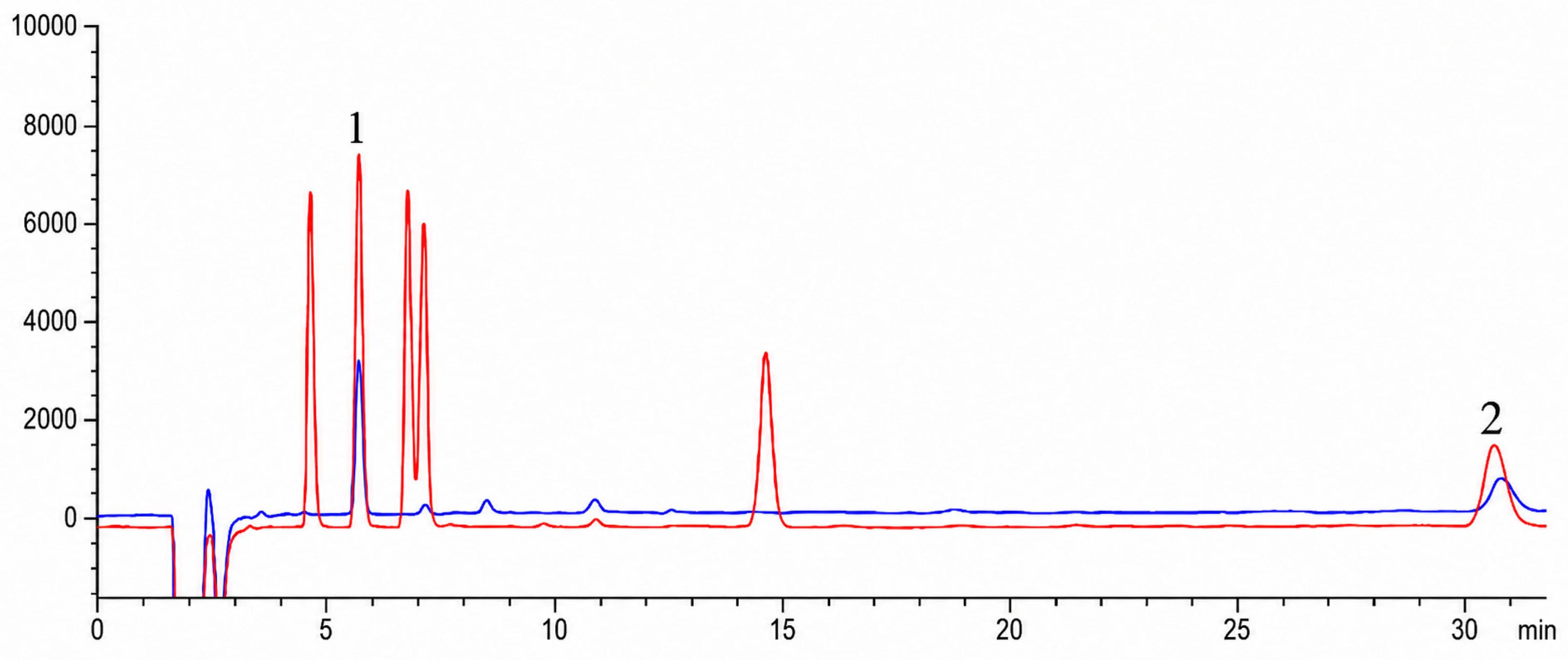
HPLC-RID analysis of bile acid extracts from the fermentation broth (Figure 1) revealed a major product peak co-eluting with the authentic UDCA standard (retention time, RT= 5.76 min), together with unreacted LCA (RT = 30.63 min), a minor peak co-eluting with 7-keto-LCA and two minor unknown peaks. Under the optimized fed-batch conditions described in Section 3.6.4, relative peak-area integration (n = 3) yielded 78 ± 3% UDCA, 12 ± 2% residual LCA, 6 ± 1% unidentified peak tentatively assigned as 7-keto-LCA based on chromatographic co-elution and 4 ± 1% minor unknown species, indicating a UDCA selectivity of 78% among the detected bile acids.
HRMS (ESI) of the purified product yielded m/z 391.2888 [M−H] and 783.5816 [2M−H] (Supplementary Figure S2). 1H NMR (600 MHz, DMSO-d6): δ 11.94 (s, 1H, COOH), 4.43 (d, J = 4.6 Hz, 1H, 3-OH), 3.86 (d, J = 6.9 Hz, 1H, 7-OH), 3.33–3.24 (m, 2H, H-3 and H-7), 0.91–0.77 (m, 6H, 21-CH3 and 19-CH3), and 0.61 (s, 3H, 18-CH3) (Supplementary Figure S3). 13C NMR (151 MHz, DMSO-d6): δ 175.37, 70.20, 69.93, 56.31, 55.16, 43.56, 43.47, 42.66, 39.20, 38.19, 37.74, 35.32, 34.23, 31.24, 30.72, 28.65, 27.19, 23.78, 21.33, 18.77, 14.56, and 12.50 (Supplementary Figure S4). All spectral features were in agreement with published data for UDCA [21,24], confirming the identity of the biotransformation product and, by inference, the C-7β hydroxylation activity of P450FE.

3.2. Physiological Characterization of F. equiseti HG18

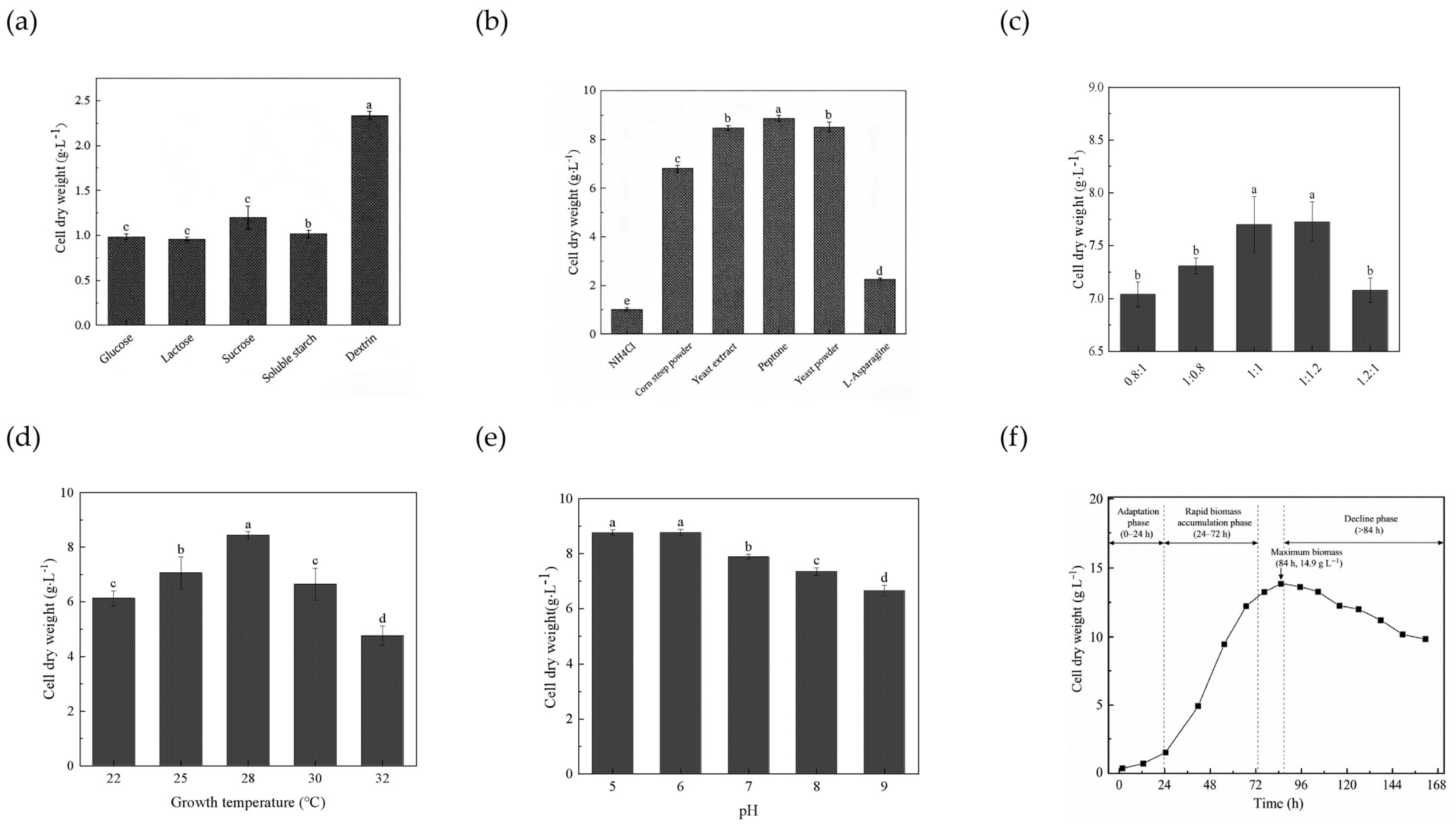
On the basis of DCW measurements (Figure 2), dextrin was identified as the preferred carbon source (DCW 2.33 ± 0.05 g L−1) and peptone as the preferred nitrogen source (DCW 8.85 ± 0.11 g L−1). The optimal C/N ratio was 1:1, and the optimal temperature and initial pH for growth were 28 °C and 6.0, respectively. The biomass accumulation profile of F. equiseti HG18 showed an initial adaptation phase (0–24 h), followed by a rapid biomass accumulation phase (24–72 h), reaching a maximum biomass of 14.9 g L−1 at 84 h before entering a decline phase. Therefore, cultures at 36 h, corresponding to the active biomass accumulation stage, were selected as seed material.

3.3. Single-Factor Optimization of Fermentation Parameters

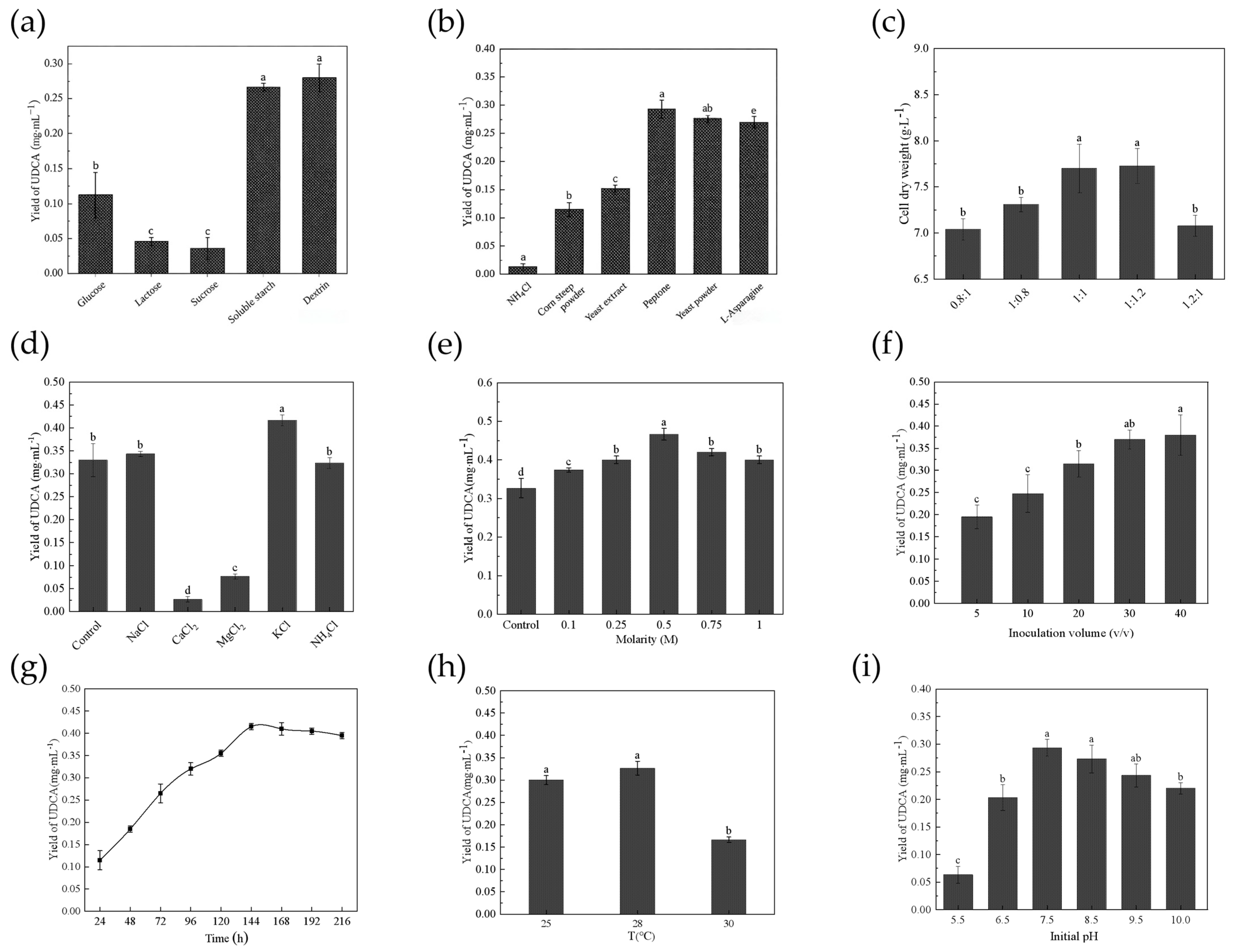
Single-factor screening (Figure 3) identified dextrin (0.28 ± 0.02 mg mL−1) and peptone (0.29 ± 0.02 mg mL−1) as the carbon and nitrogen sources giving the highest UDCA titers (Figure 3a,b). The effects of the C/N ratio and metal-ion supplementation were also evaluated (Figure 3c,d). Supplementation with 0.5 mol L−1 KCl further increased the titer by 45%, to 0.47 ± 0.02 mg mL−1 (p < 0.01, Figure 3e), consistent with previous observations in F. equiseti M41 [21]. The highest UDCA titer was observed at an inoculum size of 40% (v/v) (0.38 ± 0.05 mg mL−1), whereas 30% (v/v) inoculum resulted in a comparable UDCA titer (0.37 ± 0.02 mg mL−1) without significant difference (Figure 3f). Considering that further increasing the inoculum size did not provide a statistically significant improvement in UDCA production, and that a lower inoculum volume can reduce seed consumption, 30% (v/v) was selected as the optimal inoculum size for subsequent experiments. Maximum product accumulation was reached at 144 h, and the optimal conversion temperature and initial pH were 28 °C and 7.5, respectively (Figure 3g–i). The higher pH optimum for biotransformation (pH 7.5) than for growth (pH 6.0) is consistent with the increased aqueous solubility of LCA under mildly alkaline conditions, which is expected to improve substrate availability to the mycelia.

3.4. Optimization of Substrate Delivery and Enzyme Induction

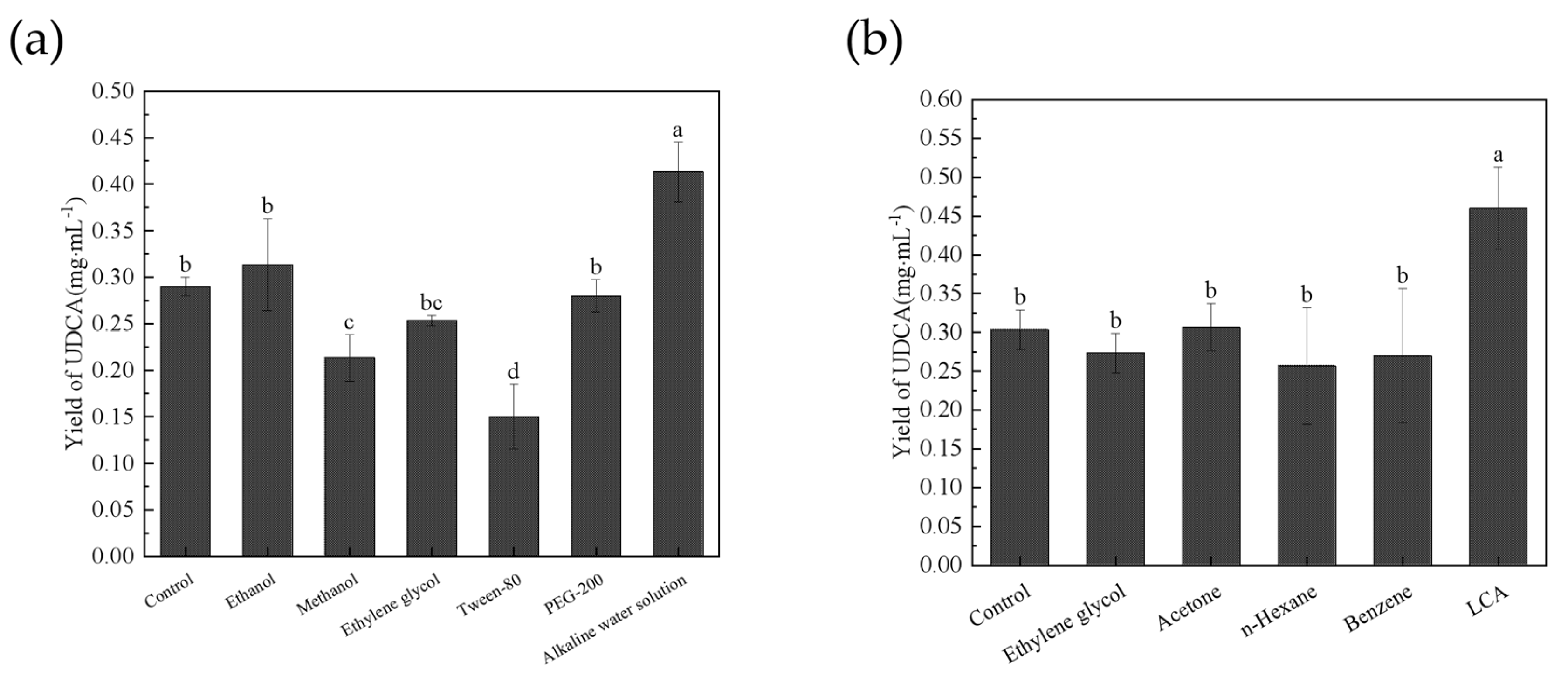
Among seven substrate delivery methods tested (Figure 4a), alkaline potassium phosphate buffer (pH 10.0) gave the highest UDCA titer (0.41 ± 0.03 mg mL−1), a 41% increase over 1% (v/v) DMSO (p < 0.01). Induction screening during seed cultivation (Figure 4b) showed that supplementation with 0.2% (w/v) LCA increased the final UDCA titer by 53% (p < 0.01), in agreement with previous transcriptomic data indicating an -8-fold LCA-induced upregulation of P450FE [27].

3.5. Statistical Optimization by Plackett–Burman Screening and Box–Behnken Response Surface Methodology

The PB design (Table 1) identified the following three factors as significant (p < 0.05): KCl concentration, initial pH, and yeast extract concentration. The corresponding ANOVA results for the PB screening model are provided in Table S2. Although peptone showed the best performance in the single-factor nitrogen-source screening and was retained as the principal nitrogen source, yeast extract was included as an additional medium component in the PB design. Under the multifactor conditions, yeast extract showed a significant effect on UDCA production, whereas peptone did not. Therefore, yeast extract was selected for further optimization. A subsequent 17-run BBD (Table 2) was performed, and the response was fitted to the following second-order polynomial in coded units (Equation (2)):
Y = 0.574 + 0.02375 X1 + 0.04125 X2 + 0.0325 X3 + 0.0075 X1 X2 + 0.02 X1 X3 + 0.025 X2 X3 − 0.04575 X12 − 0.14575 X22 − 0.03625 X32
where Y is the UDCA yield (mg/mL), and X1, X2, and X3 are the coded values of KCl concentration (M), initial pH, and yeast extract concentration (g∙L−1), respectively.
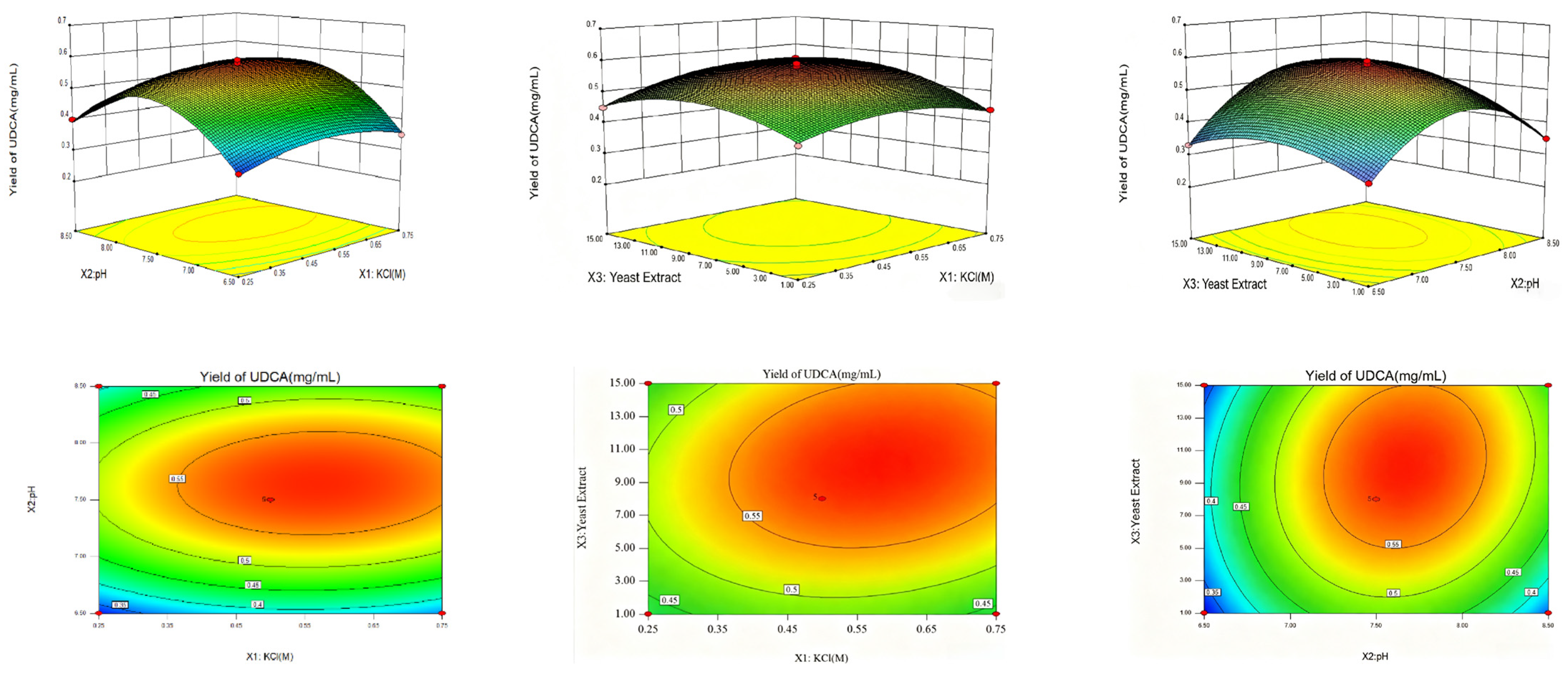
ANOVA of the fitted model (Table 3) indicated that the quadratic model was highly significant (F = 71.4, p < 0.0001), with R2 = 0.9892, adjusted R2 = 0.9754, and predicted R2 = 0.9487, while the lack of fit was not significant (p = 0.774). The response surface and contour plots showed that the optimal regions of the three significant factors were located near the central levels, which was consistent with the single-factor optimization results (Figure 5). The predicted optimum (0.59 mol L−1 KCl, pH 7.68 and 10.44 g L−1 yeast extract) was validated experimentally under the rounded conditions of 0.59 mol L−1 KCl, pH 7.6 and 10.5 g L−1 yeast extract, which yielded a UDCA titer of 0.59 ± 0.02 mg mL−1 (n = 3), in close agreement with the predicted value of 0.57 mg mL−1. Although 0.59 mol L−1 KCl represents a relatively high salt concentration and may impose osmotic stress, F. equiseti HG18 maintained favorable biotransformation performance under this condition, suggesting that the beneficial effect of K+ outweighed any adverse osmotic effect within the tested range.

3.6. Bioreactor Scale-Up and Two-Stage Fed-Batch Operation

3.6.1. Optimization of Process Parameters in a 3 L Bioreactor

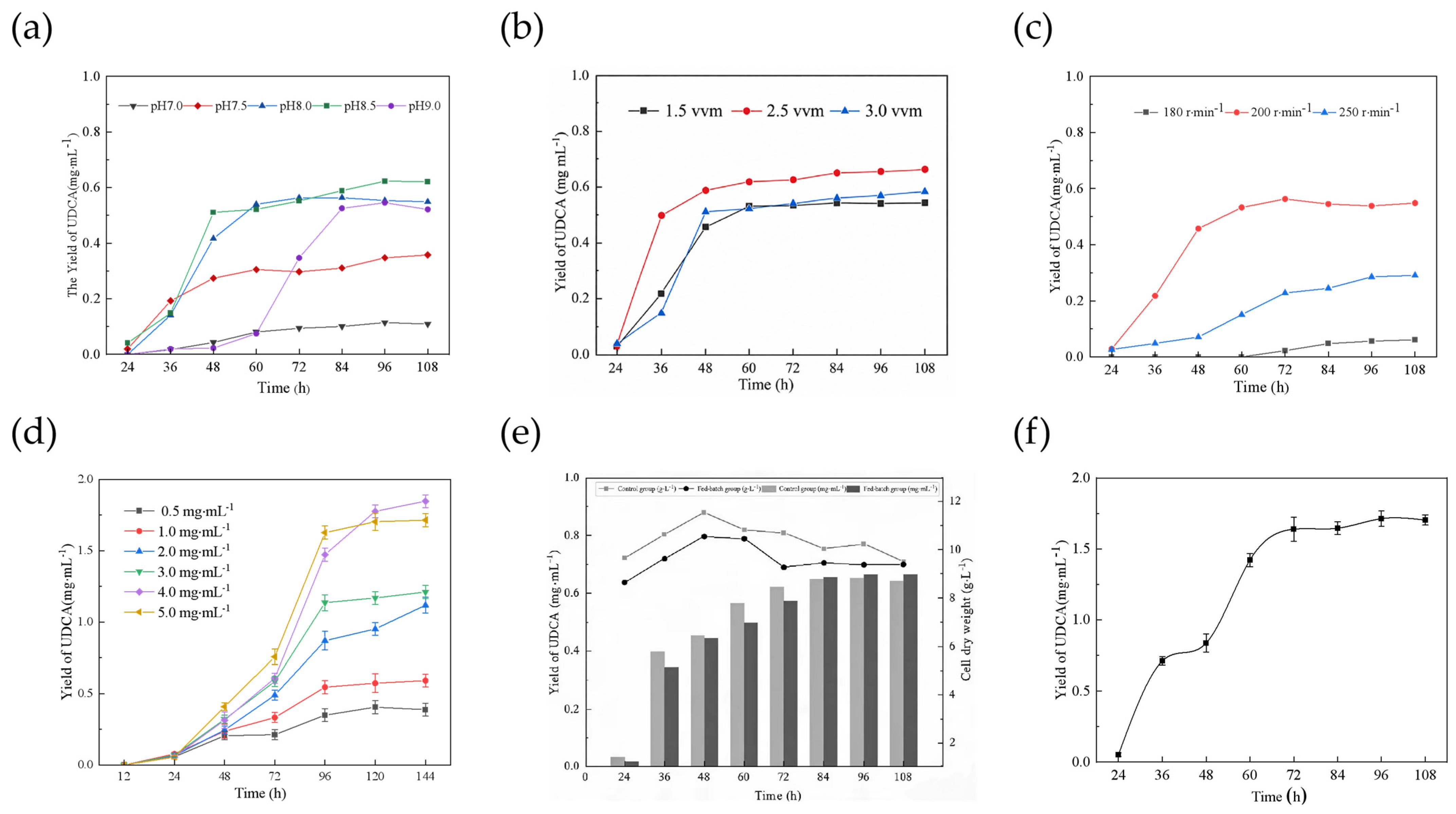
Among the tested pH conditions in the 3 L bioreactor (1 L working volume), the effects of individual bioreactor parameters were independently evaluated. The high UDCA production reaching 0.62 mg mL−1, which was observed at pH 8.5 (Figure 6a), indicates that the F. equiseti HG18 retained good biotransformation activity under mildly alkaline conditions. Aeration at 2.5 vvm and agitation at 200 rpm were identified as the optimal operating conditions within the tested ranges, under which 0.65 mg mL−1 UDCA (62% UDCA molar yield) was obtained (Figure 6b,c). Under the optimized bioreactor conditions, the fermentation time was reduced to 96 h compared with 144 h in shake-flask cultivation.

3.6.2. Effect of Initial LCA Loading on Biotransformation Performance

Experiments performed at 0.5–5.0 mg mL−1 initial LCA (Figure 6d) showed that the final UDCA titer increased with substrate loading up to 4.0 mg mL−1 LCA (1.85 mg mL−1 UDCA), whereas the UDCA molar yield decreased progressively from 75% at 0.5 mg mL−1 to 44% at 4.0 mg mL−1. This inverse relationship between substrate loading and UDCA molar yield indicates a substrate-loading-dependent loss of catalytic efficiency. Given the extremely low aqueous solubility of LCA, several factors may contribute to this behavior, including substrate-associated inhibition, increased solid-phase accumulation and mass-transfer resistance, and possible membrane-associated toxicity. Therefore, the present data are interpreted as being consistent with substrate-related inhibitory effects rather than as establishing a specific kinetic inhibition mechanism. An initial LCA loading of approximately 2.0 mg mL−1 was consequently selected as a practical operating range for subsequent feeding experiments.

3.6.3. Assessment of Nutrient Feeding

Carbon/nitrogen nutrient feeding did not significantly improve UDCA accumulation or DCW compared with the non-fed control (Figure 6e), suggesting that additional nutrient supplementation was not beneficial under the optimized conditions. Therefore, subsequent process optimization focused on substrate delivery strategies to increase substrate-loading capacity and overall product accumulation.

3.6.4. Two-Stage Fed-Batch Biotransformation

Under the two-stage intermittent feeding regime (2.0 mg mL−1 LCA added at 0 and 48 h), the instantaneous substrate concentration was kept within the non-inhibitory range identified in Section 3.6.2. UDCA accumulated to 0.87 mg mL−1 by 48 h and reached 1.71 ± 0.05 mg mL−1 at 96 h (Figure 6f), corresponding to a volumetric productivity of 17.8 mg L−1 h−1 and a UDCA molar yield of 41% (Table 4). Compared with the 1.0 mg mL−1 bioreactor batch condition, the two-stage process substantially increased substrate-handling capacity and overall product accumulation.

4. Discussion

The process data reveal a clear substrate-loading-dependent decrease in conversion efficiency. First, the shake-flask titer plateaued at 0.59 mg mL−1 despite exhaustive medium and induction optimization, indicating that further gains cannot be obtained by adjusting nutritional or physicochemical parameters alone. Second, the UDCA molar yield decreased monotonically as the initial LCA concentration was raised (Figure 6d), whereas carbon/nitrogen supplementation had no significant effect on UDCA accumulation or biomass, and aeration was maintained above the optimized level, so nutrient supply and oxygen transfer were not obviously limiting under these conditions. Related effects, including LCA solubility limits, external mass-transfer resistance associated with the solid-phase substrate, possible product inhibition and progressive loss of mycelial activity, cannot be excluded on the basis of the present data and would require Haldane-type kinetic fitting and dedicated mass-transfer analysis to resolve. Nevertheless, the observed dependence is sufficient to define a practical operating window (2 mg mL−1) within which conversion is preserved, and this observation was used directly to design the two-stage fed-batch operation in Section 3.6.4.

4.1. Comparison with Reported UDCA Bioproduction Systems

Reported microbial UDCA systems can be grouped into three categories-engineered whole cells, purified enzymatic cascades and natural fungal systems-which pursue different technological objectives and are therefore not directly comparable on the basis of titer or conversion alone (Table 5).
Recombinant E. coli expressing CYP107D1 mutants reaches 60% molar conversion [20] but depends on genetic modification and on a complex heterologous electron-transfer chain. Immobilized 7α/7β-HSDH cascades achieve high productivity at the expense of enzyme purification, multi-reactor configuration and cofactor recycling [35]. Wild-type F. equiseti HG18, in contrast, performs LCA 7β-hydroxylation in a single step in a standard stirred-tank bioreactor, without external cofactors or genetic modification. Engineered whole-cell and enzyme-cascade systems generally achieve higher conversion rates and productivities than the present fungal process. Recombinant P450 systems, however, require heterologous enzyme and redox-partner expression, whereas HSDH-based cascades may involve enzyme preparation, cofactor regeneration, and multienzyme or multireactor configurations. In contrast, the wild-type F. equiseti HG18 system performs direct LCA 7β-hydroxylation in a single whole-cell step, with intracellular cofactor regeneration and without external cofactor supplementation or genetic modification. These features simplify the overall biocatalytic process configuration, although the lower conversion and product titer remain important limitations compared with engineered systems.
G. zeae M23 achieved a higher UDCA titer of 3.52 g L−1 at shake-flask scale [24], whereas the present HG18 process has been validated in a stirred-tank bioreactor with a defined substrate-feeding strategy. These systems therefore represent different stages and objectives of process development and should not be compared solely on the basis of titer. While recombinant systems and enzyme cascades have achieved higher conversions or productivities, the contribution of the present work is therefore not the achievement of the highest fungal UDCA titer, but the integration of statistical medium optimization, defined stirred-tank operation, and substrate-loading-informed fed-batch feeding into a single bioprocess development workflow for the wild-type F. equiseti HG18 catalyst.

4.2. Alignment Between Process Behavior and the Catalytic Mechanism

The observed response of UDCA titer to key operating variables is consistent with the previously reported P450FE-mediated 7β-hydroxylation route [27]. In particular, the −53% titer improvement conferred by 0.2% (w/v) LCA pre-induction during seed cultivation parallels the substrate-dependent upregulation of P450FE reported previously at the transcriptomic level [27], which suggests that induction of the catalytic machinery, rather than biomass accumulation, is the primary determinant of biotransformation capacity. A minor peak co-eluting with 7-keto-LCA was consistently observed; however, its structural identity cannot be established by retention-time matching alone and requires confirmation by LC–MS/MS using an authentic standard. Fungal P450 monooxygenases generally require NAD (P) H-derived electrons supplied through redox partners such as cytochrome P450 reductase (CPR). Therefore, intracellular NADPH regeneration and electron-transfer efficiency may represent additional constraints on P450FE-mediated LCA hydroxylation; however, these factors were not specifically investigated in the present study.

4.3. Substrate Mass Transfer, Inhibition and the Rationale for Fed-Batch Feeding

Owing to the extremely low aqueous solubility of LCA, the biotransformation operates as a heterogeneous mycelium–solid substrate–liquid system. Elevated initial substrate loadings therefore increase solid accumulation and surface mass-transfer resistance, both of which are expected to compound any membrane-associated substrate-related inhibitory effect. The monotonic decrease in UDCA molar yield with increasing initial LCA concentration is consistent with such combined effects, including the toxicity of hydrophobic substrates on fungal cell membranes [36,37], although the individual contributions of solubility, mass transfer and inhibition were not resolved in the present study. Nonetheless, dividing the substrate charge into two doses of 2.0 mg mL−1 maintained the instantaneous concentration within the operating window identified in Section 3.6.2 and led to a four-fold increase in cumulative substrate capacity while retaining a favorable UDCA molar yield, providing an empirical validation of the mitigation strategy rather than a formal kinetic model. Moreover, the process data indicate that substrate-related inhibitory effect contributes substantially to the loss of conversion at elevated instantaneous LCA loadings, and that maintaining the substrate concentration within the empirically identified operating window through staged feeding is a practical means of improving productivity beyond that obtained by medium optimization alone. This evidence provides an engineering basis for further process development of fungal whole-cell LCA hydroxylation to biotransformation of other poorly soluble steroid substrates.
Owing to the extremely low aqueous solubility of LCA, increasing substrate loading is expected to increase solid-phase accumulation and the associated mass-transfer burden, while potentially enhancing substrate-associated toxicity. These effects provide plausible explanations for the progressive decline in UDCA molar yield observed with increasing initial LCA concentration (Figure 6d), although their individual contributions could not be resolved in the present study. Based on this experimentally observed loading-dependent behavior, the substrate charge was divided into two 2.0 mg mL−1 additions rather than being supplied as a single high initial dose. This feeding strategy was therefore used as a process-engineering response to the empirically identified operating range, rather than as proof of a specific substrate-inhibition mechanism. The staged operation enabled a cumulative LCA loading of 4.0 mg mL−1 and a UDCA titer of 1.71 mg mL−1 within 96 h, demonstrating that controlled substrate delivery can increase process throughput beyond that obtained by medium optimization alone. Although the lower UDCA molar yield observed under the two-stage feeding strategy indicates reduced substrate utilization efficiency at increased substrate loading, the strategy was intended to enhance overall process throughput rather than maximize molar yield alone. By increasing the cumulative LCA loading from 1.0 to 4.0 mg mL−1, the two-stage feeding strategy increased the UDCA titer to 1.71 mg mL−1 and achieved a volumetric productivity of approximately 17.8 mg L−1 h−1. Thus, the lower molar yield represents a trade-off between substrate utilization efficiency and increased product titer and productivity under higher substrate loading. Future process improvement should therefore focus on enhancing substrate utilization and reducing residual LCA while maintaining the increased process throughput.

4.4. Limitations and Prospect

Several limitations of this work should also be acknowledged. The absolute titer obtained here remains below those reported for engineered enzyme cascades and mutant fungal strains such as G. zeae M23. The relative contributions of substrate-related inhibitory effects, solubility and mass-transfer resistance were not disentangled by kinetic modeling. The metabolic identity of the putative 7-keto-LCA intermediate has not been unambiguously established. The transferability of the present process to other fungal whole-cell systems has not been experimentally verified and remains a proposition to be tested. The 3 L bioreactor experiments represent laboratory-scale stirred-tank validation rather than demonstration of industrial scalability, and further scale-up will require evaluation of mixing, oxygen transfer, and substrate-delivery performance at larger working volumes. Future work will focus on closing the remaining titer gap relative to engineered enzyme cascades and mutant fungal strains through P450FE engineering, host metabolic engineering, and validation in larger bioreactors. The identity and metabolic role of the putative 7-keto-LCA intermediate will also be clarified by LC–MS/MS analysis using authentic standards. Beyond catalyst engineering, further process development should explore process intensification, continuous or repeated-batch operation, and techno-economic evaluation, including substrate cost, volumetric productivity, and downstream processing requirements. Finally, the transferability of the present process-development strategy to other fungal whole-cell steroid biotransformation remains to be experimentally validated.

5. Conclusions

A systematic bioprocess development for the wild-type fungal whole-cell catalyst F. equiseti HG18 has been carried out, in which statistical medium optimization, stirred-tank scale-up and a substrate-loading-informed two-stage fed-batch operation were combined. Under the optimized condition, UDCA titer reached 1.71 mg mL−1, corresponding to a volumetric productivity of 17.8 mg L−1 h−1 and a UDCA molar yield of 41%. The principal contribution of this study is therefore the establishment of an integrated process-development framework for a wild-type fungal whole-cell UDCA catalyst, encompassing statistical medium optimization, stirred-tank operation and substrate-loading-informed fed-batch feeding. Despite these improvements, further increases in titer and reductions in downstream processing complexity and production cost will be required for practical application. Future work should therefore combine P450FE and host metabolic engineering with process intensification, larger-scale validation, and techno-economic evaluation. More broadly, this study provides a process-development framework for fungal whole-cell biotransformation of poorly soluble steroid substrates.

Supplementary Materials

The following supporting information can be downloaded at: https://www.mdpi.com/article/10.3390/fermentation12090437/s1, Figure S1. Morphological characteristics and phylogenetic analysis of strain HG18; Table S1. Primers used in this study; Figures S2–S4 HRMS, 1H NMR, and 13C NMR spectra of purified UDCA; Table S2. ANOVA analysis of the Plackett–Burman screening model.

Author Contributions

Conceptualization, S.L.; methodology, F.L. and X.M.; investigation, F.L., X.M. and Y.Y.; resources, S.L.; data curation, F.L. and Y.Y.; writing—original draft preparation, Y.Y. and F.L.; writing—review and editing, S.L.; supervision, S.L. All authors have read and agreed to the published version of the manuscript.

Funding

This research was funded by the Fundamental Research Funds for the Central Universities (grant no. 2025ZYGXZR104).

Institutional Review Board Statement

Not applicable.

Informed Consent Statement

Not applicable.

Data Availability Statement

The data supporting the findings of this study are available within the article and Supplementary Materials. Additional raw data, including chromatographic and spectroscopic data, are available from the corresponding author upon reasonable request.

Acknowledgments

The authors thank Lei Zhang, MOE International Joint Research Laboratory on Synthetic Biology and Medicines, School of Biology and Biological Engineering, South China University of Technology, for financial and technical support.

Conflicts of Interest

The authors declare no conflicts of interest.

Abbreviations

The following abbreviations are used in this manuscript:
BBDBox–Behnken design
CACholic acid
CDCAChenodeoxycholic acid
DCWDry cell weight
E-factorEnvironmental factor
HRMSHigh-resolution mass spectrometry
HSDHHydroxysteroid dehydrogenase
LCALithocholic acid
PBPlackett–Burman
PBCPrimary biliary cholangitis
RTRetention time
UDCAUrsodeoxycholic acid

References

  1. Song, P.; Zhang, X.; Feng, W.; Xu, W.; Wu, C.; Xie, S.; Yu, S.; Fu, R. Biological Synthesis of Ursodeoxycholic Acid. Front. Microbiol. 2023, 14, 1140662. [Google Scholar] [CrossRef] [Scilit] [PubMed]
  2. Shah, R.A.; Kowdley, K.V. Current and Potential Treatments for Primary Biliary Cholangitis. Lancet Gastroenterol. Hepatol. 2020, 5, 306–315. [Google Scholar] [CrossRef] [Scilit] [PubMed]
  3. Marschall, H.-U. Ursodeoxycholic Acid for Intrahepatic Cholestasis in Pregnancy. Lancet 2019, 394, 810–812. [Google Scholar] [CrossRef] [Scilit] [PubMed]
  4. Haal, S.; Guman, M.S.S.; Boerlage, T.C.C.; Acherman, Y.I.Z.; De Brauw, L.M.; Bruin, S.; De Castro, S.M.M.; Van Hooft, J.E.; Van De Laar, A.W.J.M.; Moes, D.E.; et al. Ursodeoxycholic Acid for the Prevention of Symptomatic Gallstone Disease after Bariatric Surgery (UPGRADE): A Multicentre, Double-Blind, Randomised, Placebo-Controlled Superiority Trial. Lancet Gastroenterol. Hepatol. 2021, 6, 993–1001. [Google Scholar] [CrossRef] [Scilit] [PubMed]
  5. West, R.J.H.; Ugbode, C.; Fort-Aznar, L.; Sweeney, S.T. Neuroprotective Activity of Ursodeoxycholic Acid in CHMP2B Models of Frontotemporal Dementia. Neurobiol. Dis. 2020, 144, 105047. [Google Scholar] [CrossRef] [Scilit] [PubMed]
  6. Golden, J.M.; Escobar, O.H.; Nguyen, M.V.L.; Mallicote, M.U.; Kavarian, P.; Frey, M.R.; Gayer, C.P. Ursodeoxycholic Acid Protects against Intestinal Barrier Breakdown by Promoting Enterocyte Migration via EGFR- and COX-2-Dependent Mechanisms. Am. J. Physiol.-Gastrointest. Liver Physiol. 2018, 315, G259–G271. [Google Scholar] [CrossRef] [Scilit] [PubMed]
  7. Hanafi, N.I.; Mohamed, A.S.; Sheikh Abdul Kadir, S.H.; Othman, M.H.D. Overview of Bile Acids Signaling and Perspective on the Signal of Ursodeoxycholic Acid, the Most Hydrophilic Bile Acid, in the Heart. Biomolecules 2018, 8, 159. [Google Scholar] [CrossRef] [Scilit] [PubMed]
  8. Goossens, J.-F.; Bailly, C. Ursodeoxycholic Acid and Cancer: From Chemoprevention to Chemotherapy. Pharmacol. Ther. 2019, 203, 107396. [Google Scholar] [CrossRef] [Scilit] [PubMed]
  9. Tonin, F.; Arends, I.W.C.E. Latest Development in the Synthesis of Ursodeoxycholic Acid (UDCA): A Critical Review. Beilstein J. Org. Chem. 2018, 14, 470–483. [Google Scholar] [CrossRef] [Scilit] [PubMed]
  10. He, X.-L.; Wang, L.-T.; Gu, X.-Z.; Xiao, J.-X.; Qiu, W.-W. A Facile Synthesis of Ursodeoxycholic Acid and Obeticholic Acid from Cholic Acid. Steroids 2018, 140, 173–178. [Google Scholar] [CrossRef] [Scilit] [PubMed]
  11. Li, J.; Amatuni, A.; Renata, H. Recent Advances in the Chemoenzymatic Synthesis of Bioactive Natural Products. Curr. Opin. Chem. Biol. 2020, 55, 111–118. [Google Scholar] [CrossRef] [Scilit] [PubMed]
  12. You, Z.-N.; Chen, Q.; Shi, S.-C.; Zheng, M.-M.; Pan, J.; Qian, X.-L.; Li, C.-X.; Xu, J.-H. Switching Cofactor Dependence of 7β-Hydroxysteroid Dehydrogenase for Cost-Effective Production of Ursodeoxycholic Acid. ACS Catal. 2019, 9, 466–473. [Google Scholar] [CrossRef] [Scilit]
  13. Yang, B.-Y.; You, Z.-N.; Xue, J.-T.; Pan, J.; Li, C.-X.; Xu, J.-H. Clean Enzymatic Production of Ursodeoxycholic Acid Enabled by a Newly Identified NADH-Dependent 7β-Hydroxysteroid Dehydrogenase. Mol. Catal. 2023, 537, 112946. [Google Scholar] [CrossRef] [Scilit]
  14. Huang, B.; Yang, K.; Amanze, C.; Yan, Z.; Zhou, H.; Liu, X.; Qiu, G.; Zeng, W. Sequence and Structure-Guided Discovery of a Novel NADH-Dependent 7β-Hydroxysteroid Dehydrogenase for Efficient Biosynthesis of Ursodeoxycholic Acid. Bioorg. Chem. 2023, 131, 106340. [Google Scholar] [CrossRef] [Scilit] [PubMed]
  15. Zheng, M.-M.; Chen, K.-C.; Wang, R.-F.; Li, H.; Li, C.-X.; Xu, J.-H. Engineering 7β-Hydroxysteroid Dehydrogenase for Enhanced Ursodeoxycholic Acid Production by Multiobjective Directed Evolution. J. Agric. Food Chem. 2017, 65, 1178–1185. [Google Scholar] [CrossRef] [Scilit] [PubMed]
  16. Xie, X.; Huang, R.; Zhang, W.; Zhang, R. Semi-Rational Engineering of 7β-Hydroxysteroid Dehydrogenase Enhances Forward Reaction Activity towards Ursodeoxycholic Acid Synthesis. Int. J. Biol. Macromol. 2025, 293, 139329. [Google Scholar] [CrossRef] [Scilit] [PubMed]
  17. Zhang, X.; Fan, D.; Hua, X.; Zhang, T. Large-Scale Production of Ursodeoxycholic Acid from Chenodeoxycholic Acid by Engineering 7α- and 7β-Hydroxysteroid Dehydrogenase. Bioprocess Biosyst. Eng. 2019, 42, 1537–1545. [Google Scholar] [CrossRef] [Scilit] [PubMed]
  18. Zhang, X.; Xin, J.; Liu, M.; Zhang, Y.; Luan, H.; Feng, W.; Wang, F.; Xu, W.; Song, P. Cost-Effective Whole-Cell Biosynthesis of Ursodeoxycholic Acid Using Engineered Escherichia Coli with a Multienzyme Cascade. Front. Microbiol. 2025, 16, 1538237. [Google Scholar] [CrossRef] [Scilit] [PubMed]
  19. Pedrini, P.; Andreotti, E.; Guerrini, A.; Dean, M.; Fantin, G.; Giovannini, P.P. Xanthomonas Maltophilia CBS 897.97 as a Source of New 7β- and 7α-Hydroxysteroid Dehydrogenases and Cholylglycine Hydrolase: Improved Biotransformations of Bile Acids. Steroids 2006, 71, 189–198. [Google Scholar] [CrossRef] [Scilit] [PubMed]
  20. Grobe, S.; Badenhorst, C.P.S.; Bayer, T.; Hamnevik, E.; Wu, S.; Grathwol, C.W.; Link, A.; Koban, S.; Brundiek, H.; Großjohann, B.; et al. Engineering Regioselectivity of a P450 Monooxygenase Enables the Synthesis of Ursodeoxycholic Acid via 7β-Hydroxylation of Lithocholic Acid. Angew. Chem. Int. Ed. 2021, 60, 753–757. [Google Scholar] [CrossRef] [Scilit] [PubMed]
  21. Sawada, H.; Kulprecha, S.; Nilubol, N.; Yoshida, T.; Kinoshita, S.; Taguchi, H. Microbial Production of Ursodeoxycholic Acid from Lithocholic Acid by Fusarium equiseti M41. Appl. Environ. Microbiol. 1982, 44, 1249–1252. [Google Scholar] [CrossRef] [Scilit] [PubMed]
  22. Sun, L.; Fa, Y. Screening of Strains Producing Ursodeoxycholic Acid from Lithocholic Acid and Identification of Product. Acta Microbiol. Sin. 1995, 35, 197–203. [Google Scholar]
  23. Kollerov, V.V.; Monti, D.; Deshcherevskaya, N.O.; Lobastova, T.G.; Ferrandi, E.E.; Larovere, A.; Gulevskaya, S.A.; Riva, S.; Donova, M.V. Hydroxylation of Lithocholic Acid by Selected Actinobacteria and Filamentous Fungi. Steroids 2013, 78, 370–378. [Google Scholar] [CrossRef] [Scilit] [PubMed]
  24. Kollerov, V.; Donova, M. Ursodeoxycholic Acid Production by Gibberella Zeae Mutants. AMB Expr. 2022, 12, 105. [Google Scholar] [CrossRef] [Scilit] [PubMed]
  25. Wang, H.; Abe, I. Recent Developments in the Enzymatic Modifications of Steroid Scaffolds. Org. Biomol. Chem. 2024, 22, 3559–3583. [Google Scholar] [CrossRef] [Scilit] [PubMed]
  26. Yi, G.; Zou, H.; Long, T.; Osire, T.; Wang, L.; Wei, X.; Long, M.; Rao, Z.; Liao, G. Novel Cytochrome P450s for Various Hydroxylation of Steroids from Filamentous Fungi. Bioresour. Technol. 2024, 394, 130244. [Google Scholar] [CrossRef] [Scilit] [PubMed]
  27. Zhou, Z.-R.; Liu, F.; Li, S.; Dong, C.-Z.; Zhang, L. A Fungal P450 Enzyme from Fusarium Equiseti HG18 with 7β-Hydroxylase Activity in Biosynthesis of Ursodeoxycholic Acid. J. Steroid Biochem. Mol. Biol. 2024, 240, 106507. [Google Scholar] [CrossRef] [Scilit] [PubMed]
  28. Torbati, M.; Arzanlou, M.; Da Silva Santos, A.C. Fungicolous Fusarium Species: Ecology, Diversity, Isolation, and Identification. Curr. Microbiol. 2021, 78, 2850–2859. [Google Scholar] [CrossRef] [Scilit] [PubMed]
  29. Nilsson, R.H.; Anslan, S.; Bahram, M.; Wurzbacher, C.; Baldrian, P.; Tedersoo, L. Mycobiome Diversity: High-Throughput Sequencing and Identification of Fungi. Nat. Rev. Microbiol. 2019, 17, 95–109. [Google Scholar] [CrossRef] [Scilit] [PubMed]
  30. Crous, P.W.; Lombard, L.; Sandoval-Denis, M.; Seifert, K.A.; Schroers, H.-J.; Chaverri, P.; Gené, J.; Guarro, J.; Hirooka, Y.; Bensch, K.; et al. Fusarium: More than a Node or a Foot-Shaped Basal Cell. Stud. Mycol. 2021, 98, 100116. [Google Scholar] [CrossRef] [Scilit] [PubMed]
  31. Gerwien, F.; Skrahina, V.; Kasper, L.; Hube, B.; Brunke, S. Metals in Fungal Virulence. FEMS Microbiol. Rev. 2018, 42, fux050. [Google Scholar] [CrossRef] [Scilit] [PubMed]
  32. Li, H.; Shi, W.; Li, C.; Zhang, X.; Gong, J.; Shi, J.; Koffas, M.A.G.; Xu, Z. Impact of Ethylene Glycol on DHEA Dihydroxylation in Colletotrichum Lini: Increasing the Expression of Cytochrome P450 and 6-Phosphogluconate Dehydrogenase and Enhancing the Generation of NADPH. Biochem. Eng. J. 2021, 166, 107860. [Google Scholar] [CrossRef] [Scilit]
  33. Świzdor, A.; Janeczko, T.; Panek, A. Modification of B-Nor Steroids Mediated by Filamentous Fungus Fusarium Culmorum: Focus on 15α-Hydroxylase Activity. Int. J. Mol. Sci. 2024, 25, 11913. [Google Scholar] [CrossRef] [Scilit] [PubMed]
  34. Szpisják-Gulyás, N.; Al-Tayawi, A.N.; Horváth, Z.H.; László, Z.; Kertész, S.; Hodúr, C. Methods for Experimental Design, Central Composite Design and the Box–Behnken Design, to Optimise Operational Parameters: A Review. Acta Aliment. 2023, 52, 521–537. [Google Scholar] [CrossRef] [Scilit]
  35. Zheng, M.; Chen, F.; Li, H.; Li, C.; Xu, J. Continuous Production of Ursodeoxycholic Acid by Using Two Cascade Reactors with Co-immobilized Enzymes. ChemBioChem 2018, 19, 347–353. [Google Scholar] [CrossRef] [Scilit] [PubMed]
  36. Zhao, W.; Xie, H.; Zhang, X.; Wang, Z. Crystal Substrate Inhibition during Microbial Transformation of Phytosterols in Pickering Emulsions. Appl. Microbiol. Biotechnol. 2022, 106, 2403–2414. [Google Scholar] [CrossRef] [Scilit] [PubMed]
  37. Bertelmann, C.; Mock, M.; Schmid, A.; Bühler, B. Efficiency Aspects of Regioselective Testosterone Hydroxylation with Highly Active CYP450 -based Whole-cell Biocatalysts. Microb. Biotechnol. 2024, 17, e14378. [Google Scholar] [CrossRef] [Scilit] [PubMed]
Figure 1. HPLC-RID chromatograms of mixed bile acid standards (red) and a representative F. equiseti HG18 fermentation extract (blue). 1: UDCA (RT = 5.76 min); 2: LCA (RT = 30.63 min).
Fermentation 12 00437 g001
Figure 2. Effect of (a) carbon, (b) nitrogen, (c) C/N ratio, (d) growth temperature, (e) pH, and (f) growth time course of F. equiseti HG18. Different lowercase letters indicate a significant difference between different factors (p ≤ 0.05).
Fermentation 12 00437 g002
Figure 3. Effect of fermentation parameters on UDCA production by F. equiseti HG18. (a) carbon source; (b) nitrogen source; (c) C/N ratio; (d) metal ions; (e) K+ concentration; (f) inoculum size; (g) fermentation time; (h) temperature; (i) initial pH. Different lowercase letters indicate a significant difference between different factors (p ≤ 0.05).
Fermentation 12 00437 g003
Figure 4. Effects of (a) substrate dispersion methods and (b) inducer types at 0.2% (w/v) on UDCA production by F. equiseti HG18. Different lowercase letters indicate a significant difference between different factors (p ≤ 0.05).
Fermentation 12 00437 g004
Figure 5. 3D response surface plots and contour plots showing interactive effects of KCl, initial pH, and yeast extract on UDCA titer.
Fermentation 12 00437 g005
Figure 6. Bioreactor optimization and scale-up. (ac): process parameter optimization (pH, aeration, and agitation, respectively); (d): biotransformation kinetics across initial LCA loadings; (e): effects of batch feeding on growth and UDCA yield; (bars represent UDCA titer, whereas lines represent cell dry weight (DCW) during cultivation). (f): time course of the two-stage fed-batch process.
Fermentation 12 00437 g006
Table 1. The Plackett–Burman matrices.
StdABCDEFGHJKLUDCA (mg mL−1)
111−1111−1−1−11−10.43
2−111−1111−1−1−110.40
31−111−1111−1−1−10.46
4−11−111−1111−1−10.38
5−1−11−111−1111−10.42
6−1−1−11−111−11110.43
71−1−1−11−111−1110.47
811−1−1−11−111−110.37
9111−1−1−11−111−10.38
10−1111−1−1−11−1110.36
111−1111−1−1−11−110.47
12−1−1−1−1−1−1−1−1−1−1−10.40
Note: A: initial pH (6.5/8.5); B: KCl concentration (0.25/0.75 mol L−1); C: temperature (25/28 °C); D: inoculum size (10/30%, v/v); E: yeast extract concentration (1/15 g L−1); F: peptone concentration (12/18 g L−1); G: dextrin concentration (16/25 g L−1); H: fermentation time (5/7 d). J, K, and L represent dummy factors 1, 2, and 3, respectively. The values in parentheses indicate the low (−1) and high (+1) levels, respectively.
Table 2. Box–Behnken design matrix with experimental and predicted UDCA titers.
StdX1X2X3UDCA (mg mL−1)
ExperimentalPredicted
1−1−100.330.32
21−100.350.36
3−1100.40.39
41100.450.45
5−10−10.420.43
610−10.440.44
7−1010.450.45
81010.550.54
90−1−10.320.32
1001−10.350.35
110−110.330.33
120110.460.46
130000.550.57
140000.560.57
150000.580.57
160000.590.57
170000.590.57
Note: X1: KCl concentration (0.25/0.50/0.75 mol L−1); X2: initial pH (6.5/7.5/8.5); X3: yeast extract concentration (1.0/8.0/15.0 g L−1). The values in parentheses represent the low (−1), center (0), and high (+1) levels, respectively.
Table 3. ANOVA analysis of the Plackett–Burman screening model.
FactorSum of SquaresdfMean SquareF-Valuep-ValueSignificance
Model0.1690.01771.36<0.0001Significant
X10.00510.00518.640.0035**
X20.01410.01456.220.0001**
X30.00810.00834.900.0006**
X1X22.25 × 10−412.25 × 10−40.930.3672
X1X30.00210.0026.6100.037*
X2X30.00310.00310.320.0148*
X120.00910.00936.400.0005**
X220.08910.089369.39<0.0001**
X320.01710.01769.56<0.0001**
Residual0.00272.42 × 10−4
Lack of Fit3.75 × 10−431.25 × 10−40.380.7744Not significant
Pure Error0.00143.30 × 10−4
Total0.1616
Note: R2 = 0.9892; Adj R2 = 0.9754; * p < 0.05; ** p < 0.01.
Table 4. Performance metrics of F. equiseti HG18 across optimization stages.
StageLCA Dosage (mg mL−1)Time (h)UDCA Titer (mg mL−1)Productivity (mg L−1 h−1)UDCA Molar Yield (%)
Initial shake-flask1.01440.19 ± 0.011.318.2
Optimized shake-flask1.01440.59 ± 0.024.156.6
3 L bioreactor batch1.0960.65 ± 0.036.862.3
3 L bioreactor fed-batch4.0 (2 + 2)961.71 ± 0.0517.841.0
Table 5. Comparison of the F. equiseti HG18 bioprocess with representative UDCA bioproduction systems.
System/CatalystSubstrate/LoadingTiter/YieldFeatures/AdvantagesLimitationsRef.
Engineered E. coli (CYP107D1 mutant)LCA/1.0 g L−1Molar conv. −60%Single-step whole-cell systemGMO regulation; complex P450 redox partner engineering[20]
7α/7β-HSDH enzymatic cascadeCDCA/100 mmol L−1>90% within 2 hHigh rate; low E-factor (<10)Purified enzymes; cofactor supply; multi-step operation[18,35]
G. zeae M23 (mutant strain)LCA/4.0 g L−13.52 g L−1Highest reported fungal titerShake-flask only; no scale-up engineering[24]
F. equiseti HG18 (this work)LCA/4.0 g L−1 (fed-batch)1.71 g L−1 (17.8 mg L−1 h−1)Wild-type strain; 3 L bioreactor; single-step; no external cofactorTiter lower than engineered enzyme cascadesThis work
Disclaimer/Publisher’s Note: The statements, opinions and data contained in all publications are solely those of the individual author(s) and contributor(s) and not of MDPI and/or the editor(s). MDPI and/or the editor(s) disclaim responsibility for any injury to people or property resulting from any ideas, methods, instructions or products referred to in the content.