Article

Bioconversion of Beet Molasses to Exopolysaccharides by High-Sucrose-Tolerant Lactic Acid Bacteria: Strain Screening, Fermentation Optimization and In Vitro Bioactivity Evaluation


1
College of Food Science, Sichuan Agricultural University, Ya’an 625014, China
2
Xinjiang Engineering Research Center of Beet Sugar, COFCO Sugar Holding Co., Ltd., Changji 831100, China
3
School of Food and Health, Beijing Technology and Business University, Beijing 100048, China
*
Author to whom correspondence should be addressed.
Fermentation 2026, 12(10), 464; https://doi.org/10.3390/fermentation12100464
Submission received: 1 September 2026 / Revised: 29 September 2026 / Accepted: 29 September 2026 / Published: 1 October 2026

Abstract

Production of functional exopolysaccharides (EPS) by lactic acid bacteria (LAB) is a promising strategy for the value-added utilization of beet molasses. This study aimed to isolate high-sucrose-tolerant LAB strains from beet molasses and evaluate their potential for crude EPS production. A total of 13 LAB strains were initially isolated from beet molasses, among which seven isolates were selected as high-sucrose-tolerant candidates based on their growth performance under high-sucrose conditions. Further crude EPS-production screening showed that two isolates, Leuconostoc mesenteroides C4 and Pediococcus pentosaceus T1, produced relatively high crude EPS yields. These two isolates were then identified by 16S rRNA gene sequence analysis. Further, L. mesenteroides C4 was selected as a candidate, and preliminary OFAT screening followed by Box–Behnken response surface methodology (RSM) was employed to optimize the conditions for crude EPS production. The antioxidant activity and antibiofilm activity of the crude EPS-C4 fraction were also evaluated. The RSM-predicted optimal conditions for crude EPS production by L. mesenteroides C4 were 350 g/L of beet molasses, an initial pH of 6.0, a fermentation time of 30 h, a fermentation temperature of 37 °C, an inoculum size of 2.3%, a loading volume of 85%, and a shaking speed of 108 r/min. Under these optimal conditions, a maximum crude EPS yield of 16.99 ± 0.22 g/L was obtained, which was approximately 5.0-fold higher than the yield obtained under the initial fermentation condition (3.40 g/L). Furthermore, crude EPS-C4 exhibited in vitro free-radical-scavenging activity against DPPH and ABTS+ radicals, with scavenging rates of 97.00% and 67.35% at 6.0 mg/mL, respectively. The crude EPS-C4 fraction also inhibited biofilm formation by Escherichia coli and Staphylococcus aureus, with inhibition rates of 60.68% and 68.69% at 8.0 mg/mL, respectively. These findings provide preliminary evidence that beet molasses can be used as a substrate for LAB-EPS production and suggest that crude EPS-C4 is a promising candidate for further purification, structural characterization, safety assessment, and application-oriented evaluation.

1. Introduction

Sugar beet (Beta vulgaris L.) is the second largest industrial crop for sugar manufacturing after sugarcane worldwide [1]. Recent market statistics show that sugar beet contributes approximately 25.7% to the global white sugar supply, with the worldwide output of beet sugar reaching 26 million tonnes in 2024 [2]. Beet molasses, a dark-brown and viscous syrup, is produced from concentrated sugar beet juice during sugar crystallization processing [3]. It contains abundant sugars, predominantly sucrose accompanied by minor amounts of glucose and fructose, together with proteins and minerals including potassium, sodium, and calcium [4]. Owing to its nutritional richness and low cost, beet molasses is widely used as an animal feed additive and an economical substrate for microbial growth and metabolite synthesis [5]. In recent years, beet molasses has attracted growing attention in low-cost microbial fermentation. Numerous studies have verified its feasibility for producing value-added bioproducts, including amino acids, organic acids, enzymes, and microbial polysaccharides such as xanthan gum [6,7].
Natural polysaccharides are widely applied in the food, pharmaceutical, and other industries [8,9]. Traditional industrial polysaccharide production primarily depends on extraction from plant and animal raw materials. However, this approach is severely restricted by seasonal and geographical limitations of raw material sources, resulting in unstable product supply and long production cycles [10]. Chemical or enzymatic modification, in turn, relies on costly catalysts or reagents, produces by-products, and offers limited structural diversity [11,12]. Microbial exopolysaccharides (EPS) are high-molecular-weight extracellular polymer metabolites synthesized by diverse microorganisms, including bacteria, fungi, algae and archaea [13,14]. Compared with other polysaccharide sources, microbial EPS has emerged as a promising alternative that is independent of climate and geography and allows large-scale, controllable production [15,16]. Lactic acid bacteria (LAB) are generally regarded as safe (GRAS) microbes with no health risk in the food industry. Accordingly, LAB-derived EPS (LAB-EPS) are considered preferable natural polysaccharide resources compared with EPS produced by other bacteria. Recent reviews have further emphasized that LAB-derived EPS possess multiple biological activities, including antioxidant, immunomodulatory, antimicrobial, prebiotic, and gut microbiota-regulating effects, which support their application in functional foods and pharmaceutical products [17,18]. In food systems, LAB-EPS can act as clean-label texturizers, stabilizers, thickeners, and potential prebiotic ingredients, thereby improving product texture while meeting the demand for natural additives [19]. The production of functional LAB-EPS and their derivatives has therefore gained attention because of their extensive application in the food and pharmaceutical industries [20]. Despite the identification of numerous LAB-EPS-producing strains with good functional activities from various sources, the high cost and low production yield remain major limitations on their industrial-scale fermentation and applications [21]. Carbohydrate-rich fermented matrices and by-products are increasingly recognized as valuable reservoirs of EPS-producing microorganisms, particularly LAB genera such as Leuconostoc, Lactobacillus, and Pediococcus [22]. Notably, multiple former Lactobacillus species have been taxonomically reclassified into novel genera based on the updated classification criteria reported by Zheng et al. [23]. Accordingly, screening more efficient strains from unrecognized environmental samples and reducing fermentation costs remain necessary for the future commercial application of LAB-EPS.
The use of low-cost agro-industrial substrates for LAB-EPS production has attracted increasing research interest [24]. Specifically, recent studies have demonstrated that sugar-industry molasses can serve as a cost-effective substrate to reduce LAB-EPS production costs. Besides being cheap, concentrated beet molasses supplies both fermentable sugars and minerals that support LAB growth and EPS biosynthesis. High-density fermentation using concentrated beet molasses represents a significant strategic objective for the efficient utilization of molasses resources [25]. Nevertheless, concentrated molasses not only supplies sugars but also contains high levels of potassium, sodium, and other inorganic salts [26], which markedly increase osmotic pressure upon addition to the fermentation medium. This hyperosmotic stress impairs cell viability and often lowers the productivity of desired value-added bioproducts [25,27]. Screening osmotic-stress-tolerant LAB is therefore an effective strategy to mitigate this problem.
Although beet molasses has been recognized as an abundant agro-industrial by-product and a potential low-cost substrate for microbial fermentation, studies on EPS-producing LAB adapted to its high-sugar, high-osmotic environment remain limited. We hypothesized that LAB isolated from beet molasses, being naturally adapted to the high-sugar and high-osmotic-pressure environment of molasses, possess greater tolerance to high sugar concentrations and can therefore produce EPS at higher molasses concentrations. Our previous study demonstrated that EPS-producing LAB could be isolated from beet molasses and that EPS production could be improved through fermentation optimization [28]. Compared with our previous study using beet molasses from a single sugar factory, the present work used beet molasses collected from three different sugar factories as isolation sources and obtained EPS-producing LAB strains with different taxonomic identities. The aim of this study was therefore to screen beet molasses-derived high-sucrose-tolerant LAB strains with higher crude EPS production, and taxonomically characterize two candidate isolates. Furthermore, we also optimized EPS fermentation conditions for Leuconostoc mesenteroides C4, and evaluated the in vitro antioxidant and antibiofilm activities of crude EPS-C4.

2. Materials and Methods

2.1. Materials, Chemicals and Culture Media

Beet molasses samples used for LAB isolation were collected from three sugar manufacturing subsidiaries of COFCO Sugar Holdings Co., Ltd., located in Tacheng, Changji, and Yining in the Xinjiang region of China. These three molasses samples were used only as isolation sources to obtain indigenous LAB strains from different beet-molasses environments. For all subsequent culture-medium preparation, sugar-tolerance screening, crude EPS-production screening, fermentation optimization, and bioactivity-related experiments, the beet molasses used as the medium component was obtained from the Changji sugar subsidiary (Changji, China). The physicochemical characteristics of beet molasses from the same Changji sugar factory were reported in our previous study [29]. The reagents used in this study, including glucose, concentrated sulfuric acid, phenol, ethanol, and trichloroacetic acid, were analytical grade and purchased from Chron Chemical Co., Ltd. (Chengdu, China).
Beet molasses–yeast extract (BY) broth (g/L): 40.0 g of beet molasses, 20.0 g of yeast extract, 20.0 g of K2HPO4·3H2O, 0.2 g of MgSO4·7H2O, 0.01 g of MnSO4·H2O, 0.01 g of FeSO4·7H2O, 0.01 g of CaCl2, and 0.01 g of NaCl. We adapted this formulation from a previously reported method [29] with minor modifications. Unless otherwise stated, all concentrations reported for BY broth refer to the final concentrations in the culture medium after dissolution and volume adjustment with deionized water. The medium was sterilized at 121 °C for 15 min.

2.2. Screening of High-Sucrose-Tolerant LAB Strains

The isolation of LAB was performed using the spread plate technique [28]. Briefly, 5.0 g of beet molasses was suspended in 45 mL of sterile distilled water and then subjected to 10-fold serial dilution. A 200 μL aliquot of the appropriately diluted samples was then spread onto de Man, Rogosa, and Sharpe (MRS) agar plates supplemented with 0.5% CaCO3, and incubated at 37 °C for 48 h. The colonies with a ropy and mucous phenotype and a clear zone surrounding them were preliminarily identified as potential LAB-EPS-producing isolates [30,31]. Individual colonies were purified on MRS agar plates using the successive quadrant streaking method. The purified isolates, identified as Gram-positive, oxidase-negative and catalase-negative according to the criteria described by Mokoena et al. [32], were selected and then inoculated into MRS broth supplemented with various concentrations of sucrose (250 g/L, 300 g/L, 350 g/L and 400 g/L). The initial inoculum of each candidate isolate was adjusted to approximately 1 × 10⁶ CFU/mL prior to inoculation. After 24 h incubation at 37 °C, growth ability under high-sucrose conditions was evaluated by measuring the optical density at 600 nm (OD600). The OD600 threshold of 1.30 under 400 g/L sucrose was defined based on the results reported by Kim et al. [33] to identify isolates with strong osmotic tolerance.

2.3. Crude EPS Yield Assessment

To perform a quantitative estimation of crude EPS production by LAB isolates, overnight cultures of the candidate isolates grown in MRS broth at 37 °C were inoculated into BY broth at an inoculum size of 2%. After 48 h of fermentation at 30 °C, the crude EPS in the culture broth was extracted according to a previously reported method with minor modifications [20]. Then, 10 mL of culture broth was centrifuged at 2610× g and 4 °C for 15 min to remove the bacteria. Trichloroacetic acid was then added to the cell-free supernatant at a final concentration of 6% (w/v) to remove the residual protein. After centrifugation at 2610× g and 4 °C for 30 min, the supernatant was mixed with three volumes of precooled absolute ethanol and incubated overnight at 4 °C to precipitate the crude EPS. Finally, the sample was centrifuged at 2610× g for 20 min, and the obtained crude EPS precipitate was dissolved in 10 mL of deionized water, then dialyzed for 72 h (cut-off of 8000–14,000 Da) and lyophilized. The crude EPS yield of each isolate was estimated by measuring the total carbohydrate content with the phenol–sulfuric acid colorimetric method [34] using glucose as a reference standard.

2.4. Identification of Two Candidate EPS-Producing LAB Strains

These two LAB strains were identified based on morphological examination, biochemical tests and molecular analysis based on 16S rRNA gene sequences. To examine the colony characteristics, the strains were streaked onto MRS agar plates and cultured at 37 °C for 48 h. The colony morphology, including size, shape, elevation, margin, color and texture, was observed. The cellular morphological characteristics were identified by Gram staining and microscopic visualization [35]. The biochemical test was performed using LAB biochemical identification test kits (Hopebio, Qingdao, China), which included esculin hydrate, cellobiose, maltose, mannitol, salicin, sorbitol, sucrose, raffinose, inulin, lactose, and hippuric acid sodium salt.
The genomic DNA of the strains was extracted using the TSINGKE Bacterial Genome DNA Kit. For the amplification of the 16S rRNA gene, the prokaryotic universal primers 27-F (5′-AGAGTTTGATCMTGGCTCAG-3′) and 1492-R (5′-TACGGYTACCTTG TTACGACTT-3′) were selected [36]. The PCR reaction mixture was prepared in a total volume of 25 μL, containing 12.5 μL of 2× TSE 101 PCR Gold Mix, 1.0 μL of primer 27-F, 1.0 μL of primer 1492-R, 1.0 μL of DNA template, and 9.5 μL of nuclease-free water. The PCR procedure was as follows: pre-denaturation at 98 °C for 5 min, denaturation at 98 °C for 10 s, annealing at 55 °C for 15 s, extension at 72 °C for 30 s, 39 cycles, extension at 72 °C for 10 min, and storage at 4 °C. The PCR products were then subjected to 1.0% agarose gel electrophoresis at a voltage of 150 V for 30 min. The amplified products were sent to Tsingke Biotechnology Co., Ltd. (Chengdu, China) for Sanger sequencing. The 16S rRNA sequences were submitted to NCBI (https://blast.ncbi.nlm.nih.gov/Blast.cgi, accessed on 15 June 2024) for standard nucleotide web-based NCBI BLAST+ homology comparison analysis. A phylogenetic tree based on 16S rRNA sequences from selected strains and matching type strains was constructed by the Neighbor-Joining method using MEGA 7.0 software with 1000 bootstrap replicates [37].

2.5. Growth Characteristics Assessment of Two Candidate Strains

To evaluate the preliminary stress-tolerance profiles of the two candidate strains, endpoint growth was determined by measuring OD600 after 24 h incubation under selected sucrose, pH, NaCl and temperature conditions. For sucrose tolerance, MRS broth was supplemented with sucrose at 250, 300, 350 and 400 g/L, because this assay aimed to compare strain growth under high-sucrose osmotic stress relevant to concentrated beet molasses rather than under ordinary low-sugar conditions. For acid tolerance, the initial pH of MRS broth was adjusted to 4.0, 4.5, 5.0 and 5.5. The effect of broader initial pH values on crude EPS production was evaluated separately in the OFAT experiment. For salt/osmotic tolerance, NaCl was added at 50, 100, 150 and 200 g/L, focusing on growth response under high-osmotic stress conditions. Temperature tolerance was evaluated at 25, 30, 35 and 40 °C. All cultures were inoculated with 2% (v/v) overnight cultures adjusted to approximately 1 × 10⁸ CFU/mL and incubated for 24 h before OD600 measurement. All growth-characterization experiments were performed using three independent biological replicates.

2.6. Optimization for Crude EPS Production by L. mesenteroides C4

2.6.1. OFAT Optimization Experiment Design

After comprehensive consideration of sucrose tolerance performance and crude EPS yield, L. mesenteroides C4 was selected as a candidate LAB strain for the bioconversion of beet molasses to LAB-EPS. Appropriate range selection of fermentation factors was preliminarily performed by using the OFAT method. The OFAT experiment was used only for preliminary single-factor screening and range selection; it did not evaluate interaction effects among fermentation factors, which were subsequently assessed using the Box–Behnken RSM design. To evaluate the effects of fermentation factors on crude EPS yield by L. mesenteroides C4 in 250 mL Erlenmeyer flasks, BY broth was used as the basal fermentation medium, and levels of the seven examined factors were set as follows: initial pH value (4.0, 5.0, 6.0, 7.0, 8.0), inoculum size (1.5%, 2.0%, 2.5%, 3.0%, 3.5%), fermentation temperature (25 °C, 30 °C, 34 °C, 37 °C, 40 °C), fermentation time (18 h, 24 h, 30 h, 40 h, 48 h), shaking speed (0 r/min, 30 r/min, 60 r/min, 90 r/min, 120 r/min), loading volume (40%, 55%, 70%, 85%, 100%) and beet molasses content (100 g/L, 200 g/L, 300 g/L, 400 g/L, 500 g/L). When one factor was varied in the OFAT experiments, the remaining factors were kept constant at the following basal levels unless otherwise specified: initial pH of 7.0, inoculum size of 2.0%, fermentation time of 24 h, fermentation temperature of 35 °C, shaking speed of 90 r/min, loading volume of 70%, and beet molasses content of 350 g/L. All the experiments were carried out in triplicate. The yields of crude EPS were the dependent variable and were determined using the method detailed in Section 2.3.

2.6.2. Response Surface Methodology Optimization

Based on the results of the OFAT experiments, three major influencing factors, namely, loading volume (X1), shaking speed (X2) and inoculum size (X3), were selected as independent variables. These factors were selected because they showed relatively clear effects on crude EPS yield in the OFAT experiments and were readily adjustable in shake-flask fermentation, whereas the other factors were fixed at their favorable OFAT levels to limit the experimental scale. The levels of X1, X2 and X3 were selected around the favorable ranges observed in the OFAT results, considering both crude EPS yield response and practical operability. A response surface methodology based on the Box–Behnken design was employed to determine the optimal fermentation variables. The three independent variables were tested at three levels (−1, 0, and +1) with 17 experimental runs. The factors and levels are presented in Table 1. The crude EPS yield (Y) was taken as the response value. The constant levels of remaining factors were maintained as follows: beet molasses content 350 g/L, fermentation time 30 h, fermentation temperature 37 °C, and initial pH 6.0. The experimental design and data analysis were performed using Design Expert 13.0 software. The experimental values of the predicted optimal conditions were verified and compared with the predicted values in triplicate.

2.7. Antioxidant Activity Assays of Crude EPS-C4

2.7.1. DPPH Radical Scavenging Assay

The 1,1-diphenyl-2-picrylhydrazyl (DPPH) radical scavenging capability of the crude EPS-C4 was determined following the method described by Wang et al. [38]. Briefly, 2 mL of crude EPS solution (1, 2, 3, 4, 5, 6 mg/mL) and 2 mL of 0.2 mmol/L DPPH solution (dissolved in acetate-buffered methanol) were added to the test tubes and then vortexed well. After incubation at 25 °C for 30 min in the dark, the absorbance was measured at 517 nm with a 756S UV/VIS spectrophotometer (Shanghai Lengguang Technology Co., Ltd., Shanghai, China). The equivalent concentration of ascorbic acid (vitamin C, VC) was used as a positive control. All assays were conducted in triplicate. The DPPH radical scavenging rate was calculated according to the following formula by Tarannum et al. [39].
DPPH   scavenging   rate   % = ( 1 − A Sample − A Blank A Control )   × 100
where ASample is the absorbance of the crude EPS solution mixed with an equal volume of DPPH radical solution, ABlank represents the absorbance of acetate-buffered methanol mixed with an equal volume of crude EPS solution, and AControl is the absorbance of distilled water mixed with an equal volume of DPPH radical solution.

2.7.2. ABTS+ Radical Scavenging Assay

The ABTS+ radical scavenging activity was determined using the method outlined by Wang et al. [38] with minor modifications. Briefly, 2 mL crude EPS solution with various concentrations (1, 2, 3, 4, 5, 6 mg/mL) was mixed with 4 mL 7 mmol/L ABTS+ working solution (prepared using 2.4 mmol/L potassium persulfate) and incubated for 6 min at room temperature in the dark. The sample absorbance was measured at 734 nm. The following formula was used to calculate the ABTS+ radical scavenging rate.
ABTS + scavenging   rate   ( % )   = ( 1 − A sample − A blank A control ) × 100
where ASample is the absorbance of the crude EPS solution mixed with two volumes of ABTS+ radical cation solution, ABlank is the absorbance of the crude EPS solution mixed with two volumes of distilled water, and AControl is the absorbance of distilled water mixed with two volumes of ABTS+ radical cation solution.

2.8. Determination of Antibiofilm Activity

The crystal violet staining method described by Tarannum, Hossain, Ali, Das, Dhar and Nafiz [39] was used to evaluate the antibiofilm activity of crude EPS-C4 against Escherichia coli ATCC25922 and Staphylococcus aureus ATCC29215. Briefly, freeze-dried crude EPS was dissolved in deionized water to obtain crude EPS solutions at final crude EPS concentrations in a range of 1.0 to 16.0 mg/mL, and then filtered through a 0.22 μm membrane filter for further analysis. The strain cultures grown overnight at 37 °C in LB broth were adjusted to an OD600 of 0.05, equivalent to approximately 106 CFU/mL. Afterwards, 100 μL of the adjusted bacterial suspension was mixed with 100 μL of crude EPS solutions with various concentrations in a 96-well plate to attain a final concentration of crude EPS from 0.5 to 8.0 mg/mL. After incubation at 37 °C for 24 h, the culture in each well was discarded, and the wells were rinsed three times with PBS (pH 7.2) to remove the non-adherent cells. Next, 200 μL methanol was added to fix the biofilm cells at 25 °C for 15 min. Subsequently, the methanol was discarded, and the wells were then dried at room temperature for 20 min. Thereafter, 200 μL of 0.01 mol/L crystal violet was added, followed by a 20 min incubation for staining. After rinsing with sterile water until the residual liquid was colorless, 200 μL of glacial acetic acid (33%, v/v) was added to resolubilize the cell-bound crystal violet. After incubation for 30 min, the OD values of the dissolved dye were recorded at 590 nm using a microplate reader. The biofilm inhibition rate was calculated according to the following formula [40].
Biofilm inhibition rate ( % )   = ( 1 − A 1 A 0 ) × 100
where A0 is the absorbance value of the control group; A1 is the absorbance value of the group supplemented with crude EPS-C4.

2.9. Statistical Analysis

All experiments were performed independently in triplicate, and the results were presented as mean ± standard deviation. Data were subjected to one-way analysis of variance followed by the post hoc Tukey test for multiple comparison analysis with a confidence level of 0.05 using SPSS 27.0 software.

3. Results

3.1. Screening of LAB Strains Isolated from Beet Molasses for Tolerance to High Sucrose Concentrations

A total of 13 LAB strains were isolated from beet molasses sourced from three sugar subfactories belonging to COFCO Sugar Holdings Co., Ltd. (Changji, China). The sucrose tolerance of the resulting strains was assessed by estimating their optical density at 600 nm (OD600) after 24 h incubation in MRS broth supplemented with a sucrose concentration range of 250–400 g/L. As shown in Table 2, the OD600 values of the tested strains exhibited varying trends with increasing sucrose concentrations in the medium broth. For unidentified LAB isolates C3, Y2 and Y3, and the subsequently identified isolate L. mesenteroides C4, the OD600 values initially increased and then decreased with increasing sucrose concentration, reaching maximum values at 350 g/L sucrose. The remaining nine strains showed a gradually decreasing trend in the OD600 with increasing sucrose concentration. These strain-dependent responses may reflect differences in sucrose utilization and osmotic adaptation. Previous studies have demonstrated physiological responses to sucrose stress in Lactobacillus plantarum and metabolic adjustments under osmotic stress in Lacticaseibacillus rhamnosus. Such adaptations may contribute to the observed OD600 patterns, although the underlying mechanisms require further investigation. Among these LAB isolates, seven isolates, namely T1, T5, T6, C2, C4, C5 and Y5, displayed OD600 values greater than 1.30 in the presence of 400 g/L sucrose. Among them, T1 and C4 were subsequently identified as P. pentosaceus T1 and L. mesenteroides C4, respectively, whereas the remaining isolates were retained as unidentified LAB isolates because full 16S rRNA identification was not performed for all screened isolates. Among these isolates, strain C4 showed the highest OD600 value at 400 g/L sucrose (2.08) and the smallest relative decrease in OD600 from 250 to 400 g/L sucrose (10.7%). Therefore, C4 was considered the most stable high-sucrose-tolerant candidate under the tested conditions, rather than being selected only based on the 10.7% decrease. In addition, C4 also produced the highest crude EPS yield among the screened isolates; therefore, it was selected for subsequent fermentation optimization. Therefore, these seven isolates were retained as candidate strains for subsequent crude EPS-production evaluation.

3.2. Evaluation of Crude EPS Production by Seven High-Sucrose-Tolerant LAB Strains

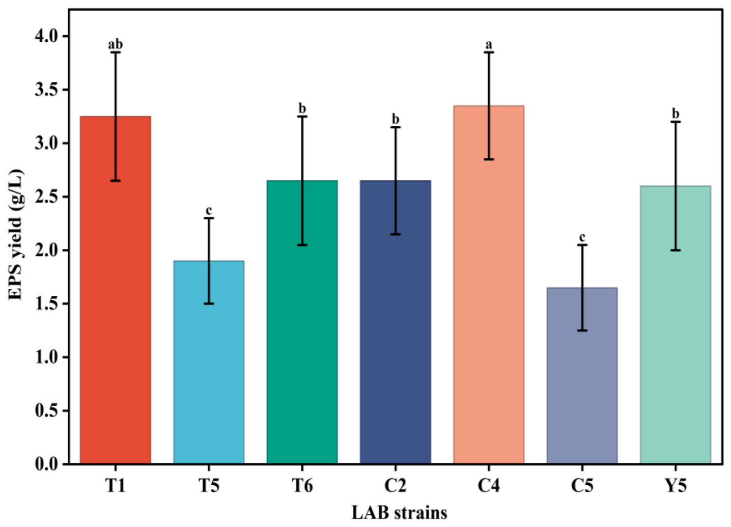
Seven of these strains with the highest sucrose tolerance were inoculated in BY broth containing beet molasses and incubated at 30 °C for 48 h. The crude EPS production yields are presented in Figure 1. The crude EPS production of these seven LAB strains ranged from 1.64 to 3.40 g/L. Among them, the crude EPS yields of the top two crude EPS-producing strains, T1 and C4, reached 3.27 g/L and 3.40 g/L, respectively. Although both T1 and C4 produced relatively high crude EPS yields, C4 showed a slightly higher crude EPS yield than T1 (3.40 vs. 3.27 g/L). In addition, C4 exhibited stronger high-sucrose tolerance, as indicated by the highest OD600 value at 400 g/L sucrose and the smallest relative decrease in OD600 from 250 to 400 g/L sucrose among the tested isolates. Therefore, C4 was selected for subsequent fermentation optimization based on its combined performance in crude EPS production and high-sucrose tolerance, rather than crude EPS yield alone. These findings indicated that these two indigenous LAB strains have great potential to produce crude EPS from beet molasses.

3.3. Identification of Strains T1 and C4

3.3.1. Morphological and Biochemical Characterization

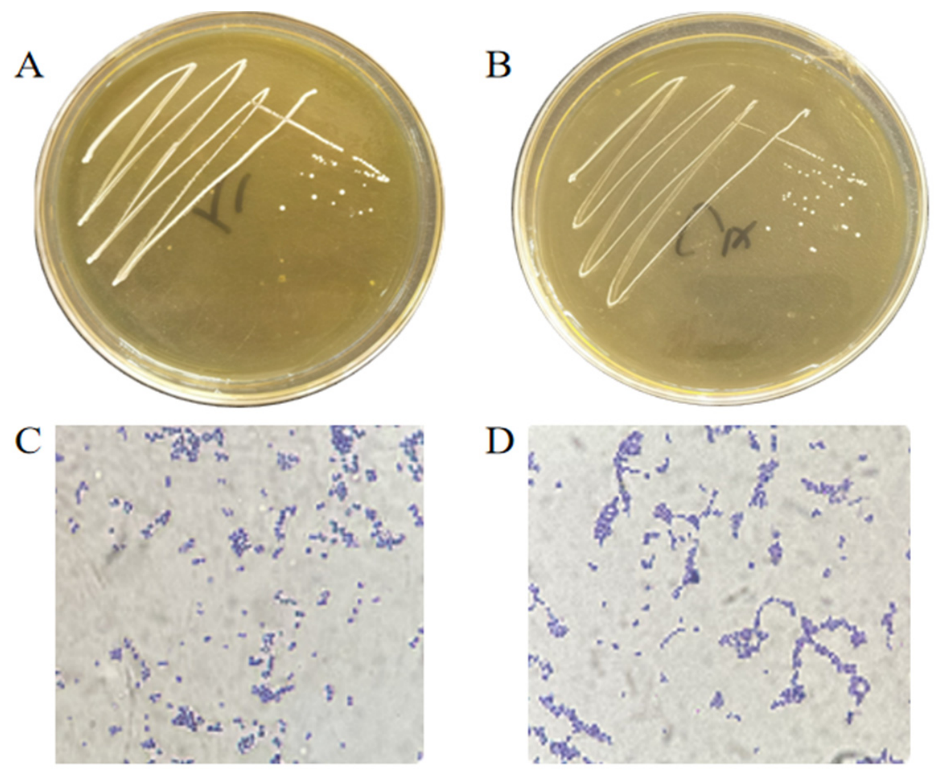
The colonies on MRS agar plates and Gram-staining results of these two strains are presented in Figure 2. The strains T1 and C4 exhibited similar morphological characteristics of round, milky white, small colonies with smooth, tidy edges as well as bulging and smooth surfaces (Figure 2A,B). The Gram-staining results presented in Figure 2C,D show that both strains were Gram-positive cells with spherical, coccoid or round shapes and no spore formation. The cocci of strain C4 formed long chains. The biochemical tests (Table 3) showed that the two strains were both oxidase- and catalase-negative, and positive for carbohydrate fermentation of cellobiose, maltose, mannitol, salicin, sorbitol, sucrose, inulin and lactose. The phenotypic characteristics of these two strains were consistent with the expected typical phenotype of LAB.

3.3.2. Bacterial Taxonomic Identification Using 16S rRNA Gene

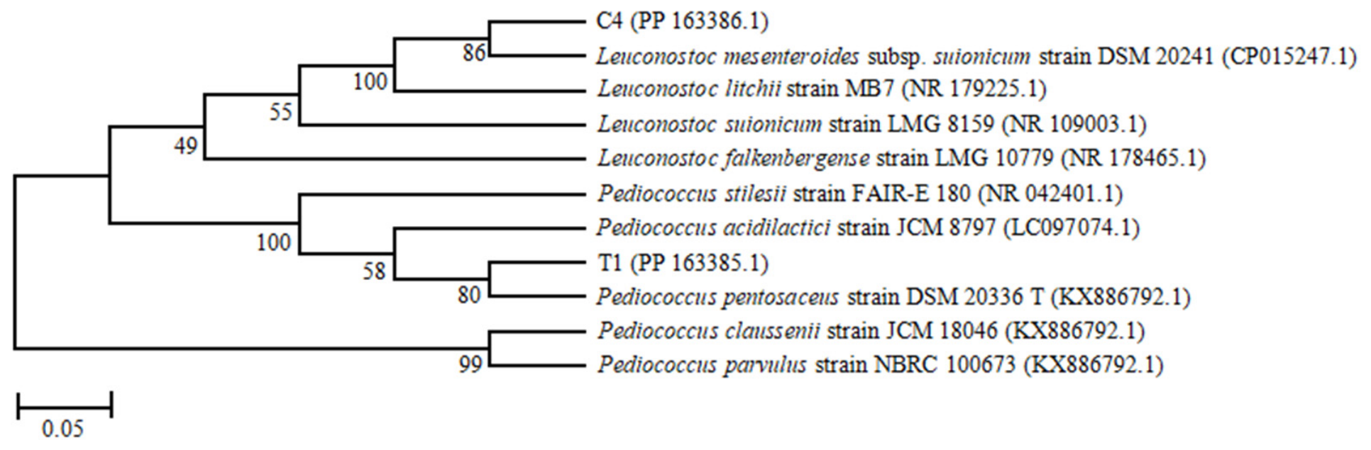
In order to further clarify the taxonomic status of these two LAB strains, the 16S rRNA genes were sequenced, and the results showed that the partial sequence lengths of the 16S rRNA gene of C4 and T1 were 1467 bp (PP 163386.1) and 1315 bp (PP 163385.1), respectively. A 16S rRNA gene sequence similarity threshold of 98.7% is commonly used to identify bacteria at the species level [41]. The Blast comparison results showed that strain C4 exhibited 99.9% 16S rRNA sequence similarity with Leuconostoc mesenteroides DSM 20242 (GenBank accession: CP015247.1), and strain T1 displayed 100% 16S rRNA sequence similarity with Pediococcus pentosaceus DSM 20336 T (GenBank accession: KX886792.1). Furthermore, the Neighbor-Joining phylogenetic tree based on 16S rRNA sequences from the nearest type strains is illustrated in Figure 3. The strain C4 clustered on the same branch of the phylogenetic tree with the type strain of Leuconostoc mesenteroides T DSM 20242, supported by a bootstrap value of 98%. Strain T1 clustered on the same branch of the phylogenetic tree with the type strain of Pediococcus pentosaceus DSM 20336 T, supported by a bootstrap value of 80%. The 98% bootstrap value for the C4/L. mesenteroides cluster indicates strong support for this phylogenetic placement, whereas the 80% bootstrap value for the T1/P. pentosaceus cluster indicates moderate support. Therefore, strains C4 and T1 were putatively assigned to L. mesenteroides and P. pentosaceus, respectively, based on 16S rRNA gene sequence similarity and phylogenetic analysis. Because the assignment was mainly based on 16S rRNA gene sequence analysis, further genomic evidence would be required for definitive species-level identification. Taken together, based on their morphological, biochemical, and 16S rRNA gene sequence characteristics, these two LAB strains were putatively identified as L. mesenteroides C4 and P. pentosaceus T1, respectively.

3.4. Growth Characteristics of Two LAB Strains

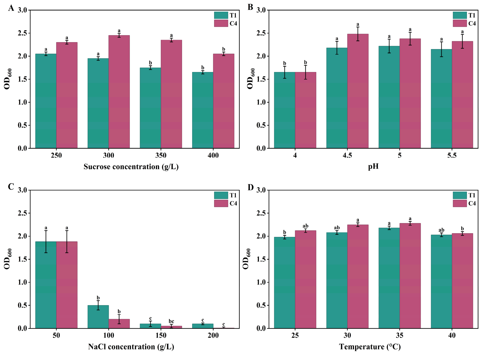
The preliminary stress-tolerance profiles, based on OD600 after 24 h incubation, are shown in Figure 4. As shown in Figure 4A, L. mesenteroides C4 showed higher OD600 values than P. pentosaceus T1 in MRS broth containing 250–400 g/L of sucrose, indicating stronger tolerance to high-sucrose osmotic stress under the tested conditions. In the acid-tolerance assay, both strains showed better endpoint growth at pH 4.5–5.5 than at pH 4.0 (Figure 4B). This assay was designed to evaluate acidic adaptation, whereas the broader pH range for crude EPS production was evaluated separately in the OFAT experiment. In the NaCl-stress assay, both strains grew at 50 g/L NaCl but showed sharply reduced growth at 100–200 g/L NaCl (Figure 4C), suggesting limited tolerance to very high NaCl concentrations. In the temperature-tolerance assay, both strains showed measurable endpoint growth at 25–40 °C, and relatively higher OD600 values were observed at 30–35 °C under the tested conditions (Figure 4).

3.5. Fermentation Optimization for Crude EPS Production of L. mesenteroides C4

3.5.1. OFAT Optimization

After comprehensive consideration of sucrose tolerance and crude EPS yield, L. mesenteroides C4 was selected as a candidate LAB strain for the bioconversion of beet molasses to LAB-EPS. The OFAT method was used to preliminarily optimize the fermentation conditions affecting crude EPS production, including varying initial pH, inoculum size, shaking speed, loading volume, fermentation temperature, fermentation time and beet molasses content in 250 mL Erlenmeyer flasks.
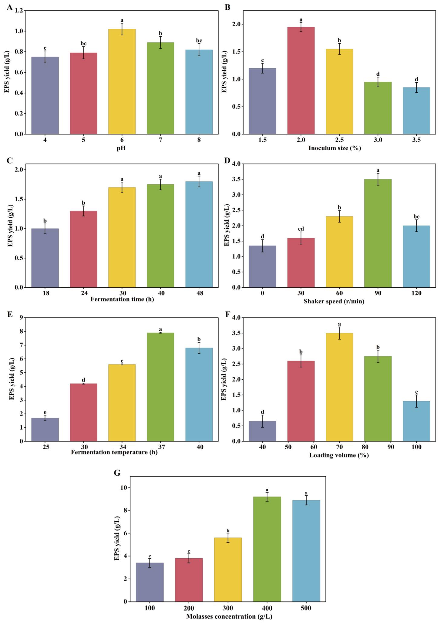
Figure 5 illustrates the effects of various factors on crude EPS production of L. mesenteroides C4. The crude EPS yield increased gradually and achieved its maximum at an initial pH value of 6.0 (Figure 5A), indicating that strain C4 displayed better crude EPS production in a slightly acidic environment. This finding was consistent with a previous report by Oleksy-Sobczak et al. [42] with respect to optimization of EPS synthesized by two Lactobacillus-type strains. The effect of inoculum size on EPS production was investigated within the range of 1.5–3.5%. As shown in Figure 5B, crude EPS yield increased with increasing inoculum size and reached the maximum value of 1.95 g/L at an inoculum size of 2.0%. However, further increasing the inoculum size led to a decrease in crude EPS production. This decline may be attributed to rapid nutrient consumption and intensified competition among bacterial cells at excessive inoculum levels, which could limit carbon flux toward EPS biosynthesis. Similar strain-dependent effects of fermentation parameters on EPS production have been reported for Leuconostoc mesenteroides XR1 during RSM-based optimization [43]. Therefore, an inoculum size of 2.0% was selected for subsequent optimization experiments.
Shaker speed and loading volume are crucial physical factors that influence the dissolved oxygen content of the culture medium under laboratory conditions [44]. As shown in Figure 5D, the lowest crude EPS yield was observed under static conditions, possibly because insufficient agitation limited oxygen transfer and nutrient mixing in the medium. Moderate agitation improved crude EPS production, and the highest yield was achieved at a shaking speed of 90 r/min. However, further increasing the shaking speed above 90 r/min decreased crude EPS yield. This decrease may be associated with excessive agitation, which could alter oxygen transfer, increase shear stress, or affect cellular metabolic activity [45,46]. Similar to shaker speed, loading volume is also one of the major factors influencing microbial EPS accumulation under liquid culture conditions. It can be seen from Figure 5F that the crude EPS production showed a trend of increasing first and then decreasing with increasing loading volume, and the highest yield was observed at the 70% loading volume. Therefore, the appropriate loading volume was determined to be 70%.
Temperature plays a major role in the production of microbial EPS. As shown in Figure 5E, the EPS production showed a trend of increasing first and then decreasing with the fermentation temperature. The maximum crude EPS yield was obtained at a fermentation temperature of 37 °C. A significant decrease in crude EPS production was observed with a higher temperature, which may be attributed to the inhibition of crude EPS synthesis-related enzyme expression and activity at high temperatures. Most previous studies have identified that the appropriate temperature for dextran or heteropolysaccharide production by Leuconostoc sp. employing sucrose as a carbon source was approximately 25 °C or even lower [47,48]. This observed difference might be caused by strain source and carbon types in the culture broth. As the fermentation time increased from 18 to 48 h, crude EPS production increased gradually and reached a plateau after 30 h (Figure 5C). No significant difference in crude EPS yield was observed among 30, 40, and 48 h (p > 0.05), as indicated by the same lowercase letters in Figure 5C. Therefore, considering fermentation efficiency and production cost, 30 h was selected as the preferred fermentation time for subsequent experiments.
Based on the fermentation levels obtained above, we determined the effect of different concentrations of beet molasses on crude EPS production, and the result is illustrated in Figure 5G. The crude EPS production increased gradually as the molasses concentration increased from 100 to 350 g/L, and the maximum crude EPS yield was obtained at a molasses concentration of 350 g/L. However, the yield of crude EPS did not increase significantly; rather, it decreased slightly as the amount of molasses increased. The slight decrease in crude EPS yield at higher molasses concentrations may be associated with increased osmotic pressure, excessive substrate concentration, or inhibitory molasses-derived compounds such as melanoidins. Accordingly, the optimal beet molasses concentration was selected to be 350 g/L.

3.5.2. Box–Behnken Design Optimization

Optimization of loading volume (X1), shaking speed (X2), and inoculum size (X3) for crude EPS production by L. mesenteroides C4 was performed using response surface methodology. Table 4 lists the designed experimental conditions and the corresponding crude EPS yields. Through multiple regression analysis of the 17 experimental runs, the following second-order polynomial equation was obtained to describe the relationship between crude EPS yield (Y) and the tested variables:
Y = 16.87 + 0.8599X1 + 0.9384X2 + 0.4627X3 + 1.40X1X2 + 2.58X1X3 − 0.0883X2X3 − 2.62X12 − 0.5630X22 − 1.18X32.
As shown in Table 5, a complex interaction relationship was observed between the investigated factors and the response value. In this model, the linear coefficients of loading volume (X1) and shaking speed (X2) were both extremely significant (p < 0.01), while inoculum size (X3) also reached a significant level (p < 0.05). The coefficient of determination (R2 = 0.9780) and adjusted R2 (0.9497) indicated that the fitted model explained most of the variation in crude EPS yield. In addition, the lack-of-fit test was not significant (p = 0.0554 > 0.05), suggesting that the model adequately fitted the experimental data within the tested range. The coefficient of variation (CV) was 3.59%, indicating acceptable experimental precision. Therefore, the model was considered suitable for predicting and optimizing crude EPS production under the present experimental conditions.
Further analysis of the ANOVA results and regression coefficients showed that the interaction terms X1X2 and X1X3 were both highly significant (p < 0.01). Among the tested interaction terms, X1X3 had a larger regression coefficient and F value than X1X2, suggesting that the interaction between loading volume and inoculum size had a relatively stronger effect on crude EPS production within the tested experimental range. However, this result should be interpreted together with the response-surface trends rather than as evidence that inoculum size alone was the dominant factor.
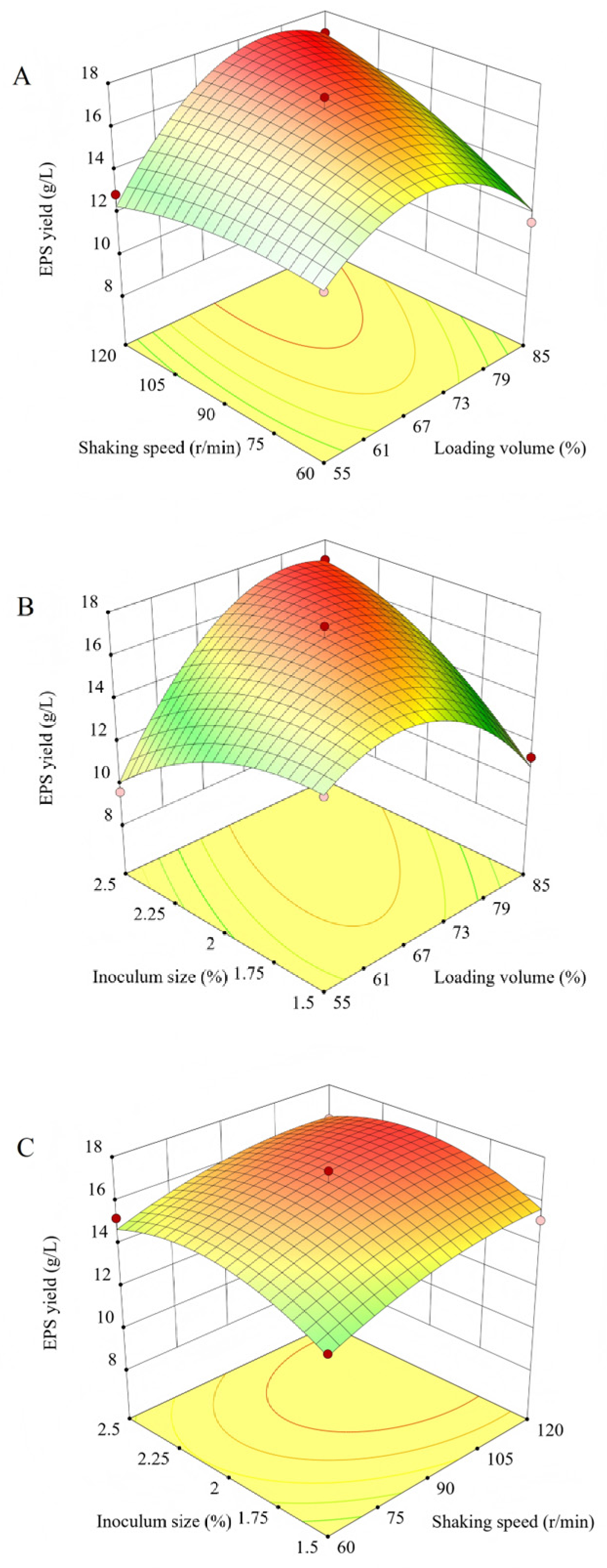
The response surface and contour plots shown in Figure 6 further illustrate the interaction effects of the tested variables. Crude EPS yield increased initially and then decreased as the levels of loading volume, shaking speed, and inoculum size increased, indicating the presence of optimal ranges for these parameters. Loading volume and shaking speed are closely associated with oxygen transfer and mixing efficiency in shake-flask fermentation. Insufficient agitation or inappropriate loading volume may limit oxygen availability and nutrient distribution, whereas excessive shaking may increase shear stress and disturb cellular metabolism, thereby reducing EPS synthesis [49]. Similarly, an appropriate inoculum size can shorten the lag phase and promote EPS accumulation, while an excessive inoculum may accelerate nutrient consumption and lead to reduced EPS production.
Based on the model prediction, the optimal conditions for crude EPS production were a loading volume of 85%, a shaking speed of 108.01 r/min, and an inoculum size of 2.33%. For operational convenience, validation experiments were conducted at a loading volume of 85%, a shaking speed of 108 r/min, and an inoculum size of 2.3%. Under these optimized conditions, the experimental crude EPS yield reached 16.99 ± 0.22 g/L, which was close to the predicted value of 17.79 g/L, with a relative error of less than 4.5%. Compared with the initial crude EPS yield of C4 before optimization (3.40 g/L), crude EPS production increased approximately 5.0-fold under the optimized conditions, confirming the effectiveness of RSM in optimizing crude EPS production from beet molasses by L. mesenteroides C4.

3.6. In Vitro Antioxidant Activity of Crude EPS-C4

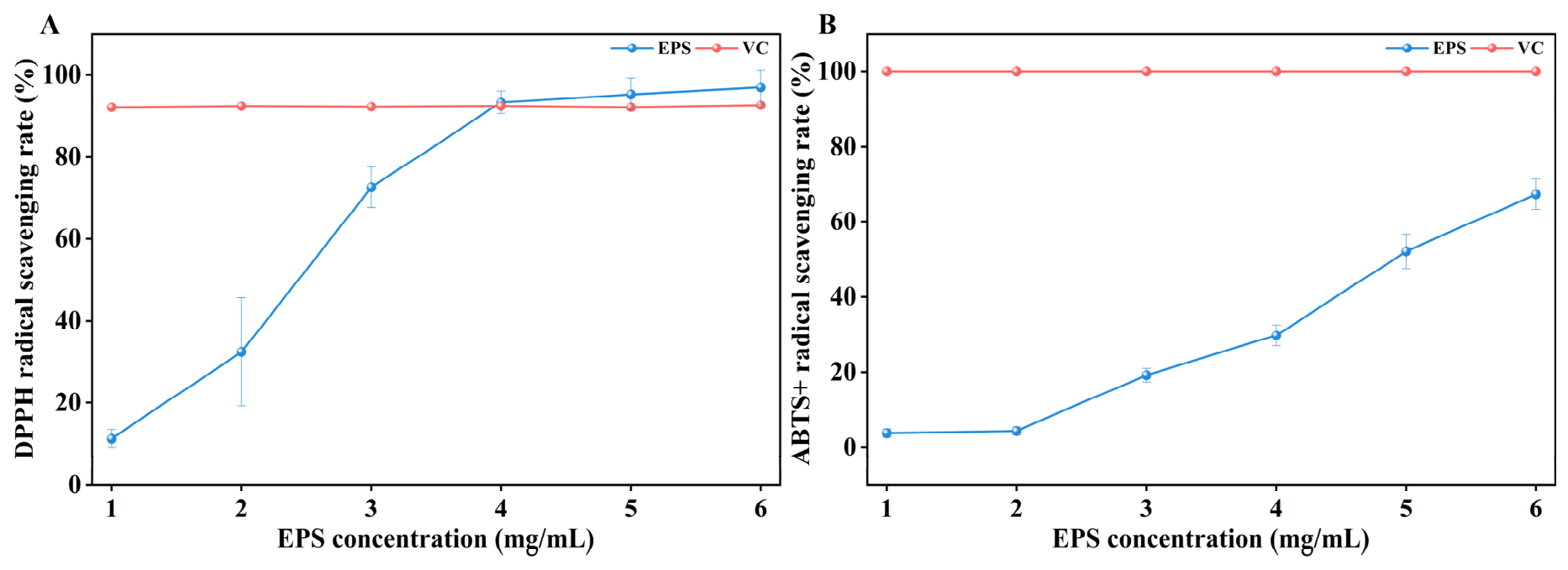
The DPPH and ABTS+ radical-scavenging activities of crude EPS-C4 at concentrations ranging from 1.0 to 6.0 mg/mL are illustrated in Figure 7. Crude EPS-C4 exhibited concentration-dependent in vitro radical-scavenging activity. The DPPH radical-scavenging rate increased with crude EPS-C4 concentration and reached 97.00% at 6.0 mg/mL. At concentrations of 4.0–6.0 mg/mL, the DPPH radical-scavenging rate of crude EPS-C4 was numerically comparable to that of the vitamin C positive control. In addition, the ABTS+ radical-scavenging rate of crude EPS-C4 increased with concentration and reached 67.35% at 6.0 mg/mL. These results indicate that crude EPS-C4 showed measurable in vitro antioxidant activity under the tested assay conditions.

3.7. Antibiofilm Activity of Crude EPS-C4

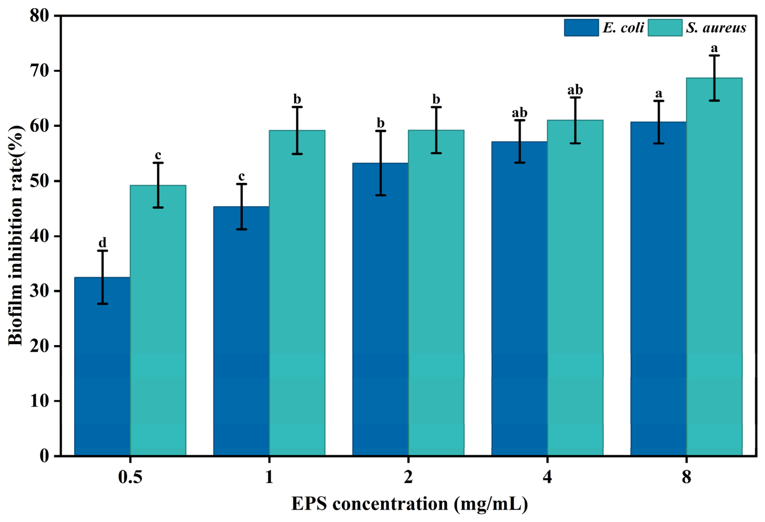
The antibiofilm activity of crude EPS-C4 against two biofilm-forming bacterial strains, E. coli ATCC 25922 and S. aureus ATCC 29215, is illustrated in Figure 8. The biofilm inhibition rates for both strains increased in a dose-dependent manner with increasing crude EPS concentration. At the maximum tested concentration of 8.0 mg/mL, crude EPS-C4 showed inhibition rates of 60.68% and 68.69% against E. coli and S. aureus biofilms, respectively. These results indicate that crude EPS-C4 showed preliminary in vitro antibiofilm activity against the tested biofilm-forming strains. However, because only two bacterial strains were evaluated, broader antibiofilm potential should be confirmed using additional bacterial species and purified EPS fractions.

4. Discussion

Although EPS-producing microorganisms are traditionally regarded as undesirable contaminants in the beet sugar industry because they can convert sucrose in beet juice into glucan and other exopolysaccharides, thereby increasing juice viscosity and reducing sugar yield and quality [50], these microorganisms may also represent valuable microbial resources for molasses valorization. In this study, beet molasses was used to isolate high-sucrose-tolerant LAB with EPS-producing potential, and the screening results identified L. mesenteroides C4 as a promising candidate for crude EPS production from this agro-industrial by-product.
Microbial EPS exhibit a variety of physiological and technological functions. Traditional fermented foods harbor abundant EPS-producing LAB, and LAB-derived EPS are generally considered attractive for food-related applications because of the long history of LAB use in fermented foods. In recent years, an increasing number of studies have reported diverse biological functions of LAB-EPS, including antioxidant, antimicrobial, prebiotic, and texture-improving properties. Nevertheless, low biosynthesis yield, high production cost, and downstream recovery difficulties remain major limitations for the industrial development of LAB-EPS [21,31]. Therefore, screening efficient EPS-producing LAB strains and optimizing fermentation conditions are important strategies for improving LAB-EPS productivity.
Response surface methodology has been widely used to improve EPS production by LAB and Leuconostoc strains. Previous studies have also shown that fermentation optimization can improve EPS production under strain- and substrate-dependent conditions [44]. For instance, the production of strain L. mesenteroides TDS2-19 increased by 31.24% after response surface optimization [51]. Jiang et al. [52] obtained a Pediococcus pentosaceus from grain vinegar, and its optimized crude EPS yield reached 15.16 g/L. In the present study, the isolate putatively identified as L. mesenteroides C4 produced 16.99 ± 0.22 g/L crude EPS under the optimized beet-molasses fermentation conditions. This result suggests that strain C4 has relatively high crude EPS-producing potential under the present experimental conditions.
The growth-characterization assay in this study was intended as a preliminary evaluation of stress tolerance rather than a complete optimization of growth conditions. Therefore, very low sucrose concentrations and low-NaCl controls were not included. In addition, although beet molasses is characterized by a relatively high potassium-ion content, NaCl was used as a general osmotic-stress agent commonly adopted in LAB salt-tolerance assays [26]. Thus, NaCl-derived osmotic stress may not fully simulate the potassium-rich ionic environment of beet molasses. Future studies should include broader sucrose and salt gradients, KCl-based osmotic stress, and wider pH ranges to establish a more complete physiological profile of the selected strains.
The relatively high crude EPS production by L. mesenteroides C4 should be interpreted as a strain-specific phenotype rather than a species-exclusive characteristic. EPS biosynthesis in LAB is closely related to the presence, organization, and expression of genes encoding glycosyltransferases, Glucansucrases/dextransucrases, and other eps-cluster components, which can vary markedly among genera, species, and even strains [53]. In sucrose-rich media, Leuconostoc strains can synthesize glucan-type EPS, particularly dextran, through extracellular Glucansucrase/dextransucrase using sucrose as the glycosyl donor [51]. Therefore, the high-sucrose beet-molasses environment may favor strains carrying active sucrose-dependent glucan-synthesis systems [54]. EPS may also serve as a protective extracellular matrix under osmotic stress, helping cells adapt to adverse environments [55]. However, genome sequencing, eps-gene analysis, and Glucansucrase activity assays were not performed in this study; therefore, this mechanism remains inferential and should be confirmed in future work.
Previous antioxidant research reported that EPS from L. mesenteroides DRP105 exhibited DPPH and ABTS radical scavenging rates of 41.52% and 32.35% at a concentration of 5 mg/mL, respectively [56]. In this study, crude EPS-C4 showed DPPH and ABTS radical scavenging rates of 97.00% and 67.35% at 6 mg/mL, respectively, indicating measurable in vitro radical-scavenging activity under the tested conditions. However, this comparison should be considered descriptive because differences in strain origin, EPS extraction method, sample purity, concentration, and assay conditions may affect the measured antioxidant activity.
The crude EPS-C4 fraction also inhibited biofilm formation by the tested biofilm-forming E. coli and S. aureus strains. LAB-EPS may inhibit biofilm formation through several possible mechanisms, including interference with initial bacterial adhesion, competition for surface-binding sites, disruption of extracellular matrix formation, or modulation of quorum-sensing-related processes [57]. Nevertheless, only two bacterial strains were evaluated in this study; broad-spectrum antibiofilm activity could be tested in future. In addition, a crude EPS-C4-only blank control for crystal violet absorbance at 590 nm was not included in the present antibiofilm assay; therefore, possible interference of crude EPS-C4 with the crystal violet signal cannot be completely excluded.
It should also be emphasized that crude EPS-C4 was evaluated as a crude EPS fraction obtained from beet-molasses fermentation. Molasses-based fermentation systems contain complex non-sugar components, including pigments, proteins, salts, Maillard reaction products, and other soluble compounds [58], which may be co-precipitated during EPS recovery. Therefore, further separation and purification of the crude EPS is required to identify the specific active components responsible for the observed antioxidant and antibiofilm activities and to characterize their molecular properties.
Taken together, beet molasses can serve as a low-cost substrate for producing crude LAB-EPS, and crude EPS-C4 may be considered a candidate bioactive polymer for further investigation. However, the present results only demonstrate preliminary in vitro antioxidant and antibiofilm activities. Practical application in food or health-related products will require further purification, structural characterization, safety assessment, and application-oriented validation. In particular, the molecular weight, monosaccharide composition, functional groups, microscopic morphology, water solubility, and water-holding capacity of crude EPS-C4 should be investigated in future work to clarify the relationship between its structure and biological activity.

5. Conclusions

In this study, a total of 13 LAB strains were isolated from beet molasses obtained from different sugar factories in Xinjiang, China, among which seven isolates were selected as high-sucrose-tolerant candidates. Among them, one potential high-sucrose-tolerant and high-crude-EPS-producing strain, C4, was putatively identified as Leuconostoc mesenteroides based on 16S rRNA gene sequence analysis. In the preliminary growth-characterization assay, strain C4 maintained good endpoint growth under 400 g/L of sucrose and 50 g/L of NaCl. Furthermore, the optimal fermentation conditions for the production of crude EPS by L. mesenteroides C4 using beet molasses were as follows: initial pH value of 6.0, loading volume of 85%, shaker speed of 108 r/min, fermentation temperature of 37 °C, inoculum size of 2.3%, 350 g/L of molasses, and a fermentation time of 30 h. Under the optimal conditions, the maximum crude EPS yield of 16.99 ± 0.22 g/L was recorded. The crude EPS-C4 fraction showed DPPH and ABTS+ radical scavenging rates of 97.00% and 67.35% at 6.0 mg/mL. Additionally, the crude EPS-C4 fraction inhibited biofilm formation by E. coli and S. aureus, with inhibition rates of 60.68% and 68.69% at 8.0 mg/mL, respectively. Taken together, crude EPS-C4 obtained from beet-molasses fermentation showed preliminary in vitro antioxidant and antibiofilm activities under the tested conditions. These findings indicate that crude EPS-C4 may be considered a candidate bioactive polymer for further investigation. Further purification, structural characterization, safety assessment, and application-oriented validation are required before its practical application can be considered.

Author Contributions

Conceptualization, S.Z. and G.S.; methodology, G.R. and H.Z.; software, H.Z.; formal analysis, C.Z. and G.R.; investigation, Y.W., C.Z. and G.R.; data curation, C.Z. and G.R.; writing—original draft preparation, Y.W.; writing—review and editing, G.S.; resources, H.Z.; supervision, S.Z.; funding acquisition, G.S.; visualization, Y.W. All authors have read and agreed to the published version of the manuscript.

Funding

This work was supported by the Bofeng Elite Program Project of Xinjiang Changji Hui Autonomous Prefecture, China (2022015) and Students’ Research Interesting Training Support by Sichuan Agricultural University, China (2023243).

Institutional Review Board Statement

Not applicable.

Informed Consent Statement

Not applicable.

Data Availability Statement

The data presented in this study are available upon request from the corresponding author.

Conflicts of Interest

Author Han Zhang was employed by COFCO Sugar Holding Co., Ltd. The remaining authors declare that the research was conducted in the absence of any commercial or financial relationships that could be construed as a potential conflict of interest.

References

  1. Tayyab, M.; Wakeel, A.; Mubarak, M.U.; Artyszak, A.; Ali, S.; Hakki, E.E.; Mahmood, K.; Song, B.; Ishfaq, M. Sugar Beet Cultivation in the Tropics and Subtropics: Challenges and Opportunities. Agronomy 2023, 13, 1213. [Google Scholar] [CrossRef] [Scilit]
  2. Industry Research Co. Granulated Sugar Market Size, Share, Growth, and Industry Analysis, By Type (Cane Sugar, Beet Sugar), By Application (Bakery, Beverage, Candy, Others), Regional Insights and Forecast to 2035. Available online: https://www.industryresearch.biz/market-reports/granulated-sugar-market-111413 (accessed on 21 September 2026).
  3. Adbhai, A.R.; Dewanjee, S.; Patel, K.G.; Karmakar, N. Sugar Beet Molasses Production and Utilization. In Sugar Beet Cultivation, Management and Processing; Misra, V., Srivastava, S., Mall, A.K., Eds.; Springer Nature: Singapore, 2022; pp. 885–904. [Google Scholar]
  4. Geremew Kassa, M.; Asemu, A.M.; Belachew, M.T.; Satheesh, N.; Abera, B.D.; Alemu Teferi, D. Review on the Application, Health Usage, and Negative Effects of Molasses. CyTA J. Food 2024, 22, 2321984. [Google Scholar] [CrossRef] [Scilit]
  5. Zhang, S.; Wang, J.; Jiang, H. Microbial Production of Value-Added Bioproducts and Enzymes from Molasses, a by-Product of Sugar Industry. Food Chem. 2021, 346, 128860. [Google Scholar] [CrossRef] [Scilit] [PubMed]
  6. Ozdal, M.; Başaran Kurbanoglu, E. Use of Chicken Feather Peptone and Sugar Beet Molasses as Low Cost Substrates for Xanthan Production by Xanthomonas campestris MO-03. Fermentation 2019, 5, 9. [Google Scholar] [CrossRef] [Scilit]
  7. Mustafa, G.; Arshad, M.; Bano, I.; Abbas, M. Biotechnological Applications of Sugarcane Bagasse and Sugar Beet Molasses. Biomass Convers. Biorefin. 2023, 13, 1489–1501. [Google Scholar] [CrossRef] [Scilit]
  8. Alam, M.; Kaur, S.; Dar, B.N.; Nanda, V. Classification, Techno-Functional Properties, and Applications of Diverse Hydrocolloids in Fruits-Based Products: A Concise Review. J. Food Sci. 2025, 90, e70119.1–e70119.26. [Google Scholar] [CrossRef] [Scilit] [PubMed]
  9. Rhein-Knudsen, N.; Ale, M.; Meyer, A. Seaweed Hydrocolloid Production: An Update on Enzyme Assisted Extraction and Modification Technologies. Mar. Drugs 2015, 13, 3340–3359. [Google Scholar] [CrossRef] [Scilit] [PubMed]
  10. Netrusov, A.I.; Liyaskina, E.V.; Kurgaeva, I.V.; Liyaskina, A.U.; Yang, G.; Revin, V.V. Exopolysaccharides Producing Bacteria: A Review. Microorganisms 2023, 11, 1541. [Google Scholar] [CrossRef] [Scilit] [PubMed]
  11. Nachtigall, C.; Surber, G.; Herbi, F.; Wefers, D.; Jaros, D.; Rohm, H. Production and Molecular Structure of Heteropolysaccharides from Two Lactic Acid Bacteria. Carbohydr. Polym. 2020, 236, 116019. [Google Scholar] [CrossRef] [Scilit] [PubMed]
  12. Zannini, E.; Waters, D.M.; Coffey, A.; Arendt, E.K. Production, Properties, and Industrial Food Application of Lactic Acid Bacteria-Derived Exopolysaccharides. Appl. Microbiol. Biotechnol. 2016, 100, 1121–1135. [Google Scholar] [CrossRef] [Scilit] [PubMed]
  13. Abarquero, D.; Renes, E.; Fresno, J.M.; Tornadijo, M.E. Study of Exopolysaccharides from Lactic Acid Bacteria and Their Industrial Applications: A Review. Int. J. Food Sci. Technol. 2022, 57, 16–26. [Google Scholar] [CrossRef] [Scilit]
  14. Prasad, S.; Purohit, S.R. Microbial Exopolysaccharide: Sources, Stress Conditions, Properties and Application in Food and Environment: A Comprehensive Review. Int. J. Biol. Macromol. 2023, 242, 124925. [Google Scholar] [CrossRef] [Scilit] [PubMed]
  15. Freitas, F.; Torres, C.A.V.; Reis, M.A.M. Engineering Aspects of Microbial Exopolysaccharide Production. Bioresour. Technol. 2017, 245, 1674–1683. [Google Scholar] [CrossRef] [Scilit] [PubMed]
  16. Chaisuwan, W.; Jantanasakulwong, K.; Wangtueai, S.; Phimolsiripol, Y.; Chaiyaso, T.; Techapun, C.; Phongthai, S.; You, S.; Regenstein, J.M.; Seesuriyachan, P. Microbial Exopolysaccharides for Immune Enhancement: Fermentation, Modifications and Bioactivities. Food Biosci. 2020, 35, 100564. [Google Scholar] [CrossRef] [Scilit]
  17. Li, Y.; Zhang, W.; Pan, X.; Xiao, Y.; Liu, L.; Ding, F.; Li, G.; Zhao, L. Recent Advances in Exopolysaccharides from Lactic Acid Bacteria: Acquisition, Characterization, Bioactivities, and Applications. Food Biosci. 2026, 79, 108830. [Google Scholar] [CrossRef] [Scilit]
  18. Zang, J.; Yan, B.; Liu, Z.; Tang, D.; Liu, Y.; Chen, J.; Yin, Z. Current State, Challenges and Future Orientations of the Applications of Lactic Acid Bacteria Exopolysaccharide in Foods. Food Microbiol. 2025, 126, 104678. [Google Scholar] [CrossRef] [Scilit] [PubMed]
  19. Qiu, Y.; Wang, T.; Yang, Q.; Liu, X.; Song, C.; Du, R. Lactic Acid Bacteria Exopolysaccharides as Next-Generation Clean-Label Texturizers and Prebiotics in Dairy Systems. Fermentation 2026, 12, 245. [Google Scholar] [CrossRef] [Scilit]
  20. Liang, S.; Wang, X.; Li, C.; Liu, L. Biological Activity of Lactic Acid Bacteria Exopolysaccharides and Their Applications in the Food and Pharmaceutical Industries. Foods 2024, 13, 1621. [Google Scholar] [CrossRef] [Scilit] [PubMed]
  21. Zhang, K.; Liu, S.; Liang, S.; Xiang, F.; Wang, X.; Lian, H.; Li, B.; Liu, F. Exopolysaccharides of Lactic Acid Bacteria: Structure, Biological Activity, Structure-Activity Relationship, and Application in the Food Industry: A Review. Int. J. Biol. Macromol. 2024, 257, 128733. [Google Scholar] [CrossRef] [Scilit] [PubMed]
  22. Oviedo-León, J.F.; Higuera, A.R.; Yáñez-Fernández, J.; Hernández-Sánchez, H.; Castro-Rodríguez, D.C. Exploring Exopolysaccharides Produced in Indigenous Mexican Fermented Beverages and Their Biotechnological Applications. Fermentation 2025, 11, 463. [Google Scholar] [CrossRef] [Scilit]
  23. Zheng, J.; Wittouck, S.; Salvetti, E.; Franz, C.M.A.P.; Harris, H.M.B.; Mattarelli, P.; O’Toole, P.W.; Pot, B.; Vandamme, P.; Walter, J.; et al. A Taxonomic Note on the Genus Lactobacillus: Description of 23 Novel Genera, Emended Description of the Genus Lactobacillus Beijerinck 1901, and Union of Lactobacillaceae and Leuconostocaceae. Int. J. Syst. Evol. Microbiol. 2020, 70, 2782–2858. [Google Scholar] [CrossRef] [Scilit] [PubMed]
  24. Sampaio, I.C.F.; Rodrigues, P.D.; De Moura, I.V.L.; Silva, M.D.S.; Widmer, L.F.; Quintella, C.M.; Ramos-de-Souza, E.; Almeida, P.F.D. Valorization of Produced Water from Oilfields for Microbial Exopolysaccharide Synthesis in Stirred Tank Bioreactors. Fermentation 2026, 12, 39. [Google Scholar] [CrossRef] [Scilit]
  25. Wang, W.-Y.; Wang, B.-P.; Su, H.-S.; Wei, M.-M.; Wei, Y.-T.; Niu, F.-X. Key Role of K+ and Ca2+ in High-Yield Ethanol Production by S. Cerevisiae from Concentrated Sugarcane Molasses. Microb. Cell Fact. 2024, 23, 123. [Google Scholar] [CrossRef] [Scilit] [PubMed]
  26. Palmonari, A.; Cavallini, D.; Sniffen, C.J.; Fernandes, L.; Holder, P.; Fagioli, L.; Fusaro, I.; Biagi, G.; Formigoni, A.; Mammi, L. Short Communication: Characterization of Molasses Chemical Composition. J. Dairy Sci. 2020, 103, 6244–6249. [Google Scholar] [CrossRef] [Scilit] [PubMed]
  27. Saisriyoot, M.; Thanapimmetha, A.; Suwaleerat, T.; Chisti, Y.; Srinophakun, P. Biomass and Lipid Production by Rhodococcus opacus PD630 in Molasses-Based Media with and without Osmotic-Stress. J. Biotechnol. 2019, 297, 1–8. [Google Scholar] [CrossRef] [Scilit] [PubMed]
  28. Long, X.; Hou, X.; Li, S.; Chen, A.; Zhang, Z.; Shen, G. Fermentation Optimization and In-Vitro Antioxidant Activity of Exopolysaccharides Produced by Leuconostoc suionicum LSBM1 Using Sugar Beet Molasses. Sugar Tech. 2024, 26, 1405–1414. [Google Scholar] [CrossRef] [Scilit]
  29. Ren, G.; Luo, W.; Gan, H.; Zhang, H.; Hou, X.; Li, S.; Chen, A.; Zhang, Z.; Pan, L.; Zhao, S.; et al. Bioconversion of Sugar Beet Molasses into Functional Exopolysaccharides by Beet Juice-Derived Lactic Acid Bacteria. Food Biosci. 2024, 62, 105139. [Google Scholar] [CrossRef] [Scilit]
  30. Ruas-Madiedo, P.; De Los Reyes-Gavilán, C.G. Invited Review: Methods for the Screening, Isolation, and Characterization of Exopolysaccharides Produced by Lactic Acid Bacteria. J. Dairy Sci. 2005, 88, 843–856. [Google Scholar] [CrossRef] [Scilit] [PubMed]
  31. Yadav, M.K.; Song, J.H.; Vasquez, R.; Lee, J.S.; Kim, I.H.; Kang, D.-K. Methods for Detection, Extraction, Purification, and Characterization of Exopolysaccharides of Lactic Acid Bacteria—A Systematic Review. Foods 2024, 13, 3687. [Google Scholar] [CrossRef] [Scilit] [PubMed]
  32. Mokoena, M.P. Lactic Acid Bacteria and Their Bacteriocins: Classification, Biosynthesis and Applications against Uropathogens: A Mini-Review. Molecules 2017, 22, 1255. [Google Scholar] [CrossRef] [Scilit] [PubMed]
  33. Kim, G.-H.; Awais, M.; Kim, T.-M.; Chi, W.-J.; Lee, J.M.; Kim, J.-H. Autochthonous Lactiplantibacillus plantarum IBT11 Modulates Gut Bacterial Community Structure and Immune-Related Gene Expression in Apis mellifera. J. Asia Pac. Entomol. 2026, 29, 102595. [Google Scholar] [CrossRef] [Scilit]
  34. Zeng, C.; Ye, G.; Li, G.; Cao, H.; Wang, Z.; Ji, S. RID Serve as a More Appropriate Measure than Phenol Sulfuric Acid Method for Natural Water-Soluble Polysaccharides Quantification. Carbohydr. Polym. 2022, 278, 118928. [Google Scholar] [CrossRef] [Scilit] [PubMed]
  35. Stephen, J.M.; Saleh, A.M. Homofermentative Lactobacilli Isolated from Organic Sources Exhibit Potential Ability of Lactic Acid Production. Front. Microbiol. 2023, 14, 1297036. [Google Scholar] [CrossRef] [Scilit] [PubMed]
  36. Fugl, A.; Berhe, T.; Kiran, A.; Hussain, S.; Laursen, M.F.; Bahl, M.I.; Hailu, Y.; Sørensen, K.I.; Guya, M.E.; Ipsen, R.; et al. Characterisation of Lactic Acid Bacteria in Spontaneously Fermented Camel Milk and Selection of Strains for Fermentation of Camel Milk. Int. Dairy J. 2017, 73, 19–24. [Google Scholar] [CrossRef] [Scilit]
  37. Shu, G.; Mei, S.; Chen, L.; Zhang, B.; Guo, M.; Cui, X.; Chen, H. Screening, Identification, and Application of Selenium-Enriched Lactobacillus in Goat Milk Powder and Tablet. J. Food Process. Preserv. 2020, 44, e14470. [Google Scholar] [CrossRef] [Scilit]
  38. Wang, J.; Zhang, J.; Guo, H.; Cheng, Q.; Abbas, Z.; Tong, Y.; Yang, T.; Zhou, Y.; Zhang, H.; Wei, X.; et al. Optimization of Exopolysaccharide Produced by Lactobacillus Plantarum R301 and Its Antioxidant and Anti-Inflammatory Activities. Foods 2023, 12, 2481. [Google Scholar] [CrossRef] [Scilit] [PubMed]
  39. Tarannum, N.; Hossain, T.J.; Ali, F.; Das, T.; Dhar, K.; Nafiz, I.H. Antioxidant, Antimicrobial and Emulsification Properties of Exopolysaccharides from Lactic Acid Bacteria of Bovine Milk: Insights from Biochemical and Genomic Analysis. LWT 2023, 186, 115263. [Google Scholar] [CrossRef] [Scilit]
  40. Juanit, T.; Payel, R.; Arabinda, G.; Megha, M.; Kumar Sil, S.; Deeplina, D. Prebiotic Levan Type Fructan from Bacillus subtilis PR-C18 as a Potent Antibiofilm Agent: Structural Elucidation and in Silico Analysis. Carbohydr. Res. 2024, 538, 109075. [Google Scholar] [CrossRef] [Scilit] [PubMed]
  41. Rossi-Tamisier, M.; Benamar, S.; Raoult, D.; Fournier, P.-E. Cautionary Tale of Using 16S rRNA Gene Sequence Similarity Values in Identification of Human-Associated Bacterial Species. Int. J. Syst. Evol. Microbiol. 2015, 65, 1929–1934. [Google Scholar] [CrossRef] [Scilit] [PubMed]
  42. Oleksy-Sobczak, M.; Klewicka, E.; Piekarska-Radzik, L. Exopolysaccharides Production by Lactobacillus rhamnosus Strains—Optimization of Synthesis and Extraction Conditions. LWT 2020, 122, 109055. [Google Scholar] [CrossRef] [Scilit]
  43. Tian, H.; Wang, W.; Liu, W.; Lv, Z.; Wang, L. Exopolysaccharide from Leuconostoc mesenteroides XR1: Yield Optimization, Partial Characterization and Properties. Int. J. Biol. Macromol. 2024, 279, 135225. [Google Scholar] [CrossRef] [Scilit] [PubMed]
  44. Guo, L.; Guo, Y.; Wu, P.; Liu, S.; Gu, C.; Yolandani; Wu, M.; Ma, H.; He, R. Enhancement of Polypeptide Yield Derived from Rapeseed Meal with Low-Intensity Alternating Magnetic Field. Foods 2022, 11, 2952. [Google Scholar] [CrossRef] [Scilit] [PubMed]
  45. Dulay, R.M.R.; Cabrera, E.C.; Kalaw, S.P.; Reyes, R.G. Optimization of Submerged Culture Conditions for Mycelial Biomass Production of Fourteen Lentinus Isolates from Luzon Island, Philippines. Biocatal. Agric. Biotechnol. 2021, 38, 102226. [Google Scholar] [CrossRef] [Scilit]
  46. Rahman, S.S.A.; Pasupathi, S.; Karuppiah, S. Conventional Optimization and Characterization of Microbial Dextran Using Treated Sugarcane Molasses. Int. J. Biol. Macromol. 2022, 220, 775–787. [Google Scholar] [CrossRef] [Scilit] [PubMed]
  47. Ge, Z.; Wang, D.; Zhao, W.; Wang, P.; Dai, Y.; Dong, M.; Wang, J.; Zhao, Y.; Zhao, X. Structural and Functional Characterization of Exopolysaccharide from Leuconostoc citreum BH10 Discovered in Birch Sap. Carbohydr. Res. 2024, 535, 108994. [Google Scholar] [CrossRef] [Scilit] [PubMed]
  48. Farinazzo, F.S.; Fernandes, M.T.C.; Mauro, C.S.I.; Garcia, S. Statistical Optimization of Exopolysaccharide Production by Leuconostoc pseudomesenteroides JF17 from Native Atlantic Forest Juçara Fruit. Prep. Biochem. Biotechnol. 2022, 52, 245–252. [Google Scholar] [CrossRef] [Scilit] [PubMed]
  49. Gu, J.; Zhang, H.; Zhang, J.; Wen, C.; Zhou, J.; Yao, H.; He, Y.; Ma, H.; Duan, Y. Optimization, Characterization, Rheological Study and Immune Activities of Polysaccharide from Sagittaria sagittifolia L. Carbohydr. Polym. 2020, 246, 116595. [Google Scholar] [CrossRef] [Scilit] [PubMed]
  50. Joshi, S.; Bruni, G.O.; Zimmerman, T.; Terrell, E.; Salter, J.; Kashem, M.N.H.; Nam, S. Phenotypic Variation in Growth and Biofilm Formation of Leuconostoc Spp. from Sugar Beet Factories. Front. Microbiol. 2026, 16, 1745936. [Google Scholar] [CrossRef] [Scilit] [PubMed]
  51. Du, R.; Xing, H.; Yang, Y.; Jiang, H.; Zhou, Z.; Han, Y. Optimization, Purification and Structural Characterization of a Dextran Produced by L. mesenteroides Isolated from Chinese Sauerkraut. Carbohydr. Polym. 2017, 174, 409–416. [Google Scholar] [CrossRef] [Scilit] [PubMed]
  52. Liao, J.-J.; Tan, C.-Y.; Liang, L.; Luo, Y.-Z.; Li, H.-L.; Chen, Y.-Y.; Pei, X.-D.; Wang, C.-H. Screening and Identification of Selenium-Enriched Purine-Lowering Lactobacillus plantarum and Lactococcus lactis with High Stress Tolerance and Antioxidant Capacity. Food Biosci. 2024, 60, 104439. [Google Scholar] [CrossRef] [Scilit]
  53. Zeidan, A.A.; Poulsen, V.K.; Janzen, T.; Buldo, P.; Derkx, P.M.F.; Øregaard, G.; Neves, A.R. Polysaccharide Production by Lactic Acid Bacteria: From Genes to Industrial Applications. FEMS Microbiol. Rev. 2017, 41, S168–S200. [Google Scholar] [CrossRef] [Scilit] [PubMed]
  54. van Hijum, S.A.; Kralj, S.; Ozimek, L.K.; Dijkhuizen, L.; van Geel-Schutten, I.G. Structure-Function Relationships of Glucansucrase and Fructansucrase Enzymes from Lactic Acid Bacteria. Microbiol. Mol. Biol. Rev. 2006, 70, 157–176. [Google Scholar] [CrossRef] [Scilit] [PubMed]
  55. Sukarno, A.S.; Dong, A.; Gumulya, Y.; Xiang, Y.; Nam, Y.; Lim, J.; Seo, J.-H.; Lee, H.-W.; Kinoshita, H.; Marcellin, E.; et al. Exopolysaccharide Biosynthesis and Regulation in Leuconostoc Mesenteroides Influences Water Holding Capacity of Fermented Plant-Based Milks. Int. J. Food Microbiol. 2026, 457, 111797. [Google Scholar] [CrossRef] [Scilit] [PubMed]
  56. Xing, H.; Du, R.; Zhao, F.; Han, Y.; Xiao, H.; Zhou, Z. Optimization, Chain Conformation and Characterization of Exopolysaccharide Isolated from Leuconostoc mesenteroides DRP105. Int. J. Biol. Macromol. 2018, 112, 1208–1216. [Google Scholar] [CrossRef] [Scilit] [PubMed]
  57. Werning, M.L.; Hernández-Alcántara, A.M.; Ruiz, M.J.; Soto, L.P.; Dueñas, M.T.; López, P.; Frizzo, L.S. Biological functions of exopolysaccharides from lactic acid bacteria and their potential benefits for humans and farmed animals. Foods 2022, 11, 1284. [Google Scholar] [CrossRef] [Scilit] [PubMed]
  58. Djordjević, M.; Šereš, Z.; Došenović, T.; Šoronja-Simović, D.; Maravić, N.; Kukić, D.; Nikolić, I.; Djordjević, M. Sugar Beet Molasses Purification by Bentonite Addition: Analysis of Quality Enhancement and Treatment Conditions. LWT 2018, 93, 142–149. [Google Scholar] [CrossRef] [Scilit]
Figure 1. Crude EPS production by seven high-sucrose-tolerant LAB strains. Data are presented as mean ± SD (n = 3). Different lowercase letters on the bars denote significant differences (p < 0.05).
Fermentation 12 00464 g001
Figure 2. Colony morphology (A,B) and Gram-staining microscopic images (C,D) of two LAB strains. (A,C): strain T1; (B,D): strain C4. The Gram-staining microscopic images in panels (C,D) were observed under 100× magnification.
Fermentation 12 00464 g002
Figure 3. Phylogenetic tree of two high-sucrose-tolerant LAB strains based on 16S rRNA gene sequence. Bootstrap values are shown at the nodes of the tree. The bar indicates 5% sequence divergence.
Fermentation 12 00464 g003
Figure 4. Endpoint growth, determined by OD600 after 24 h incubation, of two high-sucrose-tolerant LAB strains under different sucrose concentrations (A), initial pH values (B), NaCl concentrations (C), and temperatures (D). Data are presented as mean ± SD (n = 3). Different lowercase letters on the bars denote significant differences (p < 0.05).
Fermentation 12 00464 g004
Figure 5. Effects of initial pH (A), inoculum size (B), fermentation time (C), shaking speed (D), fermentation temperature (E), loading volume (F) and molasses concentration (G) on the crude EPS production by L. mesenteroides C4. Data are presented as mean ± SD (n = 3). Different lowercase letters on the bars denote significant differences (p < 0.05).
Fermentation 12 00464 g005
Figure 6. Response surface plots and contour lines of effects of interaction of various factors on crude EPS yield. Loading volume and shaking speed (A), loading volume and inoculum size (B), shaking speed and inoculum size (C). The color gradient represents the magnitude of the response value; red indicates the highest value and green indicates the lowest value.
Fermentation 12 00464 g006
Figure 7. Antioxidant activity of crude EPS-C4. DPPH radical scavenging activity (A) and ABTS+ radical scavenging activity (B). Data are presented as mean ± SD (n = 3).
Fermentation 12 00464 g007
Figure 8. Antibiofilm effect of crude EPS-C4 against biofilm-forming E. coli and S. aureus strains. Data are presented as mean ± SD (n = 3). Different lowercase letters on the bars denote significant differences (p < 0.05).
Fermentation 12 00464 g008
Table 1. Factors and levels for Box–Behnken response surface design.
VariablesRange and Levels
−101
X1 Loading volume (%)557085
X2 Shaking speed (r/min)6090120
X3 Inoculum size (%)1.52.02.5
Table 2. OD600 values of the candidate LAB strains isolated from beet molasses in MRS broth with different concentrations of sucrose cultured at 37 °C for 24 h.
Beet Molasses Sample SourceLAB StrainsSucrose Concentration (g/L)
250300350400
Tacheng sugar factoryT12.07 ± 0.05 cd1.95 ± 0.07 c1.75 ± 0.06 d1.66 ± 0.05 b
T52.03 ± 0.06 cde1.87 ± 0.05 de1.59 ± 0.06 e1.30 ± 0.05 e
T62.21 ± 0.05 b1.86 ± 0.04 e1.49 ± 0.05 f1.38 ± 0.06 d
T82.20 ± 0.05 b1.93 ± 0.06 cd1.62 ± 0.04 e1.21 ± 0.05 f
Changji sugar factoryC11.72 ± 0.03 g1.54 ± 0.03 f1.44 ± 0.05 f1.26 ± 0.04 ef
C21.97 ± 0.04 e1.88 ± 0.04 de2.04 ± 0.05 b1.66 ± 0.06 b
C31.86 ± 0.05 f2.18 ± 0.04 b1.42 ± 0.05 f1.26 ± 0.03 ef
C42.33 ± 0.06 a2.45 ± 0.04 a2.37 ± 0.04 a2.08 ± 0.05 a
C52.08 ± 0.05 c1.93 ± 0.03 cd1.91 ± 0.05 c1.70 ± 0.04 b
Yining sugar factoryY20.34 ± 0.05 i0.82 ± 0.05 h0.54 ± 0.06 h0.41 ± 0.03 h
Y30.26 ± 0.05 j0.49 ± 0.04 i0.58 ± 0.05 h0.22 ± 0.04 i
Y41.49 ± 0.06 h1.36 ± 0.05 g1.16 ± 0.04 g1.00 ± 0.05 g
Y52.01 ± 0.04 de1.82 ± 0.05 e1.58 ± 0.03 e1.55 ± 0.05 c
Different lowercase letters indicate significant differences among strains (p < 0.05). Data are presented as mean ± SD (n = 3).
Table 3. Biochemical test results of strains T1 and C4.
Biochemical Test ItemStrain T1Strain C4
Gram staining++
Oxidase−−
Catalase−−
Esculin hydrate++
Maltose++
Mannitol++
Salicin++
Sorbitol−−
Sucrose++
Raffinose−−
Inulin++
Lactose++
Hippuricase−−
Note: + indicates positive; − indicates negative.
Table 4. Box–Behnken response surface design and response value of crude EPS yield of L. mesenteroides C4.
RunX1 Loading Volume (%)X2 Shaking Speed (r/min)X3 Inoculum Size (%)Crude EPS Yield (g/L)
11 (85)0 (90)1 (2.5)17.03
2−1 (55)0 (90)1 (2.5)9.63
3−1 (55)−1 (60)0 (2.0)13.24
40 (70)0 (90)0 (2.0)16.68
50 (70)0 (90)0 (2.0)16.84
60 (70)1 (120)1 (2.5)16.33
70 (70)0 (90)0 (2.0)16.72
81 (85)−1 (90)0 (2.0)11.64
90 (70)1 (120)−1 (1.5)15.18
101 (85)0 (90)−1 (1.5)11.35
11−1 (55)1 (60)0 (2.0)12.93
120 (70)−1 (90)1 (2.5)15.25
131 (85)1 (120)0 (2.0)16.94
14−1 (55)0 (90)−1 (1.5)14.27
150 (85)0 (105)0 (2.5)17.39
160 (85)0 (105)0 (2.5)16.72
170 (85)−1 (90)−1 (2.0)13.74
Table 5. Analysis of variance for regression model.
SourceSum of SquaresDegree of FreedomMean SquareF Valuep ValueSignificance
Model87.9199.7734.56<0.0001**
X15.9115.9120.930.0026**
X27.0517.0524.930.0016**
X31.7111.716.060.0433*
X1X27.8617.8627.830.0012**
X1X326.64126.6494.28<0.0001**
X2X30.0310.030.110.7495
X1228.82128.82101.99<0.0001**
X221.3311.334.720.0663
X325.8715.8720.780.0026**
Residual1.9870.2826
Lack of fit1.6330.54246.180.0554Not significant
Pure error0.3540.0877
Cor total89.8916
Note: * and ** correspond to p < 0.05 and p < 0.01, respectively.
Disclaimer/Publisher’s Note: The statements, opinions and data contained in all publications are solely those of the individual author(s) and contributor(s) and not of MDPI and/or the editor(s). MDPI and/or the editor(s) disclaim responsibility for any injury to people or property resulting from any ideas, methods, instructions or products referred to in the content.