Article

Cutinase Production in Komagataella phaffii (Pichia pastoris): Performance Differences Between Host Strains

by
Andrea Sabido-Ramos
,
Montserrat Tagle-Gil
,
Krystel Estefany León-Montes
,
José Augusto Castro-Rodríguez
and
Amelia Farrés
*
Departamento de Alimentos y Biotecnología, Facultad de Química, Universidad Nacional Autónoma de México (UNAM), Mexico City 04510, Mexico
*
Author to whom correspondence should be addressed.
Fermentation 2025, 11(8), 483; https://doi.org/10.3390/fermentation11080483
Submission received: 11 July 2025 / Revised: 14 August 2025 / Accepted: 19 August 2025 / Published: 20 August 2025
(This article belongs to the Section Microbial Metabolism, Physiology & Genetics)

Abstract

The Pichia system has been exploited for decades as a host for recombinant protein production, but there is still an information gap regarding problems that may arise with its use. The application of strains based on the methanol-induced alcohol oxidase 1 (AOX1) promoter may represent a safety issue, and its performance varies among strains. In this study, the ability of a Komagataella phaffii MutS KM71H strain to produce recombinant cutinases was evaluated and compared to that of the more widely used Mut+ X-33 strain. The effects of the nature of the cutinase (ANCUT1 and ANCUT3, from Aspergillus nidulans), methanol level, and inoculum concentrations were evaluated in shake flasks containing a complex medium. Higher activities and volumetric cutinase productivity were observed at lower induction cell densities (0.5%) for the MutS KM71H aox1::pPICZα-A-ANCUT1 strain, while a higher one (2%) yielded better results in KM71H aox1::pPICZα-A-ANCUT3. The best inoculum and inducer conditions for both strains yielded similar results. The behavior of the different cutinases in the MutS or Mut+ genetic background was opposed: strain KM71H aox1::pPICZα-A-ANCUT3 produced 19% more activity than strain X-33 aox1::pPICZα-A-ANCUT3, while the ANCUT1 containing strain produced significantly higher activity in the X-33 Mut+ strain. These results indicate that MutS strains are viable host options without the complications of rapidly growing methanol strains. The effect of the gene structure being expressed is a phenomenon that needs further exploration.

1. Introduction

Komagataella phaffii, also known as Pichia pastoris [1], stands as a leading expression system for heterologous protein production in eukaryotes. Its widespread use stems from its notable advantages, including efficient protein folding and glycosylation, minimal endotoxin production, resistance to phage infection, and suitability for high-density cultivation, which together allow for the synthesis of target proteins in high yields [2,3]. This versatility has enabled K. phaffii to produce a diverse array of industrial enzymes, such as xylanase, mannanase, lipase, β-glucosidase, pectinase, protease, glucoamylase, phytase, myrosinase, and cutinase, with applications spanning biofuel production to food processing. Beyond industrial enzymes, K. phaffii is a proven platform for biopharmaceuticals, exemplified by recombinant antibodies, anticoagulant drugs like heparin, and various therapeutic enzymes. Its capabilities further extend to the biosynthesis of functional biopolymers, such as high-molecular-weight levan, and aromatic compounds like resveratrol and naringenin [4,5]. The impact of K. phaffii is evident in its commercial success, with over 70 attributed commercial products, more than 300 licensed industrial processes, and over 5000 recombinant proteins produced, achieving commercial adoption across sectors including biofuels, food, and animal feed. Notably, it facilitates the large-scale manufacture of recombinant therapeutics, such as insulin precursors and other vital biopharmaceuticals [6,7]. Despite these significant advancements and widespread adoption, the K. phaffii expression platform continues to face considerable challenges. Key limitations include difficulties with efficient protein folding and secretion, particularly for complex or heavily glycosylated proteins, and the presence of non-human glycosylation patterns that can compromise therapeutic efficacy. Additional obstacles encompass metabolic burden, genetic instability, the necessity for stringent process optimization, and challenges related to downstream processing and maintaining stress tolerance during high-density fermentations [6,8]. Ongoing research and development efforts in strain engineering, systems biology, and bioprocess optimization are actively addressing these limitations, aiming to enhance the scalability and reliability of K. phaffii as a robust industrial cell factory.
For achieving heterologous gene expression in K. phaffii, several host strains have been developed utilizing various promoters and markers, such as glyceraldehyde-3-phosphate dehydrogenase (GAP) or alcohol oxidase 1 (AOX1) [2,4]. The AOX1 promoter, which requires methanol as an inducer, is one of the most widely used and is central to this study. Currently, three main phenotypes of K. phaffii are employed: Mut+, possessing intact AOX1 and AOX2 genes; MutS, a mutant with an inactivated AOX1 gene; and Mut, where both AOX1 and AOX2 genes are disrupted. Among these, Mut+ and MutS strains are predominantly used due to their ability to metabolize methanol as a sole carbon source. In contrast, the Mut phenotype is less favored for optimal production owing to its deficient methanol metabolism, which leads to significantly slower recombinant protein expression rates compared to Mut+ strains [4,9,10]. Specifically, the MutS KM71H strain has its chromosomal AOX1 gene deleted and replaced with the Saccharomyces cerevisiae ARG4 gene [11]. Consequently, KM71H relies on the weaker AOX2 gene for growth on methanol, resulting in a slower growth rate compared to the Mut+ strain. Conversely, the Mut+ wild-type strain, while consuming a high amount of methanol, can experience growth inhibition due to the accumulation of toxic metabolic byproducts like formaldehyde and hydrogen peroxide, particularly when these exceed a tolerable limit [12].
The existing literature presents varied conclusions regarding the optimal methanol concentrations for heterologous protein production across different Mut+ and MutS K. phaffii strains. For instance, a MutS KM71H strain engineered to produce human DNA topoisomerase I demonstrated a peak total protein concentration at 10 g/L methanol, outperforming the Mut+ X33 [12]. Similarly, a Pichia methanolica MutS strain, like K. phaffii, achieved maximal 2N-transferrin concentration with 7 g/L methanol [13]. Furthermore, the biosynthesis of a membrane-bound Catechol-O-methyltransferase was successfully achieved using both Mut+ X33 and MutS KM71H strains under varying methanol concentrations (0.5% and 0.25%) and mixed-feed strategies in shake flask cultures [14]. Studies have also explored controlled dissolved oxygen (D.O.) and methanol (MeOH) concentrations, suggesting that Mut+ strains favor “high-D.O./low-MeOH” induction conditions, while MutS strains prefer a “high-MeOH/low-D.O.” environment for heterologous protein production [15].
The inconsistent outcomes across various studies highlight that optimizing methanol concentration is highly context-dependent, lacking a singular universal conclusion. This underscores the necessity for targeted research, particularly for enzymes of significant interest, such as cutinases. These enzymes, recognized for their capacity to hydrolyze ester bonds, possess considerable biotechnological and industrial importance, primarily owing to their critical contributions to sustainable practices. Current research highlights their pivotal role in plastic biodegradation and recycling, particularly engineered cutinases capable of depolymerizing synthetic polymers like polyethylene terephthalate (PET) under mild conditions, thereby facilitating efficient plastic recycling and supporting a circular economy [16]. Beyond plastics, cutinases find applications in the textile industry for bioscouring and fabric quality enhancement. In the detergent and food industries, their broad substrate specificity enables effective fat and stain removal and flavor modification. Moreover, cutinases are valuable in producing fine chemicals and in biosynthesis, catalyzing esterification and transesterification reactions with high stereo- and regioselectivity [17,18]. Their versatility as environmentally friendly catalysts promotes sustainable industrial processes by reducing reliance on harsh chemicals and minimizing energy consumption. Ultimately, cutinases are instrumental in fostering innovative solutions for plastic waste management and advancing green chemistry, offering more sustainable alternatives to conventional industrial methods [19,20]. While phytopathogenic fungi, bacteria, yeast, and pollen have been identified as cutinase sources [21,22,23,24], wild-type strains often suffer from low productivity and long culture periods. Consequently, recombinant technologies and the use of engineered microorganisms have become essential for enhancing cutinase production, stability, and activity. Many different microorganisms have been engineered to overexpress cutinases, and several reviews [18,25] provide comprehensive summaries on this matter as mentioned in Table S1 [26,27,28,29,30,31,32,33,34,35,36,37,38,39,40,41,42,43,44,45,46,47,48,49,50,51,52,53,54,55,56,57,58,59,60].
In the present study, the selected enzymes are cutinases from Aspergillus nidulans FGSC A4, which possesses a genome encoding four distinct cutinolytic enzymes that have demonstrated active cutinase function [61,62]. These enzymes have distinct roles in cutin degradation: ANCUT3 is a constitutive enzyme, ANCUT1 and ANCUT2 are inducible, and ANCUT4 is stress-responsive. Specifically, ANCUT3 expression increases upon detection of cutin, initiating its degradation. This initial degradation releases cutin monomers, which then induce the expression of ANCUT1 and ANCUT2. Concurrently, ANCUT4 expression is observed under oxidative stress conditions, functioning as a plant defense mechanism. ANCUT1 is notable for its high expression levels in the presence of cutin, likely due to its endolytic cleavage ability, which enhances cutin’s susceptibility to further degradation [62]. Both ANCUT1 and ANCUT2 have been thoroughly characterized, confirming their ability as carboxylic ester hydrolases to degrade polyester plastics such as PET and poly(lactic acid) [27,28,51] alongside other applications [63]. While ANCUT3 and ANCUT4 cutinases also degrade these polymers, further analysis is needed to fully characterize their mechanisms, as they do so at a lower rate [64,65]. Previous research on the evolutionary relationships among A. nidulans cutinases shows two subgroups: ANCUT1 (AN5309), ANCUT2 (AN7541), and ANCUT3 (AN7180) belong to one subgroup, characterized by six cysteine amino acid residues. The second, more genetically distant, subgroup includes ANCUT4 (AN10346) and other aspergilli with only four cysteine amino acid residues [66].
This study specifically focuses on the heterologous production of ANCUT1 and ANCUT3, both belonging to the same subgroup. Although their mature proteins share 50% sequence identity, this similarity does not translate to a complete structural identity (Table S2). Previous investigations have revealed notable differences in their hydrophobic properties and electrostatic potential, suggesting variations in substrate interaction and cellular environment behavior [28]. By comparing the expression of these two genes that produce similar enzymes, this study aims to enhance our understanding of the performance of heterologous cutinase-producing strains. This is particularly significant because most of the existing literature has concentrated on comparing the expression of a single protein across different hosts. Therefore, this research sheds light on the subtle but potentially crucial differences that can arise between two closely related proteins originating from distinct strains. Furthermore, this study attempted to determine whether MutS strains, which rely on the weaker AOX2 gene for methanol metabolism and exhibit slower growth rates, could be viable and potentially advantageous alternatives to Mut+ strains based on their ability to produce two different cutinases, especially considering the safety concerns and performance variability associated with the methanol-induced AOX1 promoter in Mut+ strains. The study emphasizes the importance of considering both strain-specific characteristics and process parameters (methanol concentration and inoculum size) when optimizing cutinase production in K. phaffii rather than a universal approach. The findings suggest that ANCUT1 production might benefit from strategies focusing on optimizing lower methanol concentrations, while ANCUT3 production might warrant strategies addressing oxygen transfer limitations at high cell densities. Ultimately, the observed structural differences between ANCUT1 and ANCUT3, despite their sequence identity, are proposed as potential explanations for their varying functions and biochemical properties.

2. Materials and Methods

2.1. Strains and Media

Escherichia coli DH5α was used for DNA manipulations. E. coli transformants were selected on low-salt Luria–Bertani plates with 25 μg/mL of Zeocin. K. phaffii X-33 (Mut+) and KM71H (MutS, Arg+ (arg4 aox1::ARG4)) strains from Invitrogen (Carlsbad, CA, USA), were used for recombinant gene expression. Competent cells were prepared according to the manufacturer’s instructions [67]. K. phaffii transformants were selected on YPDS plates with 100 μg/mL of Zeocin. The following media were employed in K. phaffii cell cultures: YPD medium (1% yeast extract, 2% peptone, and 2% dextrose); YPDS medium (YPD medium supplemented with 1 M sorbitol); buffered complex medium containing glycerol (BMGY): 100 mM potassium phosphate buffer [pH 6.0], 1.34% yeast nitrogen base, 4 × 10⁻⁴ g/L of biotin, and 1% glycerol); and buffered complex medium containing methanol (BMMY): 100 mM potassium phosphate buffer [pH 6.0], 1.34% yeast nitrogen base, 4 × 10⁻⁴ g/L of biotin, and 0.5% methanol).

2.2. Construction of Plasmids, Transformation, and Yeast Selection

The sequences encoding the mature proteins ANCUT1 (AN5309, NCBI: XM_657821.2, and Gene ID: 2871597) and ANCUT3 (AN7180, NCBI: XM_659692.1, and Gene ID: 2869828) of Aspergillus nidulans FGSC A4 were commercially synthesized with codon optimization for expression in K. phaffii (GenScript, Piscataway, NJ, USA). The nucleotide sequence encoding the signal peptide was removed from the synthetic DNA [68]. Both genes were individually cloned into the EcoRI (5′ end) and NcoI (3′ end) sites of the pPICZα-A vector (Invitrogen) through enzyme digestion and ligation. The pPICZα-A is a linear expression vector where the expression of ANCUT is controlled by the K. phaffii AOX1 promoter. The expression vector pPICZα-A was linearized with PmeI and transformed into MutS KM71H and Mut+ X-33 strains, as described previously [62], targeting the 5′ AOX1 region for the ANCUT gene integration. Briefly, 100 μL of cells grown overnight (OD600 = 1.3–1.5) were transformed with 5 μg of linearized DNA after 5 min of incubation on ice. Electroporation was performed in a Bio-Rad GenePulser (Hercules, CA, USA) following the preset K. phaffii protocol (2 kV, 25 μF, 200 Ω), immediately followed by adding 1 mL of sorbitol (1 M). The transformed cells were allowed to recover for two hours at 30 °C before plating the cells on YPD selective media.
The ANCUT1 and ANCUT3 transformants were selected using the Zeocin resistance gene (Streptoalloteichus hindustanus ble). The deletion of the AOX1 region was confirmed by using the 5′ and 3′ Pichia primers (Invitrogen).
The expressed amino acid sequences of the genes used in this study are provided as a supplementary method (Method S1).

2.3. Production of Recombinant Cutinases ANCUT1 and ANCUT3 in K. phaffii in Shake Flasks

To optimize recombinant cutinase production, two sets of experiments were conducted: methanol induction optimization and inoculum concentration effects. Reactivated recombinant clones were subcultured in YPD medium (1% yeast extract, 2% peptone, and 2% dextrose), harvested by centrifugation (3000× g, 10 min), and cultured in a 250 mL Pyrex® Erlenmeyer flask with 25 mL BMGY medium (buffered glycerol/methanol complex containing 1% yeast extract; 2% peptone; 100 mM KH2PO4 buffer pH 6; 1.34% yeast nitrogen base without amino acid; 4 × 10⁻⁵% biotin; 1% (v/v) glycerol for BMGY; and methanol for BMMY) at 29 °C, pH 6, and 300 rpm (New Brunswick™ Innova® 40R, Hauppauge, NY, USA). Upon reaching an OD600 of 2.0–5.0 A.U., cells were harvested by centrifugation, resuspended in 10 mL YP medium, and used to inoculate BMMY medium to an OD600 of 1.0. Methanol was added at final concentrations of 0.5, 1.0, 1.5, 2.0, or 3.0% (v/v) and replenished every 24 h. For the optimal inoculum concentration, 0.1, 0.2, 0.5, 1.0, and 2.0% (v/v) of the 25 mL volume were added to the BMMY medium containing 0.5% methanol for ANCUT1 or 1.5% methanol for ANCUT3. All cultures were incubated under the same conditions. Samples were collected at 0, 24, 48, and 72 h intervals and centrifuged to separate the cultured supernatant, which was used to determine enzymatic activity, protein concentration, protein profiles, and zymography analysis. Culture growth was monitored by OD600. The dry cell weight concentration was estimated from OD600 using the following equation developed in the working group:
Dry cell weight (g/L) = 0.5198 xOD600 − 0.1528

2.4. Enzymatic Assays

Carboxylesterase activity was quantified by measuring the conversion of the substrate p-nitrophenyl butyrate (p-NPB Sigma Aldrich, St. Louis, MO, USA) to p-nitrophenol (p-NP), as previously reported [62]: 170 μL of 50 mM phosphate buffer pH 7.2 + 20 μL of 1mM p-NPB in ethanol + 10 μL of supernatant-containing samples. In the negative control, the supernatant was replaced by the buffer. Each enzymatic assay was performed in triplicate. The yield of the reaction was measured at 1 min intervals over 10 min. A programmed protocol in the software Gen5 1.10 version provided with the Epoch spectrophotometer (BioTeK, Winooski, VT, USA) was used. One activity unit was defined as the amount of enzyme required to convert 1 μmol of p-NPB to p-NP per minute under the specified conditions. A calibration curve correlating optical density with p-NP concentration was used to estimate the formation of p-NP. The standard curve was prepared in ethanol with p-NP concentrations ranging from 25 to 200 μmol, and a molar extinction coefficient of 4900 cm−1 M−1 was obtained.

2.5. Protein Assay

Proteins were recovered from the supernatant fraction of yeast cultures using Amicon Ultra-15 centrifugal filter units (Merck Millipore, Burlington, MA, USA). The protein concentration was quantified according to [69] using a commercial kit (Bio-Rad Laboratories, Richmond, CA, USA) and bovine serum albumin as a protein standard. The reaction mixture, containing 160 µL of enzyme or water as a blank with 40 µL of the Bradford reagent, was incubated at room temperature for 5 min in a microplate. The absorbance was read at a wavelength of 595 nm, as specified in a protocol in the software Gen5 1.10 version provided with the Epoch spectrophotometer (BioTeK, Winooski, VT, USA).

2.6. SDS-PAGE and Zymograms

SDS–PAGE was carried out using 14% acrylamide gels, as previously described by [62,70]. The molecular weight of the proteins was determined by comparing their mobility with that of a mixture of six proteins ranging in size from 14 to 97 kDa (Bio-Rad Laboratories, Richmond, CA, USA). Proteins were visualized by staining the gels with Coomassie Blue R-250. The gels were documented using the Gel Doc imaging system and analyzed (Figure S1) using ImageLab 4.0 software version (Bio-Rad Laboratories, Richmond, CA, USA). After the electrophoretic separation, esterase activity against α–napthyl acetate was detected using zymography. To monitor esterase activity, the gel was submerged for 30 min at room temperature in 50 mM phosphate buffer (pH 7.0) + 0.5% Triton X-100, and then the gel was washed with distilled water and submerged for 30 min in buffer A [50 mM phosphate buffer (pH 7.0) and 3 mM α-naphthyl acetate], and then buffer B [50 mM phosphate buffer (pH 7.0) and 1 mM Fast Red TR base] was added. Activity is evidenced by the formation of dark red bands in the gel [66].

2.7. Statistical Analysis

Analysis of variance (ANOVA) was performed to determine the statistical significance of the factors where the confidence level was set at 95%, which depicts that all factors with p < 0.05 were considered significant (Method S2).

3. Results

3.1. Effect of Methanol on Growth and Cutinase Production in K. phaffii KM71H Strains

The cutinase production capacity of strains KM71H aox1::pPICZα-A-ANCUT1 (KM71H ANCUT1) and KM71H aox1::pPICZα-A-ANCUT3 (KM71H ANCUT3) was assayed in cultures grown in shake flasks with the BMMY medium supplemented with 0.5, 1.0, 1.5, 2.0, and 3.0% of methanol at 29 °C. Under these conditions, a similar growth pattern is observed in both strains during the first 24 h, while the methanol concentration has a minimal effect on biomass. However, after 24 h, a reduction in biomass is evident as the methanol concentration increases, being more pronounced at concentrations above 1%, while a 3.0% methanol concentration results in a significant reduction in biomass in both strains (Figure 1a, b). Despite the similarity in the effect of methanol on growth, the optimal concentration for cutinase production differs between the two enzymes. Among the different methanol concentrations evaluated for both strains (Figure S2), it was observed that ANCUT1 production is maximal with 0.5% methanol, reaching 0.18 mg/mL of soluble protein, a volumetric activity of 188 U/mL, a specific activity of 1037 U/mg, and a volumetric cutinase productivity (Qp) of 3917 U/Lh at 48 h (Table 1).
By contrast, ANCUT3 production is optimal with a 1.5% methanol concentration, resulting in a volumetric activity of 204 ± 10 U/mL, a specific activity of 2184 ± 48 U/mg, and a Qp of 4250 ± 208 U/Lh at 48 h (Table 1). This suggests that the optimal methanol concentration for cutinase production is specific to each enzyme and requires individual optimization.
Strain KM71H ANCUT3 exhibited a superior performance at higher methanol concentrations, showing a volumetric activity and Qp of 2.5, 2.0, and 1.4 times higher at 1.5, 2.0, and 3.0% methanol concentrations, respectively, compared to strain KM71H ANCUT1 (Figure S2). In addition, strain KM71H ANCUT3 consistently displayed a greater biomass and specific activity across all methanol concentrations despite lower soluble protein concentrations than strain KM71H ANCUT1 (Table 1).
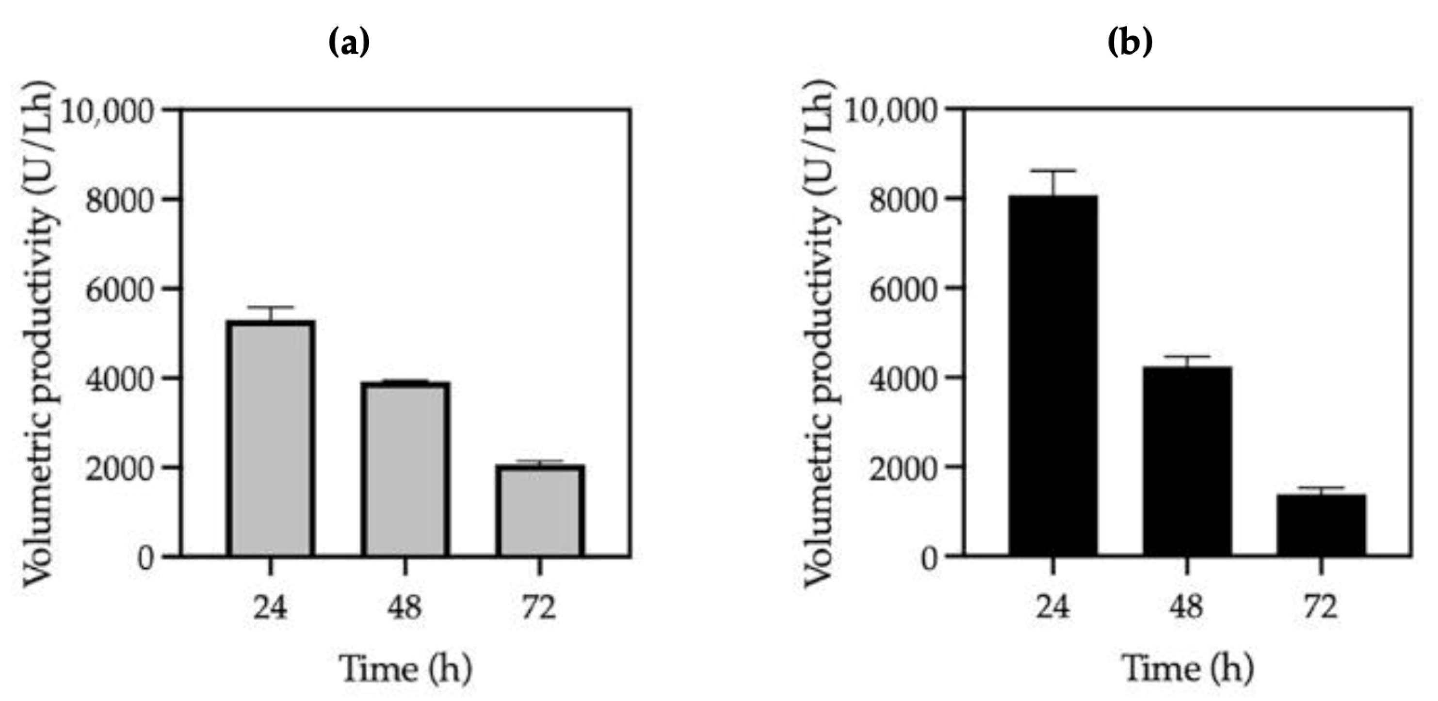
At optimal methanol concentrations (0.5% for KM71H ANCUT1 and 1.5% for KM71H ANCUT3), strain KM71H ANCUT3 showed a 1.6-fold higher biomass and 2-fold higher specific activity. While the volumetric activity and Qp of both strains at 48 h were similar and 8.0% higher for strain KM71H ANCUT3, the latter showed a 1.5-fold increase at 24 h (Figure 2).

3.2. Effect of Inoculum Concentration on Cutinase Production in KM71H Strains

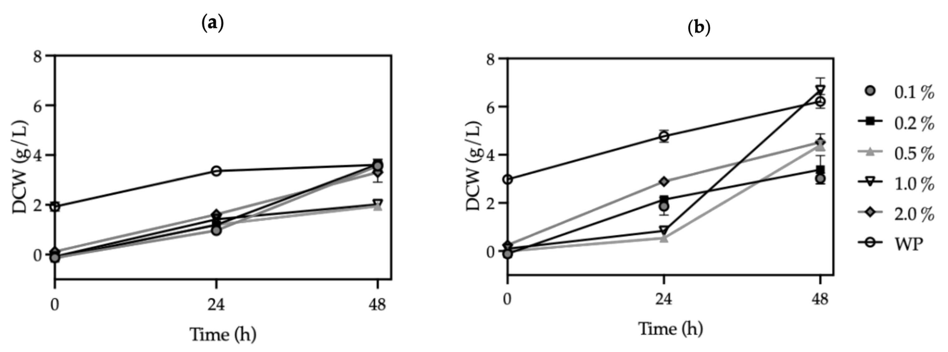
To determine the effect of the inoculum concentration on the cutinase production capacity of strains KM71H ANCUT1 and KM71H ANCUT3, they were grown in shake flasks with the BMMY medium supplemented with 0.5 and 1.5% methanol concentrations, respectively, at 29 °C with inoculum concentrations of 0.1, 0.2, 0.5, 1.0, and 2.0%. During the first 24 h, the cultures at the different inoculum concentrations behaved similarly. Subsequently, at 48 h, it was observed that cultures with 0.5 and 1.0% inoculum concentrations were the ones that reached the lowest biomass. On the other hand, when the pellet was added in its entirety, no increase in biomass was observed compared to the 0.1 and 0.2% inoculum concentrations (Figure 3a). Among the different inoculum concentrations evaluated (Figure S3), optimal ANCUT1 production was achieved with a 0.5% inoculum concentration, yielding a volumetric activity of 188 ± 10 U/mL, a specific activity of 1237 ± 145 U/mg, and a Qp of 3917 ± 208 U/Lh at 48 h (Table 2). As can be seen, these values are very similar to those presented previously when the effect of methanol on the growth and production of cutinases was evaluated; the difference lies in the biomass at which both experiments started (Figure 1a and Figure 3a).
Different growth behaviors were observed in the KM71H ANCUT3 strain. For instance, at 0.1 and 0.2% inoculum concentrations, the lowest amount of biomass was achieved, while cultures with 0.5 and 1.0% inoculum concentrations showed a similar growth profile, where a lag phase was observed during the first 24 h of culturing followed by exponential growth. Cultures with a 2.0% inoculum concentration or the whole pellet produced the highest biomass during the first 24 h and maintained linearity for up to 48 h of culturing (Figure 3b). Among the different inoculum concentrations evaluated (Figure S3), the optimal ANCUT3 production was achieved at a 2.0% inoculum concentration, resulting in 0.15 ± 0.00 mg/mL of soluble protein, a volumetric activity of 189 ± 6 U/mL, a specific activity of 1281 ± 36 U/mg, and a Qp of 3938 ± 125 U/Lh at 48 h (Table 2). While volumetric cutinase productivity and specific activities were similar, optimal inoculum concentrations and growth profiles differed significantly, suggesting distinct optimal cultivation conditions for each cutinase.
Strain KM71H ANCUT1 shows a better performance at lower inoculum concentrations, reaching the highest values at 0.5 and 1.0% inoculum concentrations (Figure S3) for most stoichiometric parameters, except for biomass and soluble protein concentration, which are 1.6 and 1.9-fold, respectively, when a 0.5% methanol concentration is used (Table 2). On the other hand, strain KM71H ANCUT3 responds better to higher inoculum concentrations (Figure S2), showing the highest values at a 2.0% inoculum concentration for almost all stoichiometric parameters, except for biomass concentration (Table 2).
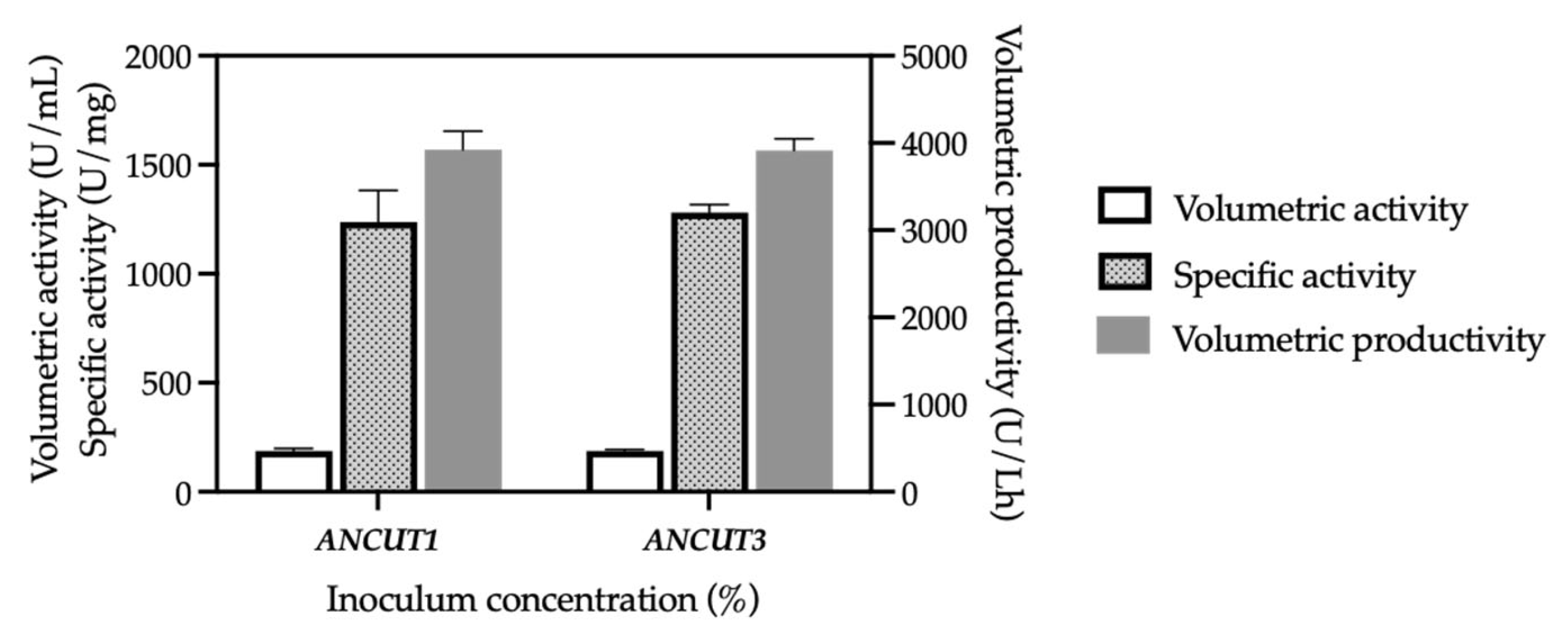
Both strains exhibited a comparable performance at their respective optimal inoculum concentrations (0.5% for KM71H ANCUT1 and 2.0% for KM71H ANCUT3) (Figure 4, Table 2).

3.3. Comparing Cutinase Production in K. phaffi MutS and Mut+ Strains

The cutinase production capacity of strain X-33 aox1::pPICZα-A-ANCUT1 (X-33 ANCUT1) and X-33 aox1::pPICZα-A-ANCUT3 (X-33 ANCUT3) was performed under conditions optimized for each KM71H strain: 0.5% methanol + 0.5% inoculum for KM71H ANCUT1 and X-33 ANCUT1 and 1.5% methanol + 2.0% inoculum for KM71H ANCUT3 and X-33 ANCUT3.
The growth difference between K. phaffii X-33 (Mut+) and KM71H (MutS) strains is evident when the ANCUT3 gene is expressed, showing a 1.7-fold increase in biomass for the X-33 ANCUT3 strain compared to the KM71H ANCUT3 strain at 48 h (Table 3). However, when the ANCUT1 gene is present in either phenotype, strains have no significant growth difference (Figure 5).
After 48 h of the production phase, strain KM71H ANCUT1 accumulated 24% more soluble protein but 43% lower volumetric activity and Qp and a 56% lower specific activity compared to strain X-33 ANCUT1 (Figure 6a and Table 3).
On the other hand, at a 1.5% methanol concentration, strains KM71H ANCUT3 and X-33 ANCUT3 accumulated the same amount of protein (Table 3), but KM71H ANCUT3 showed a 19% increase in the rest of the stoichiometric parameters compared to strain X-33 ANCUT3 (Figure 6b).

4. Discussion

Our results demonstrate a complex interplay of factors influencing cutinase production in K. phaffii KM71H strains, specifically underlining the impact of cutinase type, methanol concentration, and inoculum size. In this sense, the methanol concentration in cultures using K. phaffii as an expression system is one of the most relevant parameters influencing recombinant protein production [12] since it is both a carbon source and an inducer. Methanol may not be sufficient to initiate AOX1 transcription at low levels, while at high levels (3.0 g/L), it may be toxic to the cell [71]. For this reason, in this study, it was essential to determine the ideal concentration to produce cutinases.
The initial observation of similar growth patterns within the first 24 h across varying methanol concentrations highlights the strains’ initial metabolic robustness. However, the subsequent decrease in biomass with increasing methanol concentrations (>1%) indicates a toxicity threshold, consistent with previous research [71]. This toxicity is likely due to the accumulation of methanol metabolites, which impact cell viability.
Despite the shared sensitivity to high methanol levels, the optimal methanol concentration for cutinase production differs significantly between the KM71H ANCUT1 and KM71H ANCUT3 strains. The former performs best at lower methanol concentrations (0.5%), while the latter benefits from higher concentrations (1.5%). This suggests that the two cutinases have different metabolic demands or sensitivities to methanol or its byproducts. The higher specific activity of KM71H ANCUT3 at its optimal methanol concentration (Table 1) implies a more efficient enzyme production process under those conditions, while the observation that ANCUT1 achieves a higher specific activity and Qp at lower methanol concentrations is crucial from a process engineering perspective. Lower methanol concentrations result in reduced costs, decreased evaporation losses, and potentially simpler downstream processing. This suggests a potential economic and practical advantage for ANCUT1 production.
In addition, the results underline significant differences in optimal inoculum concentrations. While KM71H ANCUT1 production is maximized with a lower inoculum concentration (0.5%), KM71H ANCUT3 exhibits a superior performance at a significantly higher inoculum concentration (2.0%). These fundamental differences could be attributed to various factors, including nutrient competition, metabolic byproduct accumulation, or inherent differences in the growth kinetics of the strains expressing different cutinases. For instance, the higher biomass observed for KM71H ANCUT1 at 2.0% and the WP inoculum without a corresponding increase in other parameters, except for a rise in soluble protein, suggests a possible limitation in nutrient utilization or the accumulation of inhibitory byproducts or an insufficient oxygen transfer at high cell densities. Conversely, the optimal performance of KM71H ANCUT3 at a high inoculum concentration may reflect its ability to efficiently utilize resources under high-cell-density conditions (Table 2).
These results underline a critical trade-off between biomass and productivity, emphasizing the importance of carefully balancing inoculum size to avoid detrimental effects on cutinase activity.
Importantly, despite the contrasting optimal inoculum levels and methanol concentrations, both KM71H ANCUT1 and KM71H ANCUT3 strains ultimately yield a comparable overall cutinase production (as measured by volumetric activity, specific activity, and Qp). In summary, the former performs best at lower methanol (0.5%) and inoculum concentrations (0.5%), while the KM71H ANCUT3 strain requires higher levels of both (2.0% inoculum + 1.5% methanol).
The observed differences between KM71H ANCUT1 and KM71H ANCUT3 strains suggest that these parameters need to be individually optimized for each enzyme to achieve maximal productivity. This result highlights the critical need for a strain-specific optimization strategy rather than a one-size-fits-all approach.
Furthermore, these results reinforce the nuanced nature of cutinases within the broader class of esterases. While all cutinases exhibit esterase activity, their defining characteristic lies in their specialized ability to hydrolyze biopolyesters like cutin [62]. This study’s finding that ANCUT1 and ANCUT3, despite both being cutinases, show differential optimal conditions for production implicitly highlights the specific structural and functional adaptations that distinguish them. Unlike conventional esterases that typically hydrolyze water-soluble, short-chain esters and triglycerides primarily composed of fatty acids shorter than C6, cutinases possess unique active site configurations that enable efficient interactions with complex polymeric substrates including the hydrolysis of synthetic polyesters and triacylglycerols of varying chain lengths (ranging from C2–C14), probably with promiscuous polyesterase activity [24,51]. Nevertheless, their essential characteristic and defining feature remains their highly efficient action on cutin, setting them apart from other esterolytic enzymes. In addition, this substrate profile often positions cutinases as enzymatic intermediates between typical lipases and more generalized esterases. However, unlike lipases, cutinases have the catalytic triad located in a shallow binding cleft. This open and accessible active site enables them to efficiently engage with and hydrolyze high-molecular-weight hydrophobic molecules without the need for surface activation [72].
The differences observed between ANCUT1 and ANCUT3 production profiles may reflect subtle differences in their active site accessibility or substrate binding mechanisms, which are crucial for their specific cutinolytic function. A combination of these criteria, particularly substrate specificity, interfacial activation behavior (or its absence), and activity in low-water organic solvents, is often used to differentiate between esterases, lipases, and cutinases accurately.
After studying the performance of the KM71H strains producing cutinases ANCUT1 and ANCUT3, we decided to evaluate the ability of strain X-33 to produce the previously mentioned esterases and thus compare the differences between both strains of K. phaffii.
Methanol assimilation capacity plays a crucial role and has a significant impact on the overall efficiency of protein production within the cell. Specifically, the wild-type X-33 strain, which is characterized by the Mut+ phenotype, demonstrates a remarkable ability to achieve rapid growth when utilizing methanol as its primary carbon source for energy and essential nutrients. While K. phaffii KM71H presents a significantly different metabolic profile compared to the X-33 strain. A key difference lies in the fact that KM71H exhibits a deficiency in AOX1 gene activity. Because of this deficiency, KM71H is only capable of encoding the AOX2 enzyme, resulting in a considerably slower growth rate when cultured in a methanol-containing medium [14]. This metabolic constraint explains the observed differences in growth characteristics between the two strains. Specifically, the KM71H strain consumes a reduced amount of methanol in comparison to the X-33 strain. Consequently, the OD600 values are significantly lower in KM71H cultures when compared to those of the X-33 strain, reflecting a reduced biomass accumulation due to the limited methanol utilization. The comparison of Mut+ (X-33) and MutS (KM71H) strains reveals a significant impact of the methanol assimilation capacity on cutinase production, with Mut+ strains exhibiting a distinct response based on the specific cutinase expressed and its known sensitivity to high methanol levels. This sensitivity arises due to the accumulation of toxic byproducts generated during methanol metabolism, such as formaldehyde and peroxide. These compounds induce a state of increased oxidative stress within the cells, ultimately compromising their viability and hindering optimal protein production [12,71,73,74].
The superior performance of strain X-33 ANCUT1 is expected, as efficient methanol utilization leads to higher biomass and enzyme production. This strain achieved a 1.8-fold increase in volumetric activity and Qp and a 2.3-fold increase in specific activity compared to strain KM71H ANCUT1, using an inducer concentration of 0.5%. The latter is the lowest concentration used in this study. This result is promising as it may allow the use of lower methanol concentrations. Generally, the maximum product concentrations are reached at considerably lower methanol concentrations in fed-batch systems [71].
Strain KM71H ANCUT1 shows higher soluble protein levels but significantly lower volumetric activity, specific activity, and Qp compared to strain X-33 ANCUT1, a result that implies that a substantial portion of the produced protein is inactive or insoluble in the MutS strain. In contrast, KM71H ANCUT3, despite its much slower growth on methanol compared to the Mut+ strain, does not decrease its recombinant protein production, as reported elsewhere [75]. The unexpected comparable or even superior performance of this MutS strain is noteworthy, showing a 19% increase in volumetric activity, specific activity, and Qp, suggesting a more efficient conversion of biomass into cutinase than its Mut+ counterpart (Figure 6b). This challenges the assumption that efficient methanol utilization is always necessary for high cutinase yields, suggesting that slower growth rates in MutS strains may mitigate challenges associated with high-cell-density cultures during scale-up, as previously reported [76]. In addition, optimal growth conditions do not always correlate with maximal protein expression, and strain-specific metabolic and regulatory factors likely play a role. In the case of the ANCUT1-expressing strains, although X-33 and KM71H exhibit similar growth under optimal conditions, the higher ANCUT1 production in X-33 may be attributed to its Mut+ phenotype and fully functional AOX1 promoter, enabling a stronger methanol-induced expression. By contrast, KM71H relies instead on the weaker AOX2 promoter, which can lead to reduced transcriptional activity and protein yield.
Conversely, for the ANCUT3-expressing strains, the reduced production in X-33 ANCUT3, despite superior growth, may reflect a metabolic burden or bottleneck in secretion. Rapid cell growth can sometimes outpace the protein folding and processing capacity, lowering secretion efficiency. In KM71H, the slower growth may provide a more favorable environment for the proper folding and secretion of ANCUT3, leading to higher overall yields. We believe that future studies incorporating transcriptomic and proteomic analyses would help clarify these mechanistic differences.
In summary, the data underscore the importance of optimizing both methanol concentration and inoculum size for efficient cutinase production. The observed differences between production levels of KM71H ANCUT1 and KM71H ANCUT3 suggest that these parameters need to be individually optimized for each enzyme to achieve maximal productivity. Furthermore, the methanol assimilation capacity (Mut+/MutS) is cutinase-dependent, challenging assumptions about the direct relationship between rapid growth and high yield. These results highlight the importance of understanding the interplay between the expression system and the expressed protein.
A more detailed study about the structure and stability of mRNA and the newly synthesized protein might offer explanations for this phenomenon, as the coding macromolecules for these enzymes appear to be very different. For instance, the greater thermodynamic stability of the secondary structure of ANCUT1, the higher the GC content in ANCUT3, and the differences in CpG/GpC frequencies, as illustrated in Table S2, are all factors that could influence how these sequences interact with other molecules and, ultimately, their biological role. For example, these differences could affect the binding of transcription factors or DNA methylation, which in turn would affect the amount of protein produced from a gene. In this sense, it has been reported that modifying poly (dA:dT) tracts within the AOX1 promoter influences nucleosome organization, which in turn affects promoter strength and, consequently, protein expression [77]. Therefore, even though both cutinases are under the control of the same AOX1 promoter, subtle differences in the local DNA sequence (poly (dA:dT) tracts) around their respective coding sequences influence nucleosome positioning and accessibility to transcription factors and/or regulatory elements dictating their expression levels. This structural difference may explain the varying production levels observed in Figure 6, where ANCUT1 expression leads to more pronounced differences between the strains than ANCUT3 expression. Studies to corroborate this hypothesis are beyond the scope of this work. Experiments to examine parameters relating to scale up in bioreactors will be the subject of a subsequent publication.

5. Conclusions

This study provides crucial insights into optimizing recombinant cutinase production in K. phaffii, emphasizing that a universal approach is insufficient given the unique requirements of different enzymes and host strains. Our primary objective was to evaluate the performance of the MutS KM71H strain in producing recombinant cutinases ANCUT1 and ANCUT3 compared to the more commonly used Mut+ X-33, while also assessing the effects of methanol concentration and inoculum size. We demonstrated that while both KM71H strains (ANCUT1 and ANCUT3) exhibited similar initial growth responses and shared a methanol toxicity threshold, their optimal conditions for peak cutinase production diverged significantly. Specifically, KM71H ANCUT1 achieved maximal production at 0.5% methanol, yielding 188 U/mL volumetric activity, suggesting that lower methanol concentrations are economically and practically advantageous for this enzyme. In contrast, KM71H ANCUT3 production was optimal at a higher 1.5% methanol concentration, reaching 204 U/mL volumetric activity. This highlights inherent differences in the metabolic demands or sensitivities of the enzymes expressed to methanol and its byproducts. Furthermore, optimal inoculum concentrations varied markedly between the two KM71H strains: ANCUT1 performed best at a 0.5% inoculum concentration, while ANCUT3 exhibited superior performance at a significantly higher one (2.0%). Despite these differences in optimal production conditions, both KM71H strains ultimately yielded a comparable overall cutinase production (based on volumetric activity, specific activity, and Qp) at their respective optimized parameters, underscoring the critical need for individualized process optimization for each cutinase. A key finding addressing our objective of evaluating MutS viability as a host came from the comparison between MutS (KM71H) and Mut+ (X-33) strains. While X-33 ANCUT1 demonstrated a superior performance with a 43% higher volumetric activity and Qp and 56% higher specific activity compared to KM71H ANCUT1, the KM71H ANCUT3 strain remarkably showed a 19% increase in volumetric activity, specific activity, and Qp over X-33 ANCUT3. This unexpected, similar, or even superior performance of the MutS strain for ANCUT3 challenges the assumption that efficient methanol utilization (characteristic of Mut+ strains) is always necessary for high cutinase yields, suggesting that slower growth rates in MutS strains may offer advantages, such as mitigating challenges associated with high-cell-density cultures. In conclusion, our results strongly advocate strain- and enzyme-specific optimization strategies for cutinase production in K. phaffii. The observed performance differences, influenced by the methanol assimilation capacity and the specific cutinase expressed, challenge generalized assumptions about growth–yield relationships. Future research incorporating proteomic and metabolomic analyses will allow us to fully elucidate the underlying mechanistic differences driving these diverse production outcomes.

Supplementary Materials

The following supporting information can be downloaded at https://www.mdpi.com/article/10.3390/fermentation11080483/s1, Table S1: Recombinant cutinase-producing microorganisms; and Table S2: Gene and protein sequences from ANCUT1 and ANCUT3 from Aspergillus nidulans; Method S1: Nucleotide and expressed amino acid sequences; and Method S2: Statistical analysis (ANOVA); and Figure S1: SDS-PAGE. Protein profile (left side) and zymogram (right side) of the ultrafiltrate supernatant sample from KM71H and X-33 strains; and Figure S2: Effect of methanol concentrations on volumetric and specific activities of KM71H MutS strains; and Figure S3: Effect of inoculum concentrations on volumetric and specific activities of KM71H ANCUT1 and ANCUT3 strains.

Author Contributions

Conceptualization, A.F. and A.S.-R.; methodology, M.T.-G., K.E.L.-M., and J.A.C.-R.; validation, A.S.-R., M.T.-G., and K.E.L.-M.; formal analysis, A.S.-R., M.T.-G., K.E.L.-M., and J.A.C.-R.; investigation, A.S.-R., M.T.-G., K.E.L.-M., and J.A.C.-R.; writing—original draft preparation, A.S.-R. and A.F.; writing—review and editing, A.S.-R. and A.F.; funding acquisition, A.F. All authors have read and agreed to the published version of the manuscript.

Funding

This research was funded by Chemistry Faculty PAIP 5000-9095 and PAPIIT IT203118 DGAPA-UNAM. A.S.-R. is a postdoctoral SECIHTI fellow, grant 229329; J.A.C.-R. received SECIHTI scholarship 750551.

Data Availability Statement

The original contributions presented in the study are included in the article/Supplementary Materials, and further inquiries can be directed to the corresponding author.

Acknowledgments

The authors appreciate the technical support of Cindy Estrada and Sandra Pérez Munguía.

Conflicts of Interest

The authors declare no conflicts of interest. The funders had no role in the design of the study; in the collection, analyses, or interpretation of data; in the writing of the manuscript; or in the decision to publish the results.

References

  1. Yamada, Y.; Matsuda, M.; Maeda, K.; Mikata, K. The phylogenetic relationships of methanol-assimilating yeasts based on the partial sequences of 18S and 26S ribosomal RNAs: The proposal of Komagataella gen. nov. (Saccharomycetaceae). Biosci. Biotechnol. Biochem. 1995, 59, 439–444. [Google Scholar] [CrossRef] [PubMed]
  2. Khlebodarova, T.M.; Bogacheva, N.V.; Zadorozhny, A.V.; Bryanskaya, A.V.; Vasilieva, A.R.; Chesnokov, D.O.; Pavlova, E.I.; Peltek, S.E. Komagataella phaffii as a platform for heterologous expression of enzymes used for Industry. Microorganisms 2024, 12, 346. [Google Scholar] [CrossRef] [PubMed]
  3. Xu, R.; Chen, Z.; Chen, Y.; Wang, X.; Zhang, Y.; Li, X.; Wang, F. Multiple strategies for high-efficiency expression of Thermomyces lanuginosus lipase in Pichia pastoris and production of biodiesel in solvent-free system. Fuel 2023, 333, 126246. [Google Scholar] [CrossRef]
  4. Juturu, V.; Wu, J.C. Heterologous protein expression in Pichia pastoris: Latest research progress and applications. ChemBioChem 2018, 19, 7–21. [Google Scholar] [CrossRef] [PubMed]
  5. Shrivastava, A.; Pal, M.; Sharma, R.K. Pichia as yeast cell factory for production of industrially important bio-products: Current trends, challenges, and future prospects. J. Bioresour. Bioprod. 2022, 7, 209–218. [Google Scholar] [CrossRef]
  6. Barone, G.D.; Emmerstorfer-Augustin, A.; Biundo, A.; Pisano, I.; Coccetti, P.; Mapelli, V.; Camattari, A. Industrial production of proteins with Pichia pastoris—Komagataella phaffii. Biomolecules 2023, 13, 1085. [Google Scholar] [CrossRef]
  7. Unver, Y.; Dagci, I. Komagataella phaffii (Pichia pastoris) as a powerful yeast expression system for biologics broduction. Front. Biosci. (Elite Ed) 2024, 16, 19. [Google Scholar] [CrossRef]
  8. Lv, W.; Cai, M. Advancing recombinant protein expression in Komagataella phaffii: Opportunities and challenges. FEMS Yeast Res. 2025, 25, foaf010. [Google Scholar] [CrossRef]
  9. Looser, V.; Bruhlmann, B.; Bumbak, F.; Stenger, C.; Costa, M.; Camattari, A.; Fotiadis, D.; Kover, K. Cultivation strategies to enhance productivity of Pichia pastoris: A review. Biotechnol. Adv. 2015, 33, 1177–1193. [Google Scholar] [CrossRef]
  10. Liu, W.C.; Zhou, F.; Xia, D.; Shiloach, J. Expression of multidrug transporter P-glycoprotein in Pichia pastoris affects the host’s methanol metabolism. Microb. Biotechnol. 2019, 12, 1226–1236. [Google Scholar] [CrossRef]
  11. Cregg, J.M. Expression in the methylotrophic yeast Pichia pastoris. In Gene expression systems: Using nature e for the art of expression; Fernandez, J., Hoeffler, J., Eds.; Academic Press: San Diego, CA, USA, 1998; pp. 157–191. [Google Scholar]
  12. Ruo, P.A.; Leong, S.T.; Mooi, K.C.; Noorizan, M.; Boon, Y. Comparing the expression of human DNA topoisomerase I in KM71H and X33 strains of Pichia pastoris. Electron. J. Biotechnol. 2016, 21, 9–17. [Google Scholar] [CrossRef]
  13. Mayson, B.E.; Kilburn, D.G.; Zamost, B.L.; Raymond, C.K.; Lesnicki, G.J. Effects of methanol concentration on expression levels of recombinant protein in fed-batch cultures of Pichia methanolica. Biotechnol. Bioeng. 2003, 81, 291–298. [Google Scholar] [CrossRef] [PubMed]
  14. Pedro, A.Q.; Oppolzer, D.; Bonifácio, M.J.; Maia, C.J.; Queiroz, J.A.; Passarinha, L.A. Evaluation of MutS and Mut+ Pichia pastoris strains for membrane-bound catechol-O-methyltransferase biosynthesis. Appl. Biochem. Biotechnol. 2015, 18, 3840–3855. [Google Scholar] [CrossRef] [PubMed]
  15. Jia, L.; Gao, M.; Yan, J.; Chen, S.; Sun, J.; Hua, Q.; Ding, J.; Shi, Z. Evaluation of the sub-optimal induction strategies for heterologous proteins production by Pichia pastoris Mut+/MutS strains and related transcriptional and metabolic analysis. World J. Microbiol. Biotechnol. 2018, 34, 180. [Google Scholar] [CrossRef]
  16. Castro-Rodríguez, J.A.; Rodríguez-Sotres, R.; Farres, A. Determinants for an efficient enzymatic catalysis in Poly(Ethylene Terephthalate) degradation. Catalysts 2023, 13, 591. [Google Scholar] [CrossRef]
  17. Pio, T.F.; Macedo, G.A. Chapter 4 Cutinases: Properties and industrial applications. Adv. Appl. Microbiol. 2009, 66, 77–95. [Google Scholar] [CrossRef]
  18. Oliveira de Torres, C.; de Assis, M.A.; Mazutti, M.A.; Guimarães Pereira, G.C.; de Oliveira, D. Production of recombinant cutinases and their potential applications in polymer hydrolysis: The current status. Process Biochem. 2023, 134, 30–46. [Google Scholar] [CrossRef]
  19. Arnau, C.; Ramon, R.; Casas, C.; Valero, F. Optimization of the heterologous production of a Rhizopus oryzae lipase in Pichia pastoris system using mixed substrates on controlled fed-batch bioprocess. Enzym. Microb. Technol. 2010, 46, 494–500. [Google Scholar] [CrossRef]
  20. Wu, D.; Yu, X.W.; Wang, T.C.; Wang, R.; Xu, Y. High yield Rhizopus chinenisis prolipase production in Pichia pastoris: Impact of methanol concentration. Biotechnol. Bioprocess Eng. 2011, 16, 305–311. [Google Scholar] [CrossRef]
  21. Dutta, K.; Sen, S.; Veeranki, V.D. Production, characterization and applications of microbial cutinases. Process Biochem. 2009, 44, 127–134. [Google Scholar] [CrossRef]
  22. Nimchua, T.; Punnapayak, H.; Zimmermann, W. Comparison of the hydrolysis of polyethylene terephthalate fibers by a hydrolase from Fusarium oxysporum LCH I and Fusarium solani f. sp. pisi. Biotechnol. J. 2007, 2, 361–364. [Google Scholar] [CrossRef]
  23. Carvalho, C.M.L.; Aires-Barros, M.R.; Cabral, J.M.S. Cutinase structure, function and biocatalytic applications. Electron. J. Biotechnol. 1998, 1, 160–173. [Google Scholar] [CrossRef]
  24. Chen, S.; Su, L.; Chen, J.; Wu, J. Cutinase: Characteristics, preparation, and application. Biotechnol. Adv. 2013, 31, 1754–1767. [Google Scholar] [CrossRef] [PubMed]
  25. Wu, S.; Hooks, D.; Brightwell, G. Current understanding on the heterogenous expression of plastic depolymerising enzymes in Pichia pastoris. Bioengineering 2025, 12, 68. [Google Scholar] [CrossRef] [PubMed]
  26. Koschorreck, K.; Liu, D.; Kazenwadel, C.; Schmid, R.D.; Hauer, B. Heterologous expression, characterization and site-directed mutagenesis of cutinase CUTAB1 from Alternaria brassicicola. Appl. Microbiol. Biotechnol. 2010, 87, 991–997. [Google Scholar] [CrossRef]
  27. Farrés, A.; Peña-Montes, C.; Hernández-Domínguez, E.E.; Morales-García, S.L.; Sánchez-Sánchez, M.; Solís-Báez, I. Cutinasas Recombinantes de Aspergillus Nidulans Para Degradación de Poliésteres. WO-2017/204615 A2, 30 November 2017. [Google Scholar]
  28. Alvarado, E.; Castro, R.; Castro-Rodríguez, J.A.; Navarro, A.; Farres, A. Poly(lacticacid) degradation by recombinant cutinases from Aspergillus nidulans. Polymers 2024, 16, 1994. [Google Scholar] [CrossRef]
  29. Ahmed Al-Tammar, K.; Omar, O.; Abdul Murad, A.M.; Abu Bakar, F.D. Expression and characterization of a cutinase (AnCUT2) from Aspergillus niger. Open Life Sci. 2016, 11, 29–38. [Google Scholar] [CrossRef]
  30. Nyyssölä, A.; Pihlajaniemi, V.; Järvinen, R.; Mikander, S.; Kontkanen, H.; Kruus, K.; Kallio, H.; Buchert, J. Screening of microbes for novel acidic cutinases and cloning and expression of an acidic cutinase from Aspergillus niger CBS 513.88. Enzym. Microb. Technol. 2013, 52, 272–278. [Google Scholar] [CrossRef]
  31. Shirke, A.N.; Basore, D.; Butterfoss, G.L.; Bonneau, R.; Bystroff, C.; Gross, R.A. Toward rational thermostabilization of Aspergillus oryzae cutinase: Insights into catalytic and structural stability. PROTEINS: Struct. Funct. Bioinform. 2016, 84, 60–72. [Google Scholar] [CrossRef]
  32. Murguiondo, C.; Barriuso, J.; Prieto, A. Optimized enzymatic PLA hydrolysis by a recombinant fungal cutinase: A step toward a closed PLA cycle. Int. J. Biol. Macromol. 2025, 301, 140482. [Google Scholar] [CrossRef]
  33. Hu, X.; Zhaoying, G.; Zhanyong, W.; Tingting, S.; Lei, Y.; Ping, L. Enzymatic degradation of poly(butylene succinate) by cutinase cloned from Fusarium solani. Polym. Degrad. Stab. 2016, 134, 211–219. [Google Scholar] [CrossRef]
  34. Kwon, M.A.; Kim, H.S.; Yang, T.H.; Song, B.K.; Song, J.K. High-level expression and characterization of Fusarium solani cutinase in Pichia pastoris. Protein Expr. Purif. 2009, 68, 104–109. [Google Scholar] [CrossRef] [PubMed]
  35. Lienqueo, M.E.; Salazar, O.; Calado, C.R.C.; Fonseca, L.P.; Cabral, J.M.S. Influence of tryptophan tags on the purification of cutinase, secreted by a recombinant Saccharomyces cerevisiae, using cationic expanded bed adsorption and hydrophobic interaction chromatography. Biotechnol. Lett. 2008, 30, 1353–1358. [Google Scholar] [CrossRef] [PubMed]
  36. Gu, S.; Liu, C.; Zhang, W.; Qu, M.; Li, Y.; Zang, Y.; Xiong, X.; Pan, K.; Zhao, X. Characteristics of a recombinant Fusarium verticillioides cutinase and its effects on enzymatic hydrolysis of rice straw. Int. J. Biol. Macromol. 2021, 171, 382–388. [Google Scholar] [CrossRef] [PubMed]
  37. Seman, W.M.K.W.; Bakar, S.A.; Bukhari, N.A.; Gaspar, S.M.; Othman, R.; Nathan, S.; Mahadi, N.M.; Jahim, J.; Murad, A.M.A.; Bakar, F.D.A. High level expression of Glomerella cingulata cutinase in dense cultures of Pichia pastoris grown under fed-batch conditions. J. Biotechnol. 2014, 184, 219–228. [Google Scholar] [CrossRef]
  38. Hong, R.; Sun, Y.; Su, L.; Gu, L.; Wang, F.; Wu, J. High-level expression of Humicola insolens cutinase in Pichia pastoris without carbon starvation and its use in cotton fabric bioscouring. J. Biotechnol. 2019, 304, 10–15. [Google Scholar] [CrossRef]
  39. Duan, X.; Liu, Y.; You, X.; Jiang, Z.; Yang, S.; Yang, S. High-level expression and characterization of a novel cutinase from Malbranchea cinnamomea suitable for butyl butyrate production. Biotechnol. Biofuels 2017, 10, 223. [Google Scholar] [CrossRef]
  40. Wang, G.Y.; Michailides, T.J.; Hammock, B.D.; Lee, Y.-M.; Bostock, R.M. Molecular cloning, characterization, and expression of a redox-responsive cutinase from Monilinia fructicola (Wint.) Honey. Fungal Genet. Biol. 2002, 35, 261–276. [Google Scholar] [CrossRef]
  41. Vázquez-Alcántara, L.; Oliart-Ros, R.M.; García-Bórquez, A.; Peña-Montes, C. Expression of a Cutinase of Moniliophthora roreri with Polyester and PET-Plastic Residues Degradation Activity. Microbiol. Spectr. 2021, 9, e0097621. [Google Scholar] [CrossRef]
  42. Nyyssölä, A.; Pihlajaniemi, V.; Häkkinen, M.; Kontkanen, H.; Saloheimo, M.; Nakari-Setälä, T. Cloning and characterization of a novel acidic cutinase from Sirococcus conigenus. Appl. Microbiol. Biotechnol. 2014, 98, 3639–3650. [Google Scholar] [CrossRef]
  43. Gamerith, C.; Vastano, M.; Ghorbanpour, S.M.; Zitzenbacher, S.; Ribitsch, D.; Zumstein, M.T.; Sander, M.; Acero, E.H.; Pellis, A.; Guebitz, G.M. Enzymatic degradation of aromatic and aliphatic polyesters by P. pastoris expressed cutinase 1 from Thermobifida cellulosilytica. Front. Microbiol. 2017, 8, 938. [Google Scholar] [CrossRef] [PubMed]
  44. Carbios, S.B. Method for Recycling Plastic Products. U.S. Patent 10, 512 B2, 28 March 2019. [Google Scholar]
  45. Ulaganathan, N.; Dhodduraj, K.; Chandukishore, T.; Prabhu, A.A. Production of thermostable recombinant cutinase using mixed food waste: A sustainable approach toward environmental remediation. Chem. Eng. J. 2024, 496, 153892. [Google Scholar] [CrossRef]
  46. Su, L.; Hong, R.; Wu, J. Enhanced extracellular expression of gene-optimized Thermobifida fusca cutinase in Escherichia coli by optimization of induction strategy. Process Biochem. 2015, 50, 1039–1046. [Google Scholar] [CrossRef]
  47. Chen, S.; Liu, Z.; Chen, J.; Wu, J. Study on improvement of extracellular production of recombinant Thermobifida fusca cutinase by Escherichia coli. Appl. Biochem. Biotechnol. 2011, 165, 666–675. [Google Scholar] [CrossRef]
  48. Shirke, A.N.; Su, A.; Jones, J.A.; Butterfoss, G.L.; Koffas, M.A.G.; Kim, J.R.; Gross, R.A. Comparative thermal inactivation analysis of Aspergillus oryzae and Thiellavia terrestris cutinase: Role of glycosylation. Biotechnol. Bioeng. 2016, 114, 63–73. [Google Scholar] [CrossRef]
  49. Liu, M.; Yang, S.; Long, L.; Cao, Y.; Ding, S. Engineering a chimeric lipase-cutinase (Lip-Cut) for efficient enzymatic deinking of waste paper. BioResources 2018, 13, 981–996. [Google Scholar] [CrossRef]
  50. Duan, X.; Jiang, Z.; Liu, Y.; Yan, Q.; Xiang, M.; Yang, S. High-level expression of codon-optimized Thielavia terrestris cutinase suitable for ester biosynthesis and biodegradation. Int. J. Biol. Macromol. 2019, 135, 768–775. [Google Scholar] [CrossRef]
  51. Castro-Rodríguez, J.A.; Ramírez-Gonzalez, K.F.; Franco-Guerrero, F.; Sabido-Ramos, A.; Abundio-Sanchez, I.F.; Rodríguez-Sotres, R.; Rodríguez-Romero, A.; Farres, A. ANCUT1, a fungal cutinase MgCl2 -activated by a non-essential activation mechanism for Poly(ethylene terephthalate) hydrolysis. Catalysts 2025, 15, 757. [Google Scholar] [CrossRef]
  52. Li, X.; Shi, B.; Huang, J.W.; Zeng, Z.; Yang, Y.; Zhang, L.; Min, J.; Chen, C.C.; Guo, R.T. Functional tailoring of a PET hydrolytic enzyme expressed in Pichia pastoris. Bioresour. Bioprocess. 2023, 10, 26. [Google Scholar] [CrossRef]
  53. Fritzsche, S.; Hübner, H.; Oldiges, M.; Castiglione, K. Comparative evaluation of the extracellular production of a polyethylene terephthalate degrading cutinase by Corynebacterium glutamicum and leaky Escherichia coli in batch and fed-batch processes. Microb. Cell Fact. 2024, 23, 274. [Google Scholar] [CrossRef]
  54. Ma, H.-N.; Hsiang, C.-C.; Ng, I.-S. Tailored expression of ICCM cutinase in engineered Escherichia coli for efficient polyethylene terephthalate hydrolysis. Enzym. Microb. Technol. 2024, 179, 110476. [Google Scholar] [CrossRef]
  55. Tournier, V.; Topham, C.M.; Gilles, A.; David, B.; Folgoas, C.; Moya-Leclair, E.; Kamionka, E.; Desrousseaux, M.-L.; Texier, H.; Gavalda, S.; et al. An engineered PET depolymerase to break down and recycle plastic bottles. Nature 2020, 580, 216–219. [Google Scholar] [CrossRef]
  56. Sulaiman, S.; You, D.J.; Kanaya, E.; Koga, Y.; Kanaya, S. Crystal structure and thermodynamic and kinetic stability of metagenome- derived LC-cutinase. Biochemistry 2014, 53, 1858–1869. [Google Scholar] [CrossRef]
  57. Sulaiman, S.; Yamato, S.; Kanaya, E.; Kim, J.J.; Koga, Y.; Takano, K.; Kanaya, S. Isolation of a novel cutinase homolog with polyethylene terephthalate-degrading activity from leaf-branch compost by using a metagenomic approach. Appl. Environ. Microbiol. 2012, 78, 1556–1562. [Google Scholar] [CrossRef] [PubMed]
  58. Oh, Y.-R.-; Jang, Y.A.; Song, J.K.; Eom, G.T. Secretory production of an engineered cutinase in Bacillus subtilis for efficient biocatalytic depolymerization of polyethylene terephthalate. Bioprocess. Biosyst. Eng. 2022, 45, 711–720. [Google Scholar] [CrossRef] [PubMed]
  59. Shirke, A.N.; White, C.; Englaender, J.A.; Zwarycz, A.; Butterfoss, G.L.; Linhardt, R.J.; Gross, R.A. Stabilizing leaf and branch compost cutinase (LCC) with glycosylation: Mechanism and effect on PET hydrolysis. Biochemistry 2018, 57, 1190–1200. [Google Scholar] [CrossRef] [PubMed]
  60. Dutta, K.; Krishnamoorthy, H.; Dasu, V.V. Novel cutinase from Pseudomonas cepacia NRRL B 2320: Purification, characterization and identification of cutinase encoding genes. J. Gen. Appl. Microbiol. 2013, 59, 171–184. [Google Scholar] [CrossRef]
  61. Bermúdez-García, E.; Peña-Montes, C.; Castro-Rodríguez, J.A.; González-Canto, A.; Navarro-Ocaña, A.; Farrés, A. ANCUT2, a Thermo-alkaline cutinase from Aspergillus nidulans and its potential applications. Appl. Biochem. Biotechnol. 2017, 182, 1014–1036. [Google Scholar] [CrossRef]
  62. Bermúdez-García, E.; Peña-Montes, C.; Martins, I.; Pais, J.; Pereira, C.; Sánchez, S.; Farrés, A. Regulation of the cutinases expressed by Aspergillus nidulans and evaluation of their role in cutin degradation. Appl. Microbiol. Biotechnol. 2019, 103, 3863–3874. [Google Scholar] [CrossRef]
  63. Peña-Montes, C.; Bermúdez-García, E.; Castro-Ochoa, D.; Vega-Pérez, F.; Esqueda-Domínguez, K.; Castro-Rodríguez, J.A.; González-Canto, A.; Segoviano-Reyes, L.; Navarro-Ocaña, A.; Farrés, A. ANCUT1, a novel thermoalkaline cutinase from Aspergillus nidulans and its application on hydroxycinnamic acids lipophilization. Biotechnol. Lett. 2024, 46, 409–430. [Google Scholar] [CrossRef]
  64. Llanos-Reyes, A.C. Aplicación de las Cutinasas Recombinantes Ancut3 y Ancut4 Provenientes de Aspergillus nidulans en la Degradación de Poliésteres. Master’s Thesis, Universidad Nacional Autónoma de México, Ciudad de México, México, 2018. [Google Scholar]
  65. Lovera-Espinosa, D.; Torres-Jiménez, M.K. Optimización de Condiciones Para Mejorar la Producción y Degradación del Tereftalato de Polietileno (PET) por la Cutinasa ANCUT3 Expresada en Pichia pastoris. Bachelor’s Thesis, Universidad Nacional Autónoma de México, Ciudad de México, México, 2019. [Google Scholar]
  66. Castro-Ochoa, D.; Peña-Montes, C.; González-Canto, A.; Alva- Gasca, A.; Esquivel-Bautista, R.; Navarro-Ocaña, A.; Farrés, A. ANCUT2, an extracellular cutinase from Aspergillus nidulans induced by olive oil. Appl. Biochem. Biotechnol. 2012, 166, 1275–1290. [Google Scholar] [CrossRef] [PubMed]
  67. Invitrogen. pPICZα A, B, and C Pichia Expression Vectors for Selection on ZeocinTM and Purification of Secreted, Recombinant Proteins; Catalog, (V195-20): Carlsbad, CA, USA, 2010; pp. 1–38. Available online: https://tools.thermofisher.com/content/sfs/manuals/ppiczalpha_man.pdf (accessed on 10 July 2025).
  68. Esqueda-Domínguez, K.L. Producción, Identificación y Caracterización de una Cutinasa de Aspergillus nidulans PWI. Master’s Thesis, Universidad Nacional Autónoma de México, Ciudad de México, México, 2012. [Google Scholar]
  69. Bradford, M.M. A rapid and sensitive method for the quantitation of microgram quantities of protein utilizing the principle of protein-dye binding. Anal. Biochem. 1976, 72, 248–254. [Google Scholar] [CrossRef] [PubMed]
  70. Laemmli, U.K. Cleavage of structural proteins during the assembly of the head of bacteriophage T4. Nature 1970, 227, 680–685. [Google Scholar] [CrossRef] [PubMed]
  71. Zhang, W.; Inan, M.; Meagher, M.M. Fermentation strategies for recombinant protein expression in the methylotrophic yeast Pichia pastoris. Biotechnol. Bioprocess. Eng. 2000, 5, 275–287. [Google Scholar] [CrossRef]
  72. Chahinian, H.; Sarda, L. Distinction between esterases and lipases: Comparative biochemical properties of sequence-related carboxylesterases. Protein Pept. Lett. 2009, 16, 1149–1161. [Google Scholar] [CrossRef]
  73. Higgins, D.R.; Cregg, J.M. Methods in Molecular Biology: Pichia Protocols; Humana Press: Totowa, NJ, USA, 1998; pp. 1–15. [Google Scholar]
  74. Viader-Salvado, J.M.; Castillo-Galvan, M.; Fuentes-Garibay, J.A.; Iracheta-Cardenas, M.M.; Guerrero- Olazaran, M. Optimization of five environmental factors to increase beta-propeller phytase production in Pichia pastoris and impact on the physiological response of the host. Biotechnol. Prog. 2013, 29, 1377–1385. [Google Scholar] [CrossRef]
  75. Orman, M.A.; Çalık, P.; Özdamar, T.H. The influence of carbon sources on recombinant-human- growth-hormone production by Pichia pastoris is dependent on phenotype: A comparison of Muts and Mut+ strains. Biotechnol. Appl. Biochem. 2009, 52, 245–255. [Google Scholar] [CrossRef]
  76. Pla, I.A.; Damasceno, L.M.; Vannelli, T.; Ritter, G.; Batt, C.; Shuler, M.L. Evaluation of Mut+ and MutS Pichia pastoris phenotypes for high level extracellular scFv expression under feedback control of the methanol concentration. Biotechnol. Prog. 2006, 22, 881–888. [Google Scholar] [CrossRef]
  77. Yang, J.; Cai, H.; Liu, J.; Zeng, M.; Chen, J.; Cheng, Q.; Zhang, L. Controlling AOX1 promoter strength in Pichia pastoris by manipulating poly (dA:dT) tracts. Sci. Rep. 2018, 8, 1401. [Google Scholar] [CrossRef]
Figure 1. Effect of methanol concentrations on the growth of K. phaffii strains (a) KM71H ANCUT1 and (b) ANCUT3. Cultures were performed in shake flasks with BMMY medium at 29 °C. Error bars represent the standard deviation (n = 3).
Fermentation 11 00483 g001
Figure 2. Time course profile of volumetric cutinase productivity (Qp). (a) KM71H ANCUT1 and (b) KM71H ANCUT3. Cultures were performed in shake flasks with BMMY at 29 °C and supplemented with 0.5 and 1.5% methanol concentrations, respectively. Error bars represent the standard deviation (n = 3).
Fermentation 11 00483 g002
Figure 3. Effect of inoculum concentrations on the growth of K. phaffii strains. (a) KM71H ANCUT1 and (b) KM71H ANCUT3. Cultures were performed in shake flasks with BMMY medium at 29 °C and supplemented with 0.5 and 1.5% methanol concentrations, respectively. Error bars represent the standard deviation (n = 3). W.P.: whole pellet.
Fermentation 11 00483 g003
Figure 4. Comparison of stoichiometric parameters between KM71H ANCUT1 and KM71H ANCUT3 strains at 48 h. Cultures were performed in shake flasks with BMMY medium at 29 °C and supplemented with 0.5% methanol and 0.5% inoculum concentrations for ANCUT 1 and 1.5% methanol and 2.0% inoculum concentrations for ANCUT 3. Error bars represent the standard deviation (n = 3).
Fermentation 11 00483 g004
Figure 5. Growth profile of MutS and Mut+ cutinase-producing strains of K. phaffii. (a) KM71H ANCUT1 and X-33 ANCUT1. (b) KM71H ANCUT3 and X-33 ANCUT3. Cultures were performed in shake flasks with BMMY medium at 29 °C and supplemented with 0.5% methanol and 0.5% inoculum concentrations for ANCUT1 and 1.5% methanol and 2.0% inoculum concentrations for ANCUT3. Error bars represent the standard deviation (n = 3).
Fermentation 11 00483 g005
Figure 6. Comparison of stoichiometric parameters between MutS and Mut+ cutinase-producing strains of K. phaffii. (a) KM71H ANCUT1 and X-33 ANCUT1. (b) KM71H ANCUT3 and X-33 ANCUT3. Cultures were performed in shake flasks with BMMY medium at 29 °C and supplemented with 0.5% methanol and 0.5% inoculum concentrations for ANCUT1 and 1.5% methanol and 2.0% inoculum concentrations for ANCUT3. Error bars represent the standard deviation (n = 3).
Fermentation 11 00483 g006
Table 1. Comparison of stoichiometric parameters of KM71H aox1::pPICZα-A-ANCUT derivative strains at 0.5, 1.0, 1.5, 2.0, and 3.0% methanol concentrations in shake flasks *. Means and standard deviations of three technical replicate measurements are shown (n = 3).
StrainMethanol (%)Optical
Density
(600 nm)
Biomass (g/L)Protein Concentration (mg/mL)Volumetric
Activity (U/mL)
Specific
Activity (U/mg)
Productivity (U/Lh)
KM71H aox1::pPICZα-A-ANCUT10.57.78 ± 0.243.89 ± 0.130.18 ± 0.00188 ± 11037 ± 333917 ± 30
1.08.46 ± 0.524.11 ± 0.270.17 ± 0.01182 ± 71077 ± 873793 ± 148
1.58.19 ± 1.354.10 ± 0.700.15 ± 0.0080 ± 2535 ± 411673 ± 46
2.06.17 ± 0.473.05 ± 0.240.17 ± 0.0086 ± 1499 ± 71792 ± 25
3.05.26 ± 0.172.58 ± 0.090.15 ± 0.0080 ± 7523 ± 701673 ± 141
KM71H aox1::pPICZα-A-ANCUT30.58.97 ± 1.564.51 ± 0.810.10 ± 0.00132 ± 81262 ± 602729 ± 167
1.012.65 ± 2.076.42 ± 1.08 0.10 ± 00158 ± 91666 ± 2233271 ± 189
1.512.21 ± 0.116.20 ± 0.060.09 ± 0.00204 ± 102184 ± 484250 ± 208
2.010.91 ± 0.285.52 ± 0.150.10 ± 0.00174 ± 121721 ± 1723625 ± 250
3.06.84 ± 0.233.40 ± 0.120.09 ± 0.00113 ± 111129 ± 1452354 ± 229
* Data from 48 h of induction with an initial inoculum concentration of ≈ 1.0 OD600. Highlighted in bold are the data with the best parameters for each strain.
Table 2. Comparison of stoichiometric parameters of KM71H aox1::pPICZα-A-ANCUT derivative strains at 0.1, 0.2, 0.5, 1.0, and 2.0% inoculum concentrations in shake flasks *. Means and standard deviations of three technical replicate measurements are shown (n = 3).
StrainInoculum (%)Optical
Density
(600 nm)
Biomass (g/L)Protein Concentration (mg/mL)Volumetric Activity (U/mL)Specific Activity (U/mg)Productivity (U/Lh)
KM71H aox1::pPICZα-A-ANCUT1
(0.5% methanol)
0.17.14 ± 0.253.56 ± 0.130.13 ± 0.0099 ± 12779 ± 1062068 ± 247
0.27.27 ± 0.293.63 ± 0.150.13 ± 0.00140 ± 41106 ± 712910 ± 77
0.58.06 ± 0.164.04 ± 0.080.15 ± 0.01188 ± 101237 ± 1453921 ± 213
1.08.32 ± 0.184.17 ± 0.090.16 ± 0.02179 ± 91139 ± 1073720 ± 187
2.013.10 ± 1.476.66 ± 0.770.28 ± 0.0199 ± 6357 ± 352073 ± 131
WP14.23 ± 0.867.25 ± 0.450.10 ± 0.0199 ± 61022 ± 1322073 ± 131
KM71H aox1::pPICZα-A-ANCUT3
(1.5% methanol)
0.16.10 ± 0.443.02 ± 0.230.14 ± 0.00114 ± 5839 ± 442369 ± 109
0.26.8 ± 1.133.38 ± 0.590.13 ± 0.0090 ± 5688 ± 541867 ± 106
0.58.76 ± 0.404.40 ± 0.210.11 ± 0.00110 ± 81018 ± 972294 ± 163
1.013.15 ± 0.986.68 ± 0.510.10 ± 0.01116 ± 71155 ± 1252413 ± 137
2.08.99 ± 0.664.52 ± 0.350.15 ± 0.00188 ± 61281 ± 363916 ± 131
WP12.25 ± 0.556.22 ± 0.280.11 ± 0.00101 ± 7905 ± 472096 ± 150
* Data obtained after 48 h of induction. Highlighted in bold are the data with the best parameters for each strain. W.P.: whole pellet.
Table 3. Comparison of stoichiometric parameters between the MutS (KM71H ANCUT) and the Mut+ (X-33 ANCUT) derivative strains in shake flasks *. Means and standard deviations of three technical replicate measurements are shown (n = 3).
StrainMethanol (%)Inoculum (%)Optical
Density (600 nm)
Biomass
(g/L)
Protein Concentration (mg/mL)Volumetric
Activity
(U/mL)
Specific
Activity (U/mg)
Productivity (U/Lh)
KM71H ANCUT10.50.58.06 ± 0.164.04 ± 0.080.15 ± 0.01188 ± 101237 ± 1453921 ± 213
X-33ANCUT19.42 ± 0.584.74 ± 0.300.12 ± 0.00331 ± 82831 ± 1446887 ± 176
KM71H ANCUT31.52.08.99 ± 0.664.52 ± 0.350.15 ± 0.00188 ± 61281 ± 363916 ± 131
X-33ANCUT315.19 ± 0.877.74 ± 0.450.15 ± 0.00152 ± 81043 ± 613159 ± 175
* Data from 48 h of induction. Highlighted in bold are the data from strains with the best parameters.
Disclaimer/Publisher’s Note: The statements, opinions and data contained in all publications are solely those of the individual author(s) and contributor(s) and not of MDPI and/or the editor(s). MDPI and/or the editor(s) disclaim responsibility for any injury to people or property resulting from any ideas, methods, instructions or products referred to in the content.