Article

Diversity and Biological Activity of Secondary Metabolites Produced by the Endophytic Fungus Penicillium ochrochlorae

1
Chongqing Key Laboratory of Molecular Biology of Plant Environmental Adaptations, College of Life Science, Chongqing Normal University, Chongqing 401331, China
2
Key Scientific Research Base of Pest and Mold Control of Heritage Collection (State Administration of Cultural Heritage), Chongqing China Three Gorges Museum, Chongqing 400060, China
3
Chongqing Key Laboratory of Optical Fiber Sensor and Photoelectric Detection, Chongqing Engineering Research Center of Intelligent Optical Fiber Sensing Technology, Chongqing University of Technology, Chongqing 400054, China
*
Author to whom correspondence should be addressed.
Fermentation 2025, 11(7), 394; https://doi.org/10.3390/fermentation11070394
Submission received: 10 May 2025 / Revised: 2 July 2025 / Accepted: 8 July 2025 / Published: 10 July 2025

Abstract

In order to investigate bioactive natural products derived from the endophytic fungus Penicillium ochrochloron SWUKD4.1850, a comprehensive study focusing on secondary metabolites was conducted. This research led to the isolation of twenty distinct compounds, including a novel nortriterpenoid (compound 20), alongside nineteen compounds that had been previously characterized (compounds 119). The chemical structures of these compounds were elucidated using spectroscopic techniques and nuclear magnetic resonance (NMR) analyses. Compounds 117 were isolated for the first time as metabolites of P. ochrochloron. Except for compounds 114, significant structural similarity was discerned between the metabolites of the endophytic fungus and those of the host plant. Compound 20 is noted as the inaugural instance of a naturally occurring 27-nor-3,4-secocycloartane schinortriterpenoid, while compound 17 was identified in fungi for the first time. An antifungal assay showed that compound 10 displayed a broader antifungal spectrum and a stronger inhibitory effect towards four important plant pathogens, at inhibitory rates of 74.9 to 85.3%. The in vitro radical scavenging activities of compounds 1, 3, 8, 15, and 16 showed higher antioxidant activity than vitamin C. Moreover, a cytotoxic assay revealed that compound 20 had moderate cytotoxicity against the HL-60, SMMC-7721, and MCF-7 cell lines (IC50 6.5–17.8 μM). Collectively, these findings indicate that P. ochrochloron has abundant secondary metabolite synthesis ability in microbial metabolism and that these metabolites have good biological activity and have the potential to enhance plant disease resistance.

1. Introduction

Natural products serve as a vital reservoir for the development of novel pharmaceuticals, contributing to approximately two-thirds of small molecule drugs [1,2]. Fungi, in particular, have been extensively recognized for their exceptional ability to produce small molecule natural products; indeed, 28.32% of natural products are derived from fungal sources. They play a pivotal role in the discovery of bioactive compounds with unique structures [3,4]. These natural products exhibit a diverse array of chemical structures, including alkaloids, terpenoids, polyketides, steroids, benzene derivatives, and peptides [5], which encompass a wide range of structurally distinct scaffolds. This diversity endows them with extensive pharmacological activities, making them widely applicable in various fields such as medicine, agriculture, and food [6]. As a result, fungi have emerged as a primary and optimal source for the discovery and development of natural products.
Endophytic fungi emerge as promising reservoirs of bioactive natural compounds. Interestingly, plants that have adapted to host environmental conditions harbor a plethora of microbes producing a variety of bioactive natural products that can be assessed for potential biological activity [7]. Considering that endophytes have had long-term interactions with the host during the evolutionary adaptation process, endophytes possess the capability to synthesize analogs of host compounds and to modify and transform host-derived metabolites [8,9]. For instance, the endophytic fungus Ceriporia lacerate hs-zjut-c13a, isolated from Huperzia serrata, has been shown to convert huperzine into five distinct compounds and three sesquiterpenoids, with huptremules A-D exhibiting characteristics of functional metals and exogenous substrates (HupA) [10]. Similarly, the endophytic fungus Irpex lacteus has been reported to transform huperzine A into 8α,15α-epoxyhuperzine A [11]. Furthermore, co-fermentation of the endophytic fungus Umbelopsis dimorpha with the host plant Kadsura angustifolia and wheat bran has resulted in the production of a variety of new host analogs, including triterpenes, sesquiterpenes, and monoterpenes [12]. These findings underscore the potential of metabolic mimicry between endophytes and host plants as a promising strategy for the discovery of novel bioactive compounds.
Endophytic fungi, especially the Penicillium species, possess unique metabolic pathways to produce secondary metabolites with novel structures and potent biological activities, such as cytotoxic, antifungal, antibacterial, anti-inflammatory, and ɑ-glucosidase-inhibitory activities [13]. These secondary metabolites with novel structures and potent bio-activities will continue to guide the separation or synthesis of structurally novel and biologically active compounds and will offer leading compounds for the development and innovation of pharmaceuticals and pesticides [14]. In recent years, we have been focused on the exploration of secondary metabolites derived from Penicillium species associated with Schisandraceae plants [15]. P. ochrochloron has been extensively recognized as a producer of mycotoxins, including penitrems A-F, bromopenitrem A, bromopenitrem F, and dehydropenitrem D [16]. Additionally, it synthesizes compounds with antimicrobial properties, such as 2,3,4-trihydroxybutanamide and erythritol [17]. Our prior investigations have demonstrated that the endophytic fungus P. ochrochloron SWUKD4.1850 exhibits notable secondary metabolism and biotransformation capabilities. When cultured with host plant materials, it can metabolize them to generate host analogs, transform triterpenes into oxygenated terpenes, and enhance their biological activity [18,19,20]. Furthermore, a range of bioactive resorcylic acid lactones has been successfully isolated from the solid culture of P. ochrochloron, including 5α, 6β-acetonide-aigialomycin B, penochrochlactones A-B, 4-O-desmethyl-aigialomycin B, penochrochlactones C-D, aigialomycin F [21], aigialomycin A [22], aigialomycin B [22], aigialomycin D [22], zeaenol [23], and oxozeaenol [24]. In this study, continuing our previous work [25], we update the chemical diversity and biological activity of secondary metabolites produced by P. ochrochloron SWUKD4.1850. The current research details the isolation, structural characterization, and evaluation of the cytotoxic, antioxidant, and antifungal effects of all metabolites.

2. Materials and Methods

2.1. General

Analytical TLC was carried out on silica gel GF254 plates, which were obtained from Qingdao Marine Chemical Factory, Qingdao, China. Visualization of chromatographic spots was achieved through dual detection: initial observation under a UV254 lamp followed by post-spray thermal development. The derivatization procedure involved spraying plates with 5% sulfuric acid in ethanol solution prior to heat treatment for color development. Column chromatographic separations utilized two distinct stationary phases: 100–200 mesh silica gel (Qingdao Marine Chemical, Factory, China) for normal phase chromatography and Sephadex LH-20 (Amersham Pharmacia Biotech AB, Uppsala, Sweden) for size-exclusion chromatography. Optical rotation measurements were performed on a Jasco P-1020 polarimeter (JASCO Corporation, Tokyo, Japan), providing specific rotation data for chiral compound analysis. Infrared spectral data were acquired using a Bruker Tensor-27 spectrophotometer (Bruker Company, Kalkar, Germany), with samples prepared as potassium bromide pellets to facilitate mid-infrared transmission measurements. Ultraviolet–visible absorption spectra were recorded on a Shimadzu UV2401PC spectrophotometer, enabling quantitative and qualitative analysis of chromophoric groups. Nuclear magnetic resonance (NMR) spectroscopy was performed on Bruker spectrometers operating at 400 MHz (AM-400) and 600 MHz (AVANCE III-600) (Bruker BioSpin Gm BH, Rhein-stetten, Germany), acquiring both 1D proton/carbon spectra and 2D correlation datasets (HSQC, HMBC, NOESY) for structural elucidation. Electron ionization mass spectrometry (EI-MS) data were generated using a VG AutoSpec 3000 mass spectrometer (VG Instruments, East Sussex, UK), while high-resolution mass spectrometry (HR-MS) with electrospray ionization-time of flight (ESI-TOF) detection provided accurate mass measurements for molecular formula determination.

2.2. Fungal Material

The endophytic fungal strain P. ochrochloron SWUKD4.1850 was first extracted from healthy K. angustifolia plants gathered in early June 2012 in Maguan County, Yunnan Province, China. Identification of isolate SWUKD4.1850 as P. ochrochloron was achieved through detailed examination of its morphological features and sequencing of the ITS region, as outlined in our earlier study [18,19]. Public access to genetic resource information is facilitated by the deposition of this fungal strain’s genomic sequencing data in GenBank under accession number KX346178.

2.3. Fungal Fermentation, Chemical Extraction, and Purification

P. ochrochloron SWUKD4.1850 was cultivated on PDA at 28 °C for 5 days, after which the agar containing the mycelia was sliced into twenty 5 mm pieces and placed into 500 mL Erlenmeyer flasks with 100 g of sterilized moist wheat bran medium, which was modified to 60–70% (w/v) using distilled water with 1% (w/v) CaCO3. After 50 days of culture at 28 °C, the fermented rice medium was dried at 60 °C and extracted with methanol (5 × 1000 mL) three times. All dried methanol extracts were extracted with acetone for 4 days, repeated thrice, until TLC analysis showed no visible spots in the extracts. The solvent was evaporated under reduced pressure at 60 °C to obtain 76 g of metabolic crude extract.
The crude extract was separated by MCI column chromatography with a CH3OH-H2O gradient (1:9–1:0, 25 mL/min, 12 h), and the metabolites were divided into eight components (fraction A-H). The Fr.D component (550 mg) was loaded onto a methanol gel column and eluted with CH3OH-H2O 4:6 (v/v) to obtain three subfractions (Fr.D1~Fr.D3). Compounds 1 (55 mg), 3 (5 mg), and 5 (15 mg) were obtained by gradient elution on a silica gel chromatography column (CH2CL2-CH3OH, 3:7–4:6). Fr.E (450 mg) was loaded onto a methanol gel column and eluted with CH3OH-H2O 1:1 to obtain four fractions (Fr.E1~Fr.E4). Fr.E1 (175 mg) was further separated by HPLC (Agilent Eclipse XDB-C8, 250 × 9.4 mm, 5 µm column, 2.0 mL/min) and eluted with 25–60% MeOH/H2O for 30 min, then with 100% MeOH for 35 min, to yield compounds 2 (tR 9.87 min, 7.6 mg), 4 (tR 11.39 min, 15 mg), 6 (tR 14.23 min, 14 mg), 11 (tR 18.42 min, 4.1 mg), 12 (tR 20.14 min, 12.3 mg), and 9 (tR 23.16 min, 15 mg). Fr.E3 (400 mg) was further separated by HPLC (Agilent Eclipse XDB-C8, 250 × 9.4 mm, 5 µm column, 2.0 mL/min) and eluted with 30–70% MeOH/H2O for 30 min, then with 100% MeOH for 35 min, to yield compounds 8 (tR 12.46 min, 5.1 mg), 7 (tR 16.53 min, 11 mg), 18 (tR 19.28 min, 50 mg), 19 (tR 21.43 min, 9 mg), and 20 (tR 25.01 min, 7 mg). The component Fr.F (914 mg) was separated on a silica gel chromatography column (CH2CL2-CH3OH, 6:4–4:6) and further separated by HPLC (35–75% CH3OH-H2O, 2 mL/min, 30 min), then eluted with 100% MeOH for 35 min, to yield compounds 10 (tR 8.52 min, 5.0 mg), 15 (tR 10.13 min, 7 mg), 16 (tR 13.40 min, 7.9 mg), 17 (tR 16.18 min, 4.9 mg), 13 (tR 19.12 min, 6.9 mg), and 14 (tR 22.69 min, 44.8 mg).

2.4. Spectroscopic Data of the New Compound

Penioch A (20): colorless oil; [ α ] D 24.2 + 13.1° (c 0.16, CHCl3); λmax (log ε) 212 (3.87) nm; IR (KBr) υmax 3451, 2937, 1743, 1633, 1440, 1384, 1269, 1191, 1038, 886 cm−1; 1H and 13C NMR data (see Table 1), HRESIMS m/z 479.3493 [M + Na]+ (calcd for C30H48O3Na, 479.3496).

2.5. Antifungal Activity Assay

The antifungal activity of the compounds was initially tested against four significant plant pathogenic fungi: Verticillium dahliae, Botrytis cinerea, Rhizoctonia solani, and Fusarium oxysporum. These fungal strains were provided by Chongqing Normal University. Mycelial radial growth inhibition assays were conducted on potato dextrose agar (PDA) to assess pathogen sensitivity to the test compounds. Briefly, 6 mm diameter mycelial plugs were excised from the leading edge of colonies that had been actively growing for 4 days using a flame-sterilized cork borer. Each plug was placed centrally in a 90 mm diameter Petri dish, with the mycelial surface in direct contact with the agar medium. Antifungal activities were evaluated at a concentration of 50 µg/mL using the mycelial growth inhibition rate method [26]. The preparation of a negative control involved adding 20 mL of methanol, with the process of solvent evaporation carried out in a laminar flow cabinet to uphold sterility. The plates underwent incubation at 28 °C for a duration of 3 to 5 days, with incubation duration adjusted according to each specie’s growth rate, until negative control colonies completely covered the plate surface. Carbendazim and boscalid served as positive control compounds [27,28]. Measurements of colony diameters for each treatment were conducted in two perpendicular directions, and each condition was tested in triplicate.

2.6. Antioxidative Assay

The radical scavenging activity of DPPH (1,1-diphenyl-2-picrylhydrazyl) (Sigma-Aldrich, St. Louis, MO, USA) was used to assess antioxidant activity [29]. For each assay, 10 mM of each compound isolated from P. ochrochloron SWUKD4.1850 (1 µL) was evaluated, followed by 49 µL of MeOH in each well. Subsequently, 50 µL of 0.3 mM DPPH solution (Sigma-Aldrich) was added. The final concentration was tested to 0.1 mM. The mixture was incubated for 10 min, and antioxidant activity was evaluated by measuring UV at 550 nm using a Dynatech MR 500 plate reader (Thermo Fisher Scientific, Waltham, MA, USA). The percentage of inhibition was calculated for each sample at a concentration of 100 µg/mL. The IC50 value was tested with different concentrations of samples that showed 50% antioxidant activity. Vitamin C and vitamin E were used as positive controls to confirm the assay system.

2.7. Cytotoxic Assay

MTT assay was used to assess the in vitro cytotoxic effects of the test compounds on four human cancer cell lines––HL-60 (human myeloid leukemia), SMMC-7721 (hepatocellular carcinoma), A-549 (lung adenocarcinoma), and MCF-7 (breast adenocarcinoma)––following a previously established protocol [30]. The Cell Bank of the Shanghai Institute of Biochemistry and Cell Biology provided all the cell lines. Cell cultures were sustained in DMEM (Dulbecco’s modified eagle medium) enriched with 10% FBS, 100 U/mL penicillin, and 100 μg/mL streptomycin and incubated at 37 °C in a humidified environment with 5% CO2. For cytotoxicity testing, cells were seeded into 96-well plates at densities of 6000 cells/well (HL-60, SMMC-7721, A-549) or 9000 cells/well (MCF-7) and allowed to adhere overnight. Compounds for testing, dissolved in pure (AR) dimethyl sulfoxide (DMSO) and sterilized by passing through 0.45 μm syringe filters, were added in a series of dilutions, and the plates were maintained under standard culture conditions for a duration of 72 h. Following treatment, each well received 20 μL of MTT solution at 0.5 mg/mL in phosphate-buffered saline, and the plates were incubated for an additional 4 h to facilitate formazan crystal formation. Subsequently, 150 μL of DMSO was added to solubilize the crystals, and absorbance was measured using a microplate reader at 545 nm and 690 nm. Relative cell viability was calculated based on formazan production, and the following formula was used to calculate the percentage of cell proliferation inhibition: inhibition (%) = (ODDMSOODcompound)/ODDMSO × 100%. Cisplatin, with a purity of at least 98%, was used as the positive control. Three independent replicates were performed for all experiments.

3. Results

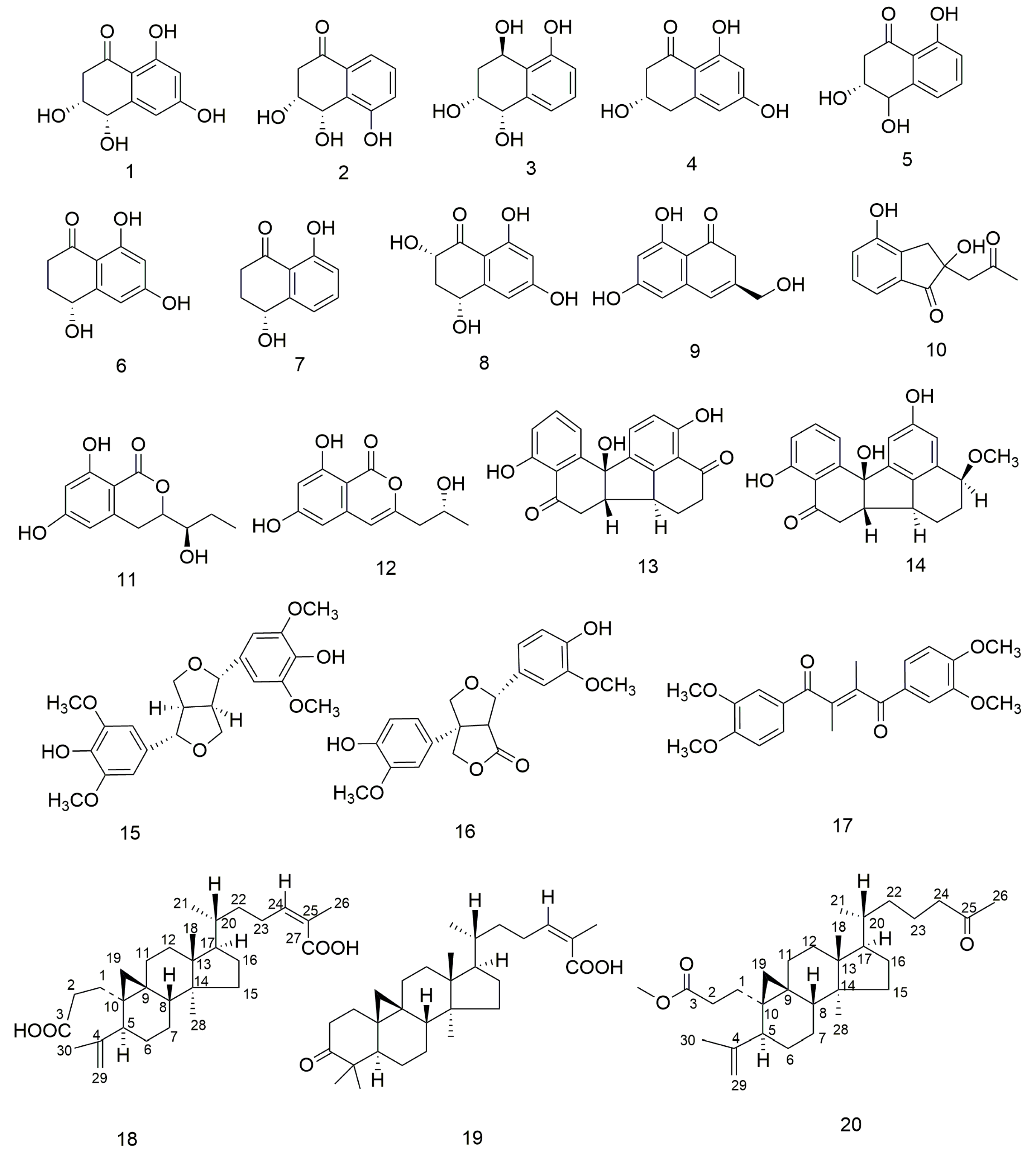
3.1. Structure Elucidation

The metabolic crude extract of P. ochrochloron SWUKD4.1850 led to the isolation of one previously undescribed nortriterpenoid, penioch A (20), alongside nineteen previously identified compounds (119). The known compounds were identified as having the structures 4-hydroxyscytalone (1) [31], 3,4,5-trihydroxy-1-tetralone (2) [32], 1,2,3,4-tetrahydro-1,2,4,5-naphthalenetetrol (3) [33], scytalone (4) [34], 3,4,8-trihydroxytetralone (5) [35], plecmillin F (6) [36], regiolone (7) [37], hydroxyisosclerone (8) [38], pyrenomycin (9) [39], indandione B (10) [40], orthosporin (11) [41], eupenicillin A (12) [42], daldinone C (13) [43], daldinone D (14) [43], sringaresinol (15) [44], ketopinoresinol (16) [45], (Z)-1,4-bis (3′,4′-dimethoxyphenyl)-2,3-dimethylbut-2-ene-1,4-dione (17) [46], nigranoic acid (18) [47], and schisandronic acid (19) [48] (Figure 1), by evaluating their optical rotation, NMR, and MS characteristics compared to the literature.
Compound 20 was obtained as colorless oil (CHCl3) and possessed the molecular formula C30H48O3 derived from its HR-ESI-MS analysis (m/z 479.3493 [M + Na]+, calcd for C30H48O3Na 479.3496), indicating seven unsaturated bonds. The 1H-NMR spectrum (Table 1) of 20 showed signals for four singlet methyls at H 0.92 (3H, s), 0.95 (3H, s), 1.67 (3H, s), and 2.14 (3H, s), one doublet methyl at H 0.88 (3H, d, J = 6.3 Hz), one methoxyl at H 3.64 (3H, s), two olefinic protons at H 4.73 (1H, s) and 4.81 (1H, s), and a pair of characteristic methylene protons from cycloartane [H 0. 40 and 0.72 (each 1H, d, J = 4.4 Hz)]. Using 13C-NMR along with DEPT experiments, 30 carbon resonances were identified, corresponding to one methoxyl, five methyls, thirteen methylenes (one olefinic), four methines, and seven quaternary carbons (two carbonyls and one olefinic). The molecular formula indicated seven hydrogen deficiency indices, yet only one ketone at C 209.6 (C-25), one carboxyl group at δC 174.5 (C-3), and two olefinic carbons at δC 149.5 (C-4) and 111.5 (C-5) were found, revealing that 20 had a structure with four cycles. Detailed examination of the 1D and 2D (Figure S1) NMR data revealed that 20 possessing the same 3,4-secocycloartane framework as the predominant triterpenoid-nigranoic acid (18) [49]. However, the analysis also uncovered notable differences in functional groups and structures between them. The resonances of the trisubstituted C-24/C-25 double bond and the characteristic C-27 carbonyl carbon resonance in the side chain of 18 were absent in penioch A. Instead, methyl ketone resonances were observed for 20. The 1H-1H COSY spectrum analysis, beginning with a methyl doublet at δH 0.88 (CH3–21), identified a spin system comprising CH3-21/H-20/H2-22/H2-23/H2-24. Coupled with the HMBC correlations of CH3-26 with C-24 (δC 44.3, t) and C-25 (δC 209.6, s) and CH3-21 with C-17 (δC 52.0, d), C-20 (δC 35.9, d), and C-22 (δC 24.9, t), this supported the assignment of 20 as a 27-nor-3,4-secocycloartane schinortriterpenoid. Furthermore, thorough examination of its NOESY spectrum and similar chemical shifts, it was established that the relative configuration of 20 matches those of 18 and 19 (Figure 2). Consequently, the configuration of 20 was confirmed as depicted, and this compound has been designated as penioch A (20).

3.2. Antifungal Assay

For antifungal activity assessment (Table 2), compounds 120 were tested against four significant agricultural phytopathogenic fungi (V. dahliae, B. cinerea, R. solani, F. oxysporum) at 50 μg/mL. Among all tested compounds, only compound 10 showed a wider antifungal range and a more potent inhibitory effect against all the plant pathogens examined; the inhibitory rate was 74.9 to 85.3%. For B. cinerea, compound 4 showed the best inhibition rate, which was 87.4% at 50 μg/mL and better than boscalid. In a similar fashion, compounds 912 demonstrated notable activity, with inhibitory rates between 60.2% and 78.4%. For R. solani, compound 4 showed the best inhibition rate, which was 85.3% at 50 μg/mL. For F. oxysporum, compound 11 achieved an inhibition rate of 87.9% at 50 μg/mL, outperforming boscalid. In a similar manner, compound 12 achieved an 82.6% inhibition rate, indicating strong antifungal activity. However, other compounds (1520) showed weak antifungal activity.

3.3. Antioxidant Analysis

The natural products extracted from Penicillium spp. have been reported to have good antioxidant activity [29]. In this research, the DPPH (1,1-diphenyl-2-picrylhydrazyl) free radical assay was used to evaluate the antioxidant properties of compounds 120 (Table 3). Among them, compounds 1820 exhibited over 90% scavenging activity at a concentration of 100 µM. Notably, the activities of compounds 1 (IC50 of 30.1 µM), 3 (IC50 of 33.5 µM), 8 (IC50 of 29.7 µM), 15 (IC50 of 25.7 µM), and 16 (IC50 of 21.8 µM) were higher than that of vitamin C. Compounds 2 (IC50 of 53.9 µM), 4 (IC50 of 42.9 µM), 5 (IC50 of 43.1 µM), 6 (IC50 of 50.9 µM), and 9 (IC50 of 52.1 µM) exhibit moderate antioxidant activity, with higher activity than vitamin E.

3.4. Cytotoxicity Assay

Numerous natural products extracted from Schisandraceae family plants are known to lower the risk of liver diseases and various cancers [15,49,50]. Therefore, all the compounds (120) were evaluated for in vitro cytotoxicity against the HL-60, SMMC-7721, A549 and MCF-7 cell lines, with cisplatin serving as the positive control. Consequently, compound 20 showed moderate cytotoxic activity, with IC50 values ranging from 6.5 to 9.3 μM for the HL-60, SMMC-7721, and MCF-7 cell lines. However, compounds 1, 3, 8, 15, and 16 showed weak cytotoxic activity, with IC50 values ranging from 23.4 to 39.2 μM for the HL-60, SMMC-7721, A549, and MCF-7 cell lines. Compounds 2, 47, 914, and 1719 were not active (IC50 > 40 μM) in any of the cell lines (Table 4).

4. Discussion

Endophytic fungi, particularly those inhabiting specialized host environments, have been proven as promising and prolific sources of bioactive secondary metabolites with potent bioactivity applications [15]. Host plants, as unique habitats, offer favorable conditions for the colonization of fungi. These fungi are able to produce natural compounds with novel structures and significant activity [1,50]. In continuation of our work [25], we undertook a comprehensive study that concentrated on compounds 120 derived from P. ochrochloron originating from the host plant K. angustifolia. In the thin-layer chromatography (TLC) analysis of various samples (Figure S2), the methanol crude extract derived from the solid fermentation product of P. ochrochloron exhibited significant differences compared to the methanol crude extracts of its co-fermented substrates, wheat bran (Figure S2 (a)) and rice (Figure S2 (b)). These findings corroborate the assertion that P. ochrochloron actively metabolized and synthesized a substantial quantity of secondary metabolites, as well as chemical constituents analogous to those of the host plant (Figure S2 (b,N)). These compounds were categorized primarily by their structural classifications, which included polyketides (114), lignans (1517), and schitriterpenoids/norschitriterpenoids (1820). Among these compounds, compounds 114 are precursors or intermediate products of resorcylic acid lactones, which were isolated from P. ochrochloron SWUKD4.1850 in our previous study [25]. According to literature reports, polyketide compounds similar to compounds 114 were previously extracted from the culture filtrates of various endophytic fungi, including Leptosphaeria maculans [51], Pyricularia oryzae [52], Phaeosphaeria sp. [53], and Xylaria sp. [54,55]. Additionally, three lignans (1517) and three schitriterpenoids/norschitriterpenoids (1820) have been reported as the principal bioactive constituents of the host plant K. angustifolia [18,19,46,47,48]. Notably, compound 20 possesses a novel structural skeleton, as the first 27-nor-3,4-secocycloartane schinortriterpenoid, with limited prior reports on the isolation of compounds with this particular skeletal structure. Our research confirmed the substantial potential of P. ochrochloron in the production of secondary metabolites, highlighting its considerable untapped potential for medicinal and agrochemical applications.
Numerous studies have reported that secondary metabolites of Penicillium species have a wide range of biological activities, including antibacterial, antifungal, anticancer, antioxidant, antiviral, immune-suppressive, cholesterol-lowering, and anti-inflammatory effects [56,57,58]. Notably, in this study, compounds 120 from P. ochrochloron showed a broad spectrum of biological activity. Polyketides represent a significant class of secondary metabolites produced by Penicillium species, distinguished by their notable structural and functional diversity [13]. Polyketide compounds 4 and 912 exhibited significant antifungal activity. Notably, compound 10 demonstrated a broader spectrum of antifungal efficacy and a more potent inhibitory effect against all tested plant pathogens, with inhibition rates ranging from 74.9% to 85.3%. Compound 4 achieved the highest inhibition rate of 85.3% at a concentration of 50 μg/mL against R. solani. Compound 11 attained an inhibition rate of 87.9% at 50 μg/mL against F. oxysporum. Additionally, the lignans of benzo[j]fluoranthene metabolites have been reported to have good antioxidant activity [59]. Compound 17 was identified in fungi for the first time and exhibited antioxidant activity higher than vitamin E. Moreover, numerous natural products extracted from Schisandraceae family plants are known to lower the risk of liver diseases and various cancers [46,47,48]. Compound 20 showed moderate cytotoxic activity, with IC50 values ranging from 6.5 to 9.3 μM for the HL-60, SMMC-7721 and MCF-7 cell lines. In summary, Penicillium species are a significant source of novel compounds and a diverse array of secondary metabolites exhibiting a broad spectrum of biological activities. In this research, active compounds have only been studied at the in vitro level, with a lack of thorough investigation into their in vivo efficacy and the mechanisms of action. In our further studies, we should also focus on the mechanism of action of active compounds to obtain active lead compounds. This strategy will facilitate the extraction of effective lead compounds, supporting advancements in medicine and agriculture, and ultimately contributing to the safeguarding of human life and health [60].
Medicinal plants have long attracted considerable attention from pharmaceutical researchers due to their significant therapeutic properties. Ecologically, endophytic fungi residing within these plants are recognized for their substantial influence on host plant growth and development, affecting factors such as biomass accumulation and productivity. Emerging evidence indicates that the bioactive compounds contributing to the medicinal efficacy of specific plants may be more closely linked to their endophytic communities than to the host plants themselves [61,62,63]. Numerous studies have demonstrated that endophytic bacteria and fungi can synthesize bioactive compounds traditionally attributed to their host medicinal plants. Compounds 1520 exhibit significant structural similarities between metabolites of endophytic fungi and those of host plants. This result revealed evident structural likenesses between metabolites derived from the host plant K. angustifolia and those of the endophytic fungus P. ochrochloron. Consequently, our study presents a promising alternative to direct extraction from plant tissues, a practice that has significantly contributed to the endangerment of numerous medicinal plant species due to over-exploitation. The International Union for Conservation of Nature and the World Wildlife Foundation have reported that about 15,000 out of the 50,000 to 80,000 known medicinal plant species face the threat of extinction, compounded by the degradation of their natural habitats [64]. This concern extends to the broader field of natural product research, where genetic engineering technologies provide an environmentally sustainable approach for producing bioactive compounds from microorganisms [4]. By leveraging these biotechnological methods, researchers can enhance the production of valuable metabolites while reducing reliance on endangered plant resources [65,66]. In this regard, analysis of endophytic fungi collected from medicinal plants, such as those in the Schisandra and Kadsura species, represents a fertile area of research with significant potential for drug discovery. Integrating traditional knowledge, scientific understanding of plant pharmacology, and microbial biotechnology offers a holistic approach to developing bioactive compounds while preserving biodiversity.

Supplementary Materials

The following supporting information can be downloaded at: https://www.mdpi.com/article/10.3390/fermentation11070394/s1. Figure S1: 1H-NMR, 13C-NMR, DEPT, HSQC, HMBC, 1H-1H COSY, NOESY MS and HRESIMS spectra of compound 20. Figure S2: Profiles of thin layer chromatography (TLC) analysis from treated different samples.

Author Contributions

J.H. designed the experiments, performed the experiments, and analyzed the data. D.Q. wrote and revised the paper. All authors have read and agreed to the published version of the manuscript.

Funding

This work was financially supported by the Young Scientists Fund of the National Natural Science Foundation of China [grant number 32401615], the Scientific and Technological Research Program of Chongqing Municipal Education Commission [grant numbers KJQN202400511], and the Self Supporting Project of Chongqing China Three Gorges Museum [grant number 3GM2022-KTZ07].

Institutional Review Board Statement

Not applicable.

Informed Consent Statement

Not applicable.

Data Availability Statement

The original contributions presented in this study are included in the article/Supplementary Material. Further inquiries can be directed to the corresponding author.

Conflicts of Interest

The authors declare no conflicts of interest.

Abbreviations

The following abbreviations are used in this manuscript:
NMRNuclear magnetic resonance
MTT3-[4,5-dimethylthiazol-2-yl-2,5-diphenyltetrazolium] bromide
DMSODimethyl sulfoxide
DPPH1,1-Diphenyl-2-picrylhydrazyl
DMEMDulbecco’s modified eagle medium
TLCThin-layer chromatography

References

  1. Hoshino, S. Exploring new natural products by utilizing untapped secondary metabolic pathways in actinomycetes. J. Nat. Med. 2025, 79, 465–476. [Google Scholar] [CrossRef] [PubMed]
  2. Schueffler, A.; Anke, T. Fungal natural products in research and development. Nat. Prod. Rep. 2014, 31, 1425–1448. [Google Scholar] [CrossRef]
  3. Carroll, A.R.; Copp, B.R.; Davis, R.A.; Keyzers, R.A.; Prinsep, M.R. Marine natural products. Nat. Prod. Rep. 2022, 39, 1122–1171. [Google Scholar] [CrossRef] [PubMed]
  4. Powar, P.V.; Chaudhari, S. Unexploited potentials of endophytic fungi: Patents review on endophytic fungi related to secondary bioactive compounds. Pharmacogn. Res. 2023, 15, 217–225. [Google Scholar] [CrossRef]
  5. Newman, D.J.; Cragg, G.M. Natural products as sources of new drugs over the nearly four decades from 01/1981 to 09/2019. J. Nat. Prod. 2020, 83, 770–803. [Google Scholar] [CrossRef] [PubMed]
  6. Yin, Y.; Tan, Q.; Wu, J.; Chen, T.; Yang, W.; She, Z.; Wang, B. The Polyketides with antimicrobial activities from a mangrove endophytic fungus Trichoderma lentiforme ML-P8-2. Mar. Drugs 2023, 21, 566. [Google Scholar] [CrossRef]
  7. Jha, P.; Kaur, T.; Chhabra, I.; Panja, A.; Paul, S.; Kumar, V.; Malik, T. Endophytic fungi: Hidden treasure chest of antimicrobial metabolites interrelationship of endophytes and metabolites. Front. Microbiol. 2023, 14, 1227830. [Google Scholar] [CrossRef]
  8. Jayaweera, S.L.D.; Van, T.T.H.; Dias, D.A. Antifungal natural products originating from endophytic and rhizospheric microbes isolated from coastal vegetation. J. Xenobiotics 2025, 15, 32. [Google Scholar] [CrossRef]
  9. Abd-Elsalam, K.A.; Almoammar, H.; Hashem, A.H.; Abuqamar, S.F. Endophytic Fungi: Exploring Biodiversity and Bioactive Potential; Springer: Singapore, 2025; pp. 1–42. [Google Scholar]
  10. Ying, Y.M.; Shan, W.G.; Zhan, Z.J. Biotransformation of huperzine a by a fungal endophyte of Huperzia serrata furnished sesquiterpenoid-alkaloid hybrids. J. Nat. Prod. 2014, 77, 2054–2059. [Google Scholar] [CrossRef]
  11. Ying, Y.M.; Xu, Y.L.; Yu, H.F. Biotransformation of huperzine A by Irpex lacteus-a fungal endophyte of Huperzia serrata. Fitoterapia 2019, 138, 104341. [Google Scholar] [CrossRef]
  12. Qin, D.; Wang, L.; Han, M.; Wang, J.Q.; Song, H.C.; Yan, X.; Duan, X.X.; Dong, J.Y. Effects of an endophytic fungus Umbelopsis dimorpha on the secondary metabolites of host-Plant Kadsura angustifolia. Front. Microbiol. 2018, 9, 2845. [Google Scholar] [CrossRef]
  13. Zhou, G.; Cai, J.; Wang, B.; Diao, W.; Zhong, Y.; Pan, S.; Xiong, W.; Huang, G.; Zheng, C. Secondary metabolites from the mangrove ecosystem-derived fungi Penicillium spp.: Chemical diversity and biological activity. Mar. Drugs 2025, 23, 7. [Google Scholar] [CrossRef] [PubMed]
  14. Udayan, E.; Gnanadoss, J.J. Potential of endophytic fungi as therapeutics: Antibiotics, antiviral and anticancer properties. Res. J. Biotechnol. 2023, 18, 132–145. [Google Scholar] [CrossRef]
  15. Wen, J.; Okyere, S.K.; Wang, S.; Wang, J.C.; Xie, L.; Ran, Y.N.; Hu, Y.C. Endophytic fungi: An effective alternative source of plant-derived bioactive compounds for pharmacological studies. J. Fungi 2022, 8, 205. [Google Scholar] [CrossRef]
  16. Nielsen, J.; Smedsgaard, J. Fungal metabolite screening: Database of 474 mycotoxins and fungal metabolites for dereplication by standardised liquid chromatography-UV-mass spectrometry methodology. J. Chromatogr. A 2003, 1002, 111–136. [Google Scholar] [CrossRef] [PubMed]
  17. Rancic, A.; Sokovic, M.; Karioti, A.; Vukojevic, J.; Skaltsa, H. Isolation and structural elucidation of two secondary metabolites from the filamentous fungus Penicillium ochrochloron with antimicrobial activity. Environ. Toxicol. Pharmacol. 2006, 22, 80–84. [Google Scholar] [CrossRef]
  18. Qin, D.; Shen, W.Y.; Wang, J.Q.; Han, M.J.; Chai, F.N.; Duan, X.X.; Guo, J.L.; Gao, T.C.; Zuo, S.H.; Dong, J.Y. Enhanced production of unusual triterpenoids from Kadsura angustifolia fermented by a symbiont endophytic fungus, Penicillium sp. SWUKD4.1850. Phytochemistry 2019, 158, 56–66. [Google Scholar] [CrossRef]
  19. Qin, D.; Shen, W.Y.; Gao, T.C.; Zuo, S.H.; Song, H.C.; Xu, J.R.; Yu, B.H.; Peng, Y.J.; Guo, J.L.; Tang, W.W.; et al. Kadanguslactones A-E, further oxygenated terpenoids from Kadsura angustifolia fermented by a symbiotic endophytic fungus, Penicillium ochrochloron SWUKD4.1850. Phytochemistry 2020, 174, 112335. [Google Scholar] [CrossRef]
  20. Huang, Q.; An, H.M.; Song, H.C.; Mao, H.Q.; Shen, W.Y.; Dong, J.Y. Diversity and biotransformative potential of endophytic fungi associated with the medicinal plant Kadsura angustifolia. Res. Microbiol. 2015, 166, 45–55. [Google Scholar] [CrossRef]
  21. Isaka, M.; Yangchum, A.; Intamas, S.; Kocharin, K.; Gareth Jones, E.B.; Kongsaeree, P.; Prabpai, S. Aigialomycins and related polyketide metabolites from the mangrove fungus Aigialus parvus BCC 5311. Tetrahedron 2009, 65, 4396–4403. [Google Scholar] [CrossRef]
  22. Isaka, M.; Suyarnsestakorn, C.; Tanticharoen, M. Aigialomycins A-E, New resorcylic macrolides from the marine mangrove fungus Aigialus parvus. J. Org. Chem. 2002, 67, 1561–1566. [Google Scholar] [CrossRef]
  23. Sugawara, F.; Kim, K.W.; Kobayashi, K.; Uzawa, J.; Strobel, G.A. Zearalenone derivatives produced by the fungus Drechslera portulacae. Phytochemistry 1992, 31, 1987–1990. [Google Scholar] [CrossRef]
  24. Sloan, A.; Tyler, N.G.; Audrey, F.A.; David, J.K.; Susan, M.; Esperanza, J.C.B.; Shen, Q.; Steven, M.S.; Mansukh, C.W.; Cedric, J.P.; et al. Resorcylic acid lactones with cytotoxic and nf-κb inhibitory activities and their structure-activity relationships. J. Nat. Prod. 2011, 74, 1126–1131. [Google Scholar]
  25. Song, H.C.; Qin, D.; Liu, H.Y.; Dong, J.Y.; You, C.; Wang, Y.M. Resorcylic acid lactones produced by an endophytic Penicillium ochrochloron strain from Kadsura angustifolia. Planta Med. 2020, 87, 225–235. [Google Scholar] [CrossRef] [PubMed]
  26. Zhang, L.; Huang, Y.; Shi, Y.; Si, H.; Luo, H.; Chen, S.; Wang, Z.; He, H.; Liao, S. Synthesis, antifungal activity and action mechanism of novel citral amide derivatives against. Pest Manag. Sci. 2024, 80, 4482–4494. [Google Scholar] [CrossRef]
  27. Li, Y.; Mu, Y.; Cao, Y.; Xu, D.; Liu, X.; Xu, G. Synthesis and evaluation of novel 1-methyl-1h-pyrazol-5-amine derivatives with disulfide moieties as potential antimicrobial agents. J. Agric. Food Chem. 2024, 72, 20658–20669. [Google Scholar] [CrossRef]
  28. Bao, A.L.; Xie, X.S.; Wang, D.Y.; Deng, Z.Q.; Chen, Y.; Liu, D.; Li, W.Y.; Tang, X.R.; Cheng, W.; Yan, Y.K. Design, synthesis and antifungal activity of novel pyrazole-amide -isothiazole derivatives as succinate dehydrogenase inhibitors. Food Chem. 2025, 464, 41465–141478. [Google Scholar] [CrossRef] [PubMed]
  29. Joo, Y.; Seo, Y.H.; Lee, S.; Shin, E.; Yeon, S.W.; Kim, S.B.; Lee, M.K. Antioxidant and tyrosinase-inhibitory activities and biological bioactivities of flavonoid derivatives from Quercus mongolica pollen. Molecules 2025, 30, 794. [Google Scholar] [CrossRef]
  30. Monks, A.; Scudiero, D.; Skehan, P.; Shoemaker, R.; Paull, K.; Vistica, D. Feasibility of a high-flux anticancer drug screen using a diverse panel of cultured human tumor cell lines. J. Natl. Cancer Inst. 1991, 83, 757–766. [Google Scholar] [CrossRef]
  31. Cimmino, A.; Maddau, L.; Masi, M.; Evidente, M.; Linaldeddu, B.T.; Evidente, A. Further secondary metabolites produced by Diplodia corticola, a fungal pathogen involved in cork oak decline. Tetrahedron 2016, 72, 6788–6793. [Google Scholar] [CrossRef]
  32. Fujimoto, Y.; Yokoyama, E.; Takahashi, T.; Uzawa, J.; Morooka, N.; Tsunoda, H.; Tatsuno, T. Studies on the metabolites of Penicillium diversum var. aureum. Chem. Pharm. Bull. 1986, 34, 1497–1500. [Google Scholar] [CrossRef] [PubMed]
  33. Ju, Z.R.; Qin, X.; Lin, X.P.; Wang, J.F.; Kaliyaperumal, K.; Tian, Y.Q.; Liu, J.; Liu, F.; Tu, Z.; Tu, Z.; et al. New phenyl derivatives from endophytic fungi Botryosphaeria sp. scsio kcf6 derived of mangrove plant Kandelia candel. Nat. Prod. Res. 2016, 30, 192–198. [Google Scholar] [CrossRef] [PubMed]
  34. Li, J.; Zhao, G.Z.; Varma, A.; Qin, S.; Li, W.J. An endophytic Pseudonocardia species induces the production of artemisinin in Artemisia annua. PLoS ONE 2012, 7, e51410. [Google Scholar] [CrossRef]
  35. Zhang, Y.; Li, X.M.; Shang, Z.; Li, C.S.; Wang, B.G. Chemical constituents of Penicillium sp. MA-37, a fungi derived from rhizospheres of mangrove plant Bruguiera gymnorrhiza. Mar. Sci. 2013, 37, 7–11. [Google Scholar]
  36. Wang, K.; Bao, L.; Ma, K.; Qi, M.; Song, F.; Yao, Y.-J.; Yin, W.-B.; Zhang, L.; Huang, Y.; Han, J.; et al. Bioactive spirobisnaphthalenes and lactones from a cup fungi Plectania sp. collected in the Tibet plateau region. Eur. J. Org. Chem. 2016, 2016, 4338–4346. [Google Scholar] [CrossRef]
  37. Meng, Z.H.; Xu, L.L.; Zhu, H.J.; Cao, F. Steroids and polyketides from the soil fungi Penicillium janthinellum xl-7. Chem. Nat. Comp. 2020, 56, 1–3. [Google Scholar] [CrossRef]
  38. Kim, K.H.; Beemelmanns, C.; Murillo, C.; Guillén, A.; Umaña, L.; Tamayo, G.; Kim, S.-N.; Clardy, J.; Cao, S. Naphthalenones and isocoumarins from a costarican fungi Xylariaceae sp. cr1546c. J. Chem. Res. 2014, 12, 722–725. [Google Scholar] [CrossRef]
  39. El-Elimat, T.; Figueroa, M.; Raja, H.A.; Graf, T.N.; Swanson, S.M.; Falkinham, J.O.; Oberlies, N.H. Biosynthetically distinct cytotoxic polyketides from Setophoma terrestris. Eur. J. Org. Chem. 2014, 2015, 109–121. [Google Scholar] [CrossRef]
  40. Wang, Y.; Liu, H.X.; Chen, Y.C.; Sun, Z.H.; Li, H.H.; Li, S.N.; Zhang, W.M. Two new metabolites from the endophytic fungus Alternaria sp. a744 derived from Morinda officinalis. Molecules 2017, 22, 765. [Google Scholar] [CrossRef]
  41. Harris, J.P.; Mantle, P.G. Biosynthesis of diaporthin and orthosporin by Aspergillus ochraceus. Phytochemistry 2001, 57, 165–169. [Google Scholar] [CrossRef]
  42. Gu, W.; Ge, H.M.; Song, Y.C.; Ding, H.; Zhu, H.L.; Zhao, X.A.; Tan, R.X. Cytotoxic benzofluoranthene metabolites from Hypoxylon truncatum ifb-18, an endophyte of Artemisia annua. J. Nat. Prod. 2007, 70, 114. [Google Scholar] [CrossRef] [PubMed]
  43. Guo, P.; Wu, C.M. Gut microbiota brings a novel way to illuminate mechanisms of natural products in vivo. Chin. Herb. Med. 2017, 9, 301–306. [Google Scholar] [CrossRef]
  44. Zhang, D.P.; Zhou, X.L.; MA, G.X.; Song, H.L.; Shi, L.L.; Wei, H.Y. A new lignin from stems of Trigonostemon lutescens. Chin. Tradit. Herb. Drugs 2020, 51, 3633–3636. [Google Scholar]
  45. Moon, S.S.; Rahman, A.A.; Kim, J.Y.; Kee, S.H. Hanultarin, a cytotoxic lignan as an inhibitor of actin cytoskeleton polymerization from the seeds of Trichosanthes kirilowii. Bioorg. Med. Chem. 2008, 16, 7264–7269. [Google Scholar] [CrossRef]
  46. Xiao, W.L.; Zhu, H.J.; Shen, Y.H.; Li, R.T.; Li, S.H.; Sun, H.D.; Zheng, Y.T.; Wang, R.R.; Lu, Y.; Wang, C.; et al. Lancifodilactone G: A unique nortriterpenoid isolated from Schisandra lancifolia and its anti-HIV activity. Org. Lett. 2005, 7, 2145–2148. [Google Scholar] [CrossRef] [PubMed]
  47. Dong, J.Y.; Chen, Y.G.; Song, H.C.; He, Y.P.; Li, L.; Zhong, Y.P.; Zhu, Y.H.; Cao, J.; Wang, L.; Zhang, K.Q. Hydroxylation of nigranoic acid to 6β-hydroxynigranoic acid by Caryospora carllicarpa YMF1.01026. Chin. Chem. Lett. 2007, 18, 165–167. [Google Scholar] [CrossRef]
  48. Dong, J.Y.; Chen, Y.G.; Song, H.C.; Zhu, Y.H.; Zhou, Y.P.; Li, L.; He, Y.P.; Cao, J.; Zhang, K.Q. Hydroxylation of the triterpenoid nigranoic acid by the fungus Gliocladium roseum YMF1.00133. Chem. Biodivers. 2007, 4, 112–117. [Google Scholar] [CrossRef]
  49. Sun, H.D.; Qiu, S.X.; Lin, L.Z.; Wang, Z.Y.; Lin, Z.W.; Pengsuparp, T.; Pezzuto, J.M.; Fong, H.H.S.; Cordell, G.A.; Farnsworth, N.R. Nigranoic acid, a triterpenoid from Schisandra sphaerandra that inhibits HIV-1 reverse transcriptase. J. Nat. Prod. 1996, 59, 525–527. [Google Scholar] [CrossRef]
  50. Aziz, B.N.; Abdel-Azeem, A.M. Antifungal activity of secondary metabolites produced by fungi. Microb. Biosyst. J. 2024, 9, 151–159. [Google Scholar] [CrossRef]
  51. Dahiya, J.S.; Rimmer, S.R. Phytoalexin accumulation in tissues of Brassica napus inoculated with Leptosphaeria maculans. Phytochemistry 1988, 27, 3481. [Google Scholar] [CrossRef]
  52. Iwasaki, S.; Muro, H.; Sasaki, K.; Nozoe, S.; Okuda, S.; Sato, Z. Isolations of phytotoxic substances produced by Pyricularia oryzae cavara. Tetrahedron Lett. 1973, 14, 3537. [Google Scholar] [CrossRef]
  53. Pittayakhajonwut, P.; Sohsomboon, P.; Dramae, A.; Suvannakad, R.; Lapanun, S.; Tantichareon, M. Antimycobacterial substances from Phaeosphaeria sp BCC8292. Planta Med. 2008, 74, 281. [Google Scholar] [CrossRef]
  54. Liu, X.; Dong, M.; Chen, X.; Mei, J.; Xin, L.; Yan, G. Antioxidant activity and phenolics of an endophytic Xylaria sp. from Ginkgo biloba. J. Nanjing Agric. Univ. 2008, 105, 548–554. [Google Scholar] [CrossRef]
  55. Arunpanichlert, J.; Rukachaisirikul, V.; Phongpaichit, S.; Supaphon, O.; Sakayaroj, J. Azaphilone and isocoumarin derivatives from the endophytic fungus Penicillium sclerotiorum PSU-A13. Nat. Prod. Res. 2016, 30, 46. [Google Scholar] [CrossRef]
  56. Galindo-Solis, J.M.; Fernandez, F.J. Endophytic fungal terpenoids: Natural role and bioactivities. Microorganisms 2022, 10, 339. [Google Scholar] [CrossRef]
  57. Sonowal, S.; Gogoi, U.; Buragohain, K.; Nath, R. Endophytic fungi as a potential source of anti-cancer drug. Arch. Microbiol. 2024, 206, 122. [Google Scholar] [CrossRef] [PubMed]
  58. Saha, S. Review on anti-inflammatory activity of natural products. Chemistryselect 2025, 10, e05885. [Google Scholar] [CrossRef]
  59. Tamam, E.E.; Mario, F.; Huzefa, A.R.; Tyler, N.G.; Audrey, F.A.; David, J.K.; Cynthia, S.D.; Mansukh, C.W.; Cedric, J.P.; Nicholas, H.O. Benzoquinones and terphenyl compounds as phosphodiesterase-4B inhibitors from a fungus of the order Chaetothyriales (MSX 47445). J. Nat. Prod. 2013, 76, 382–387. [Google Scholar]
  60. Devi, R.; Verma, R.; Dhalaria, R.; Kumar, A.; Kumar, D.; Puri, S.; Thakur, M.; Chauhan, S.; Chauhan, P.P.; Nepovimova, E.; et al. A systematic review on endophytic fungi and its role in the commercial applications. Planta 2023, 257, 70. [Google Scholar] [CrossRef]
  61. An, S.; An, J.; Lee, D.; Kang, H.N.; Kang, S.; Ahn, C.H.; Syahputra, R.A.; Ribeiro, R.I.M.A.; Kim, B. Natural products for melanoma therapy: From traditional medicine to modern drug discovery. Plants 2025, 14, 951. [Google Scholar] [CrossRef]
  62. Wang, J.T.; Zhang, P.L.; Liu, J.S.; Wang, G.K.; Xu, F.Q.; Chen, L.; Yu, Y.; Wang, G. Aspergilates A to E, second metabolites from Aspergillus sp. isolated from Paeonia, ostii. Fitoterapia 2018, 131, 204–208. [Google Scholar] [CrossRef] [PubMed]
  63. Strobel, G.; Daisy, B. Bioprospecting for microbial endophytes and their natural products. Microbiol. Mol. Biol. Rev. 2003, 67, 491–502. [Google Scholar] [CrossRef] [PubMed]
  64. Chen, S.L.; Yu, H.; Luo, H.M.; Wu, Q.; Li, C.F.; Steinmetz, A. Conservation and sustainable use of medicinal plants: Problems, progress, and prospects. Chin. Med. 2016, 11, 37. [Google Scholar] [CrossRef] [PubMed]
  65. Jia, M.; Chen, L.; Xin, H.L.; Zheng, C.J.; Rahman, K.; Han, T.; Qin, L.P. A friendly relationship between endophytic fungi and medicinal plants: A systematic review. Front. Microbiol. 2016, 7, 906–920. [Google Scholar] [CrossRef]
  66. Kusari, S.; Verma, V.C.; Lamshoft, M.; Spiteller, M. An endophytic fungus from Azadirachta indica A. Juss. that produces azadirachtin. World J. Microbiol. Biotechnol. 2012, 28, 1287–1294. [Google Scholar] [CrossRef]
Figure 1. Structure of compounds 120.
Fermentation 11 00394 g001
Figure 2. Selected 1H-1H COSY (bold), HMBC (arrow), and ROESY (double-headed arrow) correlations of compound 20.
Fermentation 11 00394 g002
Table 1. 1H NMR (600 MHz) and 13C NMR (125 MHz) spectrum data of compound 20 (δ in ppm, J in Hz).
No.Compound 20 a
δHδCNo.δHδC
11.01 (m); 1.04 (m)35.6 t161.86 (m); 1.26 (m)26.9 t
22.48 (m); 2.24 (m)31.4 t171.56 (m)52.0 d
3 174.5 s180.95 (d, 6.1)18.0 q
4 149.5 s190.72 (d, 4.4, βH); 0.40 (d, 4.4, αH)29.7 t
52.41 (m)45.8 d201.36 (m)35.9 d
61.88 (m); 1.52 (m)27.7 t210.88 (d, 6.3)18.2 q
72.02 (m); 1.08 (m)28.0 t221.28 (m); 1.07 (m)24.9 t
81.54 (m)47.7 d232.01 (m); 1.34 (m)28.9 t
9 21.3 s242.38 (m)44.3 d
10 27.0 s25 209.6 s
112.10 (m); 2.02 (m)29.9 t262.14 (s)19.8 q
121.26 (m); 1.05 (m)35.5 t27
13 45.1 s284.81 (s); 4.73 (s)111.5 t
14 48.9 s291.67 (s)19.7 q
151.64 (m); 1.36 (m)33.0 t300.92 (s)19.3 q
3-OCH33.64 (s)51.5 q
a The NMR data for compound 20 were recorded in CDCl3-d1 on a Bruker AM-600 MHz.
Table 2. In vitro antifungal activities of compounds 120 against four phytopathogenic fungi at 50 μg/mL a.
CompoundsAverage Inhibition Rate ± SD (%) b
V. dahliaeB. cinereaR. solaniF. oxysporum
144.6 ± 0.514.1 ± 0.29.3 ± 0.929.1 ± 1.2
230.4 ± 0.829.5 ± 1.211.9 ± 1.135.2 ± 0.5
334.8 ± 1.07.2 ± 0.810.3 ± 0.417.6 ± 0.5
470.3 ± 0.787.4 ± 1.047.2 ± 1.150.6 ± 0.3
536.2 ± 0.310.3 ± 06.3 ± 0.715.7 ± 1.4
623.4 ± 1.147.1 ± 0.336.3 ± 1.028.1 ± 0.1
733.6 ± 0.665.2 ± 0.255.1 ± 0.532.6 ± 1.8
852.1 ± 0.713.9 ± 021.7 ± 1.27.4 ± 1.9
963.6 ± 060.2 ± 1.658.3 ± 1.335.2 ± 0.2
1074.9 ± 1.278.4 ± 1.485.3 ± 0.982.5 ± 0.1
1146.8 ± 1.274.8 ± 0.724.5 ± 0.187.9 ± 1.5
1237.2 ± 0.570.3 ± 0.930.4 ± 0.482.6 ± 1.2
1312.5 ± 0.835.4 ± 0.442.8 ± 1.67.2 ± 2.0
1420.0 ± 0.823.8 ± 010.1 ± 0.222.7 ± 0.1
154.72 ± 0.513.58 ± 0.210.0 ± 0.02.04 ± 0.3
162.31 ± 0.330.0 ± 0.00.0 ± 0.00.0 ± 0.0
170.0 ± 0.07.34 ± 0.313.5 ± 1.20.0 ± 0.0
180.0 ± 0.00.0 ± 0.06.1 ± 0.80.0 ± 0.0
190.0 ± 0.05.6 ± 0.80.0 ± 0.07.6 ± 0.1
209.1 ± 1.37.2 ± 1.20.0 ± 0.03.6 ± 0.8
carbendazim c98.5 ± 0.3100 ± 0.0100 ± 0.093.1 ± 1.3
boscalid c72.6 ± 0.486.3 ± 0.195.1 ± 0.676.8 ± 0.6
a All values are the mean of three replicates (n = 3). b Average value ± SD (%). c Positive control substances.
Table 3. IC50 values of compounds 120 (100 μM) regarding antioxidant activity a.
Compounds100 μMCompounds100 μM
130.1 ± 0.21259.5 ± 0.9
253.9 ± 0.81360.4 ± 0.2
333.5 ± 1.21462.1 ± 2.3
442.9 ± 0.21525.7 ± 1.6
543.1 ± 1.61621.8 ± 2.1
650.9 ± 2.31762.1 ± 3.4
768.4 ± 0.818>100
829.7 ± 0.519>100
952.1 ± 1.920>100
1080.2 ± 1.8Vitamin E b58.8 ± 0.6
1160.6 ± 0.7Vitamin C b35.7 ± 0.8
a Data are shown as average value ± SD. b Positive control substances.
Table 4. Cytotoxicity of compounds 120 against human tumor cell lines (IC50 values in μM) a.
CompoundsHL-60SMMC-7721A549MCF-7
135.1 ± 0.639.2 ± 0.538.7 ± 0.138.7 ± 0.8
2>40>40>40>40
330.1 ± 1.235.8 ± 3.8>40 37.2 ± 0.5
4>40>40>40>40
5>40>40>40>40
6>40>40>40>40
7>40>40>40>40
825.8 ± 2.638.2 ± 2.133.7 ± 1.536.4 ± 0.6
9>40>40>40>40
10>40>40>40>40
11>40>40>40>40
12>40>40>40>40
13>40>40>40>40
14>40>40>40>40
1537.2 ± 0.93 26.5 ± 0.6334.6 ± 0.3738.1 ± 0.39
1632.4 ± 0.2523.4 ± 3.0130.8 ± 1.4531.2 ± 0.45
17>40>40>40>40
18>40>40>40>40
19>40>40>40>40
209.31 ± 0.046.50 ± 0.16>4017.83 ± 0.01
cisplatin b6.84 ± 0.015.80 ± 0.214.72 ± 0.193.66 ± 0.51
a Data are shown as average value ± SD. b Positive control substances.
Disclaimer/Publisher’s Note: The statements, opinions and data contained in all publications are solely those of the individual author(s) and contributor(s) and not of MDPI and/or the editor(s). MDPI and/or the editor(s) disclaim responsibility for any injury to people or property resulting from any ideas, methods, instructions or products referred to in the content.

