Article

Isolation and Functional Characterization of Yeasts from Fermented Plant Based Products

by
Dilara Devecioglu
,
Anı Kuscu
and
Funda Karbancioglu-Guler
*
Department of Food Engineering, Faculty of Chemical and Metallurgical Engineering, Istanbul Technical University, Maslak, Istanbul 34469, Türkiye
*
Author to whom correspondence should be addressed.
Fermentation 2025, 11(6), 305; https://doi.org/10.3390/fermentation11060305
Submission received: 6 May 2025 / Revised: 22 May 2025 / Accepted: 23 May 2025 / Published: 26 May 2025

Abstract

Yeasts isolated from fermented plant-based sources—boza, pickles, and chickpeas—were evaluated for probiotic potential. Among 18 colonies, seven isolates showing no hemolytic activity were selected and identified: Pichia kudriavzevii (four isolates), Kazachstania exigua, Hanseniaspora uvarum, and Saccharomyces cerevisiae. Most isolates were able to survive under low pH and bile salt conditions, simulating the environment of the gastrointestinal tract (5.62–8.59 log CFU/mL) and tolerated NaCl concentrations up to 6–8% (w/v). All isolates exhibited antibiotic resistance; however, their susceptibility to antifungals varied. While P. kudriavzevii DD_B_M88 exhibited the highest hydrophobicity (63.07%), isolate auto-aggregation increased to 53–95% after 24 h. Most isolates showed a great capability to co-aggregate with pathogens and inhibited them significantly (up to 98.72%), except for S. cerevisiae DD_NB_M90. The selected three isolates and their cell-free supernatants, up to a certain concentration, showed no significant cytotoxicity on Caco-2 cell line. Eventually, six isolates, excluding S. cerevisiae DD_NB_M90, fulfilled the probiotic criteria, and can serve as probiotic starter cultures for alternative food production.

Graphical Abstract

1. Introduction

The growing awareness and preference for healthy foods have significantly increased the demand for fermented functional foods containing probiotic microorganisms. According to the Food and Agriculture Organization (FAO), probiotics are defined as “live microorganisms, which, when consumed in adequate amounts, confer a health effect on the host.” [1]. Moreover, probiotic microorganisms are known as generally recognized as safe (GRAS) because of their positive health effects.
Probiotic microorganisms are mainly classified into lactic acid bacteria (LAB), such as Bifidobacterium, Streptococcus, and Lactobacillus species, and yeasts like Saccharomyces cerevisiae var. boulardii and Kluyveromyces fragilis, which have been approved for human use by the European Food Safety Authority [2]. There are different yeast species whose probiotic potential has been investigated in the literature, including Pichia, Saccharomyces, Debaryomyces, Kluyveromyces, and Candida [3,4,5,6]. However, the number of studies about probiotic characteristics of yeast cultures are limited compared to LAB.
Probiotic microorganisms are expected to possess several essential properties. After consumption of probiotic food products, depending on the hydrophobicity of the microorganism, the ability of microorganisms to retain and maintain their viability in the host cell shows positive effects such as reducing the attachment of pathogenic microorganisms [7]. Survival through digestion requires resistance to low pH and bile salts, enabling probiotics to overcome gastric acidity and intestinal bile salt barriers. The resistance of probiotics to antibiotics protects the health of the intestinal microflora during antibiotic therapy [7,8]. Furthermore, thanks to their antimicrobial or antagonistic activity, probiotics inhibit pathogenic bacteria and strengthen the immune system of the host cell [3]. Probiotics should also demonstrate auto-aggregation and co-aggregation with pathogens, which provides a competitive advantage against other microorganisms [9].
With the increasing demand for probiotic foods, identifying alternative sources and probiotic cultures has become a key area of research. Boza is a traditional Turkish fermented beverage that contains both lactic acid bacteria and yeast in its natural microbiota. With the growing interest in natural and probiotic ingredients, it has attracted considerable attention from consumers [10,11]. Although certain studies have indicated that pickles possess a microbiota comprising probiotic microorganisms, the majority of the existing literature has predominantly focused on lactic acid bacteria [12,13]. Chickpea (Cicer arietinum L.), which is thought to have originated in Southeastern Türkiye approximately 7000–7500 years ago, is a legume of notable importance in the region [14]. Beyond its widespread use as a staple food, it is also traditionally incorporated into various homemade pickle preparations [15]. Among the three aforementioned products, boza contains vital amino acids, minerals, vitamins, dietary fiber, and beneficial phytochemicals due to its diverse ingredients, making it highly valuable for human nutrition [16]. Pickles, through their bioactive compounds, exhibit notable health-promoting effects that should not be overlooked [17]. Chickpeas, on the other hand, are recognized as a good source of protein and mineral nutrients, further contributing to a balanced and nutritious diet [18].
Although numerous studies have focused on the isolation of various yeast strains from different sources, research on their probiotic characteristics remains relatively limited. Moreover, there is a notable gap in the literature regarding cytotoxicity analyses, which are essential for future research, particularly in terms of potential applications for human consumption. Addressing these gaps, the present study contributes to the literature by examining traditional Turkish fermented foods and aims to increase the originality by examining the possible probiotic cultures from traditional fermented foods. Yeast cultures isolated from traditional fermented plant-based products, including pickles, boza, and chickpeas, were evaluated for their probiotic potential through a series of several analyses: survival in extreme conditions, antimicrobial, aggregation and hydrophobicity properties, and resistance to antibiotics/antifungal agents as well as cytotoxicity on Caco-2 cells.

2. Materials and Methods

2.1. Sample Collection and Preparation

Harvested chickpea samples were collected from two different provinces of Türkiye (Silivri and Bilecik). Homemade Boza and homemade vegetable pickles stored at refrigerated temperature in dark conditions were used. The production procedures for the utilized products are presented in Figure 1.

2.2. Fermentation and Isolation of Yeast Strains

The broken chickpea samples in sterile distilled water, including 0.1% and 0.05% salt, were fermented for 22 h, and a certain amount of fermented liquid sample was collected. Homemade boza and pickles (10 g) were homogenized with 90 mL of peptone water. Serially diluted samples (100 µL) were inoculated onto Sabouraud 2% dextrose agar (SDA, Merck No: 1.03873, Darmstadt, Germany) supplemented with 0.1% (w/v) chloramphenicol by the spread plate technique. After incubation at 25 °C for 3 days, randomly selected colonies were purified. Before molecular identification, hemolytic activity of the purified isolates was determined by inoculation onto sheep blood agar (5% sheep blood, w/v), while both Staphylococcus aureus ATCC 25923 and Salmonella Typhimurium ATCC 14028 were used as the control group [19]. All isolates that demonstrated no hemolytic activity were stored at −40 °C in SDB containing 50% sterile glycerol.

2.3. Molecular Identification

According to the manufacturer’s instructions, pure yeast DNA was extracted from the samples by G-spinTM Total DNA Extraction Mini Kit (iNtRON Biotechnology, Seongnam Republic of Korea). PCR reactions were carried out by using 5 µL of Mg free Taq DNA polymerase buffer, 3 µL of MgCl2 (25 mM), 5 µL of deoxynucleotide triphosphates (10 × ) (2 mM each), 10 picomole/µL of each primer ITS1-5.8S-ITS2 (ITS AB28: 5′ ATA TGC TTA AGT TCA GCG GGT 3′ and ITS-TW81: 5′ GTT TCC GTA GGT GAA CCT GC 3′) (Sentobiolab, Ankara Türkiye), and 1.25 U of Taq DNA polymerase (Thermo Scientific, EP0402, Vinius, Lithuania) were used. PCR analysis was performed following the following steps: pre-denaturation for 5 min at 95 °C; 35 denaturation cycles were carried out for 1 min at 95 °C; annealing procedure was carried out for 1 min at a suitable temperature for each primer pair; extension procedure was carried out for 1 min at 72 °C; and the final extension procedure was carried out for 10 min at 72 °C. All PCR products obtained were separated by electrophoresis on agarose 3.0% gels to confirm PCR.
PCR products were purified with an EXO-SAP purification kit, and a DTCS cycle sequencing kit was used for DNA sequencing reactions. Finally, PCR products were analyzed using the ABI 3500 XL Genetic Analyzer (Applied Biosystems, Carlsbad, CA, USA). To differentiate species, forward and reverse sequences obtained from Sanger sequencing were compared with NCBI BLAST and aligned in BioEdit sequence alignment editor v7.2.5 (12 November 2013).
The sequences of the related species retrieved from GenBank and the phylogenetic tree for identified species was drawn using MEGA 11 by the evolutionary distance data method.

2.4. Evaluation of Probiotic Properties of Yeast Isolates

The cultured selected seven isolates were centrifuged (4000 rpm, 4 °C, 5 min), and cell-free supernatants (CFS) were collected for further analysis. The obtained cell pellets were washed twice with sterile saline solution (0.85% NaCl) and resuspended to reach a cell concentration of 7–8 McFarland (OD600: 0.7–0.9). The growth of isolates at different temperatures and NaCl concentrations, tolerance to low pH and bile salt, cell auto-aggregation, cell co-aggregation, cell surface hydrophobicity, antibiotic susceptibility, and antimicrobial activity assays were conducted.

2.4.1. Tolerance to Different Incubation Conditions

The growth of yeasts at different temperatures (10, 20, 25, 30, 37, and 42 °C) and their survival in the presence of NaCl (0, 1, 2, 4, 5, 6, 8, and 10% w/v) were determined by adapting the methods of [20] and [21]. Briefly, cultures were spotted onto SDA plates and incubated at selected temperatures. For NaCl tolerance, cultures were spotted onto SDA plates including NaCl at different concentrations. The presence of growth on the 3rd day of incubation was evaluated as a positive result for both analyses.
Tolerance to low pH (pH 2.0 adjusted by 1 M HCl) and to bile salts (1% w/v) were determined by the modified methods of [22]. Yeast cultures were inoculated for pH-adjustment or bile salt containing SDB medium with a final concentration of 108 CFU/mL. A medium free from bile salt at a pH of 6.5 was used as a control. Viability analysis was carried out for 0 h and 3 h incubation at 25 °C by the spread plate method. After the incubation at 25 °C for 48 h, cell viability was calculated in terms of the log (CFU/mL).

2.4.2. Evaluation of Cell Surface Properties

The adhesion characteristics of the cultures were determined to evaluate the adherence potential of microorganisms to surface hydrocarbons, according to the procedure described by [23]. First, 3 mL of cell suspension at the adjusted concentration was mixed with 0.6 mL of chloroform, an electron donor and an acidic solvent. The mixture was vortexed for 2 min and allowed to stand; then incubated at 25 °C for 1 h for aqueous and organic phase separation. The aqueous phase was carefully removed. Absorbance at 600 nm (0 and 1 h) was measured by a UV–vis spectrophotometer (Biotek, Synergy HT, Winooski VT, USA). A decrease in absorbance was used to quantify hydrophobicity (%), calculated by Equation (1)):
P e r c e n t   h y d r o p h o b i c i t y = 1 A t A 0 × 100
where At represents the absorbance at 1 h incubation and A0 represents the absorbance at t = 0.
Cell auto-aggregation and co-aggregation abilities of the yeast isolates were performed in the procedure described by [9] with slight modifications. For auto-aggregation, 4 mL of cell suspension was vortexed. For co-aggregation, 2 mL of yeast cell suspension and 2 mL of pathogen cell suspension at the concentration of 1 × 10⁸ CFU/mL (Escherichia coli ATCC 25922, Staphylococcus aureus ATCC 25923 or Candida albicans ATCC 10231 which are among the major food-borne and human pathogens) were mixed. Then the prepared solutions were incubated at 25 °C without mixing. During incubation, the absorbance of the samples was measured at 600 nm at 0, 2, 4, and 24 h. The percentage of the co-aggregation/auto-aggregation was calculated (Equation (2)):
A u t o a g g r e g a t i o n / C o a g g r e g a t i o n ( % ) = 1 A t A 0 × 100
where At represents the absorbance at 2, 4, and 24 h incubation and A0 represents the absorbance at t = 0.

2.4.3. Antibiotic/Antifungal Agent Susceptibility Assay

Resistance of yeast isolates against antibiotic/antifungal agents was determined by the disc diffusion method with the most commonly used antibiotic discs—ampicillin (10 μg), gentamicin (10 μg), kanamycin (30 μg), streptomycin (10 μg), vancomycin (30 μg)—and antifungal discs—amphotericin B (20 µg), nystatin (100 µg), ketoconazole (10 µg), itraconazole (10 µg), fluconazole (25 µg)—in the literature. Briefly, the yeast cell suspension (100 μL) was spread on SDA plates, and discs were placed to the medium. After incubation at 25 °C for 24 h, the inhibition zone diameters were measured around the discs [23], and the antibiotic susceptibility of yeast isolates was evaluated according to [24].

2.4.4. Antimicrobial Activity and Characterization

The antimicrobial activity of cell-free supernatant (CFS) was evaluated against E. coli ATCC 25922, S. aureus ATCC 25923, and C. albicans ATCC 10231. The pathogen cell suspensions (1 × 10⁸ CFU/mL, 20 µL), 162 µL of CFS, and 18 µL Mueller Hinton Broth (MHB, Merck No: 1.10293), Germany) for E. coli ATCC 25922 and S. aureus ATCC 25923 or 18 µL SDB for C. albicans ATCC 10231 were added to 96 well plates. While positive control included 18 µL MHB or SDB, 162 µL saline solution, and 20 µL pathogen cell suspension, the blank included 18 µL MHB or SDB, 162 µL CFS, and 20 µL saline solution. Absorbance at 620 nm for 0, 24, and 48 h was measured. The inhibition ability of CFS was calculated (Equation (3)):
Inhibition   ( % )   = 1 A 1 A 0 × 100
where At represents the absorbance in the presence of CFS and A0 represents the absorbance in the absence of CFS.
The produced antimicrobial substances were further tested for the production of organic acid by the Agilent Technologies 1100 HPLC system. The protein concentration was determined by using the Bradford method [25]. The free radical scavenging ability of the CFS was assessed by 2,2-diphenyl-1-picrylhydrazyl (DPPH; Sigma-Aldrich, St. Louis, MO, USA) [26].

2.5. Cytotoxicity Assay

The cytotoxicity of the selected three isolates (DD_T_M78, DD_T_M79, and DD_T_M82) and their CFSs on Caco-2 cells were determined using the 3-(4,5-dimethylthiazol-2-yl)-2,5 diphenyltetrazolium bromide (MTT) method with slight modifications [27]. Initially, Caco-2 cells were cultured in 75 mm cell culture flasks containing the MEM medium (minimum essential medium) supplemented with 20% fetal bovine serum, 1% penicillin–streptomycin, 1% non-essential amino acid mixture, and 1% sodium pyruvate and incubated at 37 °C in a humidified atmosphere with 5% CO2. Then, Caco-2 cells (1 × 10⁴ cells/mL) added to each well of the 96 well plates, and after 24 h, isolates (108, 107, 106, 105, 104, and 103 CFU/mL) and their CFSs (50, 40, 30, 20, 10, 5%, v/v) prepared in the cell culture medium were added separately. At the end of the following 24 h incubation under a humidified atmosphere of 37 °C in a cell incubator containing 5% CO2, 10 µL of MTT (5 mg/mL) was added to each well and incubated for 2 h. Then, the formed formazan crystals were solubilized with dimethyl sulfoxide. The optical density in the wells exposed to isolates or CFSs (OD1) and in the control group (OD0) were determined at 570 nm with a microplate reader. The viability percentage (%) was calculated by the following equation:
C e l l   v i a b i l i t y % = O D 1 O D 0 × 100

2.6. Statistical Analysis

All the experiments were performed at least in duplicate, and one-way analysis of variance was performed using SPSS Statistical Software (Version 24) to analyze the resulting data. The significant differences between the data were assessed using Tukey’s multiple comparison tests at a 0.05 significance level. Results were expressed as mean values ± standard deviations.

3. Results and Discussion

3.1. Isolation and Identification of Yeast Isolates

A total of 18 colonies were collected from fermented sources based on morphology. The total yeast count in the samples ranged between 106 and 108 CFU/mL or CFU/g. Among these colonies, seven yeast isolates were selected based on the absence of hemolytic activity—four isolates from homemade pickles, two isolates from boza, and one isolate from chickpea—to investigate their probiotic potential. All selected isolates were able to grow at 25 °C on SDA supplemented with 0.1% chloramphenicol. A phylogenetic tree was constructed based on their ITS sequences utilizing the neighboring method. The results showed that the four isolates belonged to Pichia kudriavzevii, and the others were Kazachstania exigua, Hanseniaspora uvarum, and Saccharomyces cerevisiae (Figure 2 and Table S1). Differences in the composition of fermented products may also cause differentiation in the microflora. While 11 different genera were isolated from boza in a study, most of the isolates belonged to Pichia fermentans [28].

3.2. Probiotic Properties of Yeast Isolates

3.2.1. Tolerance to Different Incubation Conditions

The growth of the isolates was screened at different temperatures and in the presence of NaCl, and they could mostly grow in a wide temperature range and tolerate the presence of NaCl (Table S2). Certain microorganisms are inhibited by application of NaCl as a preservative; hence, probiotic culture is not able to demonstrate its activity in the case of sensitivity to NaCl [21]. While 10% NaCl was a limit that prevented the growth of all isolates, DD_T_M80 was least resistant to NaCl and could not grow at 4% or above. Apart from this isolate, DD_NB_M90 was likewise unable to tolerate 8% NaCl in the medium. Certain physiological alterations (such as ion toxicity and cell homeostasis) may occur in cells as a result of osmotic stress [29]. As is widely known, salt is a common ingredient during food production, and its capacity to tolerate the hyperosmotic stress of yeast makes it more appealing, particularly during fermentation [30]. In order to be evaluated as probiotics, microorganisms are expected to be resistant to body temperature. Apart from 25–30 °C, where yeasts show optimum growth, the ability of all isolates to grow at 37 °C makes them preferable as probiotics. Their reported ability to withstand low temperatures also suggests that they may be used for various future studies. Although the isolates included in the study, such as P. kudriavzevii [29], and H. uvarum [31], were shown to have salt tolerance and grow at human body temperature, as [20] pointed out, variations can be seen since tolerances to incubation conditions may vary depending on the isolates.
Survival of cultures under low pH and the presence of bile salts was evaluated as an indicator of the microorganisms’ probiotic ability [9]. Probiotic microorganisms should tolerate approximately pH 2, which is considered strong stomach acidity. There was no significant impact of pH before incubation on the viability of isolates except for S. cerevisiae DD_NB_M90; however, mostly no increase in yeast concentrations was observed throughout the incubation period at low pH (Table 1). The ability of yeast genera like Pichia, Kluveromyces, Saccharomyces, and Hanseniaspora to survive at pH as low as 2.0 was shown in different studies [2,20]. All the same, the lowest survival was recorded by S. cerevisiae DD_NB_M90 at pH 2.0 at the first contact and during the incubation period, contrary to studies that reported the probiotic ability of S. cerevisiae [32]. High survival at pH 2.0 was presented by P. kudriavzevii, but a significant difference between the same species was also shown [6]. For this reason, it can be seen that there may be differences even in the same species from a common source. It has been shown that microorganisms can acquire an acid-tolerant phenotype through modifications of the cell wall and cell membrane [33]. To withstand pH stress, some microorganisms employ various mechanisms to maintain pH homeostasis, including restricting proton entry, redirecting ion flow, actively exporting protons, and preserving membrane fluidity by affecting lipid composition [34]. In light of the diversity of these underlying mechanisms, the observed variability among individual isolates is expected. Moreover, the ability of yeast isolates to maintain viability compared to lactic acid bacteria offers a significant advantage. This resilience ensures that yeast isolates remain effective for longer periods under various conditions.
In addition to low pH, analysis for tolerance to bile salt, another important criterion for evaluation as a probiotic, was assessed. Probiotic microorganisms are exposed to bile salt by passing it to the small intestine from the stomach, which is secreted for lipid digestion. Several adverse effects of bile salts on microorganisms, including disruption of microorganism membranes and denaturation of cellular proteins, have revealed the necessity of probiotic microorganisms to be resistant to bile salts. Since it is known that the bile salt concentration varies between 0.2–2.0% in the small intestine [20], the effect of 1% bile salt concentration on the viability of yeasts was examined. P. kudriavzevii DD_T_M78 was the only isolate that showed no significant difference in viability with bile salt. S. cerevisiae DD_NB_M90 preserved viability (8.47–8.59 log CFU/mL) by having a high tolerance to bile salt contrary to the effect of low pH. The variation in the sensitivity of microorganisms to acidity and the presence of bile salt may be the reasons for this difference. The probiotic evaluation of isolates should be conducted considering all possible features; however, the slight resistance to low pH may be ignored with the development of the system that provides direct passage to the small intestine. Like the current study, P. kudriavzevii resists the bile salt environment depending on the concentration [4].

3.2.2. Cell Surface Properties

The hydrophobicity, auto-aggregation, and co-aggregation abilities of yeast isolates were determined to obtain information about the cell surface properties of probiotics. One of the key physical interactions for microbial adherence to epithelial cells has been thought to be hydrophobicity [4,35]. The chemical and physical characteristics of the cell surface have an impact on hydrophobicity as well. Van der Waals, hydrophobic interactions, polar interactions, hydrogen bonds, and particular chemical interactions are all responsible for the adhesion seen as hydrocarbon adherence [35]. In this study, the hydrophobicity of yeast isolates ranged from 16 to 63% for chloroform, a type of acidic solvent (Table 2). Cell surface hydrophobicity between 30–40% could be accepted as a probiotic characteristic [9]. Accordingly, S. cerevisiae DD_NB_M90 showed the lowest hydrophobicity (16.88%), and P. kudriavzevii DD_B_M88 showed the highest hydrophobicity (63.07%) to chloroform. Various authors reported that each species exhibited varied hydrophobicity to chloroform. For example, while P. kudriavzevii Y33 showed 27.02% hydrophobicity [36], the result was 46.185% in another study [37].
For adhesion to epithelial cells and cell lines, it is critical to auto-aggregate. Like Ref. [6] most yeast cultures displayed high auto-aggregation with an increasing trend through incubation (Table 2). At the end of the incubation, isolates demonstrated significant auto-aggregation potential with mean values ranging from 53% to 95%. In general, it can be commented that there is a correlation between the cell-surface characteristics of auto-aggregation and hydrophobicity, which are crucial for host epithelial attachment [9]. Although the majority of the isolates showed high auto-aggregation, the hydrophobicity of DD_B_M88 and DD_NB_M90 varied greatly among isolates, and the same trend between both attributes was not always maintained in accordance with [5].
For preliminary screening and choosing the appropriate probiotic culture, in vitro examination to co-aggregate with probable intestinal pathogens should be utilized [4]. As the incubation period was extended up to 24 h, the co-aggregation ability of yeast isolates increased significantly (Table 2). The lowest co-aggregation ability was observed in DD_NB_M90 with all pathogens (0.77–57.45%), while the highest ability was seen in P. kudriavzevii isolates. Although the employment of antagonists to inhibit pathogens has been well documented, the capacity of yeasts has received less attention. Therefore, while this study contributes to the literature on probiotic yeasts, their strong capacity to co-aggregate, which may enable them to survive, has also been shown. The presence of particular molecules involved is likely the cause of their ability to co-aggregate and stick to the surface of epithelial cells. It is worth noting that this ability is a strain-dependent mechanism, not just one that relates to genera or species, which explains the variations in the isolates’ abilities.

3.2.3. Antibiotic/Antifungal Agent Susceptibility Assay

Throughout antibiotic treatments for microbial infections, probiotic strains with antibiotic resistance ensure the maintenance of healthy gut microbiota [8]. Therefore, the antibiotic susceptibility of yeasts was evaluated against five commonly used antibiotics and five antifungals (Table 3). Herewith, all tested yeasts resisted all the examined antibiotics. DD_T_M80, DD_B_M85, and DD_NB_M90 were not affected, although a certain range of inhibition zones was measured against antibiotic discs for these three cultures; they were considered resistant since their inhibition zones were below the limit considered to be the resistant zone. Similarly, P. kudriavzevii KT000037 showed resistance to gentamicin, kanamycin, streptomycin, and vancomycin [4]. In addition to antibiotics, sensitivity to antifungal agents also carries importance. While the majority of isolates showed higher sensitivity to nyastatin, their resistance to amphotericin B was found to be much more similar to [26].

3.2.4. Antimicrobial Activity
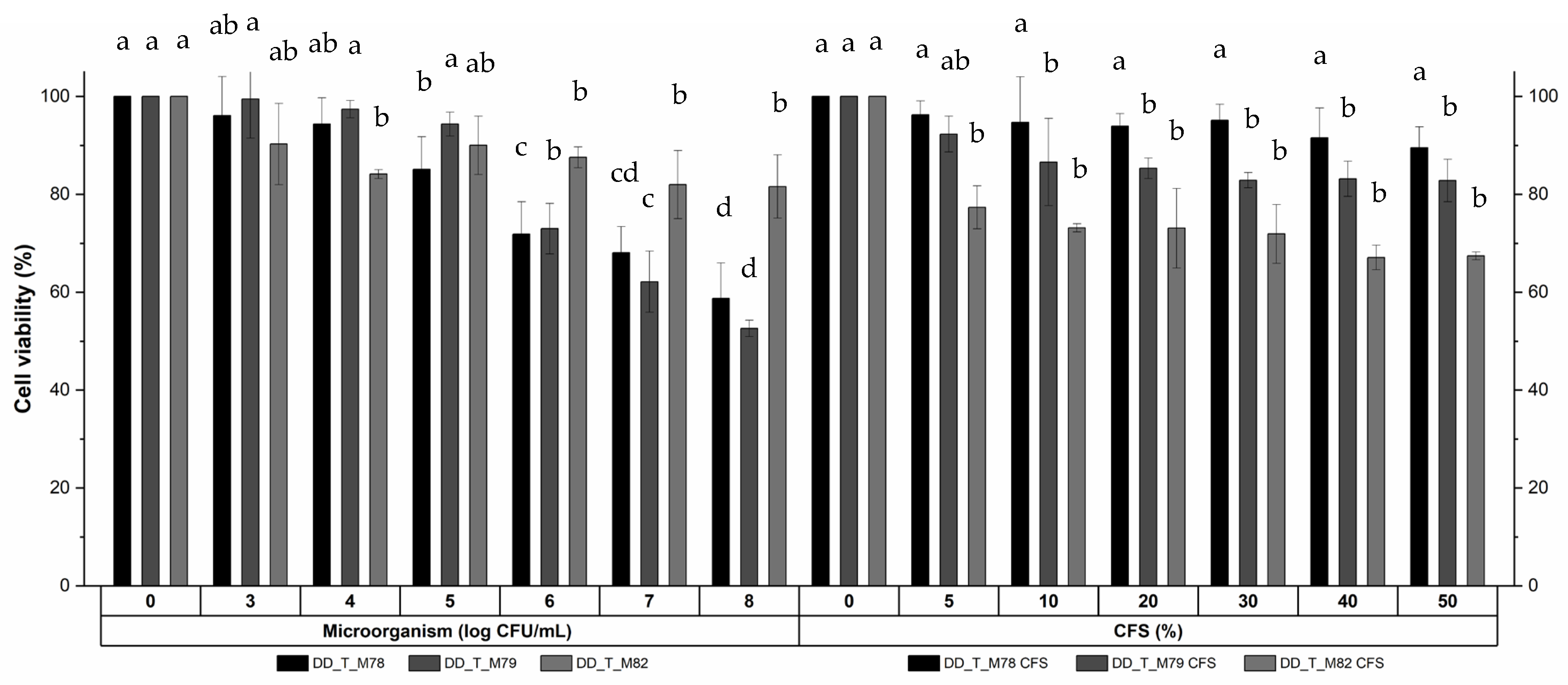
It was specified that cell-free supernatant (CFS) from several probiotics is composed of various bioactive components which show biological activities including antimicrobial. Although S. cerevisiae DD_NB_M90 has no antimicrobial activity, the other six isolates exhibited activity against E. coli and S. aureus aside from C. albicans (Table 4).
While the inhibitory effect against S. aureus was greater (31–97%) among isolates, the inhibition effect against E. coli ranged between 5–98%. Even though antimicrobial studies with the CFSs of yeasts are limited, there is a research with lactic acid bacteria suggesting that E. coli is more sensitive than S. aureus [38], as well as studies showing that E. coli is more resistant [39]. Ref. [38] claims that antimicrobial resistance also varies depending on the type of probiotics, and there is no interpretation as to which pathogen is more resistant. While the DPPH radical scavenging activity of CFS of the isolates varied from 14.29 to 44.36% (Table 4), the capacity of kefir-derived yeast cells was lower, between 6.85 and 29.64% [26]. The trends of increase or decrease in antimicrobial and antioxidant activities can vary independently. The highest DPPH radical scavenging activity was observed in DD_T_M80 (44.36%), indicating its efficacy compared to others; additionally, its antimicrobial activity was found to be significantly higher than that of the other samples, particularly at the 24 h incubation. It has been shown that organic acids can be produced during the development of microorganisms, and organic acids play a crucial role in the antimicrobial activity of yeasts by lowering the pH and creating an inhospitable environment for pathogenic microorganisms [40]. Studies on this subject have mainly progressed with lactic acid bacteria, and the amount of organic acid produced by yeast isolates was lower (Table 4). However, their detected inhibition effects were noteworthy, which indicates that their probiotic characteristics such as co-aggregation ability may be associated with their antimicrobial activity, similar to [41]. Among the isolates, P. kudriavzevii strains and S. cerevisiae DD_NB_M90 were found to produce lactic acid (0.116–0.197 g/L), and citric acid was produced to a certain extent by all isolates (0.067–0.293 g/L). The amount of lactic acid is especially higher in lactic acid bacteria with probiotic potential, and in addition to organic acids, protein content may have an impact on the activities. While the amount of protein produced by Lactobacillus plantarum S61 (198.65 µg/mL) was higher than the CFS of yeast isolates, the antimicrobial activity of proteinaceous compounds may vary depending on their molecular weight [42]. Therefore, there is a potential for further investigation with prospective purification.

3.2.5. Cytotoxicity

The cytotoxicity of six different concentrations of three isolates (108, 107, 106, 105, 104, and 103 CFU/mL) and their corresponding CFSs (50, 40, 30, 20, 10, 5%, v/v) on the Caco-2 cell line are presented in Figure 3. Among these isolates selected based on prior analyses, the viability of Caco-2 cells exposed to DD_T_M82 was higher than that of cells exposed to the other isolates, while the CFS of DD_T_M78 showed no statistically significant difference in Caco-2 cell viability compared to the control (p > 0.05). The study conducted by Saccharomyces boulardii RC009 and the Vero cell line showed that the toxic effect increased after the 106 CFU/mL, and cell viability decreased to below 20% at the 108 CFU/mL concentration [43]. In contrast, the current study reported higher viability, ranging from 52.61% to 81.62%. The cytotoxicity of extracellular secreted metabolites exhibited a significant variation depending on the cell line used [44]; cell viability exposed to CFSs of DD_T_M78, DD_T_M79, and DD_T_M82 varied between values of 67.43% and 96.29%.

4. Conclusions

In conclusion, potential probiotic yeasts were characterized in several aspects. Among the seven isolates examined, S. cerevisiae DD_NB_M90 showed limited probiotic potential. While the majority of isolates exhibited limited tolerance to low pH, bile salt and NaCl tolerance, they displayed great hydrophobicity, auto-aggregation, co-aggregation ability, antimicrobial activity, and resistance to antibiotic/antifungal agents. These findings highlight the potential of yeasts as starter cultures and their possible suitability for developing probiotic-rich foods. However, further in vivo studies could be employed to investigate the distinctive characteristics of these isolates, particularly regarding gut colonization, and overall host interactions, etc. The results emphasize that probiotic characteristics are strain-specific, and future research should focus on the genetic and phenotypic traits underlying these variations, particularly for applications in beverages and cereal-based bakery products.

Supplementary Materials

The following supporting information can be downloaded at: https://www.mdpi.com/article/10.3390/fermentation11060305/s1, Table S1: Molecular identification of yeast isolates by comparing the genomic sequences in the GenBank database using the BLAST algorithm; Table S2: The growth of yeast isolates under different conditions.

Author Contributions

Conceptualization, D.D. and F.K.-G.; methodology, D.D. and A.K.; investigation, D.D., A.K. and F.K.-G.; resources, F.K.-G., writing—original draft preparation, D.D.; writing—review and editing F.K.-G. All authors have read and agreed to the published version of the manuscript.

Funding

This work was financially supported by the Scientific Research Council of Istanbul Technical University under grant number MDK-2021-43273.

Institutional Review Board Statement

Not applicable.

Informed Consent Statement

Not applicable.

Data Availability Statement

The authors confirm that the data supporting the findings of this study are available within the article. Raw data supporting the findings of this study are available from the corresponding author, upon reasonable request.

Acknowledgments

The authors thank to Gulay Ozkan for her contribution in performing cytotoxicity analysis. The graphical abstract was created with BioRender.com.

Conflicts of Interest

The authors declare no conflicts of interest.

References

  1. Food and Agriculture Organization of the United Nations; World Health Organization. Probiotics in Food: Health and Nutritional Properties and Guidelines for Evaluation; FAO food and nutrition paper; Food and Agriculture Organization of the United Nations: Rome, Italy; World Health Organization: Rome, Italy, 2006; ISBN 978-92-5-105513-7. [Google Scholar]
  2. Menezes, A.G.T.; Ramos, C.L.; Cenzi, G.; Melo, D.S.; Dias, D.R.; Schwan, R.F. Probiotic Potential, Antioxidant Activity, and Phytase Production of Indigenous Yeasts Isolated from Indigenous Fermented Foods. Probiotics Antimicrob. Proteins 2020, 12, 280–288. [Google Scholar] [CrossRef] [PubMed]
  3. Ansari, F.; Alian Samakkhah, S.; Bahadori, A.; Jafari, S.M.; Ziaee, M.; Khodayari, M.T.; Pourjafar, H. Health-Promoting Properties of Saccharomyces cerevisiae var. boulardii as a Probiotic; Characteristics, Isolation, and Applications in Dairy Products. Crit. Rev. Food Sci. Nutr. 2023, 63, 457–485. [Google Scholar] [CrossRef] [PubMed]
  4. Chelliah, R.; Ramakrishnan, S.R.; Prabhu, P.R.; Antony, U. Evaluation of Antimicrobial Activity and Probiotic Properties of Wild-Strain Pichia kudriavzevii Isolated from Frozen idli Batter. Yeast 2016, 33, 385–401. [Google Scholar] [CrossRef]
  5. Merchán, A.V.; Benito, M.J.; Galván, A.I.; Ruiz-Moyano Seco de Herrera, S. Identification and Selection of Yeast with Functional Properties for Future Application in Soft Paste Cheese. LWT 2020, 124, 109173. [Google Scholar] [CrossRef]
  6. Ogunremi, O.R.; Agrawal, R.; Sanni, A.I. Development of Cereal-Based Functional Food Using Cereal-Mix Substrate Fermented with Probiotic Strain—Pichia kudriavzevii OG32. Food Sci. Nutr. 2015, 3, 486–494. [Google Scholar] [CrossRef]
  7. Binetti, A.; Carrasco, M.; Reinheimer, J.; Suárez, V. Yeasts from Autochthonal Cheese Starters: Technological and Functional Properties. J. Appl. Microbiol. 2013, 115, 434–444. [Google Scholar] [CrossRef]
  8. Gotcheva, V.; Hristozova, E.; Hristozova, T.; Guo, M.; Roshkova, Z.; Angelov, A. Assessment of Potential Probiotic Properties of Lactic Acid Bacteria and Yeast Strains. Food Biotechnol. 2002, 16, 211–225. [Google Scholar] [CrossRef]
  9. Simões, L.A.; Cristina de Souza, A.; Ferreira, I.; Melo, D.S.; Lopes, L.A.A.; Magnani, M.; Schwan, R.F.; Dias, D.R. Probiotic Properties of Yeasts Isolated from Brazilian Fermented Table Olives. J. Appl. Microbiol. 2021, 131, 1983–1997. [Google Scholar] [CrossRef]
  10. Usal, M.; Özgölet, M.; Arici, M.; Törnük, F. Enzymatic and Antimicrobial Activities of Lactic Acid Bacteria and Yeasts Isolated from Boza, a Traditional Fermented Grain Based Beverage. Food Biosci. 2024, 61, 104681. [Google Scholar] [CrossRef]
  11. Altay, F.; Karbancıoglu-Güler, F.; Daskaya-Dikmen, C.; Heperkan, D. A Review on Traditional Turkish Fermented Non-Alcoholic Beverages: Microbiota, Fermentation Process and Quality Characteristics. Int. J. Food Microbiol. 2013, 167, 44–56. [Google Scholar] [CrossRef]
  12. Guney, D.; Başdoğan, M.G.B.; Sengun, I. Probiotic Characterisation of Lactic Acid Bacteria Isolated from Pickles and Their Potential Application as Presumptive Probiotic Starter Culture in Cucumber Pickles. J. Food Meas. Charact. 2025, 19, 2077–2097. [Google Scholar] [CrossRef]
  13. Akmal, U.; Ghori, I.; Elasbali, A.M.; Alharbi, B.; Farid, A.; Alamri, A.S.; Muzammal, M.; Asdaq, S.M.B.; Naiel, M.A.E.; Ghazanfar, S. Probiotic and Antioxidant Potential of the Lactobacillus spp. Isolated from Artisanal Fermented Pickles. Fermentation 2022, 8, 328. [Google Scholar] [CrossRef]
  14. Yilmaz, D.; Sagiroglu, H.C. Development of Measurement System for Grain Loss of Some Chickpea Varieties. Measurement 2015, 66, 73–79. [Google Scholar] [CrossRef]
  15. Yalim, S.; Özdemır, Y. Effects of Preparation Procedures on Ascorbic Acid Retention in Pickled Hot Peppers. Int. J. Food Sci. Nutr. 2003, 54, 291–296. [Google Scholar] [CrossRef]
  16. Kumari, V.B.C.; Huligere, S.; MK, J.; Goh, K.W.; Desai, S.M.; HL, K.; Ramu, R. Characterization of Lactobacillus spp. as Probiotic and Antidiabetic Potential Isolated from Boza, Traditional Fermented Beverage in Turkey. Int. J. Microbiol. 2024, 2024, 2148676. [Google Scholar] [CrossRef]
  17. Ahmada Kh, A.; Abdo, A.; Khan, S.; Aleryani, H.; Mi, S.; Wang, X. Advancing Pickling Techniques to Enhance Bioactive Compounds and Probiotic Content in Pickled Vegetables. Food Rev. Int. 2025, 1–27. [Google Scholar] [CrossRef]
  18. Adak, M.S.; Kayan, N.; Gunes, A.; Inal, A.; Alpaslan, M.; Cicek, N.; Guzelordu, T. Effect of Harvest Timing on Yield and Mineral Nutritional Value of Kabuli Type Chickpea Seeds. J. Plant Nutr. 2007, 30, 1397–1407. [Google Scholar] [CrossRef]
  19. Fernández-Pacheco, P.; Ramos Monge, I.M.; Fernández-González, M.; Poveda Colado, J.M.; Arévalo-Villena, M. Safety Evaluation of Yeasts With Probiotic Potential. Front. Nutr. 2021, 8, 659328. [Google Scholar] [CrossRef]
  20. Gürkan Özlü, B.; Terzi, Y.; Uyar, E.; Shatila, F.; Yalçın, H.T. Characterization and Determination of the Potential Probiotic Yeasts Isolated from Dairy Products. Biologia 2022, 77, 1471–1480. [Google Scholar] [CrossRef]
  21. Pundir, R.K.; Kaur, S.R.N.; Kaur, A. Probiotic Potential of Lactic Acid Bacteria Isolated from Food Samples: An in Vitro Study. J. Appl. Pharm. Sci. 2013, 3, 85–93. [Google Scholar] [CrossRef]
  22. Topçu, K.C.; Kaya, M.; Kaban, G. Probiotic Properties of Lactic Acid Bacteria Strains Isolated from Pastırma. LWT 2020, 134, 110216. [Google Scholar] [CrossRef]
  23. Hossain, M.N.; Afrin, S.; Humayun, S.; Ahmed, M.M.; Saha, B.K. Identification and Growth Characterization of a Novel Strain of Saccharomyces boulardii Isolated From Soya Paste. Front. Nutr. 2020, 7, 27. [Google Scholar] [CrossRef] [PubMed]
  24. Wayne, P.A. Clinical and Laboratory Standards Institute Performance Standards for Antimicrobial Susceptibility Testing; Twenty Third Informational Supplement. Clin. Lab. Stand. Inst. 2013, 31, 100–121. [Google Scholar]
  25. Bradford, M.M. A Rapid and Sensitive Method for the Quantitation of Microgram Quantities of Protein Utilizing the Principle of Protein-Dye Binding. Anal. Biochem. 1976, 72, 248–254. [Google Scholar] [CrossRef]
  26. Goktas, H.; Dikmen, H.; Demirbas, F.; Sagdic, O.; Dertli, E. Characterisation of Probiotic Properties of Yeast Strains Isolated from Kefir Samples. Int. J. Dairy Technol. 2021, 74, 715–722. [Google Scholar] [CrossRef]
  27. Yasmin, I.; Saeed, M.; Khan, W.A.; Khaliq, A.; Chughtai, M.F.J.; Iqbal, R.; Tehseen, S.; Naz, S.; Liaqat, A.; Mehmood, T.; et al. In Vitro Probiotic Potential and Safety Evaluation (Hemolytic, Cytotoxic Activity) of Bifidobacterium Strains Isolated from Raw Camel Milk. Microorganisms 2020, 8, 354. [Google Scholar] [CrossRef]
  28. Caputo, L.; Quintieri, L.; Baruzzi, F.; Borcakli, M.; Morea, M. Molecular and Phenotypic Characterization of Pichia Fermentans Strains Found among Boza Yeasts. Food Res. Int. 2012, 48, 755–762. [Google Scholar] [CrossRef]
  29. Pongcharoen, P. The Ability of Pichia kudriavzevii to Tolerate Multiple Stresses Makes It Promising for Developing Improved Bioethanol Production Processes. Lett. Appl. Microbiol. 2022, 75, 36–44. [Google Scholar] [CrossRef]
  30. Alkalbani, N.S.; Osaili, T.M.; Al-Nabulsi, A.A.; Olaimat, A.N.; Liu, S.-Q.; Shah, N.P.; Apostolopoulos, V.; Ayyash, M.M. Assessment of Yeasts as Potential Probiotics: A Review of Gastrointestinal Tract Conditions and Investigation Methods. J. Fungi 2022, 8, 365. [Google Scholar] [CrossRef]
  31. Piraine, R.E.A.; Retzlaf, G.M.; Gonçalves, V.S.; Cunha, R.C.; Conrad, N.L.; Bochman, M.L.; Leite, F.P.L. Brewing and Probiotic Potential Activity of Wild Yeasts Hanseniaspora uvarum PIT001, Pichia kluyveri LAR001 and Candida intermedia ORQ001. Eur. Food Res. Technol. 2023, 249, 133–148. [Google Scholar] [CrossRef]
  32. Motey, G.A.; Johansen, P.G.; Owusu-Kwarteng, J.; Ofori, L.A.; Obiri-Danso, K.; Siegumfeldt, H.; Larsen, N.; Jespersen, L. Probiotic Potential of Saccharomyces cerevisiae and Kluyveromyces marxianus Isolated from West African Spontaneously Fermented Cereal and Milk Products. Yeast 2020, 37, 403–412. [Google Scholar] [CrossRef] [PubMed]
  33. Castro-López, C.; Romero-Luna, H.E.; García, H.S.; Vallejo-Cordoba, B.; González-Córdova, A.F.; Hernández-Mendoza, A. Key Stress Response Mechanisms of Probiotics During Their Journey Through the Digestive System: A Review. Probiotics Antimicrob. Proteins 2023, 15, 1250–1270. [Google Scholar] [CrossRef] [PubMed]
  34. Bustos, A.Y.; Taranto, M.P.; Gerez, C.L.; Agriopoulou, S.; Smaoui, S.; Varzakas, T.; Enshasy, H.A.E. Recent Advances in the Understanding of Stress Resistance Mechanisms in Probiotics: Relevance for the Design of Functional Food Systems. Probiotics Antimicrob. Proteins 2025, 17, 138–158. [Google Scholar] [CrossRef]
  35. Zoumpourtikoudi, V.; Pyrgelis, N.; Chatzigrigoriou, M.; Tasakis, R.N.; Touraki, M. Interactions among Yeast and Probiotic Bacteria Enhance Probiotic Properties and Metabolism Offering Augmented Protection to Artemia Franciscana against Vibrio Anguillarum. Microb. Pathog. 2018, 125, 497–506. [Google Scholar] [CrossRef] [PubMed]
  36. Lata, P.; Kumari, R.; Sharma, K.B.; Rangra, S. Savitri In Vitro Evaluation of Probiotic Potential and Enzymatic Profiling of Pichia kudriavzevii Y33 Isolated from Traditional Home-Made Mango Pickle. J. Genet. Eng. Biotechnol. 2022, 20, 132. [Google Scholar] [CrossRef]
  37. Helmy, E.A.; Soliman, S.A.; Abdel-Ghany, T.M.; Ganash, M. Evaluation of Potentially Probiotic Attributes of Certain Dairy Yeast Isolated from Buffalo Sweetened Karish Cheese. Heliyon 2019, 5, e01649. [Google Scholar] [CrossRef]
  38. Lee, J.Y.; Kim, Y.; Kim, J.-I.; Lee, H.-Y.; Moon, G.-S.; Kang, C.-H. Improvements in Human Keratinocytes and Antimicrobial Effect Mediated by Cell-Free Supernatants Derived from Probiotics. Fermentation 2022, 8, 332. [Google Scholar] [CrossRef]
  39. Mitsuwan, W.; Sornsenee, P.; Romyasamit, C. Lacticaseibacillus spp.; Probiotic Candidates from Palmyra Palm Sugar Possesses Antimicrobial and Anti-Biofilm Activities against Methicillin-Resistant Staphylococcus aureus. Vet. World 2022, 15, 299–308. [Google Scholar] [CrossRef]
  40. Reuben, R.C.; Roy, P.C.; Sarkar, S.L.; Alam, R.-U.; Jahid, I.K. Isolation, Characterization, and Assessment of Lactic Acid Bacteria toward Their Selection as Poultry Probiotics. BMC Microbiol. 2019, 19, 253. [Google Scholar] [CrossRef]
  41. Rahmani, B.; Alimadadi, N.; Attaran, B.; Nasr, S. Yeasts from Iranian Traditional Milk Kefir Samples: Isolation, Molecular Identification and Their Potential Probiotic Properties. Lett. Appl. Microbiol. 2022, 75, 1264–1274. [Google Scholar] [CrossRef]
  42. Abouloifa, H.; Rokni, Y.; Hasnaoui, I.; Bellaouchi, R.; Gaamouche, S.; Ghabbour, N.; Karboune, S.; Ben Salah, R.; Brasca, M.; D’hallewin, G.; et al. Characterization of Antimicrobial Compounds Obtained from the Potential Probiotic Lactiplantibacillus Plantarum S61 and Their Application as a Biopreservative Agent. Braz. J. Microbiol. 2022, 53, 1501–1513. [Google Scholar] [CrossRef] [PubMed]
  43. Poloni, V.L.; Bainotti, M.B.; Vergara, L.D.; Escobar, F.; Montenegro, M.; Cavaglieri, L. Influence of Technological Procedures on Viability, Probiotic and Anti-Mycotoxin Properties of Saccharomyces boulardii RC009, and Biological Safety Studies. Curr. Res. Food Sci. 2021, 4, 132–140. [Google Scholar] [CrossRef] [PubMed]
  44. Sharma, M.; Chandel, D.; Shukla, G. Antigenotoxicity and Cytotoxic Potentials of Metabiotics Extracted from Isolated Probiotic, Lactobacillus rhamnosus MD 14 on Caco-2 and HT-29 Human Colon Cancer Cells. Nutr. Cancer 2020, 72, 110–119. [Google Scholar] [CrossRef]
Figure 1. The production procedures of (A)—fermented chickpea, (B)—boza, (C)—pickles.
Fermentation 11 00305 g001
Figure 2. Phylogenetic tree of the yeast isolates. The evolutionary distances were computed using the maximum composite likelihood method. The percentage of trees in which the associated taxa clustered together is shown next to the branches. Evolutionary analyses were conducted in Mega 11.
Fermentation 11 00305 g002
Figure 3. Cytotoxicity of probiotic isolates and their CFSs. a–d Values within the same sample with different superscript small letters are significantly different (p < 0.05).
Fermentation 11 00305 g003
Table 1. The viability of yeast isolates at low pH and in the presence of bile salt in log (CFU/mL).
MicroorganismControlpH 2.01% Bile Salt
Initial3 hInitial3 hInitial3 h
DD_B_M857.78 ± 0.01 b,B8.28 ± 0.06 b,A7.68 ± 0.08 a,B7.84 ± 0.13 a,B7.25 ± 0.04 e,A7.36 ± 0.08 e,A
DD_T_M807.63 ± 0.04 c,B8.07 ± 0.08 c,A7.54 ± 0.07 a,b,B7.64 ± 0.11 a,b,B7.24 ± 0.04 e,B7.64 ± 0.03 d,A
DD_T_M787.16 ± 0.04 e,A7.35 ± 0.02 e,A7.26 ± 0.06 c,d,A7.18 ± 0.08 c,d,A7.62 ± 0.05 c,B7.99 ± 0.08 b,c,A
DD_T_M797.17 ± 0.01 e,B7.60 ± 0.04 d,A7.16 ± 0.04 d,B7.06 ± 0.06 d,B7.40 ± 0.03 d,B7.82 ± 0.03 b,c,A
DD_T_M827.49 ± 0.04 c,d,B7.68 ± 0.06 d,A7.39 ± 0.03 b,c,B7.40 ± 0.01 b,c,B7.82 ± 0.03 b,B8.09 ± 0.08 b,A
DD_B_M887.40 ± 0.05 d,B7.77 ± 0.04 d,A7.35 ± 0.04 b,c,d,B7.40 ± 0.03 b,c,B7.34 ± 0.02 d,e,B7.69 ± 0.03 d,A
DD_NB_M908.52 ± 0.03 a,B8.70 ± 0.03 a,A6.02 ± 0.06 e,C5.62 ± 0.02 e,D8.47 ± 0.04 a,B8.59 ± 0.05 a,A,B
a–e Values within the same column with different superscript small letters are significantly different (p < 0.05). A–D Values within the same row of control and condition with different superscript capital letters are significantly different (p < 0.05).
Table 2. Cell surface properties of the yeast isolates.
AnalysisMicroorganisms
DD_T_M78DD_T_M79DD_T_M80DD_T_M82DD_B_M85DD_B_M88DD_NB_M90
Hydrophobicity (%)34.31 ± 1.07 c29.74 ± 0.42 d38.59 ± 0.84 b35.40 ± 0.73 c34.74 ± 0.86 c63.07 ± 0.14 a16.88 ± 0.33 e
Auto-aggregation (%)2 h90.79 ± 0.69 a,B93.89 ± 1.12 a,A76.40 ± 1.89 b,C79.40 ± 1.85 b,B67.69 ± 1.13 c,C91.13 ± 1.39 a,B7.94 ± 1.25 d,B
4 h93.97 ± 0.58 a,A95.37 ± 0.51 a,A86.05 ± 1.01 b,B93.84 ± 1.10 a,A72.42 ± 0.93 c,B94.94 ± 0.22 a,A8.58 ± 0.64 d,B
24 h94.25 ± 0.27 a,A95.31 ± 1.14 a,A95.23 ± 0.78 a,A94.47 ± 0.59 a,A91.77 ± 0.53 a,A94.75 ± 0.57 a,A,B53.29 ± 3.38 b,A
Co-aggregation (%)E. coli2 h77.54 ± 0.67 b,B83.25 ± 1.60 a,B51.25 ± 0.83 c,C31.65 ± 1.84 e,B42.84 ± 1.00 d,C82.96 ± 1.10 a,A0.77 ± 0.37 f,C
4 h88.46 ± 0.68 a,b,A89.76 ± 0.95 a,A84.53 ± 0.04 b,B89.64 ± 0.63 a,A67.44 ± 0.93 c,B88.53 ± 1.97 a,b,A7.60 ± 0.80 d,B
24 h88.78 ± 0.44 a,A88.18 ± 0.69 a,A89.44 ± 0.41 a,A88.88 ± 0.30 a,A89.22 ± 1.05 a,A88.77 ± 0.91 a,A52.12 ± 1.03 b,A
S. aureus2 h86.55 ± 0.61 a,B80.76 ± 0.83 c,B76.95 ± 0.08 d,C44.12 ± 0.69 f,B48.84 ± 0.87 e,C83.88 ± 0.14 b,B8.58 ± 0.61 g,C
4 h88.02 ± 0.10 a,A,B88.46 ± 0.95 a,A84.02 ± 0.90 b,B90.73 ± 1.07 a,A75.36 ± 0.62 c,B88.01 ± 1.17 a,A13.20 ± 0.35 d,B
24 h89.01 ± 0.40 a,A88.90 ± 0.72 a,A88.60 ± 1.60 a,A87.64 ± 1.05 a,A88.09 ± 0.87 a,A88.18 ± 0.06 a,A57.45 ± 0.77 b,A
C. albicans2 h78.61 ± 1.34 b,B87.49 ± 0.83 a,A50.92 ± 0.21 c,C89.64 ± 0.75 a,A43.17 ± 0.13 d,C86.94 ± 1.34 a,A8.60 ± 0.68 e,C
4 h89.40 ± 2.00 a,A91.77 ± 2.06 a,A81.04 ± 0.42 b,B91.17 ± 1.05 a,A67.61 ± 1.48 c,B89.91 ± 1.13 a,A13.47 ± 1.42 d,B
24 h89.75 ± 0.62 a,A88.97 ± 0.42 a,A90.44 ± 0.88 a,A89.57 ± 1.58 a,A89.43 ± 1.70 a,A89.45 ± 0.62 a,A56.99 ± 0.09 b,A
a–g Values within the same raw for an analysis with different superscript small letters are significantly different (p < 0.05). A–C Values within the same column for an analysis with different superscript capital letters are significantly different (p < 0.05).
Table 3. Antibiotic/antifungal susceptibility of the yeast isolates.
AgentsInhibition Zone (mm)
DD_T_M78DD_T_M79DD_T_M80DD_T_M82DD_B_M85DD_B_M88DD_NB_M90
Antibiotics
Ampicillin (10 µg)- *-7.25 ± 0.05-9.17 ± 0.24-7.73 ± 0.17
Gentamycin (10 µg)----6.12 ± 0.08-9.30 ± 0.05
Kanamycin (30 µg)--7.97 ± 0.12-6.17 ± 0.29-12.17 ± 0.29
Streptomycin (10 µg)--9.77 ± 0.21-6.83 ± 0.15-7.27 ± 0.25
Vancomycin (30 µg)--6.17 ± 0.24-6.60 ± 0.17-10.33 ± 0.15
Antifungals
Amphotericin B (20 µg)6.25 ± 0.257.38 ± 1.1911.75 ± 1.096.00 ± 0.009.00 ± 0.006.50 ± 0.006.75 ± 0.25
Nystatin (100 µg)10.50 ± 0.8718.00 ± 0.7114.75 ± 0.4320.25 ± 0.4320.75 ± 0.8319.25 ± 2.2818.38 ± 0.41
Ketoconazole (10 µg)9.00 ± 0.717.75 ± 1.2513.50 ± 0.506.38 ± 0.416.00 ± 0.006.50 ± 0.508.00 ± 1.00
Itraconazole (10 µg)10.75 ± 0.8312.25 ± 0.8310.17 ± 0.6210.75 ± 0.437.50 ± 0.8713.75 ± 0.8310.00 ± 0.00
Fluconazole (25 µg)13.50 ± 1.5014.25 ± 0.8314.38 ± 0.4110.75 ± 1.096.00 ± 0.007.00 ± 0.006.00 ± 0.00
- *: no inhibition zone.
Table 4. Antimicrobial properties and characterization of the CFSs of yeast isolates.
AnalysisMicroorganisms
DD_T_M78DD_T_M79DD_T_M80DD_T_M82DD_B_M85DD_B_M88DD_NB_M90
Inhibition (%) E. coli24 h22.72 ± 1.47 c.B31.13 ± 1.42 b,B98.72 ± 0.28 a,A24.64 ± 1.43 c,B34.06 ± 1.63 b,B7.02 ± 0.05 d,C-1
48 h18.69 ± 1.46 c.B27.11 ± 0.83 b,C92.82 ± 0.50 a,B19.44 ± 0.93 c,C25.10 ± 1.45 b,C5.54 ± 0.28 d,C-
S. aureus24 h95.48 ± 1.60 a.b.A84.67 ± 0.47 c,A97.72 ± 0.69 a,A94.34 ± 0.47 b,A96.93 ± 0.68 a,b,A39.53 ± 0.64 d,A-
48 h96.35 ± 0.96 a.A85.87 ± 0.76 b,A97.31 ± 0.60 a,A97.34 ± 0.80 a,A97.59 ± 0.86 a,A31.01 ± 1.38 c,B-
CFS CharacterizationProtein (µg BSA/mL)48.97 ± 1.42 a14.81 ± 1.04 b13.14 ± 1.04 b,c15.64 ± 1.42 b10.08 ± 0.68 c,d9.81 ± 0.79 d11.19 ± 0.39 c,d
DPPH (% inhibition)34.02 ± 0.82 b30.33 ± 0.94 c44.36 ± 1.62 a23.81 ± 0.94 d19.92 ± 1.55 e32.08 ± 1.97 b,c14.29 ± 0.61 f
Lactic acid (g/L)0.121 ± 0.002 c0.116 ± 0.003 cND20.144 ± 0.004 b,cND0.197 ± 0.006 a0.154 ± 0.014 b
Citric acid (g/L)0.127 ± 0.001 c0.105 ± 0.011 c0.067 ± 0.005 d0.187 ± 0.006 b0.120 ± 0.001 c0.293 ± 0.002 a0.281 ± 0.005 a
Tartaric acid (g/L)0.035 ± 0.000 a0.018 ± 0.001 bND0.018 ± 0.003 b0.012 ± 0.001 b0.020 ± 0.001 bND
1 No inhibition, 2 Not detected. a–f Values within the same row with different superscript small letters are significantly different (p < 0.05). A–C Values within the same column for inhibition effect of a CFS with different superscript capital letters are significantly different (p < 0.05).
Disclaimer/Publisher’s Note: The statements, opinions and data contained in all publications are solely those of the individual author(s) and contributor(s) and not of MDPI and/or the editor(s). MDPI and/or the editor(s) disclaim responsibility for any injury to people or property resulting from any ideas, methods, instructions or products referred to in the content.

