Article

Catabolism Mechanism and Growth-Promoting Effect of Xylooligosaccharides in Lactiplantibacillus plantarum Strain B20

School of Biological Engineering, Henan University of Technology, Zhengzhou 450001, China
*
Authors to whom correspondence should be addressed.
Fermentation 2025, 11(5), 280; https://doi.org/10.3390/fermentation11050280
Submission received: 7 April 2025 / Revised: 8 May 2025 / Accepted: 9 May 2025 / Published: 13 May 2025

Abstract

:
Prebiotics are food ingredients that result in specific changes in the composition and/or activity of the gastrointestinal microbiota, thus conferring benefits upon host health. Xylooligosaccharides (XOS) are prebiotic fibers made from xylan. Commercial XOS are mixtures of oligosaccharides containing β-1,4–linked xylose residues. Though they are widely added to foods at different doses, the molecular mechanisms of the catabolism and growth promotion of XOS in the innate gut microbes Lactobacillus spp. remain unknown. In this study, we evaluated the growth-promoting effect using a human fecal isolate, Lactiplantibacillus plantarum strain B20 (Lb. plantarum B20). Assays of bacterial growth and lactic acid production showed stronger growth promotion of XOS than other oligosaccharides did, in a dose- and fraction-dependent pattern. Using the Lb. plantarum strain SK151 genome as a reference, bioinformatic analysis failed to identify any previously characterized genes responsible for the uptake and catabolism of XOS. However, transcriptomic analysis of Lb. plantarum B20 yielded numerous differentially expressed genes (DEGs) during fermentation of XOS. Among these, an oligopeptide ABC transporter (RS03575-03595, composed of five proteins) and a hydrolase (RS06170) were significantly upregulated. Molecular docking analysis indicated that the substrate-binding protein RS03575 may mediate the import of XOS into the cell. Enzymatic assays further demonstrated that RS06170 possesses β-xylosidase activity and can effectively degrade XOS. In addition, functional enrichment analysis suggested that the growth-promoting effect of XOS may be attributed to the upregulation of genes involved in cellular component biogenesis and cell division, potentially through modulation of ribosome function and carbohydrate metabolism in Lb. plantarum B20. These results provide valuable insights into the mechanisms by which XOS promote growth and highlight potential targets for enhancing prebiotic–probiotic interactions.

1. Introduction

Xylooligosaccharides (XOS) are currently sold as prebiotics or functional ingredients in the food industry due to various health-promoting effects [1]. Acting as prebiotics, XOS primarily benefit human health by selectively stimulating the growth of beneficial microorganisms, particularly Bifidobacterium spp. and Lactobacillus spp., which are the most prominent probiotic genera used in food and pharmaceutical products. The growth-promoting effect of XOS on Bifidobacterium spp. has already been widely recognized [2]. However, different results have been reported for Lactobacillus (Lb.) spp. when using XOS to promote growth. For example, different fermentation capacities of XOS in Lb. plantarum S26, Lb. brevis S27, and Lb. sakei S16 have been demonstrated [3]. A recent report even showed limited prebiotic activity on Lb. plantarum and Lb. fermentum under anaerobic conditions when XOS produced from sugarcane bagasse was supplemented [4]. In fact, the growth-promoting effect of XOS in Lactobacillus is strain/species-specific, as many of them did not efficiently metabolize XOS [5].
Commercially available XOS are mixtures of xylose oligomers, normally with degrees of polymerization (DP) ranging from 2 to 7. Different chemical/structural characteristics of XOS are definitely affecting their properties/effects. For example, all tested Lactobacillus spp. strains showed preference for XOS fractions with lower DPs, e.g., efficiently metabolizing short-chain oligomers such as xylobiose (XOS2) and xylotriose (XOS3) [3]. Similarly, our previous work demonstrated that the growth-promoting effects of XOS on different Bifidobacterium species were both fraction- and dose-dependent, indicating that tailoring supplementation to match the metabolic preference could significantly enhance the efficacy of XOS as a prebiotic intervention [2]. Therefore, to achieve optimal growth stimulation of specific probiotic strains, careful evaluation of both the dose and the polymerization profile of XOS used is essential.
Despite the widespread commercialization of both Lactobacillus spp. and XOS, the molecular mechanism underlying the growth-promoting effect of XOS in Lactobacillus spp. remains largely elusive. To promote growth, XOS need to be metabolized by bacteria, which includes the uptake and degradation processes. Multiple ABC transporters and degrading enzymes for XOS have been characterized in Bifidobacterium spp. and other genera [6,7]. In B. animalis subsp. lactis BB-12, XOS utilization was predicted to be facilitated by three steps: (1) binding of XOS at the cell surface and transport across the membrane by ABC-type oligosaccharide transporters, (2) intracellular degradation of XOS to D-xylose by xylanases and xylosidases, and (3) conversion of D-xylose to xylulose 5-phosphate by two other enzymes [8]. However, such information in Lactobacillus is still scarce [9]. This is largely because only species belonging to the group of heterofermentative Lactobacillus possess a low capacity to degrade XOS. For example, three β-xylosidases, namely xynB1 (encoded by LVIS_0375), xynB2 (encoded by LVIS_2285), and abf3 (encoded by LVIS_1748), have been reported in Lb. brevis DSM20054 [10]. In particular, xynB1 showed degrading activity against XOS2 and XOS3. In addition, D-xylose can be converted to D-xylulose 5-phosphate (X5P) by xylose isomerase (xylA) and xylulose kinase (xylB) in L. pentosus MD353 [11]. Through the phosphoketolase pathway (PK pathway), X5P is finally converted into lactic acid and acetic acid. Notably, it was found that β-xylosidases classified into the GH43 glycosyl hydrolase family from Lactobacillus share high amino acid identity with enzymes from bifidobacteria. However, whether such transporters and enzymes are present in Lb. plantarum is unknown.
To investigate the effects of XOS on Lb. plantarum, this paper thoroughly evaluated the relationship between XOS doses, pure oligomer fractions, and growth-promoting effects, using a human fecal isolate, Lactiplantibacillus plantarum B20 (Lb. plantarum B20) in vitro. Strains of Lb. plantarum are typical probiotics used as starter cultures in different foods and possess versatile benefits to human health [12]. To gain molecular insights into the mechanism underlying XOS catabolism and its growth-promoting effects on Lb. plantarum, comprehensive genomic and transcriptomic analyses were conducted. Additionally, the activity of a critical enzyme involved in the catabolism of XOS was also tested. Furthermore, the enzymatic activity of a key catalyst involved in the XOS degradation pathway was assessed. Data obtained in this study may provide useful information for the application of XOS as functional food ingredients and shed light on how Lb. plantarum B20 responds to XOS at a molecular level.

2. Material and Methods

2.1. Chemicals and Reagents

Analytic-grade monosaccharides including D-xylose, D-fructose, D-glucose, and D-galactose were purchased from a chemical supplier (Macklin Inc., Shanghai, China). Food-grade oligosaccharides including fructooligosaccharide (FOS), galactooligosaccharide (GOS), and polydextrose (PDX), were purchased from commercial providers. Henan Heagreen Bio-technology Co., Ltd. (Zhoukou, China) produced the XOS sample (95% syrup). The composition was determined (42.03% XOS2, 27.9% XOS3, 13.59% XOS4, and 14.87% oligomers with DP ≥ 5) by high-performance liquid chromatography. Heagreen also provided the pure standards of XOS fractions with defined DPs (DP2–DP6) in powder form. All carbohydrates were separately dissolved in distilled water at a final concentration of 20% (w/v). The solutions were sterilized by a 0.22 μm Millipore Filter Membrane (Sangon Biotech Co. Ltd, Shanghai, China) and stored at 4 °C within one week of preparation. Detailed information about the carbohydrates used in this study is provided in Table S1.

2.2. Bacterial Strain and Growth Conditions

Lb. plantarum B20 was isolated from the feces of healthy adults in 2017 and identified primarily by 16S rDNA sequencing. A glycerol stock of the strain was activated in MRS broth (Cat. No. HB0384-1, Qingdao Hopebiol Co., Ltd., Qingdao, China) that supported good growth. The growth of the bacteria was carried out statically in a sealed jar using the MGC AnaeroPack (MitsubishiTM, Tokyo, Japan) at 37 °C for 24 h.

2.3. Compare the Growth-Promoting Effect of Different Oligosaccharides on Lb. plantarum B20

A modified MRS broth that did not have the addition of glucose was manually prepared according to the standard recipe. The broth was sub-packed in glass tubes (5 mL each) and used as a basis for the following test. Different solutions of sugars (20%, w/v, 100 μL) sterilized by filtration were added separately into glass tubes to a final concentration of 0.4%. Then, freshly overnight-cultured cells of Lb. plantarum B20 were inoculated at 1% (v/v), to an initial OD600 of approximately 0.05. All tubes were incubated anaerobically at 37 °C for 24 h as described above. At set time points (12 h and 24 h), the tubes were fully vortexed, and 1 mL of culture fluids transferred into 1.5 mL Eppendorf tubes were used for further assays of growth and lactic acid production. Three independent experiments were performed, and each sample was assayed in triplicate.

2.4. Dose- and Fraction-Dependent Effect of XOS on Lb. plantarum B20

For the XOS dose test, 0, 0.1%, 0.2%, 0.4%, 0.8%, and 1.6% (w/v) XOS were added into 5 mL modified MRS broth, respectively. For the test of XOS fractions, xylose and XOS fractions with different DPs (DP2–DP6) were separately added at 0.4%. This concentration was selected because Lactobacillus strains are more effectively stimulated when more than 0.2% XOS was supplemented [5]. In both experiments, overnight grown culture of Lb. plantarum B20 were inoculated at 1% (v/v) into the respective media. Cultures were incubated under anaerobic conditions at 37 °C for 24 h as previously mentioned. At two time points (12 h and 24 h), 1 mL of each culture was collected for subsequent assays of bacterial growth and lactic acid production, as previously described. Each experiment was performed in triplicate and three samples from each treatment were used for analysis.

2.5. Quantification of Bacterial Growth and Lactic Acid Production

To assess the bacterial growth and lactic acid production, culture samples were collected at specified time points (as previously mentioned). The samples were centrifuged at 10,000× g for 2 min and cell pellets were washed twice using ddH2O to remove residual medium components. Rinsed cell pellets were resuspended in 1 mL ddH2O and used for growth assay in a transparent 96-well plate in triplicate (200 μL in each well). The optical density was monitored at 600 nm (OD600) using a microtitor reader (SpectraMax i3, Molecular Devices, San Jose, CA, USA). Water was used as a blank control. Bacterial cells collected from the medium without any sugar were used as basal controls. The absorbance readings for each sample were subtracted by basal controls. After centrifugation of culture fluids, supernatants were transferred into new tubes for lactic acid detection. The supernatants were pretreated by precipitation in an equal volume of tungstic acid solution to remove protein and glucose. Lactic acid was determined by a simple colorimetric assay in hot sulfuric acid. Briefly, in a 1.5 mL tube, 0.01 g calcium hydroxide powder, 100 μL pretreated sample, 900 μL dH2O, and 160 μL 20% CuSO4 solution were mixed and then incubated for 3 min in boiling water. After the mixture reached room temperature, the tube was centrifuged at 3000× g for 5 min. Then, 50 μL supernatant was moved into a new tube and 600 μL sulfuric acid was carefully added. The tube was incubated for 5 min in boiling water. After the mixture reached room temperature, 12.5 μL 1.5% p-Hydroxybiphenol solution was supplemented. After colorization for 15 min, the tube was incubated in boiling water for a further 5 min. After the tube reached room temperature, absorbance (Abs. 565 nm) was measured in a transparent 96-well plate in triplicate (200 μL in each well) using the above-mentioned microtitor reader. Pure L-lactic acid was used for calculating standard curves.

2.6. Genomic Analysis of XOS Catabolism in Lb. plantarum

To investigate the genetic basis of XOS catabolism in Lb. plantarum, the high-quality genome of strain SK151 (Assembly: GCF_003269405.1) was selected as the reference due to its higher genome coverage and completeness. A multi-tiered bioinformatic approach was employed to identify key genetic components involved in the uptake and degradation of XOS. Initially, genes encoding carbohydrate transport systems including ABC transporters, sugar permeases, and members of the major facilitator superfamily (MFS) were retrieved from the annotated genome using the keyword “transporter” followed by manual refinement. Subsequently, to pinpoint transport systems specifically associated with XOS uptake, protein sequences of well-characterized XOS-specific ABC transporters from other bacterial species were used as queries in BLASTp searches against the Lb. plantarum genome. Additionally, enzymes involved in XOS hydrolysis and downstream xylose metabolism were explored using homologous protein sequences as queries. For enhanced sensitivity and domain-level resolution, DELTA-BLAST was performed against the NCBI non-redundant protein database.

2.7. RNA Isolation and Sequencing of Lb. plantarum B20

To investigate the transcriptional responses to XOS, Lb. plantarum B20 cells recovered from overnight cultures were inoculated (5% v/v) into the modified MRS broth supplemented with either 1.6% xylose (group N), 0.4% XOS (group TL), or 1.6% XOS (group TH). The cultures were incubated at 37 °C for 12 h under static conditions in sealed jars. Subsequently, total RNA was extracted from cells harvested from three independent biological replicates via centrifugation at 10,000× g for 2 min at 4 °C using TRIzol® Reagent (ThermoFisher Scientific, Waltham, MA, USA), according to the manufacturer’s instructions. The concentration was measured by Nanodrop2000. The purity was detected by agarose gel electrophoresis and the RNA Integrity Number (RIN) was detected by Agilent 2100. The rRNA was removed using the Ribo-Zero Magnetic Kit (EpiCentre Biotechnologies, Madison, WI, USA). Obtained RNA was then used to construct cDNA libraries using the TruSeqTM Total RNA Library Prep Kit (Illumina, San Diego, CA, USA). RNA sequencing was carried out using HiSeq 4000 SBS Kit (Illumina, USA) by a commercial service provider (Origingene Biotech Co., Ltd., Shanghai, China). After obtaining raw reads, adapter, higher N-ratio (>10%) reads, reads smaller than 25 bp, and low-quality reads were filtered out using Cutadapt v1.16 (http://cutadapt.readthedocs.io/, accessed on 18 September 2021) and FastqStat. jar (Origingene, v1.0). Clean reads were aligned to the reference genome of L. plantarum SK151 (Assembly: GCF_003269405.1) using Hisat2 and the quality was evaluated by RSeQC (http://rseqc.sourceforge.net, v2.6.4, accessed on 19 September 2021). The raw RNA-Seq data were deposited in the public domain of the NCBI Sequence Read Archive (SRA) under BioProject Accession ID: PRJNA1243018.

2.8. Bioinformatic Analysis of Transcriptomic Data

High-quality clean reads obtained from RNA sequencing were subjected to functional annotation using the BlastX tool against multiple databases, including NR, STRING (Search Tool for the Retrieval of Interacting Genes), GO (Gene Ontology), COG (Clusters of Orthologous Groups of proteins), KEGG (Kyoto Encyclopedia of Genes and Genomes) using the Diamond v0.9.19.120 with E value < 1 × 10⁻⁵, and CAZY (Carbohydrate-active enzymes, http://www.cazy.org, accessed on 8 May 2025) using hmmscan v5.0 (https://www.ebi.ac.uk/Tools/hmmer/search/hmmscan, accessed on 8 May 2025). Gene expression levels were normalized by transcripts per million reads (TPM) including both known and novel predicted RNA, using software Salmon v0.11.3 (https://salmon.readthedocs.io, accessed on 8 May 2025). Differentially expressed genes (DEGs) between the control and treatment groups were screened using edgeR v3.24 (http://www.bioconductor.org/packages/release/bioc/html/edgeR.html, accessed on 8 May 2025), with the default threshold of a false discovery rate p < 0.05 and |log2FC| > 1. Enrichment analyses of GO and KEGG pathways for DEGs were conducted by Origingene using self-developed software (http://www.origin-gene.com, accessed on 10 December 2021). Operon was also predicted by Rockhopper (http://cs.wellesley.edu/~btjaden/Rockhopper, accessed on 10 December 2021).

2.9. Quantitative Real-Time Polymerase Chain Reaction (qPCR) Validation

To validate the transcriptomic data, 10 DEGs (7 upregulated and 3 downregulated) identified from the RNA-Seq analysis were selected for qPCR. Briefly, total RNA was isolated from Lb. plantarum B20 cells grown in modified MRS containing 1.6% xylose or XOS for 12 h using TRIzol® Reagent. Then, it was reversely transcribed into cDNA by HiScript 1st strand cDNA Synthesis Kit (Vazyme, Nanjing, China). The cDNA was used as a template for qPCR quantification. The AceQ qPCR SYBR Green Master Mix (without ROX) (Vazyme, Nanjing, China) was used according to the manufacturer’s protocols for amplification in a CFX96 TouchTM Real-Time PCR Detection System (Bio-Rad, Hercules, CA, USA). The parameters were as follows: 95 °C pre-denaturation for 5 min; 40 cycles of 10 s at 95 °C, and 30 s at 60 °C. A melting curve analysis was followed after amplification with a temperature gradient of 0.5 °C/s from 60 °C to 95 °C to confirm specific amplification. Primers were designed by Primer Premier version 6 (Premier Biosoft Inc., Palo Alto, CA, USA) and synthesized by Origingene (Shanghai, China), as supplied in Table S2. Lastly, the relative mRNA expression was evaluated using the 2−ΔΔCt method, with a housekeeping gene GAPDH (gene id: RS01560) as reference [13].

2.10. Characterization of DEGs and Heterologous Expression of an XOS Inducible Hydrolase

The subcellular localization of some DEGs was predicted using DeepLocPro-1.0 (https://services.healthtech.dtu.dk/services/DeepLocPro-1.0, accessed on 11 March 2023) and the functional domains were analyzed using the Simple Modular Architecture Research Tool (SMART, https://smart.embl.de/, accessed on 11 March 2023). The 3D structures were predicted by Phyre 2.2 using the NORMAL mode (https://www.sbg.bio.ic.ac.uk/phyre2, accessed on 1 December 2024). The top model with 99.9% confidence and more than 90% coverage was presented [14]. The binding potentials between XOS3 and the substrate-binding protein (RS03575) or hydrolase (RS06170) were evaluated by Autodock Vina (https://www.swissdock.ch, accessed on 7 December). The complete gene of RS06170 was amplified by polymerase chain reaction (PCR) using high-fidelity DNA polymerase with specific primers (Table S2), using the genomic DNA extracted from Lb. plantarum B20 as template. The genomic DNA was extracted from overnight cultivated bacteria using a TIANamp Bacteria DNK Kit (Tiangen Biotech. Co. Ltd., Beijing, China). The AceQ qPCR SYBR Green Master Mix (without ROX) (Vazyme, Nanjing, China) was used according to the manufacturer’s protocols for amplification in a CFX96 TouchTM Real-Time PCR Detection System (Bio-Rad, CA, USA). The PCR reaction was conducted on a themocycler (LongGene Scientific Instruments Co. Ltd., Hangzhou, China) with the following parameters: 95 °C pre-denaturation for 5 min; 35 cycles of 10 s at 95 °C, 10 s at 55 °C, 30 s at 72 °C, and a final extension at 72 °C for 5 min. The specific PCR fragment with a size of 753 bp was cloned into the expression plasmid pET-28a by double enzyme digestion and subsequent ligation. Then, the ligate was transformed into E. coli DH5α by chemical transformation and subcloned into E. coli BL21 for heterologous overexpression as described elsewhere [15]. Positive transformants grown on antibiotic agar were screened by colony PCR. After sequencing validation, a single clone with correct ORF was propagated in LB medium containing 50 μg/mL Kan, until OD600 reached 0.8 (37 °C, 180 rpm). Protein expression was induced by the addition of 0.8 mM IPTG for 16 h under 25 °C.

2.11. Enzymatic Activity Assays

The activities of endo-xylanases and β-xylosidase in both supernatants and cellular extracts were measured using specific commercial kits. Briefly, 24 h–incubated culture fluids were centrifuged at 6000× g for 10 min at 4 °C. The supernatants were transferred to new clean tubes. The cell pellets were re-suspended in 1 mL sample solutions and homogenized with 0.2 g glass beads (diameter ≤ 106 μm, Sigma-Aldrich, St Louis, MO, USA) using a tissue grinder (SCIENTZ-24, Ningbo, China). After centrifugation at 10,000× g for 10 min at 4 °C to remove the debris, the supernatants were collected as cell crude extracts for the following assays. The total protein concentration in the cell crude extract was measured using the Bradford Protein Assay Kit with bovine serum albumin as the standard, according to the user guidelines (Beyotime Biotechnology, Shanghai, China).
The activity of xylanase was determined using an Acidic Xylanase Assay Kit (Cat. BC2600, Solarbio Co. Ltd., Beijing, China), in which beechwood xylan was used as the substrate that can be converted to reductive sugars and quantified by reading absorbance at 540 nm after reaction with 3,5-dinitrosalicylic acid. One unit of xylanase was defined as the amount of enzyme required to release 1 μmol of xylose per min under 50 °C and pH 4.8. The activity of β-xylosidase was determined using a Micro β-xylosidase Assay Kit (Cat. BC2620, Solarbio Co. Ltd., Beijing, China), in which p-nitrophenyl-β-D-xylopyranoside (pNPX) was used as the substrate that can be converted to p-nitrophenol and quantified by reading at Abs. 405 nm. One unit (U) of β-xylosidase was defined as the amount of enzyme required to release 1 μmol of nitrophenol per min under 45 °C and pH 7.4. Specific activity was expressed as U/mg in cell crude extracts of wet biomass. All absorbance was monitored on transparent 96-well microtitor plates using a Multimode Reader Spark® (Tecan, Groedig, Austria). For each assay, triplicate measurements were conducted to obtain the mean activity value.

2.12. Statistical Analysis

Statistical analysis was performed using SPSS 19.0 (SPSS Inc., Chicago, IL, USA). Data were presented as mean ± standard deviation (SD). Significant differences were assessed using the t-test and Tukey’s one-way analysis of variance (ANOVA) when at p < 0.05 for the statistical significance.

3. Results and Discussion

3.1. XOS Show Dose- and Fraction-Dependent Growth-Promoting Effects in Lb. plantarum B20

Using four different oligosaccharides as the sole carbohydrate, their fermentability by Lb. plantarum B20 was evaluated in batch cultures. Strain B20 demonstrated robust growth on all sugars, with XOS inducing the strongest growth response. Notably, cultures supplemented with XOS showed a significant increase in OD600 compared to those supplemented with xylose (Figure 1A). In contrast, the growth on PDX was less pronounced, likely due to the preference of Lb. plantarum B20 for glucose. The most efficient fermentation of XOS can also be demonstrated by assaying the production of lactic acid. As shown, comparing to growth on their corresponding monomers, the relative production of lactic acid is above 1 only when XOS are used as a substrate (Figure 1B). Taken together, these findings indicate that Lb. plantarum B20 preferentially utilizes glucose, fructose, and galactose but exhibits the most substantial growth-promoting effect in the presence of XOS.
To further test the dose-response effect and mainly determine which fraction responded to the growth-promoting effect of XOS, growth was monitored in the presence of different amounts or pure fractions in comparison to that grown with xylose. For B20, a constant increase in relative growth suggested a positive relationship between the growth-promoting effect and the XOS dose (0–1.6%) in Lb. plantarum B20 (Figure 1C). All fractions of XOS stimulated higher growth than xylose, but XOS3 showed the highest (Figure 1D). As a major component, XOS3 also efficiently promotes the growth of different Bifidobacterium spp. [2]. Also, it was found that XOS3 performed better than XOS2 in improving gut histomorphology [16]. In fact, the total proportion of XOS2 and XOS3 is often considered as a quality standard during the production of XOS, as they are the major bioactive components. Anyhow, assessing the relationship between the growth-promoting effect and XOS doses and its fractions will provide basic standards for the quality control of XOS production and its effective application. The utilization of XOS by these lactic acid bacteria, including Lb. plantarum, Lb. brevis, and Lb. Sakei, has also been tested by HPLC, with preferences for fractions. Similarly, the added oligosaccharides induced the LAB to form end-products of typical mixed-acid fermentation [3].

3.2. XOS Induced a Large Number of DEGs in Lb. plantarum B20

Statistics of RNA-seq data showed that as much as 13,000,000 average reads were produced for each sample, with unique mapped ratios > 90% (Table S3). Annotation of transcripts yielded a variable number of genes in different databases, ranging from 1604 in KEGG to 2922 in NR (Table S4). Principal component analysis (PCA) assessed the clustering of samples, which showed obvious differences between the control (N) and XOS-treated (TH and TL) groups (Figure 2A). The Pearson’s correlation analysis between the three biological replicates of each experimental group displayed a higher correlation coefficient (R2 > 0.75) (Figure 2B). Under the thresholds of FDR < 0.05 and |log2FC| ≥ 1, 908, 669 DEGs were detected in the presence of high (TH group) and low (TL group) doses of XOS, respectively (Figure S1). In particular, there were 164 upregulated genes (Figure 2C) and 106 downregulated genes (Figure 2D) in both low- and high-level XOS-treated groups.
To obtain a first clue on these DEGs, the top 20 upregulated genes in Lb. plantarum B20 in both TH and TL groups were summarized by sorting the value of log2FC. As shown in Table 1, an alpha/beta hydrolase (RS06170) and a ribonuclease R (RS01605) were listed as the two most upregulated genes (mean |log2FC| > 6) when XOS were supplemented as a substrate. Ribonuclease R (RNase R) is a 3′ to 5′ hydrolytic exoribonuclease able to digest highly structured RNA [17]. A recent study demonstrated that RNase R binds first to the 30S platform to facilitate the degradation of the functionally important anti-Shine–Dalgarno sequence and the decoding-site helix [18]. The protein has three functional domains, namely CSP, RNB, and S1. The CSP is a cold shock protein domain, involved in nucleic acid binding. Proteins with the CSP domain are well conserved from bacteria to higher organisms and have nucleic acid–binding properties [19]. Of note, the expression of an ABC transporter ATP-binding protein (RS03590) and a ParA family protein (RS11975) was also significantly improved. The superfamily of ABC transporters is found in all domains of life, with most of them importing or exporting their substrates in an ATP-dependent manner across biological membranes [20]. ParA involved in bacterial DNA segregation and divisome positioning makes protein waves on the nucleoid or membrane to segregate chromosomes and position the divisome [21] In addition, it seems that the expression of three of the top 20 upregulated genes (RS06170, RS11975, and RS07280) was induced by XOS, as no reads have been obtained in the N group that supplemented xylose as a substrate.
To validate the RNA-Seq data, the expression levels of 10 representative candidate genes were analyzed via qPCR. In three separate samples, the regulation patterns for the selected genes in the TL group, including seven upregulated (RS01605, RS06170, RS07205, RS11975, RS12305, RS13070, and RS03590) and three downregulated (RS05130, RS12800, and RS14210), were consistent with the RNA-Seq data (Figure S2). However, significant differences in the relative fold changes were observed. Overall, the results confirmed that the quality of RNA-Seq is acceptable and the data are comparable.

3.3. Genomic Insight into XOS Catabolism in Lb. plantarum

Although we showed effective catabolism of XOS by Lb. plantarum B20, there were no genes annotated as XOS transporters or xylanase, xylosidase, xylose isomerase, and xylulose kinase on the reference genome of Lb. plantarum SK151 (Accession: NZ_CP030105). Bacteria can import and metabolize structurally different sugars, sugar derivatives, and oligosaccharides occurring in nature. Different uptake mechanisms and the broad range of overlapping substrate specificities allow bacteria to quickly adapt to and colonize changing environments [22]. Bacterial representative sugar transporters include ATP-dependent cassette (ABC) transporters, major facilitator (MFS) superfamily proton symporters, sodium solute symporters (SSS), and the bacterial PEP-dependent phosphotransferase system [23]. For Lb. plantarum SK151, as many as 22 genes were annotated as sugar transporters, 48 as MFS transporters, and 25 as ABC transporter substrate-binding proteins on the genome. Of note, among these 25 proteins, 8 had been annotated as peptide-binding proteins, 9 with unknown substrate specificity, and the other 8 proteins with known substrate specificity (Table S5). In addition, 17 proteins were annotated as alpha/beta fold family hydrolases.
To further screen genes potentially involved in XOS catabolism, some representative proteins were used for BLASTp analysis within Lb. plantarum (taxid: 1590), using the NR protein sequences database. It was reported that B. lactis BB-12 imports XOS with an ABC transporter composed by BIF_00212/00257/00258 and degrades it using four enzymes, including two endo-1,4-xylanases encoded by BIF_00928/00633 and two β-xylosidases encoded of BIF_00405/00092 [8]. For B. lactis BL-04, a sugar-binding protein (BALAC_0514) and two different ABC transporter permeases (BALAC_0515/0516) have been investigated [6,24]. For the uptake and degradation of xyloside in Corynebacteria, an ABC transporter composed of xylEFG and a β-xylosidase encoded by xylD have also been characterized [25]. Glycosidases for the hydrolysis of arabinoxylan oligosaccharides have also been reported in Lb. brevis, namely xynB1 and xynB2 [10].
As summarized in Table 2, for the uptake, three characterized XOS-binding proteins and six ABC transporter permeases were used as queries, and all yielded hits with identities ranging from 23.23% to 34.78%. For the degradation of XOS, hits were found with identities ranging from 22.44% to 37.01% using two endo-1,4-β-xylanases and 5 β-xylosidases as queries. A recent study showed robust growth kinetics of Lb. brevis YT108 on XOS as the sole carbon source, with a growth profile comparable to that on glucose, achieving a pH reduction to 4.68 and identifying three key gene clusters (xylCDEPFRT, xylHTG, and xylABT) and a key enzyme (1,4-β-xylosidase) involved in XOS metabolism [26]. However, it is obvious that Lb. plantarum may have a different uptake system and degradation enzymes for XOS, due to generally very low genetic identity. Anyhow, for the conversion of xylose, Lb. plantarum have similar enzymes to other genera/species; e.g., hits with identities > 98% were found for two queried xylose isomerases and one xylulose kinase.

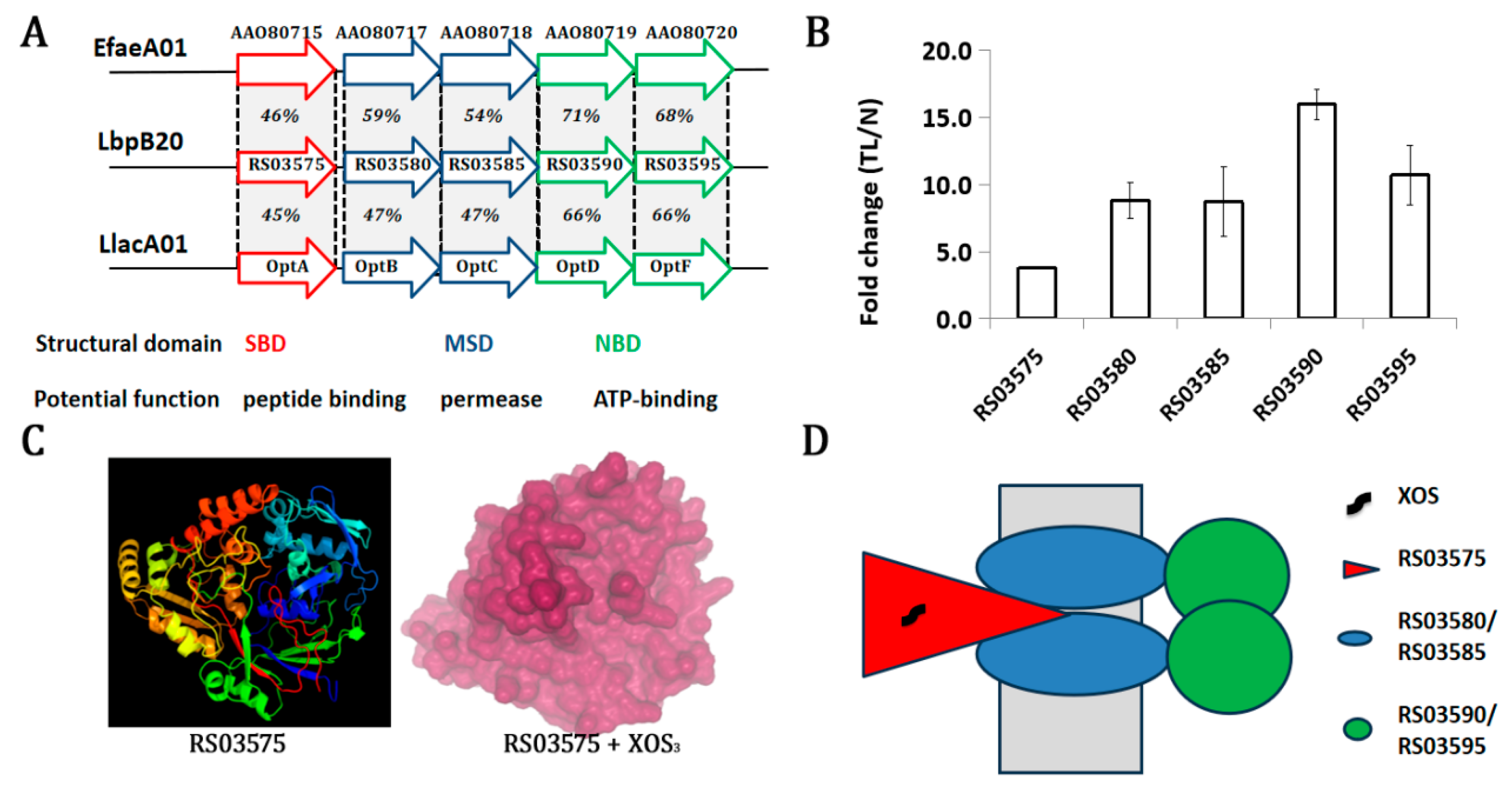
3.4. An XOS-Inductive ABC Transporter May Mediate Its Uptake

Among these DEGs, RS03575 was significantly upregulated and annotated as an ABC transporter ATP-binding protein. Four adjacent genes (RS03580, RS03585, RS03590, and RS03595) were also upregulated and encode two ABC permeases and two ATP-binding cassette domain-containing proteins. Transcript prediction by Rockhopper indicates that these four genes are co-transcribed as an operon. This operon and an upstream nearby gene RS03575 formed a gene cluster, which was annotated as an oligopeptide ABC transporter in the STRING database. The top hit names for these five transcripts are oppA_Bacsu, oppB_Bacsu, oppC_Bacsu, oppD_Bacsu, and oppF_Strmu, respectively, in the Swissprot database, with identities ranging from 48% to 81%. Alignment in the validated ABC transporter database (ABCdb, https://www-abcdb.biotoul.fr, accessed on 8 October 2023) showed that the transporter (RS03575-03595) is similar to EfaeA01 (from Enterococcus faecalis strain V583) and LlacA01 (from Lactococcus lactis strain IL1403). It is worth noting that the substrate-binding protein RS03575 has lower identities than other components (46% and 45%, Figure 3A). In fact, the sugar-binding lipoprotein (XynE) of the ABC transporter interacted with different xylosaccharides, as demonstrated by isothermal titration calorimetry in Geobacillus stearothermophilus [27]. However, pairwise sequence alignment with EMBOSS Needle suggests only a 14.7% identity between RS03575 and XynE (GenBank accession: DQ868502.2), indicating that they are different (https://www.ebi.ac.uk/jdispatcher/psa/emboss_needle, accessed on 8 October 2023).
All components of the transporter were significantly upregulated in the TL group, which added 0.4% XOS as substrate (Figure 3B). The transporters oppABCDF, EfaeA01, and LlacA01 were all identified as peptide/oligopeptide ABC importers [28]. For oppABCDF, OppA is responsible for the capture of peptides from the external medium. Two integral, highly hydrophobic, membrane-spanning proteins, OppB and OppC, form a channel through the membrane used for peptide translocation [29]. However, as demonstrated in the hyperthermophilic bacterium Thermotoga maritima, several putative oligopeptide ABC transporters bind oligosaccharides with micromolar to nanomolar affinities and are used primarily for oligosaccharide transport [30]. High-throughput data mining and molecular docking confirm the multi-specificity of carbohydrate ABC transporters and they share subunits to transport different substrates. For example, ABC transporters mediating the uptake of sugar and phospholipid precursors are evolutionarily linked in Thermus thermophilus HB8 [31]. Moreover, a previously unidentified ABC sugar transporter TP6568, found within Streptomyces avermitilis, possesses a broad substrate specificity. It could not only promote the uptake of diverse monosaccharides and disaccharides but also enhance the utilization of industrial carbon sources such as starch, sucrose, and dextrin [32]. These results indicate that some ABC transporters might be moonlighting proteins that can be explored by bacteria for different substrate importation.
Structural analysis by Phyre2.2 showed that RS03575 can be modeled with 100% confidence and 93% coverage (508 residues) by the single highest-scoring template c5kztB with a PDB title “Listeria monocytogenes OppA bound to peptide”. Considering XOS3 is a major growth stimulator and the probiotic bacteria Lb. brevis can completely uptake it, we evaluated the binding potential to XOS with it as a ligand [33]. Docking with XOS3 by AutoDock Vina calculated a negative affinity (−6.656 kcal/mol), and the substrate can be completely embedded by RS03575 (Figure 3C). Khangwal reported that beta-D-xylosidase from B. adolescentis showed good effectual binding toward XOS2 with a binding energy of −4.2 kcal/mol [34]. A smaller energy value between RS03575 and XOS3 suggests that the binding affinity is strong. In addition, the interactions were stabilized by several hydrogen bonds; e.g., five hydrogen bonds can be formed between XOS3 and residues in the center of protein RS03575 (Figure S3). Precisely, Tyr326, Thr398, Leu505, Tyr532, and Asn538 are the major interacting residues that form hydrogen bonds. Similar to a recent study, the substrate-binding protein RS03575 has a long and narrow pocket accommodating XOS3 [35]. It was found that hydrophobic interactions provide thermodynamic stability to folded proteins and hydrogen bonds connect secondary structure elements, thereby contributing fundamentally to the binding of protein and ligand [36]. These data indicate that RS03575 has a high binding potential to XOS. Therefore, it is quite possible that the ABC transporter RS03575-03595 mediates XOS uptake in Lb. plantarum B20. Potentially, RS03575, containing a substrate-binding domain (SBD) localized in the cytoplasmic membrane, binds XOS, while the permeases RS03580/RS03585, containing membrane-spanning domains (MSDs), form a channel for importation. The ATP-binding proteins RS03590/RS03595, containing a nucleotide-binding domain (NBD), release XOS from permeases by providing energy (Figure 3D).
In fact, ABC transporters involved in the uptake of different oligomers have already been reported. It was confirmed that B. kashiwanohense strains utilize short- and long-chain xylan-derived and human milk oligosaccharides and possess genes for xylosidase and specific ABC transporter substrate-binding proteins that contribute to the utilization of versatile oligosaccharides [37]. Similarly, a gene cluster inuABCDEF (Ldb1381-1386) encoding an ABC transporter in Lb. delbrueckii JCM 1002T was induced when it was grown on inulin, suggesting that polymerized inulin-type fructans might be taken up by the ABC transporter [38]. In addition, in the ABC transporter for cellodextrin transportation in Clostridium thermocellum, it was identified that the ATPase-encoding gene clo1313_2554 is located outside the transporter gene cluster [39]. Thus, these previously published studies strongly support the hypothesis that the uptake of XOS in Lb. plantarum B20 is mediated by the ABC transporter RS03575-03595.

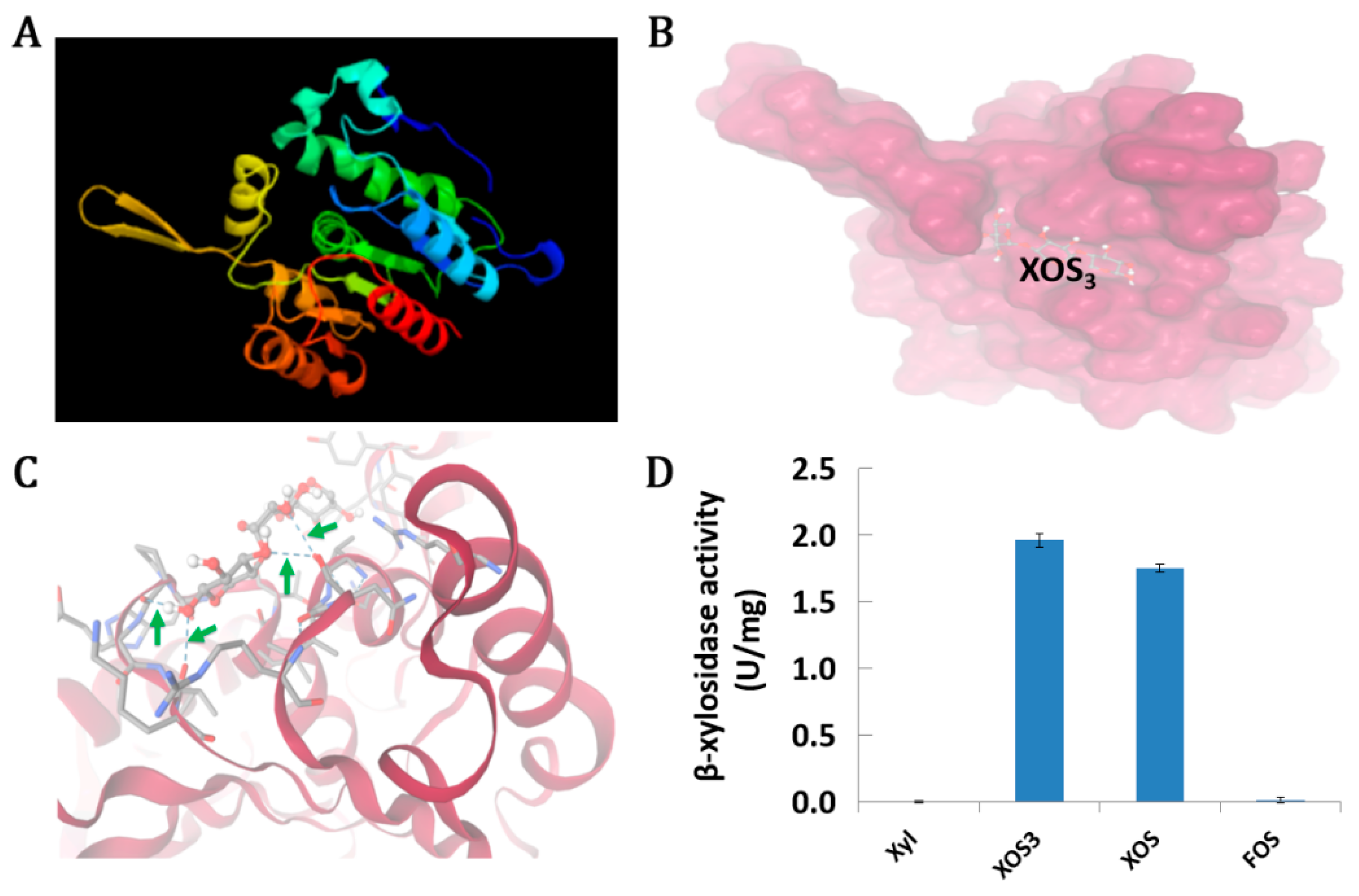
3.5. An Unknown Hydrolase RS06170 Can Degrade XOS

Through a keyword “xylanase” or “xylosidase” screen of the transcripts of Lb. plantarum B20 grown in the presence of XOS and xylose, it was found that four genes were potentially involved in XOS degradation as they were annotated as possessing endo-1,4-β-xylanase activity or xylan endo-1,3-β-xylosidase activity (Table S6). However, one gene is not expressed, and the difference in expression is insignificant in the other three genes. In contrast, we observed that an uncharacterized hydrolase (RS06170, as shown in Table 1) was significantly upregulated in the presence of XOS. Therefore, we analyzed the gene with a couple of bioinformatic tools. Subcellular location predicted that the protein is in the cytoplasm. The molecular weight of the protein is 28.8. SMART analysis indicates RS06170 has a hydrolase_4 domain. The domain is found in bacteria, and the majority of the members in this family carry the exopeptidase active-site residues. Through Phyre2.2 analysis, the protein has been modeled with 99.9% confidence and 92% coverage by the single highest-scoring template c6gocA with a PDB title “methylesterase bt1017” (Figure 4A). Further molecular docking simulation with XOS3 by AutoDock Vina indicates potential affinity (−5.489 kcal/mol), and the substrate can enter the groove of the protein (Figure 4B). In addition, four hydrogen bonds can be formed between XOS3 and residues on the surface of protein RS06170 (Figure 4C). It was predicted that XylA interacted with XOS3 by several amino acids in which the interaction is stabilized by four hydrogen bonds and several van der Waals interactions [40]. For RS06170, four hydrogen bonds can also be formed between XOS3 and residues on the surface of this protein. These residues include Val221, Lys234, ASN243, and Tyr248. Binding on the surface may facilitate the release of intermediate, thereby accelerating the catalysis.
Generally, endo-xylanases act on the backbone of the β-1,4-linked xylan, liberating xylooligomers, whereas β-xylosidases (EC 3.2.1.37) are active on these latter oligomers releasing xylose [41]. To catabolize XOS, β-xylosidases belonging to the GH43 family catalyze the reduction in XOS to free xylose using an inverting mechanism [42]. Despite the recognized potential of XOS as a prebiotic to target beneficial components of the human gut microbiota, very little is known about the enzymes used by lactobacilli to hydrolyze XOS. Although β-xylosidase activity has been detected from the cell crude extracts of Lb. brevis, Lb. plantarum, and Lb. sakei [3], the enzymes of β-xylosidase have only been characterized from three Lactobacillus species, e.g., one from Lb. brevis NCDC01, one (xylA) from Lb. rossiae DSM 15814T, and one from Lb. reuteri ATCC 53608 [41,43,44].
To further investigate the function of RS0170, the full-length coding sequence was cloned and expressed in E. coli BL21. As shown, the protein can be successfully overexpressed under IPTG induction (Figure S4). Enzymatic assays with recombinant E. coli crude cell extract indicate that RS06170 exhibits β-xylosidase activity but lacks xylanase activity. Consistent with the bioinformatic prediction, RS06170 is an intracellular enzyme, as no β-xylosidase activity can be detected from the culture supernatants. Substrate specificity assays demonstrate that RS06170 effectively degrades XOS and XOS3 but not FOS (Figure 4D). Further blast of the protein with all characterized β-xylosidases from lactobacilli as mentioned above demonstrates that they have little conservation. In fact, β-xylosidases are a group of structurally diverse enzymes with varying specificities due to the diversity of the organisms that produce them and the heterogeneity of their substrates. At least eight different glycoside hydrolase (GH) families possessing β-xylosidase activity in the CAZy database have been reported. Based on structural similarity, RS06170 is more likely to belong to GH5. Enzymes within this family have typical (β/α)8 TIM-barrels and catalyze the reaction via retention of the anomeric carbon configuration [42]. In addition, SMART analysis indicates that RS06170 has a hydrolase_4 domain. This domain is found in bacteria and the majority of the members in this family carry the exopeptidase active-site residues. Therefore, the protein identified in this study represents a novel β-xylosidase responsible for XOS catabolism in Lb. plantarum.

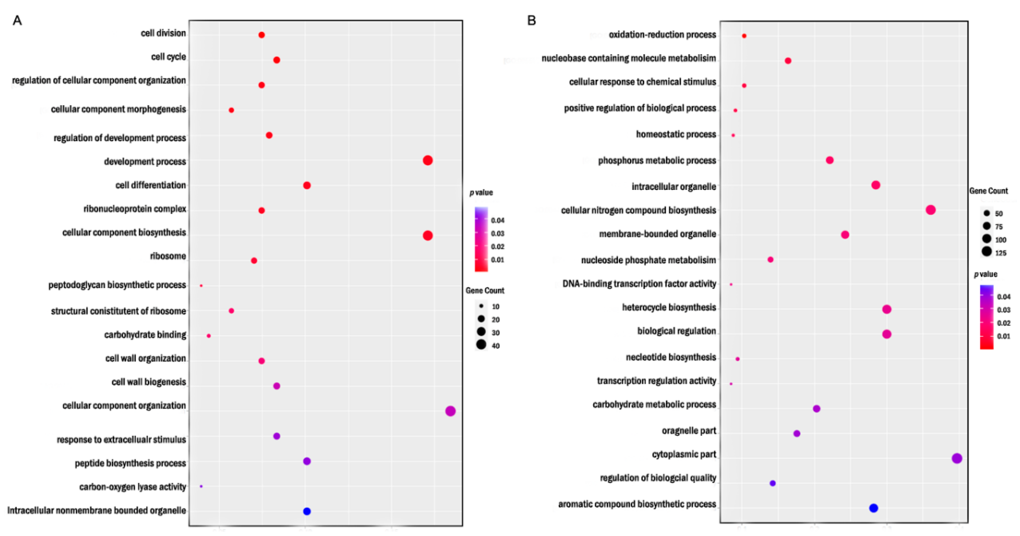
3.6. DEGs Were Enriched in Different GO Terms Under Different Levels of XOS

To explore the biological functions associated with DEGs under varying concentration of XOS, gene ontology (GO) enrichment analysis was performed. At 0.4% XOS, DEGs were significantly enriched in biological processes such as cell division, cell cycle, and cell differentiation (Figure 5A). Bacterial cell division is a replication process known as binary fission. To start genetic information synthesis, DNA must first be unwound by special proteins. Although the DNA in prokaryotes usually exists in a ring, it can become quite tangled when it is being used by the cell. To copy the DNA efficiently, it must be stretched out. The two strands of DNA separate into two different sides of the prokaryote cell. The cell then becomes longer and divides in the middle. Except for FtsA, the master regulator of bacterial division, numerous proteins that comprise the dynamic divisome coordinate membrane constriction with the synthesis of a division septum [45,46].
For Lb. plantarum B20 in the presence of 0.4% XOS, there are a large number of genes involved in cell division that were upregulated, including FtsL (RS07970) and a FtsW/RodA/SpoVE family protein (RS08805). As parts of the divisome, FtsL and FtsW interact with and are involved in coordinating the assembly and progression of the division process [47]. Additionally, as many as 39 genes involved in cellular component biogenesis are differentially expressed when XOS are used as a carbon source. The interference with the cell cycle by XOS has also been recently reported in E. coli, though in an opposite way [48].
When treated with 1.6% XOS, DEGs were significantly enriched in processes such as oxidation-reduction processes, nucleobase-containing molecules metabolism, and cellular response to chemical stimulus (Figure 5B). It is known that the reducing power and the growth rate increase in parallel (over considerable ranges) to their respective optima during the adaptation of cells to utilize a given carbon substrate. In general, oxidation can result in damage to cell functions, while reduction mitigates the damage of oxidative stress [49]. In cell biology, different stress response regulons are activated in bacteria, depending on the type of stressors. As a glycan, the hydrolysis of XOS involves multiple oxidation/reduction and elimination/hydration steps, each catalyzed by enzyme modules. In fact, homologues of these enzymes are common, and such alternative stepwise mechanisms appear to constitute abundant pathways for glycan degradation as part of the metabolism of carbohydrates in bacteria [50].

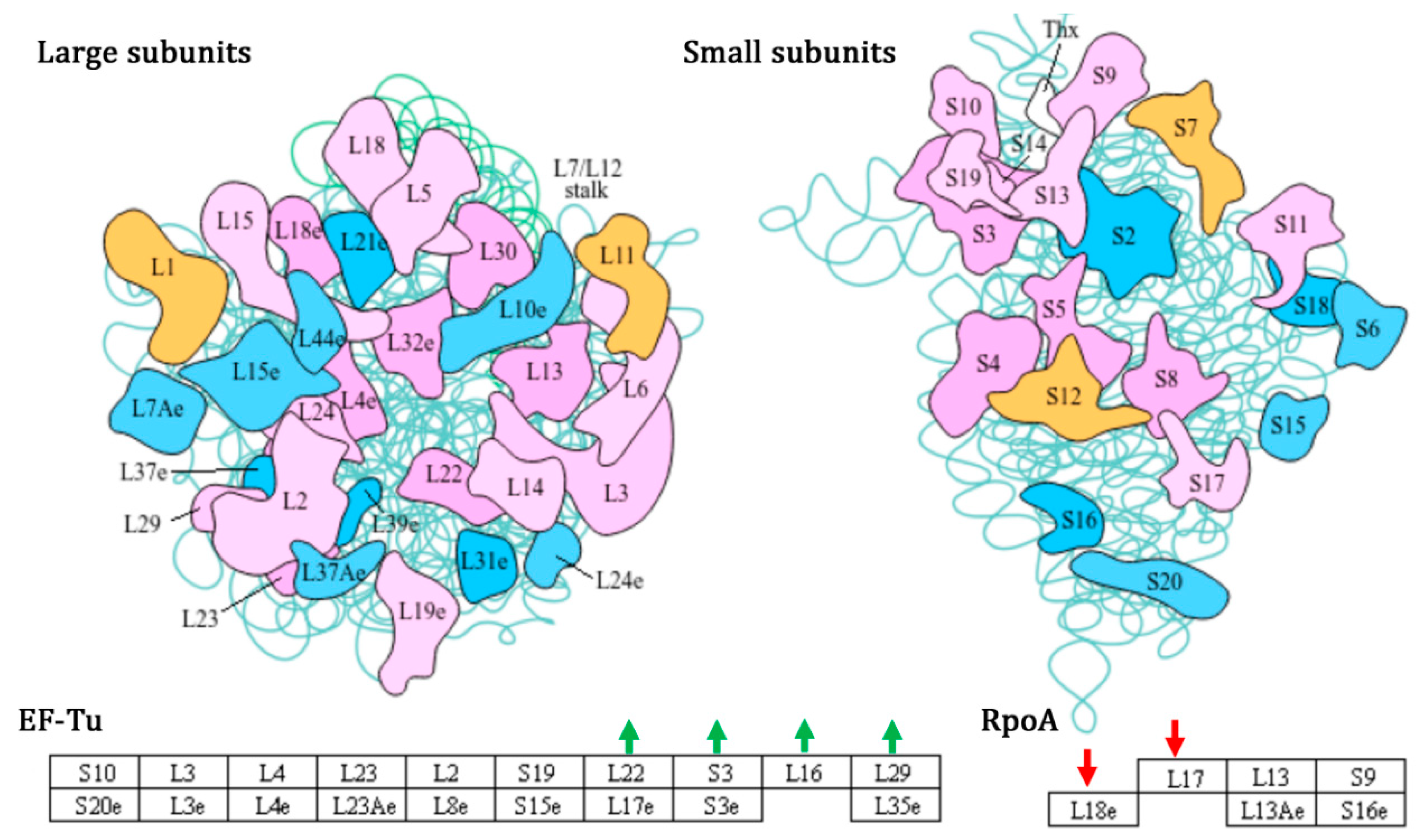
3.7. XOS-Promoted Growth of Lb. plantarum B20 by Impacting Ribosome and Carbohydrate Metabolism

The KEGG enrichment showed that XOS influences many pathways in Lb. plantarum B20 in both the TH and TL groups (Figure S5). In particular, many DEGs are found in the ribosome pathway (ko03010). Ribosomes are universally conserved ribonucleoprotein complexes involved in the decoding of the genetic information contained in messenger RNAs into proteins [51]. Accordingly, ribosome biogenesis is a fundamental cellular process required for functional ribosome homeostasis and to preserve satisfactory gene expression capability. It was postulated that the inhibition of growth of bacteria exposed to antibiotics can be almost uniquely due to reducing the number of ribosomes contributing to protein synthesis, i.e., the number of effective ribosomes [52]. On the other hand, the stimulation of growth and the higher replication rate can be potentially due to increasing the number of ribosomes or improving the function of ribosomes. In bacteria, cellular growth rate is connected to ribosome abundance, and cell size maintenance under nutrient perturbations requires a balanced trade-off between ribosomes and division protein synthesis [53]. Numerous studies have shown that cellular growth, stimulated by nutrients and/or growth factor signaling, is a prerequisite for cell cycle progression in most types of cells [54]. This seems true for Lb. plantarum B20 in the presence of XOS, as many genes related to ribosomes are upregulated. For example, four ribosomal proteins RS02640-RS02655 (rplV/L22, rpsC/S3, rplP/L16, rmpC/L29) as elongation factors Tu (EF-Tu) are all significantly upregulated, while RS02695 (rplR/L18) and RS02750 (rplQ/L17) are downregulated (Figure 6). In fact, in addition to Lb. plantarum, XOS also had a proliferative effect on Lb. brucelli, Lb. acidophilus, and Lb. rhamnosus [1].
It was also found that DEGs are enriched exclusively in carbohydrate metabolism, including glycolysis/gluconeogenesis (ko0010), starch and sucrose metabolism (ko00500), galactose metabolism (ko00052), and pyruvate metabolism (ko00620). Carbohydrates are degraded in central metabolic pathways to fuel cells with energy and building blocks to synthesize all biomolecules [55]. The process is controlled by large and densely interconnected regulatory networks. Efficient allocation of energy resources to key physiological functions allows living organisms to grow and thrive in diverse environments and adapt to a wide range of perturbations [56]. It was believed that a strong carbohydrate utilization ability is important for Lb. plantarum gastrointestinal tract colonization and probiotic effects [12]. During glycolysis, four genes, including RS02900 (L-lactate dehydrogenase, ldh), RS11185 (glycoside hydrolase family protein), RS12800 (phosphoenolpyruvate carboxykinase, pck), and RS13850 (acetaldehyde-CoA/alcohol dehydrogenase, adhE), are all downregulated in the presence of XOS. LDH and AdhE are key metabolic enzymes essential for glycolysis. LDH catalyzes the reversible conversion of pyruvate, the end product of glycolysis, to lactate, while AdhE converts acetyl-CoA to ethanol via an acetaldehyde intermediate during ethanol fermentation in an anaerobic environment [57]. Beyond gluconeogenesis, PCK is a rate-limiting enzyme that catalyzes the reversible conversion of oxaloacetic acid into phosphoenolpyruvate providing metabolites for several metabolic pathways [58,59]. In addition, the pathways of the phosphotransferase system (PTS) and oxidative phosphorylation were also influenced by XOS, indicating broader metabolic reconfiguration in response to this carbon source.

4. Conclusions

This study shows that only XOS have an obvious growth-promoting effect in Lb. plantarum B20, and the effect is both dose- and fraction-dependent. A supplement of XOS of at least 0.2% is needed for significant growth promotion of Lb. plantarum B20. All pure fractions of XOS stimulate growth, but XOS3 shows the strongest effect. XOS regulate the expression of a large number of genes in Lb. plantarum B20. The majority of DEGs are enriched in different GOs when the levels of XOS are different. Based on genomic and transcriptomic data, the uptake of XOS is potentially mediated by an ABC transporter composed of a five-gene cluster. Enzymatic tests indicated that Lb. plantarum B20 can degrade XOS by a novel alpha/beta hydrolase RS06170. Functional enrichment analysis suggests that the growth-promoting effect of XOS is partially due to the upregulation of many genes involved in cell division and cellular component biogenesis by impacting ribosome and carbohydrate metabolism.

Supplementary Materials

The following supporting information can be downloaded at: https://www.mdpi.com/article/10.3390/fermentation11050280/s1, Table S1. Composition and source of different carbohydrates used in the study. Table S2. The primers used in this study. Table S3. Summary of RNA-Seq data. Table S4. Summary of gene transcripts annotated in different databases in Lb. plantarum B20. Table S5. Summary of ABC transporter substrate-binding proteins on the genome of Lb. plantarum SK151. Table S6. Analysis of gene transcripts potentially involved in xylan metabolism in Lb. plantarum B20. Figure S1. Venn diagram of DEGs in Lb. plantarum B20 in the presence of XOS. Figure S2. qPCR validation of RNA-seq data using 7 up-regulated and 3 down-regulated representative genes. Figure S3. Hydrogen bonds formation between RS03575 and XOS3. Green arrows indicate hydrogen bonds between RS03575 residues and XOS3 molecule. Figure S4. SDS-PAGE detection of heterologous overexpressed RS06170 in E. coli BL21. Figure S5. KEGG enrichment of DEGs in the presence of 0.4% (A) or 1.6% (B) XOS.

Author Contributions

Conceptualization, Z.S.; methodology, Y.S. and H.W.; software, Z.N.; validation, H.W.; formal analysis, Y.S.; investigation, Y.S.; resources, Z.S.; data curation, Z.N. and Z.S.; writing—original draft, Y.S.; writing—review and editing; supervision; funding acquisition, Z.S. and C.L. All authors have read and agreed to the published version of the manuscript.

Funding

This study was supported by a Major Special Science and Technology Project of Henan Province (No. 231100110300) and the National Key R&D Project (No. 2022YFD2101405).

Institutional Review Board Statement

Not applicable.

Informed Consent Statement

Not applicable.

Data Availability Statement

The original contributions presented in this study are included in the article/Supplementary Materials. Further inquiries can be directed to the corresponding authors.

Acknowledgments

The authors acknowledge Erting Liu from Henan Heagreen Biotechnology Co., Ltd. for providing XOS samples. The authors also thank Muhammad Imran for an extensive language polishing during the revision of this paper.

Conflicts of Interest

The authors declare no conflict of interest.

References

  1. Tian, S.; Yang, Z.; Yan, F.; Xue, X.; Lu, J. Preparation of Xylooligosaccharides from Rice Husks and Their Structural Characterization, Antioxidant Activity, and Probiotic Properties. Int. J. Biol. Macromol. 2024, 271, 132575. [Google Scholar] [CrossRef]
  2. Sun, Z.; Yue, Z.; Liu, E.; Li, X.; Li, C. Assessment of the Bifidogenic and Antibacterial Activities of Xylooligosaccharide. Front. Nutr. 2022, 9, 858949. [Google Scholar] [CrossRef]
  3. Iliev, I.; Vasileva, T.; Bivolarski, V.; Momchilova, A.; Ivanova, I. Metabolic Profiling of Xylooligosaccharides by Lactobacilli. Polymers 2020, 12, 2387. [Google Scholar] [CrossRef]
  4. Puja, B.K.; Mallick, S.; Dey, T.; Chanda, S.; Ghosh, S. Xylooligosaccharide Recovery from Sugarcane Bagasse Using β-Xylosidase-Less Xylanase, BsXln1, Produced by Bacillus stercoris DWS1: Characterization, Antioxidant Potential and Influence on Probiotics Growth under Anaerobic Conditions. Int. J. Biol. Macromol. 2025, 285, 138307. [Google Scholar] [CrossRef]
  5. Li, Z.; Summanen, P.H.; Komoriya, T.; Finegold, S.M. In vitro study of the prebiotic xylooligosaccharide (XOS) on the growth of Bifidobacterium spp and Lactobacillus spp. Int. J. Food Sci. Nutr. 2015, 66, 919–922. [Google Scholar] [CrossRef]
  6. Ejby, M.; Fredslund, F.; Vujicic-Zagar, A.; Svensson, B.; Slotboom, D.J.; Abou Hachem, M. Structural Basis for Arabinoxylo-Oligosaccharide Capture by the Probiotic Bifidobacterium animalis subsp. lactis Bl-04. J. Mol. Microbiol. Biotechnol. 2013, 90, 1100–1112. [Google Scholar] [CrossRef]
  7. Mukherjee, S.; Lodha, T.D.; Madhuprakash, J. Comprehensive Genome Analysis of Cellulose and Xylan-Active CAZymes from the Genus Paenibacillus: Special Emphasis on the Novel Xylanolytic Paenibacillus sp. LS1. Microbiol. Spectrum. 2023, 11, e05028-22. [Google Scholar] [CrossRef]
  8. Gilad, O.; Jacobsen, S.; Stuer-Lauridsen, B.; Pedersen, M.B.; Garrigues, C.; Svensson, B. Combined Transcriptome and Proteome Analysis of Bifidobacterium animalis Subsp. lactis BB-12 Grown on Xylo-Oligosaccharides and a Model of Their Utilization. Appl. Environ. Microbiol. 2010, 76, 7285–7291. [Google Scholar] [CrossRef]
  9. Zúñiga, M.; Yebra, M.J.; Monedero, V. Complex Oligosaccharide Utilization Pathways in Lactobacillus. Curr. Issues Mol. Biol. 2021, 40, 49–80. [Google Scholar] [CrossRef] [PubMed]
  10. Michlmayr, H.; Hell, J.; Lorenz, C.; Böhmdorfer, S.; Rosenau, T.; Kneifel, W. Arabinoxylan Oligosaccharide Hydrolysis by Family 43 and 51 Glycosidases from Lactobacillus brevis DSM 20054. Appl. Environ. Microbiol. 2013, 79, 6747–6754. [Google Scholar] [CrossRef] [PubMed]
  11. Lokman, B.C.; Santen, P.V.; Verdoes, J.C.; Kruse, J.; Leer, R.J.; Posno, M.; Pouwels, P.H. Organization and Characterization of Three Genes Involved in D-Xylose Catabolism in Lactobacillus pentosus. Molec. Gen. Genet. 1991, 230, 161–169. [Google Scholar] [CrossRef] [PubMed]
  12. Cui, Y.; Wang, M.; Zheng, Y.; Miao, K.; Qu, X. The Carbohydrate Metabolism of Lactiplantibacillus plantarum. Int. J. Mol. Sci. 2021, 22, 13452. [Google Scholar] [CrossRef]
  13. Livak, K.J.; Schmittgen, T.D. Analysis of relative gene expression data using real-time quantitative PCR and the 2(-Delta Delta C(T)) Method. Methods 2001, 25, 402–408. [Google Scholar] [CrossRef] [PubMed]
  14. Powell, H.R.; Islam, S.A.; David, A.; Sternberg, M.J.E. Phyre2.2: A Community Resource for Template-Based Protein Structure Prediction. J. Mol. Biol. 2025, 168960. [Google Scholar] [CrossRef] [PubMed]
  15. Ma, J.; Sun, Z.; Ni, Z.; Qi, Y.; Sun, Q.; Hu, Y.; Li, C. Molecular Identification and Engineering a Salt-Tolerant GH11 Xylanase for Efficient Xylooligosaccharides Production. Biomolecules. 2024, 14, 1188. [Google Scholar] [CrossRef]
  16. Das, R.; Mishra, P.; Mishra, B.; Jha, R. Effects of in ovo feeding of xylobiose and xylotriose on growth performance, carcass traits, ileal histomorphometry, and immune-related gene expression in broiler chickens. Anim. Feed Sci. Technol. 2024, 313, 115998. [Google Scholar] [CrossRef]
  17. Pavankumar, T.L. RNase R vs. PNPase: Selecting the best-suited exoribonuclease for environmental adaptation. Extremophiles 2024, 28, 35. [Google Scholar] [CrossRef]
  18. Dimitrova-Paternoga, L.; Kasvandik, S.; Beckert, B.; Granneman, S.; Tenson, T.; Wilson, D.N.; Paternoga, H. Structural basis of ribosomal 30S subunit degradation by RNase R. Nature 2024, 626, 1133–1140. [Google Scholar] [CrossRef]
  19. Suman; Chaudhary, M.; Nain, V. In Silico Identification and Evaluation of Bacillus Subtilis Cold Shock Protein B (cspB)-Like Plant RNA Chaperones. J. Biomol. Struct. Dyn. 2021, 39, 841–850. [Google Scholar] [CrossRef]
  20. Bilsing, F.L.; Anlauf, M.T.; Hachani, E.; Khosa, S.; Schmitt, L. ABC Transporters in Bacterial Nanomachineries. Int. J. Mol. Sci. 2023, 24, 6227. [Google Scholar] [CrossRef]
  21. Pulianmackal, L.T.; Vecchiarelli, A.G. Positioning of Cellular Components by the ParA/MinD Family of ATPases. Curr. Opin. Microbiol. 2024, 79, 102485. [Google Scholar] [CrossRef] [PubMed]
  22. Goh, Y.J.; Klaenhammer, T.R. Genetic Mechanisms of Prebiotic Oligosaccharide Metabolism in Probiotic Microbes. Annu. Rev. Food Sci. Technol. 2015, 6, 137–156. [Google Scholar] [CrossRef]
  23. Jeckelmann, J.-M.; Erni, B. Transporters of Glucose and Other Carbohydrates in Bacteria. Pflugers Arch-Eur. J. Physiol. 2020, 472, 1129–1153. [Google Scholar] [CrossRef] [PubMed]
  24. Andersen, J.M.; Barrangou, R.; Hachem, M.A.; Lahtinen, S.J.; Goh, Y.J.; Svensson, B.; Klaenhammer, T.R. Transcriptional Analysis of Oligosaccharide Utilization by Bifidobacterium lactis Bl-04. BMC Genomics 2013, 14, 312. [Google Scholar] [CrossRef]
  25. Watanabe, A.; Hiraga, K.; Suda, M.; Yukawa, H.; Inui, M. Functional Characterization of Corynebacterium alkanolyticum β-Xylosidase and Xyloside ABC Transporter in Corynebacterium glutamicum. Appl. Environ. Microbiol. 2015, 81, 4173–4183. [Google Scholar] [CrossRef]
  26. Chen, Y.; Zhou, J.; Li, X.; Li, X.; Qiu, S.; Xu, Z.; Yang, J.; Zhu, Y.; Zhang, X.; Yan, L. Phenotypic and Genomic Characterization of Levilactobacillus brevis YT108: A Potential Probiotic Strain Capable of Metabolizing Xylo-Oligosaccharides. Lett. Appl. Microbiol. 2025, 78, ovaf014. [Google Scholar] [CrossRef] [PubMed]
  27. Shulami, S.; Zaide, G.; Zolotnitsky, G.; Langut, Y.; Feld, G.; Sonenshein, A.L.; Shoham, Y. A Two-Component System Regulates the Expression of an ABC Transporter for Xylo-Oligosaccharides in Geobacillus searothermophilus. Appl. Environ. Microbiol. 2007, 73, 874–884. [Google Scholar] [CrossRef]
  28. Masulis, I.S.; Sukharycheva, N.A.; Kiselev, S.S.; Andreeva, Z.S.; Ozoline, O.N. Between Computational Predictions and High-Throughput Transcriptional Profiling: In Depth Expression Analysis of the OppB Trans-Membrane Subunit of Escherichia coli OppABCDF Oligopeptide Transporter. Res. Microbiol. 2020, 171, 55–63. [Google Scholar] [CrossRef]
  29. Yang, X.; Hu, T.; Liang, J.; Xiong, Z.; Lin, Z.; Zhao, Y.; Zhou, X.; Gao, Y.; Sun, S.; Yang, X.; et al. An oligopeptide permease, OppABCD, requires an iron-sulfur cluster domain for functionality. Nat. Struct. Mol. Biol. 2024, 31, 1072–1082. [Google Scholar] [CrossRef]
  30. Nanavati, D.M.; Thirangoon, K.; Noll, K.M. Several Archaeal Homologs of Putative Oligopeptide-Binding Proteins Encoded by Thermotoga maritima Bind Sugars. Appl. Environ. Microbiol. 2006, 72, 1336–1345. [Google Scholar] [CrossRef]
  31. Chandravanshi, M.; Sharma, A.; Dasgupta, P.; Mandal, S.K.; Kanaujia, S.P. Identification and characterization of ABC transporters for carbohydrate uptake in Thermus thermophilus HB8. Gene 2019, 696, 135–148. [Google Scholar] [CrossRef] [PubMed]
  32. Dong, Z.; Li, L.; Du, G.; Zhang, Y.; Wang, X.; Li, S.; Xiang, W. A previously unidentified sugar transporter for engineering of high-yield Streptomyces. Appl. Microbiol. Biotechnol. 2024, 108, 72. [Google Scholar] [CrossRef] [PubMed]
  33. Mathew, S.; Aronsson, A.; Karlsson, E.N.; Adlercreutz, P. Xylo- and arabinoxylooligosaccharides from wheat bran by endoxylanases, utilisation by probiotic bacteria, and structural studies of the enzymes. Appl. Microbiol Biotechnol. 2018, 102, 3105–3120. [Google Scholar] [CrossRef]
  34. Khangwal, I.; Skariyachan, S.; Uttarkar, A.; Muddebihalkar, A.G.; Niranjan, V.; Shukla, P. Understanding the Xylooligosaccharides Utilization Mechanism of Lactobacillus brevis and Bifidobacterium adolescentis: Proteins Involved and Their Conformational Stabilities for Effectual Binding. Mol. Biotechnol. 2022, 64, 75–89. [Google Scholar] [CrossRef]
  35. Saberi, M.; Dekkers, R.; Passerini, L.; Huber, M.; Overhand, M.; Ubbink, M. Terminal spin labeling of xylotriose strongly affects interactions in the active site of xylanase BcX. J. Biomol. NMR, 2025; advance online publication. [Google Scholar] [CrossRef]
  36. Ferenczy, G.G.; Kellermayer, M. Contribution of hydrophobic interactions to protein mechanical stability. Comput. Struct. Biotechnol. J. 2022, 20, 1946–1956. [Google Scholar] [CrossRef]
  37. Orihara, K.; Yahagi, K.; Saito, Y.; Watanabe, Y.; Sasai, T.; Hara, T.; Tsukuda, N.; Oki, K.; Fujimoto, J.; Matsuki, T. Characterization of Bifidobacterium kashiwanohense that utilizes both milk- and plant-derived oligosaccharides. Gut Microbes 2023, 15, 2207455. [Google Scholar] [CrossRef] [PubMed]
  38. Tsujikawa, Y.; Ishikawa, S.; Sakane, I.; Yoshida, K.I.; Osawa, R. Identification of genes encoding a novel ABC transporter in Lactobacillus delbrueckii for inulin polymers uptake. Sci. Rep. 2021, 11, 16007. [Google Scholar] [CrossRef]
  39. Yan, F.; Dong, S.; Liu, Y.J.; Yao, X.; Chen, C.; Xiao, Y.; Bayer, E.A.; Shoham, Y.; You, C.; Cui, Q.; et al. Deciphering Cellodextrin and Glucose Uptake in Clostridium thermocellum. mBio 2022, 13, e0147622. [Google Scholar] [CrossRef]
  40. Skariyachan, S.; Khangwal, I.; Niranjan, V.; Kango, N.; Shukla, P. Deciphering effectual binding potential of xylo-substrates towards xylose isomerase and xylokinase through molecular docking and molecular dynamic simulation. J. Biomol. Struct. Dyn. 2021, 39, 3948–3957. [Google Scholar] [CrossRef]
  41. Singh, R.P.; Bhaiyya, R.; Thakur, R.; Niharika, J.; Singh, C.; Latousakis, D.; Saalbach, G.; Nepogodiev, S.A.; Singh, P.; Sharma, S.C.; et al. Biochemical Basis of Xylooligosaccharide Utilisation by Gut Bacteria. Int. J. Mol. Sci. 2022, 23, 2992. [Google Scholar] [CrossRef] [PubMed]
  42. Rohman, A.; Dijkstra, B.W.; Puspaningsih, N.N.T. β-Xylosidases: Structural Diversity, Catalytic Mechanism, and Inhibition by Monosaccharides. Int. J. Mol. Sci. 2019, 20, 5524. [Google Scholar] [CrossRef] [PubMed]
  43. Lasrado, L.D.; Gudipati, M. Purification and Characterization of β-D-Xylosidase from Lactobacillus brevis Grown on Xylo-Oligosaccharides. Carbohydr. Polym. 2013, 92, 1978–1983. [Google Scholar] [CrossRef]
  44. Pontonio, E.; Mahony, J.; Di Cagno, R.; O’Connell Motherway, M.; Lugli, G.A.; O’Callaghan, A.; De Angelis, M.; Ventura, M.; Gobbetti, M.; van Sinderen, D. Cloning, Expression and Characterization of a β-D-Xylosidase from Lactobacillus rossiae DSM 15814T. Microb. Cell Fact. 2016, 15, 72. [Google Scholar] [CrossRef]
  45. Mahone, C.R.; Goley, E.D. Bacterial Cell Division at a Glance. J. Cell Sci. 2020, 133, jcs237057. [Google Scholar] [CrossRef]
  46. Cameron, T.A.; Margolin, W. Insights into the Assembly and Regulation of the Bacterial Divisome. Nat. Rev. Microbiol. 2024, 22, 33–45. [Google Scholar] [CrossRef] [PubMed]
  47. Villanelo, F.; Ordenes, A.; Brunet, J.; Lagos, R.; Monasterio, O. A Model for the Escherichia coli FtsB/FtsL/FtsQ Cell Division Complex. BMC Struct. Biol. 2011, 11, 28. [Google Scholar] [CrossRef]
  48. Ren, L.; Ye, H.; Fang, J.; Cao, Q.; Zhang, C.; Dong, Z.; Feng, D.; Zuo, J.; Wang, W. Xylooligosaccharide Interferes with the Cell Cycle and Reduces the Antibiotic Tolerance of Avian Pathogenic Escherichia coli by Associating with Its Potential Antimetabolic Actions. Poult. Sci. 2024, 103, 104405. [Google Scholar] [CrossRef]
  49. Fasnacht, M.; Polacek, N. Oxidative Stress in Bacteria and the Central Dogma of Molecular Biology. Front. Mol. Biosci. 2021, 8, 671037. [Google Scholar] [CrossRef]
  50. Nasseri, S.A.; Lazarski, A.C.; Lemmer, I.L.; Zhang, C.Y.; Brencher, E.; Chen, H.-M.; Sim, L.; Panwar, D.; Betschart, L.; Worrall, L.J.; et al. An Alternative Broad-Specificity Pathway for Glycan Breakdown in Bacteria. Nature 2024, 631, 199–206. [Google Scholar] [CrossRef]
  51. Jüttner, M.; Ferreira-Cerca, S. Ribosome Biogenesis: Methods and Protocols, 1st ed.; Humana Press: New York, NY, USA, 2022; pp. 3–22. ISBN 978-01-4939-2685-7. [Google Scholar]
  52. Levin, B.R.; McCall, I.C.; Perrot, V.; Weiss, H.; Ovesepian, A.; Baquero, F. A Numbers Game: Ribosome Densities, Bacterial Growth, and Antibiotic-Mediated Stasis and Death. mBio 2017, 8, e02253-16. [Google Scholar] [CrossRef] [PubMed]
  53. Serbanescu, D.; Ojkic, N.; Banerjee, S. Nutrient-Dependent Trade-Offs between Ribosomes and Division Protein Synthesis Control Bacterial Cell Size and Growth. Cell Rep. 2020, 32, 108183. [Google Scholar] [CrossRef] [PubMed]
  54. Øvrebø, J.I.; Ma, Y.; Edgar, B.A. Cell growth and the cell cycle: New insights about persistent questions. BioEssays News Rev. Mol. Cell. Dev. Biol. 2022, 44, e2200150. [Google Scholar] [CrossRef] [PubMed]
  55. Durica-Mitic, S.; Göpel, Y.; Görke, B. Carbohydrate Utilization in Bacteria: Making the Most Out of Sugars with the Help of Small Regulatory RNAs. Microbiol. Spectr. 2018, 6, 229–248. [Google Scholar] [CrossRef]
  56. Cylke, A.; Serbanescu, D.; Banerjee, S. Energy allocation theory for bacterial growth control in and out of steady state. bioRxiv 2024. [CrossRef]
  57. Pony, P.; Rapisarda, C.; Terradot, L.; Marza, E.; Fronzes, R. Filamentation of the Bacterial Bi-Functional Alcohol/Aldehyde Dehydrogenase AdhE is Essential for Substrate Channeling and Enzymatic Regulation. Nat. Commun. 2020, 11, 1426. [Google Scholar] [CrossRef]
  58. Yu, S.; Meng, S.; Xiang, M.; Ma, H. Phosphoenolpyruvate Carboxykinase in Cell Metabolism: Roles and Mechanisms beyond Gluconeogenesis. Mol. Metab. 2021, 53, 101257. [Google Scholar] [CrossRef]
  59. Latorre-Muro, P.; Baeza, J.; Hurtado-Guerrero, R.; Hicks, T.; Delso, I.; Hernández-Ruiz, C.; Velázquez-Campoy, A.; Lawton, A.J.; Angulo, J.; Denu, J.M.; et al. Self-Acetylation at the Active Site of Phosphoenolpyruvate Carboxykinase (PCK1) Controls Enzyme Activity. J. Biol. Chem. 2021, 296, 100205. [Google Scholar] [CrossRef]
Figure 1. The growth-promoting effect of different oligosaccharides in Lb. plantarum B20: (A) relative growth of OD600 in the presence of 0.4% different oligosaccharides; (B) relative lactic acid production in the supernatant after fermentation with 0.4% (w/v) different oligosaccharides; (C) relative growth of OD600 supplemented with 0–1.6% XOS, respectively; (D) relative growth of OD600 supplemented with 0.4% different XOS fractions. Strain B20 was inoculated in modified 5 mL MRS broth supplemented with corresponding sugars. Samples were collected after 12 and 24 h of inoculation. All data were the mean of three independent experiments assayed in triplicates and relative to that of samples collected from the modified MRS broth supplemented with corresponding monosaccharide. The red dashed lines indicate the relative values when corresponding monomers were used as substrates. Significance was analyzed by Tukey’s one-way analysis of variance (ANOVA) when relative values are higher than 1 (p < 0.05). *, significantly stimulated by XOS after incubation for 12 h; #, significantly stimulated by XOS after incubation for 24 h; FOS, fructooligosaccharide; GOS, galactooligosaccharide; PDX, polydextrose; XOS, xylooligosaccharide; Xyl, xylose; XOS2, xylobiose; XOS3, xylotriose; XOS4, xylotetraose; XOS5, xylopentaose; XOS6, xylohexaose.
Fermentation 11 00280 g001
Figure 2. The transcriptomic data of Lb. plantarum B20 in the presence of different levels XOS: (A) principal component analysis (PCA) of nine samples from different treatments; (B) heatmap demonstration of nine samples from three groups; (C) Venn diagram of upregulated DEGs in two XOS-treated groups; (D) Venn diagram of downregulated DEGs in two XOS treated groups. N1-3, samples treated with 1.6% xylose; TH1-3, XOS treated samples at high level (1.6%); TL1-3, XOS-treated samples at low level (0.4%).
Fermentation 11 00280 g002
Figure 3. Predicted ABC transporter for XOS uptake in Lb. plantarum B20: (A) BLAST analysis of the gene cluster (RS03575-RS03595) with reported ABC transporters; (B) the transcription of these genes in the presence of 0.4% XOS; (C) the predicted structure and molecular docking between XOS2 and substrate-binding protein RS03575; (D) proposed uptake model of the ABC transporter. EfaeA01, ABC transporter from Enterococcus faecalis strain V583; LlacA01, ABC transporter from Lactococcus lactis strain IL1403; LbpB20, ABC transporter from Lb. plantarum B20; SBD, substrate-binding domain; MSD, membrane-spanning domain; NBD, nucleotide-binding domain.
Fermentation 11 00280 g003
Figure 4. Analysis of the hydrolase RS06170 and activity assays in vitro: (A) predicted structure by Phyre 2.2; (B) molecular docking of RS06170 and XOS3 by Autodock Vina; (C), hydrogen bonds formation between RS06170 and XOS3; (D) activities of the heterologous protein RS06170 with different substrates. The 3D structures were predicted using the NORMAL mode. The top model with 99.9% confidence and more than 90% coverage was presented. Green arrows indicate hydrogen bonds. FOS, fructooligosaccharide; XOS, xylooligosaccharide; Xyl, xylose; XOS3, xylotriose.
Fermentation 11 00280 g004
Figure 5. GO enrichment of DEGs in Lb. plantarum B20 in the presence of different levels XOS: (A) GO dotplot of DEGs in the presence of 0.4% XOS (TL vs. N); (B) GO dotplot of DEGs in the presence of 1.6% XOS (TH vs. N). Only the top 20 GO terms with p < 0.05 were demonstrated.
Fermentation 11 00280 g005
Figure 6. Analysis of the Lb. plantarum B20 ribosome pathway in the presence of 0.4% XOS. In the upside, images are the large and small subunits of ribosome. In the downside, boxes are ribosomal proteins. Green arrows indicate the upregulation of corresponding genes; red arrows indicate the downregulation of corresponding genes when bacteria are grown on medium supplemented with XOS.
Fermentation 11 00280 g006
Table 1. Top 20 upregulated genes in Lb. plantarum B20 in the presence of XOS at different doses.
Seq. idlog2FC(TH/N)p-Valuelog2FC(TL/N)p-ValueFunction
RS061707.70.0008.10.000alpha/beta fold hydrolase
RS016056.90.0006.80.000ribonuclease R
RS119755.80.0007.20.000ParA family protein
RS017906.90.0003.70.002hypothetical protein HMPREF0531_11053
RS123053.70.0005.00.000AI-2E family transport protein
RS130703.30.0005.10.000UDP-glucose 4-epimerase GalE
RS054704.20.0004.10.000ACP S-malonyltransferase
RS072055.00.0003.10.000Chaperone protein DnaK
RS030355.20.0002.90.016trans-hexaprenyltranstransferase, component II
RS130653.70.0003.80.000UDP-glucose-hexose-1-phosphate uridylyltransferase
RS054853.10.0054.30.000acetyl-CoA carboxylase, biotin carboxyl carrier protein
RS150653.80.0003.40.008DUF2089 family protein
RS035902.50.0003.90.000ABC transporter ATP-binding protein
RS050802.20.0014.00.000hypothetical protein
RS047903.00.0222.70.00650S ribosomal protein L35
RS039952.70.0002.50.000hypothetical protein HMPREF0531_12577
RS026502.40.0002.50.00450S ribosomal protein L16
RS011502.30.0062.40.029recombination protein RecR
RS072802.10.0452.60.022YlxR family protein
RS119702.00.0002.50.000ParB/RepB/Spo0J family partition protein, DNA-binding protein
Table 2. Genomic analysis of transporters and enzymes potentially involved in XOS catabolism in Lb. plantarum (taxid: 1590).
FunctionQueryDescriptionTop HitIdentity (%)Reference
UptakeBIF_00212Sugar-binding proteinWP_063847148.123.23[8]
BIF_00257
BIF_00258
ABC transporter permeasePOD88560.1
WP_072539333.1
28.38
34.78
BALAC_0514Sugar-binding proteinWP_063847148.123.23[6]
BALAC_0515
BALAC_0516
ABC transporter permeasePOD88560.1
WP_072539333.1
28.38
34.78
[24]
xylE (AJY53615.1)Solute-binding proteinWP_063847148.125.63[25]
xylF (AJY53616.1)
xylG (AJY53617.1)
ABC transporter permeasePOD88560.1
WP_072539333.1
27.27
32.91
DegradationBIF_00928
BIF_00633
Endo-1,4-β-xylanaseWP_097558172.1
WP_063486650.1
37.01
22.44
[8]
BIF_00405
BIF_00092
β-xylosidaseBBM21911.1
WP_063486650.1
27.49
30.11
xylD (AJY53618.1)β-xylosidaseWP_114648652.130.03[25]
xynB1 (LVIS_0375)β-xylosidaseBBM21911.127.96[10]
xynB2 (LVIS_2285) BBM21911.129.17
ConversionBIF_00501Xylose isomeraseWP_021337395.170.14[8]
BIF_00829Xylulose kinaseWP_021337394.126.28
BALAC_0521Xylulose kinaseWP_021337394.126.28[24]
xylA (P21938.1)Xylose isomeraseWP_021337395.199.11[11]
xylB (P21939.1)Xylulose kinaseWP_021337394.198.20
Disclaimer/Publisher’s Note: The statements, opinions and data contained in all publications are solely those of the individual author(s) and contributor(s) and not of MDPI and/or the editor(s). MDPI and/or the editor(s) disclaim responsibility for any injury to people or property resulting from any ideas, methods, instructions or products referred to in the content.

