Article

Characterization, Production, and Application of Antifungal Metabolites from Probiotic Levilactobacillus and Lactiplantibacillus Strains Isolated from Fermented Olives

1
Research Unit of Microbiology, Biomolecules, and Biotechnology, Laboratory of Chemistry-Physics and Biotechnology of Molecules and Materials, Faculty of Sciences and Techniques—Mohammedia, Hassan II University of Casablanca, Casablanca 28806, Morocco
2
Laboratory of Bioresources, Biotechnology, Ethnopharmacology and Health, Faculty of Sciences, Mohammed Premier University, Oujda 60000, Morocco
3
Laboratory of Natural Resources and Environment, Polydisciplinary Faculty of Taza, Sidi Mohamed Ben Abdellah University, B. P 1223, Taza 35000, Morocco
4
Research Unit Bioprocess and Biointerfaces, Laboratory of Industrial Engineering and Surface Engineering, National School of Applied Sciences, Sultan Moulay Slimane University, Mghila, Beni Mellal 23000, Morocco
5
Department of Food Science and Agricultural Chemistry, Macdonald Campus, McGill University, 21111 Lakeshore, Sainte-Anne-de-Bellevue, QC H9X 3V9, Canada
*
Author to whom correspondence should be addressed.
Fermentation 2025, 11(12), 661; https://doi.org/10.3390/fermentation11120661
Submission received: 31 October 2025 / Revised: 21 November 2025 / Accepted: 23 November 2025 / Published: 26 November 2025
(This article belongs to the Special Issue Recent Trends in Lactobacillus and Fermented Food, 3rd Edition)

Abstract

In this study, the characterization, production, and application of antifungal metabolites obtained from the Levilactobacillus (L. brevis S27) and two Lactiplantibacillus (L. pentosus S42 and L. plantarum S62) strains were evaluated. The lactic acid bacteria (LAB) cells showed antifungal activity against molds. The cell-free supernatant (CFS) of Levilactobacillus and Lactiplantibacillus presented antimicrobial activity against fungi and foodborne pathogenic bacteria, and the antifungal activity was significantly (p < 0.05) higher than that of the antibacterial activity. Moreover, the antifungal metabolites were characterized as proteinaceous compounds that remained stable under both high and low temperatures and demonstrated activity across a broad pH range. Additionally, metabolite production was significantly higher (p < 0.05) at an initial pH of 5 when incubated at either 25 °C or 37 °C. All strains and their CFSs exhibited strong bio-preservative effects against Penicillium digitatum in yogurt and against Aspergillus niger on orange fruit. Consequently, these Lactobacilli strains and their antifungal metabolites represent a novel approach to biocontrol in the agri-food industry and agricultural products.

1. Introduction

Fungal growth, caused by organisms like Aspergillus, Penicillium, and Fusarium, can spoil a vast array of foods, including grains, nuts, fruits, and dairy products. Fungal spoilage is one of the most significant barriers to the widespread acceptance of food products by consumers, resulting in substantial economic losses and significant food waste. Fungal contamination can occur at various steps in food processing [1], and beyond their negative impact on food quality, some fungal genera, such as Aspergillus, Penicillium, Alternaria, and Fusarium, can produce dangerous metabolites, such as mycotoxins, that can have a toxic effect on humans and animals [2]. Mycotoxins represent a major problem in food security and can induce liver cancer, reduce immunity, and have a negative economic impact [3,4]. Moreover, controlling fungi spoilage and prolonging product shelf life through the application of chemical preservatives can increase fungi resistance, harming health and the environment [5]. Chemical preservatives can pose several health risks, including allergic reactions; asthma; hypersensitivity; digestive disturbances; disruption of gut microbiota; potential neurotoxicity in certain compounds; and potential long-term carcinogenic, mutagenic, or genotoxic effects [6]. The interest in research on a biological alternative to chemical preservation to control spoilage and mycotoxin production caused by fungi has increased in recent years. Consequently, lactic acid bacteria (LAB) are commonly applied for bio-preservation approaches.
Recently, Zheng [7] reclassified the LAB genera into 25 new genera and distinguished between different lactobacilli. Many LAB species are generally recognized as safe (GRAS) [8] and do not have any risks to human health [9]. The application of lactic acid bacteria in the food, agricultural, and human health industries should be further guided by research [9]. Thus, these microorganisms have been applied to various food products [10] and influence the various biotechnological and techno-functional properties of food products [11]. Moreover, LAB species could improve the safety properties and prolong the shelf life of fermented products based on their ability to inhibit spoilage microbes [12]. Therefore, LAB have the ability to produce various antimicrobial compounds such as bacteriocins, organic acids, hydrogen peroxide (H2O2), exopolysaccharides (EPSs), and other compounds [13]. They also improve nutritional content and sensory attributes and extend the shelf life of various food products [13,14]. The antifungal metabolites from LAB can be used as biocontrol agents and are one of the best approaches applied in food preservation [15,16].
Generally, the production of antifungal metabolites by LAB is influenced by many factors reported in previous works, including the growth temperature, initial pH, growth medium, nutritional factors, and other factors [17,18,19]. Moreover, the effectiveness of the bio-conservation effect can be influenced by these factors [20]. Therefore, studies on factors influencing the production of antifungal metabolites from LAB strains are still lacking. Based on this research approach to a new natural preservative, this work aims to characterize antifungal metabolites and highlight the main factors influencing the production of antifungal metabolites by Lactobacilli strains and their application to yogurt and orange fruit as bio-preservation agents against fungi.

2. Materials and Methods

2.1. Microorganisms and Growth Conditions

Levilactobacillus (L. brevis S27) and Lactiplantibacillus (L. pentosus S42 and L. plantarum S62) strains, selected from fermented olives [21], were used in this work and grown in de Man Rogosa Sharpe Medium (MRS) broth (Biokar, Allonne, France) for 24 h at 30 °C before experimental use.
The indicator fungal strains (Aspergillus niger, Penicillium digitatum, Fusarium oxysporum, and Rhizopus oryzae) were grown on YEGA medium (10 g/L glucose, 10 g/L yeast extract, and 20 g/L agar) at 25 °C for 2–5 days, and yeasts (Candida pelliculosa and Rhodotorula glutinis UMP 22) were grown on YEG medium (10 g/L glucose and 10 g/L yeast extract) at 25 °C for 48 h before experimental use.
Pathogenic bacteria (Staphylococcus aureus ATCC 6538, Pseudomonas aeruginosa ATCC 49189, Listeria monocytogenes ATCC 19117, and Salmonella enterica ATCC 14028) were grown on Muller–Hinton broth (MH) at 37 °C for 24 h before experimental use.

2.2. Antifungal Activity of Levilactobacillus and Lactiplantibacillus Cells

The antifungal activity of cells from Levilactobacillus and Lactiplantibacillus strains was conducted using the method described by Magnusson and Schnurer [22]. Two streaks, 2 cm each, of strains were inoculated on MRS agar and then incubated at 37 °C for 48 h. The resulting colonies were overlaid with soft YEGA (0.75% agar) containing 105 spores/mL of mold after incubation at 37 °C for 48 h. The inhibition zones were determined around each Lactobacillus strain streak after incubation at 25 °C for 72 h, based on the following criteria: (−), absence of inhibition zone around each steak; (+), inhibition zone between 0.1 and 3% of the surface of Petri dishes; (++), inhibition zone between 3 and 8% of the surface of Petri dishes; and (+++), inhibition zone greater than 8% of the surface of Petri dishes. All tests were performed in triplicate.

2.3. Fermentation and Preparation of the Cell-Free Supernatant (CFS)

The fermentation was carried out by inoculating MRS broth (500 mL) with 1% (v/v) of each Lactobacilli strain and incubating for 48 h at 30 °C. The cell-free supernatant (CFS) of Levilactobacillus and Lactiplantibacillus was obtained after centrifugation at 8000× g for 10 min and sterilized using a 0.20 µm pore size filter.

2.4. Antimicrobial Activity of CFS from Levilactobacillus and Lactiplantibacillus

2.4.1. Well Diffusion Assay

The antimicrobial activity of CFS from the Levilactobacillus and Lactiplantibacillus strains was determined using a well diffusion assay. Briefly, 105 CFU/mL of R. glutinis UMP 22 or C. pelliculosa and pathogenic bacteria (S. enterica, S. aureus, L. monocytogenes, or P. aeruginosa) were evenly spread on YEG or Mueller–Hinton (MH) broth (Biokar, France). The wells, cut in the agar using a sterile filter pipette tip, were charged with 100 μL of the CFS. The measurement of inhibition zones was determined in mm after incubation for 24 h at 25 °C/37 °C for yeast and bacteria, respectively. All tests were conducted in triplicate.

2.4.2. Mycelium and Biomass Inhibition

The mycelium and biomass inhibition of fungi was evaluated in the presence of CFS. Briefly, 10% (v/v) of CFS from Levilactobacillus and Lactiplantibacillus strains was added to the YEGA and YEG medium and inoculated with 105 spores/mL of mold, and the medium without CFS was used as a control. All tests were incubated at 25 °C for 5 days, and the mycelium and biomass inhibition was calculated, in triplicate, using the formula:
Mycelium or Biomass inhibition (%) = (1 − (Fungal colony diameter (mm)
in assay/Fungal biomass (g) in assay)/(Fungal colony diameter (mm) in control/
Fungal biomass (g) in control)) × 100

2.5. Characterization of Antifungal Metabolites from Levilactobacillus and Lactiplantibacillus

The CFS from Levilactobacillus and Lactiplantibacillus strains was adjusted with the solutions of sodium hydroxide (2 M) and hydrochloric acid (2 M) to different pH values (4.5, 5, 5.5, 6, 6.5, and 7.0). In total, 1 mg/mL each of different enzymes (Sigma-Aldrich, St. Louis, MO, USA), including catalase, protease, and proteinase K, was added to neutral CFS. The stability of the antifungal metabolites was measured by the heat treatments of the CFS at 80 °C and 100 °C for 10 min, placed under sterile conditions (121 °C, 15 min), and stored at 4 °C for 4 weeks. The antifungal activity of all treated CFSs was measured in triplicate.

2.6. Production of Antifungal Metabolites from Levilactobacillus and Lactiplantibacillus

2.6.1. Influence of Temperature

Levilactobacillus and Lactiplantibacillus strains were inoculated in MRS broth (100 mL) with 1% (v/v) of each strain and incubated at different temperatures (25 °C, 30 °C, 37 °C, and 45 °C) for 24 h. The antifungal activity of all CFSs was measured in triplicate.

2.6.2. Influence of pH

Levilactobacillus and Lactiplantibacillus strains were inoculated in MRS broth adjusted at initial pH values of 4, 5, 6, 7, and 8, adjusted with solutions of sodium hydroxide (2 M) and hydrochloric acid (2 M) with 1% (v/v) of each strain. The antifungal activity of all CFSs was measured in triplicate after incubation at 30 °C for 24 h.

2.6.3. Influence of Agitation

The overnight cultures of the Levilactobacillus and Lactiplantibacillus strains were inoculated in MRS broth with 1% (v/v) of each strain, and the culture was incubated in an incubator shaker under the agitation of 50, 100, and 200 rpm at 30 °C for 24 h. The antifungal activity of all CFSs was realized in triplicate.

2.6.4. Influence of NaCl

The overnight culture of Levilactobacillus and Lactiplantibacillus strains was inoculated with 1% (v/v) in MRS broth containing 2, 4, 6, 8, 10, and 12% (w/v) of sodium chloride and incubated at 30 °C for 24 h. The antifungal activity of CFS was measured in triplicate.

2.7. Application of Levilactobacillus and Lactiplantibacillus and Their CFS as Bio-Preservative Agents

2.7.1. Application of CFS from Levilactobacillus and Lactiplantibacillus to Yogurt

The application of the bio-preservation effect of the Levilactobacillus and Lactiplantibacillus strains was conducted and comprised four yogurt products. Raw cow milk, 200 mL, was mixed with 3% (w/v) skim milk and then homogenized and pasteurized at 80 °C for 10 min. After cooling to 43 °C, the control yogurt was produced with an inoculation of 1% (w/v) commercial yogurt as a starter culture. The treatment yogurt received a combination of 1% (w/v) starter culture with a final concentration of 107 colony-forming units (CFUs)/mL of L. brevis S27, L. pentosus S42, and L. plantarum S62, 1% (w/v) as bio-preservative cultures. All the yogurt treatments were inoculated with 105 spores/mL of P. digitatum and incubated at 43 °C for 6 h; then, the yogurt treatment was cooled and stored at 4 °C for 15 days.

2.7.2. Application of Levilactobacillus and Lactiplantibacillus to Orange Fruit

The potential application of CFS as a bio-preservation agent was conducted using the orange fruit model. The orange fruits were washed, dried, and disinfected with an ethanol solution (90%) with UV exposition for 20 min. Briefly, a volume of 50 μL of 105 spores/mL of A. niger and 50 μL CFS of L. brevis S27, L. pentosus S42, and L. plantarum S62 was introduced into small wounds (3 mm wide × 5 mm deep), created aseptically with a sterile puncher. The control was maintained using sterile distilled water instead of CFS and inoculated in the same conditions as the assays. The growth of A. niger on orange fruits was examined visually during storage at room temperature for 15 days. All treatments were applied in triplicate.

2.8. Statistical Analysis

The GraphPad Prism 8 software (San Diego, CA, USA) was used to perform the one-way ANOVA analysis, which compared the means with a significant difference of p < 0.05. Additionally, the data were displayed as means ± standard deviation, and the groups of means were determined by using the Student–Newman–Keuls (S-N-K) comparison post hoc test.

3. Results

3.1. Antifungal Activity of Levilactobacillus and Lactiplantibacillus Cells

The results for the antifungal activity of the Levilactobacillus and Lactiplantibacillus strains producing antifungal metabolites are presented in Table 1 and Figure 1a. The cells of L. pentosus S42 and L. plantarum S62 presented high inhibition against A. niger, P. digitatum, and F. oxysporum and moderate inhibition against R. oryzae. However, the cells of L. brevis S27 presented moderate inhibition against A. niger and P. digitatum, low inhibition against F. oxysporum, and no activity against R. oryzae.

3.2. Antimicrobial Activity of CFS from Levilactobacillus and Lactiplantibacillus

The results of the antimicrobial activity obtained for CFS from the Levilactobacillus and Lactiplantibacillus strains against yeasts and molds are presented in Table 2 and Figure 1b. All the CFSs of each Lactobacilli strain demonstrated antifungal activity, with the inhibition diameter ranging between 22.78 and 23.63 mm and 19.78 and 20.63 mm against R. glutinis and C. pelliculosa, respectively (Figure 1b). However, Table 3 presents the inhibition percentage of biomass and mycelium growth obtained with the CFS of the Levilactobacillus and Lactiplantibacillus strains. The inhibition of the biomass values ranged between 80.00 and 84.50%, 74.69 and 78.82%, 78.90 and 86.13%, and 57.66 and 60.44% against P. digitatum, F. oxysporum, A. niger, and R. oryzae, respectively. Moreover, the inhibition of mycelium growth ranged between 47.55 and 55.59%, 43.28 and 49.57%, 42.50 and 50.08%, and 40.15 and 45.64% against P. digitatum, F. oxysporum, A. niger, and R. oryzae, respectively. On the other hand, the results for the antibacterial activity against the foodborne pathogenic bacteria presented in Table 2 revealed the significant antibacterial activity of CFS from Levilactobacillus and Lactiplantibacillus against all pathogenic bacteria tested, with inhibition diameters ranging between 17.1 and 17.15 mm, 16.7 and 16.85 mm, 17.05 and 17.1 mm, and 16.53 and 16.9 mm against S. aureus, S. enterica, L. monocytogenes, and P. aeruginosa, respectively. Furthermore, these strains showed a capacity to produce extracellular antifungal and antibacterial metabolites, showing high activities against yeasts and a broad spectrum of molds and bacteria. Among these strains, S62 exhibited the highest (p < 0.05) antifungal and antibacterial activity against all indicator strains (yeasts, molds, and bacteria).

3.3. Characterization of Antifungal Metabolites from Levilactobacillus and Lactiplantibacillus

The results of antifungal metabolites characterization from the Levilactobacillus and Lactiplantibacillus strains are presented in Table 4. The CFS of each Lactobacilli strain exhibited the highest and similar antifungal activity at acidic pH (4, 4.5, and 5), with inhibition diameters ranging between 19.78 and 20.63 mm, 19.5 and 20.2 mm, and 18.30 and 19.05 mm, respectively. However, the inhibition diameters of CFS at pH 7 (neutralized) decreased to a diameter between 13.85 and 15.1 mm (Table 4). The antifungal activity showed a significant (p < 0.05) reduction after the neutralization of pH and showed high activity at acidic pH. Consequently, the antifungal activity was reduced at neutral pH and was highly active at acidic pH. Moreover, the inhibition activity of CFS from the Levilactobacillus and Lactiplantibacillus strains was resistant to treatments of 80 °C and 100 °C for 10 min and sterilization conditions, and it was nearly stable during storage in a refrigerator (Table 4). However, the catalase enzyme treatment of the CFS did not affect the activity, which confirms that the antifungal metabolites, other than hydrogen peroxide, and the antifungal activity disappeared after the treatment of the CFS with protease and proteinase K.

3.4. Production of Antifungal Metabolites from Levilactobacillus and Lactiplantibacillus

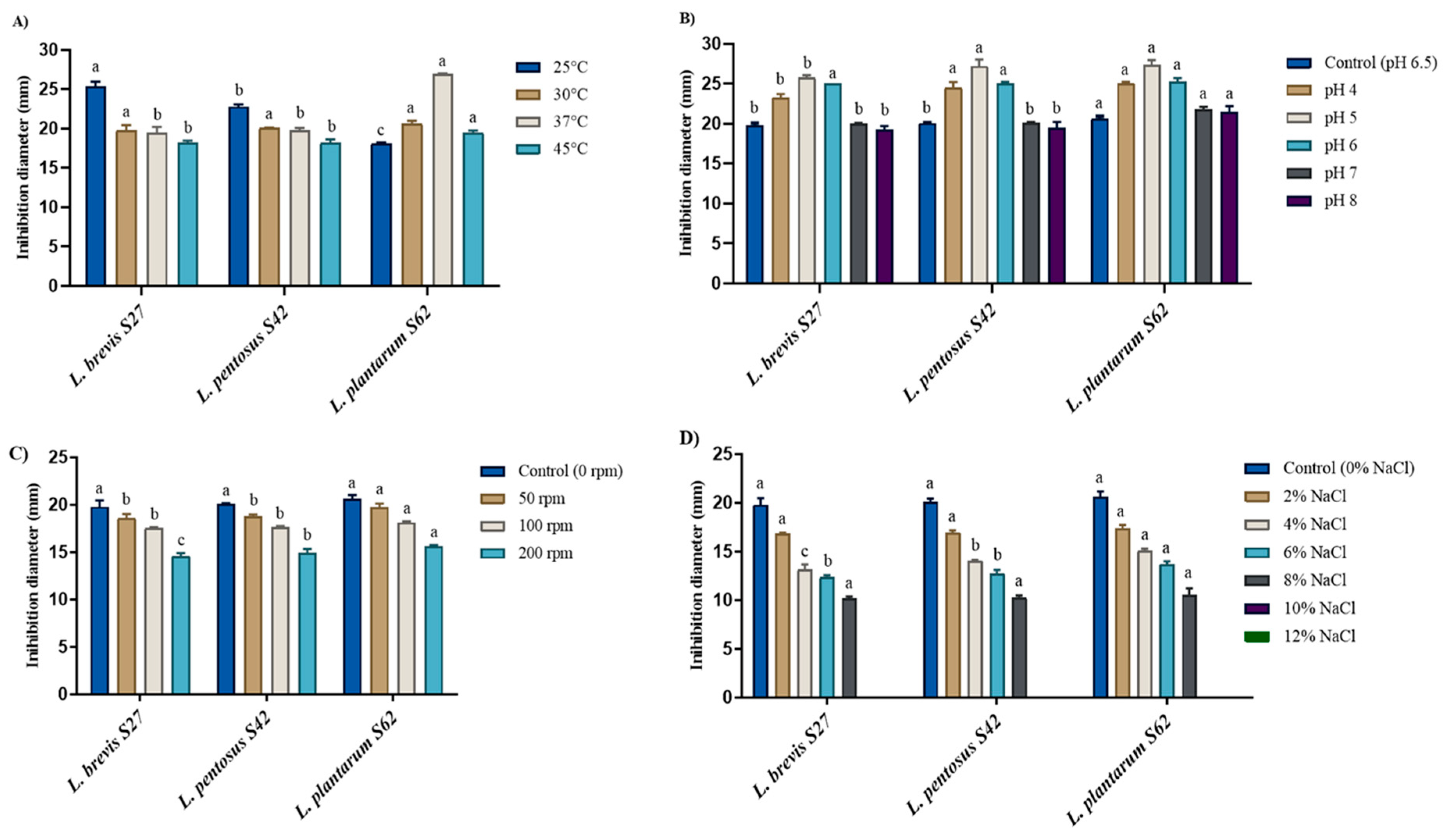
The results for the influence of different factors on antifungal metabolite production from Levilactobacillus and Lactiplantibacillus are presented in Figure 2. The results in Figure 2A show the highest production (p < 0.05) at 25 °C from L. brevis S27 and L. pentosus S42; meanwhile, L. plantarum S62 has a higher production (p < 0.05) at 37 °C. However, all strains showed that high production was detected at an initial pH of 5 (Figure 2B). The production was significantly higher (p < 0.05) at pH 4 and 5, with inhibition diameter ranges of 23.3–251 mm and 25.8–27.4 mm, respectively, compared with the control (pH 6.5) at 19.78–20.62 mm. Also, no significant difference (p < 0.05) in the production of antifungal metabolites was observed between the control and pH 7 and 8. Our results show that maximum production was detected at an initial pH of 5 and at a temperature of 25 °C for S27 and S42 and 37 °C for S62. On the other hand, the results for the effect of agitation (Figure 2C) clearly showed a decrease in antifungal metabolite production with an increase in agitation for all Levilactobacillus and Lactiplantibacillus strains. The inhibition diameters between the control (without agitation) and the cultures that underwent agitation (50 rpm, 100 rpm, and 200 rpm) were significantly different (p < 0.05). Hence, the agitation affected the production of antifungal metabolites unfavorably. However, the presence of NaCl concentration in the medium, until 8% NaCl was added, had a negative effect on antifungal metabolite production by reducing inhibition (Figure 2D). However, none of the Levilactobacillus and Lactiplantibacillus strains produced antifungal metabolites in 10% and 12% NaCl (Figure 2D), and the production of antifungal metabolites was reduced significantly (p < 0.05) by an increase in the NaCl concentration in the MRS medium.

3.5. Application of Levilactobacillus and Lactiplantibacillus Strains on Yogurt

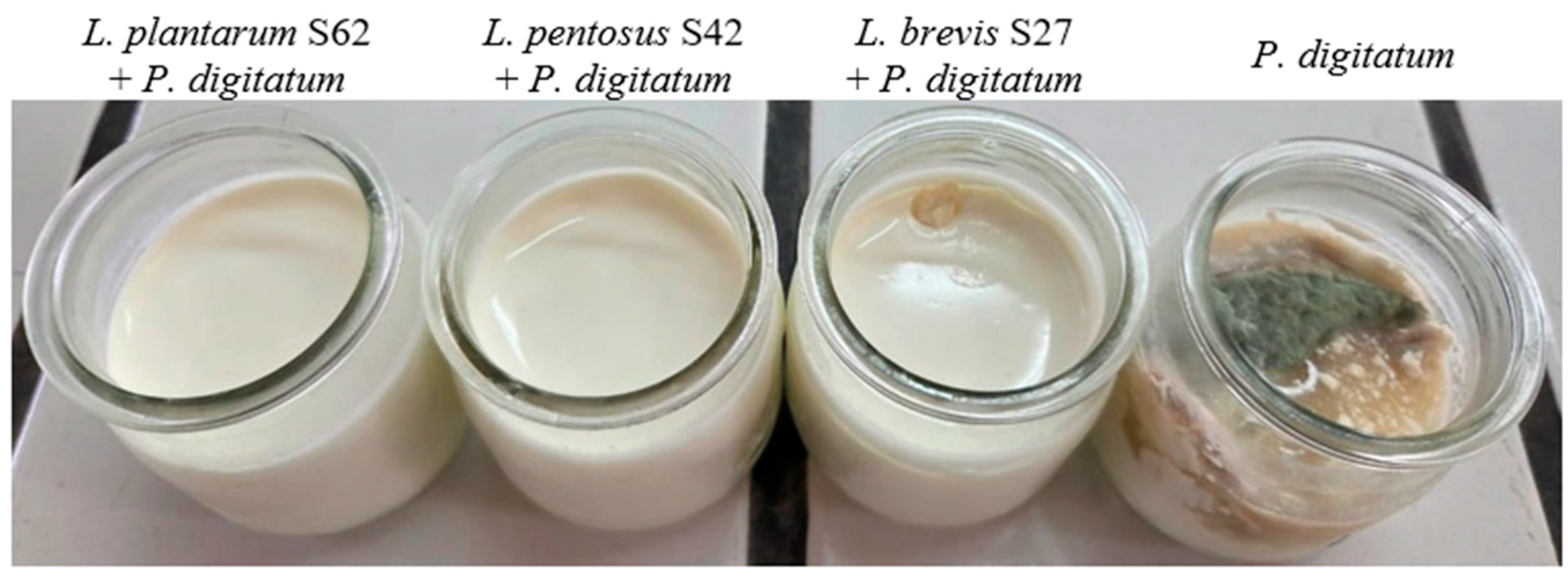
The antifungal properties of the Levilactobacillus and Lactiplantibacillus strains were further evaluated on yogurt contaminated with P. digitatum, and the results are presented in Figure 3. The control without the addition of Lactobacillus strains showed a total spoilage of yogurt after storage at 4 °C for 15 days. By contrast, in the presence of L. pentosus S42 and L. plantarum S62, no turquoise mycelia or spores were observed in yogurt over 15 days of storage at 4 °C, except for the presence of L. brevis S27; a small amount of spoilage was observed, suggesting that the Levilactobacillus and Lactiplantibacillus strains could effectively inhibit the growth of P. digitatum on yogurt.

3.6. Application of CFS from Levilactobacillus and Lactiplantibacillus on Orange Fruit

Figure 4 reports the results of the application of antifungal metabolites from the Levilactobacillus and Lactiplantibacillus strains on orange fruits against A. niger and during storage at room temperature. After 15 days of incubation, the fruits in the control (without CFS) were spoiled by A. niger. However, the CFS shows a reduction in A. niger growth after storage for 15 days. The use of CFS from L. plantarum S62 led to high inhibition of A. niger on orange fruits after storage for 15 days. Nevertheless, in the presence of CFS from L. pentosus S42 and L. brevis S27, a small amount of spoilage from A. niger was present on the surface of the orange fruits. All CFSs of the Levilactobacillus and Lactiplantibacillus strains demonstrated a bio-preservation effect in the orange models against A. niger during storage.

4. Discussion

The Levilactobacillus and Lactiplantibacillus cells used in this study demonstrated antifungal activity against molds. This result agrees with the result obtained by Magnusson and Schnurer [22] and confirms those of Guimaraes [23] and Riolo [24], which exhibited the inhibition of fungi by LAB cells. This study was conducted to exploit the high antifungal activity of Levilactobacillus and Lactiplantibacillus cells to control spoilage caused by fungi.
The CFS of the Levilactobacillus and Lactiplantibacillus strains showed high inhibition of yeast, molds (mycelia and biomass), and pathogenic bacteria. The studies of [5] and Arrioja-Bretón [25] demonstrated that the CFS of LAB strains inhibits fungi and highly inhibits the growth of the mycelia and biomass of molds [26,27]. Luz [4] found that the peptide reduced 58% and 73% of the biomass growth of fungi. Furthermore, these results were obtained by Arena [28] and Gharbi [29], who confirmed that CFS exhibited high antibacterial activity against pathogenic bacteria. These findings demonstrated that the antifungal metabolites produced by Levilactobacillus and Lactiplantibacillus are responsible for antibacterial activity against pathogenic bacteria. Therefore, these metabolites showed a large inhibition spectrum, including bacteria, yeast, and mold, and could be applied to prevent the spoilage of different food products.
The characterization of antifungal metabolites shows activity at a high pH and temperature and the disappearance of proteolytic enzymes. These results confirm those obtained by Volentini [27] and Chen [30]. The reduction in antifungal activity at neutral pH can be related to the neutralization of organic acids [16,31]. However, the activity was stable in hot and cold temperatures, which agrees with the studies conducted by [16,32], and the antifungal activity of LAB was greatly influenced by temperature and humidity [33]. Therefore, the disappearance of antifungal activity after treatment with protease and proteinase K enzymes demonstrated the possibility that antifungal metabolites have a proteinaceous nature. Furthermore, the Levilactobacillus and Lactiplantibacillus strains demonstrated a significant reduction (p < 0.05) after the neutralization of pH and the treatment with protease and proteinase K enzymes, suggesting that antifungal metabolites produced as proteinaceous substances are stable and resist hot and cold conditions. Hence, our results demonstrated that the antifungal metabolites we produced can be applied as bio-preservation agents during food processing and storage.
LAB can produce several kinds of antifungal metabolites, and each metabolite has its inhibitory properties regarding target microorganisms (yeasts and molds) [34]. The antifungal mechanisms of CFS from LAB disrupt the cell membrane integrity and increase cell membrane permeability [33]. Our study demonstrates that the antifungal metabolite is a proteinaceous substance. Moreover, previous studies have shown that the action mechanisms of peptide and proteinaceous compounds cause damage to the mycelial cell wall and hyphal size reduction in fungi and dissipate the proton gradient across the cell membrane [15,35].
High levels of antifungal metabolite production resulted at pH 5 and at 25 °C and 37 °C. Previous works based on initial pH and incubation temperature have shown that maximum bacteriocin production takes place at an initial pH between 5.0 and 7.0 and a temperature of 28 °C [36], and antifungal metabolites are at 30 °C and a pH range from 6.8 to 7.6 [37]. In addition, other works have shown that pH 7 and 37 °C allow for the high production of antifungal metabolites [5,32,38]. Moreover, Lactobacillus plantarum produces many antifungal metabolites at different temperatures and pH 6.5 [39,40]. Rouse [41] demonstrated that temperatures between 25 °C and 30 °C and pH between 4 and 8 can lead to optimal production of the antifungal metabolites. Our findings demonstrated that the factors (initial pH and temperature) are dependent on Lactobacillus strains. In this context, the production of antifungal metabolites decreased with an increase in NaCl concentrations and agitation. Previous studies have demonstrated that NaCl reduces the production of bacteriocin by LAB [37,42]. Consequently, our Levilactobacillus and Lactiplantibacillus strains produced high concentrations of NaCl (8%), indicating their possible use as starter cultures and bio-control agents in fermented vegetables such as olives. Hence, studies of other factors and their combinations may lead to the optimization of antifungal metabolite production for their applications as bio-preservative agents or bio-pesticides in controlling post-harvest spoilage.
The application of Levilactobacillus and Lactiplantibacillus strains as a bio-preservative agent in yogurt production demonstrated the total inhibition of P. digitatum spoilage. These results confirm the bio-preservative effect of LAB strains against fungi (yeast and molds) on yogurt. The inhibitory effects of Lactiplantibacillus strains against yeasts have been determined in yogurt [26] and can improve the functional properties of the product [43]. In addition, the application of L. plantarum on yogurt has shown the total inhibition of Penicillium sp. [44]. Mélo [45] demonstrates the ability of LAB strains to be natural bio-protective agents for large-scale yogurt manufacturing. Our findings indicate that the Levilactobacillus and Lactiplantibacillus strains could be used as bio-preservative agents in dairy products such as yogurt.
All CFSs of the Levilactobacillus and Lactiplantibacillus strains demonstrated a bio-preservation effect in the orange models against A. niger during storage. This study confirms the potential of the antifungal activity of CFS from Lactobacillus strains to control the spoilage of orange fruits [30]. Other studies by Volentini [27] and Ma [46] showed that LAB and its CFS inhibit the disease caused by Penicillium species in citrus fruits. The antifungal metabolites of L. plantarum S62 appeared to be more effective than other strains in orange against A. niger spoilage, and the high effectiveness of antifungal activity from CFS against spoilage fungi may be due to the high activity of CFS in acidic pH and/or the synergic effect between the essential oil in the orange peel and antifungal metabolites. Our findings indicate the possibility of applying the antifungal metabolites of L. plantarum S62 as bio-preservative agents or bio-pesticides in controlling post-harvest spoilage.

5. Conclusions

The Levilactobacillus and Lactiplantibacillus strains exhibited an ability to reduce and inhibit the growth of different microorganisms (mold, yeast, and bacteria). Their antifungal metabolites were stable under stress conditions like hot and cold temperatures and acidic pH. Therefore, their high production was obtained at an initial pH of 5 and at temperatures of 25 °C and 37 °C. However, an increase in NaCl concentration and agitation decreased the production of these metabolites. In addition, Levilactobacillus and Lactiplantibacillus strains had highly effective antifungal activity against fungi on yogurt and orange fruits. Consequently, this biological alternative (Lactobacillus strains or antifungal metabolites) could be applied as a bio-preservative agent in the agri-food industry or as a bio-pesticide in agriculture and could be developed as an active package to preserve foods during storage.

Author Contributions

H.A., conceptualization, methodology, validation, and writing—original draft preparation; S.G. and I.H., methodology; N.G., N.E.B., and Y.R., formal analysis; S.K. and A.A., investigation and writing—review and editing. All authors have read and agreed to the published version of the manuscript.

Funding

This research received no external funding.

Institutional Review Board Statement

Not applicable.

Informed Consent Statement

Not applicable.

Data Availability Statement

The original contributions presented in the study are included in the article, further inquiries can be directed to the corresponding author.

Conflicts of Interest

The authors declare no conflicts of interest.

References

  1. Yang, J.; Li, J.; Jiang, Y.; Duan, X.; Qu, H.; Yang, B.; Chen, F.; Sivakumar, D. Natural occurrence, analysis, and prevention of mycotoxins in fruits and their processed products. Crit. Rev. Food Sci. Nutr. 2014, 54, 64–83. [Google Scholar] [CrossRef]
  2. Abdel-Nasser, A.; Hathout, A.S.; Badr, A.N.; Barakat, O.S.; Fathy, H.M. Extraction and characterization of bioactive secondary metabolites from lactic acid bacteria and evaluating their antifungal and antiaflatoxigenic activity. Biotechnol. Rep. 2023, 38, e00799. [Google Scholar] [CrossRef]
  3. Freire, F.D.C.O.; Da Rocha, M.E.B. Impact of mycotoxins on human health. In Fungal metabolites; Springer: Berlin/Heidelberg, Germany, 2017; pp. 239–261. [Google Scholar]
  4. Luz, C.; Saladino, F.; Luciano, F.B.; Mañes, J.; Meca, G. In vitro antifungal activity of bioactive peptides produced by Lactobacillus plantarum against Aspergillus parasiticus and Penicillium expansum. LWT—Food Sci. Technol. 2017, 81, 128–135. [Google Scholar] [CrossRef]
  5. Shehata, M.G.; Badr, A.N.; El Sohaimy, S.A.; Asker, D.; Awad, T.S. Characterization of antifungal metabolites produced by novel lactic acid bacterium and their potential application as food biopreservatives. Ann. Agric. Sci. 2019, 64, 71–78. [Google Scholar] [CrossRef]
  6. Warner, J.O. Artificial food additives: Hazardous to long-term health? Arch. Dis. Child. 2024, 109, 882–885. [Google Scholar] [CrossRef]
  7. Zheng, J.; Wittouck, S.; Salvetti, E.; Franz, C.M.; Harris, H.M.; Mattarelli, P.; O’toole, P.W.; Pot, B.; Vandamme, P.; Walter, J. A taxonomic note on the genus Lactobacillus: Description of 23 novel genera, emended description of the genus Lactobacillus Beijerinck 1901, and union of Lactobacillaceae and Leuconostocaceae. Int. J. Syst. Evol. Microbiol. 2020, 70, 2782–2858. [Google Scholar] [CrossRef]
  8. EFSA. Update of the list of QPS-recommended biological agents intentionally added to food or feed as notified to EFSA 15: Suitability of taxonomic units notified to EFSA until September 2021. EFSA J. 2022, 20, e07045. [Google Scholar] [CrossRef]
  9. Akpoghelie, P.O.; Edo, G.I.; Ali, A.B.M.; Yousif, E.; Zainulabdeen, K.; Owheruo, J.O.; Isoje, E.F.; Igbuku, U.A.; Essaghah, A.E.A.; Makia, R.S.; et al. Lactic acid bacteria: Nature, characterization, mode of action, products and applications. Process Biochem. 2025, 152, 1–28. [Google Scholar] [CrossRef]
  10. Leroy, F.; De Vuyst, L. Lactic acid bacteria as functional starter cultures for the food fermentation industry. Trends Food Sci. Technol. 2004, 15, 67–78. [Google Scholar] [CrossRef]
  11. Abouloifa, H.; Gaamouche, S.; Ghabbour, N.; El Guerrouj, B.; Karboune, S.; Saalaoui, E.; Asehraou, A. Lactic acid bacteria from Moroccan traditional foods: Techno-functional, health-promoting, nutraceutical value and application as a starter and bio-preservative agent in the food products. Bioresour. Technol. Rep. 2024, 27, 101941. [Google Scholar] [CrossRef]
  12. Yang, H.; Hao, L.; Jin, Y.; Huang, J.; Zhou, R.; Wu, C. Functional roles and engineering strategies to improve the industrial functionalities of lactic acid bacteria during food fermentation. Biotechnol. Adv. 2024, 74, 108397. [Google Scholar] [CrossRef] [PubMed]
  13. Gajendran, V.P.; Rajamani, S. Recent Advancements in Harnessing Lactic Acid Bacterial Metabolites for Fruits and Vegetables Preservation. Probiotics Antimicrob Proteins 2025, 17, 2673–2689. [Google Scholar] [CrossRef] [PubMed]
  14. Banicod, R.J.S.; Tabassum, N.; Javaid, A.; Kim, Y.M.; Khan, F. Lactic Acid Bacteria-Derived Secondary Metabolites: Emerging Natural Alternatives for Food Preservation. Probiotics Antimicrob. Proteins 2025. [Google Scholar] [CrossRef]
  15. Siedler, S.; Balti, R.; Neves, A.R. Bioprotective mechanisms of lactic acid bacteria against fungal spoilage of food. Curr. Opin. Biotechnol. 2019, 56, 138–146. [Google Scholar] [CrossRef]
  16. Fugaban, J.I.I.; Jung, E.S.; Todorov, S.D.; Holzapfel, W.H. Evaluation of antifungal metabolites produced by lactic acid bacteria. Probiotics Antimicrob. Proteins 2023, 15, 1447–1463. [Google Scholar] [CrossRef]
  17. Batish, K.K.; Utpal, R.; Ram, L.; Grover, S. Antifungal Attributes of Lactic Acid Bacteria—A Review. Crit. Rev. Biotechnol. 1997, 17, 209–225. [Google Scholar] [CrossRef]
  18. Dalié, D.K.D.; Deschamps, A.M.; Richard-Forget, F. Lactic acid bacteria—Potential for control of mould growth and mycotoxins: A review. Food Control 2010, 21, 370–380. [Google Scholar] [CrossRef]
  19. Matevosyan, L.A.; Bazukyan, I.L.; Trchounian, A.H. Antifungal activity of lactic acid bacteria isolates and their associations: The effects of Ca and Mg divalent cations. Curr. Microbiol. 2020, 77, 959–966. [Google Scholar] [CrossRef]
  20. Souza, L.V.; Martins, E.; Moreira, I.; de Carvalho, A.F. Strategies for the Development of Bioprotective Cultures in Food Preservation. Int. J. Microbiol. 2022, 2022, 6264170. [Google Scholar] [CrossRef] [PubMed]
  21. Abouloifa, H.; Rokni, Y.; Bellaouchi, R.; Ghabbour, N.; Karboune, S.; Brasca, M.; Salah, R.B.; Chihib, N.E.; Saalaoui, E.; Asehraou, A. Characterization of probiotic properties of antifungal Lactobacillus strains isolated from traditional fermenting green olives. Probiotics Antimicrob Proteins 2020, 12, 683–696. [Google Scholar] [CrossRef]
  22. Magnusson, J.; Schnurer, J. Lactobacillus coryniformis subsp. coryniformis strain Si3 produces a broad-spectrum proteinaceous antifungal compound. Appl. Environ. Microbiol. 2001, 67, 1–5. [Google Scholar] [CrossRef]
  23. Guimaraes, A.; Santiago, A.; Teixeira, J.A.; Venancio, A.; Abrunhosa, L. Anti-aflatoxigenic effect of organic acids produced by Lactobacillus plantarum. Int. J. Food Microbiol. 2018, 264, 31–38. [Google Scholar] [CrossRef]
  24. Riolo, M.; Luz, C.; Santilli, E.; Meca, G.; Cacciola, S.O. Antifungal activity of selected lactic acid bacteria from olive drupes. Food Biosci. 2023, 52, 102422. [Google Scholar] [CrossRef]
  25. Arrioja-Bretón, D.; Mani-López, E.; Palou, E.; López-Malo, A. Antimicrobial activity and storage stability of cell-free supernatants from lactic acid bacteria and their applications with fresh beef. Food Control 2020, 115, 107286. [Google Scholar] [CrossRef]
  26. Liang, N.; Zhao, Z.; Curtis, J.M.; Ganzle, M.G. Antifungal cultures and metabolites of lactic acid bacteria for use in dairy fermentations. Int. J. Food Microbiol. 2022, 383, 109938. [Google Scholar] [CrossRef]
  27. Volentini, S.I.; Olmedo, G.M.; Grillo-Puertas, M.; Rapisarda, V.A.; Hebert, E.M.; Cerioni, L.; Villegas, J.M. Biological control of green and blue molds on postharvest lemon by lactic acid bacteria. Biol. Control 2023, 185, 105303. [Google Scholar] [CrossRef]
  28. Arena, M.P.; Silvain, A.; Normanno, G.; Grieco, F.; Drider, D.; Spano, G.; Fiocco, D. Use of Lactobacillus plantarum strains as a bio-control strategy against food-borne pathogenic microorganisms. Front. Microbiol. 2016, 7, 464. [Google Scholar] [CrossRef]
  29. Gharbi, Y.; Fhoula, I.; Ruas-Madiedo, P.; Afef, N.; Boudabous, A.; Gueimonde, M.; Ouzari, H.-I. In-vitro characterization of potentially probiotic Lactobacillus strains isolated from human microbiota: Interaction with pathogenic bacteria and the enteric cell line HT29. Ann. Microbiol. 2018, 69, 61–72. [Google Scholar] [CrossRef]
  30. Chen, H.; Ju, H.; Wang, Y.; Du, G.; Yan, X.; Cui, Y.; Yuan, Y.; Yue, T. Antifungal activity and mode of action of lactic acid bacteria isolated from kefir against Penicillium expansum. Food Control 2021, 130, 108274. [Google Scholar] [CrossRef]
  31. Chen, O.; Hong, Y.; Ma, J.; Deng, L.; Yi, L.; Zeng, K. Screening lactic acid bacteria from pickle and cured meat as biocontrol agents of Penicillium digitatum on citrus fruit. Biol. Control 2021, 158, 104606. [Google Scholar] [CrossRef]
  32. Falguni, P.; Shilpa, V.I.J.; Mann, B. Production of proteinaceous antifungal substances from Lactobacillus brevis NCDC 02. Int. J. Dairy Technol. 2010, 63, 70–76. [Google Scholar] [CrossRef]
  33. Wu, X.; Liu, Y.; Guo, Z.; Ji, N.; Sun, Q.; Liu, T.; Li, Y. Antifungal activities of Pediococcus pentosaceus LWQ1 and Lactiplantibacillus plantarum LWQ17 isolated from sourdough and their efficacies on preventing spoilage of Chinese steamed bread. Food Control 2025, 168, 110940. [Google Scholar] [CrossRef]
  34. Abouloifa, H.; Hasnaoui, I.; Rokni, Y.; Bellaouchi, R.; Ghabbour, N.; Karboune, S.; Brasca, M.; Abousalham, A.; Jaouadi, B.; Saalaoui, E. Antifungal activity of lactic acid bacteria and their application in food biopreservation. In Advances in applied microbiology, Elsevier: Amsterdam, 2022; Volume 120, pp. 33–77.
  35. Muhialdin, B.J.; Algboory, H.L.; Kadum, H.; Mohammed, N.K.; Saari, N.; Hassan, Z.; Meor Hussin, A.S. Antifungal activity determination for the peptides generated by Lactobacillus plantarum TE10 against Aspergillus flavus in maize seeds. Food Control 2020, 109, 106898. [Google Scholar] [CrossRef]
  36. Barman, S.; Ghosh, R.; Mandal, N.C. Production optimization of broad spectrum bacteriocin of three strains of Lactococcus lactis isolated from homemade buttermilk. Ann. Agrar. Sci. 2018, 16, 286–296. [Google Scholar] [CrossRef]
  37. Batish, V.K.; Lal, R.; Grover, S. Studies on environmental and nutritional factors on production of antifungal substance by Lactobacillus acidophilus R. Food Microbiol. 1990, 7, 199–206. [Google Scholar] [CrossRef]
  38. Bustos, A.Y.; Font de Valdez, G.; Gerez, C.L. Optimization of phenyllactic acid production by Pediococcus acidilactici CRL 1753. Application of the formulated bio-preserver culture in bread. Biol. Control 2018, 123, 137–143. [Google Scholar] [CrossRef]
  39. Sangmanee, P.; Hongpattarakere, T. Inhibitory of multiple antifungal components produced by Lactobacillus plantarum K35 on growth, aflatoxin production and ultrastructure alterations of Aspergillus flavus and Aspergillus parasiticus. Food Control 2014, 40, 224–233. [Google Scholar] [CrossRef]
  40. Muthusamy, K.; Soundharrajan, I.; Srisesharam, S.; Kim, D.; Kuppusamy, P.; Lee, K.D.; Choi, K.C. Probiotic characteristics and antifungal activity of Lactobacillus plantarum and its impact on fermentation of Italian Ryegrass at low moisture. Appl. Sci. 2020, 10, 417. [Google Scholar] [CrossRef]
  41. Rouse, S.; Harnett, D.; Vaughan, A.; van Sinderen, D. Lactic acid bacteria with potential to eliminate fungal spoilage in foods. J. Appl. Microbiol. 2008, 104, 915–923. [Google Scholar] [CrossRef] [PubMed]
  42. Barbosa, M.S.; Jurkiewicz, C.; Landgraf, M.; Todorov, S.D.; Franco, B.D.G.M. Effect of proteins, glucose and NaCl on growth, biosynthesis and functionality of bacteriocins of Lactobacillus sakei subsp. sakei 2a in foods during storage at 4 °C: Tests in food models. LWT—Food Sci. Technol. 2018, 95, 167–171. [Google Scholar] [CrossRef]
  43. Ramos, I.M.; Navajas Porras, B.; Delgado-Osorio, A.; Rufián-Henares, J.Á.; Poveda, J.M. Impact of Lactiplantibacillus plantarum UCLM56 on the bioactive properties of sheep’s milk yogurt before and after in vitro digestion and fermentation. LWT 2025, 228, 118101. [Google Scholar] [CrossRef]
  44. Wang, Q.; Zhang, F.; Zhang, Y.; Zhang, Y.; Wang, H.; Song, J.; Suo, H. A novel strain Lactiplantibacillus plantarum LPP95 isolated from Chinese pickles: Antifungal effect, mechanism, and potential application in yogurt. Food Biosci. 2024, 58, 103640. [Google Scholar] [CrossRef]
  45. Mélo, E.D.d.; e Silva, P.I.S.; do Oriente, S.F.; Almeida, R.D.; Pessoa, J.M.; França, K.B.; de Gusmão, T.A.S.; de Gusmão, R.P.; Lisboa Oliveira, H.M.; Nascimento, A.P.S. Effect of Commercial Bioprotective Lactic Cultures on Physicochemical, Microbiological, and Textural Properties of Yogurt. Fermentation 2024, 10, 585. [Google Scholar] [CrossRef]
  46. Ma, J.; Hong, Y.; Deng, L.; Yi, L.; Zeng, K. Screening and characterization of lactic acid bacteria with antifungal activity against Penicillium digitatum on citrus. Biol. Control 2019, 138, 104044. [Google Scholar] [CrossRef]
Figure 1. Antifungal activity of Levilactobacillus and Lactiplantibacillus strains. (a) Antifungal activity of L. brevis S27 against A. niger. (b) Antifungal activity of CFS from Levilactobacillus and Lactiplantibacillus strain against R. glutinis UMP22.
Fermentation 11 00661 g001
Figure 2. Factors influencing the production of antifungal metabolites with Levilactobacillus and Lactiplantibacillus strains. (A) Temperature, (B) initial pH, (C) agitation, and (D) NaCl concentration. a,b,c Means of each parameter with different lower case letters differed significantly (p < 0.05).
Fermentation 11 00661 g002
Figure 3. Bio-preservative effect of probiotic Levilactobacillus and Lactiplantibacillus strains against P. digitatum obtained on yogurt after 15 days of incubation at 4 °C.
Fermentation 11 00661 g003
Figure 4. Bio-preservative effect of CFS of probiotic Levilactobacillus and Lactiplantibacillus strains against A. niger obtained on orange fruits after 15 days of incubation at room temperature.
Fermentation 11 00661 g004
Table 1. Antifungal activity of Levilactobacillus and Lactiplantibacillus strains against fungi using the overlay method.
LAB StrainInhibition Zone
A. nigerP. digitatumF. oxysporumR. oryzae
L. brevisS27+++++
L. pentosusS42+++++++++++
L. plantarumS62+++++++++++
(−), absence of an inhibition zone around each steak; (+), inhibition zone between 0.1 and 3% of the surface of Petri dishes; (++), inhibition zone between 3 and 8% of the surface of Petri dishes; (+++), Inhibition zone greater than 8% of the surface of Petri dishes.
Table 2. Antibacterial and antifungal activity of CFS from Levilactobacillus and Lactiplantibacillus strains against pathogenic bacteria and yeast.
LAB
Strain
Inhibition Zone (mm)
Pathogenic BacteriaYeast
L. monocytogenes ATCC 19117S. aureus ATCC 6538P. aeruginosa ATCC 49189S. enterica ATCC 14028R. glutinis UMP22C. pelliculosa
L. brevisS2717.05 a ± 0.0717.1 a ± 0.1416.7 ab ± 0.1416.53 b ± 0.3922.78 b ± 0.2119.78 b ± 0.73
L. pentosusS4217.05 a ± 0.0717.15 a ± 0.0716.75 a ± 0.3516.65 ab ± 0.2123.06 ab ± 0.0720.06 b ± 0.34
L. plantarumS6217.1 a ± 0.1417.15 a ± 0.0716.85 a ± 0.0716.9 a ± 0.1423.63 a ± 0.1420.63 a ± 0.40
Values are mean ± standard error of triplicates. a,b Means in the same column for each parameter with different lowercase letters differed significantly (p < 0.05).
Table 3. Biomass and mycelium inhibition of mold using CFS from Levilactobacillus and Lactiplantibacillus strains.
LAB StrainsInhibition (%)
BiomassMycelium
A. nigerP. digitatumF. oxysporumR. oryzaeA. nigerP. digitatumF. oxysporumR. oryzae
L. brevisS2778.90 c ± 0.7180.00 c ± 0.5474.69 b ± 0.7257.66 b ± 0.6342.50 c ± 0.1447.55 c ± 0.0743.28 c ± 0.4940.15 b ± 0.14
L.pentosusS4282.77 b ± 0.4582.20 b ± 0.4575.26 b ± 0.5657.19 b ± 0.5248.70 b ± 0.0749.25 b ± 0.2844.55 b ± 0.2840.07 b ± 0.28
L. plantarumS6286.13 a ± 0.1384.50 a ± 0.5078.82 a ± 0.7160.44 a ± 0.6150.08 a ± 0.1455.59 a ± 0.6449.57 a ± 0.0745.64 a ± 0.49
Values are mean ± standard error of triplicates. a,b,c Means in the same column for each parameter with different lowercase letters differed significantly (p < 0.05).
Table 4. Characterization of antifungal metabolites produced by Levilactobacillus and Lactiplantibacillus strains.
ConditionsLAB Strains
L. brevis S27L. pentosus S42L. plantarum S62
CFS without treatment (pH 4)19.78 b ± 0.7320.06 b ± 0.3420.63 a ± 0.40
Effect of pH   
4.519.5 a ± 0.6919.8 a ± 0.2820.2 a ± 0.28
518.3 b ± 0.4218.6 b ± 0.2819.05 a ± 0.07
5.515.9 b ± 0.2816.05 b ± 0.0716.25 a ± 0.07
614.1 b ± 0.1415.25 a ± 0.3515.35 a ± 0.4
6.513.85 c ± 0.0714.6 b ± 0.4215.1 a ± 0.14
713.8 c ± 0.1414.4 b ± 0.1415.05 a ± 0.07
Effect of Temperature   
80 °C/10 min19.78 b ± 0.7320.06 b ± 0.3420.63 a ± 0.40
100 °C/10 min19.6 b ± 0.4220.0 b ± 0.2820.63 a ± 0.28
Sterilization19.5 b ± 0.4219.8 b ± 0.2820.5 a ± 0.42
Effect of storage (4 °C)   
Week 119.78 b ± 0.7320.06 b ± 0.3420.63 a ± 0.40
Week 219.55 a ± 0.6319.85 a ± 0.3520.3 a ± 0.14
Week 318.6 b ± 0.2819.0 a ± 0.419.2 a ± 0.14
Week 417.6 b ± 0.2818.0 a ± 0.1418.2 a ± 0.14
Effect ofenzymes   
pH7 + Catalase13.75 c ± 0.0714.45 b ± 0.2115.1 a ± 0.14
pH7 + ProteaseNDNDND
pH7 + Proteinase KNDNDND
pH7 + Lipase13.7 c ± 0.4214.48 b ± 0.1715.08 a ± 0.02
Values are mean ± standard error of triplicates. a,b,c Means in the same rows (Lactobacilli strains) for each parameter with different lowercase letters differed significantly (p < 0.05). ND: not detected.
Disclaimer/Publisher’s Note: The statements, opinions and data contained in all publications are solely those of the individual author(s) and contributor(s) and not of MDPI and/or the editor(s). MDPI and/or the editor(s) disclaim responsibility for any injury to people or property resulting from any ideas, methods, instructions or products referred to in the content.