Article

Beneficial Effects of Probiotics on Liver Injury Caused by Chronic Alcohol Consumption

1
Business School, Honghe University, Honghe 661199, China
2
College of Food Science and Technology, Yangzhou University, Yangzhou 225127, China
3
Key Laboratory of Dairy Biotechnology and Safety Control, Yangzhou 225127, China
*
Author to whom correspondence should be addressed.
Fermentation 2024, 10(3), 127; https://doi.org/10.3390/fermentation10030127
Submission received: 12 January 2024 / Revised: 8 February 2024 / Accepted: 21 February 2024 / Published: 23 February 2024
(This article belongs to the Section Probiotic Strains and Fermentation)

Abstract

:
Alcoholic liver injury is a serious risk to human health. Probiotics have become a popular form of treatment. Lacticaseibacillus casei Grx12 and Limosilactobacillus fermentum Grx07 isolated from the gut of long-lived people in Rugao, Jiangsu, were studied to determine their protective effects and possible mechanisms of action on alcoholic liver injury. The results showed that rat serum ALT and AST were restored, and liver injury was reduced after the probiotics intervention. The level of antioxidant enzymes and antioxidants such as SOD, GSH and GSH-Px in the rat liver was significantly increased (p < 0.05), which reduces the level of MDA, a peroxidation product in the liver, and thus alleviates liver oxidative stress. L. casei Grx12 and L. fermentum Grx07 also could significantly enhance the expression of Nrf2 protein in the rat liver to regulate the anti-oxidative stress response in the body and cells (p < 0.05). The levels of ADH, Na+-K+-ATPase and Ca2+-ATPase in the rat liver were significantly increased (p < 0.05), which enhanced the body’s metabolism of alcohol. The rat serum LPS and liver TNF-α, IL-6, VEGF, TGF-β1 and NF-κB levels were significantly reduced (p < 0.05), indicating that the probiotics could relieve liver inflammation. The results of this study indicate that L. casei Grx12 and L. fermentum Grx07 have certain protective effects on alcoholic liver injury in rats, likely because of their antioxidant properties and ability to prevent oxidative stress and relieve inflammation.

1. Introduction

Chinese liquor has a long history of production and drinking in China. Alcohol production and per capita alcohol consumption have increased significantly, leading to a significant increase in the prevalence of acute and chronic alcoholic liver injury in recent years [1]. Alcoholic liver disease has become the most common liver disease after hepatitis A, hepatitis B and other types of viral hepatitis [2]. Alcoholic liver injury can gradually develop into alcoholic fatty liver, alcoholic hepatitis, alcoholic liver fibrosis and alcoholic cirrhosis, and even induce liver cancer [3]. More than 90% of ingested alcohol is metabolized in the liver and will generate excessive free radicals, causing oxidative stress, which in turn leads to liver damage [4]. The intestinal barrier can be damaged due to long-term alcohol intake. The amount of Lipopolysaccharides (LPS) in the portal vein is increased, thereby activating Kupffer cells to produce inflammatory cytokines that exacerbate liver injury [5]. Therefore, it is of great significance to find measures that can relieve oxidative stress and inflammation for the prevention and treatment of alcoholic liver injury.
Abstinence from alcohol is the key factor and the most fundamental way to treat alcoholic liver injury. Treatment of alcoholic liver injury through medication is also commonly used today [6]. Its mechanism of action mainly focuses on lipid-lowering, antioxidant, anti-inflammatory, anti-fibrosis effects and improving ethanol metabolism [7]. Although medication has been shown to have some effect, most medications also have an effect on liver function and most need to be metabolized in the liver to be effective, as alcohol has already caused damage to the liver, and the metabolism of medications may in turn increase the burden on the liver [8]. Moreover, most drugs can only work in one way, for example, antioxidant drugs can improve liver oxidative stress to play the role of hepatoprotection, but they have no alleviating effects on alcohol-induced endotoxemia, so it cannot have a comprehensive protective effect.
Probiotics have the functions of anti-oxidation [9], regulating the gut microbiota [10] and immune regulation [11]. Probiotics can exert their beneficial effects from multiple targets. With the deepening of research into them, the use of probiotics to prevent and treat alcoholic liver injury has gradually attracted the attention of researchers [12]. Lactobacillus (Lacticaseibacillus) rhamnosus GG can inhibit the overgrowth of intestinal Gram-negative bacteria by producing antibacterial substances, thereby reducing LPS levels and alleviating alcoholic liver injury in rats [13]. This is one of the earliest studies on using probiotics to alleviate alcoholic liver injury. In recent studies, feeding rats with Probio-M8-fermented milk effectively maintained the stability of the gut microbiota, reduced liver inflammation and oxidative stress and mitigated liver damage in alcoholic liver disease (ALD) [14]. Lactobacillus plantarum HFY09 intervention in ethanol-induced mice led to decreases in serum triglyceride (TG), total cholesterol (TC), aspartate aminotransferase (AST), alanine aminotransferase (ALT), hyaluronidase and precollagen III, and increases in liver alcohol dehydrogenase (ADH) [15]. Although researchers have found that some probiotics can relieve alcoholic liver injury, its mechanism of action in treating alcoholic liver injury is still unclear. Therefore, there is still a need for further comprehensive studies on the protective effects of probiotics against alcoholic liver injury.
Human-derived probiotics have more advantages than of non-human-derived probiotics [16]. It is safer and more effective to use human-derived probiotics for alleviating alcoholic liver injury. L. casei Grx12 and L. fermentum Grx07 were selected from the intestines of long-lived people in Rugao, Jiangsu. Previous studies have shown that these two human-derived probiotics have a strong antioxidant capacity in vitro [17]. In this study, we used a rat model of alcoholic liver injury to investigate the protective effects of human-derived probiotics on the rat liver and to analyze their multi-target mechanisms of action.

2. Materials and Methods

2.1. The Probiotic Bacteria

L. casei Grx12 (Grx12) and L. fermentum Grx07 (Grx07) were isolated and preserved by Jiangsu Key Lab of Dairy Biological Technology and Safety Control, China. The bacterial fluid of Grx12 and Grx07 after activation in MRS liquid medium for 3 generations was centrifuged at 6000 rpm for 10 min to collect the resulting pellet. The bacterial body was washed with sterilized saline twice, and 10% sterilized skimmed milk was added into the bacterial precipitate as a cryoprotecting agent to adjust its bacterial number to 1.0 × 10⁹ CFU/mL; it was then mixed well to confirm the number of viable bacteria with MRS solid medium. Bacterial cultures were stored at −80 °C and defrosted at 30 °C, in a water bath, prior to use.

2.2. Animals and Treatment

We purchased 50 healthy male Wistar rats, which were 5 weeks old and weighed about 150 g at the start of the experiment, from the Comparative Medical Center at the Yangzhou University, Jiangsu, China. Rats (10 per group) were randomly divided into control group (Control), model group (Model), drug group (Drug), L. casei Grx12 group (Grx12) and L. fermentum Grx07 group (Grx07). Gavage was used twice a day and the interval between gavages was 8 h. The specific gavage methods are shown in Table 1. The drug group used Dongbaogantai (Compound Methionine and Choline Bitartrate Tablets) which were manufactured by Tonghua Dongbao Pharmaceutical Co., Ltd (Tonghua, China). Each tablet contained 0.1 g of methionine and 0.1 g of chline bitartrate solution. An alcoholic liver injury model was established by gavaging the rats with Chinese liquor with increasing alcohol concentration. For the Chinese liquor we used the commercially available Beijing Erguotou which is manufactured by Niulanshan DISTILLERY Factory (Beijing, China). The alcohol concentration of the Chinese liquor used for gavage gradually increased from 30% to 40% across 2 weeks, and was maintained at 40% until the 6th week; it was then increased to 45% until the end of the experiment [18]. The total duration of the experiment was 10 weeks.
All rats were housed under a 12 h light/12 h dark cycle in a controlled room with a temperature of 23 ± 3 °C and a humidity of 50 ± 10%, and were allowed free access to food and water. All rats were euthanized following anesthesia under 1% sodium pentobarbital.

2.3. Biochemical Analysis of Serum and Liver

Serum samples were extracted from venous plexus on the eye socket by centrifugation at 2000 rpm for 15 min at 4 °C. Serum aminotransferase levels (ALT and AST) were measured using enzymatic assay kits. Serum LPS levels were determined using ELISA kits from Shanghai Hualan Bioengineering Institute (Shanghai, China).
Livers were immediately excised from sacrificed mice and homogenized with ice-cold physiological saline. Then, the homogenate was centrifuged at 3000 rpm for 15 min at 4 °C. The supernatant was continued for further analysis. The liver MDA, SOD, GSH, GSH-Px, ADH, Na+-K+-ATPase and Ca2+-ATPase activities were measured using commercially available kits purchased from Nanjing Jiancheng Bioengineering Institute (Nanjing, China). The liver TNF-α, IL-6, VEGF, TGF-β1 and NF-κB levels were determined using ELISA kits purchased from Nanjing Jiancheng Bioengineering Institute (Nanjing, China).

2.4. Pathological Observation of the Rat Liver

Liver samples were fixed in 2.5% glutaraldehyde for more than 2 h. Transmission electron microscopy was used to observe liver slices.

2.5. Determination of Liver Cell Index

The rat liver tissue was ground and filtered through a 200-mesh screen to make the sample a single cell suspension. The hepatocytes were washed three times with PBS solution (pH 7.4) and centrifuged at 1500 rpm for 5 min each time. Finally, pre-chilled PBS was added to resuspend the cells for flow cytometry testing (adjust the cell concentration to 1 × 10⁶ cells/mL).

2.6. Hepatocyte Apoptosis

Assay was performed using Annexin V-FITC Apoptosis Detection Kit I purchased from Shanghai Beyotime Biotechnology Institute (Shanghai, China). Then, the 0.5 mL cell suspension was centrifuged and mixed with 500 μL Binding Buffer. Then, it was added to 5 μL Annexin V-FITC and 5 μL PI, and stored at room temperature in the dark for 5 min. The liver cells in the control group with only 500 μL of Binding Buffer but no Annexin V-FITC and PI added were used as negative controls. Flow cytometry was used to detect hepatocyte apoptosis with an excitation wavelength of 488 nm and an emission wavelength of 530 nm [19].

2.7. Determination of Antioxidant Specific Protein Nrf2 in Rat Liver

We added 1 μL each of Nrf2 monoclonal antibody and secondary antibody to 0.1 mL single-cell suspension. After incubation at room temperature in the dark for 30 min, it was washed once with PBS. Before testing, it was added to 0.1 mL PBS and filter through a 500-mesh screen. In the determination of protein immunofluorescence markers, a background control and a negative control of antibodies were set [20].
Calculation formula: I = Log (x − mode) × 340

2.8. Statistical Analysis

All data were statistically processed using SPSS16.0. The data were expressed as “mean ± standard deviation”, and one-way ANOVA was used for significance test. p < 0.05 was considered statistically significant.

3. Results

3.1. General State of Rats

The rats in the Control group were very active and in good condition, with normal feeding, drinking, urination and defecation, and soft and shiny fur. After being gavage with alcohol, the rats clearly appeared to be intoxicated in about half an hour, showing slow movement, unstable crawling, stagnation and half-open eyes, and most of the rats in the model group were more seriously intoxicated, unable to move their bodies, and fell into a deep sleep more quickly; some of the rats in the probiotic group and the drug group were able to eat and drink, but after one hour, all of those in the alcohol group fell into a deep sleep. After prolonged gavage with alcohol, the rats’ fur was rough and lost its luster, and some of them had loose stools. However, after 2 weeks of continuous gavage in the probiotics group, the rats’ stools were more shaped than those in the Model group, and in the middle and late stages of the experiment, the shape of the stools was no different from those of the Control group.

3.2. Human-Derived Probiotics Alleviate the Liver Injury

Pathological observation of the rats’ livers revealed that the hepatocyte nuclei of the rats in the Control group were round or oval, and the nuclear membranes were intact, clear and continuous. The organelle structure was normal. There was no clear distribution of lipid droplets in the cytoplasm. A large number of lipid droplets of different sizes were distributed around the nuclei in the hepatocytes of the Model group, and the density of the lipid droplets was greater than that of the droplets in the Control group. The mitochondria and endoplasmic reticulum of the Drug group were basically normal, and endocytotic lipid droplets could be seen in the nuclei. In the two probiotics groups, the number of lipid droplets in liver cells decreased, and the mitochondrial morphology and nuclear membrane tended to be normal. Particularly in the Grx12 group, the nuclear chromatin distribution of hepatocytes was relatively uniform, and the nuclear membrane was shaped smoothly, almost similar to the Control group (Figure 1A).
Compared with the Control group, the serum AST level in the Model group was significantly increased (p < 0.05). After treatment with drugs and probiotics Grx12 and Grx07, the serum AST levels in the Drug, Grx12 and Grx07 groups were significantly reduced (p < 0.05) (Figure 1B). There was no significant change in the serum ALT between groups (p > 0.05) (Figure 1C).

3.3. Human-Derived Probiotics Reduce the Hepatocyte Apoptosis

After long-term intake of large amounts of alcohol, the percentage of hepatocyte apoptosis in the Model group increased significantly (p < 0.05). After treatment with drugs and probiotics, hepatocyte apoptosis was significantly improved (p < 0.05). Among the groups, the effects of probiotics were significantly better than those of drugs (p < 0.05) (Figure 2).

3.4. Human-Derived Probiotics Restore ADH and ATPase

The ADH, Na+-K+-ATPase and Ca2+-ATPase activities in the rats’ livers the Model group were significantly reduced (p < 0.05) compared with the Control group. The treatments with drug and probiotics significantly increased the levels of ADH, Na+-K+-ATPase and Ca2+-ATPase in the rats’ livers (p < 0.05). The effect of Grx12 was significantly better than that of the drugs and Grx07 (p < 0.05) (Figure 3).

3.5. Human-Derived Probiotics Alleviate Liver Oxidative Stress

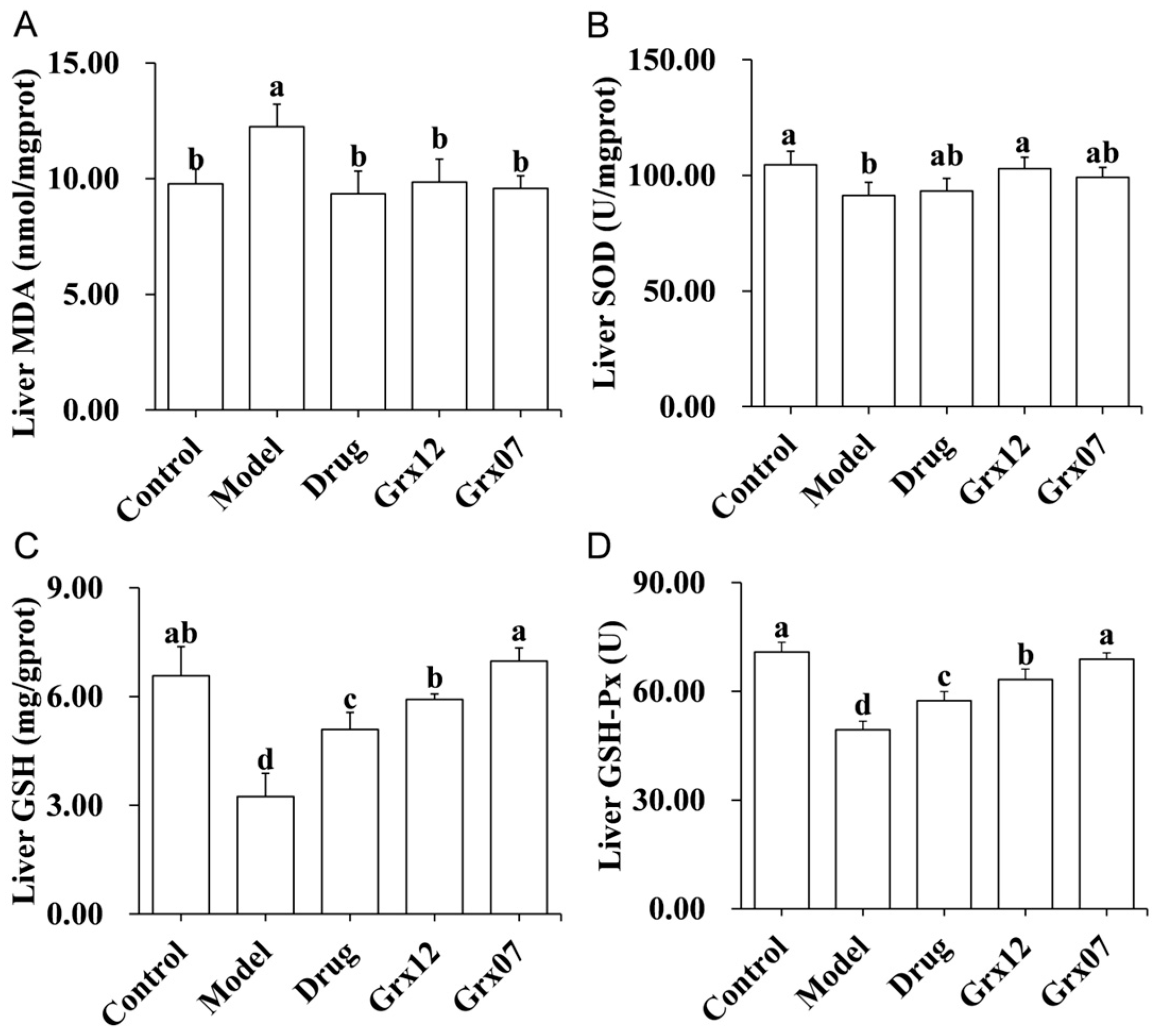
Long-term intake of large amounts of alcohol resulted in a significant reduction in the levels of SOD, GSH and GSH-Px, which exert antioxidant functions in the rat liver (p < 0.05) (Figure 4B–D). Correspondingly, the level of lipid peroxidation product, MDA, was increased significantly in the livers of the rats in the Model group (p < 0.05) (Figure 4A). The treatments with drugs and probiotics could significantly increase the levels of SOD, GSH and GSH-Px, and reduce the level of MDA (Figure 4). The effect of Grx07 on the levels of GSH and GSH-Px was significantly better than that of the drugs or Grx12 (p < 0.05) (Figure 4C,D).
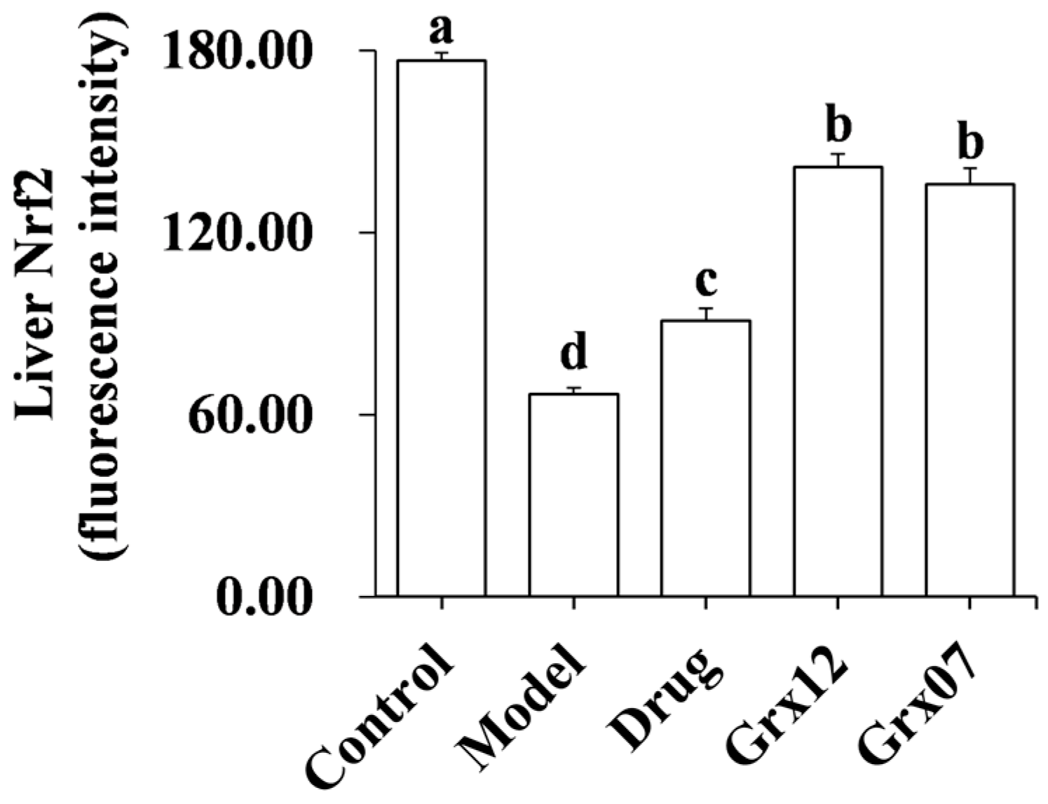
The protein expression of Nrf2 in the rat liver of the group Model was significantly lower than that of the group Control. After treatment with the drug and probiotics, the protein expression of Nrf2 in rat liver of the group Drug, Grx12 and Grx07 were significantly increased (p < 0.05). The effects of probiotics were significantly better than drugs (p < 0.05) (Figure 5).

3.6. Human-Derived Probiotics Alleviate Liver Inflammation

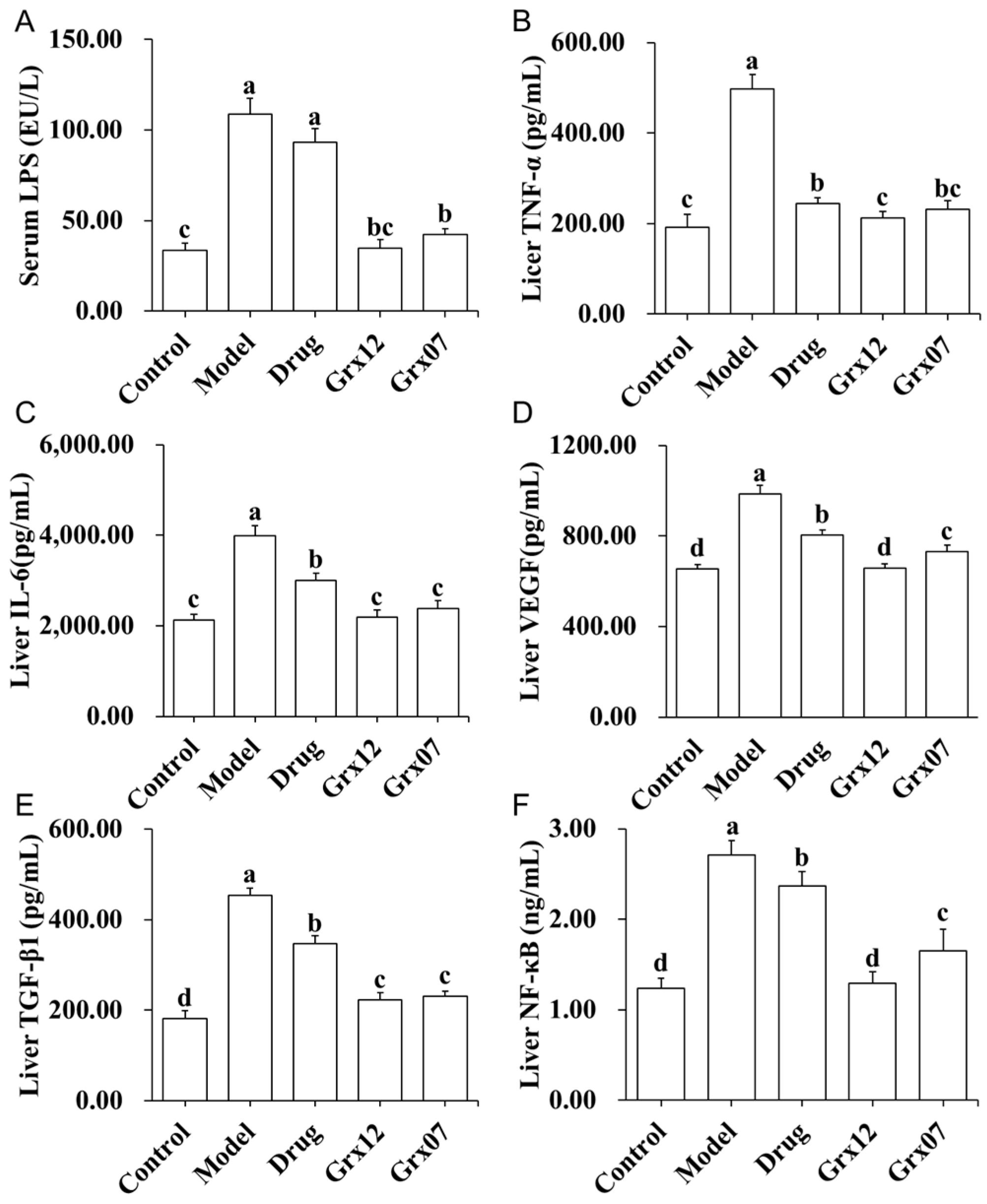
Compared with the Control group, the serum LPS levels in the Model and Drug groups were increased significantly (p < 0.05). After treatment with probiotics, the serum LPS levels in the Grx12 and Grx07 groups were significantly reduced (p < 0.05) (Figure 6A).
The levels of TNF-α, IL-6, VEGF, TGF-β1 and NF-κB in the livers of rats in the Model group were significantly higher than those in the Control group (p < 0.05). After the treatments with drugs and probiotics, these inflammatory factors were significantly reduced (p < 0.05) (Figure 6B–F). The effects of the probiotics were significantly better than those of the drugs (p < 0.05) (Figure 6). The effect of Grx12 on the levels of VEGF and NF-κB was significantly better than that of Grx07 (p < 0.05) (Figure 6D,F).

4. Discussion

Alcoholic liver injury is a liver disease that is caused by long-term heavy drinking and progressively develops, which can seriously jeopardize health and even life in the later stages; its pathogenesis is complicated, and there is still a lack of ideal treatment means. Probiotics have a wide range of physiological functions; it is an antioxidant, regulating intestinal flora and enhancing immunity. The use of probiotics to alleviate alcoholic liver injury has become a hot research topic. In this study, according to the pathogenesis of alcoholic liver injury and the corresponding protective mechanism, probiotics isolated from the intestinal tract of the population in Rugao Longevity Village, Jiangsu Province, were used as the research object to study their protective effects and mechanism of action on alcoholic liver injury through the establishment of a rat model of alcoholic liver injury.
The development and progression of alcoholic liver disease is a complex process and multiple factors play an important role in its pathogenesis. The liver is the main organ for ethanol metabolism, and more than 90% of ethanol is metabolized in the liver [21]. The liver will be severely damaged after long-term heavy intake of alcohol, leading to the deformation and necrosis of liver cells. Furthermore, sugar, protein, fat, etc. cannot normally be metabolized by the liver, and the liver’s detoxification function will be affected. The levels of ALT and AST in the serum are often used to reflect liver cell damage and determine the degree of damage [22]. When the liver is damaged, the transaminase in the liver cells enters the serum, causing an increase in ALT and AST in the serum. In addition, alcohol indices apoptosis of liver cells. The apoptosis of liver cells plays an important role in the occurrence and development of alcoholic liver disease [23], and the degree of liver-cell apoptosis is closely related to the severity of liver injury [24]. In this study, after supplementing rats with Grx12 and Grx07, it was found that lipid droplets in liver cells were reduced, liver cell apoptosis was alleviated and the level of AST in serum was significantly reduced. All this shows that alcoholic liver damage in rats can be alleviated by probiotics. It is worth noting that probiotics are better than the drug used in relieving liver cell apoptosis.
Alcohol has three main metabolic pathways in the body, one of which is the ADH pathway in the hepatocytes [25]. Reduced ADH activity affects the normal metabolism of alcohol in the liver, resulting in a large accumulation of ethanol and its metabolites in the liver and causing liver injury. ADH levels in the liver were greatly reduced after long-term intake of large amounts of alcohol in rats, while drug and probiotic interventions significantly increased hepatic ADH content. Among them, the Grx12 had the best effect. This suggests that probiotics are able to metabolize alcohol by enhancing its metabolism. However, several steps in the metabolism of alcohol produce very harmful reactive oxygen species, which inhibit the antioxidant capacity of liver cells, reduce SOD and increase lipid peroxidation. Lipid peroxidation can activate phospholipase C and phospholipase D to decompose mitochondrial membrane phospholipids, which change the fluidity and permeability of mitochondria, and make the lipid microenvironment of membrane Na+-K+-ATPase and Ca2+-ATPase abnormal. Thus, mitochondrial structure and function become abnormal, and apoptosis is induced [26]. In this study, after supplementing rats with Grx12 and Grx07, the activities of Na+-K+-ATPase and Ca2+-ATPase returned to normal levels. This demonstrates the role of probiotics in modulating lipid peroxidation.
Liver lipid peroxidation can be caused by the long-term high intake of alcohol, which is an important cause of alcoholic liver injury [27]. As the product of free radicals, MDA acts on lipids to produce peroxidation. Its level can reflect the degree of lipid peroxidation and the metabolism of free radicals in the body, and indirectly reflect the severity of liver cells being attacked by free radicals [28]. SOD is the most important enzyme for the body to clear superoxide anion radicals [29]. GSH can not only regulate the redox reaction in the body, and resist lipid peroxidation, but also has a detoxification function [30]. GSH-Px is an important peroxidase-decomposing enzyme with GSH as a substrate, which plays an important role in protecting organelles from oxidative damage [31]. Under normal conditions, SOD, GSH, GSH-Px and other antioxidants work together to effectively remove free radicals and maintain the body’s redox balance [27]. However, long-term intake of alcohol encourages the body to produce excessive free radicals, which significantly reduces or even exhausts the GSH level and also reduces the activity of SOD and GSH-Px in liver cells, which causes lipid peroxidation [27]. Nrf2 is an important transcription factor that regulates the anti-oxidative stress response in the body and cells, and plays a key role in the defense mechanism induced by cellular anti-oxidative stress and exogenous toxic substances [32]. In this study, after supplementing rats with Grx12 and Grx07, the level of MDA in the livers of the rats was significantly reduced, while the activity of SOD and GSH-Px, the level of GSH and the expression of the Nrf2 protein were significantly increased. This suggests that probiotics can improve the body’s oxidative stress state and relieve lipid peroxidation.
In the pathogenesis of alcoholic liver disease, in addition to causing lipid peroxidation, alcohol can also cause damage to the intestinal barrier, leading to endotoxin displacement, which in turn stimulates inflammatory cells to release various cytokines and inflammatory mediators, resulting in liver cell being damage and inflammation in the liver [21,33]. LPS is the lipopolysaccharide component on the outer membrane of the cell wall of Gram-negative bacteria, which can cause inflammation [34]. Cytokines are the main component of the body’s defense system. They are not only the product of immune responses, but also enhance immune response and promote liver cell damage. TNF-α and IL-6 are considered to reflect the severity of the inflammatory response and play a vital role in the occurrence and development of liver injury. TNF-α has a direct cytotoxic effect on the one hand, which can cause hepatocyte necrosis, and on the other hand, it can cause microcirculation disorders and lead to hepatocyte necrosis. At the same time, TNF-α also stimulates other inflammatory factors such as IL-6, causing a cascade amplification reaction, further aggravating liver damage [35]. VEGF can increase capillary permeability and promote liver cell regeneration [36]. VEGF increases in the acute phase of liver inflammation, and enhances the release of cytokines including IL-1, IL-6, TNF-α, etc., thus playing a key role in the acute inflammation phase [37]. TGF-β1 is a factor that promotes liver fibrosis and extracellular matrix degradation, can induce hepatocyte apoptosis and necrosis and ultimately promotes the occurrence and development of ALD [38]. As a multifunctional cellular transcription factor, NF-κB is associated with liver inflammation, fibrosis, liver cell regeneration, and apoptosis [39]. When stimulated by cytokines, endotoxins, oxygen free radicals, etc., NF-κB is activated and produces a large number of inflammatory mediators. In this study, after long-term alcohol intake, the serum LPS level increased significantly, causing endotoxemia and inducing severe inflammation in the liver. In addition, the increase in lipid peroxidation products (such as MDA) also stimulated the body’s immune cell response, induced the expression of inflammatory cytokines and led to liver inflammation. Supplementing with Grx12 and Grx07 reduced serum LPS and liver cytokine levels. This may show that supplementation with probiotics and colonization in the intestine can effectively inhibit harmful flora and greatly reduce the number of Gram-negative bacteria. Therefore, the endotoxin level is also significantly reduced, thereby alleviating the inflammatory response in the liver. At the same time, the mitigating effects of probiotics on lipid peroxidation also alleviates inflammation to a certain extent. In addition, probiotics can also directly regulate the NF-κB and other signaling pathways to promote a immune response in the intestinal mucosa, regulate the secretion of cytokines, inhibit the expression of inflammatory factors, reduce apoptosis and improve inflammation-related diseases. It is worth noting that these two human-derived probiotics have similar action modes but different degrees of effects.

5. Conclusions

This study has confirmed that both L. casei Grx12 and L. fermentum Grx07 have a certain protective effect on alcoholic liver injury in rats. The protective mechanism is related to their good antioxidant capacity, which can effectively relieve alcohol-induced lipid peroxidation. In addition, L. casei Grx12 and L. fermentum Grx07 can also relieve liver inflammation to alleviate alcoholic liver injury by reducing LPS translocation and regulating cytokines.

Author Contributions

Conceptualization, J.S., H.Q. and D.L.; methodology, J.S. and X.C.; software, J.S.; validation, J.S. and Y.W.; formal analysis, J.S.; investigation, D.C.; resources, Y.H.; data curation, J.S.; writing—original draft preparation, J.S. and Y.H.; writing—review and editing, J.S., H.Q., Y.H. and R.G.; visualization, J.S.; supervision, R.G.; project administration, X.C.; funding acquisition, R.G. and Y.H. All authors have read and agreed to the published version of the manuscript.

Funding

This research was funded by National Natural Science Foundation of China (31972094, 32272362) and City-school cooperation to build science and Technology Innovation Platform (YZ2020265).

Institutional Review Board Statement

The study was conducted in accordance with the U.S. National Institutes of Health guidelines for the care and use of laboratory animals (NIH Publication No. 85-23 Rev. 1985), and approved by the Animal Care Committee of the Center for Disease Control and Prevention (Jiangsu, China) (No. 202103262, 5 March 2021).

Informed Consent Statement

Not applicable.

Data Availability Statement

The data supporting the findings reported here are available upon reasonable request from the corresponding author.

Acknowledgments

This study was also helped and supported by other members of the Key Laboratory of Dairy Biotechnology and Safety Control.

Conflicts of Interest

The authors declare no conflicts of interest.

References

  1. Malnick, S.D.H.; Alin, P.; Somin, M.; Neuman, M.G. Fatty Liver Disease-Alcoholic and Non-Alcoholic: Similar but Different. Int. J. Mol. Sci. 2022, 23, 16226. [Google Scholar] [CrossRef]
  2. Xiao, J.; Wang, F.; Wong, N.-K.; Lv, Y.; Liu, Y.; Zhong, J.; Chen, S.; Li, W.; Koike, K.; Liu, X.; et al. Epidemiological Realities of Alcoholic Liver Disease: Global Burden, Research Trends, and Therapeutic Promise. Gene Expr. 2020, 20, 105–118. [Google Scholar] [CrossRef]
  3. Seitz, H.K.; Neuman, M.G. The History of Alcoholic Liver Disease: From an Unrecognized Disease to One of the Most Frequent Diseases in Hepatology. J. Clin. Med. 2021, 10, 858. [Google Scholar] [CrossRef]
  4. Mackus, M.; Van De Loo, A.J.; Garssen, J.; Kraneveld, A.D.; Scholey, A.; Verster, J.C. The Role of Alcohol Metabolism in the Pathology of Alcohol Hangover. J. Clin. Med. 2020, 9, 3421. [Google Scholar] [CrossRef]
  5. Slevin, E.; Baiocchi, L.; Wu, N.; Ekser, B.; Sato, K.; Lin, E.; Ceci, L.; Chen, L.; Lorenzo, S.R.; Xu, W.; et al. Kupffer Cells: Inflammation Pathways and Cell-Cell Interactions in Alcohol-Associated Liver Disease. Am. J. Pathol. 2020, 190, 2185–2193. [Google Scholar] [CrossRef] [PubMed]
  6. Singal, A.K.; Bataller, R.; Ahn, J.; Kamath, P.S.; Shah, V.H. ACG Clinical Guideline: Alcoholic Liver Disease. Am. J. Gastroenterol. 2018, 113, 175–194. [Google Scholar] [CrossRef]
  7. Ohashi, K.; Pimienta, M.; Seki, E. Alcoholic liver disease: A current molecular and clinical perspective. Liver Res. 2018, 2, 161–172. [Google Scholar] [CrossRef] [PubMed]
  8. Kong, L.-Z.; Chandimali, N.; Han, Y.-H.; Lee, D.-H.; Kim, J.-S.; Kim, S.-U.; Kim, T.-D.; Jeong, D.K.; Sun, H.-N.; Lee, D.S.; et al. Pathogenesis, Early Diagnosis, and Therapeutic Management of Alcoholic Liver Disease. Int. J. Mol. Sci. 2019, 20, 2712. [Google Scholar] [CrossRef] [PubMed]
  9. Wang, Y.; Wu, Y.; Wang, Y.; Xu, H.; Mei, X.; Yu, D.; Wang, Y.; Li, W. Antioxidant Properties of Probiotic Bacteria. Nutrients 2017, 9, 15. [Google Scholar] [CrossRef]
  10. Sanders, M.E.; Merenstein, D.J.; Reid, G.; Gibson, G.R.; Rastall, R.A. Probiotics and prebiotics in intestinal health and disease: From biology to the clinic. Nat. Rev. Gastroenterol. Hepatol. 2019, 16, 605–616. [Google Scholar] [CrossRef]
  11. Plaza-Diaz, J.; Ruiz-Ojeda, F.J.; Gil-Campos, M.; Gil, A. Mechanisms of Action of Probiotics. Adv. Nutr. 2019, 10, S49–S66. [Google Scholar] [CrossRef]
  12. Mishra, G.; Singh, P.; Molla, M.; Yimer, Y.S.; Dinda, S.C.; Chandra, P.; Singh, B.K.; Dagnew, S.B.; Assefa, A.N.; Ewunetie, A. Harnessing the potential of probiotics in the treatment of alcoholic liver disorders. Front. Pharmacol. 2023, 14, 1212742. [Google Scholar] [CrossRef]
  13. Nanji, A.A.; Khettry, U.; Sadrzadeh, S.M. Lactobacillus feeding reduces endotoxemia and severity of experimental alcoholic liver (disease). Proc. Soc. Exp. Biol. Med. 1994, 205, 243–247. [Google Scholar] [CrossRef]
  14. He, Q.; Yang, C.; Kang, X.; Chen, Y.; Zhang, T.; Zhang, H.; Kwok, L.-Y. Intake of Bifidobacterium lactis Probio-M8 fermented milk protects against alcoholic liver disease. J. Dairy. Sci. 2022, 105, 2908–2921. [Google Scholar] [CrossRef]
  15. Gan, Y.; Tong, J.; Zhou, X.; Long, X.; Pan, Y.; Liu, W.; Zhao, X. Hepatoprotective Effect of Lactobacillus plantarum HFY09 on Ethanol-Induced Liver Injury in Mice. Front. Nutr. 2021, 8, 684588. [Google Scholar] [CrossRef]
  16. Zhang, L.; Qu, H.; Liu, X.; Li, Q.; Liu, Y.; Wang, W.; Chen, D.; Xiao, L.; Gu, R. Comparison and selection of probiotic Lactobacillus from human intestinal tract and traditional fermented food in vitro via PCA, unsupervised clustering algorithm, and heat-map analysis. Food Sci. Nutr. 2022, 10, 4247–4257. [Google Scholar] [CrossRef]
  17. Liu, D.; Huang, Y.; Gu, R.; Su, W. Screening of antioxidative lactic acid bacteria from the intestinal tract of the longevity village people of Rugao. Sci. Technol. Food Ind. 2014, 35, 153–157. [Google Scholar]
  18. Sun, F.; Xie, M.; Zhu, L.; Xue, J.; Gu, Z. Inhibitory effect of osthole on alcohol-induced fatty liver in mice. Dig. Liver Dis. 2009, 41, 127–133. [Google Scholar] [CrossRef] [PubMed]
  19. Teng, Y.H.; Li, J.P.; Liu, S.L.; Zou, X.; Fang, L.H.; Zhou, J.Y.; Wu, J.; Xi, S.Y.; Chen, Y.; Zhang, Y.Y.; et al. Autophagy Protects from Raddeanin A (RA)-Induced Apoptosis in 2 SGC-7901 Human Gastric Cancer Cells. Evid.-Based Complement. Altern. Med. 2016, 2016, 9406758. [Google Scholar] [CrossRef] [PubMed]
  20. Qing, R.; Huang, Z.; Tang, Y.; Xiang, Q.; Yang, F. Cordycepin alleviates lipopolysaccharide-induced acute lung injury via Nrf2/HO-1 pathway. Int. Immunopharmacol. 2018, 60, 18–25. [Google Scholar] [CrossRef]
  21. Bajaj, J.S. Alcohol, liver disease and the gut microbiota. Nat. Rev. Gastroenterol. Hepatol. 2019, 16, 235–246. [Google Scholar] [CrossRef]
  22. Zahid, M.; Arif, M.; Rahman, M.A.; Mujahid, M. Hepatoprotective and antioxidant activities of Annona squamosa seed extract against alcohol-induced liver injury in Sprague Dawley rats. Drug Chem. Toxicol. 2020, 43, 588–594. [Google Scholar] [CrossRef]
  23. Hengartner, M.O. The biochemistry of apoptosis. Nature 2000, 407, 770–776. [Google Scholar] [CrossRef]
  24. Ma, X.-Y.; Zhang, M.; Fang, G.; Cheng, C.-J.; Wang, M.-K.; Han, Y.-M.; Hou, X.-T.; Hao, E.-W.; Hou, Y.-Y.; Bai, G. Ursolic acid reduces hepatocellular apoptosis and alleviates alcohol-induced liver injury via irreversible inhibition of CASP3 in vivo. Acta Pharmacol. Sin. 2020, 42, 1101–1110. [Google Scholar] [CrossRef]
  25. Teschke, R. Alcoholic Liver Disease: Alcohol Metabolism, Cascade of Molecular Mechanisms, Cellular Targets, and Clinical Aspects. Biomedicines 2018, 6, 106. [Google Scholar] [CrossRef]
  26. Begum, R.; Thota, S.; Abdulkadir, A.; Kaur, G.; Bagam, P.; Batra, S. NADPH oxidase family proteins: Signaling dynamics to disease management. Cell Mol. Immunol. 2022, 19, 660–686. [Google Scholar] [CrossRef] [PubMed]
  27. Li, S.; Tan, H.-Y.; Wang, N.; Zhang, Z.-J.; Lao, L.; Wong, C.-W.; Feng, Y. The Role of Oxidative Stress and Antioxidants in Liver Diseases. Int. J. Mol. Sci. 2015, 16, 26087–26124. [Google Scholar] [CrossRef] [PubMed]
  28. Ayala, A.; Muñoz, M.F.; Argüelles, S. Lipid peroxidation: Production, metabolism, and signaling mechanisms of malondialdehyde and 4-hydroxy-2-nonenal. Oxid. Med. Cell Longev. 2014, 2014, 360438. [Google Scholar] [CrossRef]
  29. Fukai, T.; Ushio-Fukai, M. Superoxide dismutases: Role in redox signaling, vascular function, and diseases. Antioxid. Redox Signal 2011, 15, 1583–1606. [Google Scholar] [CrossRef]
  30. Kim, H.H.; Choi, S.E.; Jeong, W.I. Oxidative stress and glutamate excretion in alcoholic steatosis: Metabolic synapse between hepatocyte and stellate cell. Clin. Mol. Hepatol. 2020, 26, 697–704. [Google Scholar] [CrossRef] [PubMed]
  31. Hess, J.A.; Khasawneh, M.K. Cancer metabolism and oxidative stress: Insights into carcinogenesis and chemotherapy via the non-dihydrofolate reductase effects of methotrexate. BBA Clin. 2015, 3, 152–161. [Google Scholar] [CrossRef] [PubMed]
  32. Wang, L.; Zhang, X.; Xiong, X.; Zhu, H.; Chen, R.; Zhang, S.; Chen, G.; Jian, Z. Nrf2 Regulates Oxidative Stress and Its Role in Cerebral Ischemic Stroke. Antioxidants 2022, 11, 2377. [Google Scholar] [CrossRef] [PubMed]
  33. Seitz, H.K.; Bataller, R.; Cortez-Pinto, H.; Gao, B.; Gual, A.; Lackner, C.; Mathurin, P.; Mueller, S.; Szabo, G.; Tsukamoto, H. Alcoholic liver disease. Nat. Rev. Dis. Primers 2018, 4, 16. [Google Scholar] [CrossRef] [PubMed]
  34. Yang, F.; Yang, X.R. Kinetic analysis of interaction between lipopolysaccharide and biomolecules. Chin. J. Anal. Chem. 2007, 35, 677–680. [Google Scholar] [CrossRef]
  35. Sana, M.; Rashid, M.; Rashid, I.; Akbar, H.; Gomez-Marin, J.E.; Dimier-Poisson, I. Immune response against toxoplasmosis-some recent updates RH: Toxoplasma gondii immune response. Int. J. Immunopathol. Pharmacol. 2022, 36, 3946320221078436. [Google Scholar] [CrossRef]
  36. Kim, J.M.; Kim, H.-G.; Han, J.-M.; Lee, J.-S.; Lee, H.-W.; Choi, M.-K.; Son, C.-G. The herbal formula CGX ameliorates the expression of vascular endothelial growth factor in alcoholic liver fibrosis. J. Ethnopharmacol. 2013, 150, 892–900. [Google Scholar] [CrossRef]
  37. Zhang, C.; Ellis, J.L.; Yin, C. Inhibition of vascular endothelial growth factor signaling facilitates liver repair from acute ethanol-induced injury in zebrafish. Dis. Model. Mech. 2016, 9, 1383–1396. [Google Scholar] [CrossRef]
  38. Buko, V.; Kuzmitskaya, I.; Kirko, S.; Belonovskaya, E.; Naruta, E.; Lukivskaya, O.; Shlyahtun, A.; Ilyich, T.; Zakreska, A.; Zavodnik, I. Betulin attenuated liver damage by prevention of hepatic mitochondrial dysfunction in rats with alcoholic steatohepatitis. Physiol. Int. 2019, 106, 323–334. [Google Scholar] [CrossRef]
  39. Baker, R.G.; Hayden, M.S.; Ghosh, S.J.C.M. NF-κB, inflammation, and metabolic disease. Cell Metab. 2011, 13, 11–22. [Google Scholar] [CrossRef]
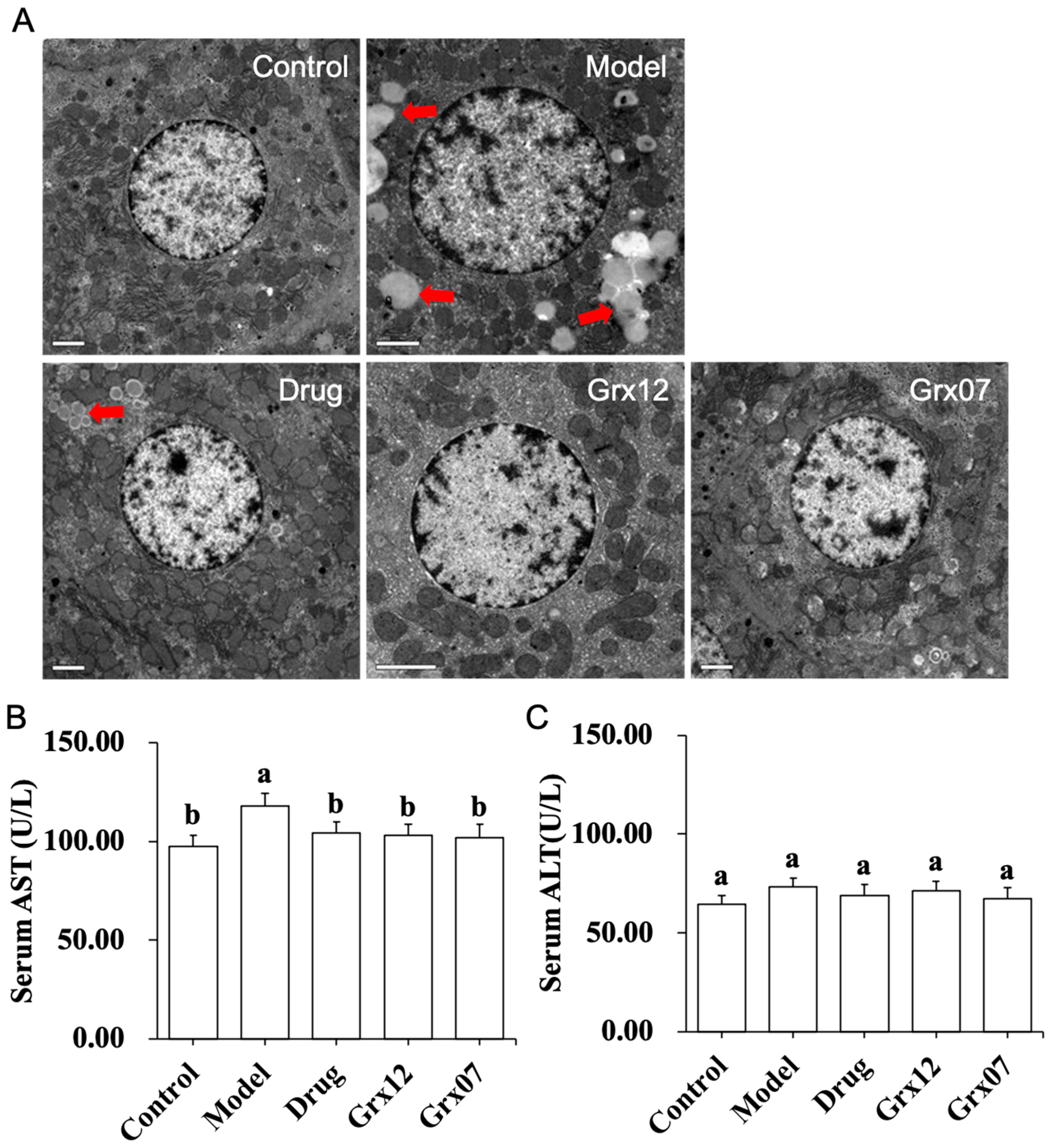
Figure 1. Effect of human-derived probiotics on the liver injury of rats. (A) Pathological observation of the rat liver. (B) The serum AST. (C) The serum ALT. Different letters indicate significant differences (p < 0.05). The red clippings mark the lipid droplets around the hepatocytes.
Fermentation 10 00127 g001
Figure 2. Effect of human-derived probiotics on the hepatocyte apoptosis of rats. Different letters indicate significant differences (p < 0.05).
Fermentation 10 00127 g002
Figure 3. Effect of human-derived probiotics on the ADH and ATPase in the liver of rats. (A) Liver Na+-K+-ATPase activity. (B) Liver Ca2+-ATPase activity. (C) Liver ADH activity. Different letters indicate significant differences (p < 0.05).
Fermentation 10 00127 g003
Figure 4. Effect of human-derived probiotics on oxidative stress in rats. (A) Liver MDA level. (B) Liver SOD level. (C) Liver GSH level. (D) Liver GSH-Px level. Different letters indicate significant differences (p < 0.05).
Fermentation 10 00127 g004
Figure 5. Effect of human-derived probiotics on the expression of Nrf2 in the liver of rats. Different letters indicate significant differences (p < 0.05).
Fermentation 10 00127 g005
Figure 6. Effect of human-derived probiotics on the inflammation of rats. (A) Serum LPS level. (B) Liver TNF-α level. (C) Liver IL-6 level. (D) Liver VEGF level. (E) Liver TGF-β1 level. (F) Liver NF-κB level. Different letters indicate significant differences (p < 0.05).
Fermentation 10 00127 g006
Table 1. The grouping and treatment of animals.
GroupGavage (0.1 mL/10 g Rat Body Weight)
MorningAfternoon
Controlsalinesaline
ModelChinese liquorsaline
DrugChinese liquorDongbaogangtai (0.06 g/mL methionine and chline bitartrate solution)
Grx12Chinese liquor109 CFU/Grx12 bacterial suspension
Grx07Chinese liquor109 CFU/mL Grx07 bacterial suspension
Disclaimer/Publisher’s Note: The statements, opinions and data contained in all publications are solely those of the individual author(s) and contributor(s) and not of MDPI and/or the editor(s). MDPI and/or the editor(s) disclaim responsibility for any injury to people or property resulting from any ideas, methods, instructions or products referred to in the content.