Article

The Antarctic Yeast Sporobolomyces roseus AL103 as a Promising Source of Health-Promoting Biologically Active Compounds

by
Snezhana Rusinova-Videva
1,
Maya M. Zaharieva
2,
Dilyana Hristova
3,
Stefka Nachkova
4,
Margarita Kambourova
5,
Hristo Najdenski
6,* and
Spiro Konstantinov
3
1
Laboratory of Eukaryotic Cell Biology, Department of Biotechnology, The Stephan Angeloff Institute of Microbiology—Bulgarian Academy of Sciences, 4000 Plovdiv, Bulgaria
2
Laboratory of Cytotoxicity and Signal Transduction, Department of Infectious Microbiology, The Stephan Angeloff Institute of Microbiology—Bulgarian Academy of Sciences, 1113 Sofia, Bulgaria
3
Department of Pharmacology, Pharmacotherapy and Toxicology, Faculty of Pharmacy, Medical University of Sofia, 1000 Sofia, Bulgaria
4
Department of Analytical Chemistry and Computer Chemistry, Faculty of Chemistry, University of Plovdiv “Paisii Hilendarski”, 4000 Plovdiv, Bulgaria
5
Laboratory of Extremophilic Microorganisms, Department of General Microbiology, The Stephan Angeloff Institute of Microbiology—Bulgarian Academy of Sciences, 1113 Sofia, Bulgaria
6
Laboratory of Bacterial Virulence, Resistance and New Antimicrobial Agents, Department of Infectious Microbiology, The Stephan Angeloff Institute of Microbiology—Bulgarian Academy of Sciences, 1113 Sofia, Bulgaria
*
Author to whom correspondence should be addressed.
Fermentation 2024, 10(10), 508; https://doi.org/10.3390/fermentation10100508
Submission received: 31 August 2024 / Revised: 30 September 2024 / Accepted: 30 September 2024 / Published: 2 October 2024
(This article belongs to the Section Microbial Metabolism, Physiology & Genetics)

Abstract

:
Antarctic yeasts represent a poorly explored source of novel bioactive compounds with antineoplastic activity and a favorable toxicological profile. The present paper presents the newest data on the antiproliferative and antimicrobial potential of extracts obtained from the psychrophilic strain AL103 of the species Sporobolomyces roseus. The capacity of AL103 to grow under different cultivation conditions, including in a bioreactor system with optimal biomass quantities of approximately 6.0 g/L, was demonstrated. A comparative examination of the metabolic profiles (GC-MS-based) of yeast extracts revealed a wide variety of synthesized molecules responsible for the different levels of antineoplastic activity depending on the tissue origin of the malignant cell lines. Concentration response curves were generated by the MTT dye reduction test. The respective IC50 values were extrapolated and found between 35.3 and 163 µg/mL. The antibacterial potential of both extracts was evaluated with the broth microdilution test against four referent pathogenic bacterial strains. The estimated minimal inhibitory concentrations revealed a moderate antibacterial activity. According to the GC-MS results, both extracts are rich in long-chain fatty acids which are known for their antibacterial properties. In conclusion, the Antarctic strain AL103 possesses promising potential for further pharmacological investigations aiming to elucidate its application as a health-promoting food additive or/and as a source of biologically active compounds.

1. Introduction

Antarctica is a geographical area representing the largest polar laboratory on the planet. Psychrophilic yeasts growing in Antarctica have a unique metabolism that allows them to withstand the extremely low temperatures [1]. Some of the established biochemical features related to the adaptation of cold-adapted yeast include increased synthesis of unsaturated fatty acids and specific antifreeze proteins. An accumulation of glycerol and trehalose and an increase in the production of exopolysaccharides, sphingolipids and carotenoids were also observed [2]. The intracellular synthesis of a variety of molecules has been reported by various authors. Metabolic profiles show the presence of amino acids, alcohols, organic acids and lipids, while some substances such as carotenoids and CoQ10 demonstrate distinct photoprotective and antioxidant activity [3,4,5]. In the available literature, information on the different biological activities of the representatives of Antarctic yeasts or their metabolites is scarce. The Antarctic yeast species Sporobolomyces roseus (strain AL103) which is the subject of this study belongs to the genus Sporobolomyces. Due to its high content of lipids, carotenoids, different types of enzymes, esters and other compounds, some mesophilic representatives of Sporobolomyces have been proposed for industrial applications as unconventional yeasts. There are different patents in which they are included because of their good biological activities [6]. In view of the biological activity of the psychrophilic species Srorobolomyces or its metabolites and the scarcity of the existing and limited reports [3,7,8], it is extremely important that the data be supplemented with new studies.
On the other hand, neoplastic diseases represent a major health and economic burden worldwide, being the second cause of death on the European continent and in the USA [9,10]. In Europe, more than 3.7 million new cases and 1.9 million deaths are registered every year, which comprise approximately 25% of the cancer cases globally (EU Policy on Cancer). According to the European Commission’s estimates from 2020 to 2022, cancer incidence rose by 2.3%, whereas mortality went up by 2.4% [11]. According to the last update of the WHO, the most common neoplasm cancers are breast (12.4%), colorectal (11.8%), lung (11.1%), prostate (10.6%) and bladder (4.7%) [12,13]. Among hematological malignancies, multiple myeloma comprised 4.9% of cases and Hodgkin lymphoma comprised 2.6% of cases. B- and T-cell lymphomas represented 3% of all cases of lymphoma registered in 2012 [14]. With the growing incidence in almost every country, the prevention of cancer is one of the most significant public health challenges of the 21st century [15]. Due to the heterogeneous nature of malignant diseases and the constantly arising problems with systemic toxicity and resistance toward numerous clinically applied chemotherapeutics, there is an urgent need for a continuous search for new antineoplastic agents.
In the last decade, the inhibition of cancer cells by natural compounds and extracts has attracted much attention. Antineoplastic potential has been observed in different fungal species. Methanol and ethyl acetate extracts obtained from Fomitopsis sp., Alternaria alternata and Phomposis sp. showed selective cytotoxic activity in breast and colon cancer cell lines [16]. Aspergillus fumigatus extract was found to exhibit antineoplastic effects in MCF-7 breast cancer cells by inhibiting cell proliferation and inducing apoptosis [17]. Yeasts belong to the eukaryotic kingdom of fungi and comprise 5–10% of estimated fungal species [18]. Notably, published studies on the anticancer activity of yeast are important but still scarce. Recently, the cytotoxicity of Pichia kudriavzevii AS-12 metabolites in colorectal cancer cell lines (HT-29 and Caco-2) was reported [19]. These metabolites activate both the extrinsic and intrinsic apoptosis pathways, interfering with pro- and antiapoptotic proteins of the Bcl-2 family, e.g., Bad and Bcl-2, which are involved in apoptosis regulation. In addition, Fas receptor activation takes place, initiating the cascade of initiator caspase-8 and -9 as well as the consequent executor caspase-3. The measured cytotoxic effects of these metabolites in HT-29 cells were comparable with the S-phase-specific clinically approved cytostatic drug 5-fluorouracil. Klyuiveromyces marxianus AS41 metabolites were highly active in gastric carcinoma cells and their anti-tumor effects were found to be selective [20]. As probable modes of action, the downregulation of Bcl-2 and the upregulation of Bad were discussed. Concomitant involvement of the intrinsic and extrinsic apoptosis cascades was suggested in the same study [20]. Other authors found that bioactive phenolic metabolites isolated from the apple yeast extract had antineoplastic effects (IC50 concentrations ranging from 31 to 38 µg/mL) against breast (MCF-7) and cervical (HeLa) cancer cells without significant inhibition of normal human embryonic kidney HEK-293 cells [21]. Andrade et al. [22] reported the presence of a selective cytotoxic effect of a β-carotene-containing yeast biomass produced by the yeast strain Rhodotorula glutinis against breast cancer MCF-7 cells and leukemic HL-60 cells. The initial data on the antineoplastic and proapoptotic activity in our previous studies of S. roseus AL103 determine the strain’s potential for additional biological activities. The extracts used demonstrated that the urothelial bladder carcinoma cell line T-24 was more sensitive than the non-tumor cell line CCL-1 [8].
Based on scientific reports on the antimicrobial activity of metabolites of yeasts from the genus Sporobolomyces, this study is of great interest. The antimicrobial properties of yeasts were discovered a long time ago. In particular, the data on Sporobolomyces roseus suggest an inhibitory effect of two compounds against Staphylococcus aureus, as evidenced with a bioassay on a thin-layer chromatography plate (TCL) [23]. Sporobolomyces sp. isolated from the phyllosphere surface of rice plant was found to produce an antimicrobial carotenoid pigment which was effective against E. coli and S. aureus [24]. Carotenoids and other hydrophobic organic compounds are the main carriers of the antimicrobial activity of the so-called red yeasts to which the genus Sporobolomyces also belongs. However, studies on the antibacterial activity of red yeasts, e.g., Sporobolomyces sp., are still scarce, especially on those isolated from Antarctica species. Therefore, further studies on their antimicrobial potential should be welcome, keeping in mind the urgent need for new antimicrobial agents with novel mechanisms of action in the era of the emerging antibiotic resistance.
The proven biological activities of the above-listed yeast species led us to investigate for the first time the antineoplastic and antimicrobial activities of extracts obtained from the Antarctic yeast Sporobolomyces roseus AL103 on a panel of different cancer cell lines. Aiming to analyze and compare the specific metabolic profile of the cell extracts, S. roseus AL103 was cultivated under different conditions in flasks and a bioreactor.

2. Materials and Methods

2.1. Biological Material

The strain AL103 of the yeast species Sporobolomyces roseus was isolated from a soil sample from Antarctica (Livingston Island). The sample was taken at a depth of 10 cm in the soil, and it aqueous suspension was cultivated for around 14–17 days on malt agar at 4 °C. The sequence was deposited in the National Center for Biotechnology Information (NCBI) under number ON567313. The sequence is based on a molecular genetic analysis of ITS1-5.8S-ITS4 regions of it. The producer was described in our previous studies with good cytotoxicity against malignant cell lines [8].

2.2. Media and Growth Conditions

The culture medium was prepared as in our preview studies [25]. Two types of submerged cultivation were carried out—in flasks and in a bioreactor cultivation system. The 500 mL flask contained 50 mL of medium. The pH (Hamilton, Bonaduz AG, Switzerland) and oxygen (Hamilton, Bonaduz AG, Switzerland) electrodes were used for the corresponding measurements in a Sartorius bioreactor with a working volume of 5 L. Mechanical stirring was carried out at 220 rpm with a rotary shaker and 400 rpm with a turbine stirrer in the bioreactor. The monitoring of the cultivation process in the bioreactor cultivation conditions was performed by using the software program BioPAT® MFCS/DA 3.0. Each of the cultivation processes was carried out at 22 °C for 120 h.

2.3. Biomass Production and Extracts Preparation

Before freeze-drying, the biomass was collected and separated by centrifugation at 6000× g for 30 min. The dry weight of the biomass prepared by the Antarctic yeast strain was determined after drying at 105 °C until a constant weight was reached. A total of 1 g of freeze-dried yeast biomass was used for extraction. Methanol (100%) was used as the extraction reagent and followed by double ultrasonic extractions for 20 min [26,27]. The solutions were dried by vacuum evaporation to produce dry extracts. DMSO was used to dissolve the dry extracts and in subsequent applications.

2.4. Gas Chromatography/Mass Spectrometry (GC/MS) Profiling

Samples of 0.5 g were used for derivatization, and 100.0 µL methoxamine chloride in pyridine (20.0 mg/mL) was added to the samples. The samples were incubated at 70 °C for 90 min. Then, 50.0 µL BSTFA (silylating reagent) was added before the next incubation cycle at 70 °C for 30 min. One microliter of each sample was used for the chromatography analysis. It was carried out by using the Agilent GC 7890A gas chromatograph (Agilent Technologies Inc., Santa Clara, CA, USA) and the Agilent MD 5975C, mass spectral detector supplied with a DB-5MS column (length 30 m; diameter 0.32 mm; film coating thickness 0.25 µm). The initial temperature was 100 °C for 2 min, and the temperature was increased to 180 °C at a step of 15 °C/min. That step was followed by retention for 1 min and an increase in the temperature to 300 °C at a step of 5 °C/min and with retention for 10 min. The injector and detector were maintained at a temperature of 250 °C. Helium was used as a carrier gas (1.0 mL/min). The mass detector scan range was m/z = 50–550. Identification of the components present in the samples was made by comparing the retention time and Kovat’s index (RI) with those of standard substances and mass spectral data from the libraries of the Golm Metabolome Database (http://gmd.mpimp-golm.mpg.de/ accessed on 20 January 2024) and the National Institute of Standards and Technology (NIST 08), USA.

2.5. Cell Lines and Culture Maintenance

The malignant cell lines HD-MY-Z (DSMZ, ACC 346, B-cell Hodgkin lymphoma), SKW-3 (DSMZ, ACC 53, T-cell leukemia, derivative of KE-37), U-266 (DSMZ, ACC 9, multiple myeloma) and Cal-29 (DSMZ, ACC 515, bladder carcinoma) were purchased from the German Collection for Cell Cultures and Microorganisms, Leibnitz Institute, DSMZ GmbH, Braunschweig, Germany. The cell lines HuT-78 (ATCC® TIB-161™, cutaneous T-cell lymphoma, Sézary syndrome), MJ (G11) (ATCC® CRL-8294™, cutaneous T-cell lymphoma, Mycosis fungoides), RPMI-8226 (ATCC® CCL-155™, multiple myeloma) and CCL-1 (NCTC clone 929 (L cell, L-929, derivative of Strain L), 159 ATCC® CCL-1™, non-tumoral cell line) were delivered from the American Type Culture Collection (ATCC, Manassas, VA, USA). All cell lines were maintained in culture media and conditions recommended by the respective biobank (ATCC or DSMZ) that they were purchased from. HD-MY-Z, SKW-3, U-266, RPMI-8226 and Cal-29 were cultured in 90% RPMI-1640 (#RPMI-HA, Capricorn, Germany) supplemented with 10% (v/v) fetal bovine serum (#FBS-HI-12A, Capricorn, Germany). CCL-1 was maintained in MEM (#MEM-A, Capricorn, Germany) supplemented with 10% horse serum (#HOS-1A, Capricorn, Germany) and 2 mM L-glutamine (#GLN-B, Capricorn, Germany). HuT-78 cells were grown in modified Dulbecco’s medium (#DMEM-HHSTA, Capricorn, Germany) with 4 mM L-glutamine. All cell lines were cultivated in a humidified atmosphere (≥95%) at 37 °C and 5% (v/v) CO2.

2.6. MTT Assay

The antineoplastic activity of the S. roseus extracts was measured according to ISO 10993-5, Annex C, which is based on the MTT dye reduction assay [28,29]. Briefly, cells were seeded in 96-well plates at a volume of 100 µL/well and a cell density depending on the cell line as follows—MJ: 10.2 × 10⁵ cells/well; RPMI: 8.0 × 10⁵ cells/well; HD-MY-Z: 3.0 × 10⁵ cells/well; SKW-3: 4.0 × 10⁵ cells/well; U-266: 4.5 × 10⁵ cells/well; Cal-29: 2.5 × 10⁵ cells/well; HuT-78: 7.7 × 10⁵ cells/well. The cells were cultivated for 24 h until entering the log phase of cell growth. After 24 h of incubation, the cells entered the log phase of the growth curve and were exposed for 72 h to twofold dilutions of both extracts in concentrations ranging between 25 and 400 μg/mL. Cell viability was measured after incubation of the samples with MTT solution (final concentration 0.1 mg/mL) for 120 min at 37 °C. The dye was reduced by the mitochondrial dehydrogenases in the survived cells to a water-insoluble product formazan, which forms violet crystals. The latter was dissolved in an equivalent volume of an organic solvent (2-propranolol acidified with 5% formic acid). The absorbance was measured at a wavelength λ = 550 nm (reference filter 690 nm) on a microplate, ELISA Reader ELx800 (Bio-Tek Instruments Inc., Santa Clara, CA, USA), against a blank solution containing the organic solvent with the used culture medium and MTT solution. The results were calculated with the GraphPad Prizm software (version 10.3.0), and the dose–response inhibition model was based on four parameters: Y = 100/(1 + 10^((LogIC50-X) × HillSlope)). The percentage of the survived cell fraction is presented on the Y axis (“% of the untreated control”) after converting the absorbance of each sample to a percentage of the untreated control, which was taken as 100%.

2.7. Bacterial Strains and Growth Conditions

Four referent bacterial strains of the species Staphylococcus aureus ATCC 29213, Enterococcus faecalis ATCC 29212, Escherichia coli 25922 and Pseudomonas aeruginosa ATCC 27853 (recommended in [30]) were used. The strains were purchased from the American Type Culture Collection, Manassas, Virginia, USA. The bacterial cultures were maintained in BHI (Himedia/India) in an incubator at 37 °C. The broth microdilution test (BMD) was performed in Mueller Hinton Broth (MHB, Thermo Scientific-Oxoid, Hampshire, RG24 8PW, United Kingdom, UK) for estimation of the antibacterial activity.

2.8. Broth Microdilution Test and Redox Activity

The antibacterial activity of the yeast extracts was evaluated according to the ISO standard 20776/1-2006 based on the BMD test. Briefly, twofold serial dilutions of the extracts from 5 down to 0.01 mg/mL were prepared in triplicate by using 96-well plates to obtain a final volume of 50 µL/well. An equivalent volume of the relevant bacterial strain in suspension with a density of 5 × 10⁵ CFU/mL was added to each well. The plates were incubated at 37 °C for 24 h. The lowest drug concentration which inhibited the visible bacterial growth was accepted as the minimal inhibitory concentration (MIC). Gentamycin (0.008–4 mg/L) and penicillin (0.008–4 mg/L) (Gibco, Life Technologies Ltd., Paisley, UK) were used as reference antibiotics (positive control). The recommendations of EUCAST (European Committee on Antimicrobial Susceptibility Testing) were followed for the results’ analysis (Breakpoint Tables for Bacteria). PBS served as a negative control and MHB as a blank solution.
The redox (metabolic) activity of the bacteria was measured in the same plates after evaluation of the MICs. Briefly, MTT (3-(4,5-dimethylthiazolyl-2)-2,5-diphenyltetrazolium bromide) solution (10 µL) was added to each well and the plates were incubated for 120 min at 37 °C [31]. The MTT dye (#M2128, Sigma® Life Science, Steinheim, Germany) was reduced by the membrane located on the bacterial enzyme complex NADH–ubiquinone reductase (H+-translocation) to the insoluble compound formazan. The formazan crystals were dissolved in 2-propanol containing 5% formic acid, and the absorbance was measured at 550 nm (Absorbance Microplate Reader Lx800, Bio-Tek Instruments Inc., USA). A respective mixture of MHB, MTT and solvent served as a blank solution.

3. Results

3.1. Submerged Cultivation of Sporobolomyces roseus AL103

The growth of the S. roseus AL103 strain was followed for 120 h (Figure 1 and Figure S1a). The accumulated biomass reached 4.8 g/L (dw) after 48 h of cultivation in the flasks. During the stationary phase, the biomass was almost unchanged and 5.0 g/L was harvested at 96 h. The registered productivity was 0.05 g/L/h. The pH tracking curve of the culture medium showed a sharp decrease to pH 2.0–2.2, with a baseline of pH 5.3 after the 24th hour of fermentation (Figure 1).

3.2. Bioreactor Cultivation of Sporobolomyces roseus AL103

Cultivation in the bioreactor was characterized by a longer exponential phase (96 h) compared to the flask cultures (Figure 2A and Figure S1b). At the end of the exponential phase, an optimal amount of accumulated biomass was observed—appr. ≈ 6.4 g/L. The performance of the fermentation process in the bioreactor showed that the maximum biomass was accumulated after 96 h of cultivation and the amount did not change significantly after 96 h (Figure 2A). Based on the results obtained, the productivity of the strain was determined to be 0.066 g/L/h.

3.3. GC/MS Profiling

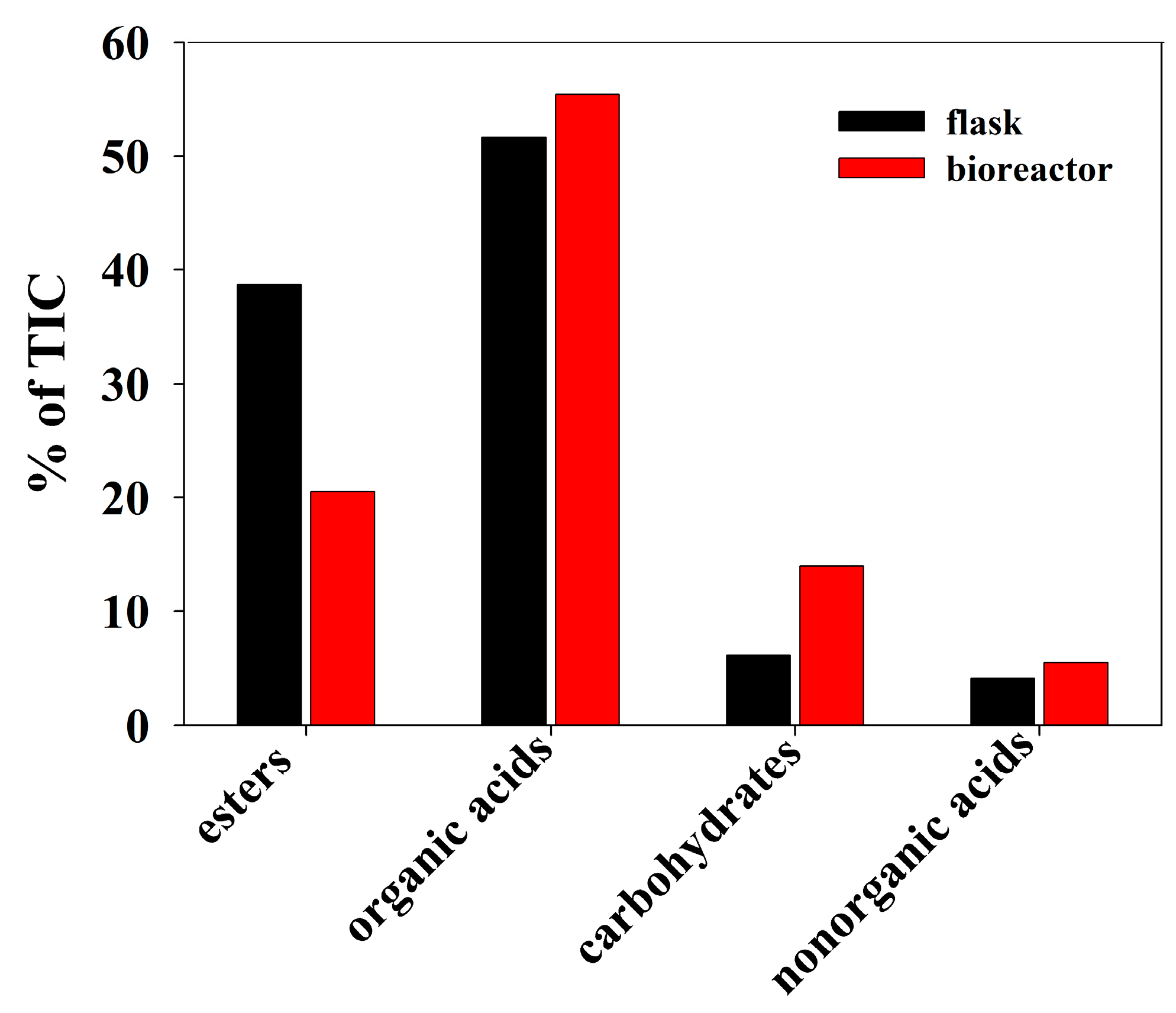
The analysis of the methanol extracts showed the presence of several groups of substances varying in quantity among the different types of cultures. As can be seen (Table 1, Figure S1d,e), thirty-two compounds were identified. The most abundant compounds (above 10%) from both cultivation conditions were fatty acids. Some other organic acids, phosphoric acid and some monosaccharides were quantified in lower amounts. In the extract obtained from flask cultivation, 11-octadecenoic acid methyl ester and palmitic acid predominated, being in higher amounts compared to the sample originating from the bioreactor. Concerning oleic acid, it prevailed in the bioreactor culture. Regarding carbohydrates, fructose and glucose in the extracts of the bioreactor biomass both exceeded those from flask cultivation, whereas arabinose had a higher content in the flask culture (Figure 3).

3.4. Antiproliferative Activity of Yeast Extracts on Malignant and Non-Tumorigenic Cell Lines

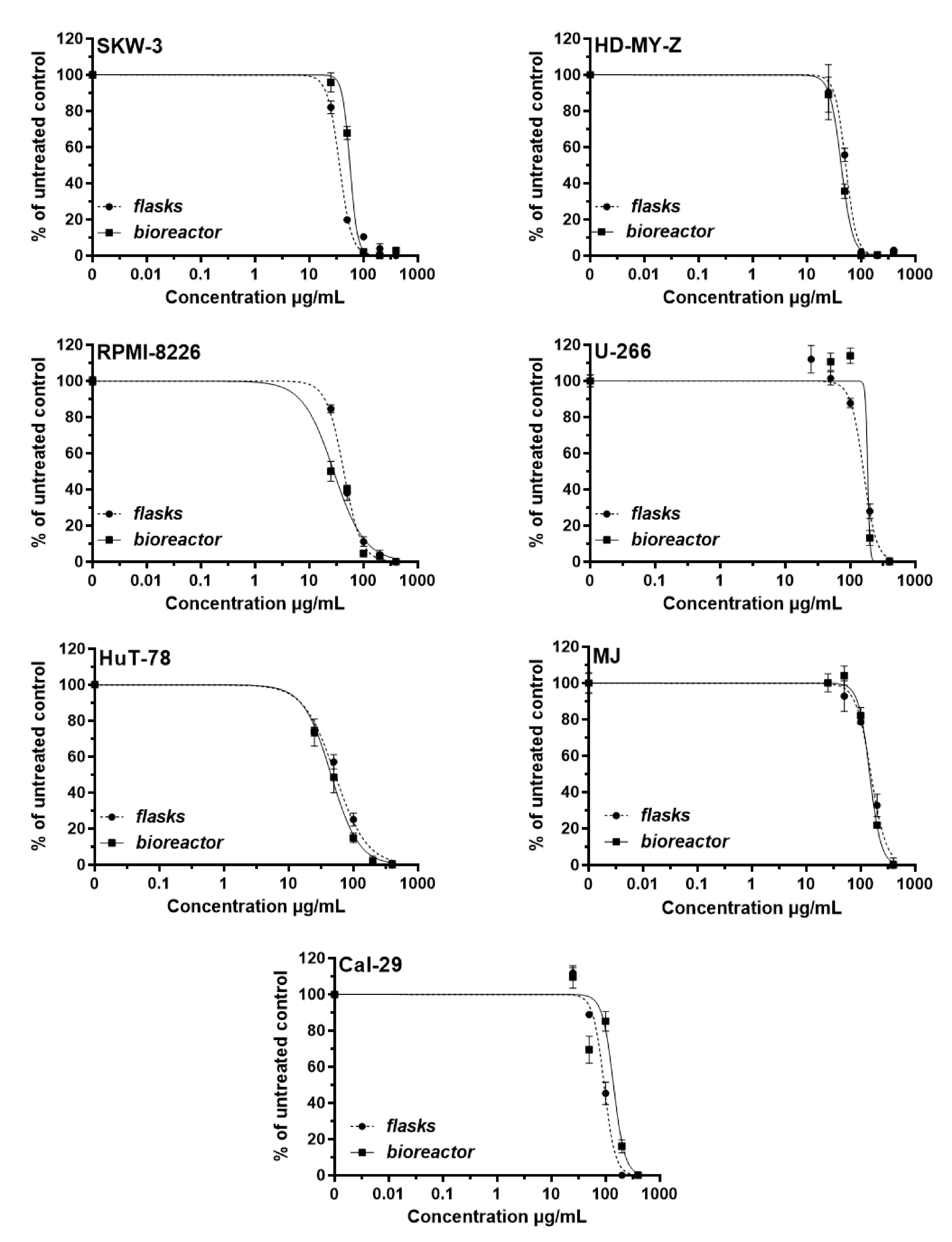
A methanol extract was obtained from S. roseus AL103 and the cell lines HuT-78, HD-MY-Z, RPMI-8226, SKW-3 (KE-37), U-266, T-24, MJ, Cal-29 and CCL-1 were exposed to it for a period of 72 h. The tested concentrations ranged between 25 and 400 µg/mL. The calculated median inhibitory concentrations served as a criterion for the evaluation of the antiproliferative activity of the extracts. The results are presented in Table 2 and Figure 4.
Most sensitive to the in vitro effect of the extract of S. roseus AL103 cultivated in flasks were the cell lines SKW-3 (IC50 = 35.3 µg/mL) and RPMI-8226 (IC50 = 42.33 µg/mL), followed by HD-MY-Z and HuT-78. The bioreactor culture extract was more cytotoxic for RPMI-8226, HD-MY-Z and HuT-78 cells, followed by SKW-3. The least sensitive cell line was U-266 followed by MJ and Cal-29.

3.5. Minimal Inhibitory Concentrations (MICs)

The MICs of the two tested yeast extracts are presented in Table 3. The extract of S. roseus AL103 cultivated in the flasks was most active against S. aureus, whereas the extract of S. roseus AL103 cultivated in the bioreactor achieved an MIC of 5 mg/mL against all tested strains except for the P. aeruginosa strain, which was not affected.

3.6. Inhibition of the Bacterial Metabolic Activity

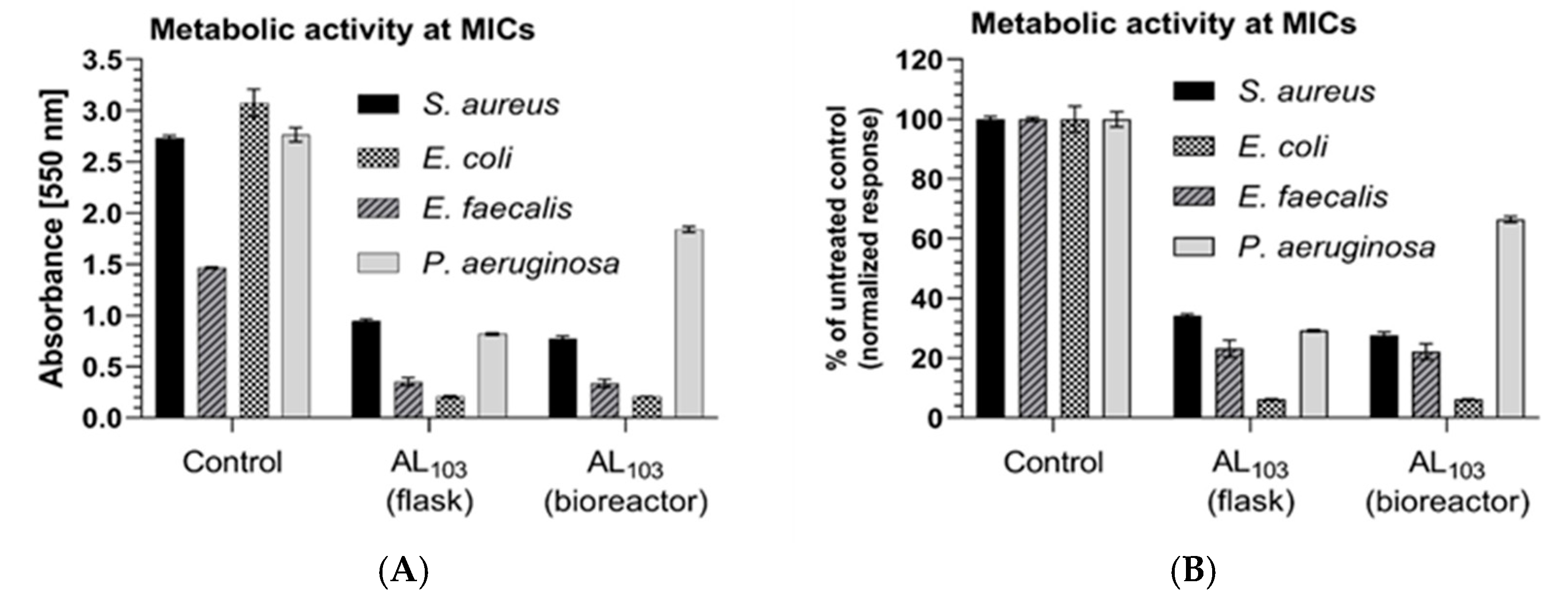
The effects of the two yeast extracts on the metabolic activity of the treated bacteria are shown in Figure 5, where the values for the MIC are presented. The lower redox activity of the bacterial enzyme complex NADH–ubiquinone reductase (H+-translocation) is related to a stronger bacteriostatic effect. As visible in Figure 5, the tested extracts are inhibited at different levels but do not fully inhibit the metabolic activity of the bacteria.

4. Discussion

The biotechnological need for a higher quantity of yeast extract regarding its future application as an antineoplastic tool determined our interest in cultivation not only in flasks but also in bioreactors. The slower culture growth of the yeast strain in the bioreactor and the correspondingly longer process could be explained by the fact that low oxygen solubility limits the growth rate. Enhanced metabolite production under dissolved oxygen (DO) limitation was observed by other authors [32]. The decrease in the pH of the process is typical for the biosynthesis of exopolysaccharides by Antarctic yeasts, and is the advantage of the process regarding natural protection against contamination (Figure 1) [33].
The observed increase in the biomass originating from the bioreactor culture (≈33.0%) compared to the flask culture could be explained by a more homogeneous environment with better access of each cell to the nutrient medium components in the bioreactor. The course of the pH remained unchanged in comparison with the flask culture experiments (Figure 1 and Figure 2A). The low pH was an advantage of the cultivation process because it is a natural protective factor against microbiological contamination. The curve of oxygen consumption in the exchange processes showed a short interval of sharp depletion in the exponential growth phase of S. roseus AL103 followed by lower levels of consumption in the stationary phase (Figure 2B). The culture did not experience a period of oxygen starvation. Through dissolved oxygen levels, at any point in the process, we can predict the presence or absence of cell growth disturbances. That is, the construction of the curve helps us to indirectly observe the development of the culture. A similar biomass yield was reported for the psychrophilic EPS producer Cryptococcus laurentii AL65 under the same cultivation conditions [34]. The observed comparatively short process is not typical for psychrophilic yeasts, as their processes usually last 7–14 days [35,36]. The selected temperature was suitable for various cultivation processes with Antarctic yeasts [25,34]. The established prolongation of the process, higher biomass yield, culture volume and low pH during cultivation to prevent microbial contamination are essential biotechnological advantages of the described process.
The observed quantitative differences in the metabolic composition of the extracts derived from flask and bioreactor cultivation biomass were probably due to the more intensive aeration and stirring during the bioreactor process compared to flask cultivation. The observed data should be of use in designing subsequent studies for the identification and isolation of active metabolites and, in particular, for the characterization of the synergistic effects between them.
Some metabolites detected in the extracts have been found to be anti-inflammatory agents, precursors of biologically active substances, preservatives and cosmetic additives (Table 1). Indole-3-acetic (IAA) acid is recognized as an auxin-type plant hormone. IAA toxicity has been described in T24 bladder carcinoma, breast carcinoma, nasopharyngeal FaDu carcinoma, G361 melanoma cells, pancreatic cancer and others. A dose-dependent and time-dependent effect of IAA on apoptosis induction in human hematopoietic tumors has been demonstrated [37]. IAA possesses a cytotoxic effect if it is stimulated by other substances, such as horseradish peroxidase (HRP). IAA also stimulates the production of reactive oxygen species (ROS), such as O-2 and H2O2. This causes structural changes in plasma membranes and initiates cellular damage and apoptosis [38]. For 2-ketoglutaric acid, inhibition of the G1 phase of the cell cycle and subsequent apoptosis of osteosarcoma cells were observed [39]. In addition, 2-ketoglutaric acid was shown to play a role in tumor neo-angiogenesis, as well as in the regulation of the hypoxic response [40,41]. Pentadecanoic acid was described as contributing to apoptosis in breast cancer cells [42]. The net action of short-chain pentanoate and butyrate was considered useful in the context of cellular cancer immunotherapy [43].
In our study, the antineoplastic effect of the accumulated biomass was evaluated on a panel of eight malignant cell lines originating from different neoplastic diseases, including tumor and hematological malignancies and one non-malignant cell line derived from normal mouse fibroblasts. The IC50 values calculated from the MTT assay varied depending on the cell line (Table 2). The extracts from both types of cultivation showed dose-dependent cytotoxicity profiles (Figure 4), but no clear correlation between the activity of the extract and the origin of the cell lines was observed. The analysis of the data showed that cell lines originating from hematological malignancies were in general more sensitive to the antiproliferative activity of the extract as compared to solid tumor cell lines (Cal-29) originating from urinary bladder cancer, whereby the multiple myeloma cell line RPMI-8226 was the most sensitive. The IC50 values calculated for the leukemia cell line SKW-3 were 35.3 µg/mL for the flask extract and 55.9 µg/mL for the bioreactor extract. The IC50 values determined in our study are similar to the data published by other authors for the leukemia cell line U937 after treatment with crude methanol extracts obtained from three bacterial strains with antineoplastic activity (23–45 µg/mL) [44].
The CTCL cell line MJ and the multiple myeloma cell line U-266 were exceptions among the hematological cells tested, with IC50 values higher than those determined for the solid tumor cell lines (Table 2). The results in the present study partially correlate with the data published by Saber et al. [19,20], who investigated the antineoplastic activity of yeast extracts against various malignant cell lines and also observed a dose-dependent cytotoxicity. The sensitivity of RPMI-8226 toward the extract obtained from the S. roseus AL103 (IC50 was 42.33 µg/mL and 28.27 µg/mL for flask and bioreactor) cell extract was comparable with that reported for an ethyl acetate extract from Aspergillus sp. F-21 toward the cervical cancer cell line HeLa (IC50 was 20.37 μg/mL) and the breast cancer cell line MCF-7 (IC50 was 44.75 μg/mL) [45]. Both yeast extracts exhibited selective cytotoxicity on the malignant cell lines included in this study. The same type of S. carnicolor extract used as an agent in the treatment of HCT116 demonstrated IC50 > 200 μg/mL [46].
The different concentrations of metabolites present in both extracts were reflected in differences in the in vitro antineoplastic activity. Most likely, the culture conditions are responsible for the production of various metabolites and affect their amount in the obtained extracts. Despite the low percentage of some of the proven cytotoxic substances in the extracts, their combination may result in synergistic interactions, thus leading to significantly higher inhibition of tumor cell growth.
According to the data, the inhibitory activity of S. roseus AL103 against the tested bacteria can be expected only at a concentration of 5 mg/mL or higher (with the exception of Staphylococcus aureus). The ethyl acetate extract of Sporobolomyces carnicolor showed similar MIC values in the inhibition of S. aureus and Pseudomonas aeruginosa. In this study, two- and fourfold higher MIC values against Gram-positive pathogenic bacteria were reported [46]. A pigment from an extract of Sporobolomyces sp. inhibited the growth of E. coli, S. aureus, S. faecalis, B. subtilis, Enterococcus sp. and P. aeruginosa with different zones of inhibition [24]. Detailed studies related to the biological activity of the strain of S. roseus demonstrated its use as a biocontrol marker for limiting the growth of Penicillium expansum by half and its metabolite patulin by 75% [47]. The antibacterial effect of both extracts tested in this study is moderate. The extracts are rich in organic acids (Figure 3). The GC-MS analysis gives a comprehensive view of the type of fatty acids contained in their composition. Certain fatty acids such as hexadecatetraenoic acid, 3-hydroxydodecanoic acid, N-tridecanoic acid, lauric acid and others are known to inhibit the growth and biofilm formation of Gram-positive bacteria and, to a lesser extent, Gram-negative bacteria. They have a wide range of applications as health-promoting food additives in the food industry, as preservation agents and in agriculture [48,49,50]. The molecular targets of the fatty acids are still unclear. They target inner membrane components and more easily penetrate Gram-positive bacteria due to the lack of an outer membrane. Their mode of action depends on the chemical structure, chain length, degree of saturation and site of action and is expressed in terms of nutrient uptake inhibition, the formation of peroxidation products, binding to electron carriers, etc., which finally lead to cell lysis and death [48,51,52,53]. Keeping in mind these facts, we could assume that, although moderate, the antibacterial potential of both extracts tested in this study is promising for future investigations after optimization of the culture conditions regarding the enhancement of antimicrobial fatty acid production.
In the present study, 32 metabolites were identified in cell extracts of the Antarctic representative S. roseus AL103. The demonstrated biological activity and the literature referenced allowed us to consider some metabolites as cytotoxic to cancer cell lines and others as antimicrobial agents (Table 1). Cell substances with a cytotoxic effect were IAA, 2-ketoglutaric acid, pentadecanoic acid, pentanoate and butyrate. Moderate cytotoxicity could be explained by the presence of hexadecatetraenoic acid, 3-hydroxydodecanoic acid, N-tridecanoic acid, lauric acid and fatty acids. Our research is focused on specific biological effects, but biotechnology makes it possible to separate and study individual metabolites for application in other areas as well.

5. Conclusions

The Antarctic strain S. roseus AL103 demonstrates considerably good cell growth in submerged cultivation. The recent study on the dynamics of the fermentation process contributes to the existing knowledge about the biosynthetic potential of these psychrophilic yeasts and points out new possibilities for optimization of the processes. The higher biomass accumulation in the bioreactor system identifies this cultivation method as more suitable than flask cultivation. The synthesis of different amounts of metabolites depending on the cultivation method was demonstrated by GC-MS. A large part of the metabolites of S. roseus were described for the first time in this study. The antineoplastic and antibacterial activity of the extracts from the psychrophilic yeast depended on the tissue origin of the cell lines and the bacterial species. Median inhibitory concentrations under 50 µg/mL were achieved in both leukemia and lymphoma cell lines. The minimal inhibitory concentrations in the bacteria demonstrated moderate antibacterial potential. The obtained results are promising for future pharmacological investigations on psychrophilic yeasts, which can be a reliable source of biologically active compounds.

Supplementary Materials

The following supporting information can be downloaded at: https://www.mdpi.com/article/10.3390/fermentation10100508/s1, Figure S1: (a) S. roseus AL103 culture in flask; (b) S. roseus AL103 culture in Biostat A plus bioreactor; (c) lyophilized biomass from S. roseus AL103; GC/MS chromatograms of (d) extract of S. roseus AL103 cultured in flasks and (e) extract of S. roseus AL103 cultured in 5 L bioreactor.

Author Contributions

Conceptualization, S.R.-V., M.M.Z., M.K., H.N. and S.K.; methodology, S.R.-V. and M.M.Z.; software, S.R.-V., M.M.Z. and S.N.; validation, S.R.-V., M.M.Z., D.H. and S.N.; formal analysis, S.R.-V., M.M.Z. and D.H.; investigation, S.R.-V., M.M.Z., D.H., S.N., S.K. and H.N.; resources, S.R.-V.; writing—original draft preparation, S.R.-V., D.H., M.M.Z., S.N. and M.K.; writing—review and editing, S.K., M.K. and H.N.; visualization, S.R.-V. and M.M.Z.; supervision, S.R.-V., M.K., M.M.Z., S.K. and H.N.; project administration, S.R.-V.; funding acquisition, S.R.-V. All authors have read and agreed to the published version of the manuscript.

Funding

This study was funded by the Bulgarian Ministry of Education and Science through the National Centre for Polar Studies, and Sofia University “St. Kliment Ohridski” in the framework of the National Program for Polar Studies 2022–2025 (grant numbers 70-25-40/23.01.2023).

Institutional Review Board Statement

Not applicable.

Informed Consent Statement

Not applicable.

Data Availability Statement

Data is contained within the article or Supplementary Material.

Acknowledgments

We thank the staff of the Bulgarian Antarctic Expedition for their logistic support. The antiproliferative activity of the extracts on HD-MY-Z, HuT-78 and MJ (MTT dye assay) was determined by using laboratory equipment donated by the Alexander von Humboldt Foundation to Maya M. Zaharieva in the framework of the program “Equipment subsidies”.

Conflicts of Interest

The authors declare no conflicts of interest.

References

  1. Buzzini, P.; Margesin, R. (Eds.) Cold-adapted yeasts: A lesson from the cold and a challenge for the XXI century. In Cold-Adapted Yeasts; Springer: Berlin/Heidelberg, Germany, 2014; pp. 3–22. [Google Scholar]
  2. Segal-Kischinevsky, C.; Romero-Aguilar, L.; Alcaraz, L.; Lopez-Ortiz, G.; Martinez-Castillo, B.; Torres-Ramirez, N.; Sandoval, G.; Gonzalez, J. Yeasts inhabiting extreme environments and their biotechnological applications. Microorganisms 2022, 10, 794. [Google Scholar] [CrossRef]
  3. Rusinova-Videva, S.; Kambourova, M.; Alipieva, K.; Nachkova, S.; Simova, S. Metabolic profiling of Antarctic yeasts by proton nuclear magnetic resonance-based spectroscopy. Biotechnol. Biotechnol. Eq. 2019, 1, 12–19. [Google Scholar] [CrossRef]
  4. Zlatanov, M.; Pavlova, K.; Antova, G.; Angelova-Romova, M.; Georgieva, K.; Rusinova-Videva, S. Biomass production by Antarctic yeast strains: An investigation on lipid composition. Biotechnol. Biotechnol. Eq. 2010, 24, 2096–2101. [Google Scholar] [CrossRef]
  5. Dimitrova, S.; Pavlova, K.; Lukanov, L.; Zagorchev, P. Synthesis of coenzyme Q10 and b-caroten by yeasts isolated from Antarctic soil and lichen in response to ultraviolet and visible radiations. Appl. Biochem. Biothechnol. 2010, 162, 795–804. [Google Scholar] [CrossRef] [PubMed]
  6. Kot, A.; Kieliszek, M.; Piwowarek, K.; Blazejak, S.; Mussagy, C. Sporobolomysec and Sporidiobolus-non-conventional yeasts for use in industries. Fungal Biol. Rev. 2021, 37, 41–58. [Google Scholar] [CrossRef]
  7. Barahona, S.; Yuivar, Y.; Socias, G.; Alcaino, J.; Cifuentes, V.; Baeza, M. Identification and characterization of yeast isolated from sedimentary rocks of Union Glacier at the Antarctica. Extremophiles 2016, 20, 479–491. [Google Scholar] [CrossRef]
  8. Rusinova-Videva, S.; Hristova, D.; Konstantinov, S. Antineoplastic activity of extracts of the Antarctic yeast Sporobolomyces roseus AL103 on urinary bladder cancer cells. C. R. Acad. Bulg. Sci. 2023, 76, 778–788. [Google Scholar] [CrossRef]
  9. Siegel, R.L.; Miller, K.D.; Wagle, N.S.; Jemal, A. Cancer statistics, 2023. CA Cancer J. Clin. 2023, 73, 17–48. [Google Scholar] [CrossRef]
  10. Dyba, T.; Randi, G.; Bray, F.; Martos, C.; Giusti, F.; Nicholson, N.; Gavin, A.; Flego, M.; Neamtiu, L.; Dimitrova, N.; et al. The European cancer burden in 2020: Incidence and mortality estimates for 40 countries and 25 major cancers. Eur. J. Cancer Nov. 2021, 157, 308–347. [Google Scholar] [CrossRef]
  11. ESIC. Available online: https://ecis.jrc.ec.europa.eu/ (accessed on 7 January 2024).
  12. Ferlay, J.; Colombet, M.; Soerjomataram, I.; Dyba, T.; Randi, G.; Bettio, M.; Gavin, A.; Visser, O.; Bray, F. Cancer incidence and mortality patterns in Europe: Estimates for 40 countries and 25 major cancers in 2018. Eur. J. Cancer 2018, 103, 356–387. [Google Scholar] [CrossRef]
  13. World Health Organization. Cancer Data and Statistics. 20 April 2020. 2020. Available online: http://www.euro.who.int/en/health-topics/noncommunicable-diseases/cancer/data-and-statistics (accessed on 16 January 2024).
  14. Lymphoma Coalition Europe. Lymphoma Incidence, Mortality and Survival Data for Europe. 2020. Available online: https://www.lymphomacoalition.org/europe (accessed on 20 January 2024).
  15. American Institute for Cancer Research. Global Cancer Data by Country. 20 April 2020. 2020. Available online: https://www.wcrf.org/dietandcancer/cancer-trends/data-cancer-frequency-country (accessed on 10 January 2024).
  16. Shweta, S.; Gurumurthy, B.; Ravikanth, G.; Ramanan, U.; Shivanna, M. Endophytic fungi from Miquelia dentate Bedd., produce the anti-cancer alkaloid, camptothecine. Phytomed 2013, 20, 337–342. [Google Scholar] [CrossRef] [PubMed]
  17. Li, Y.; Himaya, S.; Dewapriya, P.; Zhang, C.; Kim, S. Fumigaclavine C from a ma-rine-derived fungus Aspergillus fumigates induces apoptosis in MCF -7 breast cancer cells. Mar. Drugs 2013, 11, 5063–5086. [Google Scholar] [CrossRef] [PubMed]
  18. Türker, M. Yeast Biotechnology: Diversity and Applications. In Proceedings of the 27th VH Yeast Conference, Advances in Science and Industrial Production of Baker’s Yeast, Istanbul, Turkey, 14–15 April 2014; pp. 1–26. [Google Scholar]
  19. Saber, A.; Alipou, B.; Faghfoor, Z.; Moisavijam, A.; Khosroushahi, A. Secretion metabo-lites of probiotic yeast, Pichiakudravzevii AS-12, induces apoptosis pathways in human colorectal cancer cell lines. Nut. Res. 2017, 41, 36–46. [Google Scholar] [CrossRef] [PubMed]
  20. Saber, A.; Alipour, B.; Faghfoori, Z.; Khosroushahi, A. Secretion metabolites of dairy Kluyveromyces marxianus AS41 isolated as probiotic, induces apoptosis in different hu-man cancer cell lines and exhibit anti-pathogenic effects. J. Fun. Foods 2017, 34, 408–421. [Google Scholar] [CrossRef]
  21. Sarkate, A.; Banerjee, S.; Iqbal Mir, J.; Roy, P. Antioxidant and cytotoxic activity of bio-active phenolic metabolites isolated from the yeast-extract treated cell culture of apple. PCTOC 2017, 130, 641–649. [Google Scholar] [CrossRef]
  22. Andrade, R.; Lima, R.; Ribeaux, D.; Araújo, H.; Franco, L.; Pessoa-Júnior, A.; Cam-pos-Takaki, G. Production of β-carotene by a newly isolated Rhodotorula glutinis UCP1555 strain and cytotoxic effect evaluation. J. Chem. Eng. 2016, 10, 212–220. [Google Scholar] [CrossRef]
  23. McCormack, P.; Wildman, H.; Jeffries, P. Production of antibacterial compounds by phylloplane inhabiting yeasts and yeastlike fungi. Appl. Environ. Microbiol. 1994, 60, 927–931. [Google Scholar] [CrossRef]
  24. Manimala, M.; Murugesan, R. In vitro antioxidant and antimicrobial activity of carotenoid pigment extracted from Sporobolomyces sp. isolated from natural source. J. Appl. Natur. Sci. 2014, 6, 649–653. [Google Scholar] [CrossRef]
  25. Rusinova-Videva, S.; Ognyanov, M.; Georgiev, Y.; Petrova, A.; Dimitrova, P.; Kambourova, M. Chemical characterization and biological effect of exopolysaccharides synthesized by Antarctic yeasts Cystobasidium ongulense AL101 and Leucosporidium yakuticum AL102 on murine innate immune cells. World J. Microbiol. Biotechnol. 2023, 39, 211–231. [Google Scholar] [CrossRef]
  26. Dagnon, S.; Novakova, Z.; Bojilov, D.; Nedialkov., P.; Kouassi, C. Development of surro-gate standards approach for the determination of polyphenols in Vernonia amygdalina Del. J. Food Compos. Anal. 2019, 82, 103231. [Google Scholar] [CrossRef]
  27. Jiang, T.; Ghosh, R.; Charcosset, C. Extraction, purification and application of curcumin from plant materials—A comprehensive review. Trends Food Sci. Technol. 2021, 112, 419–430. [Google Scholar] [CrossRef]
  28. Mosmann, T. Rapid colorimetric assay for cellular growth and survival: Application to proliferation and cytotoxicity assays. J. Immunol. Methods 1983, 65, 55–63. [Google Scholar] [CrossRef]
  29. ISO 10993-5:2009; Biological Evaluation of Medical Devices—Part 5: Tests for In Vitro Cytotoxicity. International Organization for Standardization: Geneva, Switzerland, 2009.
  30. ISO20776/1-2006; Clinical Laboratory Testing and In Vitro Diagnostic Test Systems—Susceptibility Testing of Infectious Agents and Evaluation of Performance of Antimicrobial Susceptibility Test Devices—Part 1: Reference Method for Testing the In Vitro Activity of Antimicrobial Agents against Rapidly Growing Aerobic Bacteria Involved in Infectious Diseases. Technical Committee CEN/TC 140, Technical Committee ISO/TC 21. European Committee for Standardization (CEN): Brussels, Belgium, 2006; p. 19.
  31. Wang, H.; Cheng, H.; Wang, F.; Wei, D.; Wang, X. An improved 3-(4,5-dimethylthiazol-2-yl)-2,5-diphenyl tetrazolium bromide (MTT) reduction assay for evaluating the viability of Escherichia coli cells. J. Microbiol. Methods 2010, 82, 330–333. [Google Scholar] [CrossRef]
  32. Fazenda, L.; Harvey, L.; McNeil, B. Effects of dissolved oxygen on fungal morphology and process rheology during fed-batch processing of Ganoderma lucidum. J. Microbiol. Biotechnol. 2010, 20, 844–851. [Google Scholar] [CrossRef]
  33. Rusinova-Videva, S.; Pavlova, K.; Georgieva, K. Effect of different carbon sources on bio-synthesis of exopolysaccharide from Antarctic strain Cryptococcus laurentii AL62. Biotechnol. Biotechnol. Eq. 2011, 25, 80–84. [Google Scholar] [CrossRef]
  34. Rusinova-Videva, S.; Nachkova, S.; Adamov, A.; Dimitrova-Dyulgerova, I. Antarctic yeast Cryptococcus laurentii (AL65): Biomass and exopolysaccharide production and biosorp-tion of metals. J. Chem. Technol. Biotechnol. 2020, 95, 1372–1379. [Google Scholar] [CrossRef]
  35. Amaretti, A.; Raimondi, S.; Sala, M.; Roncagila, L.; De Lucia, M.; Leonardi, A.; Rossi, M. Single cell oils of the cold-adapted oleaginous yeast Rhodotorula glacialis DBVPG 4785. Microb. Cell Factories 2010, 9, 73. [Google Scholar] [CrossRef]
  36. Alchihab, M.; Destain, J.; Aguedo, M.; Majad, L.; Ghalfi, H.; Wathelet, J.-P.; Thonart, P. Pro-duction of γ–decalactone by a psychrophilic and a mesophilic strain of the yeast Rhodotorula aurantiaca. Appl. Biochem. Biotechnol. 2009, 158, 41–50. [Google Scholar] [CrossRef]
  37. Dalmazzo, L.; Santana-Lemos, B.; Jacomo, R.; Garcia, A.; Rego, E.; Da Fonseca, L.; Falcao, R. Antibody-targeted horseradish peroxidase associated with indole-3-acetic acid induces apoptosis in vitro in hematological malignancies. Leukemia Res. 2011, 35, 657–662. [Google Scholar] [CrossRef]
  38. Kim, D.; Jeon, S.; Park, K. Oxidation of Indol-3-acetic acid by horseradish peroxidase iduses apoptoses in G361 human melanoma cells. Cell. Sign 2003, 16, 81–88. [Google Scholar] [CrossRef]
  39. Kalawaj, K.; Slawinska-Brych, A.; Mizerska-Kowalska, M.; Zurek, A.; Bojarska-Junak, A.; Kandefer-Szerszen, M.; Zdzisinska, B. Alpha- ketoglutarate exerts in vitro an-ti-osteosarcoma effect through inhibition of cell proliferation, induction of apoptosis via the JNK and Caspase 9-dependent mechanism, and suppression of TGF-β and VEGF production and metastatic potential of cells. Int. J. Mol. Sci. 2020, 21, 9406. [Google Scholar] [CrossRef]
  40. Zdzisinska, B.; Zurek, A.; Kandefer-Szerszen, M. Alpha-ketoglutarate as a molecule with pleiotropic activity: Well-know and novel possibilities of therapeutic use. Arch. Immunol. Ther. Ex. 2017, 65, 21–36. [Google Scholar] [CrossRef]
  41. Abla, H.; Sollazzo, M.; Gasparre, G.; Iommarini, L.; Porcelli, A. The multifaceted con-tribution of α–ketoglutarate to tumor progression: An opportunity to exploit. Sem. Cell Dev. Biol. 2020, 98, 26–33. [Google Scholar] [CrossRef]
  42. To, N.; Nguyen, Y.; Moon, J.; Ediriweera, M.; Cho, S. Pentadecanoic acid, an odd-chain fat-ty acid, suppresses the stemness of MCF-7/SC Human Breast cancer stem-like cells through JAK2/STAT3 signaling. Nutrients 2020, 12, 1663. [Google Scholar] [CrossRef]
  43. Luu, M.; Riester, Z.; Baldrich, A.; Reichardt, N.; Yuille, S.; Busetti, A.; Klein, M.; Wempe, A.; Leister, H.; Raifer, H.; et al. Microbial short-chain fatty acids modulate CD8+ T cell responses and improve adoptive immunotherapy for cancer. Nat. Commun. 2021, 12, 4077. [Google Scholar] [CrossRef]
  44. Phonnok, S.; Uthaisang-Tanechpongtamb, W.; Wongsatayanon, T. Anticancer and apoptosis-inducing activities of microbial metabolites. Electron. J. Biotechnol. 2010, 13, 1–2. [Google Scholar] [CrossRef]
  45. Thomas, T.; Rao, J.; Subrahmanyam, M.; Chandrashekhar, H.; Maliyakkal, N.; Kisan, K.; Joseph, A.; Udupa, N. In vitro anticancer activity of microbial isolates from diverse habi-tats. Braz J. Pharm. Sci. 2011, 47, 279–287. [Google Scholar] [CrossRef]
  46. Heydari, H.; Koc, A.; Simsek, D.; Gozcelioglu, B.; Altanlar, N.; Konuklugil, B. Isolation, identification and bioactivity screening of Turkish marine-derived fungi. Farmacia 2019, 67, 780–788. [Google Scholar] [CrossRef]
  47. Sanzani, S.; Sgaramella, M.; Mosca, S.; Solfrozzo, M.; Ippolito, A. Control of Penicillium expansum by an Epiphytic basidiomycetous yeast. Horticulturae 2021, 7, 473. [Google Scholar] [CrossRef]
  48. Desbois, A.P.; Smith, V.J. Antibacterial free fatty acids: Activities, mechanisms of action and biotechnological potential. Appl. Microbiol. Biotechnol. 2010, 85, 1629–1642. [Google Scholar] [CrossRef]
  49. Zhou, J.; Velliou, E.; Hong, S.H. Investigating the effects of nisin and free fatty acid combined treatment on Listeria monocytogenes inactivation. LWT 2020, 133, 110115. [Google Scholar] [CrossRef]
  50. Zaharieva, M.M.; Zheleva-Dimitrova, D.; Rusinova-Videva, S.; Ilieva, Y.; Brachkova, A.; Balabanova, V.; Gevrenova, R.; Kim, T.; Kaleva, M.; Georgieva, A.; et al. Antimicrobial and Antioxidant Potential of Scenedesmus obliquus Microalgae in the Context of Integral Biorefinery Concept. Molecules 2022, 27, 519. [Google Scholar] [CrossRef]
  51. Huang, C.B.; Alimova, Y.; Myers, T.M.; Ebersole, J.L. Short- and medium-chain fatty acids exhibit antimicrobial activity for oral microorganisms. Arch. Oral Biol. 2011, 56, 650–654. [Google Scholar] [CrossRef]
  52. Pohl, C.H.; Kock, J.L.F.; Thibane, V.S. Antifungal free fatty acids: A review. Sci. Again. Microb. Path. 2011, 1, 61–71. [Google Scholar] [CrossRef]
  53. Shilling, M.; Matt, L.; Rubin, E.; Visitacion, M.P.; Haller, N.A.; Grey, S.F.; Woolverton, C.J. Antimicrobial effects of virgin coconut oil and its medium-chain fatty acids on Clostridium difficile. J. Med. Food 2013, 16, 1079–1085. [Google Scholar] [CrossRef]
Figure 1. Time course of cell growth and pH monitoring during cultivation of Sporobolomyces roseus AL103 at 220 rpm and 22 °C over 120 h.
Fermentation 10 00508 g001
Figure 2. (A) The time course of cell growth and pH monitoring in a 5 L Sartorius A plus bioreactor during the cultivation of Sporobolomyces roseus AL103 at 400 rpm, 22 °C and 1 L/L/min over 120 h; (B) the time course of dissolved oxygen in the culture medium during the cultivation of Sporobolomyces roseus AL103 in a bioreactor.
Fermentation 10 00508 g002
Figure 3. Distribution of major metabolite groups in methanol extracts from biomass of Sporobolomyces roseus AL103 cultivated in flask and bioreactor.
Fermentation 10 00508 g003
Figure 4. Antiproliferative effect of methanol extracts obtained from Sporobolomyces roseus AL103 after 72 h of exposure: dose–inhibition curves.
Fermentation 10 00508 g004
Figure 5. The metabolic activity of the treated bacteria at the minimal inhibitory concentrations (MICs). (A) The graph presents the raw absorbance data of the survived bacterial fractions; (B) the graph presents the survival bacterial fractions as a percentage in comparison to the untreated controls.
Fermentation 10 00508 g005
Table 1. Chemical composition of yeast extracts obtained from flask and bioreactor cultivation.
Peak.RTRIName% of TIC
FlaskBioreactor
14.6051075Glycolic acid0.7111.574
25.0411573-Hydroxypropanoic acid0.3320.074
35.30912424-Hydroxybutanoic acid0.2570.123
45.4251266Phosphoric acid4.1265.5022
57.05313902-Methyl-2-butenedioic acid0.4010.11
67.23914142,4-Dihydroxybutanoic acid0.7650.199
78.28115802-Ketoglutaric acid 0.5940.373
89.18716082,3-Dihydroxybutanedioic acid0.3220.115
99.76316312,4,5-Trihydroxypentanoic acid1.2262.522
1010.2421660Arabinose 1.270.58
1110.3881672Arabinose 2.6791.039
1210.7191720Phenylpyruvic acid 2.5084.017
1310.8521756cis-Aconitic acid 3.5382.766
1411.15918482-Keto-L-gulonic acid 0.4551.517
1511.2791870Fructose 0.995.154
1611.331881Fructose 0.5343.829
1711.7621897Glucose 0.3593.156
1811.8791905Glucose 0.2970.245
1912.5811946Pentadecanoic acid 0.2160.402
2012.7051958Methyl pentanoate2.7730.187
2113.0241970Indole-3-acetic acid 0.97291.531
2213.581981Gluconic acid 1.50880.133
2313.8321999Methyl palmitate0.120.736
2414.9032040Palmitic acid 17.93813.566
2515.712068Methyl linoleate0.5320.62
2615.832079Methyl oleate0.1550.113
2716.7092090Methyl 11-octadecenoate34.26518.625
2816.9322135Methyl stearate0.8370.246
2917.6352197Linoleic acid 3.6513.814
3017.7762202Oleic acid 14.41320.683
3118.1212236Stearic acid 1.5411.6216
3222.022441Gluconic acid-6-phosphate 0.30.245
Legend: RT—retention time; RI—Kovat’s retention index; TIC—total ion current.
Table 2. Median inhibitory concentration of two types of methanol extract from S. roseus AL103 cultivated in flasks and bioreactor on different cancer cell lines after 72 h of exposure.
Cell LinesParameters Evaluated After Both Types of Cultivation
FlasksBioreactor
IC50 (µg/mL)95% CIRIC50 (µg/mL)95% CIR
SKW-3 (KE-37)35.333.15–37.590.9655.951.69–60.450.98
HD-MY-Z52.1048.97–55.420.9743.0140.94–45.180.99
RPMI-822642.3340.0–44.790.9828.2723.77–33.610.95
U-266163.0147.7–179.90.96185.5160.5–210.50.95
HuT-7853.2348.09–58.920.9645.8942.32–49.760.98
MJ154.5136.8–174.40.95146.2131.0–163.30.97
CAL-2994.1887.81–101.00.98140.2115.7–169.90.9
Legend: IC50—50% inhibitory concentration (median); 95% CI—95% confidence interval; R—correlation coefficient.
Table 3. Minimal bactericidal concentrations of tested yeast extracts.
Bacterial StrainMICs* [mg/mL]
S. roseus × FlaskS. roseus × BioreactorReferent Antibiotics
INNValue
Staphylococcus aureus (ATCC 29213)2.55Gentamicin0.00025
Enterococcus faecalis (ATCC 29212)55Penicillin0.0025
Escherichia coli (25922)55Gentamicin0.002
Pseudomonas aeruginosa (ATCC 27853)5>5Gentamicin0.0005
Legend: *MICs—minimal inhibitory concentrations; INN—international nonproprietary name of antibiotic used as positive control.
Disclaimer/Publisher’s Note: The statements, opinions and data contained in all publications are solely those of the individual author(s) and contributor(s) and not of MDPI and/or the editor(s). MDPI and/or the editor(s) disclaim responsibility for any injury to people or property resulting from any ideas, methods, instructions or products referred to in the content.