Article

Fermentation, Identification, and Antioxidant Activity of Saponins Produced by a Wild Ginseng Endophytic Fungus Umbelopsis dimorpha Strain NSJG

1
School of Environment, Harbin Institute of Technology, Harbin 150090, China
2
School of Marine Science and Technology, Harbin Institute of Technology, Weihai 264209, China
*
Author to whom correspondence should be addressed.
Fermentation 2024, 10(1), 9; https://doi.org/10.3390/fermentation10010009
Submission received: 9 November 2023 / Revised: 15 December 2023 / Accepted: 21 December 2023 / Published: 21 December 2023

Abstract

:
Background: Endophytes from ginseng plants may produce saponins through in vitro fermentation, and some saponins possess antioxidant activity. This study aimed to enhance the total saponin yield of an endophytic fungus isolated from wild ginseng, determine saponin types, and explore whether saponin extracts from optimized fermentation protocols have antioxidant activity. Methods: Umbelopsis dimorpha strain NSJG was fermented in liquid under different conditions. The types of saponins were analyzed by Liquid Chromatography/Mass Spectrometry (LC/MS). The antioxidant activity of saponin extracts was evaluated by measuring their hemolytic inhibition rate on erythrocytes. Results: The endophytic fungus strain NSJG produced 1.236 mg/mL total saponins through liquid potato dextrose (PD) fermentation. The total saponin concentrations of optimal protocols G-ED-8 (3.107 mg/mL) and G-DP-8 (2.045 mg/mL) were three times and twice that of G-PD. Saponin extracts from G-ED-8 and G-DP-8 contained seven and eight types of ginsenosides and had higher hemolysis inhibition rates on erythrocytes than Vitamin C (VC), which were 36.05% and 46.69%. Conclusions: The research highlights the potential implications of wild ginseng endophytic fungus strain NSJG for the in vitro production of ginsenosides and the development of novel antioxidants.

1. Introduction

Panax ginseng C.A. Meyer is an herb belonging to the Panax genus in the Araliaceae family with a long history of use in traditional Chinese medicine [1]. Saponins are among the paramount active compounds found in this plant [2]. A growing body of research has revealed that saponins demonstrate diverse pharmacological activities, including promoting cardiovascular health [3], augmenting immune function [4], improving cognitive ability [5], and exhibiting antioxidant properties [6]. Saponins are triterpenoid compounds, classified into two distinct groups based on their structural variation: oleanane pentacyclic triterpenoid saponins and dammarane tetracyclic triterpenoid saponins [7]. Dammarane tetracyclic triterpenoid saponins can be easily hydrolyzed to protopanaxadiol-type (PPD) saponin and protopanaxatriol-type (PPT) saponin [8]. There are also dammarane-type tetracyclic triterpenoid saponins with modified C-17 side chains. PPD and PPT saponins include various monomeric saponins. For example, ginsenosides Ra1, Ra2, Ra3, Rb1, Rb2, Rb3, Rc, Rd, Rg3, F2, Rh2, Rs1, Rs2, and Rs3 belong to PPD saponins. Ginsenosides Re, Rf, Rg1, Rg2, Rh1, F1, F3, and F5 belong to PPT saponins [9,10]. The collective term for these multiple monomeric saponins is total saponins. Therefore, total saponins are typically a combination of different saponin types. Figure 1 and Tables S1 and S2 illustrate the main styles and chemical structures of distinct saponins. Generally, ginsenosides are converted into rare ginsenosides with short sugar chains in the body to exert biological activity. Rare ginsenosides, such as ginsenosides Rh1, Rh2, Rh3, Rh4, Rg3, Rg5, Rd, Rk1, Rk3, CK, F1, F2, and M1, have a lower polarity and are more easily absorbed into the blood by the body, so they have a higher medicinal value [11].
Endophytes are microorganisms that live in plants, either symbiotically or parasitically [12]. Research has shown that these microorganisms interact with their host plants and have significant effects on plant growth, health, and medicinal efficacy. The endophytic flora of ginseng contains multiple species of bacteria and fungi [13,14,15,16,17], which are capable of producing various bioactive substances that promote the growth, development, and physiological metabolism of ginseng, thus affecting the medicinal value of ginseng [18]. Endophytes may be a crucial factor in achieving the medicinal effects of ginseng [19]. For example, endophytes from Panax Noto-ginseng can synthesize and release ginsenoside enzymes, which can promote the transformation and release of ginsenosides and enhance their pharmacological activity [20]. Endophytes from Panax ginseng can directly produce ginsenosides, making them a new source of strains to obtain ginsenosides [21]. The efficient use of ginseng endophytes in saponin production can reduce the waste of precious medicinal materials and lead to breakthroughs in traditional Chinese medicine research.
Numerous studies have demonstrated that ginsenosides exhibit robust antioxidant activity by elevating superoxide dismutase and glutathione peroxidase activities as well as reducing reactive oxygen species (ROS) generation, malondialdehyde (MDA), and lactate dehydrogenase accumulation, displaying remarkable antioxidant potential [22,23,24,25,26]. Antioxidant activity evaluation can be classified as chemical antioxidant activity assessment and cellular antioxidant activity evaluation [27]. However, due to different components in extract samples, they may exhibit synergistic or antagonistic effects on each other. The chemical evaluation method determines antioxidant activity in a relatively simple way, but it does not accurately reflect the antioxidant activity of the sample [28]. Therefore, the cellular antioxidant activity evaluation was used in this study. Currently, human hepatocellular carcinoma (HepG-2), erythrocytes, gastric cancer cells, human cloned colon cancer cells, and human umbilical vein endothelial cells are cell models used to evaluate cell antioxidant activity. Mammalian mature erythrocytes have no nuclei and are sensitive to oxidative damage. Thus, they are an ideal model for determining cellular antioxidant activity [29,30]. Therefore, this experiment utilized erythrocytes as a model and measured the antioxidant activity of saponin extract samples. By observing hemolysis that occurs after oxidative damage, hemoglobin absorption in the cell membrane is employed as a measurement index to calculate the inhibition rate of hemolysis. The hemolysis inhibition rate reflects the antioxidant activity of the extract samples, with a higher inhibition rate signifying higher antioxidant activity.
In our previous research, extracts obtained from a liquid medium of potato dextrose (PD) fermented by an endophytic fungus strain NSJG isolated from wild ginseng could produce total saponins. The strain NSJG was identified as Umbelopsis dimorpha [31]. This study optimized the medium and fermentation conditions to enhance total saponin production and identified saponin styles in fermentation extracts. Finally, saponin extracts obtained by fermentation were used to study its antioxidant activity by evaluating cellular antioxidant activity. The process of utilizing endophytic fungus to produce saponins was safe and non-toxic, reducing the generation of by-products, and saving the cost of obtaining ginsenosides in large-scale production at the factory [32,33]. It is a new green means of developing natural products and has good application prospects in the development and application of antioxidants.

2. Results

2.1. Total Saponin Concentration under Different Fermentation Conditions

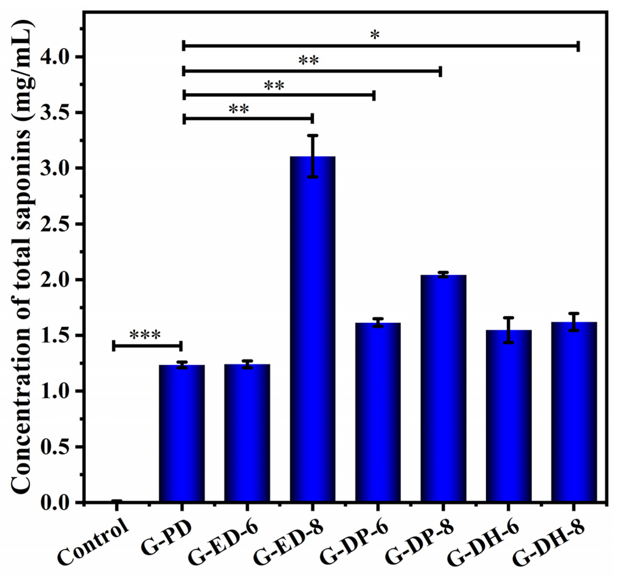
As shown in Figure 2, compared to the control group (0.009 ± 0.004 mg/mL), the endophytic fungus NSJG could produce total saponins in a conventional PD (G-PD) liquid medium at a concentration of 1.236 ± 0.025 mg/mL. There was an extremely significant statistical difference (p < 0.001) between the results of G-PD and the control group.
Six liquid fermentation protocols were designed to optimize saponin production. Total saponin concentrations in the G-ED-8 (3.107 ± 0.186 mg/mL) and G-DP-8 (2.045 ± 0.021 mg/mL) protocols were significantly increased and more than twice that of G-PD (1.236 ± 0.025 mg/mL). There were significant statistical differences (p < 0.01) between the results of G-ED-8 and G-PD, as well as the results of G-DP-8 and G-PD. Therefore, G-ED-8 and G-DP-8 were two suitable fermentation protocols for NSJG to produce high-yield saponins. As seen in Table 1, the fermentation conditions of the G-ED-8 protocol were as follows: potato concentration 104.5 mg/mL, glucose concentration 37.5 mg/mL, inoculum volume 9.4%, fermentation broth pH 2.1, fermentation temperature 28.5 °C, and fermentation time 8 d. The fermentation conditions of the G-DP-8 protocol were as follows: potato concentration 325.9 mg/mL, glucose concentration 35.3 mg/mL, inoculum volume 9.3%, fermentation broth pH 4.5, fermentation temperature 21.0 °C, and fermentation time 8 d. This result also indicated that the total saponin concentration produced by the same strain under different fermentation conditions was different. Thus, total saponin production may be changed by controlling fermentation conditions.

2.2. Identification and Analysis of Saponin Types of Fermentation Extracts

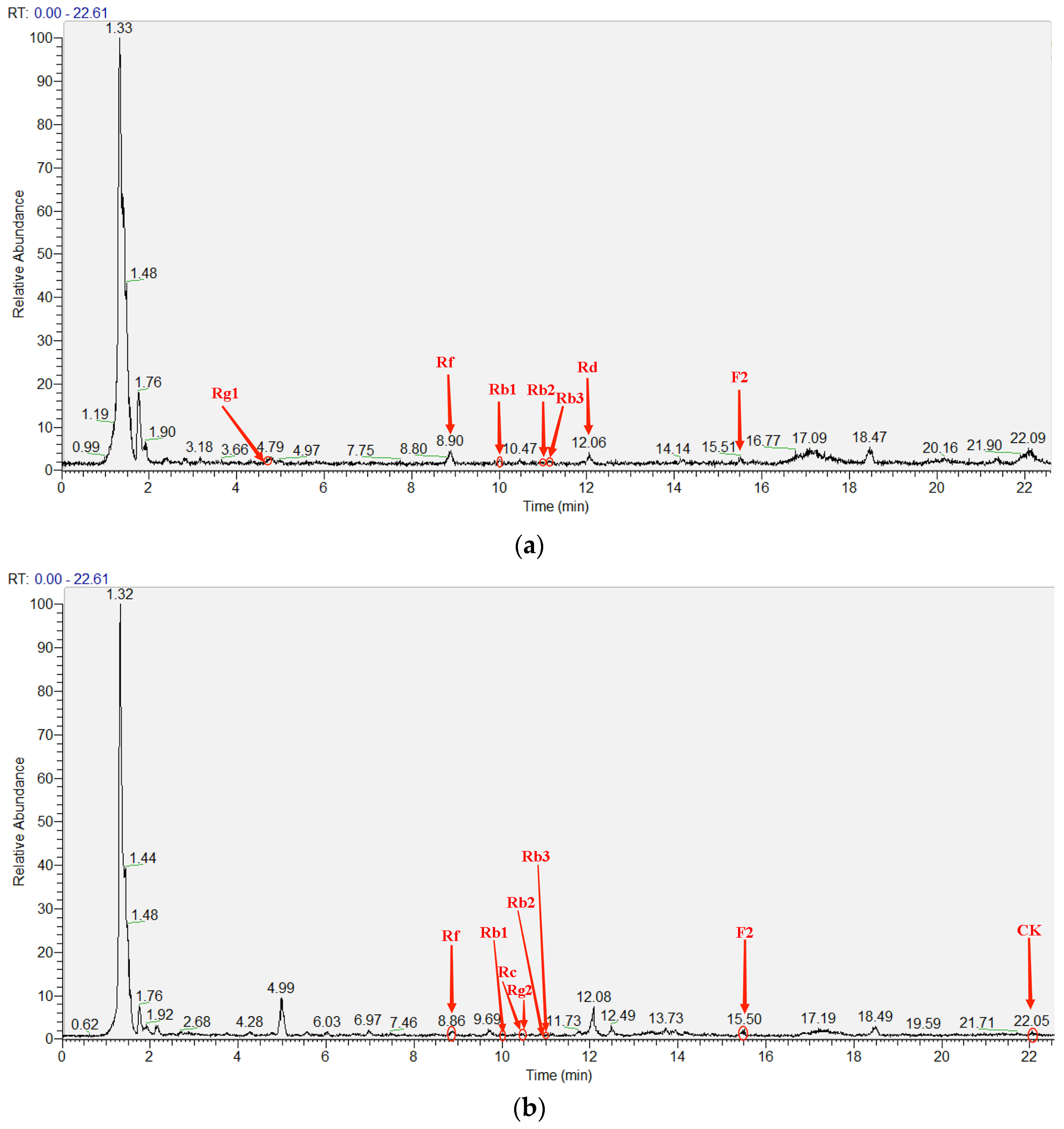
Twenty types of saponin standards could be detected within 30 min by Liquid Chromatography/Mass Spectrometry (LC/MS) in our previous experiment, of which peaks were complete [34]. The saponin types of G-ED-8 and G-DP-8 fermentation extracts were identified according to these 20 saponin standards. As shown in Figure 3a and Table 2, ginsenosides Rg1, Rf, Rb1, Rb2, Rb3, Rd, and F2 were found in the fermentation extract of G-ED-8. The concentration of ginsenoside Rb1 was 65.74 mg/L, the highest of the seven ginsenosides. According to Figure 3b and Table 3, there were eight types of saponins in the fermentation extract of G-DP-8, which were ginsenosides Rf, Rb1, Rc, Rg2, Rb2, Rb3, F2, and CK. The highest concentration of one of the eight saponins was ginsenoside Rb1, which reached 39.34 mg/L. Five types of saponins were the same in the fermentation extracts of G-ED-8 and G-DP-8. The saponins present only in the G-ED-8 fermentation extract were ginsenosides Rg1 and Rd, while the saponins present only in the G-DP-8 fermentation extract were ginsenosides Rc, Rg2, and CK. It illustrated that different fermentation conditions may result in the production of different types of saponins. Therefore, certain specific saponins can be obtained by controlling fermentation conditions. In addition, fermentation extracts from the two optimized protocols contained different rare ginsenosides. The fermentation extract of G-ED-8 contained rare ginsenoside Rd at a concentration of 2.68 mg/L and F2 at a concentration of 3.69 mg/L. The fermentation extract of G-DP-8 contained rare ginsenosides F2 and CK at concentrations of 4.53 mg/L and 0.83 mg/L, respectively.

2.3. Determination of Antioxidant Activity of Saponin Extracts

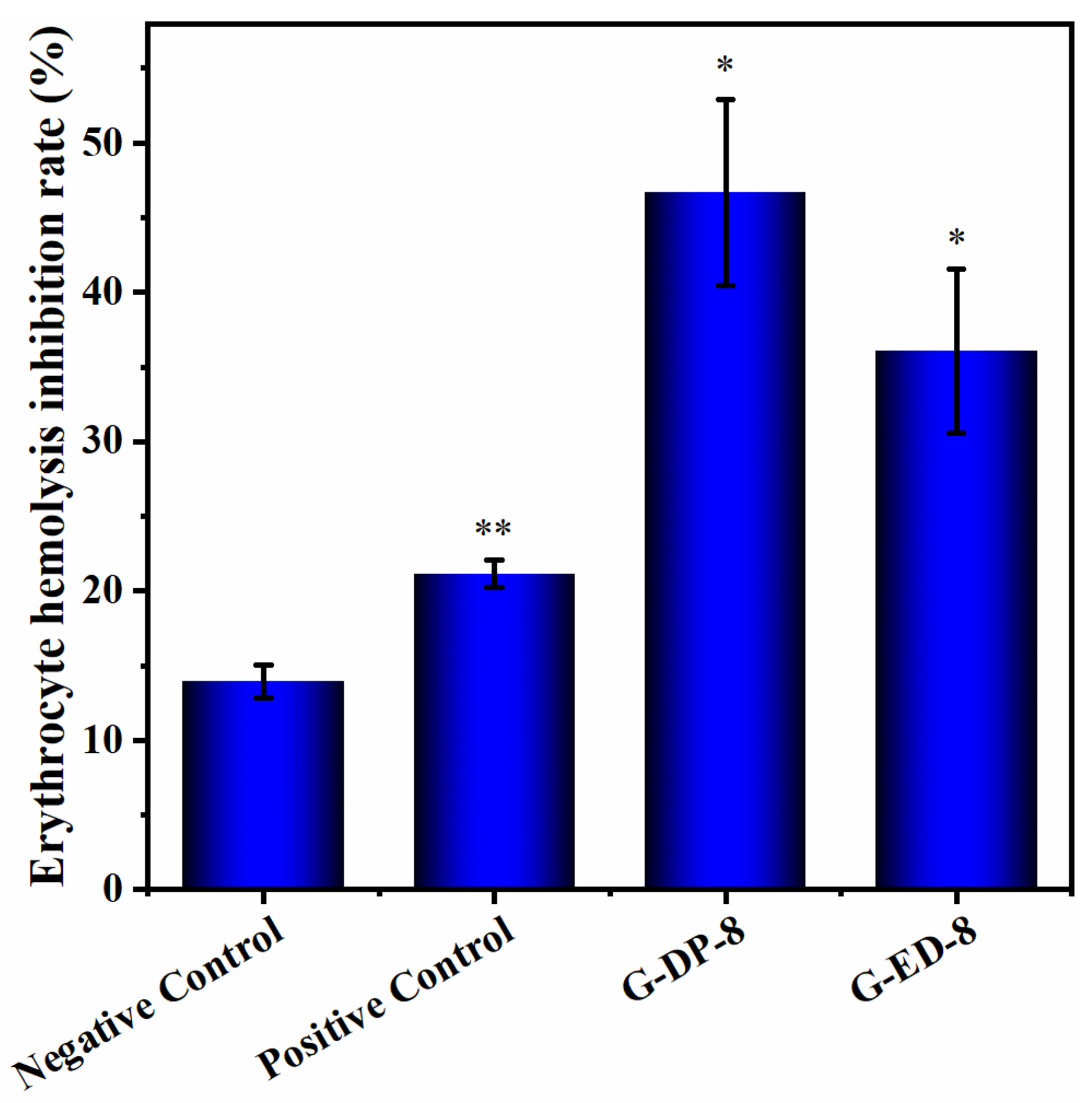
The saponin extracts of G-ED-8 and G-DP-8 were formulated into a solution of 20 μg/mL as the experimental group to determine their inhibitory rate on erythrocyte hemolysis and to evaluate their antioxidant activity. This concentration was selected based on saponin concentration conversion from the relevant literature [35]. It is non-toxic to cells at 20 μg/mL, thus eliminating the possibility of decreased metabolic activity because of cytotoxicity. As observed in Figure 4, when compared with the negative control group (13.91% ± 1.11%), saponin extracts from G-ED-8 and G-DP-8 exhibited a marked inhibitory effect on erythrocyte hemolysis, with the erythrocyte hemolysis inhibition rate being higher than that of the Vitamin C (VC) positive control group (21.12% ± 0.92%), thereby showing high antioxidant activity. The erythrocyte hemolysis inhibition rate of G-DP-8 (46.69% ± 6.23%) was higher than that of G-ED-8 (36.05% ± 5.47%), which could be attributed to the presence of a greater variety of saponins in the saponin extract of G-DP-8. There was a significant statistical difference (p < 0.01) between the positive and negative control groups. Both G-DP-8 and G-ED-8 had statistical differences (p < 0.05) compared to the negative control group.

3. Discussion

Due to the symbiotic relationship between endophytes and their host plants, the production or accumulation of host metabolites may be affected by endophytes [36]. Endophytes have been found to contribute directly to the production of host metabolites or to enhance their accumulation [18,37]. The ability of endophytic fungi to cultivate total saponins through in vitro fermentation from ginseng plants is noteworthy. Although saponin-producing endophytes are an eco-friendly and inexpensive alternative to obtaining saponins from ginseng plants, they often produce low yields. For example, according to research, the endophytic fungus Fusarium subglutinans strain Pg27 and Aspergillus protuberus strain Pg30 can produce total saponins in vitro, with the highest yields being 0.181 mg/mL and 0.144 mg/mL, respectively [21]. The total saponin concentration produced by the endophytic fungus Agrobacterium rhizogenes strain PDA-2 is only 0.073 mg/L [38]. The results of this study demonstrated that NSJG is an outstanding endophytic fungus producing a high saponin concentration (1.236 mg/mL), exceeding that of the endophytic fungi mentioned above. Six fermentation protocols were employed to optimize total saponin production by the strain NSJG, resulting in a maximum saponin concentration of 3.107 mg/mL. Thus, NSJG has excellent potential as an endophytic fungus for maximizing saponin production.
Although there are many types of ginsenosides, they all contain similar ring structures [2]. Ginsenosides can be roughly divided into dammarane and oleanane types according to the different glycoside structures [10]. Compared to common ginsenosides, rare ginsenosides that exist in trace amounts in nature are less polar and easier to be absorbed by the human body, so they have higher biological activity [13,39]. Therefore, it is necessary to develop a process for the mass production of rare ginsenosides. In this study, multiple monomeric saponins were detected in fermentation extracts from the two optimized protocols G-ED-8 and G-DP-8, with ginsenoside Rb1 accounting for the highest proportion of all saponins. The saponin extract of G-ED-8 included rare ginsenosides Rd and F2. The saponin extract of G-DP-8 contained rare ginsenosides F2 and CK. These rare ginsenosides have significant medicinal value. For instance, rare ginsenoside Rd exhibits remarkable anti-tumor properties and neuroprotection, as well as cardioprotective effects [40,41,42]. Ginsenoside F2 exhibits anti-inflammatory effects on the skin and aids in preventing obesity [43,44]. Rare ginsenoside CK possesses anti-tumor, anti-inflammatory, and anti-thrombotic effects [45,46]. Therefore, the endophytic fungus strain NSJG is an excellent strain that can produce a variety of rare saponins and has potential functional effects. Most rare ginsenosides were obtained by bio-transforming some saponins through fermentation using endophytic fungi. For example, Aspergillus niger KCCM 11239 converted ginsenoside Rb1 into rare ginsenoside Rg3 [47]. Beauveria bassiana converted white ginseng extract into ginsenosides Rb2 and Rc, as well as rare ginsenosides Rd and Rg3 [48]. Esteya vermicola CNU 120806 converted ginsenoside Rg3 to Rh2 [49]. There have also been many studies using metabolically engineered bacteria to biosynthesize certain rare ginsenosides [11]. In this study, rare ginsenosides could be directly produced through the in vitro fermentation of an endophytic fungus. It was low-cost and environmentally friendly compared to other methods of in vitro acquisition of rare ginsenosides [19]. Certain specific saponins can also be obtained by controlling fermentation conditions.
Oxidative stress is the result of an imbalance in the production and consumption of free radicals in the body, leading to a process of excessive production of prooxidative free radicals, including a significant increase in ROS generation, which is related to the pathophysiology of a variety of diseases [50]. Excessive ROS are harmful to cell structure and the function of biomolecules such as proteins, nucleic acids, lipids, and carbohydrates [51]. Lipid peroxidation of biological membranes is an important step in oxidative damage to cells. Therefore, lipid peroxidation can be used as an indicator of oxidative stress. 2,2′-azobis(2-amidinopropane hydrochloride) (AAPH) is an oxidant that thermally decomposes to produce two carbon-centered alkyl radical molecules, which then react with oxygen to generate peroxyl radical molecules and alkoxy molecules with oxidative activity [52]. Relevant research reports have shown that adding AAPH to a red blood cell suspension can induce the hemolysis of red blood cells. This hemolysis can be inhibited by antioxidants [53]. Several studies have used the AAPH-induced oxidative hemolysis model to measure the antioxidant activity of various compounds, especially complex mixtures, including studies on the antioxidant properties of various saponins [54,55]. For example, some studies have explored the antioxidant effects of different ginsenosides after the AAPH-induced hemolysis of erythrocytes and found that ginsenosides Rg2, Rg3, and Rh2 can act as prooxidants, while other ginsenosides Rg1, Rb1, Rd, Re, Rc, R1, and Rb3 can directly exert antioxidant effects [28,56]. In this study, AAPH was used at a final concentration of 40 mM to induce the hemolysis of red blood cells. Saponin extracts from G-ED-8 and G-DP-8 were used to intervene in the hemolytic process. The results revealed that both fermentation samples G-ED-8 and G-DP-8 could inhibit red blood cell hemolysis compared to PBS negative control and VC positive control groups. Furthermore, G-DP-8 showed superior antioxidant activity. These results were hypothesized to be related to a greater variety of saponins present in fermented extracts. Ginsenoside Rb1 was the highest of all saponins in the fermentation extract of G-ED-8 and G-DP-8. According to research, ginsenoside Rb1 may achieve antioxidant effects through multiple mechanisms. For example, ginsenoside Rb1 alleviated diabetic retinopathy by regulating the antioxidant function of the rat retina. It may inhibit the increase in MDA content and the decrease in glutathione (GSH) levels in rat retinas and significantly increase the Nuclear Factor erythroid 2-Related Factor 2 (Nrf2) level and the expression of glutamate cysteine ligase catalytic subunit (GCLC) and glutamate cysteine ligase modifier subunit (GCLM) in rat retinal cell nuclei [57]. Ginsenoside Rb1 could also improve arteriosclerosis and prevent diabetic vasculopathy by reducing the inhibition of diabetes-induced Adenosine 5′-monophosphate (AMP)-activated protein kinase (AMPK), regulating nicotinamide adenine dinucleotide phosphate (NADPH) oxidase, alleviating oxidative stress, and inhibiting the TGFβ1/Smad2/3 pathway [58]. The mechanism of action of the fermentation extract in this experiment remains to be further explored.

4. Materials and Methods

4.1. Strains and Chemicals

NSJG, a wild ginseng endophyte isolated from Changbai Mountain, China, was genetically identified as Umbelopsis dimorpha through internal transcribed spacer (ITS) sequencing.
The following chemicals were obtained from Shuangshuang Chemical Co. (Yantai, China): absolute methanol, absolute ethanol, N-butanol, and concentrated sulfuric acid. Chromatographically pure methanol was procured from TEDIA (Fairfield, OH, USA). Glucose and vanillin were acquired from Tianjin Standard Technology Co. (Tianjin, China). Ginsenoside standards were sourced from Master Biotechnology Co. (Chengdu, China). Dimethyl sulfoxide (DMSO) and PBS buffer solution were ordered by Solar Bio Science & Technology Co. (Beijing, China); Vitamin C was purchased from Yaanda Biotechnology Co. (Beijing, China). AAPH was supplied by Nanjing Dulai Biotechnology Co. (Nanjing, China).

4.2. Liquid Fermentation of an Endophytic Fungus under Different Fermentation Conditions

During liquid fermentation, potato concentration, glucose concentration, inoculation volume, fermentation broth pH, incubation temperature, and incubation time must be considered. The data processing system version 9.0 [59] was used to determine the optimal level of mixing of different parameters to design distinct fermentation protocols for liquid fermentation to improve saponin productivity. Several fermentation protocols containing the above six fermentation parameters are shown in Table 1.
The NSJG was subjected to in vitro fermentation using various liquid media prepared according to Table 1. A 100 mL conical flask was filled with 40 mL of each prepared medium and sterilized for 20 min at 121 °C and 0.1 MPa using a vertical pressure steam sterilizer LDZX-50KBS (Shenan, Shanghai, China). On the pre-cultured NSJG culture plate, a 5 mm hole punch was used to create holes. Five complete round hole plaques with a size of 0.5 cm × 0.5 cm were inoculated into a 100 mL conical flask. The inoculation procedure was repeated in triplicate, with one flask remaining as blank control. Fermentation culture was conducted in a full-temperature shaking incubator HZP-150 (Jinghong, Shanghai, China) at 150 rpm, with varying incubation temperatures and times for each protocol (Table 1). Once the culture has been completed, all the liquid containing mycelial balls was thoroughly dried using an electric blast dryer DHG-9070 (Yuying, Shanghai, China) and crushed until uniform for extraction of total saponins.

4.3. Extraction of Total Saponins

Both the supernatant and mycelium pellets were thoroughly dried and ground into a fine powder to obtain the fermented sample required for total saponin extraction. In a reagent bottle, anhydrous methanol was added along with 4 mL of the powdered sample, and the mixture was sonicated for 30 min in an ultrasonic instrument KM-700DE (Kunshanmeimei, Shanghai, China) with 500 W power and a frequency of 40 kHz. After sonication, the extract was centrifuged at 10,000 rpm for 10 min using a high-speed refrigerated centrifuge KDC-160HR (Zhongjia, Anhui, China) to obtain the supernatant. This process was repeated twice to improve precipitation efficiency. The collected supernatant was concentrated using a rotary evaporation instrument RE-52AA (Xiande, Shanghai, China) and then dried at 40 °C. It was then washed three times with 2 mL of water-saturated n-butanol to remove impurities. The resultant supernatant was added to a rotary evaporation flask and dried until the residue was completely evaporated, leaving behind the total saponins extract redissolved in a 2 mL methanol solution, which was stored in a brown reagent bottle at −20 °C.

4.4. Determination of Total Saponins

The concentration of total saponins in the reaction sample was determined by the color reaction of saponin acid hydrolysis and vanillin at 544 nm using a UV-Visible Spectrophotometer SP-2000UV (Unico, Shanghai, China). Oleanolic acid (OA) was used to draw a standard curve. An amount of 20 μL of total saponin solution was added into a graduated test tube and dried. Then, 5 mL of 72% (v/v) sulfuric acid and 0.5 mL of 8% (m/v) vanillin ethanol were added to the graduated test tube. The test tubes were placed in a constant-temperature water bath at 60 °C for 10 min and then in a cold water bath for 10 min. A control solution consisting of sample-free reaction solution was prepared as a blank, and the absorbance of the test sample was determined at 544 nm. The concentration of total saponins in the sample was subsequently calculated using the calibration curve [34].

4.5. Identification and Analysis of Saponin Extracts

To measure total saponin types, the LC/MS LTQ Orbitrap XL instrument (Thermos Fisher Scientific, Shanghai, China) was employed. The Accucore C18 analytical column (2.6 μm, 150 mm × 2.1 mm) was utilized to detect saponins, eluted with deionized water as Mobile Phase A and acetonitrile as Stationary Phase B. The chromatographic profile was designed as a programmed gradient, comprising 0–13 min with 77–54% A; 13–33 min with 54–32% A; 33–45 min with 32% A; 45–55 min with 32–0% A; 55–60 min with 0% A; and 60–63 min with 0–77% A [34]. The flow rate was set to 0.3 mL/min, and an injection volume of 10 μL was employed. Absorption at 203 nm was measured to detect the sample. The mass spectrometer settings included a spray voltage of 3.0 kV and a sheath gas of N2. Positive ion mode was utilized, and the scanning range was M/Z 150–1300.
A solution containing a mixture of various saponins, including pseudo-ginsenoside F11, noto-ginsenoside Fe, noto-ginsenoside Ft1, and ginsenosides Rb1, Rb2, Rb3, Rc, Rd, Re, Rf, Rg1, Rg2, Rg3, Rh1, Rh2, Rk2, CK, F1, F2, and F3, was prepared with a concentration of 0.2 mg/mL. The saponins in the solution were analyzed by comparing their charge-to-mass ratio (M/Z), peak time, and peak area with a database based on their total ion chromatogram and mass spectrum [60].
The quantitative calculation method for a particular type of saponin involves the detection of a standard sample comprising 20 saponins and the test sample (G-ED-8 and G-DP-8) via LC/MS. The peak area of saponins in the test sample was compared with the same saponins in the standard sample to calculate the concentration of a particular saponin. Equation (1) was utilized to describe the calculation formula.
CT = AT AS × VT VS × C S
where CT reflects the concentration of a particular saponin in the sample to be tested, AT represents the peak area of a particular saponin in the test sample, AS represents the peak area of that same saponin in the standard sample, VT signifies the injection volume of the sample being examined, while VS denotes the injection volume of the standard sample. Lastly, CS designates the concentration of the specific saponin in the standard sample. During the experiment, a 4 μL injection volume was used for both the sample to be tested and the standard sample. The standard sample contained 20 saponins at a concentration of 0.2 mg/mL [34].

4.6. Determination of Erythrocyte Hemolysis Inhibition Rate

Saponin extracts were used to intervene in the hemolysis of erythrocytes induced by AAPH. The higher the inhibition rate, the stronger the ability to prevent hemolysis, indicating the higher antioxidant activity of the ginsenoside extract sample.
In this study, a 3% (v/v) suspension of erythrocytes was prepared and used to determine its hemolysis inhibition rate. First, 160 μL of erythrocyte suspension was added to each well of a 96-well plate. Then 20 μL fermented total saponin solution, 20 μL PBS buffer, 20 μL VC solution, and 20 μL distilled water were added to the experimental group, negative control group, positive control group, and complete hemolysis control group, respectively. After the 96-well plate was subsequently incubated at a constant temperature of 37 °C for 40 min, 20 μL of AAPH solution (400 mM) dissolved in PBS was added to each well and followed by further incubation at 37 °C for four and a half hours. The 96-well plate was then centrifuged at 2000 rpm for 10 min using a small plate centrifuge, Plate Smart (DHS, Beijing, China), and the supernatant was transferred to a new 96-well plate for measurement. The OD value was determined at 570 nm using a VDRTEX-5MK3 microplate reader (Redian, Shanghai, China). The final action concentration of total saponins and VC is 20 μg/mL. The formula for calculating the erythrocyte hemolysis inhibition rate is described using Equation (2):
The   erythrocyte   hemolysis   inhibition   rate   ( % ) = complete   hemolysis   control   OD   value   -   sample   OD   value complete   hemolysis   control   OD   value   ×   100 %

4.7. Statistical Analysis

All experimental procedures were performed a minimum of three times to ensure accuracy and reliability. Data were reported as mean ± standard deviation (SD) (n = 3). Student’s t-test was conducted to compare two groups, while one-way Analysis of Variance (ANOVA) was used to compare multiple groups. In the figures, statistical significance was indicated by * p < 0.05, ** p < 0.01, and *** p < 0.001.

5. Conclusions

In summary, Umbelopsis dimorpha strain NSJG, an endophytic fungus capable of producing saponin through in vitro fermentation, was subjected to various fermentation protocols to optimize saponin yield. By optimizing fermentation conditions, two protocols yielded saponin production more than twice that of the initial levels. The saponin extracts from both protocols were further analyzed, revealing a wide range of ginsenosides and significant antioxidant activity. Therefore, the NSJG strain has potential application value in saponin production and can be used to produce efficient natural antioxidants for functional foods.

Supplementary Materials

The following supporting information can be downloaded at: https://www.mdpi.com/article/10.3390/fermentation10010009/s1, Table S1: Glycosyl types of protopanaxadiol (PPD) saponins; Table S2: Glycosyl types of protopanaxatriol (PPT) saponins.

Author Contributions

Conceptualization, Q.C. and P.Y.; methodology, P.Y.; software, Z.W. and Y.G.; validation, Q.C., J.W. and D.W; formal analysis, J.W. and D.W.; investigation, Z.W. and Y.G.; resources, X.G.; data curation, Q.C.; writing—original draft preparation, Q.C.; writing—review and editing, Q.C. and P.Y.; visualization, J.W.; supervision, X.G. and P.Y.; project administration, P.Y.; funding acquisition, P.Y. All authors have read and agreed to the published version of the manuscript.

Funding

This research was funded by the key scientific research and development project of Shandong province, grant number 2019GSF107038.

Institutional Review Board Statement

Not applicable.

Informed Consent Statement

Not applicable.

Data Availability Statement

The data of all results in this study are included in the manuscript. If necessary, the data can be obtained by contacting the corresponding author.

Conflicts of Interest

The authors declare no conflict of interest.

References

  1. Li, X.; Liu, J.; Zuo, T.T.; Hu, Y.; Li, Z.; Wang, H.D.; Xu, X.Y.; Yang, W.Z.; Guo, D.A. Advances and challenges in ginseng research from 2011 to 2020: The phytochemistry, quality control, metabolism, and biosynthesis. Nat. Prod. Rep. 2022, 39, 875–909. [Google Scholar] [CrossRef] [PubMed]
  2. Shi, Z.Y.; Zeng, J.Z.; Wong, A.S.T. Chemical Structures and Pharmacological Profiles of Ginseng Saponins. Molecules 2019, 24, 2443. [Google Scholar] [CrossRef] [PubMed]
  3. Kim, J.H. Cardiovascular Diseases and Panax ginseng: A Review on Molecular Mechanisms and Medical Applications. J. Ginseng Res. 2012, 36, 16–26. [Google Scholar] [CrossRef] [PubMed]
  4. Bhardwaj, J.; Chaudhary, N.; Seo, H.J.; Kim, M.Y.; Shin, T.S.; Kim, J.D. Immunomodulatory effect of tea saponin in immune T-cells and T-lymphoma cells via regulation of Th1, Th2 immune response and MAPK/ERK2 signaling pathway. Immunopharmacol. Immunotoxicol. 2014, 36, 202–210. [Google Scholar] [CrossRef] [PubMed]
  5. Ju, S.; Seo, J.Y.; Lee, S.K.; Oh, J.; Kim, J.S. Oral administration of hydrolyzed red ginseng extract improves learning and memory capability of scopolamine-treated C57BL/6J mice via upregulation of Nrf2-mediated antioxidant mechanism. J. Ginseng Res. 2021, 45, 108–118. [Google Scholar] [CrossRef] [PubMed]
  6. Chung, S.I.; Kang, M.Y.; Lee, S.C. In Vitro and In Vivo Antioxidant Activity of Aged Ginseng (Panax ginseng). Prev. Nutr. Food Sci. 2016, 21, 24–30. [Google Scholar] [CrossRef] [PubMed]
  7. Kim, Y.J.; Zhang, D.; Yang, D.C. Biosynthesis and biotechnological production of ginsenosides. Biotechnol. Adv. 2015, 33, 717–735. [Google Scholar] [CrossRef]
  8. Christensen, L.P. Ginsenosides chemistry, biosynthesis, analysis, and potential health effects. Adv. Food Nutr. Res. 2009, 55, 1–99. [Google Scholar]
  9. Wang, K.; Zhang, Z.; Li, S.; Hu, J.; Liu, T.; Jiang, Y.; Wu, J.; Lu, M.; Zhao, M.; Li, L.; et al. Transcriptome-Wide Analysis for Ginsenoside Rb3 Synthesis-Related Genes and Study on the Expression of Methyl Jasmonate Treatment in Panax ginseng. Life 2021, 11, 387. [Google Scholar] [CrossRef]
  10. Liu, H.; Lu, X.; Hu, Y.; Fan, X. Chemical constituents of Panax ginseng and Panax notoginseng explain why they differ in therapeutic efficacy. Pharmacol. Res. 2020, 161, 105263. [Google Scholar] [CrossRef]
  11. Li, M.; Ma, M.; Wu, Z.; Liang, X.; Zheng, Q.; Li, D.; An, T.; Wang, G. Advances in the biosynthesis and metabolic engineering of rare ginsenosides. Appl. Microbiol. Biotechnol. 2023, 107, 3391–3404. [Google Scholar] [CrossRef] [PubMed]
  12. Dini-Andreote, F. Endophytes: The Second Layer of Plant Defense. Trends Plant Sci. 2020, 25, 319–322. [Google Scholar] [CrossRef] [PubMed]
  13. Chu, L.L.; Bae, H. Bacterial endophytes from ginseng and their biotechnological application. J. Ginseng Res. 2022, 46, 1–10. [Google Scholar] [CrossRef] [PubMed]
  14. Eo, J.K.; Choi, M.S.; Eom, A.H. Diversity of endophytic fungi isolated from Korean ginseng leaves. Mycobiology 2014, 42, 147–151. [Google Scholar] [CrossRef] [PubMed]
  15. Park, Y.H.; Kim, Y.; Mishra, R.C.; Bae, H. Fungal endophytes inhabiting mountain-cultivated ginseng (Panax ginseng Meyer): Diversity and biocontrol activity against ginseng pathogens. Sci. Rep. 2017, 7, 16221. [Google Scholar] [CrossRef] [PubMed]
  16. Song, X.; Wu, H.; Yin, Z.; Lian, M.; Yin, C. Endophytic Bacteria Isolated from Panax ginseng Improves Ginsenoside Accumulation in Adventitious Ginseng Root Culture. Molecules 2017, 22, 837. [Google Scholar] [CrossRef] [PubMed]
  17. Cho, K.M.; Hong, S.Y.; Lee, S.M.; Kim, Y.H.; Kahng, G.G.; Lim, Y.P.; Kim, H.; Yun, H.D. Endophytic bacterial communities in ginseng and their antifungal activity against pathogens. Microb. Ecol. 2007, 54, 341–351. [Google Scholar] [CrossRef] [PubMed]
  18. Hong, C.E.; Kim, J.U.; Lee, J.W.; Lee, S.W.; Jo, I.H. Diversity of bacterial endophytes in Panax ginseng and their protective effects against pathogens. 3 Biotech 2018, 8, 397. [Google Scholar] [CrossRef]
  19. Nisa, H.; Kamili, A.N.; Nawchoo, I.A.; Shafi, S.; Shameem, N.; Bandh, S.A. Fungal endophytes as prolific source of phytochemicals and other bioactive natural products: A review. Microb. Pathog. 2015, 82, 50–59. [Google Scholar] [CrossRef]
  20. Wei, G.; Chen, Z.; Wang, B.; Wei, F.; Zhang, G.; Wang, Y.; Zhu, G.; Zhou, Y.; Zhao, Q.; He, M.; et al. Endophytes isolated from Panax notoginseng converted ginsenosides. Microb. Biotechnol. 2021, 14, 1730–1746. [Google Scholar] [CrossRef]
  21. Wu, H.; Yang, H.Y.; You, X.L.; Li, Y.H. Diversity of endophytic fungi from roots of Panax ginseng and their saponin yield capacities. Springerplus 2013, 2, 107. [Google Scholar] [CrossRef] [PubMed]
  22. Xing, X.; Dan, Y.; Xu, Z.; Xiang, L. Implications of Oxidative Stress in the Pathogenesis and Treatment of Hyperpigmentation Disorders. Oxid. Med. Cell. Longev. 2022, 2022, 7881717. [Google Scholar] [CrossRef] [PubMed]
  23. Ning, C.; Gao, X.; Wang, C.; Huo, X.; Liu, Z.; Sun, H.; Yang, X.; Sun, P.; Ma, X.; Meng, Q.; et al. Hepatoprotective effect of ginsenoside Rg1 from Panax ginseng on carbon tetrachloride-induced acute liver injury by activating Nrf2 signaling pathway in mice. Env. Toxicol. 2018, 33, 1050–1060. [Google Scholar] [CrossRef] [PubMed]
  24. Liu, H.; Chen, W.; Lu, P.; Ma, Y.; Liang, X.; Liu, Y. Ginsenoside Rg1 attenuates the inflammation and oxidative stress induced by diabetic nephropathy through regulating the PI3K/AKT/FOXO3 pathway. Ann. Transl. Med. 2021, 9, 1789. [Google Scholar] [CrossRef] [PubMed]
  25. Zhu, Y.; Zhu, C.; Yang, H.; Deng, J.; Fan, D. Protective effect of ginsenoside Rg5 against kidney injury via inhibition of NLRP3 inflammasome activation and the MAPK signaling pathway in high-fat diet/streptozotocin-induced diabetic mice. Pharmacol. Res. 2020, 155, 104746. [Google Scholar] [CrossRef]
  26. Zhao, Y.; Wang, Y.; Zhang, M.; Gao, Y.; Yan, Z. Protective Effects of Ginsenosides (20R)-Rg3 on H(2) O(2) -Induced Myocardial Cell Injury by Activating Keap-1/Nrf2/HO-1 Signaling Pathway. Chem. Biodivers. 2021, 18, e2001007. [Google Scholar] [CrossRef]
  27. He, B.; Chen, D.; Zhang, X.; Yang, R.; Yang, Y.; Chen, P.; Shen, Z. Oxidative Stress and Ginsenosides: An Update on the Molecular Mechanisms. Oxid. Med. Cell Longev. 2022, 2022, 9299574. [Google Scholar] [CrossRef]
  28. Liu, Z.Q.; Luo, X.Y.; Liu, G.Z.; Chen, Y.P.; Wang, Z.C.; Sun, Y.X. In vitro study of the relationship between the structure of ginsenoside and its antioxidative or prooxidative activity in free radical induced hemolysis of human erythrocytes. J. Agric. Food Chem. 2003, 51, 2555–2558. [Google Scholar] [CrossRef]
  29. Wolfe, K.L.; Liu, R.H. Cellular antioxidant activity (CAA) assay for assessing antioxidants, foods, and dietary supplements. J. Agric. Food Chem. 2007, 55, 8896–8907. [Google Scholar] [CrossRef]
  30. Winston, G.W.; Regoli, F.; Dugas, A.J., Jr.; Fong, J.H.; Blanchard, K.A. A rapid gas chromatographic assay for determining oxyradical scavenging capacity of antioxidants and biological fluids. Free Radic. Biol. Med. 1998, 24, 480–493. [Google Scholar] [CrossRef]
  31. Chen, Q.; Wang, J.; Gao, Y.; Wang, Z.; Gao, X.; Yan, P. Biotransformation of American Ginseng Stems and Leaves by an Endophytic Fungus Umbelopsis sp. and Its Effect on Alzheimer’s Disease Control. Nutrients 2023, 15, 4878. [Google Scholar] [CrossRef] [PubMed]
  32. Paek, K.Y.; Murthy, H.N.; Hahn, E.J.; Zhong, J.J. Large scale culture of ginseng adventitious roots for production of ginsenosides. Adv. Biochem. Eng. Biotechnol. 2009, 113, 151–176. [Google Scholar]
  33. Xu, W.; Lyu, W.; Duan, C.; Ma, F.; Li, X.; Li, D. Preparation and bioactivity of the rare ginsenosides Rg3 and Rh2: An updated review. Fitoterapia 2023, 167, 105514. [Google Scholar] [CrossRef]
  34. Chen, Q.; Wang, J.; Gao, Y.; Gao, X.; Yan, P. Optimization of Fermentation Conditions and Product Identification of a Saponin-Producing Endophytic Fungus. Microorganisms 2023, 11, 2331. [Google Scholar] [CrossRef]
  35. Li, M.; Chen, Y.; Zhang, P.; Zhang, L.; Zhou, R.; Xu, Y.; Ding, H.; Wang, Q.; Wang, Z. Semi-synthesis of Twelve Known 20Z/E Pseudo-Ginsenosides and Their Comparative Study of Antioxidative Activity in Free Radical Induced Hemolysis of Rabbit Erythrocytes. Chem. Pharm. Bull. 2018, 66, 535–540. [Google Scholar] [CrossRef]
  36. Zhao, M.; Guo, D.L.; Liu, G.H.; Fu, X.; Gu, Y.C.; Ding, L.S.; Zhou, Y. Antifungal Halogenated Cyclopentenones from the Endophytic Fungus Saccharicola bicolor of Bergenia purpurascens by the One Strain-Many Compounds Strategy. J. Agric. Food Chem. 2020, 68, 185–192. [Google Scholar] [CrossRef]
  37. Li, R.; Duan, W.; Ran, Z.; Chen, X.; Yu, H.; Fang, L.; Guo, L.; Zhou, J. Diversity and correlation analysis of endophytes and metabolites of Panax quinquefolius L. in various tissues. BMC Plant Biol. 2023, 23, 275. [Google Scholar]
  38. Yan, H.; Jin, H.; Fu, Y.; Yin, Z.; Yin, C. Production of Rare Ginsenosides Rg3 and Rh2 by Endophytic Bacteria from Panax ginseng. J. Agric. Food Chem. 2019, 67, 8493–8499. [Google Scholar] [CrossRef]
  39. He, Y.; Hu, Z.; Li, A.; Zhu, Z.; Yang, N.; Ying, Z.; He, J.; Wang, C.; Yin, S.; Cheng, S. Recent Advances in Biotransformation of Saponins. Molecules 2019, 24, 2365. [Google Scholar] [CrossRef]
  40. Liu, G.M.; Lu, T.C.; Sun, M.L.; Jia, W.Y.; Ji, X.; Luo, Y.G. Ginsenoside Rd Inhibits Glioblastoma Cell Proliferation by Up-Regulating the Expression of miR-144-5p. Biol. Pharm. Bull. 2020, 43, 1534–1541. [Google Scholar] [CrossRef]
  41. Hou, J.; Xue, J.; Lee, M.; Sung, C. Ginsenoside Rd as a potential neuroprotective agent prevents trimethyltin injury. Biomed. Rep. 2017, 6, 435–440. [Google Scholar] [CrossRef] [PubMed]
  42. Zhang, N.; An, X.; Lang, P.; Wang, F.; Xie, Y. Ginsenoside Rd contributes the attenuation of cardiac hypertrophy in vivo and in vitro. Biomed. Pharmacother. 2019, 109, 1016–1023. [Google Scholar] [CrossRef] [PubMed]
  43. Zhou, J.; Zhang, J.; Li, J.; Guan, Y.; Shen, T.; Li, F.; Li, X.; Yang, X.; Hu, W. Ginsenoside F2 Suppresses Adipogenesis in 3T3-L1 Cells and Obesity in Mice via the AMPK Pathway. J. Agric. Food Chem. 2021, 69, 9299–9312. [Google Scholar] [CrossRef] [PubMed]
  44. Park, S.H.; Seo, W.; Eun, H.S.; Kim, S.Y.; Jo, E.; Kim, M.H.; Choi, W.M.; Lee, J.H.; Shim, Y.R.; Cui, C.H.; et al. Protective effects of ginsenoside F2 on 12-O-tetradecanoylphorbol-13-acetate-induced skin inflammation in mice. Biochem. Biophys. Res. Commun. 2016, 478, 1713–1719. [Google Scholar] [CrossRef] [PubMed]
  45. Baik, I.H.; Kim, K.H.; Lee, K.A. Antioxidant, Anti-Inflammatory and Antithrombotic Effects of Ginsenoside Compound K Enriched Extract Derived from Ginseng Sprouts. Molecules 2021, 26, 4102. [Google Scholar] [CrossRef]
  46. Zhou, L.; Li, Z.K.; Li, C.Y.; Liang, Y.Q.; Yang, F. Anticancer properties and pharmaceutical applications of ginsenoside compound K: A review. Chem. Biol. Drug Des. 2022, 99, 286–300. [Google Scholar] [CrossRef] [PubMed]
  47. Chang, K.H.; Jo, M.N.; Kim, K.T.; Paik, H.D. Evaluation of glucosidases of Aspergillus niger strain comparing with other glucosidases in transformation of ginsenoside Rb1 to ginsenosides Rg3. J. Ginseng Res. 2014, 38, 47–51. [Google Scholar] [CrossRef]
  48. Gum, S.I.; Rahman, M.K.; Won, J.S.; Cho, M.K. A Distinctive Pattern of Beauveria bassiana-biotransformed Ginsenoside Products Triggers Mitochondria/FasL-mediated Apoptosis in Colon Cancer Cells. Phytother. Res. 2016, 30, 136–143. [Google Scholar] [CrossRef]
  49. Hou, J.; Xue, J.; Wang, C.; Liu, L.; Zhang, D.; Wang, Z.; Li, W.; Zheng, Y.; Sung, C. Microbial transformation of ginsenoside Rg3 to ginsenoside Rh2 by Esteya vermicola CNU 120806. World J. Microbiol. Biotechnol. 2012, 28, 1807–1811. [Google Scholar] [CrossRef]
  50. Ebrahimi, S.; Alalikhan, A.; Aghaee-Bakhtiari, S.H.; Hashemy, S.I. The redox modulatory effects of SP/NK1R system: Implications for oxidative stress-associated disorders. Life Sci. 2022, 296, 120448. [Google Scholar] [CrossRef]
  51. Droge, W. Free radicals in the physiological control of cell function. Physiol. Rev. 2002, 82, 47–95. [Google Scholar] [CrossRef] [PubMed]
  52. Motoyama, T.; Miki, M.; Mino, M.; Takahashi, M.; Niki, E. Synergistic inhibition of oxidation in dispersed phosphatidylcholine liposomes by a combination of vitamin E and cysteine. Arch. Biochem. Biophys. 1989, 270, 655–661. [Google Scholar] [CrossRef] [PubMed]
  53. Nuruki, Y.; Matsumoto, H.; Tsukada, M.; Tsukahara, H.; Takajo, T.; Tsuchida, K.; Anzai, K. Method to Improve Azo-Compound (AAPH)-Induced Hemolysis of Erythrocytes for Assessing Antioxidant Activity of Lipophilic Compounds. Chem. Pharm. Bull. 2021, 69, 67–71. [Google Scholar] [CrossRef] [PubMed]
  54. Yamamoto, Y.; Niki, E.; Kamiya, Y.; Miki, M.; Tamai, H.; Mino, M. Free radical chain oxidation and hemolysis of erythrocytes by molecular oxygen and their inhibition by vitamin E. J. Nutr. Sci. Vitaminol. 1986, 32, 475–479. [Google Scholar] [CrossRef] [PubMed]
  55. Zheng, Q.; Tan, W.; Feng, X.; Feng, K.; Zhong, W.; Liao, C.; Liu, Y.; Li, S.; Hu, W. Protective Effect of Flavonoids from Mulberry Leaf on AAPH-Induced Oxidative Damage in Sheep Erythrocytes. Molecules 2022, 27, 7625. [Google Scholar] [CrossRef] [PubMed]
  56. Ren, B.; Feng, J.; Yang, N.; Guo, Y.; Chen, C.; Qin, Q. Ginsenoside Rg3 attenuates angiotensin II-induced myocardial hypertrophy through repressing NLRP3 inflammasome and oxidative stress via modulating SIRT1/NF-kappaB pathway. Int. Immunopharmacol. 2021, 98, 107841. [Google Scholar] [CrossRef] [PubMed]
  57. Dong, C.; Liu, P.; Wang, H.; Dong, M.; Li, G.; Li, Y. Ginsenoside Rb1 attenuates diabetic retinopathy in streptozotocin-induced diabetic rats1. Acta Cir. Bras. 2019, 34, e201900201. [Google Scholar] [CrossRef] [PubMed]
  58. Zhang, X.; Wang, L.; Guo, R.; Xiao, J.; Liu, X.; Dong, M.; Luan, X.; Ji, X.; Lu, H. Ginsenoside Rb1 Ameliorates Diabetic Arterial Stiffening via AMPK Pathway. Front. Pharmacol. 2021, 12, 753881. [Google Scholar] [CrossRef]
  59. Tang, Q.Y.; Zhang, C.X. Data Processing System (DPS) software with experimental design, statistical analysis and data mining developed for use in entomological research. Insect Sci. 2013, 20, 254–260. [Google Scholar] [CrossRef]
  60. Wang, C.Q.; Yi, L.W.; Zhao, L.; Zhou, Y.Z.; Guo, F.; Huo, Y.S.; Zhao, D.Q.; Xu, F.; Wang, X.; Cai, S.Q. 177 Saponins, Including 11 New Compounds in Wild Ginseng Tentatively Identified via HPLC-IT-TOF-MS(n), and Differences among Wild Ginseng, Ginseng under Forest, and Cultivated Ginseng. Molecules 2021, 26, 3371. [Google Scholar] [CrossRef]
Figure 1. Chemical structures of different types of ginsenosides and aglycon glycosides. -GlcA represents D-Glucuronic acid; -Rha represents α-L-rhamnopyranosyl; -Ara(f) represents α-L-arabinofuranosyl; -Xyl represents β-D-xylopyranosyl; -Glc represents β-D-glucopyranosyl; -Ara(p) represents α-L-arabinopyranosyl. PPD represents protopanaxadiol-type saponin; PPT represents protopanaxatriol-type saponin.
Fermentation 10 00009 g001
Figure 2. Concentration of total saponins in fermentation extracts by potato dextrose (PD) liquid medium and six fermentation protocols. Data are presented as mean ± standard deviation (SD) (n = 3). * p < 0.05, ** p < 0.01, *** p < 0.001.
Fermentation 10 00009 g002
Figure 3. The Liquid Chromatography/Mass Spectrometry (LC/MS) results of fermentation extracts from two optimized protocols. (a) Total ion chromatogram (TIC) of LC/MS determination by fermentation extract from G-ED-8 protocol. (b) TIC of LC/MS determination by fermentation extract from G-DP-8 protocol. In the figures, RT represents the retention time of each peak. The abscissa and ordinate represent the time and relative abundance of the peak. The arrow points to the specific location of the saponin peak.
Fermentation 10 00009 g003
Figure 4. Determination of erythrocyte hemolysis inhibition rate of G-DP-8 and G-ED-8 saponin extracts. Data are presented as mean ± SD (n = 3). * p < 0.05, ** p < 0.01. The formula of the negative control group is 97.6% (v/v) phosphate buffered saline (PBS) buffer, the positive control group is 20 μg/mL (m/v) Vitamin C (VC) solution, the action concentration of G-ED-8 and G-DP-8 are both 20 μg/mL (m/v).
Fermentation 10 00009 g004
Table 1. Fermentation condition parameters for liquid fermentation optimization protocols.
ProtocolsPotato
Concentration (g/L)
Glucose
Concentration (g/L)
Inoculation Volume (%)pHIncubation
Temperature (°C)
Incubation
Time (d)
G-PD200.020.010.0Natural26.07
G-ED-6104.537.59.42.128.56
G-ED-8104.537.59.42.128.58
G-DP-6325.935.39.34.521.06
G-DP-8325.935.39.34.521.08
G-DH-698.336.12.14.231.06
G-DH-898.336.12.14.231.08
Table 2. Analysis of saponin types and concentrations in fermentation extracts of G-ED-8 protocol.
Saponin TypeRetention Time (RT) of Standards (min) [34]RT of Sample (min)Charge-to-Mass Ratio (M/Z, M + Na)Calculate Mass Errors (ppm)Peak AreaConcentration (mg/L)
Ginsenoside Rg14.784.69823.47665.221,497,9350.71
Ginsenoside Rf8.908.90823.4889−9.7114,871,6764.55
Ginsenoside Rb19.9310.021131.58872.564,720,04065.74
Ginsenoside Rb211.0110.981101.57782.903,285,8211.47
Ginsenoside Rb311.1111.131101.57792.814,875,1012.52
Ginsenoside Rd12.0812.06969.53610.724,234,2682.68
Ginsenoside F215.4715.51807.48352.976,833,6293.69
Table 3. Analysis of saponin types and concentrations in fermentation extracts of G-DP-8 protocol.
Saponin TypeRT of Standards (min) [34]RT of Sample (min)M/Z (M + Na)Calculate Mass Errors (ppm)Peak AreaConcentration (mg/L)
Ginsenoside Rf8.908.86823.47912.1912,663,8033.88
Ginsenoside Rb19.9310.021131.5881−0.622,824,37739.34
Ginsenoside Rc10.5210.441101.57862.18646,0201.15
Ginsenoside Rg210.7210.50807.48382.602,149,9775.47
Ginsenoside Rb211.0110.971101.57673.90867,8640.39
Ginsenoside Rb311.1111.111101.57802.724,500,3602.37
Ginsenoside F215.4715.5807.48402.358,396,9114.53
Ginsenoside CK22.0722.05645.43250.933,855,9520.83
Disclaimer/Publisher’s Note: The statements, opinions and data contained in all publications are solely those of the individual author(s) and contributor(s) and not of MDPI and/or the editor(s). MDPI and/or the editor(s) disclaim responsibility for any injury to people or property resulting from any ideas, methods, instructions or products referred to in the content.