
Hydrogel-Based Biosensors

 ,
,  , , , , , ,

Abstract
1. Introduction
2. Biomedical Applications
2.1. Proteins and Enzymes
2.2. Hormones and Metabolites
2.3. DNA Sensing
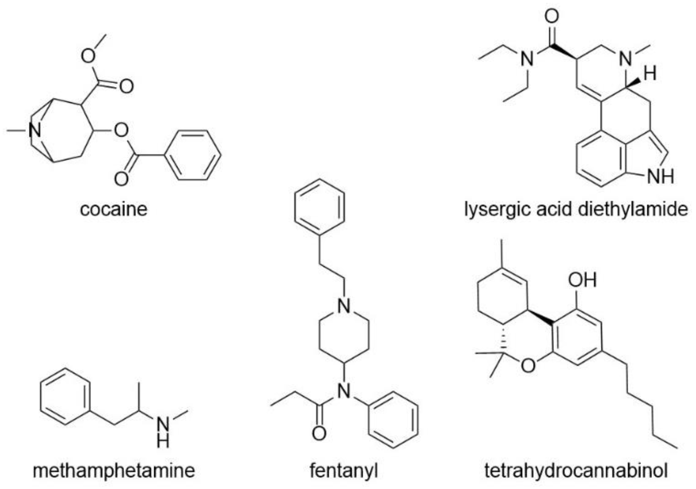
3. Drug Sensing
3.1. Sensing of Pharmaceutical Drugs
3.2. Sensing of Illicit Drugs
4. Hydrogel-Based Sensors for the Detection of Environmental Pollutants
Detection of Heavy Metals
5. Conclusions
Author Contributions
Funding
Institutional Review Board Statement
Informed Consent Statement
Data Availability Statement
Conflicts of Interest
References
- Pinelli, F.; Magagnin, L.; Rossi, F. Progress in hydrogels for sensing applications: A review. Mater. Today Chem. 2020, 17, 100317. [Google Scholar] [CrossRef]
- Buenger, D.; Topuz, F.; Groll, J. Hydrogels in sensing applications. Progr. Polym. Sci. 2012, 37, 1678–1719. [Google Scholar] [CrossRef]
- Hasanzadeh, M.; Shadjou, N.; de la Guardia, M. Nanosized hydrophobic gels: Advanced supramolecules for use in electrochemical bio- and immunosensing. TrAC 2018, 102, 210–224. [Google Scholar] [CrossRef]
- Holtz, J.H.; Asher, S.A. Polymerized colloidal crystal hydrogel films as intelligent chemical sensing materials. Nature 1997, 389, 829–832. [Google Scholar] [CrossRef] [PubMed]
- Chaubey, A.; Malhotra, B. Mediated biosensors. Biosens. Bioelectron. 2002, 17, 441–456. [Google Scholar] [CrossRef]
- Grieshaber, D.; MacKenzie, R.; Vörös, J.; Reimhult, E. Electrochemical Biosensors- Sensor Principles and Architectures. Sensors 2008, 8, 1400–1458. [Google Scholar] [CrossRef] [PubMed]
- Ronkainen, N.J.; Halsall, H.B.; Heineman, W.R. Electrochemical biosensors. Chem. Soc. Rev. 2010, 1747–1763. [Google Scholar] [CrossRef]
- Herrmann, A.; Haag, R.; Schedler, U. Hydrogels and Their Role in Biosensing Applications. Adv. Healthc. Mater. 2021, 1–25. [Google Scholar] [CrossRef]
- Soto, R.J.; Hall, J.R.; Brown, M.D.; Taylor, J.B.; Schoenfisch, M.H. In Vivo Chemical Sensors: Role of Biocompatibility on Performance and Utility. Anal. Chem. 2017, 89, 276–299. [Google Scholar] [CrossRef]
- Biomarkers Definitions Working Group. Biomarkers and surrogate endpoints: Preferred definitions and conceptual framework. Clin. Pharm. Ther. 2001, 69, 89–95. [Google Scholar] [CrossRef]
- Lin, D.; Shen, L.; Luo, M.; Zhang, K.; Li, J.; Yang, Q.; Zhu, F.; Zhou, D.; Zheng, S.; Chen, Y.; et al. Circulating tumor cells: Biology and clinical significance. Signal Transduct. Target. Ther. 2021, 6, 404. [Google Scholar] [CrossRef]
- Califf, R.M. Biomarker definitions and their applications. Exp. Biol. Med. 2018, 243, 213–221. [Google Scholar] [CrossRef]
- Aronson, J.K.; Ferner, R.E. Biomarkers-A General Review. Curr. Protoc. Pharmacol. 2017, 76, 9.23.1–9.23.17. [Google Scholar] [CrossRef]
- Das, R.; Bej, S.; Ghosh, D.; Murmu, N.C.; Hirani, H.; Banerjee, P. Stimuli-responsive discriminative detection of Cu2+ and Hg2+ with concurrent sensing of S2- from aqueous medium and bio-fluids by C N fused azophenine functionalized “smart” hydrogel assay @A potential biomarker sensor for Wilson’s disease. Sens. Act. B 2021, 341, 129925. [Google Scholar] [CrossRef]
- Ratish, N.R.; Snehasish, D.; Das, S.; Wakchaure, P.; Ganguly, B.; Chatterjee, P.B. A Highly Selective Turn-On Biosensor for Measuring Spermine/Spermidine in Human Urine and Blood. ACS Appl. Bio Mater. 2019, 2, 2374–2387. [Google Scholar] [CrossRef]
- Biswas, A.; Bornhoeft, L.R.; Banerjee, S.; You, Y.-H.; McShane, M.J. Composite Hydrogels Containing Bioactive Microreactors for Optical Enzymatic Lactate Sensing. ACS Sens. 2017, 2, 1584–1588. [Google Scholar] [CrossRef]
- Wang, H.; Wang, H.; Li, Y.; Jiang, C.; Chen, D.; Wen, Y.; Li, Z. Capillarity self-driven DNA hydrogel sensor for visual quantification of microRNA. Sens. Act. B 2020, 313, 128036. [Google Scholar] [CrossRef]
- Nimse, S.B.; Sonawane, M.D.; Song, K.-S.; Kim, T. Biomarker detection technologies and future directions. Analyst 2016, 141, 740–755. [Google Scholar] [CrossRef] [PubMed]
- Chandrawati, R. Enzyme-responsive polymer hydrogels for therapeutic delivery. Exp. Biol. Med. 2016, 241, 972–979. [Google Scholar] [CrossRef]
- West, J.L.; Hubbell, J.A. Polymeric Biomaterials with Degradation Sites for Proteases Involved in Cell Migration. Macromolecules 1999, 32, 241–244. [Google Scholar] [CrossRef]
- Purcell, B.P.; Lobb, D.; Charati, M.B.; Dorsey, S.M.; Wade, R.J.; Zellars, K.N.; Doviak, H.; Pettaway, S.; Logdon, C.B.; Shuman, J.A.; et al. Injectable and bioresponsive hydrogels for on-demand matrix metalloproteinase inhibition. Nat. Mater. 2014, 13, 653–661. [Google Scholar] [CrossRef] [PubMed]
- Kono, H.; Otaka, F.; Ozaki, M. Preparation and characterization of guar gum hydrogels as carrier materials for controlled protein drug delivery. Carbohydr. Polym. 2014, 111, 830–840. [Google Scholar] [CrossRef]
- Altintas, Z.; Tothill, I. Biomarkers and biosensors for the early diagnosis of lung cancer. Sens. Act. B 2013, 188, 988–998. [Google Scholar] [CrossRef]
- Shohatee, D.; Keifer, J.; Schimmel, N.; Mohanty, S.; Ghosh, G. Hydrogel-based suspension array for biomarker detection using horseradish peroxidase-mediated silver precipitation. Anal. Chim. Acta 2018, 999, 132–138. [Google Scholar] [CrossRef]
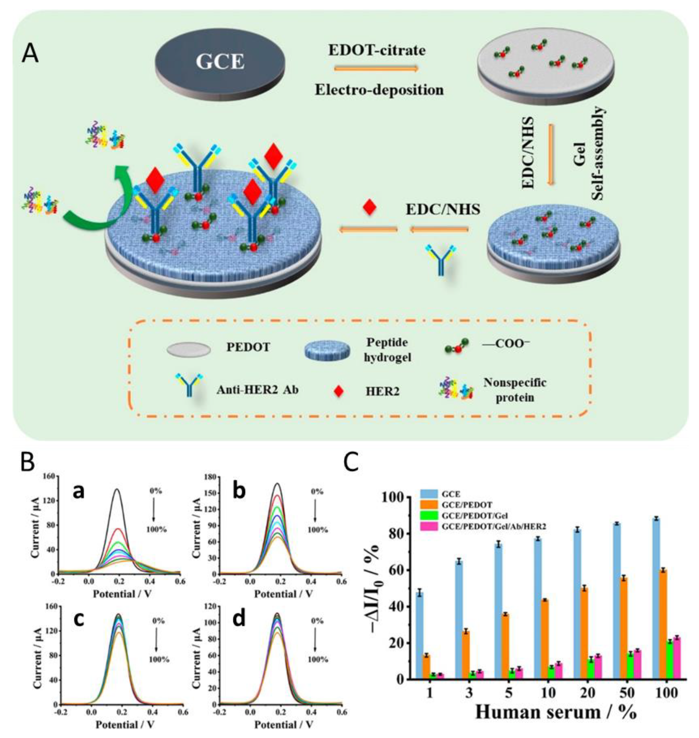
- Wang, W.; Han, R.; Chen, M.; Luo, X. Antifouling Peptide Hydrogel Based Electrochemical Biosensors for Highly Sensitive Detection of Cancer Biomarker HER2 in Human Serum. Anal. Chem. 2021, 93, 7355–7361. [Google Scholar] [CrossRef] [PubMed]
- Yamamoto, K.; Sueyoshi, K.; Hisamoto, H.; Endo, T. Development of Element Technology for 1 STEP Biomarker Protein Analysis Device Using Silver Nanoparticle-Contained Hydrogel and Reagent-Immobilized Cartridge. Electron. Comm. Jpn. 2017, 100, 45–53. [Google Scholar] [CrossRef]
- Huang, J.; Lin, Q.; Zhang, X.; He, X.; Xing, X.; Lian, W.; Zuo, M.; Zhang, Q. Electrochemical immunosensor based on polyaniline/poly (acrylic acid) and Au-hybrid graphene nanocomposite for sensitivity enhanced detection of salbutamol. Food Res. Int. 2011, 44, 92–97. [Google Scholar] [CrossRef]
- Wang, H.; Han, H.; Ma, Z. Conductive hydrogel composed of 1,3,5-benzenetricarboxylic acid and Fe3+ used as enhanced electrochemical immunosensing substrate for tumor biomarker. Bioelectrochemistry 2017, 114, 48–53. [Google Scholar] [CrossRef]
- Wang, H.; Ma, Z. Ultrasensitive amperometric detection of the tumor biomarker cytokeratin antigen using a hydrogel composite consisting of phytic acid, Pb(II) ions and gold nanoparticles. Microchim. Acta 2017, 184, 1045–1050. [Google Scholar] [CrossRef]
- Karachaliou, N.; Mayo-de-las-Casas, C.; Molina-Vila, M.A.; Rosell, R. Real-time liquid biopsies become a reality in cancer treatment. Ann. Transl. Med. 2015, 3, 1–3. [Google Scholar] [CrossRef]
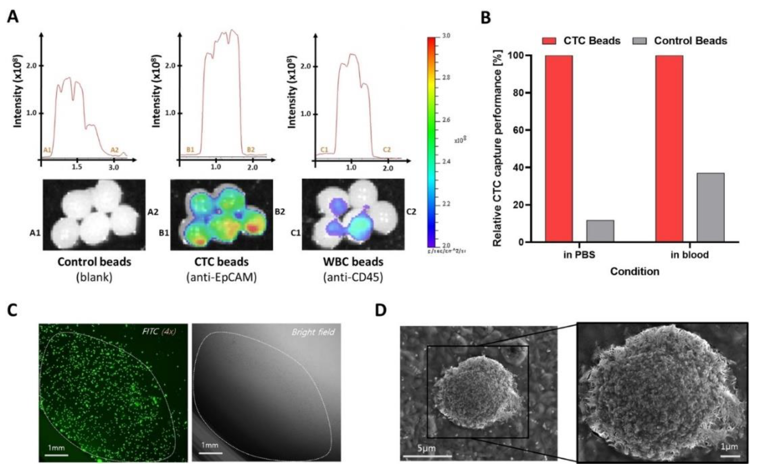
- Kang, Y.-T.; Kim, Y.J.; Rupp, B.; Purcell, E.; Hadlock, T.; Ramnath, N.; Nagrath, S. Isolation of Circulating Biomarkers for Liquid Biopsy using Immunoaffinity-Based Stimuli-Responsive Hybrid Hydrogel Beads. Anal. Sens. 2021, 1, 117–129. [Google Scholar] [CrossRef]
- Gayton, J.L. Etiology, prevalence, and treatment of dry eye disease. Clin. Ophthalmol. 2009, 3, 405–412. [Google Scholar] [CrossRef] [PubMed]
- Brewitt, H.; Sistani, F. Dry Eye Disease. Surv. Ophthal. 2001, 45, S199–S202. [Google Scholar] [CrossRef] [PubMed]
- Saldanha, I.J.; Petris, R.; Makara, M.; Channa, P.; Akpek, E.K. Impact of the COVID-19 pandemic on eye strain and dry eye symptoms. Ocul. Surf. 2021, 22, 38–46. [Google Scholar] [CrossRef]
- Hagan, S.; Tomlinson, A. Tear fluid biomarker profiling: A review of multiplex bead analysis. Ocul. Surf. 2013, 11, 219–235. [Google Scholar] [CrossRef]
- Wechsler, M.E.; Jocelyn Dang, H.K.H.; Simmonds, S.P.; Bahrami, K.; Wyse, J.M.; Dahlhauser, S.D.; Reuther, J.F.; VandeWalle, A.N.; Anslyn, E.V.; Peppas, N.A. Electrostatic and Covalent Assemblies of Anionic Hydrogel-Coated Gold Nanoshells for Detection of Dry Eye Biomarkers in Human Tears. Nano Lett. 2021, 21, 8734–8740. [Google Scholar] [CrossRef] [PubMed]
- Culver, H.R.; Wechsler, M.E.; Peppas, N.A. Label-Free Detection of Tear Biomarkers Using Hydrogel-Coated Gold Nanoshells in a Localized Surface Plasmon Resonance-Based Biosensor. ACS Nano 2018, 12, 9342–9354. [Google Scholar] [CrossRef] [PubMed]
- Yusuf, S.; McKee, M. Documenting the Global Burden of Cardiovascular Disease. Circulation 2014, 129, 1459–1462. [Google Scholar] [CrossRef]
- Ji, J.; Lu, W.; Zhu, Y.; Jin, H.; Yao, Y.; Zhang, H.; Zhao, Y. Porous Hydrogel-Encapsulated Photonic Barcodes for Multiplex Detection of Cardiovascular Biomarkers. ACS Sens. 2019, 4, 1384–1390. [Google Scholar] [CrossRef] [PubMed]
- UN Interagency Coordinating Group on Antimicrobial Resistance. No Time to Wait: Securing the Future from Drug-Resistant Infections; United Nations: New York, NY, USA, 2019. [Google Scholar]
- World Health Organization. Weekly Epidemiological Update on COVID-19—1 March 2022; Emergency Situational Updates. Available online: https://covid19.who.int (accessed on 15 November 2022).
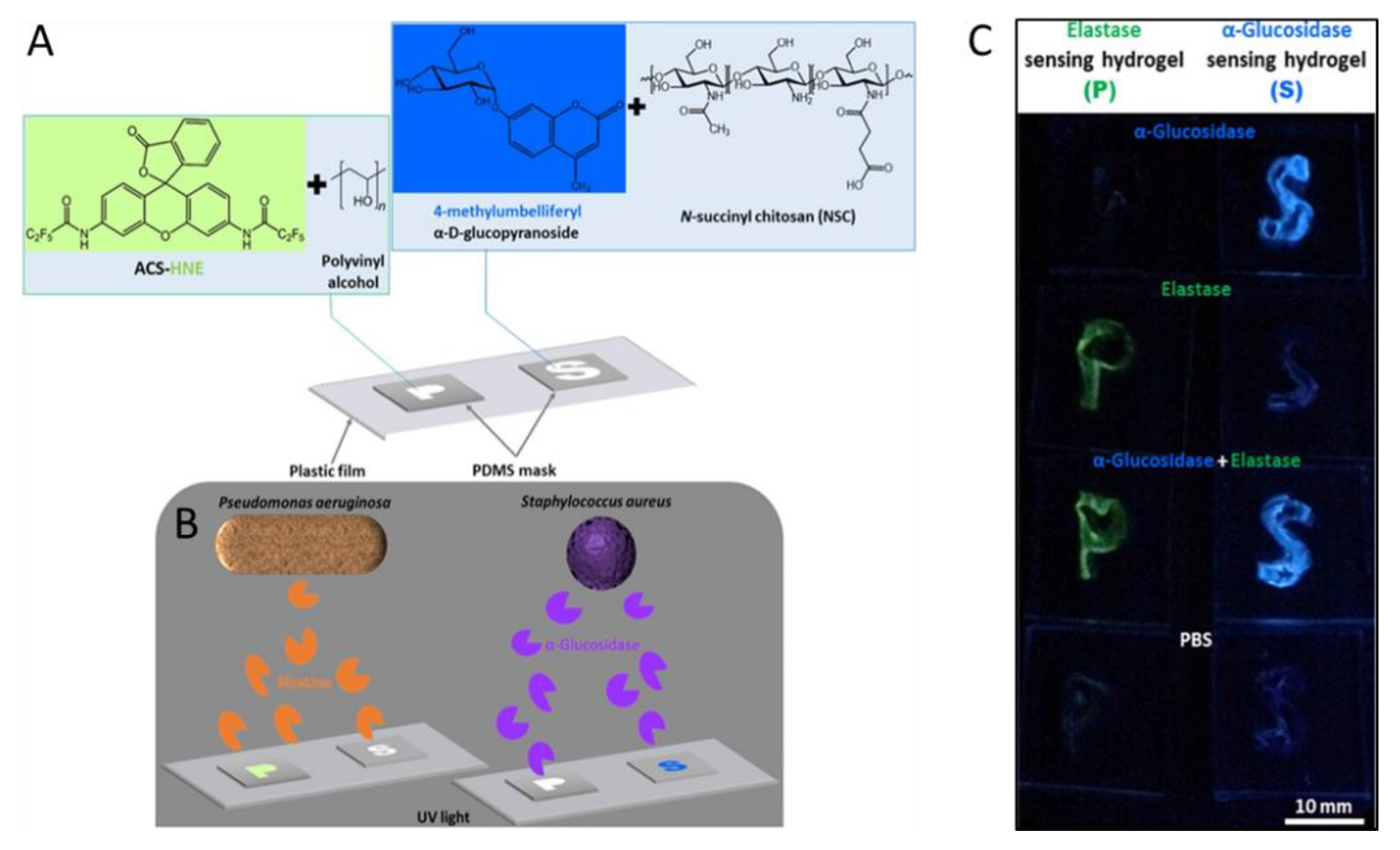
- Jia, Z.; Sukker, I.; Müller, M.; Schönherr, H. Selective Discrimination of Key Enzymes of Pathogenic and Nonpathogenic Bacteria on Autonomously Reporting Shape-Encoded Hydrogel Patterns. ACS Appl. Mater. Interfaces 2018, 10, 5175–5184. [Google Scholar] [CrossRef] [PubMed]
- Kaur, K.; Chelangat, W.; Druzhinin, S.I.; Karuri, N.W.; Müller, M.; Schönherr, H. Quantitative E. coli Enzyme Detection in Reporter Hydrogel-Coated Paper Using a Smartphone Camera. Biosensors 2021, 11, 25. [Google Scholar] [CrossRef] [PubMed]
- Horcajada, J.P.; Montero, M.; Oliver, A.; Sorlí, L.; Luque, S.; Gómez-Zorrilla, S.; Benito, N.; Grau, S. Epidemiology and treatment of multidrug-resistant and extensively drug-resistant Pseudomonas aeruginosa infections. Clin. Microbiol. Rev. 2019, 32. [Google Scholar] [CrossRef] [PubMed]
- Jia, Z.; Gwynne, L.; Sedgwick, A.C.; Müller, M.; Williams, G.T.; Jenkins, A.T.A.; James, T.D.; Schönherr, H. Enhanced Colorimetric Differentiation between Staphylococcus aureus and Pseudomonas aeruginosa Using a Shape-Encoded Sensor Hydrogel. ACS Appl. Bio Mater. 2020, 3, 4398–4407. [Google Scholar] [CrossRef] [PubMed]
- Wang, J.; Lv, M.; Xia, H.; Du, J.; Zhao, Y.; Li, H.; Zhang, Z. Minimalist Design for a Hand-Held SARS-Cov-2 Sensor: Peptide-Induced Covalent Assembly of Hydrogel Enabling Facile Fiber-Optic Detection of a Virus Marker Protein. ACS Sens. 2021, 6, 2465–2471. [Google Scholar] [CrossRef]
- Giebelhaus, R.T.; Erland, L.A.; Murch, S.J. HormonomicsDB: A novel workflow for the untargeted analysis of plant growth regulators and hormones [version 1; peer review: 1 approved with reservations]. F1000Research 2022, 11, 1191. [Google Scholar] [CrossRef]
- Bahadır, E.B.; Sezgintürk, M.K. Electrochemical biosensors for hormone analyses. Biosens. Bioelectron. 2015, 68, 62–71. [Google Scholar] [CrossRef]
- Johnstone, C.; Hendry, C.; Farley, A.; McLafferty, E. Endocrine system: Part 1. Nurs. Stand. 2014, 28, 42–49. [Google Scholar] [CrossRef]
- George, S.M.; Tandon, S.; Kandasubramanian, B. Advancements in Hydrogel-Functionalized Immunosensing Platforms. ACS Omega 2020, 5, 2060–2068. [Google Scholar] [CrossRef]
- Zhang, Q.; Wang, Y.; Mateescu, A.; Sergelen, K.; Kibrom, A.; Jonas, U.; Wei, T.; Dostalek, J. Biosensor based on hydrogel optical waveguide spectroscopy for the detection of 17β-estradiol. Talanta 2013, 104, 149–154. [Google Scholar] [CrossRef]
- Fine, T.; Leskinen, P.; Isobe, T.; Shiraishi, H.; Morita, M.; Marks, R.S.; Virta, M. Luminescent yeast cells entrapped in hydrogels for estrogenic endocrine disrupting chemical biodetection. Biosens. Bioelectron. 2006, 21, 2263–2269. [Google Scholar] [CrossRef]
- Mandon, C.A.; Blum, L.J.; Marquette, C.A. Adding Biomolecular Recognition Capability to 3D Printed Objects. Anal. Chem. 2016, 88, 10767–10772. [Google Scholar] [CrossRef]
- Zhang, A.; Guo, W.; Ke, H.; Zhang, X.; Zhang, H.; Huang, C.; Yang, D.; Jia, N.; Cui, D. Sandwich-format ECL immunosensor based on Au star@BSA-Luminol nanocomposites for determination of human chorionic gonadotropin. Biosens. Bioelectron. 2018, 101, 219–226. [Google Scholar] [CrossRef] [PubMed]
- Pourreza, N.; Ghomi, M. A novel metal enhanced fluorescence bio probe for insulin sensing based on poly vinyl alcohol-borax hydrogel functionalized by Ag dots. Sens. Act. B 2017, 251, 609–616. [Google Scholar] [CrossRef]
- Bhuniya, S.; Kim, B.H. An insulin-sensing sugar-based fluorescent hydrogel. Chem. Commun. 2006, 1842–1844. [Google Scholar] [CrossRef]
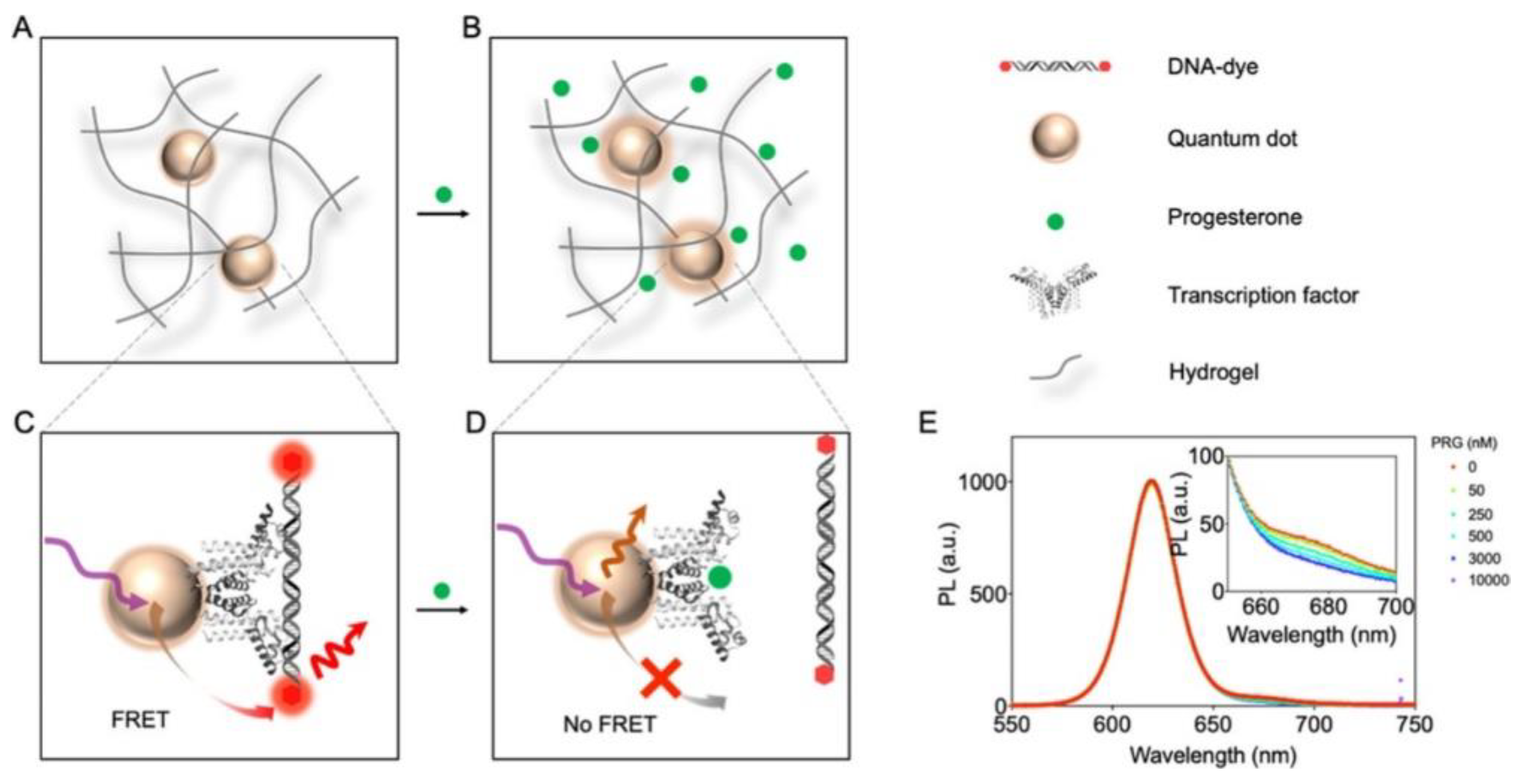
- Chen, M.; Grazon, C.; Sensharma, P.; Nguyen, T.T.; Feng, Y.; Chern, M.; Baer, R.C.; Varongchayakul, N.; Cook, K.; Lecommandoux, S.; et al. Hydrogel-embedded quantum dot−transcription factor sensors for quantitative progesterone detection. ACS Appl. Mater. Interfaces 2020, 12, 43513–43521. [Google Scholar] [CrossRef] [PubMed]
- Mills, G.B.; Moolenaar, W.H. The emerging role of lysophosphatidic acid in cancer. Nat. Rev. Cancer 2003, 3, 582–591. [Google Scholar] [CrossRef] [PubMed]
- Li, J.; Ji, C.; Lü, B.; Rodin, M.; Paradies, J.; Yin, M.; Kuckling, D. Dually Crosslinked Supramolecular Hydrogel for Cancer Biomarker Sensing. ACS Appl. Mater. Interfaces 2020, 12, 36873–36881. [Google Scholar] [CrossRef] [PubMed]
- Lamichhane, S.; Sen, P.; Dickens, A.M.; Hyötyläinen, T.; Orešič, M. An overview of metabolomics data analysis: Current tools and future perspectives. In Data Analysis for Omic Sciences: Methods and Applications; Elsevier: Amsterdam, The Netherlands, 2018; pp. 387–413. [Google Scholar]
- Teunissen, C.E.; Verheul, C.; Willemse, E.A. The use of cerebrospinal fluid in biomarker studies. Handb. Clin. Neurol. 2018, 146, 3–20. [Google Scholar] [CrossRef]
- Lu, C.; Thompson, C.B. Metabolic regulation of epigenetics. Cell Metab. 2012, 16, 9–17. [Google Scholar] [CrossRef]
- Li, L.; Wang, Y.; Pan, L.; Shi, Y.; Cheng, W.; Shi, Y.; Yu, G. A nanostructured conductive hydrogels-based biosensor platform for human metabolite detection. Nano Lett 2015, 15, 1146–1151. [Google Scholar] [CrossRef]
- Hasanah, U.; Md Sani, N.D.; Heng, L.Y.; Idroes, R.; Safitri, E. Construction of a Hydrogel Pectin-Based Triglyceride Optical Biosensor with Immobilized Lipase Enzymes. Biosensors 2019, 9, 135. [Google Scholar] [CrossRef] [PubMed]
- Deng, J.; Liang, W.; Fang, J. Liquid Crystal Droplet-Embedded Biopolymer Hydrogel Sheets for Biosensor Applications. ACS Appl. Mater. Interfaces 2016, 8, 3928–3932. [Google Scholar] [CrossRef]
- Liu, L.; Fei, T.; Guan, X.; Lin, X.; Zhao, H.; Zhang, T. Room temperature ammonia gas sensor based on ionic conductive biomass hydrogels. Sens. Act. B 2020, 320, 1–32. [Google Scholar] [CrossRef]
- Davies, S.; Spanel, P.; Smith, D. Quantitative analysis of ammonia on the breath of patients in end-stage renal failure. Kidney Int. 1997, 223–228. [Google Scholar] [CrossRef] [PubMed]
- Erfkamp, J.; Guenther, M.; Gerlach, G. Enzyme-Functionalized Piezoresistive Hydrogel Biosensors for the Detection of Urea. Sensors 2019, 19, 2858. [Google Scholar] [CrossRef] [PubMed]
- Park, H.; Park, S.Y. Smart Fluorescent Hydrogel Glucose Biosensing Microdroplets with Dual-Mode Fluorescence Quenching and Size Reduction. ACS Appl. Mater. Interfaces 2018, 10, 30172–30179. [Google Scholar] [CrossRef]
- Wei, F.; Lillehoj, P.B.; Ho, C.-M. DNA diagnostics: Nanotechnology-enhanced electrochemical detection of nucleic acids. Pediatr. Res. 2010, 67, 458–468. [Google Scholar] [CrossRef]
- Dario, P.; Carrozza, C.; Benvenuto, A.; Menciassi, A. Micro-systems in biomedical applications. J. Micromech. Microeng. 2000, 10, 235–244. [Google Scholar] [CrossRef]
- Zhao, Y.; Zhao, X.; Tang, B.; Xu, W.; Li, J.; Hu, J.; Gu, Z. Quantum-Dot-Tagged Bioresponsive Hydrogel Suspension Array for Multiplex Label-Free DNA Detection. Adv. Funct. Mater. 2010, 20, 976–982. [Google Scholar] [CrossRef]
- Baeissa, A.; Dave, N.; Smith, B.D.; Liu, J. DNA-Functionalized Monolithic Hydrogels and Gold Nanoparticles for Colorimetric DNA Detection. ACS Appl. Mater. Interfaces 2010, 2, 3594–3600. [Google Scholar] [CrossRef] [PubMed]
- Chen, C.-Y.; Ito, Y.; Chiu, Y.-C.; Wu, W.-C.; Higashihara, T.; Ueda, M.; Chen, W.-C. pH-responsive Dendritic Gelators. Chem. Lett. 2012, 41, 92–94. [Google Scholar] [CrossRef]
- Peppas, N.A.; Hilt, J.Z.; Khademhosseini, A.; Langer, R. Hydrogels in biology and medicine: From molecular principles to bionanotech-nology. Adv. Mat. 2006, 18, 1345–1360. [Google Scholar] [CrossRef]
- Le Goff, G.C.; Srinivas, R.L.; Hill, W.A.; Doyle, P.S. Hydrogel microparticles for biosensing. Eur. Polym. J. 2015, 72, 386–412. [Google Scholar] [CrossRef] [PubMed]
- Kowalczyk, A.; Wagner, B.; Karbarz, M.; Nowicka, A.M. A dual DNA biosensor based on two redox couples with a hydrogel sensing platform functionalized with carboxyl groups and gold nanoparticles. Sens. Act. B 2015, 208, 220–227. [Google Scholar] [CrossRef]
- Sun, L.; Hu, N.; Peng, J.; Chen, L.; Weng, J. Utrasensitive Detection of Mitochondrial DNA Mutation by Graphene Oxide/DNA Hydrogel Electrode. Adv. Funct. Mater. 2014, 24, 6905–6913. [Google Scholar] [CrossRef]
- Chen, M.; Hou, C.; Huo, D.; Fa, H.; Zhao, Y. A sensitive electrochemical DNA biosensor based on three-dimensional nitrogen-doped graphene and Fe3O4 nanoparticles. Sens. Act. B 2017, 239, 421–429. [Google Scholar] [CrossRef]
- Wang, Y.; Zhang, W.; Gong, C.; Liu, B.; Li, Y.; Wang, L.; Su, Z.; Wei, G. Recent advances in the fabrication, functionalization, and bioapplications of peptide hydrogels. Soft Matter. 2020, 16, 10029–10045. [Google Scholar] [CrossRef]
- Arenkov, P.; Kukhtin, A.; Gemmell, A.; Voloshchuk, S.; Chupeeva, V.; Mirzabekov, A. Protein microchips: Use for immunoassay and enzymatic reactions. Anal. Biochem. 2000, 278, 123–131. [Google Scholar] [CrossRef]
- Kivlehan, F.; Paolucci, M.; Brennan, D.; Ragoussis, I.; Galvin, P. Three-dimensional hydrogel structures as optical sensor arrays, for the detection of specific DNA sequences. Anal. Biochem. 2012, 421, 1–8. [Google Scholar] [CrossRef]
- King, P.J.S.; Saiani, A.; Bichenkova, E.V.; Miller, A.F. A de novo self-assembling peptide hydrogel biosensor with covalently immobilised DNA-recognising motifs. Chem. Commun. 2016, 52, 6697–6700. [Google Scholar] [CrossRef]
- Wang, H.; Song, S.; Hao, J.; Song, A. Hydrogels triggered by metal ions as precursors of network CuS for DNA detection. Chemistry 2015, 21, 12194–12201. [Google Scholar] [CrossRef] [PubMed]
- Mao, X.; Pan, S.; Zhou, D.; He, X.; Zhang, Y. Fabrication of DNAzyme-functionalized hydrogel and its application for visible detection of circulating tumor DNA. Sens. Act. B 2019, 285, 385–390. [Google Scholar] [CrossRef]
- Corman, V.M.; Landt, O.; Kaiser, M.; Molenkamp, R.; Meijer, A.; Chu, D.K.W.; Bleicker, T.; Brünink, S.; Schneider, J.; Schmidt, M.L.; et al. Detection of 2019 novel coronavirus (2019-nCoV) by real-time RT-PCR. Eurosurveillance 2020, 25, 2000045. [Google Scholar] [CrossRef]
- Kim, H.S.; Abbas, N.; Shin, S. A rapid diagnosis of SARS-CoV-2 using DNA hydrogel formation on microfluidic pores. Biosens. Bioelectron. 2021, 177, 113005. [Google Scholar] [CrossRef]
- Wang, R.; Li, Y. Hydrogel based QCM aptasensor for detection of avian influenza virus. Biosens. Bioelectron. 2013, 42, 148–155. [Google Scholar] [CrossRef]
- Zarch, S.M.A.; Tezerjani, M.D.; Talebi, M.; Mehrjardi, M.Y.V. Molecular biomarkers in diabetes mellitus (DM). Med. J. Islam. 2020, 34, 28. [Google Scholar]
- Diaz-Arrastia, R.; Shahim, P.; Sandsmark, D.K. Molecular biomarkers in the neurological ICU. Curr. Opin. Crit. Care 2020, 1, 103–108. [Google Scholar] [CrossRef]
- Si, Y.; Xu, L.; Wang, N.; Zheng, J.; Yang, R.; Li, J. Target miRNA-Responsive DNA Hydrogel-Based SERS Sensor Arrays for miRNAs-Marked Cancer Screening. Anal. Chem. 2020, 92, 2649–2655. [Google Scholar] [CrossRef]
- Culver, H.R.; Clegg, J.R.; Peppas, N.A. Analyte-Responsive Hydrogels: Intelligent Materials for Biosensing and Drug Delivery. Acc. Chem. Res. 2017, 50, 170–178. [Google Scholar] [CrossRef]
- Wu, Z.; Hu, X.; Tao, C.; Li, Y.; Liu, J.; Yang, C.; Shen, D.; Li, G. Direct and label-free detection of cholic acid based on molecularly imprinted photonic hydrogels. J. Mater. Chem. 2008, 18, 5452. [Google Scholar] [CrossRef]
- Meng, L.; Meng, P.; Tang, B.; Zhang, Q.; Wang, Y. Molecularly imprinted photonic hydrogels for fast screening of atropine in biological samples with high sensitivity. Forensic Sci. Int. 2013, 231, 6–12. [Google Scholar] [CrossRef] [PubMed]
- Yuan, Y.; Li, Z.; Liu, Y.; Gao, J.; Pan, Z.; Liu, Y. Hydrogel photonic sensor for the detection of 3-pyridinecarboxamide. Chemistry 2012, 18, 303–309. [Google Scholar] [CrossRef] [PubMed]
- Li, S.; Yang, M.; Zhou, W.; Johnston, T.G.; Wang, R.; Zhu, J. Dextran hydrogel coated surface plasmon resonance imaging (SPRi) sensor for sensitive and label-free detection of small molecule drugs. Appl. Surf. Sci. 2015, 355, 570–576. [Google Scholar] [CrossRef]
- Shirani, M.; Kalantari, H.; Khodayar, M.J.; Kouchak, M.; Rahbar, N. An ultra-sensitive optical aptasensor based on gold nanoparticles/poly vinyl alcohol hydrogel as acceptor/emitter pair for fluorometric detection of digoxin with on/off/on strategy. Spectrochim. Acta Part A Mol. Biomol. Spectrosc. 2021, 250, 119345. [Google Scholar] [CrossRef] [PubMed]
- Caffarel-Salvador, E.; Brady, A.J.; Eltayib, E.; Meng, T.; Alonso-Vicente, A.; Gonzalez-Vazquez, P.; Torrisi, B.M.; Vicente-Perez, E.M.; Mooney, K.; Jones, D.S.; et al. Hydrogel-Forming Microneedle Arrays Allow Detection of Drugs and Glucose In Vivo: Potential for Use in Diagnosis and Therapeutic Drug Monitoring. PLoS ONE 2015, 10, e0145644. [Google Scholar] [CrossRef]
- Bahram, M.; Hoseinzadeh, F.; Farhadi, K.; Saadat, M.; Najafi-Moghaddam, P.; Afkhami, A. Synthesis of gold nanoparticles using pH-sensitive hydrogel and its application for colorimetric determination of acetaminophen, ascorbic acid and folic acid. Colloids Surf. A Physicochem. Eng. Asp. 2014, 441, 517–524. [Google Scholar] [CrossRef]
- Wang, M.; Zhong, L.; Cui, M.; Liu, W.; Liu, X. Nanomolar Level Acetaminophen Sensor Based on Novel Polypyrrole Hydrogel Derived N-doped Porous Carbon. Electroanalysis 2019, 31, 711–717. [Google Scholar] [CrossRef]
- Kokulnathan, T.; Ramaraj, S.; Chen, S.-M.; Han-Yu, Y. Eco-Friendly Synthesis of Biocompatible Pectin Stabilized Graphene Nanosheets Hydrogel and Their Application for the Simultaneous Electrochemical Determination of Dopamine and Paracetamol in Real Samples. J. Electrochem. Soc. 2018, 165, B240–B249. [Google Scholar] [CrossRef]
- Wang, M.; Cui, M.; Zhong, L.; Qin, L.; Liu, X. Ultrahigh Sensitivity Acetaminophen Sensor Based on Network-Structured Nanocarbons. J. Electrochem. Soc. 2018, 165, H872–H880. [Google Scholar] [CrossRef]
- Havens, N.; Trihn, P.; Kim, D.; Luna, M.; Wanekaya, A.K.; Mugweru, A. Redox polymer covalently modified multiwalled carbon nanotube based sensors for sensitive acetaminophen and ascorbic acid detection. Electrochim. Acta 2010, 55, 2186–2190. [Google Scholar] [CrossRef]
- Liu, Y.; Minami, T.; Nishiyabu, R.; Wang, Z.; Anzenbacher, P. Sensing of carboxylate drugs in urine by a supramolecular sensor array. J. Am. Chem. Soc. 2013, 135, 7705–7712. [Google Scholar] [CrossRef] [PubMed]
- Laishram, R.; Maitra, U. Rapid Sensing of Specific Drugs at Sub-Ppb Levels by Using a Hybrid Organic-Inorganic Photoluminescent Soft Material. Asian J. Org. Chem. 2017, 6, 1235–1239. [Google Scholar] [CrossRef]
- Kassahun, G.S.; Griveau, S.; Juillard, S.; Champavert, J.; Ringuedé, A.; Bresson, B.; Tran, Y.; Bedioui, F.; Slim, C. Hydrogel Matrix-Grafted Impedimetric Aptasensors for the Detection of Diclofenac. Langmuir 2020, 36, 827–836. [Google Scholar] [CrossRef] [PubMed]
- Jewell, K.S.; Falås, P.; Wick, A.; Joss, A.; Ternes, T.A. Transformation of diclofenac in hybrid biofilm–activated sludge processes. Water Res. 2016, 105, 559–567. [Google Scholar] [CrossRef] [PubMed]
- Umbreen, N.; Sohni, S.; Ahmad, I.; Khattak, N.U.; Gul, K. Self-assembled three-dimensional reduced graphene oxide-based hydrogel for highly efficient and facile removal of pharmaceutical compounds from aqueous solution. J. Colloid Interface Sci. 2018, 527, 356–367. [Google Scholar] [CrossRef] [PubMed]
- Feng, Z.; Simeone, A.; Odelius, K.; Hakkarainen, M. Biobased Nanographene Oxide Creates Stronger Chitosan Hydrogels with Improved Adsorption Capacity for Trace Pharmaceuticals. ACS Sustain. Chem. Eng. 2017, 5, 11525–11535. [Google Scholar] [CrossRef]
- Godiya, C.B.; Kumar, S.; Xiao, Y. Amine functionalized egg albumin hydrogel with enhanced adsorption potential for diclofenac sodium in water. J. Hazard. Mater. 2020, 393, 122417. [Google Scholar] [CrossRef]
- Sun, X.; Agate, S.; Salem, K.S.; Lucia, L.; Pal, L. Hydrogel-Based Sensor Networks: Compositions, Properties, and Applications—A Review. ACS Appl. Bio Mater. 2021, 4, 140–162. [Google Scholar] [CrossRef]
- World Health Organization. Pharmaceuticals in Drinking Water. 2011. Available online: https://www.who.int/publications/i/item/9789241502085 (accessed on 15 November 2022).
- Villa, C.C.; Sánchez, L.T.; Valencia, G.A.; Ahmed, S.; Gutiérrez, T.J. Molecularly imprinted polymers for food applications: A review. Trends Food Sci. Technol. 2021, 111, 642–669. [Google Scholar] [CrossRef]
- Cao, Y.; Liu, G.; Zheng, B.; Wang, X.; Li, H.; Wang, G.; Zhao, L.; Wang, Y. A sulfamethoxazole molecularly imprinted two-dimensional photonic crystal hydrogel sensor. Soft Matter. 2021, 17, 4969–4978. [Google Scholar] [CrossRef]
- Zhang, R.; Wang, Y.; Yu, L.-P. Specific and ultrasensitive ciprofloxacin detection by responsive photonic crystal sensor. J. Hazard. Mater. 2014, 280, 46–54. [Google Scholar] [CrossRef] [PubMed]
- Wang, Y.; Xie, T.; Yang, J.; Lei, M.; Fan, J.; Meng, Z.; Xue, M.; Qiu, L.; Qi, F.; Wang, Z. Fast screening of antibiotics in milk using a molecularly imprinted two-dimensional photonic crystal hydrogel sensor. Anal. Chim. Acta 2019, 1070, 97–103. [Google Scholar] [CrossRef] [PubMed]
- Tan, B.; Zhao, H.; Du, L.; Gan, X.; Quan, X. A versatile fluorescent biosensor based on target-responsive graphene oxide hydrogel for antibiotic detection. Biosens. Bioelectron. 2016, 83, 267–273. [Google Scholar] [CrossRef] [PubMed]
- Gao, L.; Lian, C.; Zhou, Y.; Yan, L.; Li, Q.; Zhang, C.; Chen, L.; Chen, K. Graphene oxide-DNA based sensors. Biosens. Bioelectron. 2014, 60, 22–29. [Google Scholar] [CrossRef]
- Il’ina, A.V.; Varlamov, V.P. Chitosan-based polyelectrolyte complexes: A review. Appl. Biochem. Microbiol. 2005, 41, 5–11. [Google Scholar] [CrossRef]
- Malik, A.H.; Iyer, P.K. Conjugated Polyelectrolyte Based Sensitive Detection and Removal of Antibiotics Tetracycline from Water. ACS Appl. Mater. Interfaces 2017, 9, 4433–4439. [Google Scholar] [CrossRef]
- Ehtesabi, H.; Roshani, S.; Bagheri, Z.; Yaghoubi-Avini, M. Carbon dots—Sodium alginate hydrogel: A novel tetracycline fluorescent sensor and adsorber. J. Environ. Chem. Eng. 2019, 7, 103419. [Google Scholar] [CrossRef]
- Luo, Q.; Ren, T.; Lei, Z.; Huang, Y.; Huang, Y.; Xu, D.; Wan, C.; Guo, X.; Wu, Y. Non-toxic chitosan-based hydrogel with strong adsorption and sensitive detection abilities for tetracycline. Chem. Eng. J. 2022, 427, 131738. [Google Scholar] [CrossRef]
- Ehrbar, M.; Schoenmakers, R.; Christen, E.H.; Fussenegger, M.; Weber, W. Drug-sensing hydrogels for the inducible release of biopharmaceuticals. Nat. Mat. 2008, 7, 800–804. [Google Scholar] [CrossRef]
- Peacock, A.; Leung, J.; Larney, S.; Colledge, S.; Hickman, M.; Rehm, J.; Giovino, G.A.; West, R.; Hall, W.; Griffiths, P.; et al. Global statistics on alcohol, tobacco and illicit drug use: 2017 status report. Addiction 2018, 113, 1905–1926. [Google Scholar] [CrossRef]
- Garg, K.C.; Kumar, S. Bibliometrics of the Global Drug Abuse Research Output as Reflected by Coverage in Web of Science Core Collection during 2011–2018. J. Sci. Res. 2020, 9, 174–184. [Google Scholar] [CrossRef]
- Zaami, S.; Marinelli, E.; Varì, M.R. New Trends of Substance Abuse During COVID-19 Pandemic: An International Perspective. Front. Psychiatry 2020, 11, 700. [Google Scholar] [CrossRef]
- Groshkova, T.; Stoian, T.; Cunningham, A.; Griffiths, P.; Singleton, N.; Sedefov, R. Will the Current COVID-19 Pandemic Impact on Long-term Cannabis Buying Practices? J. Addict. Med. 2020, 14, e13-10. [Google Scholar] [CrossRef] [PubMed]
- Mattson, C.L.; Tanz, L.J.; Quinn, K.; Kariisa, M.; Patel, P.; Davis, N.L. Trends and Geographic Patterns in Drug and Synthetic Opioid Overdose Deaths—United States, 2013–2019. MMWR Morb. Mortal. Wkly. Rep. 2021, 70, 202–207. [Google Scholar] [CrossRef]
- Abuse, National Institute on Drug. Commonly Used Drugs Charts: National Institute on Drug Abuse. Available online: https://www.drugabuse.gov/drug-topics/commonly-used-drugs-charts (accessed on 15 November 2022).
- Stinson, F.S.; Grant, B.F.; Dawson, D.A.; Ruan, W.J.; Huang, B.; Saha, T. Comorbidity between DSM-IV alcohol and specific drug use disorders in the United States: Results from the National Epidemiologic Survey on Alcohol and Related Conditions. Drug Alcohol Depend. 2005, 80, 105–116. [Google Scholar] [CrossRef]
- Zima, T. Alcohol Abuse. EJIFCC 2018, 29, 285–289. [Google Scholar] [PubMed]
- Barroso, O.; Mazzoni, I.; Rabin, O. Hormone abuse in sports: The antidoping perspective. Asian J. Androl. 2008, 10, 391–402. [Google Scholar] [CrossRef] [PubMed]
- Werner, T.C.; Hatton, C.K. Performance-Enhancing Drugs in Sports: How Chemists Catch Users. J. Chem. Educ. 2011, 88, 34–40. [Google Scholar] [CrossRef]
- Fredolini, C.; Meani, F.; Alex Reeder, K.; Rucker, S.; Patanarut, A.; Botterell, P.J.; Bishop, B.; Longo, C.; Espina, V.; Petricoin, E.F.; et al. Concentration and preservation of very low abundance biomarkers in urine, such as human growth hormone (hGH), by Cibacron Blue F3G-A loaded hydrogel particles. Nano Res. 2008, 1, 502–518. [Google Scholar] [CrossRef] [PubMed]
- Denizli, A.; Pişkin, E. Dye-ligand affinity systems. J. Biochem. Biophys. Methods 2001, 49, 391–416. [Google Scholar] [CrossRef]
- United Nations. World Drug Report 2021 (Sales No. E.21.XI.8); United Nations Publications: New York, NY, USA, 2021. [Google Scholar]
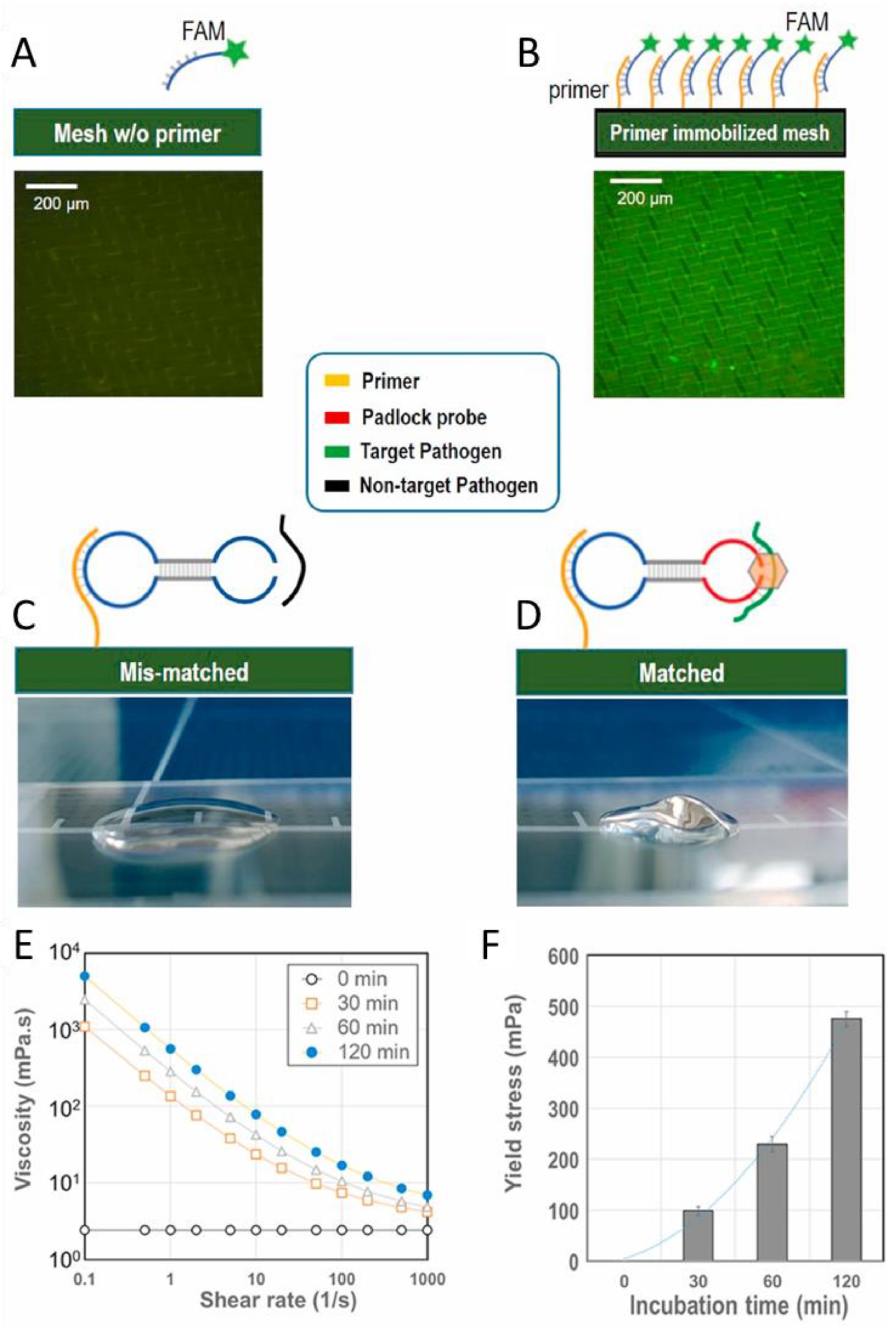
- Li, Y.; Ma, Y.; Jiao, X.; Li, T.; Lv, Z.; Yang, C.J.; Zhang, X.; Wen, Y. Control of capillary behavior through target-responsive hydrogel permeability alteration for sensitive visual quantitative detection. Nat. Commun. 2019, 10, 1036. [Google Scholar] [CrossRef] [PubMed]
- Zhu, Z.; Wu, C.; Liu, H.; Zou, Y.; Zhang, X.; Kang, H.; Yang, C.J.; Tan, W. An Aptamer Cross-Linked Hydrogel as a Colorimetric Platform for Visual Detection. Angew. Chem. Int. Ed. 2010, 122, 1070–1074. [Google Scholar] [CrossRef]
- Liu, J.; Lu, Y. Fast Colorimetric Sensing of Adenosine and Cocaine Based on a General Sensor Design Involving Aptamers and Nanoparticles. Angew. Chem. Int. Ed. 2006, 118, 96–100. [Google Scholar] [CrossRef]
- Stanley, T.H. The Fentanyl Story. J. Pain 2014, 15, 1215–1226. [Google Scholar] [CrossRef] [PubMed]
- Ling, W.; Mooney, L.; Hillhouse, M. Prescription opioid abuse, pain and addiction: Clinical issues and implications. Drug Alcohol Rev. 2011, 30, 300–305. [Google Scholar] [CrossRef]
- Barfidokht, A.; Mishra, R.K.; Seenivasan, R.; Liu, S.; Hubble, L.J.; Wang, J.; Hall, D.A. Wearable electrochemical glove-based sensor for rapid and on-site detection of fentanyl. Sens. Act. B 2019, 296, 126422. [Google Scholar] [CrossRef]
- Liu, L.; Grillo, F.; Canfarotta, F.; Whitcombe, M.; Morgan, S.P.; Piletsky, S.; Correia, R.; He, C.; Norris, A.; Korposh, S. Carboxyl-fentanyl detection using optical fibre grating-based sensors functionalised with molecularly imprinted nanoparticles. Biosens. Bioelectron. 2021, 177, 113002. [Google Scholar] [CrossRef] [PubMed]
- Chapuis-Hugon, F.; Cruz-Vera, M.; Savane, R.; Ali, W.H.; Valcarcel, M.; Deveaux, M.; Pichon, V. Selective sample pretreatment by molecularly imprinted polymer for the determination of LSD in biological fluids. J. Sep. Sci. 2009, 32, 3301–3309. [Google Scholar] [CrossRef]
- Richards, J.R.; Laurin, E.G. Methamphetamine Toxicity; StatPearls; StatPearls Publishing: Treasure Island, FL, USA, 2021. [Google Scholar]
- Vikholm-Lundin, I.; Auer, S.; Hellgren, A.-C. Detection of 3,4-methylenedioxymethamphetamine (MDMA, ecstasy) by displacement of antibodies. Sens. Actuators B Chem. 2011, 156, 28–34. [Google Scholar] [CrossRef]
- Choodum, A.; Kanatharana, P.; Wongniramaikul, W.; NicDaeid, N. A sol–gel colorimetric sensor for methamphetamine detection. Sens. Act. B 2015, 215, 553–560. [Google Scholar] [CrossRef]
- Akhoundian, M.; Alizadeh, T.; Ganjali, M.R.; Norouzi, P. Ultra-trace detection of methamphetamine in biological samples using FFT-square wave voltammetry and nano-sized imprinted polymer/MWCNTs -modified electrode. Talanta 2019, 200, 115–123. [Google Scholar] [CrossRef] [PubMed]
- Souza, M.A.; de Oliveira, K.V.; Oliveira, F.C.; Silva, L.P.; Rubim, J.C. The adsorption of methamphetamine on Ag nanoparticles dispersed in agarose gel—Detection of methamphetamine in fingerprints by SERS. Vib. Spectrosc. 2018, 98, 152–157. [Google Scholar] [CrossRef]
- Andre, C.M.; Hausman, J.-F.; Guerriero, G. Cannabis sativa: The Plant of the Thousand and One Molecules. Front. Plant Sci. 2016, 7, 19. [Google Scholar] [CrossRef] [PubMed]
- Zhang, Q.; Berg, D.; Mugo, S.M. Molecularly imprinted carbon based electrodes for tetrahydrocannabinol sensing. Inorg. Chem. Commun. 2019, 107, 107459. [Google Scholar] [CrossRef]
- Kroh, C.; Wuchrer, R.; Steinke, N.; Guenther, M.; Gerlach, G.; Härtling, T. Hydrogel-Based Plasmonic Sensor Substrate for the Detection of Ethanol. Sensors 2019, 19, 1264. [Google Scholar] [CrossRef] [PubMed]
- Dispenza, C.; Sabatino, M.A.; Alessi, S.; Spadaro, G.; D’Acquisto, L.; Pernice, R.; Adamo, G.; Stivala, S.; Parisi, A.; Livreri, P.; et al. Hydrogel films engineered in a mesoscopically ordered structure and responsive to ethanol vapors. React. Funct. Polym. 2014, 79, 68–76. [Google Scholar] [CrossRef]
- Pernice, R.; Adamo, G.; Stivala, S.; Parisi, A.; Busacca, A.C.; Spigolon, D.; Sabatino, M.A.; D’Acquisto, L.; Dispenza, C. Opals infiltrated with a stimuli-responsive hydrogel for ethanol vapor sensing. Opt. Mater. Express OME 2013, 3, 1820–1833. [Google Scholar] [CrossRef]
- Du, X.; Zhai, J.; Li, X.; Zhang, Y.; Li, N.; Xie, X. Hydrogel-Based Optical Ion Sensors: Principles and Challenges for Point-of-Care Testing and Environmental Monitoring. ACS Sens. 2021, 6, 1990–2001. [Google Scholar] [CrossRef]
- Jones, K.W. Environmental Sensors; John Wiley & Sons, Ltd.: New York, NY, USA, 1995; pp. 451–489. ISBN 978-3-527-62018-0. [Google Scholar]
- Zhang, Q.; Zhang, Y.; Wan, Y.; Carvalho, W.; Hu, L.; Serpe, M.J. Stimuli-Responsive Polymers for Sensing and Reacting to Environmental Conditions. Progr. Polym. Sci. 2021, 116, 101386. [Google Scholar] [CrossRef]
- Yu, J.; Tsow, F.; Mora, S.J.; Tipparaju, V.V.; Xian, X. Hydrogel-incorporated colorimetric sensors with high humidity tolerance for environmental gases sensing. Sens. Act. B 2021, 345, 130404. [Google Scholar] [CrossRef]
- Hu, L.; Zhang, Q.; Li, X.; Serpe, M.J. Stimuli-responsive polymers for sensing and actuation. Mater. Horiz. 2019, 6, 1774–1793. [Google Scholar] [CrossRef]
- Ye, B.-F.; Zhao, Y.-J.; Cheng, Y.; Li, T.-T.; Xie, Z.-Y.; Zhao, X.-W.; Gu, Z.-Z. Colorimetric photonic hydrogel aptasensor for the screening of heavy metal ions. Nanoscale 2012, 4, 5998. [Google Scholar] [CrossRef] [PubMed]
- Qin, J.; Dong, B.; Li, X.; Han, J.; Gao, R.; Su, G.; Cao, L.; Wang, W. Fabrication of intelligent photonic crystal hydrogel sensors for selective detection of trace mercury ions in seawater. J. Mater. Chem. C 2017, 5, 8482–8488. [Google Scholar] [CrossRef]
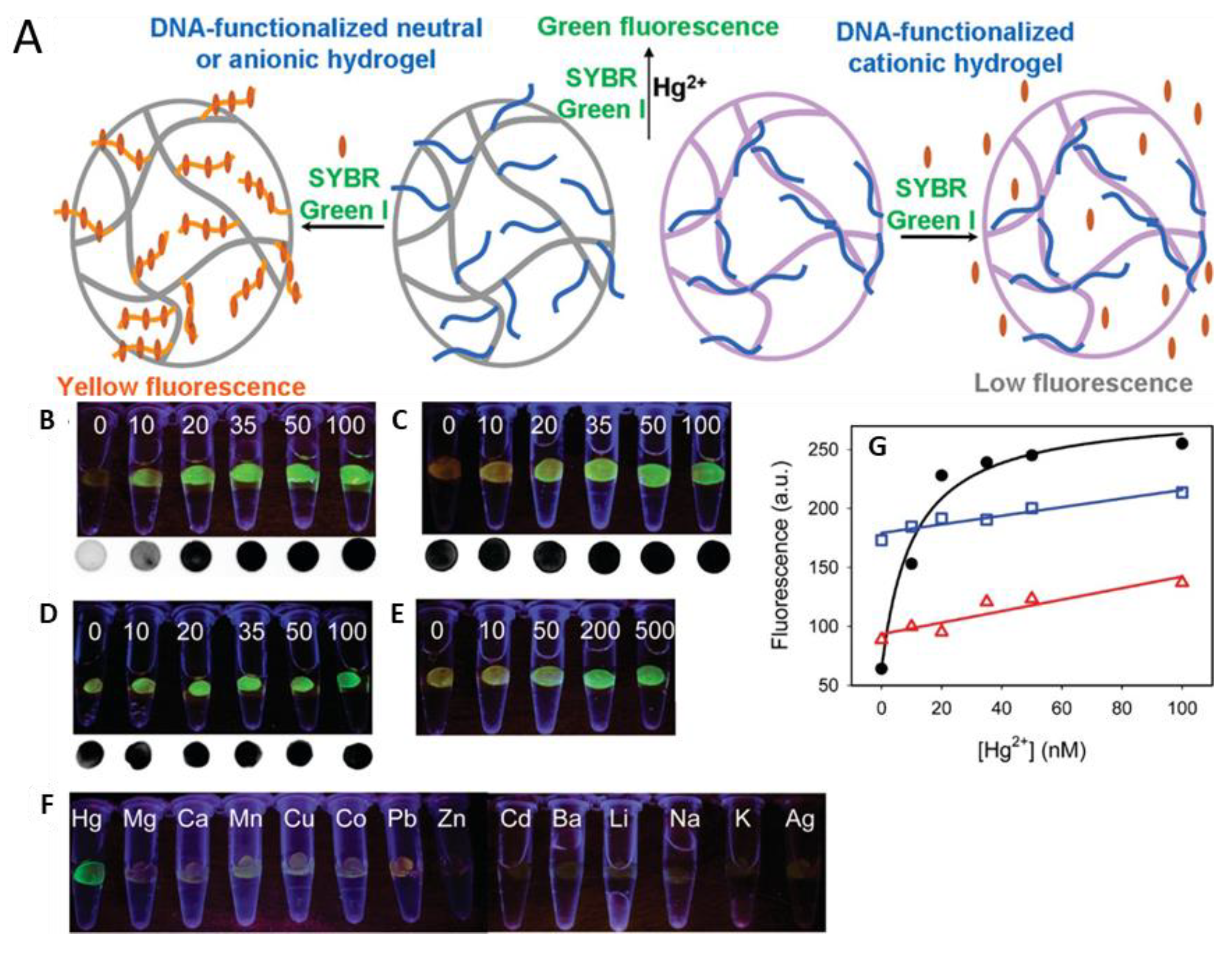
- Dave, N.; Chan, M.Y.; Huang, P.-J.J.; Smith, B.D.; Liu, J. Regenerable DNA-Functionalized Hydrogels for Ultrasensitive, Instrument-Free Mercury(II) Detection and Removal in Water. J. Am. Chem. Soc. 2010, 132, 12668–12673. [Google Scholar] [CrossRef]
- He, X.; Zhou, X.; Liu, W.; Liu, Y.; Wang, X. Flexible DNA Hydrogel SERS Active Biofilms for Conformal Ultrasensitive Detection of Uranyl Ions from Aquatic Products. Langmuir 2020, 36, 2930–2936. [Google Scholar] [CrossRef]
- Dhanjai; Sinha, A.; Kalambate, P.K.; Mugo, S.M.; Kamau, P.; Chen, J.; Jain, R. Polymer hydrogel interfaces in electrochemical sensing strategies: A review. TrAC Trends Anal. Chem. 2019, 118, 488–501. [Google Scholar] [CrossRef]
- Pramanik, K.; Sarkar, P.; Bhattacharyay, D. Semi-quantitative colorimetric and supersensitive electrochemical sensors for mercury using rhodamine b hydrazide thio derivative. J. Mol. Liq. 2019, 276, 141–152. [Google Scholar] [CrossRef]
- Jiang, C.; Li, Y.; Wang, H.; Chen, D.; Wen, Y. A portable visual capillary sensor based on functional DNA crosslinked hydrogel for point-of-care detection of lead ion. Sens. Act. B 2020, 307, 127625. [Google Scholar] [CrossRef]
- Joseph, K.A.; Dave, N.; Liu, J. Electrostatically Directed Visual Fluorescence Response of DNA-Functionalized Monolithic Hydrogels for Highly Sensitive Hg2+ Detection. ACS Appl. Mater. Interfaces 2011, 3, 733–739. [Google Scholar] [CrossRef]
- Huang, J.; Liu, X.; Li, L.; Chen, S.; Yang, J.; Yan, J.; Xu, F.; Zhang, X. Nitrogen-Doped Carbon Quantum Dot-Anchored Hydrogels for Visual Recognition of Dual Metal Ions through Reversible Fluorescence Response. ACS Sustain. Chem. Eng. 2021, 9, 15190–15201. [Google Scholar] [CrossRef]
- Jeevika, A. A simple colorimetric sensor for the recognition of copper ions based on safranin impregnated poly vinyl alcohol hydrogels. Opt. Mater. 2020, 105, 109971. [Google Scholar] [CrossRef]
- Keith, S.; Faroon, O.; Roney, N.; Scinicariello, F.; Wilbur, S.; Ingerman, L.; Llados, F.; Plewak, D.; Wohlers, D.; Diamond, G. Health Effects—Toxicological Profile for Uranium; Agency for Toxic Substances and Disease Registry (US): Atlanta, GA, USA, 2013. [Google Scholar]
- Qin, J.; Dong, B.; Cao, L.; Wang, W. Photonic hydrogels for the ultratrace sensing of divalent beryllium in seawater. J. Mater. Chem. C 2018, 6, 4234–4242. [Google Scholar] [CrossRef]
- Chandler, L.; Huang, B.; Mu, T.T. A smart handheld Raman spectrometer with cloud and AI deep learning algorithm for mixture analysis. Proc. SPIE 2019, 10983, 20–28. [Google Scholar] [CrossRef]
- Xiao, F.; Sun, Y.; Du, W.; Shi, W.; Wu, Y.; Liao, S.; Wu, Z.; Yu, R. Smart Photonic Crystal Hydrogel Material for Uranyl Ion Monitoring and Removal in Water. Adv. Funct. Mater. 2017, 27, 1702147. [Google Scholar] [CrossRef]
- Ab Latif, W.; Ara, A.; Usmani, J.A. Lead toxicity: A review. Interdiscip. Toxicol. 2015, 8, 55–64. [Google Scholar] [CrossRef]
- Huang, Y.; Ma, Y.; Chen, Y.; Wu, X.; Fang, L.; Zhu, Z.; Yang, C.J. Target-Responsive DNAzyme Cross-Linked Hydrogel for Visual Quantitative Detection of Lead. Anal. Chem. 2014, 86, 11434–11439. [Google Scholar] [CrossRef]
- Song, Y.; Zhang, Y.; Bernard, P.E.; Reuben, J.M.; Ueno, N.T.; Arlinghaus, R.B.; Zu, Y.; Qin, L. Multiplexed volumetric bar-chart chip for point-of-care diagnostics. Nat. Commun. 2012, 3, 1283. [Google Scholar] [CrossRef]
- Chu, J.; Chen, C.; Li, X.; Yu, L.; Li, W.; Cheng, M.; Tang, W.; Xiong, Z. A responsive pure DNA hydrogel for label-free detection of lead ion. Anal. Chim. Acta 2021, 1157, 338400. [Google Scholar] [CrossRef]
- Jacobi, Z.E.; Li, L.; Liu, J. Visual detection of lead(II) using a label-free DNA-based sensor and its immobilization within a monolithic hydrogel. Analyst 2012, 137, 704–709. [Google Scholar] [CrossRef]
- Lu, Y.; Wei, M.; Wang, C.; Wei, W.; Liu, Y. Enhancing hydrogel-based long-lasting chemiluminescence by a platinum-metal organic framework and its application in array detection of pesticides and D-amino acids. Nanoscale 2020, 12, 4959–4967. [Google Scholar] [CrossRef]
- Hong, W.; Chen, Y.; Feng, X.; Yan, Y.; Hu, X.; Zhao, B.; Zhang, F.; Zhang, D.; Xu, Z.; Lai, Y. Full-color CO2 gas sensing by an inverse opal photonic hydrogel. Chem. Commun. 2013, 49, 8229. [Google Scholar] [CrossRef]
- Danish, E.Y.; Bakhsh, E.M.; Akhtar, K. Design of chitosan nanocomposite hydrogel for sensitive detection and removal of organic pollutants. Int. J. Biol. Macromol. 2020, 159, 276–286. [Google Scholar] [CrossRef] [PubMed]
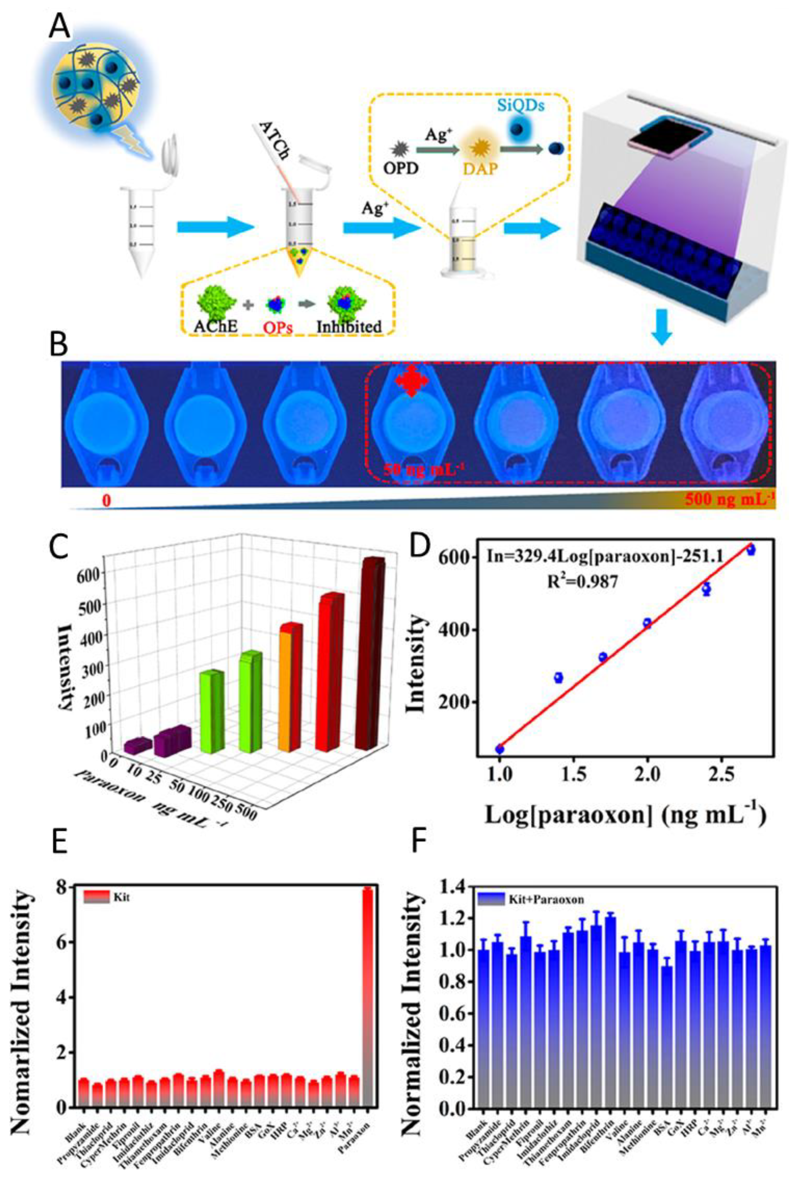
- Jin, R.; Kong, D.; Yan, X.; Zhao, X.; Li, H.; Liu, F.; Sun, P.; Lin, Y.; Lu, G. Integrating Target-Responsive Hydrogels with Smartphone for On-Site ppb-Level Quantitation of Organophosphate Pesticides. ACS Appl. Mater. Interfaces 2019, 11, 27605–27614. [Google Scholar] [CrossRef] [PubMed]
- Wu, S.; Li, D.; Gao, Z.; Wang, J. Controlled etching of gold nanorods by the Au(III)-CTAB complex, and its application to semi-quantitative visual determination of organophosphorus pesticides. Microchim. Acta 2017, 184, 4383–4391. [Google Scholar] [CrossRef]


Publisher’s Note: MDPI stays neutral with regard to jurisdictional claims in published maps and institutional affiliations. |
© 2022 by the authors. Licensee MDPI, Basel, Switzerland. This article is an open access article distributed under the terms and conditions of the Creative Commons Attribution (CC BY) license (https://creativecommons.org/licenses/by/4.0/).
Share and Cite
Völlmecke, K.; Afroz, R.; Bierbach, S.; Brenker, L.J.; Frücht, S.; Glass, A.; Giebelhaus, R.; Hoppe, A.; Kanemaru, K.; Lazarek, M.; et al. Hydrogel-Based Biosensors. Gels 2022, 8, 768. https://doi.org/10.3390/gels8120768
Völlmecke K, Afroz R, Bierbach S, Brenker LJ, Frücht S, Glass A, Giebelhaus R, Hoppe A, Kanemaru K, Lazarek M, et al. Hydrogel-Based Biosensors. Gels. 2022; 8(12):768. https://doi.org/10.3390/gels8120768
Chicago/Turabian StyleVöllmecke, Katharina, Rowshon Afroz, Sascha Bierbach, Lee Josephine Brenker, Sebastian Frücht, Alexandra Glass, Ryland Giebelhaus, Axel Hoppe, Karen Kanemaru, Michal Lazarek, and et al. 2022. "Hydrogel-Based Biosensors" Gels 8, no. 12: 768. https://doi.org/10.3390/gels8120768
APA StyleVöllmecke, K., Afroz, R., Bierbach, S., Brenker, L. J., Frücht, S., Glass, A., Giebelhaus, R., Hoppe, A., Kanemaru, K., Lazarek, M., Rabbe, L., Song, L., Velasco Suarez, A., Wu, S., Serpe, M., & Kuckling, D. (2022). Hydrogel-Based Biosensors. Gels, 8(12), 768. https://doi.org/10.3390/gels8120768