
Advances in Composite Stimuli-Responsive Hydrogels for Wound Healing: Mechanisms and Applications


Abstract

1. Introduction
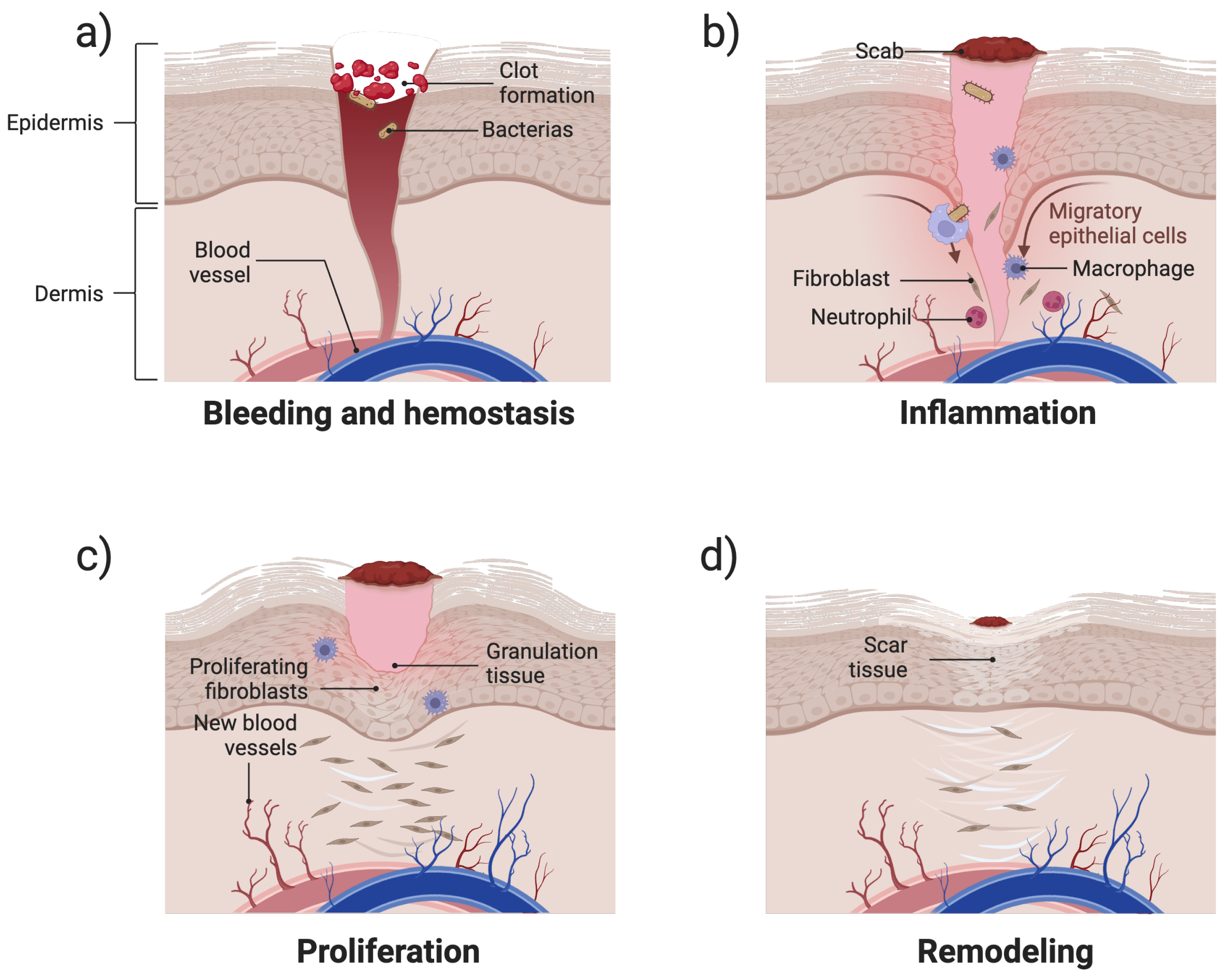
2. Biological Mechanisms of Wound Healing
3. Classification Based on Stimuli Types
3.1. Single Stimuli-Responsive Mode
3.1.1. pH-Responsive Hydrogels
3.1.2. Temperature-Responsive Hydrogels
3.1.3. Enzyme-Responsive Hydrogels
3.1.4. Photoresponsive Hydrogels
3.1.5. ROS-Scavenging Hydrogels
3.2. Multi-Stimuli Responsive Hydrogels
4. Conclusions
5. Clinical Translation and Future Perspectives
Funding
Institutional Review Board Statement
Informed Consent Statement
Data Availability Statement
Acknowledgments
Conflicts of Interest
References
- Wang, P.; Cai, F.; Li, Y.; Yang, X.; Feng, R.; Lu, H.; Bai, X.; Han, J. Emerging trends in the application of hydrogel-based biomaterials for enhanced wound healing: A literature review. Int. J. Biol. Macromol. 2024, 261, 129300. [Google Scholar] [CrossRef] [PubMed]
- Shariatzadeh, F.J.; Currie, S.; Logsetty, S.; Spiwak, R.; Liu, S. Enhancing wound healing and minimizing scarring: A comprehensive review of nanofiber technology in wound dressings. Prog. Mater. Sci. 2024, 101350. [Google Scholar] [CrossRef]
- Lindley, L.E.; Stojadinovic, O.; Pastar, I.; Tomic-Canic, M. Biology and biomarkers for wound healing. Plast. Reconstr. Surg. 2016, 138, 18S–28S. [Google Scholar] [CrossRef]
- Landén, N.X.; Li, D.; Ståhle, M. Transition from inflammation to proliferation: A critical step during wound healing. Cell. Mol. Life Sci. 2016, 73, 3861–3885. [Google Scholar] [CrossRef]
- Boodhoo, K.; Vlok, M.; Tabb, D.L.; Myburgh, K.H.; van de Vyver, M. Dysregulated healing responses in diabetic wounds occur in the early stages postinjury. J. Mol. Endocrinol. 2021, 66, 141–155. [Google Scholar] [CrossRef]
- Gomez-Florit, M.; Pardo, A.; Domingues, R.M.; Graça, A.L.; Babo, P.S.; Reis, R.L.; Gomes, M.E. Natural-based hydrogels for tissue engineering applications. Molecules 2020, 25, 5858. [Google Scholar] [CrossRef]
- Francesko, A.; Petkova, P.; Tzanov, T. Hydrogel dressings for advanced wound management. Curr. Med. Chem. 2018, 25, 5782–5797. [Google Scholar] [CrossRef] [PubMed]
- Chen, C.; Gu, Y.; Deng, L.; Han, S.; Sun, X.; Chen, Y.; Lu, J.R.; Xu, H. Tuning gelation kinetics and mechanical rigidity of β-hairpin peptide hydrogels via hydrophobic amino acid substitutions. ACS Appl. Mater. Interfaces 2014, 6, 14360–14368. [Google Scholar] [CrossRef]
- Dong, R.; Guo, B. Smart wound dressings for wound healing. Nano Today 2021, 41, 101290. [Google Scholar] [CrossRef]
- Freedman, B.R.; Hwang, C.; Talbot, S.; Hibler, B.; Matoori, S.; Mooney, D.J. Breakthrough treatments for accelerated wound healing. Sci. Adv. 2023, 9, eade7007. [Google Scholar] [CrossRef]
- Hao, Z.; Li, X.; Zhang, R.; Zhang, L. Stimuli-Responsive Hydrogels for Antibacterial Applications. Adv. Healthc. Mater. 2024, 13, 2400513. [Google Scholar] [CrossRef] [PubMed]
- Deng, Z.; Yu, R.; Guo, B. Stimuli-responsive conductive hydrogels: Design, properties, and applications. Mater. Chem. Front. 2021, 5, 2092–2123. [Google Scholar] [CrossRef]
- Gounden, V.; Singh, M. Hydrogels and wound healing: Current and future prospects. Gels 2024, 10, 43. [Google Scholar] [CrossRef]
- Liu, J.; Du, C.; Huang, W.; Lei, Y. Injectable smart stimuli-responsive hydrogels: Pioneering advancements in biomedical applications. Biomater. Sci. 2024, 12, 8–56. [Google Scholar] [CrossRef]
- Sun, Z.; Ding, Y.; Wang, Z.; Luo, H.; Feng, Q.; Cao, X. Ros-responsive hydrogel with size-dependent sequential release effects for anti-bacterial and anti-inflammation in diabetic wound healing. Chem. Eng. J. 2024, 493, 152511. [Google Scholar] [CrossRef]
- Ding, H.; Tan, P.; Fu, S.; Tian, X.; Zhang, H.; Ma, X.; Gu, Z.; Luo, K. Preparation and application of pH-responsive drug delivery systems. J. Control. Release 2022, 348, 206–238. [Google Scholar] [CrossRef]
- Zhou, H.; Zhu, Y.; Yang, B.; Huo, Y.; Yin, Y.; Jiang, X.; Ji, W. Stimuli-responsive peptide hydrogels for biomedical applications. J. Mater. Chem. B 2024, 12, 1748–1774. [Google Scholar] [CrossRef]
- Xin, H.; Maruf, D.; Akin-Ige, F.; Amin, S. Stimuli-responsive hydrogels for skin wound healing and regeneration. Emergent Mater. 2024, 1–18. [Google Scholar] [CrossRef]
- Alsaikhan, F.; Farhood, B. Recent advances on chitosan/hyaluronic acid-based stimuli-responsive hydrogels and composites for cancer treatment: A comprehensive review. Int. J. Biol. Macromol. 2024, 280, 135893. [Google Scholar]
- Chai, Q.; Jiao, Y.; Yu, X. Hydrogels for biomedical applications: Their characteristics and the mechanisms behind them. Gels 2017, 3, 6. [Google Scholar] [CrossRef]
- Kumi, M.; Ejeromedoghene, O.; Sudane, W.D.; Zhang, Z. Unlocking the biological response of smart Stimuli-Responsive hydrogels and their application in biological systems. Eur. Polym. J. 2024, 209, 112906. [Google Scholar] [CrossRef]
- Ejeromedoghene, O.; Omoniyi, A.O.; Akor, E.; Alowakennu, M.; Samson, K.A.; Abesa, S.; Zhang, Z. Progress in stimuli-responsive hydrogel composites for digital technologies. Appl. Mater. Today 2024, 37, 102088. [Google Scholar]
- Zhang, L.; Zhou, Y.; Su, D.; Wu, S.; Zhou, J.; Chen, J. Injectable, self-healing and pH responsive stem cell factor loaded collagen hydrogel as a dynamic bioadhesive dressing for diabetic wound repair. J. Mater. Chem. B 2021, 9, 5887–5897. [Google Scholar] [CrossRef] [PubMed]
- Liu, F.; Urban, M.W. Recent advances and challenges in designing stimuli-responsive polymers. Prog. Polym. Sci. 2010, 35, 3–23. [Google Scholar] [CrossRef]
- Li, Z.; Zhou, Y.; Li, T.; Zhang, J.; Tian, H. Stimuli-responsive hydrogels: Fabrication and biomedical applications. View 2022, 3, 20200112. [Google Scholar]
- Wang, Z.; Chen, R.; Yang, S.; Li, S.; Gao, Z. Design and application of stimuli-responsive DNA hydrogels: A review. Mater. Today Bio 2022, 16, 100430. [Google Scholar] [CrossRef]
- Barhoum, A.; Sadak, O.; Ramirez, I.A.; Iverson, N. Stimuli-bioresponsive hydrogels as new generation materials for implantable, wearable, and disposable biosensors for medical diagnostics: Principles, opportunities, and challenges. Adv. Colloid Interface Sci. 2023, 317, 102920. [Google Scholar] [CrossRef]
- Zhang, D.; Ren, B.; Zhang, Y.; Xu, L.; Huang, Q.; He, Y.; Li, X.; Wu, J.; Yang, J.; Chen, Q.; et al. From design to applications of stimuli-responsive hydrogel strain sensors. J. Mater. Chem. B 2020, 8, 3171–3191. [Google Scholar]
- Khalid, K.A.; Nawi, A.F.M.; Zulkifli, N.; Barkat, M.A.; Hadi, H. Aging and wound healing of the skin: A review of clinical and pathophysiological hallmarks. Life 2022, 12, 2142. [Google Scholar] [CrossRef]
- Steed, D.L. The role of growth factors in wound healing. Surg. Clin. N. Am. 1997, 77, 575–586. [Google Scholar] [CrossRef]
- Peña, O.A.; Martin, P. Cellular and molecular mechanisms of skin wound healing. Nat. Rev. Mol. Cell Biol. 2024, 25, 599–616. [Google Scholar] [CrossRef] [PubMed]
- Cioce, A.; Cavani, A.; Cattani, C.; Scopelliti, F. Role of the skin immune system in wound healing. Cells 2024, 13, 624. [Google Scholar] [CrossRef]
- Fabian, T.C. Damage control in trauma: Laparotomy wound management acute to chronic. Surg. Clin. 2007, 87, 73–93. [Google Scholar] [CrossRef] [PubMed]
- Choudhary, V.; Choudhary, M.; Bollag, W.B. Exploring skin wound healing models and the impact of natural lipids on the healing process. Int. J. Mol. Sci. 2024, 25, 3790. [Google Scholar] [CrossRef]
- Raziyeva, K.; Kim, Y.; Zharkinbekov, Z.; Kassymbek, K.; Jimi, S.; Saparov, A. Immunology of acute and chronic wound healing. Biomolecules 2021, 11, 700. [Google Scholar] [CrossRef]
- Martin, P.; Nunan, R. Cellular and molecular mechanisms of repair in acute and chronic wound healing. Br. J. Dermatol. 2015, 173, 370–378. [Google Scholar] [CrossRef]
- Tarnuzzer, R.W.; Schultz, G.S. Biochemical analysis of acute and chronic wound environments. Wound Repair Regen. 1996, 4, 321–325. [Google Scholar] [CrossRef]
- Monaco, J.L.; Lawrence, W.T. Acute wound healing: An overview. Clin. Plast. Surg. 2003, 30, 1–12. [Google Scholar] [CrossRef] [PubMed]
- Cañedo-Dorantes, L.; Cañedo-Ayala, M. Skin acute wound healing: A comprehensive review. Int. J. Inflamm. 2019, 2019, 3706315. [Google Scholar] [CrossRef]
- Lawrence, W.T. Physiology of the acute wound. Clin. Plast. Surg. 1998, 25, 321–340. [Google Scholar] [CrossRef]
- Li, J.; Chen, J.; Kirsner, R. Pathophysiology of acute wound healing. Clin. Dermatol. 2007, 25, 9–18. [Google Scholar] [CrossRef] [PubMed]
- Yang, Y.; Zhong, S.; Meng, F.; Cui, X. Multi-Functional hydrogels to promote diabetic wound Healing: A review. Chem. Eng. J. 2024, 497, 154855. [Google Scholar] [CrossRef]
- Morton, L.M.; Phillips, T.J. Wound healing and treating wounds: Differential diagnosis and evaluation of chronic wounds. J. Am. Acad. Dermatol. 2016, 74, 589–605. [Google Scholar] [CrossRef] [PubMed]
- Falanga, V.; Isseroff, R.R.; Soulika, A.M.; Romanelli, M.; Margolis, D.; Kapp, S.; Granick, M.; Harding, K. Chronic wounds. Nat. Rev. Dis. Prim. 2022, 8, 50. [Google Scholar] [CrossRef]
- Eriksson, E.; Liu, P.Y.; Schultz, G.S.; Martins-Green, M.M.; Tanaka, R.; Weir, D.; Gould, L.J.; Armstrong, D.G.; Gibbons, G.W.; Wolcott, R.; et al. Chronic wounds: Treatment consensus. Wound Repair Regen. 2022, 30, 156–171. [Google Scholar] [CrossRef] [PubMed]
- Li, L.; Wang, Y.; Huang, Z.; Xu, Z.; Cao, R.; Li, J.; Wu, B.; Lu, J.R.; Zhu, H. An additive-free multifunctional β-glucan-peptide hydrogel participates in the whole process of bacterial-infected wound healing. J. Control. Release 2023, 362, 577–590. [Google Scholar] [CrossRef]
- Werdin, F.; Tenenhaus, M.; Rennekampff, H.O. Chronic wound care. Lancet 2008, 372, 1860–1862. [Google Scholar] [CrossRef]
- Jia, D.; Li, S.; Jiang, M.; Lv, Z.; Wang, H.; Zheng, Z. Facile reactive oxygen species-scavenging supramolecular hydrogel to promote diabetic wound healing. ACS Appl. Mater. Interfaces 2024, 16, 15752–15760. [Google Scholar] [CrossRef]
- Li, S.; Renick, P.; Senkowsky, J.; Nair, A.; Tang, L. Diagnostics for wound infections. Adv. Wound Care 2021, 10, 317–327. [Google Scholar] [CrossRef]
- Cavallo, I.; Sivori, F.; Mastrofrancesco, A.; Abril, E.; Pontone, M.; Di Domenico, E.G.; Pimpinelli, F. Bacterial biofilm in chronic wounds and possible therapeutic approaches. Biology 2024, 13, 109. [Google Scholar] [CrossRef]
- Versey, Z.; da Cruz Nizer, W.S.; Russell, E.; Zigic, S.; DeZeeuw, K.G.; Marek, J.E.; Overhage, J.; Cassol, E. Biofilm-innate immune interface: Contribution to chronic wound formation. Front. Immunol. 2021, 12, 648554. [Google Scholar] [CrossRef] [PubMed]
- Darvishi, S.; Tavakoli, S.; Kharaziha, M.; Girault, H.H.; Kaminski, C.F.; Mela, I. Advances in the sensing and treatment of wound biofilms. Angew. Chem. Int. Ed. 2022, 61, e202112218. [Google Scholar] [CrossRef] [PubMed]
- Haidari, H.; Vasilev, K.; Cowin, A.; Kopecki, Z. Bacteria-Activated Dual pH- and Temperature-Responsive Hydrogel for Targeted Elimination of Infection and Improved Wound Healing. ACS Appl. Mater. Interfaces 2022, 14, 51744–51762. [Google Scholar] [CrossRef]
- Guo, J.; Yao, L.; Wang, X.; Song, R.; Yang, B.; Jin, D.; Guo, J.; Wu, G. Dual-Responsive Antibacterial Hydrogel Patch for Chronic-Infected Wound Healing. Biomacromolecules 2024, 25, 7283–7297. [Google Scholar] [CrossRef]
- Yang, Y.; Wang, J.; Huang, S.; Li, M.; Chen, J.; Pei, D.; Tang, Z.; Guo, B. Bacteria-responsive programmed self-activating antibacterial hydrogel to remodel regeneration microenvironment for infected wound healing. Natl. Sci. Rev. 2024, 11, nwae044. [Google Scholar] [CrossRef] [PubMed]
- Hurlow, J.; Wolcott, R.D.; Bowler, P.G. Clinical management of chronic wound infections: The battle against biofilm. Wound Repair Regen. 2025, 33, e13241. [Google Scholar] [CrossRef]
- Alfei, S.; Schito, G.C.; Schito, A.M.; Zuccari, G. Reactive oxygen species (ROS)-mediated antibacterial oxidative therapies: Available methods to generate ROS and a novel option proposal. Int. J. Mol. Sci. 2024, 25, 7182. [Google Scholar] [CrossRef]
- Gao, D.; Zhang, Y.; Bowers, D.T.; Liu, W.; Ma, M. Functional hydrogels for diabetic wound management. APL Bioeng. 2021, 5, 031503. [Google Scholar] [CrossRef]
- Hu, Y.; Yu, L.; Dai, Q.; Hu, X.; Shen, Y. Multifunctional antibacterial hydrogels for chronic wound management. Biomater. Sci. 2024, 12, 2460–2479. [Google Scholar] [CrossRef]
- Chen, Y.; Wang, X.; Tao, S.; Wang, Q.; Ma, P.Q.; Li, Z.B.; Wu, Y.L.; Li, D.W. Research advances in smart responsive-hydrogel dressings with potential clinical diabetic wound healing properties. Mil. Med. Res. 2023, 10, 37. [Google Scholar] [CrossRef]
- Das, I.J.; Bal, T. pH factors in chronic wound and pH-responsive polysaccharide-based hydrogel dressings. Int. J. Biol. Macromol. 2024, 279, 135118. [Google Scholar] [CrossRef] [PubMed]
- Fan, Y.; Wang, H.; Wang, C.; Xing, Y.; Liu, S.; Feng, L.; Zhang, X.; Chen, J. Advances in smart-response hydrogels for skin wound repair. Polymers 2024, 16, 2818. [Google Scholar] [CrossRef] [PubMed]
- Che, X.; Zhao, T.; Hu, J.; Yang, K.; Ma, N.; Li, A.; Sun, Q.; Ding, C.; Ding, Q. Application of chitosan-based hydrogel in promoting wound healing: A review. Polymers 2024, 16, 344. [Google Scholar] [CrossRef]
- Singh, J.; Nayak, P. pH-responsive polymers for drug delivery: Trends and opportunities. J. Polym. Sci. 2023, 61, 2828–2850. [Google Scholar]
- Liang, Y.; He, J.; Guo, B. Functional hydrogels as wound dressing to enhance wound healing. ACS Nano 2021, 15, 12687–12722. [Google Scholar]
- Lv, X.; Zhang, J.; Yang, D.; Shao, J.; Wang, W.; Zhang, Q.; Dong, X. Recent advances in pH-responsive nanomaterials for anti-infective therapy. J. Mater. Chem. B 2020, 8, 10700–10711. [Google Scholar] [CrossRef] [PubMed]
- Han, Z.; Yuan, M.; Liu, L.; Zhang, K.; Zhao, B.; He, B.; Liang, Y.; Li, F. pH-Responsive wound dressings: Advances and prospects. Nanoscale Horizons 2023, 8, 422–440. [Google Scholar] [CrossRef]
- Zhang, W.; Liu, W.; Long, L.; He, S.; Wang, Z.; Liu, Y.; Yang, L.; Chen, N.; Hu, C.; Wang, Y. Responsive multifunctional hydrogels emulating the chronic wounds healing cascade for skin repair. J. Control. Release 2023, 354, 821–834. [Google Scholar]
- Kizilel, S.; Cevik, O. PH Responsive Hybrid Hydrogel and Method of Synthesis Thereof. U.S. Patent 10,022,446, 17 July 2018. [Google Scholar]
- Tulain, U.R.; Ahmad, M.; Rashid, A.; Malik, M.Z.; Iqbal, F.M. Fabrication of Ph-responsive hydrogel and its in vitro and in vivo evaluation. Adv. Polym. Technol. 2018, 37, 290–304. [Google Scholar] [CrossRef]
- Khan, F.; Atif, M.; Haseen, M.; Kamal, S.; Khan, M.S.; Shahid, S.; Nami, S.A. Synthesis, classification and properties of hydrogels: Their applications in drug delivery and agriculture. J. Mater. Chem. B 2022, 10, 170–203. [Google Scholar] [CrossRef]
- Yang, G.; Zhang, Z.; Liu, K.; Ji, X.; Fatehi, P.; Chen, J. A cellulose nanofibril-reinforced hydrogel with robust mechanical, self-healing, pH-responsive and antibacterial characteristics for wound dressing applications. J. Nanobiotech. 2022, 20, 312. [Google Scholar] [CrossRef] [PubMed]
- Thambi, T.; Jung, J.M.; Lee, D.S. Recent strategies to develop pH-sensitive injectable hydrogels. Biomater. Sci. 2023, 11, 1948–1961. [Google Scholar] [CrossRef] [PubMed]
- Ren, H.; Zhang, Z.; Chen, X.; He, C. Stimuli-Responsive Hydrogel Adhesives for Wound Closure and Tissue Regeneration. Macromol. Biosci. 2024, 24, 2300379. [Google Scholar] [CrossRef] [PubMed]
- Tricou, L.P.; Al-Hawat, M.L.; Cherifi, K.; Manrique, G.; Freedman, B.R.; Matoori, S. Wound pH-modulating strategies for diabetic wound healing. Adv. Wound Care 2024, 13, 446–462. [Google Scholar] [CrossRef]
- Jia, X.; Dou, Z.; Zhang, Y.; Li, F.; Xing, B.; Hu, Z.; Li, X.; Liu, Z.; Yang, W.; Liu, Z. Smart responsive and controlled-release hydrogels for chronic wound treatment. Pharmaceutics 2023, 15, 2735. [Google Scholar] [CrossRef]
- Psarrou, M.; Mitraki, A.; Vamvakaki, M.; Kokotidou, C. Stimuli-responsive polysaccharide hydrogels and their composites for wound healing applications. Polymers 2023, 15, 986. [Google Scholar] [CrossRef]
- Li, Z.; Zhao, Y.; Liu, H.; Ren, M.; Wang, Z.; Wang, X.; Liu, H.; Feng, Y.; Lin, Q.; Wang, C.; et al. pH-responsive hydrogel loaded with insulin as a bioactive dressing for enhancing diabetic wound healing. Mater. Des. 2021, 210, 110104. [Google Scholar] [CrossRef]
- Suo, H.; Hussain, M.; Wang, H.; Zhou, N.; Tao, J.; Jiang, H.; Zhu, J. Injectable and pH-sensitive hyaluronic acid-based hydrogels with on-demand release of antimicrobial peptides for infected wound healing. Biomacromolecules 2021, 22, 3049–3059. [Google Scholar] [CrossRef]
- He, J.; Zhang, Z.; Yang, Y.; Ren, F.; Li, J.; Zhu, S.; Ma, F.; Wu, R.; Lv, Y.; He, G.; et al. Injectable self-healing adhesive pH-responsive hydrogels accelerate gastric hemostasis and wound healing. Nano-Micro Lett. 2021, 13, 80. [Google Scholar] [CrossRef]
- Huang, D.; Du, J.; Luo, F.; He, G.; Zou, M.; Wang, Y.; Lin, Z.; Wu, D.; Weng, Z. Injectable Hydrogels with Integrated Ph Probes and Ultrasound-Responsive Microcapsules as Smart Wound Dressings for Visual Monitoring and On-Demand Treatment of Chronic Wounds. Adv. Healthc. Mater. 2024, 13, 2303379. [Google Scholar] [CrossRef]
- Jin, S.; Mia, R.; Newton, M.A.A.; Cheng, H.; Gao, W.; Zheng, Y.; Dai, Z.; Zhu, J. Nanofiber-reinforced self-healing polysaccharide-based hydrogel dressings for pH discoloration monitoring and treatment of infected wounds. Carbohydr. Polym. 2024, 339, 122209. [Google Scholar] [CrossRef] [PubMed]
- Knopf, G.K.; Sinar, D. Flexible hydrogel actuated graphene-cellulose biosensor for monitoring pH. In Proceedings of the 2017 IEEE International Symposium on Circuits and Systems (ISCAS), Baltimore, MD, USA, 28–31 May 2017; IEEE: Piscateway, NJ, USA, 2017; pp. 1–4. [Google Scholar]
- Tymetska, S.; Shymborska, Y.; Stetsyshyn, Y.; Budkowski, A.; Bernasik, A.; Awsiuk, K.; Donchak, V.; Raczkowska, J. Thermoresponsive smart copolymer coatings based on P (NIPAM-co-HEMA) and P (OEGMA-co-HEMA) brushes for regenerative medicine. ACS Biomater. Sci. Eng. 2023, 9, 6256–6272. [Google Scholar] [CrossRef]
- Said, S.S.; Campbell, S.; Hoare, T. Externally addressable smart drug delivery vehicles: Current technologies and future directions. Chem. Mater. 2019, 31, 4971–4989. [Google Scholar]
- Abbasi, A.R.; Sohail, M.; Minhas, M.; Khaliq, T.; Kousar, M.; Khan, S.; Hussain, Z.; Munir, A. Bioinspired sodium alginate based thermosensitive hydrogel membranes for accelerated wound healing. Int. J. Biol. Macromol. 2020, 155, 751–765. [Google Scholar] [CrossRef]
- Yu, Y.; Cheng, Y.; Tong, J.; Zhang, L.; Wei, Y.; Tian, M. Recent advances in thermo-sensitive hydrogels for drug delivery. J. Mater. Chem. B 2021, 9, 2979–2992. [Google Scholar] [CrossRef]
- Gu, R.; Zhou, H.; Zhang, Z.; Lv, Y.; Pan, Y.; Li, Q.; Shi, C.; Wang, Y.; Wei, L. Research progress related to thermosensitive hydrogel dressings in wound healing: A review. Nanoscale Adv. 2023, 5, 6017–6037. [Google Scholar] [CrossRef] [PubMed]
- Khan, B.; Arbab, A.; Khan, S.; Fatima, H.; Bibi, I.; Chowdhry, N.P.; Ansari, A.Q.; Ursani, A.A.; Kumar, S.; Hussain, J.; et al. Recent progress in thermosensitive hydrogels and their applications in drug delivery area. MedComm–Biomater. Appl. 2023, 2, e55. [Google Scholar] [CrossRef]
- Castillo-Henríquez, L.; Castro-Alpízar, J.; Lopretti-Correa, M.; Vega-Baudrit, J. Exploration of bioengineered scaffolds composed of thermo-responsive polymers for drug delivery in wound healing. Int. J. Mol. Sci. 2021, 22, 1408. [Google Scholar] [CrossRef]
- Luo, J.; Zhao, X.; Guo, B.; Han, Y. Preparation, thermal response mechanisms and biomedical applications of thermosensitive hydrogels for drug delivery. Expert Opin. Drug Deliv. 2023, 20, 641–672. [Google Scholar] [CrossRef]
- Markandeywar, T.S.; Singh, D.; Narang, R.K. A Complete sojorum on thermosensitive hydrogels for wound healing: Recent developments and ongoing research. Curr. Drug Ther. 2024, 19, 151–177. [Google Scholar] [CrossRef]
- Tian, M.L.; Zhou, J.F.; Qi, X.; Shen, R. Thermo-sensitive hydrogel and their biomedical applications. IOP Conf. Ser. Earth Environ. Sci. 2021, 714, 032062. [Google Scholar] [CrossRef]
- Cao, M.; Wang, Y.; Hu, X.; Gong, H.; Li, R.; Cox, H.; Zhang, J.; Waigh, T.A.; Xu, H.; Lu, J.R. Reversible thermoresponsive peptide–PNIPAM hydrogels for controlled drug delivery. Biomacromolecules 2019, 20, 3601–3610. [Google Scholar] [CrossRef] [PubMed]
- Lacroce, E.; Rossi, F. Polymer-based thermoresponsive hydrogels for controlled drug delivery. Expert Opin. Drug Deliv. 2022, 19, 1203–1215. [Google Scholar] [CrossRef]
- Zhang, M.; Deng, F.; Tang, L.; Wu, H.; Ni, Y.; Chen, L.; Huang, L.; Hu, X.; Lin, S.; Ding, C. Super-ductile, injectable, fast self-healing collagen-based hydrogels with multi-responsive and accelerated wound-repair properties. Chem. Eng. J. 2021, 405, 126756. [Google Scholar] [CrossRef]
- Feng, T.; Wu, H.; Ma, W.; Wang, Z.; Wang, C.; Wang, Y.; Wang, S.; Zhang, M.; Hao, L. An injectable thermosensitive hydrogel with a self-assembled peptide coupled with an antimicrobial peptide for enhanced wound healing. J. Mater. Chem. B 2022, 10, 6143–6157. [Google Scholar] [CrossRef]
- Safakas, K.; Saravanou, S.F.; Iatridi, Z.; Tsitsilianis, C. Alginate-g-PNIPAM-based thermo/shear-responsive injectable hydrogels: Tailoring the rheological properties by adjusting the LCST of the grafting chains. Int. J. Mol. Sci. 2021, 22, 3824. [Google Scholar] [CrossRef] [PubMed]
- Bonetti, L.; De Nardo, L.; Farè, S. Thermo-responsive methylcellulose hydrogels: From design to applications as smart biomaterials. TIssue Eng. Part B Rev. 2021, 27, 486–513. [Google Scholar] [CrossRef]
- Tang, S.; Gong, Z.; Wang, Z.; Gao, X.; Zhang, X. Multifunctional hydrogels for wound dressings using xanthan gum and polyacrylamide. Int. J. Biol. Macromol. 2022, 217, 944–955. [Google Scholar] [CrossRef]
- Sekar, M.P.; Budharaju, H.; Sethuraman, S.; Sundaramurthi, D. Carboxymethyl cellulose-agarose-gelatin: A thermoresponsive triad bioink composition to fabricate volumetric soft tissue constructs. SLAS Technol. 2023, 28, 183–198. [Google Scholar] [CrossRef]
- Zhang, S.; Ge, G.; Qin, Y.; Li, W.; Dong, J.; Mei, J.; Ma, R.; Zhang, X.; Bai, J.; Zhu, C.; et al. Recent advances in responsive hydrogels for diabetic wound healing. Mater. Today Bio 2022, 18, 100508. [Google Scholar] [CrossRef]
- Xu, X.; Wang, Y.; Han, C.G.; Lin, J.; Shen, Q.; Lan, Y.; Long, L.; Tan, X.; Liu, J.; Liu, S.; et al. Poison Turned Panacea: Arsenic Trioxide Loaded Hydrogel for Inhibiting Scar Formation in Wound Healing. ACS Biomater. Sci. Eng. 2024, 10, 6533–6544. [Google Scholar] [CrossRef]
- Sangboonruang, S.; Semakul, N.; Manokruang, K.; Khammata, N.; Jantakee, K.; Mai-ngam, K.; Charoenla, S.; Khamnoi, P.; Saengsawang, K.; Wattananandkul, U.; et al. Multifunctional poloxamer-based thermo-responsive hydrogel loaded with human lactoferricin niosomes: In vitro study on anti-bacterial activity, accelerate wound healing, and anti-inflammation. Int. J. Pharm. X 2024, 8, 100291. [Google Scholar] [CrossRef] [PubMed]
- Zhao, Y.; Yi, B.; Hu, J.; Zhang, D.; Li, G.; Lu, Y.; Zhou, Q. Double Cross-Linked Biomimetic Hyaluronic Acid-Based Hydrogels with Thermo-Stimulated Self-Contraction and Tissue Adhesiveness for Accelerating Post-Wound Closure and Wound Healing. Adv. Funct. Mater. 2023, 33, 2300710. [Google Scholar] [CrossRef]
- Li, Z.; Huang, J.; Jiang, Y.; Liu, Y.; Qu, G.; Chen, K.; Zhao, Y.; Wang, P.; Wu, X.; Ren, J. Novel Temperature-Sensitive Hydrogel Promotes Wound Healing Through YAP and MEK-Mediated Mechanosensitivity. Adv. Healthc. Mater. 2022, 11, 2201878. [Google Scholar] [CrossRef]
- Elbediwy, A.; Vincent-Mistiaen, Z.I.; Spencer-Dene, B.; Stone, R.; Boeing, S.; Wculek, S.; Cordero, J.B.; Tan, E.; Ridgway, R.; Brunton, V.; et al. Integrin signalling regulates YAP and TAZ to control skin homeostasis. Development 2016, 143, 1674–1687. [Google Scholar] [CrossRef] [PubMed]
- Wang, X.; Wang, Q. Enzyme-laden bioactive hydrogel for biocatalytic monitoring and regulation. Accounts Chem. Res. 2021, 54, 1274–1287. [Google Scholar]
- Li, P.; Zhong, Y.; Wang, X.; Hao, J. Enzyme-regulated healable polymeric hydrogels. ACS Cent. Sci. 2020, 6, 1507–1522. [Google Scholar] [CrossRef]
- Hu, J.; Zhang, G.; Liu, S. Enzyme-responsive polymeric assemblies, nanoparticles and hydrogels. Chem. Soc. Rev. 2012, 41, 5933–5949. [Google Scholar] [CrossRef]
- Minehan, R.L.; Del Borgo, M.P. Controlled release of therapeutics from enzyme-responsive biomaterials. Front. Biomater. Sci. 2022, 1, 916985. [Google Scholar] [CrossRef]
- Khattak, S.; Ullah, I.; Sohail, M.; Akbar, M.U.; Rauf, M.A.; Ullah, S.; Shen, J.; Xu, H.T. Endogenous/exogenous stimuli-responsive smart hydrogels for diabetic wound healing. Aggregate 2025, 6, e688. [Google Scholar] [CrossRef]
- Guo, S.; Zhang, Q.; Li, X.; Wang, Q.; Li, X.; Wang, P.; Xue, Q. Bacterial-responsive biodegradable silver nanoclusters composite hydrogel for infected wound therapy. Colloids Surfaces B Biointerfaces 2025, 245, 114213. [Google Scholar] [CrossRef] [PubMed]
- Distler, T.; McDonald, K.; Heid, S.; Karakaya, E.; Detsch, R.; Boccaccini, A.R. Ionically and enzymatically dual cross-linked oxidized alginate gelatin hydrogels with tunable stiffness and degradation behavior for tissue engineering. ACS Biomater. Sci. Eng. 2020, 6, 3899–3914. [Google Scholar] [CrossRef] [PubMed]
- Jiang, T.; Liu, S.; Wu, Z.; Li, Q.; Ren, S.; Chen, J.; Xu, X.; Wang, C.; Lu, C.; Yang, X.; et al. ADSC-exo@ MMP-PEG smart hydrogel promotes diabetic wound healing by optimizing cellular functions and relieving oxidative stress. Mater. Today Bio 2022, 16, 100365. [Google Scholar] [CrossRef]
- Li, N.; Zhan, A.; Jiang, Y.; Liu, H. A novel matrix metalloproteinases-cleavable hydrogel loading deferoxamine accelerates diabetic wound healing. Int. J. Biol. Macromol. 2022, 222, 1551–1559. [Google Scholar] [CrossRef] [PubMed]
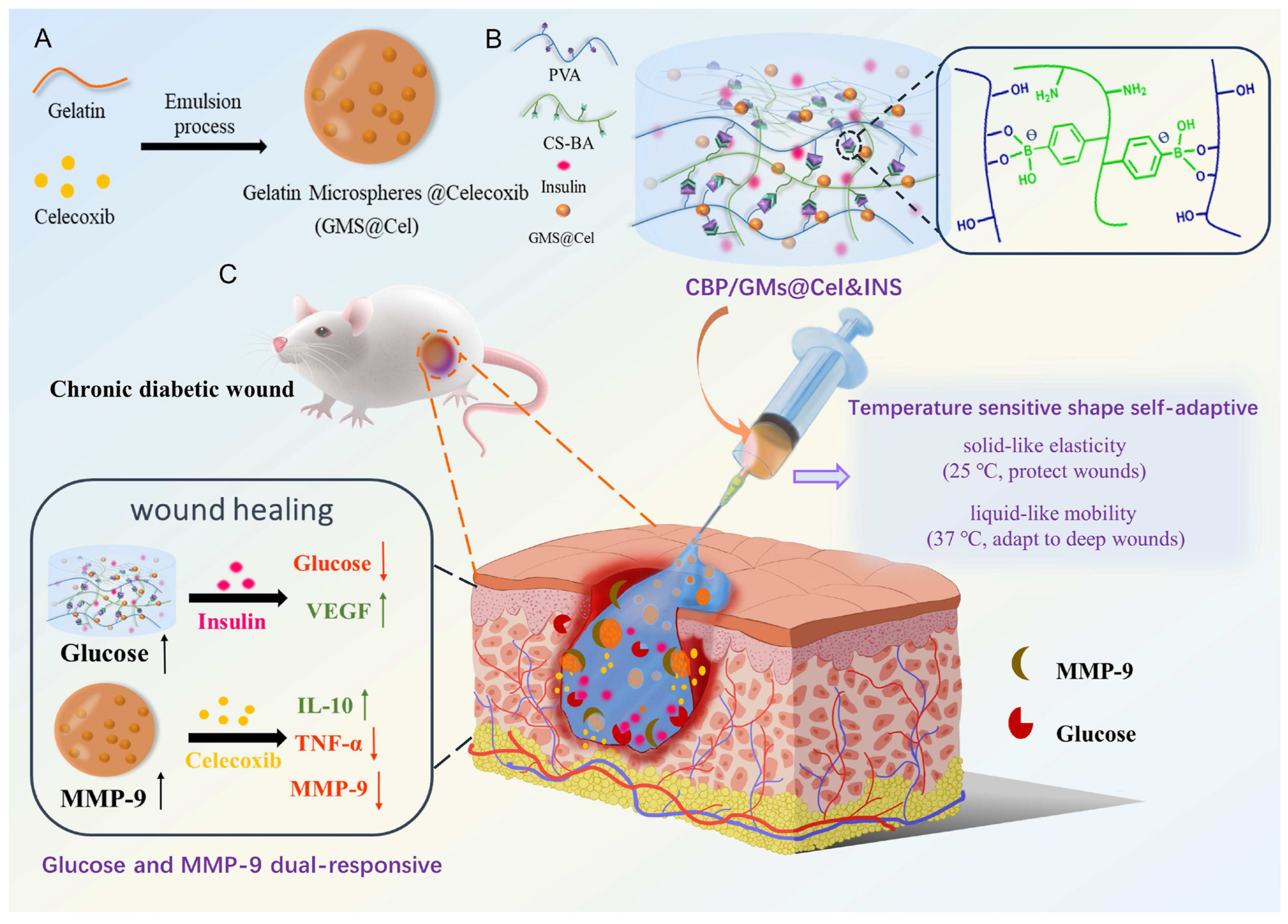
- Zhou, W.; Duan, Z.; Zhao, J.; Fu, R.; Zhu, C.; Fan, D. Glucose and MMP-9 dual-responsive hydrogel with temperature sensitive self-adaptive shape and controlled drug release accelerates diabetic wound healing. Bioact. Mater. 2022, 17, 1–17. [Google Scholar] [CrossRef]
- Sobczak, M. Enzyme-responsive hydrogels as potential drug delivery systems—state of knowledge and future prospects. Int. J. Mol. Sci. 2022, 23, 4421. [Google Scholar] [CrossRef]
- Wang, M.; Gao, B.; Wang, X.; Li, W.; Feng, Y. Enzyme-responsive strategy as a prospective cue to construct intelligent biomaterials for disease diagnosis and therapy. Biomater. Sci. 2022, 10, 1883–1903. [Google Scholar] [CrossRef]
- Maleki, A.; He, J.; Bochani, S.; Nosrati, V.; Shahbazi, M.A.; Guo, B. Multifunctional photoactive hydrogels for wound healing acceleration. ACS Nano 2021, 15, 18895–18930. [Google Scholar] [CrossRef]
- Xu, Y.; Chen, H.; Fang, Y.; Wu, J. Hydrogel combined with phototherapy in wound healing. Adv. Healthc. Mater. 2022, 11, 2200494. [Google Scholar] [CrossRef]
- He, Q.T.; Qian, P.; Yang, X.Y.; Kuang, Q.; Lin, Y.T.; Yi, W.; Tian, T.; Cai, Y.P.; Hong, X.J. Rational design of bacteria-targeted and photo-responsive MOF gel with antibacterial and anti-inflammatory function for infected wound healing. Chem. Eng. J. 2024, 493, 152760. [Google Scholar] [CrossRef]
- Liu, Y.; Xiao, Y.; Cao, Y.; Guo, Z.; Li, F.; Wang, L. Construction of chitosan-based hydrogel incorporated with antimonene nanosheets for rapid capture and elimination of bacteria. Adv. Funct. Mater. 2020, 30, 2003196. [Google Scholar] [CrossRef]
- Liu, C.; Li, Y.; Li, W.; Fan, Y.; Zhou, W.; Xiao, C.; Yu, P.; Liu, Y.; Liu, X.; Huang, Z.; et al. LSPR-enhanced photoresponsive antibacterial efficiency of Bi/MoS2-loaded fibrin gel for management of diabetic wounds. Int. J. Biol. Macromol. 2024, 277, 134430. [Google Scholar] [CrossRef]
- He, Z.; Xu, Q.; Newland, B.; Foley, R.; Lara-Sáez, I.; Curtin, J.F.; Wang, W. Reactive oxygen species (ROS): Utilizing injectable antioxidative hydrogels and ROS-producing therapies to manage the double-edged sword. J. Mater. Chem. B 2021, 9, 6326–6346. [Google Scholar] [CrossRef] [PubMed]
- Zhang, Y.; Xu, Y.; Hu, W.; Ma, X.; Hu, J.; Ye, Y.; Yang, S.; Yu, Y.; Li, N.; Zheng, D.; et al. An adhesive and self-healing ROS-scavenging hydrogel loading with hMSC-derived exosomes for diabetic wound healing. Int. J. Pharm. 2025, 672, 125315. [Google Scholar] [CrossRef]
- Huang, C.; Dong, L.; Zhao, B.; Lu, Y.; Huang, S.; Yuan, Z.; Luo, G.; Xu, Y.; Qian, W. Anti-inflammatory hydrogel dressings and skin wound healing. Clin. Transl. Med. 2022, 12, e1094. [Google Scholar] [CrossRef] [PubMed]
- Wang, Y.; Chen, C.; He, C.; Dong, W.; Yang, X.; Kong, Q.; Yan, B.; He, J. Quaternized chitosan-based biomimetic nanozyme hydrogels with ROS scavenging, oxygen generating, and antibacterial capabilities for diabetic wound repair. Carbohydr. Polym. 2025, 348, 122865. [Google Scholar] [CrossRef] [PubMed]
- Pan, X.y.; Wang, Z.h.; Wu, X.q.; Guo, C.r.; Yang, L.x.; Liu, H.r.; Wang, Y.h.; Chen, W.j.; Wang, J.j.; Nan, K.h.; et al. ROS scavenging and corneal epithelial wound healing by a self-crosslinked tissue-adhesive hydrogel based-on dual-functionalized hyaluronic acid. Int. J. Biol. Macromol. 2025, 293, 139200. [Google Scholar] [CrossRef]
- Dong, Y.; Wang, Z. ROS-scavenging materials for skin wound healing: Advancements and applications. Front. Bioeng. Biotechnol. 2023, 11, 1304835. [Google Scholar] [CrossRef]
- Cui, T.; Li, X.; He, S.; Xu, D.; Yin, L.; Huang, X.; Deng, S.; Yue, W.; Zhong, W. Instant self-assembly peptide hydrogel encapsulation with fibrous alginate by microfluidics for infected wound healing. ACS Biomater. Sci. Eng. 2020, 6, 5001–5011. [Google Scholar] [CrossRef]
- Fan, L.; He, Z.; Peng, X.; Xie, J.; Su, F.; Wei, D.X.; Zheng, Y.; Yao, D. Injectable, intrinsically antibacterial conductive hydrogels with self-healing and pH stimulus responsiveness for epidermal sensors and wound healing. ACS Appl. Mater. Interfaces 2021, 13, 53541–53552. [Google Scholar] [CrossRef]
- Su, K.; Deng, D.; Wu, X.; Song, Y.; Sun, Y.; Wang, X.; Zhang, Z.; Li, J.; Yan, Z.; Shang, X.; et al. On-demand detachable adhesive hydrogel based on dual dynamic covalent cross-linked with NIR/pH dual-responsive properties for diabetic wound healing. Chem. Eng. J. 2024, 479, 147646. [Google Scholar] [CrossRef]
- Gui, Q.; Ding, N.; Wu, H.; Liu, J.; Geng, Y.; Zhu, J.; Gao, M.; Du, A.; Yue, B.; Zhu, L. Development of a pH-Responsive Antimicrobial and Potent Antioxidant Hydrogel for Accelerated Wound Healing: A Game Changer in Drug Delivery. Adv. Biol. 2024, 9, e2400358. [Google Scholar] [CrossRef] [PubMed]
- Li, M.; Liang, Y.; He, J.; Zhang, H.; Guo, B. Two-pronged strategy of biomechanically active and biochemically multifunctional hydrogel wound dressing to accelerate wound closure and wound healing. Chem. Mater. 2020, 32, 9937–9953. [Google Scholar] [CrossRef]
- Ahovan, Z.A.; Khosravimelal, S.; Eftekhari, B.; Mehrabi, S.; Hashemi, A.; Eftekhari, S.; Milan, P.B.; Mobaraki, M.; Seifalian, A.; Gholipourmalekabadi, M. Thermo-responsive chitosan hydrogel for healing of full-thickness wounds infected with XDR bacteria isolated from burn patients: In vitro and in vivo animal model. Int. J. Biol. Macromol. 2020, 164, 4475–4486. [Google Scholar] [CrossRef]
- Shi, W.; Song, N.; Huang, Y.; He, C.; Zhang, M.; Zhao, W.; Zhao, C. Improved Cooling Performance of Hydrogel Wound Dressings via Integrating Thermal Conductivity and Heat Storage Capacity for Burn Therapy. Biomacromolecules 2022, 164, 4475–4486. [Google Scholar] [CrossRef]
- Liu, X.; Ding, Q.; Liu, W.; Zhang, S.; Wang, N.; Chai, G.; Wang, Y.; Sun, S.; Zheng, R.; Zhao, Y.; et al. A Poloxamer 407/chitosan-based thermosensitive hydrogel dressing for diabetic wound healing via oxygen production and dihydromyricetin release. Int. J. Biol. Macromol. 2024, 263, 130256. [Google Scholar] [CrossRef]
- Lim, S.; Kim, J.A.; Chun, Y.H.; Lee, H.J. Hyaluronic acid hydrogel for controlled release of heterobifunctional photocleavable linker-modified epidermal growth factor in wound healing. Int. J. Biol. Macromol. 2023, 253, 126603. [Google Scholar] [CrossRef]
- Yang, N.; Zhu, M.; Xu, G.; Liu, N.; Yu, C. A near-infrared light-responsive multifunctional nanocomposite hydrogel for efficient and synergistic antibacterial wound therapy and healing promotion. J. Mater. Chem. B 2020, 8, 3908–3917. [Google Scholar] [CrossRef]
- He, D.; Liao, C.; Li, P.; Liao, X.; Zhang, S. Multifunctional photothermally responsive hydrogel as an effective whole-process management platform to accelerate chronic diabetic wound healing. Acta Biomater. 2024, 174, 153–162. [Google Scholar] [CrossRef]
- Wei, Y.; Fu, J.; Liu, E.; Gao, J.; Lv, Y.; Li, Z. Injectable hydrogels doped with PDA nanoparticles for photothermal bacterial inhibition and rapid wound healing in vitro. RSC Adv. 2024, 14, 2778–2791. [Google Scholar] [CrossRef]
- Zhao, H.; Huang, J.; Li, Y.; Lv, X.; Zhou, H.; Wang, H.; Xu, Y.; Wang, C.; Wang, J.; Liu, Z. ROS-scavenging hydrogel to promote healing of bacteria infected diabetic wounds. Biomaterials 2020, 258, 120286. [Google Scholar]
- Pan, W.; Qi, X.; Xiang, Y.; You, S.; Cai, E.; Gao, T.; Tong, X.; Hu, R.; Shen, J.; Deng, H. Facile formation of injectable quaternized chitosan/tannic acid hydrogels with antibacterial and ROS scavenging capabilities for diabetic wound healing. Int. J. Biol. Macromol. 2022, 195, 190–197. [Google Scholar]
- Ni, Z.; Yu, H.; Wang, L.; Huang, Y.; Lu, H.; Zhou, H.; Liu, Q. Multistage ROS-responsive and natural polyphenol-driven prodrug hydrogels for diabetic wound healing. ACS Appl. Mater. Interfaces 2022, 14, 52643–52658. [Google Scholar] [CrossRef]
- Bai, J.; Chen, C.; Wang, J.; Zhang, Y.; Cox, H.; Zhang, J.; Wang, Y.; Penny, J.; Waigh, T.; Lu, J.R.; et al. Enzymatic regulation of self-assembling peptide A9K2 nanostructures and hydrogelation with highly selective antibacterial activities. ACS Appl. Mater. Interfaces 2016, 8, 15093–15102. [Google Scholar] [CrossRef]
- Zhao, M.; Kang, M.; Wang, J.; Yang, R.; Zhong, X.; Xie, Q.; Zhou, S.; Zhang, Z.; Zheng, J.; Zhang, Y.; et al. Stem Cell-Derived Nanovesicles Embedded in Dual-Layered Hydrogel for Programmed ROS Regulation and Comprehensive Tissue Regeneration in Burn Wound Healing. Adv. Mater. 2024, 36, 2401369. [Google Scholar] [CrossRef]
- Jin, R.; Hiemstra, C.; Zhong, Z.; Feijen, J. Enzyme-mediated fast in situ formation of hydrogels from dextran–tyramine conjugates. Biomaterials 2007, 28, 2791–2800. [Google Scholar] [CrossRef] [PubMed]
- Wu, Y.; Wang, Y.; Long, L.; Hu, C.; Kong, Q.; Wang, Y. A spatiotemporal release platform based on pH/ROS stimuli-responsive hydrogel in wound repairing. J. Control. Release 2022, 341, 147–165. [Google Scholar]
- Zhang, Y.; Gao, X.; Tang, X.; Peng, L.; Zhang, H.; Zhang, S.; Hu, Q.; Li, J. A dual pH-and temperature-responsive hydrogel produced in situ crosslinking of cyclodextrin-cellulose for wound healing. Int. J. Biol. Macromol. 2023, 253, 126693. [Google Scholar]
- Tian, R.; Liu, J.; Dou, G.; Lin, B.; Chen, J.; Yang, G.; Li, P.; Liu, S.; Jin, Y.; Qiu, X.; et al. Synergistic antibiosis with spatiotemporal controllability based on multiple-responsive hydrogel for infectious cutaneous wound healing. Smart Mater. Med. 2022, 3, 304–314. [Google Scholar] [CrossRef]
- Wu, J.; Xue, W.; Yun, Z.; Liu, Q.; Sun, X. Biomedical applications of stimuli-responsive “smart” interpenetrating polymer network hydrogels. Mater. Today Bio 2024, 25, 100998. [Google Scholar] [CrossRef]
- Brumberg, V.A.; Bikmulina, P.Y.; Pozdnyakov, A.A.; Heydari, Z.; Zimulkina, D.R.; Smirnova, O.A.; de Sena Pereira, F.D.A.; Nesterova, A.M.; Kotova, S.L.; Vosough, M.; et al. Scaling liver bioprinting: A guide for usage of the hepatic extracellular matrix as a bioink. Int. J. Bioprinting 2025, 11, 57–83. [Google Scholar] [CrossRef]
- Moura, D.; Rohringer, S.; Ferreira, H.P.; Pereira, A.T.; Barrias, C.; Magalhães, F.D.; Bergmeister, H.; Gonçalves, I.C. Long-term in vivo degradation and biocompatibility of degradable pHEMA hydrogels containing graphene oxide. Acta Biomater. 2023, 173, 351–364. [Google Scholar] [CrossRef] [PubMed]
- Li, Y.; Saiding, Q.; Wang, Z.; Cui, W. Engineered Biomimetic Hydrogels for Organoids. Prog. Mater. Sci. 2023, 141, 101216. [Google Scholar] [CrossRef]
- Neumann, M.; di Marco, G.; Iudin, D.; Viola, M.; van Nostrum, C.; van Ravensteijn, B.G.P.; Vermonden, T. Stimuli-Responsive Hydrogels: The Dynamic Smart Biomaterials of Tomorrow. Macromolecules 2023, 56, 8377–8392. [Google Scholar] [CrossRef]
- El-Husseiny, H.M.; Mady, E.A.; Hamabe, L.; Abugomaa, A.; Shimada, K.; Yoshida, T.; Tanaka, T.; Yokoi, A.; Elbadawy, M.; Tanaka, R. Smart/stimuli-responsive hydrogels: Cutting-edge platforms for tissue engineering and other biomedical applications. Mater. Today Bio 2021, 13, 100186. [Google Scholar] [CrossRef]


| Stimulus | Response Mechanism | Functional Advantages | Applications |
|---|---|---|---|
| pH | pH-triggered structural change or degradation | Controlled release; infection reduction in acidic environment | Inflamed/infected wounds |
| Thermo | Sol–gel transition near LCST or UCST | Injectable; body-temp responsive; in situ gelling | Burns, deep wounds |
| ROS | ROS-induced degradation or release | Antioxidant delivery; oxidative stress reduction | Chronic wounds, diabetic ulcers |
| Enzyme | Enzymatic cleavage of crosslinks | Selective release; enzyme-responsive action | Diabetic wounds, chronic inflammation |
| Light | Photo-triggered conformational or thermal effect | Non-invasive control; light-activated therapy | Superficial wounds, PDT |
| Glucose | Glucose-sensitive degradation or release | Glucose-regulated delivery; diabetic wound care | Diabetic ulcers, insulin systems |
| Electro | Electrically induced ion flow or deformation | Remote control; promotes regeneration | Electrotherapy, nerve repair |
| Magnetic | Magnetic field-induced heating or motion | Localised activation; precise targeting | Deep-tissue healing, cancer therapy |
| DNA | DNA hybridisation or displacement | Programmable release; high specificity | Gene therapy, smart dressings |
| Stimulus Type | Hydrogel Material | Mechanism | Wound Type | Reference |
|---|---|---|---|---|
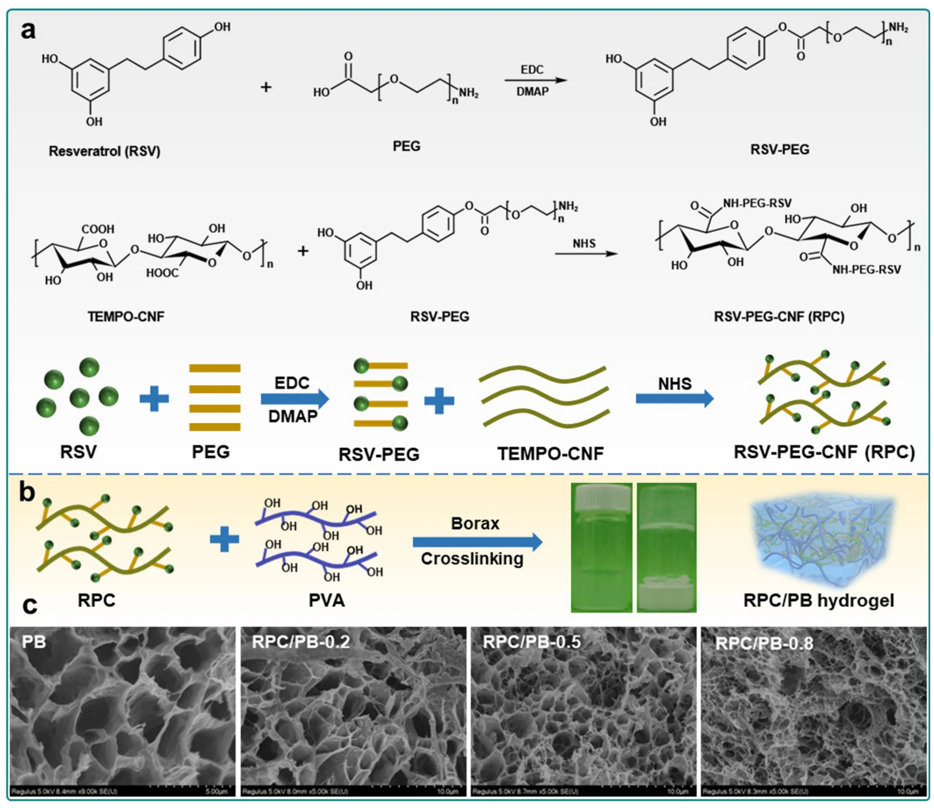
| pH | RSV-grafted CNF, PVA–borax | pH-triggered drug release via semi-IPN structure | Infected | [72] |
| Nap-GFFKH, sodium alginate | Self-assembly under pH via microfluidic mixing | Infected | [131] | |
| PER-TBA, NaCl, CS | pH-cleavable Schiff base in dynamic network | General | [132] | |
| HA-ALD, ADH, N-CS | pH-degradable hydrazone and imine bonds enabling insulin release | Diabetic | [78] | |
| CMCS, 2-FPBA, EGCG | Dual pH-cleavable Schiff and borate bonding | Diabetic | [133] | |
| HA, AMP peptide (KK(SLKL)3KK) | pH-triggered AMP release via Schiff base crosslinking | Infected | [79] | |
| HAMA, GelMA-CA, Ag+ | pH-triggered Ag+ release via polyphenol coordination | Acute | [134] | |
| Anthocyanin-based hydrogel | pH-indicated visible colour change for infection monitoring | Infected | [81] | |
| Thermo | QCS, rGO-PDA, PNIPAM | LCST-induced sol–gel transition of PNIPAM | General | [135] |
| Thermoresponsive chitosan (TCTS) | Thermo-induced phase behaviour enabling XDR bacterial suppression | Burn | [136] | |
| COL, GG, PNIPAM | Thermo/NIR-induced gelation for enhanced wound repair | General | [96] | |
| PNIPAM, HA | Thermo-induced contraction activates mechanotransduction for wound closure | Chronic | [105] | |
| PEG derivatives, CNT-OH | Thermoconductive PEG/CNT network enabling rapid heat dissipation | Burn | [137] | |
| Dihydromyricetin, CaO2NPs | Heat-softened structure for sustained delivery | Diabetic | [138] | |
| Light | HA, EGF | UV-cleavable linker for EGF release | General | [139] |
| CS, WS2, ciprofloxacin | NIR-induced photothermal gel activation | Infected | [140] | |
| Ag np, MOF, boronic acid, berberine | Visible-light-triggered ROS generation and synergistic photodynamic antibacterial activity | Infected | [122] | |
| Chitosan, AM NSs | NIR-triggered photothermal antibacterial activity via AM NSs | Infected | [123] | |
| Bi/MoS2 | Synergistic PDT and PTT under NIR for enhanced antibacterial efficacy | Diabetic | [124] | |
| PAG, AA, GelMA, CuS, LAS | NIR-triggered heating via CuS for release | Diabetic | [141] | |
| GMH, PDA | UV-cleavable Schiff base network | Infected | [142] | |
| ROS | EFM peptide hydrogel | ROS-triggered Met oxidation and gel breakdown | Diabetic | [48] |
| PVA, TVA | ROS-cleavable phenylboronic ester linkage | Diabetic | [143] | |
| QCS, TA, HCl, NaHCO3 | TA-based phenol ROS scavenging | Diabetic | [144] | |
| CQCS, PEG, hMnO2@PDA NPs | H2O2-responsive ROS scavenging via enzyme-mimetic MnO2 and antioxidant PDA | Infected | [128] | |
| PPBA, TA, PVA | ROS-cleavable boronic ester dynamic bonds | Diabetic | [145] | |
| Enzyme | 4-arm-PEG-MAL, MMP(W)x, PEG-SH, ADSC-exo | MMP-sensitive peptide cleavage | Diabetic | [115] |
| A9K2 peptide, FBS, PAO, Cu2+, NaCl | LOX-triggered peptide hydrogelation | General | [146] | |
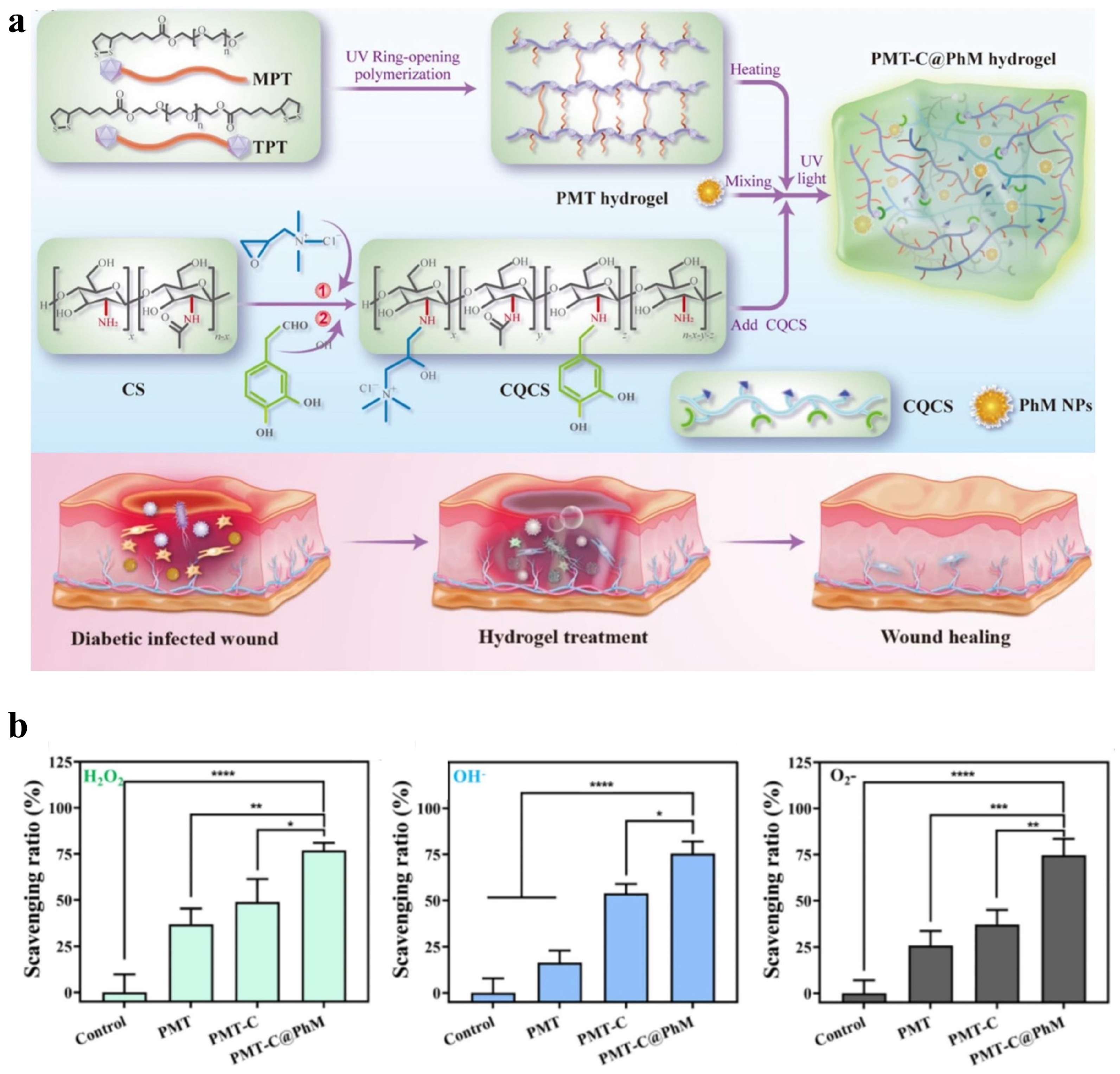
| Dual-layered hydrogel with AIE-PS and stem cell vesicles | Enzyme-triggered photosensitiser release and ROS-scavenging for staged repair | Burn | [147] | |
| Dex-TA / Dex-DG-TA | HRP-mediated enzymatic crosslinking | General | [148] |
Disclaimer/Publisher’s Note: The statements, opinions and data contained in all publications are solely those of the individual author(s) and contributor(s) and not of MDPI and/or the editor(s). MDPI and/or the editor(s) disclaim responsibility for any injury to people or property resulting from any ideas, methods, instructions or products referred to in the content. |
© 2025 by the authors. Licensee MDPI, Basel, Switzerland. This article is an open access article distributed under the terms and conditions of the Creative Commons Attribution (CC BY) license (https://creativecommons.org/licenses/by/4.0/).
Share and Cite
Ding, K.; Liao, M.; Wang, Y.; Lu, J.R. Advances in Composite Stimuli-Responsive Hydrogels for Wound Healing: Mechanisms and Applications. Gels 2025, 11, 420. https://doi.org/10.3390/gels11060420
Ding K, Liao M, Wang Y, Lu JR. Advances in Composite Stimuli-Responsive Hydrogels for Wound Healing: Mechanisms and Applications. Gels. 2025; 11(6):420. https://doi.org/10.3390/gels11060420
Chicago/Turabian StyleDing, Ke, Mingrui Liao, Yingyu Wang, and Jian R. Lu. 2025. "Advances in Composite Stimuli-Responsive Hydrogels for Wound Healing: Mechanisms and Applications" Gels 11, no. 6: 420. https://doi.org/10.3390/gels11060420
APA StyleDing, K., Liao, M., Wang, Y., & Lu, J. R. (2025). Advances in Composite Stimuli-Responsive Hydrogels for Wound Healing: Mechanisms and Applications. Gels, 11(6), 420. https://doi.org/10.3390/gels11060420